photoprotective function of betacyanin in leaves of
TRANSCRIPT

DOI: 10.1007/s11099-011-0062-7 PHOTOSYNTHETICA 49 (4): 497-506, 2011
497
Photoprotective function of betacyanin in leaves of Amaranthus cruentus L. under water stress T. NAKASHIMA*, +, T. ARAKI**, and O. UENO***
Graduate School of Bioresource and Bioenvironmental Sciences, Kyushu University, Hakozaki 6-10-1, Higashi-ku, Fukuoka 812-8581, Japan* Faculty of Agriculture, Ehime University, Tarumi 3-5-7, Matsuyama, Ehime 790-8566, Japan** Faculty of Agriculture, Kyushu University, Hakozaki 6-10-1, Higashi-ku, Fukuoka 812-8581, Japan*** Abstract The photoprotective function of leaf betacyanin in water-stressed Amaranthus cruentus plants was examined by comparing leaves of two strains which differ significantly in the amount of betacyanin. At 0, 1, and 2 days after the imposed water stress, leaves were subjected to high-light (HL) treatment to assess their photosynthetic capacity and photoinhibition susceptibility. The water stress equally reduced leaf relative water content (RWC), gas-exchange rate and chlorophyll (Chl) contents in both leaves, indicating that the severity of water stress was comparable between the strains. Consequently, the extent of photoinhibition after the HL treatment increased in both strains as water stress developed; however, it was significantly greater in acyanic leaves than in betacyanic leaves, suggesting lower photoinhibition susceptibility in the betacyanic strain. The betacyanic leaves also exhibited approximately 30% higher values for photochemical quenching coefficient (qP) during the period of water stress despite the nonphotochemical quenching coefficient (qN) did not differ significantly between the strains. These results may be partially explained by the increased amount of leaf betacyanin under water stress. Moreover, a decrease in Chl content in betacyanic leaves might have enhanced light screening effect of betacyanin by increasing relative abundance of betacyanin to Chl molecule. In addition, reduced Chl content increased light penetrability of leaves. As a result, the extent of photoinhibition at the deeper tissue was exacerbated and the Chl fluorescence emitted from these tissues was more readily detected, facilitating assessment of photoinhibition at deeper tissues where the effect of betacyanic light screening is considered to be most apparent. Our results demonstrated that leaf betacyanin contributes to total photoprotective capacity of A. cruentus leaves by lowering excitation pressure on photosystem II (PSII) via attenuation of potentially harmful excess incident light under water stress. Additional key words: betacyanin; grain amaranthus; light screening; maximum quantum yield of photosystem II; photoinhibition; water deficit. Introduction Red pigmentation in leaves at certain developmental stages or in response to particular environmental stimuli is physiological trait of plants, although its functional significance has long been a considerable debate. In these
leaves, accumulation of anthocyanins, or less commonly betacyanins, is primarily responsible for leaf reddening (Manetas 2006). Among several putative functions of leaf anthocyanins, a growing number of studies support the
———
Received 12 January 2011, accepted 15 July 2011. +Corresponding author; fax: +81 92 642 2833; e-mail: [email protected] Abbreviations: Chl – chlorophyll; DAT – day(s) after treatment; T – difference in transmission spectra between the strains; F0 and Fm – minimum and maximum fluorescence yield of dark-adapted leaves, respectively; Fv – variable fluorescence in dark-adapted state; Fm’, F0’, and Fs – maximum, minimum, and steady-state fluorescence yield of light-adapted leaves, respectively; Fv’ – variable fluorescence in light-adapted state; Fv/Fm – maximum quantum yield of PSII; gs – stomatal conductance; HL – high light; PIFv/Fm – percent inhibition of Fv/Fm after HL treatment; PN – net photosynthetic rate; PPFD – photosynthetic photon flux density; PS – photosystem; QA – primary quinone acceptor of PSII; qN and qP – nonphotochemical and photochemical quenching coefficient, respectively; ROS – reactive oxygen species; RWC – leaf relative water content. Acknowledgements: We thank Dr. M. Katsuta (National Institute of Crop Science, National Agriculture and Food Research Organization, Tsukuba, Japan) for kindly providing seeds of A. cruentus, Dr. S. Agarie (Faculty of Agriculture, Kagawa University, Kagawa, Japan) for lecturing us the method of betacyanin quantification, and Dr. S. Yamashita (Faculty of Agriculture, Kyushu University, Fukuoka, Japan) for allowing us to use the spectroscope.

T. NAKASHIMA et al.
498
protective function against photoinhibition induced by abiotic stresses such as high irradiance, low air temperature, soil nutrient deficiency and UV radiation (e.g. Burger and Edwards 1996, Gaume et al. 2001, Hughes et al. 2005). Yet, some studies reported con-trasting results in which anthocyanic leaves were simi-larly or even more susceptible to photoinhibition than acyanic counterparts (Burger and Edwards 1996, Manetas et al. 2003, Zeliou et al. 2009). On the other hand, some other possible functions relating to leaf warming, sink-source regulation and osmotic adjustment were examined in leaves displaying anthocyanin accumulation during autumn and winter seasons (Chalker-Scotte 2002, Oberbauer and Starr 2002, Hughes et al. 2005).
Betacyanins are alternative forms of nonphotosyn-thetic red pigments belonging to betalain group which replace anthocyanins in the nine families of Caryo-phyllales (Solovchenko and Merzlyak 2008). Since beta-cyanins are known to possess similar absorption spectra and antioxidant properties in vitro, they are also consi-dered to fulfill functions of anthocyanins (Solovchenko and Merzlyak 2008). Some recent studies indeed reported the protective function of leaf betacyanins under low temperature and heat stresses (Wang and Liu 2007, Shu et al. 2009, Hayakawa and Agarie 2010).
Purple amaranth (A. cruentus L.) is a betacyanin-producing C4 dicot which has been cultivated as a pseudo-cereal crop since the era of Aztecs, and recently
receives considerable attention as a source of human diet due to its high nutritional value (Escudero et al. 2004). Although Amaranthus spp. are often described as drought-tolerant plants (Liu and Stützel 2002), Hura et al. (2007) attributed significant decline of photosynthetic rate in A. cruentus under drought stress to photoinhibitory injury of photosynthetic apparatus.
Although the impact of photoinhibition on plant productivity is difficult to quantify, photoinhibitory loss of potential carbon gain has been estimated to be approxi-mately 10–32% in plant canopies (Ögren and Sjöström 1990, Zhu et al. 2004). To prevent adverse effects of photoinhibition, higher plants have developed an array of photoprotective mechanisms including light avoidance movements of leaves and chloroplasts, dissipation of absorbed energy as heat, photorespiration and reactive oxygen species (ROS) scavenging system with enzymatic and low-molecular-mass antioxidants (Takahashi and Badger 2011).
Given the possibility that leaf betacyanins afford an additional photoprotection for Amaranthus leaves under abiotic stresses, the present study examined the photoprotective potential of betacyanin in water-stressed leaves of A. cruentus L. To this end, the interstrainal comparison of photoinhibition susceptibility under water stress was performed using leaves with and without betacyanin accumulation.
Materials and methods Plant materials and growth conditions: Two contras-tive strains of A. cruentus L. differing in leaf coloration, the betacyanin-deficient (acyanic) Tohoku 1 and the betacyanin-depositing (betacyanic) Tohoku 3, were ger-minated on perforated multi-well nursery boxes filled with loam soil granules and grown for 4 weeks in a green-house at the experimental field of Kyushu University (3335’N, 13023’E) during a summer season. Seedlings with 5 or 6 fully expanded leaves were then transplanted to 5 L pots containing sandy loam soil incorporated with 0.34 g of nitrogen, phosphorus and potassium fertilizers in the forms of ammonium nitrate, calcium super-phosphate and potassium chloride, respectively. The plants were grown outdoors for 4 weeks at the mean air temperature and relative humidity of 26C and 65%, respectively, and day length of approximately 12 h. During this period, plants were provided with adequate irrigation twice a day.
Water-stress treatment was then imposed on plants of ca. 90–100 cm in height and with ca. 18–20 fully expanded leaves at their vegetative growth stage. The treatment was imposed by withholding irrigation for two consecutive days of clear weather, wherein photo-synthetic photon flux density (PPFD) at midday exceeded 1,500 μmol m–2 s–1. At 0, 1, and 2 day(s) after the treatment (DAT), following experiments were conducted
on fully matured upper leaves of the two strains.
High-light (HL) treatment: To assess photoinhibition susceptibility of acyanic and betacyanic leaves, HL treatment was given as follows. Plants were transferred to the laboratory at the night before the measurements and dark-adapted overnight until the onset of the treatment. Target leaves were placed in a leaf assimilatory chamber (PLC-4N, Shimadzu Co., Kyoto, Japan) in which air temperature and relative humidity were maintained at 30°C and 60%, respectively, and ambient air ([CO2] = 399 ± 14 μL L–1, [O2] = 21%) was pumped into the chamber at the rate of 1 L min–1. Leaves were then exposed to photoinhibitory illumination (PPFD; 1,500 μmol m–2 s–1) provided by metal halide lamp (LS-M180, Sumita Optical Glass Inc., Saitama, Japan) for 1 h, and subsequently dark-adapted for 20 min.
Chl fluorescence and gas-exchange measurements during the HL treatment: Chl fluorescence was mea-sured by using a portable Chl fluorometer (PAM-2000, Heinz Walz, Effeltrich, Germany). The measuring beam of 20 kHz at the intensity level of 9 and saturation pulse of 8,000 μmol m–2 s–1 with pulse duration of 0.8 s were used to determine fluorescence yield. In addition, external actinic light of 1,500 μmol m˗2 s–1 provided by the same

PHOTOPROTECTIVE FUNCTION OF BETACYANIN IN LEAVES
499
metal halide light source was used for simultaneous measurements of Chl fluorescence and gas-exchange rate during the HL treatment. For this purpose, the fiber-optic light guide of PAM-2000 was positioned on the side of the chamber window (6.25 cm2) at the angle of 60° without interfering the actinic light.
Chl fluorescence in dark-adapted state was measured before and after the HL treatment on the overnight and 20 min dark-adapted leaves, respectively, to determine the maximum quantum yield of PSII (Fv/Fm). Fv/Fm was calculated according to the method of van Kooten and Snel (1990), where Fv = Fm – F0, and Fm and F0 represents maximum and minimum fluorescence yield of dark-adapted leaves, respectively. Using these Fv/Fm values, percentage inhibition of Fv/Fm (PIFv/Fm) was derived as PIFv/Fm (%) = [1 – (Fv/Fm after the HL treatment/Fv/Fm be-fore the HL treatment)] 100, to parameterize the magni-tude of photoinhibition induced by the HL treatment.
At the end of the HL treatment when the gas-exchange rate reached a steady state, the maximum (Fm’) and steady state (Fs) fluorescence yields of light-adapted leaves were measured, and the minimum fluorescence yield of light-adapted leaves (F0’) was derived as F0’ = F0/(Fv/Fm + F0/Fm’) according to Oxborough and Baker (1997). Obtained values were used to calculate the photochemical quenching coefficient [qP = (Fm’Fs)/ (Fm’F0’)] and nonphotochemical quenching coefficient [qN = 1 (Fm’F0’)/Fv] as described by Genty et al. (1989).
The gas-exchange measurement was conducted using an infrared CO2/H2O gas analyzer (LI-6262, LI-COR Inc., Nebraska, USA) installed in an open gas-exchange system. Measurements were made at the end of the HL treatment, and the stomatal conductance (gs) and net photosynthetic rate (PN) were calculated according to Long and Hallgren (1985).
Six plants per strain or 5 plants only in case of betacyanic strains at 1 DAT were used. The measure-ments were made on a fully matured upper leaf of each plant and thereafter, leaves were sampled for biochemical and morphological analyses.
Leaf transmittance and transmission spectrum were measured to estimate the extent to which leaf betacyanin influences light-absorption property of leaves. Total leaf transmittance of visible light spectrum (400–700 nm) were measured with a quantum meter (QMSS, Apogee Instruments Inc., Utah, USA). The transmittance was calculated as T (%) = I/I0 100, where I and I0 represent the light intensity above and beneath a leaf, respectively. Measurements were taken from 3 prestressed plants per strain using a fully matured leaf of each plant.
Leaf transmission spectra were measured by using a spectroscope (SA-100, Lambda Vision Inc., Kanagawa, Japan) coupled with an optical quartz fiber (dia-meterm), the end of which was pointing toward a halogen light source (MHF-150L, Moritex Corp.,
Tokyo, Japan) at 10-cm distance. The background light spectrum was obtained once before the measurement with the aid of neutral density filter of known transmission spectrum. Intact leaves were then placed immediately above the optical fiber end and spectra of light passing through the leaves were measured. From the background light spectrum and leaf transmission spectra, transmittan-ces at different wavelengths were calculated as described previously. Measurements were repeated over 4 different areas of a fully matured upper leaf of each plant. The means were calculated from these measurements of 3 prestressed plants.
Leaf relative water content (RWC): For RWC determi-nation, leaf samples were obtained from the same leaves used in gas-exchange and Chl fluorescence measure-ments. Three leaf disks of 3.14 cm2 were excised from the base, mid, and top regions of a leaf lamella without primary veins and immediately weighed to determine the fresh mass. The leaf disks were then floated on distilled water for 24 h in the darkness to obtain the fully turgid mass, and dried subsequently in a 70°C oven for 48 h to measure the dry mass. The RWC of each disk was calculated as RWC = [(fresh mass dry mass)/(fully turgid mass dry mass)] 100. Finally, RWCs of the 3 individual leaf disks were averaged to represent RWC of a leaf. The means were calculated from values of 6 plants (5 plants for the betacyanic strain at 1 DAT).
Chl and betacyanin contents: For pigment quantifi-cation, leaf disks of 6 mm in diameter were obtained from the same leaf lamella used for the RWC measurement. The primary and secondary veins were excluded from sampling area. Chl was extracted from 3 leaf disks in 80% acetone for 2 days in the darkness and quantified spectrophotometrically after the method of Porra et al. (1989). Betacyanin content was determined as described by Elliot (1979) with minor modifications. Five leaf disks were ground with liquid nitrogen in a microcentrifuge tube and washed twice with 100% acetone to remove Chl. After centrifugation for 5 min at 21,500 × g at 4°C, acetone residue was removed by vacuum evaporation and thereafter, 3 mM of acetic acid was added to the precipi-tant for betacyanin extraction. The betacyanin content was determined spectrophotometrically from absorbance at 538 nm with molar extinction coefficient of 56,600 L cm–1 mol–1. The means were calculated from values of 6 plants (5 plants for the betacyanic strain at 1 DAT).
Leaf anatomy: To evaluate cellular distribution of betacyanin within a leaf, cross-sections were prepared from fully matured upper leaves of prestressed plants. Leaf segments without primary and secondary veins were vacuum infiltrated in distilled water added with a few drops of Triton-X 100 to remove air trapped in inter-cellular space, which appears as dark blots under a light microscope. The pieces of leaf samples were then hand-

T. NAKASHIMA et al.
500
sectioned by using a pith and razor blade. The cross-sections were observed under a light microscope at 200 × magnification.
Leaf segments exercised from fully matured leaves of prestressed plants were fixed in formalin acetic alcohol (FAA) fixative solution consisting of 63 % (v/v) ethanol, 4.8 % (v/v) glacial acetic acid and 5% (v/v) formalin. The thickness of whole leaf, upper epidermis, palisade mesophyll, bundle sheath, spongy mesophyll, and lower epidermis (Fig. 1) were measured for hand sections obtained from the fixed samples under the light micro-
scope. Measurements were taken from 4 different sites per cross-section and the averaged values were used as the representative of a plant. The means were calculated from values of 3 plants.
Statistical analysis: The statistical significance of inter-strainal differences was determined by Student t-test. The data are presented as means ± SE of 6 plants unless otherwise stated; however, that of betacyanic leaves at 1 DAT was presented as means ± SE of 5 plants.
Results The leaf anatomy, cellular distribution of betacyanin and pigment composition of leaves: In acyanic and betacyanic leaves, the presence of C4 Kranz anatomy consisting of palisade and spongy mesophyll cells and bundle sheath cells were clearly distinguished (Fig. 1). In acyanic leaves (Fig. 1A), betacyanin was apparently absent in any leaf tissues including epidermis. On the other hand, significant accumulation of betacyanin was observed in upper and lower epidermis as well as palisade and spongy mesophyll cells of betacyanic leaves (Fig. 1B). Consistently, the comparison of pigment composition in prestressed leaves showed approximately 50-fold higher betacyanin content in betacyanic leaves than in acyanic leaves (Table 1). In contrast, neither Chl contents nor Chl a/b ratios were significantly different between the strains. Moreover, no significant differences in thickness of whole leaf and different leaf tissues were observed between the strains (Table 2).
Transmittance and absorption spectra of leaves: The total leaf transmittances of visible light spectrum (400–700 nm) were 6.4 % and 2.4 % in acyanic and betacyanic leaves, respectively (Fig. 2). The leaf transmission spectra of the two strains were also compared to evaluate which spectral region contributed to such difference in the transmittance. Acyanic leaves showed transmission spectrum typical of green leaves with peaks at around 550 nm and somewhere over 700 nm (Burger and Edwards 1996, Hughes and Smith 2007), whereas that of betacyanic leaves was significantly lower in blue-green wavelengths (Fig. 3A). The distinct difference in trans-mission spectra of the two strains (T) was observed as a peak at 550 nm (Fig. 3B), which resembles to the absorption peak of betacyanins (538 nm; Elliot 1979). Hence, 3-fold lower transmittance in betacyanic leaves was considered due to the accumulation of betacyanin.
Changes in leaf water status and gas-exchange para-meters during the water-stress treatment: In both strains the initial values of RWC at 0 DAT were 96% (Fig. 4A), being within a range of typical values for well watered plants (Liu and Stützel 2002, Carmo-Silva et al. 2008). After imposition of water-stress treatment, RWC
in both strains fell to 72–76% at 1 DAT and below 60% at 2 DAT. Compared to relatively slow decline of RWC, gs and PN dropped drastically to 21–25% of the pre-stressed values at 1 DAT, followed by further slow reduction by 35–40% at 2 DAT (Fig. 4B, C). Both strains showed similar trends in changes of RWC and gas-exchange parameters throughout the experimental period.
Changes in Chl fluorescence parameters: As reported previously (Tezara et al. 1999, Flexas et al. 1998), water stress had no apparent effect on Fv/Fm measured before the HL treatment on overnight dark-adapted leaves, which was maintained at 0.73–0.77 throughout the experimental period. After the HL treatment, Fv/Fm decreased to 0.63 and 0.65 in prestressed leaves of the
Fig. 1. Cross-sections of acyanic (A) and betacyanic (B) leaves of Amaranthus cruentus. The composition of tissue thickness of leaves measured in Table 2 is also shown in (B). BS – bundle sheath; DC – druse-containing cell; LE – lower epidermis; PM – palisade mesophyll; SM – spongy mesophyll; UE – upper epidermis. Bars = 50 m.

PHOTOPROTECTIVE FUNCTION OF BETACYANIN IN LEAVES
501
Table 1. Comparison of chlorophyll (Chl) (a+b) content, Chl a/b ratio and betacyanin content in acyanic and betacyanic leaves of Amaranthus cruentus. ns – absence of significant difference at P<0.05; *** – presence of significant differences between the two strains by Student t-test at P<0.001. Values are means ± SE (n = 6).
Strain Chl (a+b) [g m–2] Chl a/b Betacyanin [μmol m–2]
Acyanic 0.24 ± 0.02 3.04 ± 0.06 1.27 ± 0.14 Betacyanic 0.24 ± 0.01 3.00 ± 0.06 64.27 ± 5.07 ns ns ***
Table 2. Thickness of whole leaf, upper epidermis, palisade mesophyll, bundle sheath, spongy mesophyll, and lower epidermis in pre-stressed leaves of acyanic and betacyanic strains of Amaranthus cruentus. ns – absence of significant differences between the two strains by Student t-test at P<0.05 level. Values are means ± SE (n = 3).
Strain Whole leaf [μm]
Upper epidermis [μm]
Palisade mesophyll [μm]
Bundle sheath [μm]
Spongy mesophyll [μm]
Lower epidermis [μm]
Acyanic 185.0 ± 8.6 14.2 ± 1.7 36.0 ± 1.8 85.2 ± 4.1 32.5 ± 2.5 12.7 ± 0.6 Betacyanic 174.0 ± 9.7 15.6 ± 0.8 31.9 ± 1.5 80.2 ± 5.0 31.3 ± 3.4 12.5 ± 0.6 ns ns ns ns ns ns
Fig. 2. Total leaf transmittance of visible light (400700 nm) in acyanic and betacyanic leaves of Amaranthus cruentus. Measurements were made under full sunlight at midday. Means ± SE (n = 3). *** – presence of significant differences between the two strains by Student t-test at P< 0.001 acyanic and betacyanic strains, respectively (Fig. 5A). The extent of reduction in Fv/Fm after the HL treatment was increased by the imposition of water stress, resulting in Fv/Fm below 0.6 at 2 DAT. However, betacyanic leaves retained relatively high Fv/Fm than acyanic leaves during water stress. Although the calculated PIFv/Fm increased in both strains after the imposition of water stress, PIFv/Fm of betacyanic leaves stayed 20–25% lower than that of acyanic leaves (Fig. 5B).
The significant interstrainal difference was also observed in qP (Fig. 5C), which denotes the proportion of excitation energy captured by open traps and being converted to chemical energy in the PSII reaction center (Krause and Weis 1991). The changes in qP of both strains paralleled with the decreasing trend of PN. However, betacyanic leaves retained 0.27 at 1 DAT and
0.2 at 2 DAT, being approximately 30% higher than that of acyanic leaves. In contrast, qN, a parameter reflecting the proportion of energy dissipated mainly as heat (Buschmann 1999), increased slightly and similarly in both strains at 1 DAT, without showing further increase
Fig. 3. Transmission spectra in acyanic and betacyanic leaves of Amaranthus cruentus. Measurements were made under a halo-gen light (A) and the difference in the transmission spectra calculated as T = transmittance of acyanic leaves – trans-mittance of betacyanic leaves (B). Values are means of 3 plants.

T. NAKASHIMA et al.
502
at 2 DAT (Fig. 5D). When the PIFv/Fm was replotted against the RWC (Fig. 6), linear correlations were observed in both strains and the slope was steeper in acyanic leaves than in betacyanic ones.
Changes in Chl and betacyanin contents during water stress treatment: As shown in Table 1, both Chl contents and Chl a/b ratios were almost equivalent in both leaves.
After the imposition of water stress, the Chl content and Chl a/b ratio decreased gradually at a similar rate in both strains (Fig. 7A,B). On the other hand, betacyanin content in betacyanic leaves showed approximately 1.5-fold increase at 2 DAT relative to that in prestressed leaves (Fig. 7C) whereas that in acyanic leaves did not change during the treatment.
Discussion To assess photoprotective potential of leaf betacyanin by comparing two strains that differ in leaf coloration, minimizing variations in physiological traits of compared leaves, except for the amount of betacyanin, is a funda-mental importance (Manetas 2006). In this regard, the acyanic and betacyanic leaves of A. cruentus showed comparable Chl contents and Chl a/b ratios (Table 1), implying that the potential light-harvesting capacities in both leaves were similar. Moreover, the rate of CO2
assimilation and heat dissipation via xanthophyll cycle, two major fates for absorbed energy, did not differ significantly between the strains (Figs. 4C, 5D). It is therefore plausible to assume that the potential risk of photoinhibition was roughly equal for the strains and that the amount of betacyanin was a major physiological trait differing between the strains. In addition, the anatomical analysis confirmed a similarity in the internal structure of both leaves, further supporting comparability of the strains (Table 2).
Yet, alternative electron sinks such as photorespira-tion and Mehler reaction coupled with an enzymatic antioxidant system, both of which quench a significant portion of energy in stressed C3 plants (Asada 2000, Ripley et al. 2007), was largely ignored in the present study. In C4 plants, however, the contribution of photo-respiration as an alternative electron sink accounts only for a few percent of linear electron flow through PSII and remains very low under water stress (Ripley et al. 2007, Carmo-Silva et al. 2008). Moreover, Ripley et al. (2007) reported that alternative electron sinks including Mehler reaction in a C4 subspecies of Alloteropsis semialata dissipated only 16% of linear electron flow under water stress, whereas it accounted for 30% in a C3 subspecies. Hence, these alternative energy sinks are considered less important in C4 leaves.
After the HL treatment, leaves of both strains were photoinhibited and its extent was exacerbated with decreasing RWC under water stress (Figs. 5A,B, 6). It is considered that photoinhibition increases largely due to reduced capacity of photosynthetic carbon reduction (Flexas and Medrano 2002). Although the detailed mechanisms of PN reduction in C4 plants are not well understood as in C3 plants yet (Ghannoum 2009), stomatal closure was considered as the initial and main cause of the PN reduction (Fig. 4B,C) and, to some extent, occurrence of metabolic limitation such as reduced
activities of enzymes involved in carboxylation and ATP synthesis may also be possible (Du et al. 1996, Tezara et al. 1999). After imposition of water stress, the enhanced heat dissipation was considered primarily responsible for offsetting a decreased energy demand (Fig. 5D) as reported in other studies (Medrano et al. 2002, Tezara et al. 2008). However, the heat dissipative capacity in both strains was apparently exhausted at 1 DAT since further increase in qN was not observed at 2 DAT. In water-stressed C3 plant Vitis vinifera, energy dissipation via thermal dissipation increased up to 90% of total energy absorbed (Medrano et al. 2002), suggesting needs of an additional photoprotection.
Fig. 4. Changes in leaf relative water content (RWC, A), stomatal conductance (gs, B) and net photosynthetic rate (PN, C) in acyanic and betacyanic leaves of Amaranthus cruentus during water-stress treatment. The gas exchange was measured under PPFD of 1,500 μmol m–2 s–1 with chamber temperature and relative humidity of 30C and 60%, respectively. Means ± SE (n = 6, except for betacyanic leaves at 1 DAT where n = 5).
PHOTOPROTECTIVE FUNCTION OF BETACYANIN IN LEAVES
503
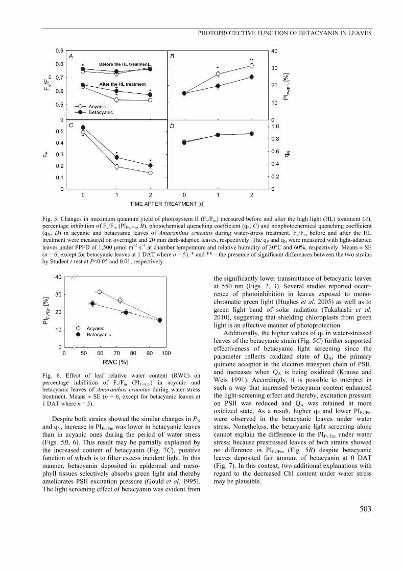
Fig. 5. Changes in maximum quantum yield of photosystem II (Fv/Fm) measured before and after the high light (HL) treatment (A), percentage inhibition of Fv/Fm (PIFv/Fm, B), photochemical quenching coefficient (qP, C) and nonphotochemical quenching coefficient (qN, D) in acyanic and betacyanic leaves of Amaranthus cruentus during water-stress treatment. Fv/Fm before and after the HL treatment were measured on overnight and 20 min dark-adapted leaves, respectively. The qP and qN were measured with light-adapted leaves under PPFD of 1,500 μmol m–2 s–1 at chamber temperature and relative humidity of 30C and 60%, respectively. Means ± SE (n = 6, except for betacyanic leaves at 1 DAT where n = 5). * and ** – the presence of significant differences between the two strains by Student t-test at P<0.05 and 0.01, respectively.
Fig. 6. Effect of leaf relative water content (RWC) on percentage inhibition of Fv/Fm (PIFv/Fm) in acyanic and betacyanic leaves of Amaranthus cruentus during water-stress treatment. Means ± SE (n = 6, except for betacyanic leaves at 1 DAT where n = 5).
Despite both strains showed the similar changes in PN and qN, increase in PIFv/Fm was lower in betacyanic leaves than in acyanic ones during the period of water stress (Figs. 5B, 6). This result may be partially explained by the increased content of betacyanin (Fig. 7C), putative function of which is to filter excess incident light. In this manner, betacyanin deposited in epidermal and meso-phyll tissues selectively absorbs green light and thereby ameliorates PSII excitation pressure (Gould et al. 1995). The light screening effect of betacyanin was evident from
the significantly lower transmittance of betacyanic leaves at 550 nm (Figs. 2, 3). Several studies reported occur-rence of photoinhibition in leaves exposed to mono-chromatic green light (Hughes et al. 2005) as well as to green light band of solar radiation (Takahashi et al. 2010), suggesting that shielding chloroplasts from green light is an effective manner of photoprotection.
Additionally, the higher values of qP in water-stressed leaves of the betacyanic strain (Fig. 5C) further supported effectiveness of betacyanic light screening since the parameter reflects oxidized state of QA, the primary quinone acceptor in the electron transport chain of PSII, and increases when QA is being oxidized (Krause and Weis 1991). Accordingly, it is possible to interpret in such a way that increased betacyanin content enhanced the light-screening effect and thereby, excitation pressure on PSII was reduced and QA was retained at more oxidized state. As a result, higher qP and lower PIFv/Fm were observed in the betacyanic leaves under water stress. Nonetheless, the betacyanic light screening alone cannot explain the difference in the PIFv/Fm under water stress; because prestressed leaves of both strains showed no difference in PIFv/Fm (Fig. 5B) despite betacyanic leaves deposited fair amount of betacyanin at 0 DAT (Fig. 7). In this context, two additional explanations with regard to the decreased Chl content under water stress may be plausible.

T. NAKASHIMA et al.
504
Fig. 7. Changes in total chlorophyll content [Chl (a+b), A], chlorophyll a/b ratio (Chl a/b, B) , betacyanin content (C) in acyanic and betacyanic leaves of Amaranthus cruentus during water stress treatment. Means ± SE (n = 6, except for betacyanic leaves at 1 DAT where n = 5) .*** denotes the presence of significant differences between the two strains by Student t-test at P<0.001.
Firstly, reduced Chl content at 1 and 2 DAT may have expedited the degree of light attenuation by increasing relative abundance of betacyanin to Chl, so that the proportion of light absorbed by Chl was decreased. The similar argument was presented by Zeliou et al. (2009) for cold-stressed citrus leaves. Secondly, the reduced Chl content was also in part involved in the alteration of light profile within leaves and conditions for Chl fluorescence measurement. That is, when concentration of Chl within a leaf is high, penetration of measuring beam, as well as of actinic light, is exclusively limited to shallower tissue layers near upper leaf surface (Schindler and Lichten-thaler 1994). Moreover, Chl fluorescence signal emitted from deeper tissue is likely to be reabsorbed by Chl in
upper tissues (Lichtenthaler et al. 2005). Such self-shading property of Chl leads to a measurement bias by which only photoinhibition at upper tissue layers is estimated. In water-stressed leaves, on the other hand, the effect of self-shading was lessened due to the reduced Chl content, allowing light to penetrate into deeper tissues within a leaf. As the result, the emission and detection of Chl fluorescence from deeper tissues were facilitated and therefore photoinhibition in these tissues was more readily detected.
Several studies reported that green light penetrates into deeper tissues than blue and red light and eventually is absorbed by Chl there (Koizumi et al. 1998, Vogelmann and Han 2000). Further, Oguchi et al. (2011) recently showed that green light induced more severe photodamage than red light at deeper tissues within capsicum leaves. Therefore, leaf betacyanin is considered to ameliorate photoinhibition at deeper tissues rather than tissues near adaxial surface. Thus, the drastic decline of Chl content permitted the assessment of photoinhibition at deeper tissues where the effect of betacyanic light screening is most apparent, explaining the presence of significant interstrainal difference in the PIFv/Fm of water-stressed leaves, but not in that of prestressed leaves.
Apart from the protective effect of betacyanin, it may also interfere with photosynthetic light acquisition, since green light is absorbed and utilized for carbon fixation at deeper tissue within a leaf (Sun et al. 1998, Terashima et al. 2009). This was evident from slightly lower quantum yield of photosynthesis in betacyanic leaves than in acyanic leaves of A. cruentus (data not shown). However, under subsaturating light conditions where light avail-ability is not a primary constraint, the loss of photosyn-thesis may be negligible. Rather, it may afford photo-protection for shade-adapted chloroplasts in deeper tissue which is considered more prone to photoinhibition (Gould et al. 2000). Such photoprotective mechanism may be particularly effective in leaves characterized by lower Chl content and reduced photosynthetic capacity, e.g. leaves under abiotic stresses or senescing process.
In summary, we conclude that leaf betacyanin provides an additional photoprotection, at least in part, by attenuating excess light in water-stressed leaves of A. cruentus, although an alternative function of beta-cyanin as a direct ROS scavenger still remains to be evaluated. Consequently, under photoinhibitory growth conditions, betacyanic strains of Amaranthus spp. may show higher tolerance to adverse effects of photo-inhibition such as ROS-mediated leaf senescence (Munné-Bosch and Alegre 2004)
References Asada, K.: The water-water cycle as alternative photon and
electron sinks. – Phil. Trans. R. Soc. Lond. B. 355: 1419-1431, 2000.
Burger, J. Edwards, G.E.: Photosynthetic efficiency, and photo-
damage by UV and visible radiation, in red versus green leaf Coleus varieties. – Plant Cell Physiol. 37: 395-399, 1996.
Buschmann, C.: Photochemical and non-photochemical quench-ing coefficients of the chlorophyll fluorescence: comparison

PHOTOPROTECTIVE FUNCTION OF BETACYANIN IN LEAVES
505
of variation and limits. – Photosynthetica 37: 217-224, 1999. Carmo-Silva, A.E., Powers, S.J., Keys, A.J., Arrabaca, M.C.,
Parry, M.A. J.: Photorespiration in C4 grasses remains slow under drought conditions. – Plant Cell Environ. 31: 925-940, 2008.
Chalker-Scott, L.: Do anthocyanins function as osmoregulators in leaf tissues? – Adv. Bot. Res. 37: 104-129, 2002.
Du, Y.C., Kawamitsu, Y., Nose, A., Hiyane, S., Murayama, S., Wasano, K., Uchida, Y.: Effects of water stress on carbon exchange rate and activities of photosynthetic enzymes in leaves of sugarcane (Saccharum sp.). – Aust. J. Plant Physiol. 23: 719-726, 1996.
Elliott, D.C.: Analysis of variability in the Amaranthus betacya-nin assay for cytokinins. – Plant Physiol. 63: 274-276, 1979.
Escudero, N.L., de Arellano M.L., Luco, J.M., Giménez, M.S., Mucciarelli, S.I.: Comparison of the chemical composition and nutritional value of Amaranthus cruentus flour and its protein concentrate. – Plant Food Human Nutr. 59:15-21, 2004.
Flexas, J., Escalona, J.M. and Medrano, H.: Down-regulation of photosynthesis by drought under field conditions in grapevine leaves. – Funct. Plant Biol. 25: 893-900, 1998.
Flexas, J., Medrano, H.: Energy dissipation in C3 plants under drought. – Funct. Plant Biol. 29: 1209–1215, 2002.
Gaume, A., Mächler, F., De León, C, Narro, L., Frossard, E.: Low-P tolerance by maize (Zea mays L.) genotypes: significance of root growth, and organic acids and acid phosphatase root exudation. – Plant Soil 228: 253-264, 2001.
Genty, B., Briantais, J.M., Baker, N.R.: The relationship between the quantum yield of photosynthetic electron transport and quenching of chlorophyll fluorescence. – Biochim. Biophys. Acta 990: 87-92, 1989.
Ghannoum, O.: C4 photosynthesis and water stress. – Ann. Bot. 103: 635-644, 2009.
Gould, K.S., Kuhn, D.N., Lee, D.W., Oberbauer, S.F.: Why leaves are sometimes red? – Nature 378: 241-242, 1995.
Gould, K.S., Markham, K.R., Smith, R.H., Goris, J.J.: Functional role of anthocyanins in the leaves of Quintinia serrata A. Cunn. – J. Exp. Bot. 51: 1107-1115, 2000.
Hayakawa, K., Agarie, S.: Physiological roles of betacyanin in halophyte, Suaeda japonica Makino. – Plant Prod. Sci. 13: 351-359, 2010.
Hughes, N.M., Neufield, H.S., Burkey, K.O.: Functional role of anthocyanins in high-light winter leaves of the evergreen herb Galax urceolata. – New Phytol. 168: 575-587, 2005.
Hughes, N.M., Smith, W.K.: Attenuation of incident light in Galax urceolata (Diapensiaceae): concerted influence of adaxial and abaxial anthocyanic layers on photoprotection. – Amer. J. Bot. 94: 784-790, 2007.
Hura, T., Hura, K., Grzesiak, M., Rzepka, A.: Effect of long-term drought stress on leaf gas exchange and fluorescence parameters in C3 and C4 plants. – Acta Physiol. Plant. 29: 103-113, 2007.
Koizumi, M., Takahashi, K., Mineuchi, K., Nakamura, T. Kano, H.: Light gradients and the transverse distribution of chlorophyll fluorescence in mangrove and Camellia leaves. – Ann. Bot. 81: 527-533, 1998.
Krause, G.H., Weis, E.: Chlorophyll fluorescence and photo-synthesis: the basics. – Annu. Rev. Plant Physiol. Plant Mol. Biol. 42: 313-349, 1991.
Lichtenthaler, H.K., Buschmann, C., Knapp, M: How to correctly determine the different chlorophyll fluorescence parameters and the chlorophyll fluorescence decrease ratio
RFD of leaves with the PAM fluorometer. – Photosynthetica 43: 379-393, 2005.
Liu, F., Stützel, H.: Leaf water relations of vegetable amaranth (Amaranthus spp.) in response to soil drying. – Eur. J. Agron. 16: 137-150, 2002.
Long, S.P., Hallgren, J.E.: Measurements of CO2 assimilation by plants in the field and the laboratory. – In: Coombs, J., Hall, D.O., Long, S.P., Scurlock, J.M.O. (ed.): Techniques in Bioproductivity and Photosynthesis. 2nd Ed. Pp. 62-94. Pregamon Press, Oxford – New York – Toronto – Sydney – Frankfurt 1985.
Manetas, Y.: Why some leaves are anthocyanic and why most anthocyanic leaves are red? – Flora 201: 163-177, 2006.
Manetas, Y., Petropoulou, Y., Psaras, G.K., Drinia, A.: Exposed red (anthocyanic) leaves of Quercus coccifera display shade characteristics. – Funct. Plant Biol. 30: 265-270, 2003.
Medrano, H., Bota, J., Abadía, A., Sampol, B., Escalona, J.M., Flexas, J.: Effects of drought on light-energy dissipation mechanisms in high-light acclimated, field grown grapevines. – Funct. Plant Biol. 29: 1197-1207, 2002.
Munné-Bosch, S., Alegre, L.: Die and let live: leaf senescence contributes to plant survival under drought stress. – Funct. Plant Biol. 31: 203-216, 2004.
Oberbauer, S.F., Starr, G.: The role of anthocyanins for photosynthesis of Alaskan artic evergreens during snowmelt. – Adv. Bot. Res. 37: 129-145, 2002.
Ögren, E., Sjöström, M.: Estimation of the effect of photo-inhibition on the carbon gain in leaves of a willow canopy. – Planta 181: 560-567, 1990.
Oguchi, R., Douwstra, P., Fujita, T., Chow, W.S., Terashima, I.: Intra-leaf gradients of photoinhibition induced by different color lights: implications for the dual mechanisms of photoinhibition and for the application of conventional chlorophyll fluorometers. – New Phytol. 191: 146-159, 2011.
Oxborough, K., Baker, N.R.: Resolving chlorophyll a fluores-cence images of photosynthetic efficiency into photochemical and non-photochemical components- calculation of qP and Fv’/Fm’ without measuring F0’. – Photosynth. Res. 54: 135-142, 1997.
Porra, R.J., Thompson, W.A., Kriedemann, P.E.: Determination of accurate extinction coefficients and simultaneous equations for assaying chlorophylls a and b extracted with four different solvents: verification of the concentration of chlorophyll standards by atomic absorption spectroscopy. – Biochim. Biophys. Acta 975: 384-394, 1989.
Ripley, B.S., Gilbert, M.E., Ibrahim, D.G., Osborne, P.C.: Drought constraints on C4 photosynthesis: stomatal metabolic limitation in C3 and C4 subspecies of Alloteropsis semialata. – J. Exp. Bot. 58: 1351-1363, 2007.
Schindler, C., Lichtenthaler, H. K.: Photosynthetic CO2 assimi-lation, chlorophyll fluorescence and zeaxanthin accumulation in field grown maple tree in the course of a sunny and a cloudy day. – J. Plant Physiol. 148: 399-412, 1994.
Shu, Z., Shao, L., Huang, H.-Y., Zeng, X.-Q., Lin, Z.-F., Chen, G.-Y., Peng, C.-L.: Comparison of thermostability of PSII between the chromatic and green leaf cultivars of Amaranthus tricolor L. – Photosynthetica 47: 548-558, 2009.
Solovchenko, A.E., Merzlyak, M.N.: Screening of visible and UV radiation as a photoprotective mechanism in plant. – Russ. J. Plant Physiol. 55: 803-822, 2008.
Sun, J., Nishio, J.N., Vogelmann, T.C.: Green light drives CO2 fixation deep within leaves. – Plant Cell Physiol. 39: 1020-1026, 1998.

T. NAKASHIMA et al.
506
Takahashi, S., Badger, M.R.: Photoprotection in plants: a new light on photosystem II damage. – Trends Plant Sci. 16: 53-60, 2011.
Takahashi, S., Milward, S. E., Yamori, W., Evans, J.R., Hillier, W., Badger, M.R.: The solar action spectrum of photosystem II damage. – Plant Physiol. 153: 988-993, 2010.
Terashima, I., Fujita, T., Inoue, T, Chow, W.S., Oguchi, R.: Green light drives leaf photosynthesis more efficiently than red light in strong white light: revising the enigmatic question of why leaves are green. – Plant Cell Physiol. 50: 684-697, 2009.
Tezara, W., Driscoll, S., Lawlor, D.W.: Partitioning of photo-synthetic electron flow between CO2 assimilation and O2 reduction in sunflower plants under water deficit. – Photosynthetica 46: 127-134, 2008.
Tezara, W., Mitchell, V.J., Driscoll, S.D. Lawlor, D.W.: Water stress inhibits plant photosynthesis by decreasing coupling factor and ATP. – Nature 401: 914-917, 1999.
van Kooten, O., Snel, J.F.H.: The use of chlorophyll fluores
cence nomenclature in plant stress physiology. – Photosynth. Res. 25: 147-150, 1990.
Vogelmann, T.C., Han, T.: Measurement of gradients of absorbed light in spinach leaves from chlorophyll fluores-cence profiles. – Plant Cell Environ. 23:1303-1311, 2000.
Wang, C.-Q., Liu, T.: Involvement of betacyanin in chilling-induced photoinhibition in leaves of Suaeda salsa. – Photosynthetica 45: 182-188, 2007.
Zeliou, K., Manetas, Y., Petropoulou, Y.: Transient winter leaf reddening in Citrus creticus characterizes weak (stress-sensitive) individuals, yet anthocyanins cannot alleviate the adverse effects on photosynthesis. – J. Exp. Bot. 60: 3031-3042, 2009.
Zhu, X., Ort, D.R., Whitmarsh, J. Long, S.P.: The slow reversibility of photosystem II thermal energy dissipation on transfer from high to low light may cause large losses in carbon gain by crop canopies: a theoretical analysis. – J. Exp. Bot. 55: 1167-1175, 2004.