photosynthetic electron xow avects h 2o2 signaling by ... · planta (2008) 228:1055–1066 doi...
TRANSCRIPT
Planta (2008) 228:1055–1066
DOI 10.1007/s00425-008-0807-0ORIGINAL ARTICLE
Photosynthetic electron Xow aVects H2O2 signaling by inactivation of catalase in Chlamydomonas reinhardtii
Ning Shao · Christoph F. Beck · Stéphane D. Lemaire · Anja Krieger-Liszkay
Received: 11 July 2008 / Accepted: 4 August 2008 / Published online: 10 September 2008© Springer-Verlag 2008
Abstract A speciWc signaling role for H2O2 in Chlamydo-monas reinhardtii was demonstrated by the deWnition of apromoter that speciWcally responded to this ROS. Expres-sion of a nuclear-encoded reporter gene driven by this pro-moter was shown to depend not only on the level ofexogenously added H2O2 but also on light. In the dark, theinduction of the reporter gene by H2O2 was much lowerthan in the light. This lower induction was correlated withan accelerated disappearance of H2O2 from the culturemedium in the dark. Due to a light-induced reduction incatalase activity, H2O2 levels in the light remained higher.Photosynthetic electron transport mediated the light-con-trolled down-regulation of the catalase activity since it wasprevented by 3-(3�4�-dichlorophenyl)-1,1-dimethylurea(DCMU), an inhibitor of photosystem II. In the presence oflight and DCMU, expression of the reporter gene was lowwhile the addition of aminotriazole, a catalase inhibitor, ledto a higher induction of the reporter gene by H2O2 in the
dark. The role of photosynthetic electron transport and thio-redoxin in this regulation was investigated by usingmutants deWcient in photosynthetic electron Xow and bystudying the correlation between NADP-malate dehydroge-nase and catalase activities. It is proposed that, contrary toexpectations, a controlled down-regulation of catalaseactivity occurs upon a shift of cells from dark to light. Thisdown-regulation apparently is necessary to maintain a cer-tain level of H2O2 required to activate H2O2-dependent sig-naling pathways.
Keywords Catalase · Chlamydomonas · Hydrogen peroxide · Signaling · Thioredoxin
AbbreviationsAPX Ascorbate peroxidaseAT AminotriazoleDCMU 3-(3�4�-Dichlorophenyl)-1, 1-dimethylureaDTT DithiothreitolMDH Malate dehydrogenaseLUC LuciferaseROS Reactive oxygen speciesTRX Thioredoxin
Introduction
Life in an oxygen-rich atmosphere has to cope with thedanger of oxidative stress. Plants are particularly exposedto oxidative stress caused by photosynthetic processes.Reactive oxygen species (ROS) are produced during nor-mal cell metabolism but their production is drasticallyenhanced when plants are exposed to stresses such as highlight, low temperatures, or drought, or combinationsthereof. Due to their high reactivity, ROS can damage
N. Shao · C. F. BeckFakultät für Biologie, Institut für Biologie III, Universität Freiburg, Schänzlestrasse 1, 79104 Freiburg, Germany
N. ShaoMax-Planck-Institut für Molekulare PXanzenphysiologie, Am Mühlenberg 1, 14476 Potsdam/Golm, Germany
S. D. LemaireInstitut de Biotechnologie des Plantes, Centre National de la Recherche ScientiWque, Université Paris-Sud, 91405 Orsay Cedex, France
A. Krieger-Liszkay (&)CEA, iBiTecS, CNRS URA 2096, Service de Bioénergétique, Biologie Structurale et Mécanisme, 91191 Gif-sur-Yvette Cedex, Francee-mail: [email protected]
123
1056 Planta (2008) 228:1055–1066
macromolecules essential for the integrity of the cell suchas lipids, proteins, and nucleic acids. Thus, the concentra-tion and accumulation of ROS needs to be strictly con-trolled. For this purpose, cells have evolved a variety ofvery eYcient non-enzymatic (e.g., ascorbate, glutathione,tocopherols) and enzymatic (e.g., catalase, ascorbate peroxi-dase, superoxide dismutase, peroxiredoxins, thioredoxins,glutaredoxins) antioxidant systems (Apel and Hirt 2004;Foyer and Noctor 2005). The requirement for a control ofROS accumulation implicates that these reactive intermedi-ates are sensed by cells in order to activate stress responsesallowing adaptation to a new set of environmental condi-tions. It is now widely accepted that ROS, and especiallyH2O2, play a major role in cellular signaling pathways andregulation of gene expression in a wide variety of organ-isms including plants (Apel and Hirt 2004; Laloi et al.2004). Several studies have used microarray analyses tostudy alterations in gene expression in response to ROS(Gadjev et al. 2006 and references therein). These studieshave demonstrated that increased ROS levels alter theexpression of a rather large set of genes (up to one-third ofthe genome). They also have shown that diVerent ROS havespeciWc signaling properties; a result supported by therecent discovery that H2O2 and singlet oxygen employ spe-ciWc targets for the activation of an ROS-responsive pro-moter (Shao et al. 2007). The mechanisms that allowtransduction of ROS signals to the nucleus remainunknown. In plants, the signaling pathways mediated byROS involve heterotrimeric G-proteins (Joo et al. 2005)and protein phosphorylation mediated by MAP kinases andprotein phosphatases (Kovtun et al. 2000; Gupta and Luan2003; Rentel et al. 2004).
Under conditions of intracellular H2O2 production ortreatment with exogenous H2O2, several plant H2O2 detoxi-fying enzymes, such as catalases and ascorbate peroxi-dases, are activated at the transcript, protein, and/or activitylevels (Gechev et al. 2002; Vandenabeele et al. 2004; Yab-uta et al. 2004; Davletova et al. 2005). Several studies havealso shown that some of these enzymes may be inactivatedby H2O2 (Miyake and Asada 1996; Shikanai et al. 1998;Hiner et al. 2000; Miyake et al. 2006).
Ascorbate peroxidases (APX) are the main H2O2 detoxi-fying enzymes in plants. Several isoforms of APX exist inplants and are located in the chloroplasts, the cytosol andperoxisomes (Shigeoka et al. 2002; Panchuk et al. 2005;Teixeira et al. 2006). Incubation of tobacco cell cultures inthe presence of an H2O2 generating system (glucose + glu-cose oxidase) has been shown to result in inhibition ofcytosolic APX activity (De Pinto et al. 2006). Also theendogenous generation of H2O2 via photosynthetic electrontransfer resulted in inhibition of chloroplastic APX activityin tobacco plants (Miyake et al. 2006). APX were recentlyfound among new putative targets of thioredoxins (TRX)
identiWed by proteomic studies (Marchand et al. 2004;Wong et al. 2004; Yamazaki et al. 2004). Moreover, puri-Wed recombinant cytosolic APX from poplar was shown tobe inhibited by reduced cytosolic thioredoxin h, reducedglutathione, and dithiothreitol. The molecular mechanismof this inhibition though remains to be determined (Gelhayeet al. 2006).
In plants, also catalases and peroxiredoxins play animportant role in detoxifying H2O2 (for review see Feiera-bend 2005; Dietz et al. 2006). Light inactivation of catalasehas been observed for enzymes of both plant and animalorigin. DiVerent isoforms of plant catalases show diVerentsensitivities towards light (Grotjohann et al. 1997; Engelet al. 2006). The action spectrum of photoinactivation isidentical to the absorption spectrum of catalase, indicatingthat the light inactivation of catalases is caused by directlight absorption by the heme moieties. However, when irra-diation was performed in the presence of chloroplasts the invitro inactivation of catalase was enhanced. Under theseconditions red light which is not absorbed by catalase butby photosynthetic pigments is suYcient to inhibit catalase(Feierabend and Engel 1986).
Catalases from Chlamydomonas, wheat, spinach, peaand potato were found among the potential targets of thi-oredoxin (TRX) identiWed by proteomic studies (Balmeret al. 2004; Lemaire et al. 2004; Maeda et al. 2004;Wong et al. 2004; Michelet et al. 2006). TRX are smalldisulWde oxidoreductases which play a major role inlight signaling and in the oxidative stress responses(Vieira Dos Santos and Rey 2006; Lemaire et al. 2007).We have previously shown that in total soluble extractsof Chlamydomonas, the activity of catalase could bemodulated by cytosolic thioredoxin h: a 50% inhibitionin catalase activity was observed after treatment with thecytosolic TRX system (NADPH, NADPH-thioredoxinreductase, TRX h) which could be mimicked by treat-ment with the strong reductant dithiothreitol (DTT)(Lemaire et al. 2004).
Hence, the question arises whether a down-regulationof the activity of ROS detoxifying enzymes, like APX andcatalases, might be necessary to allow diVusion and per-ception of H2O2 under stress conditions that would resultin the activation of genes that allow acclimation tochanges in the environment. In order answer this question,we investigated whether photosynthetic electron transfermay be involved in the regulation of H2O2 detoxifyingenzymes and thus the regulation of gene expression inresponse to H2O2. As a model system, we have chosenChlamydomonas reinhardtii which was transformed witha construct containing a Renilla reniformis luciferasereporter gene under the control of an HSP70A promoterfragment that speciWcally responds to H2O2 (Shao et al.2007).
123
Planta (2008) 228:1055–1066 1057
Materials and methods
Algal strains and culture conditions
Chlamydomonas reinhardtii strains 325 (CW15, arg7-8)and D66 (CW15) were kindly provided by R. Matagne(University of Liège, Belgium) and Arthur Grossman (Car-negie Institute, Stanford, CA, USA), respectively. Thewild-type strains CC-124 (mt¡) and WT34 (mt+) (a deriva-tive of strain 137c) were from the Chlamydomonas Genet-ics Center (Duke University, Durham, NC, USA) and theInstitut de Biologie Physico-Chimique in Paris, respec-tively. Mutants �petD (Kuras and Wollman 1994) andrbcL lacking both, the small and large subunit of Rubisco(rbcL-18-5B, Spreitzer et al. 1985) were kindly providedby Olivier Vallon (IBPC, Paris). Cultures were grownphotomixotrophically in Tris, acetate, phosphate (TAP)medium (Harris 1989) on a rotary shaker at 23–25°C undercontinuous irradiation with white light (70 �mol photonsm¡2 s¡1). TAP medium was supplemented with 100 mg l¡1
of arginine when required.
Nuclear transformation of Chlamydomonas
Plasmids used for Chlamydomonas transformation werepuriWed by PEG precipitation. Prior to transformation, theplasmid containing the ARG7 gene (pCB412) was linear-ized by EcoRI digestion. Plasmids containing the reporterconstruct, PHSP70A-�HSE -LUC (Shao et al. 2007) were line-arized by ScaI digestion. Chlamydomonas reinhardtii strain325 was cotransformed with pCB412 (selection plasmid)and the plasmid containing the PHSP70A-�HSE -LUC reporterconstruct using the glass beads method (Kindle 1990). Fortransformation, cells were grown to 3–5 £ 106 cells ml¡1
and concentrated to 3 £ 108 cells ml¡1. 1 £ 108 cells weremixed with 500 ng of linearized LUC reporter plasmid,100 ng of linearized pCB412, 0.3 g of acid-washed glassbeads, and 100 �l of 10% PEG 6000. Immediately aftervortexing for 20 s, cells were spread onto TAP-agar (1%agar) plates for the selection of arginine autotrophic clones.Transformants that harbored the LUC constructs weredetected by luciferase assay.
Bioluminescence assay
Bioluminescence assays were performed essentially asdescribed by Minko et al. (1999) at room temperature witha luminescence counter (MicroBeta TriLux) set in “Xash”mode with single auto-injection. After sampling, cells werespun down, resuspended in the same volume of samplebuVer [1.5 mM Tris-HCI (pH 7.8), 1 mM EDTA], and fro-zen at ¡80°C for at least 20 min. After thawing, 20 �l were
transferred to 96-well microtiter plates and 125 �l assaybuVer [0.1 M K2HPO4 (pH 7.6); 0.5 M NaCl; 1 mMEDTA] were added to each well. After incubation at roomtemperature for 15 min in the dark, bioluminescence wasassayed using the MicroBeta TriLux. The substrate(0.01 mM Coelenterazine, Biosynth AG, Staad, Switzer-land) was auto-injected into the wells, and luminescencewas recorded over a 20 s period, following a 1 s delay win-dow. The background was normalized using wells contain-ing only buVer or buVer with cells lacking the LUC gene.Expression of the LUC reporter gene in transgenic C. rein-hardtii cells was normalized for cell numbers. The induc-tion factor was calculated by comparison with untreatedcells.
RNA isolation and RNA-blot analyses
RNA extraction, electrophoretic separation of RNA andhybridizations were performed as described previously(von GromoV et al. 1989). The probe for LUC (accessionnumber: AY004213) was a 0.9-kb BamHI/XhoI fragmentfrom pCrLUC (Fuhrmann et al. 2004). The CBLP geneencoding a Chlamydomonas G�-like polypeptide wasemployed as loading control gene (von Kampen et al.1993).
Measurement of hydrogen peroxide
At each time point, 0.5-ml aliquots of the cultures werecentrifuged at 13,000g in a microcentrifuge. The superna-tants were mixed with an equal volume of 1 M KI. After15 min at room temperature, the mixture was assayed foriodine formation by determining the OD at 390 nm. Theabsorbance at 390 nm was stable for at least 3 h (WaVe-nschmidt et al. 1993). Concentrations were determinedusing a standard calibration curve with known amounts ofH2O2 (Sigma).
Measurement of enzyme activities
Cultures of C. reinhardtii strain D66 were grown to 1–2 £ 106 cells ml¡1, transferred in the dark for 16 h, andsubsequently exposed to light (70 �mol photons m¡2 s¡1)or maintained in the dark. When indicated, DCMU (Wnalconcentration 6 �M) was added 45 min prior to illumina-tion. Some cultures were supplemented with H2O2 (2 mMWnal concentration). Cells to be collected (1 £ 108 cells)were pelleted by centrifugation (microcentrifuge, maximalspeed) and resuspended in 200 �l 50 mM Hepes buVer (pH8). After two consecutive freeze/thawing cycles in liquidnitrogen, the enzyme activities in crude extracts of Chla-mydomonas cultures were measured.
123

1058 Planta (2008) 228:1055–1066
Catalase activity was measured polarographically at20°C with a Clark-type electrode in 50 mM Hepes buVer(pH 8) in the presence of 1 mM H2O2 as substrate using aWnal protein concentration of 5 �g ml¡1. The protein con-centration of the crude extracts was determined using theamido black assay.
Ascorbate peroxidase activity was measured photometri-cally at 290 nm in 20 mM phosphate buVer, pH 7.0, con-taining 0.5 mM ascorbate and 1 mM H2O2 as substratesusing crude extract with a Wnal protein concentration of5 �g ml¡1.
Initial NADP-malate dehydrogenase activity was mea-sured photometrically at 340 nm in 100 mM Tris/HCl,pH 7.9, containing 0.75 mM oxalacetate and 0.27 mMNADPH as substrates using crude extract with a Wnalprotein concentration of 10 �g ml¡1. To activate MDH toits maximal activity, samples were incubated with10 mM DTT, 20-�M thioredoxin (cytosolic thioredoxinh1 from Chlamydomonas) in 100 mM Tris/HCl, pH 7.9for 10 min prior to start of the assay by addition of sub-strates. The ratio between initial activity and total activ-ity gives the percentage of activated MDH at the timepoint of harvest.
Statistics
Data represent means or representative examples frommeasurements repeated 3–6 times. Typical standard errorsare shown, in Figs. 1b, 6 and 8; they are omitted for somedata for clarity.
Results
Induction of an H2O2-responsive reporter gene by H2O2 is strongly stimulated by light
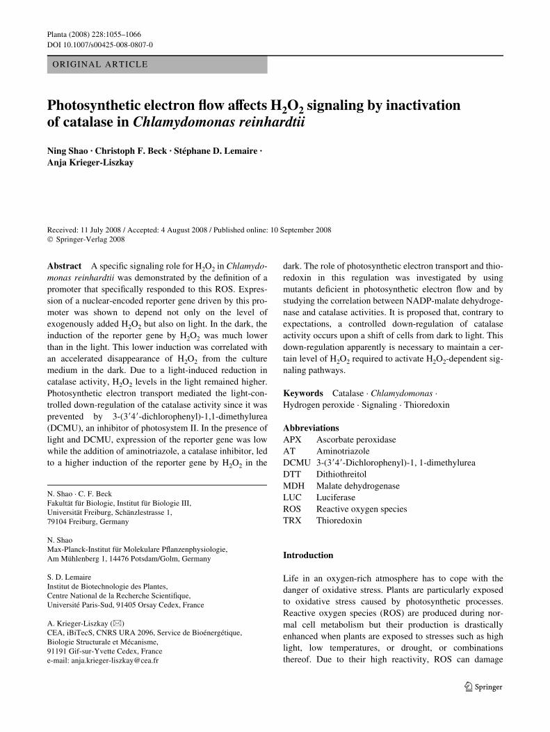
In the present work, we investigated the regulation of anH2O2-responsive reporter gene by exogenously addedH2O2. For these assays we employed transformants harbor-ing an H2O2-responsive fragment of the HSP70A promoterfused to a Renilla-derived luciferase gene (LUC) (Shaoet al. 2007). A schematic representation of the LUC
Fig. 1 a–c Response of a PHSP70A-Luc reporter gene in Chlamydo-monas transformants to the addition of H2O2 in the light and in thedark. a A truncated version (�HSE) of the HSP70A promoter constructknown to be inducible by H2O2 (Shao et al. 2007) was fused to a lucif-erase reporter gene that at its upstream end harbored sequences ofHSP70B. Dark shaded bars represent exons, light shaded bars indicateintrons. The third exon of HSP70B was fused to a Renilla luciferase re-porter gene with C. reinhardtii adapted codon usage (Fuhrmann et al.2004) (LUC), indicated by a dark grey bar. The 3� UTR of the fusiongene is encoded by the corresponding region of RBCS2. b Induction ofPHSP70A-LUC in Chlamydomonas transformants by exogenously ap-plied hydrogen peroxide. Light grown C. reinhardtii cells were incu-bated in the presence of 2 mM H2O2 with white light of an intensity of70 �mol photons m¡2 s¡1 (open circles) or in the dark (closed circles).H2O was added to light-incubated control cultures (open squares) andto control cultures transferred to the dark (closed squares). Samples forluciferase assays were taken at the time points indicated and processedas described in Sect. “Materials and methods”. The activity was nor-malized with respect to cell numbers and the induction factors werecalculated by comparison with untreated cells at time point 0 h. cDependence of the degree of induction of the PHSP70A-LUC reporterconstruct in Chlamydomonas transformants in the light on the concen-tration of exogenous H2O2. Experiments were performed as describedin b. Samples for luciferase assays were taken after incubation for 3 h(diamonds) and after 5 h (triangles)
ATG LUC
-149 -23
Time (h)
0.0 0.5 1.0 1.5 2.0
0
50
100
150
200
250
LUC
aci
tvity
(fo
ld in
duct
ion)
H2O
2 (mM)
5 HSP70B
PHSP70A-∆HSE-LUC3
RB
CS
2
0 1 2 3 4 5
0
50
100
150
200
250
LUC
act
ivity
(fo
ld in
duct
ion)
a
b
c
�
123
Planta (2008) 228:1055–1066 1059
reporter construct is presented in Fig. 1a. Transformantswith this construct did not respond to heat shock butexpression of the reporter gene is induced by hydrogen per-oxide, either produced inside the chloroplast or added exog-enously to the culture medium (Shao et al. 2007).
A strong increase in luciferase activity was observed intransformants with this construct after treatment by H2O2,but only when the cultures were incubated in the light(Fig. 1b). In the light, the activity increased about 200-foldwithin 5 h of incubation in the presence of 2 mM H2O2. Inthe dark, a distinctly weaker induction was observed. Theluc activity was 11-fold higher after 3 h, and 19-fold higherafter 5 h incubation. Light itself (70 �mol photons m¡2 s¡1)had no eVect since no induction of the reporter gene couldbe observed in the light-grown control cultures supple-mented with H2O. In the absence of H2O2, no diVerencewas observed between light-grown cultures and culturestransferred from the light to the dark. When cultures weregrown in the dark for 20 h and then shifted to light in theabsence of H2O2, a weak increase in luciferase activity wasobserved (Shao et al. 2007). This increase was 2.5- to3-fold smaller than the one observed in the presence of H2O2
in the dark.We next tested the level of expression in dependence on
the concentration of H2O2 added to the culture medium.Luciferase activity was shown to increase strongly with theconcentration of H2O2 as can be seen by plotting luciferaseactivity 3 and 5 h after H2O2 addition as a function of theconcentration of externally added H2O2 (Fig. 1c). We haveshown previously that much lower H2O2 concentrations aresuYcient to induce the reporter gene when H2O2 is pro-duced inside the chloroplast by the photosynthetic electrontransfer chain in the presence of metronidazole as electronacceptor. In this case, a high level of induction of thereporter gene was observed already after 2 h, when thedetectable H2O2 concentration in the medium was 3 �M(Shao et al. 2007).
Catalase activity contributes to a faster degradation of H2O2 in the dark
The strong induction of transformants with the PHSP70A-�HSE
-LUC reporter construct by externally added H2O2
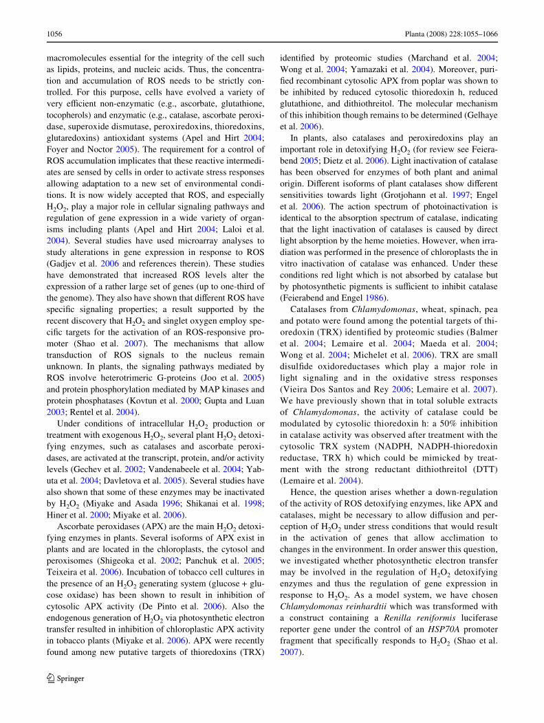
observed only in the light may possibly be linked to lowerrate of H2O2 degradation by Chlamydomonas cells incu-bated in the light than in the dark. This was tested by mea-suring the rate of H2O2 disappearance from the culturemedium kept in light and or darkness. Indeed, H2O2 wasmore rapidly degraded by cultures in the dark than in thelight (Fig. 2a). In the dark, H2O2 almost completely disap-peared from the culture medium within 4 h and more than50% of the initial H2O2 had disappeared already within 1 h.By contrast, in the light, about 75% of the H2O2 was still
present after 1 h and some H2O2 could still be detected after4 h. The slower disappearance of the H2O2 from the culturemedium in the light may possibly be explained by the con-tinuous production of H2O2 by the photosynthetic electrontransport chain. However, the Mehler reaction as potentialinternal source of H2O2 cannot explain the diVerencebetween H2O2 degradation in the light and in the dark,because little H2O2 was produced by cultures, even in thepresence of metronidazole, an artiWcial electron acceptor ofPSI (less than 2 �M H2O2 per hour of irradiation wasdetectable in the medium; Shao et al. 2007).
Fig. 2 a, b Degradation of exogenously added H2O2 by Chlamydo-monas cultures. a Time course of degradation of H2O2 added to the me-dium of Chlamydomonas cultures in the light (open circles) or in thedark (closed circles). The change in H2O2 content in cell-free mediumserved as a control (open squares). 2 mM H2O2 was added at time pointzero. Samples were taken at the times points indicated and the H2O2concentration was determined as described in Sect. “Materials andmethods”. Light-grown C. reinhardtii cells were irradiated with70 �mol photons m¡2 s¡1 of white light. b Time course of H2O2 deg-radation in the medium of Chlamydomonas cultures in the dark in theabsence (closed circles) or in the presence of 2 mM aminotriazole, acatalase inhibitor (closed triangles). The inhibitor was added 45 minprior to the addition of 2 mM H2O2. All other conditions were asdescribed in a
0.0
0.5
1.0
1.5
2.0
H2O
2 m
M
0 1 2 3 4
0.0
0.5
1.0
1.5
2.0
Time (h)
a
b
123

1060 Planta (2008) 228:1055–1066
This faster degradation of H2O2 in the dark may becaused by a light-dependent inhibition of H2O2 detoxifyingenzymes. In plants, catalases and ascorbate peroxidasesconstitute the two major classes of enzymes involved inH2O2 detoxiWcation. To test whether catalase is involved inthe observed diVerences in H2O2 degradation between lightand dark conditions, we measured the rates of H2O2 degra-dation in the dark in the presence or absence of the catalaseinhibitor aminotriazole (AT; Fig. 2b). The concentration ofH2O2 in the culture medium decreased considerably slowerin the presence of AT than in its absence. This indicatesthat, under the conditions tested, catalase appears to be oneof the major enzymes involved in H2O2 detoxiWcation inChlamydomonas cells. This result also suggested that alight-dependent regulation of catalase activity may accountfor the diVerences in H2O2 degradation observed betweenlight and dark conditions (Fig. 2) and consequently for thelight-dependence of the H2O2 inducibility of the reportergene (Fig. 1b).
Light has previously been shown to inactivate plant cata-lases (Grotjohann et al. 1997; for review see Feierabend2005). Generally, this inactivation was observed under highlight conditions (800 �mol photons m¡2 s¡1). To testwhether catalase activity was already aVected by the lowlight intensities used in the experiments shown in Figs. 1and 2, cell extracts of Chlamydomonas were illuminatedwith white light of an intensity of 70 �mol photons m-2 s¡1
for up to 5 h. Under these conditions, no signiWcant photo-inactivation of the enzyme could be observed within 5 h ofillumination (Fig. 6). We therefore concluded that a directphotoinactivation of catalase is unlikely to play a role underthe low light conditions that were employed for studyingthe induction of the reporter gene by H2O2.
The redox state of the photosynthetic electron transfer chain aVects H2O2 detoxiWcation and catalase activity
Recently, it was shown that the activity of Chlamydomonascatalase is inhibited by reduced TRX in vitro (Lemaireet al. 2004). In chloroplasts, TRXs are reduced by the pho-tosynthetic electron transfer chain in the light. The reduc-tion of TRXs may thus possibly be responsible for theinactivation of catalases; and therefore, the slower H2O2
degradation observed in the light. Such a mechanism mayaccount for the light dependence of the induction of thereporter gene by H2O2. In order to test whether photosyn-thetic electron transfer plays a role in this light dependence,we investigated the eVect of DCMU, an inhibitor of photo-system II, on the expression of the reporter gene, the degra-dation of H2O2, and the activity of H2O2 detoxifyingenzymes. In the presence of DCMU, linear photosyntheticelectron transfer is blocked and, among other componentswhich may play a role in redox regulation, chloroplastic
TRXs remain oxidized. In the presence of DCMU, only asmall increase in luciferase activity could be detected aftertreatment with H2O2 in the light (Fig. 3), indicating only aweak induction of the reporter gene. The level of luciferaseactivity observed was similar to that seen after addition ofH2O2 in the dark. This suggests that linear photosyntheticelectron Xow is required for an eYcient induction of thereporter gene by H2O2 in the light.
If this requirement is linked to a light-dependent inacti-vation of catalase, implicating TRXs or other mediators,then DCMU was predicted to aVect the rate of H2O2 degra-dation in the light and the activity of catalases. Therefore,the time course of H2O2 consumption was measured in thelight in the presence and absence of DCMU (Fig. 4). In thelight, the H2O2 concentration decreased signiWcantly fasterin the presence of DCMU than in its absence. This suggeststhat photosynthetic electron Xow accounts for the eVect oflight on the rate of H2O2 degradation in the culture medium.
To investigate the eVect of the redox state of photosyn-thetic electron Xow further, the degradation of H2O2 wastested using mutants lacking either the cytochrome b6fcomplex (�petD) or deWcient in ribulose-1,5-bisphosphatecarboxylase (Rubisco) (rbcL). As shown in Fig. 5, only asmall diVerence in the kinetics of H2O2 degradation wasobserved between dark-treated and light-treated cultures ofthe petD mutant lacking the cytochrome b6f complex. ThediVerences were statistically not signiWcant according to theStudent’s t test. In this mutant, forward electron transport toPSI is completely abolished, the plastoquinone pool stays
Fig. 3 EVect of DCMU on the induction of the PHSP70A-LUC reporterconstruct by H2O2 in Chlamydomonas transformants. Cells were incu-bated in the presence of 6 �M DCMU for 45 min prior to the additionof 2 mM H2O2. After H2O2 addition, cells were incubated for 5 h in thelight (70 �mol photons m¡2 s¡1) or in the dark. Luciferase activity wasnormalized with respect to cell numbers. The luciferase activity oflight-grown cultures in the absence of DCMU was set as one
Light Dark Light Dark Light Light
H2O2 - - + + + -
DCMU - - - - + +
0.0
0.2
0.4
0.6
0.8
1.0
Rel
ativ
e LU
C a
ctiv
ity
123

Planta (2008) 228:1055–1066 1061
reduced and components downstream from PSI remain oxi-dized. In the mutant lacking the large subunit of Rubisco,the diVerence in H2O2 degradation between light and darkis comparable to that observed in the wild-type strain. Inthis mutant, upon irradiation, the electron transport chain,including electron acceptors like NADP+ and thioredoxinsstays highly reduced.
These data suggest that photosynthetic electron Xow reg-ulates the activity of H2O2 detoxifying enzymes, possiblyinvolving TRXs. In order to test this possibility, we mea-sured catalase activity in cells opened up by freeze/thawing.
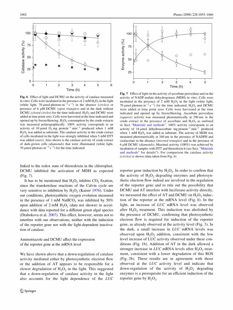
The activity could be inhibited by 75% by addition of 5-mM DTT (Fig. 6). This indicates that the catalase canundergo redox dependent inactivation. Figure 6 shows thatincubation of the cells in the light clearly inhibited theactivity of catalase. Indeed, about 40% of the activity waslost within 2 h and only 25% remained after 5 h of incuba-tion in the light. The remaining activity after 5 h illumina-tion corresponded to the activity of the catalase in thepresence of DTT. By contrast, in cells maintained in thedark, the activity remained above 85% of the initial activitythroughout the experiment. Presence of DCMU during thelight incubation protected against the loss of activity(Fig. 6), supporting the idea that the activity of H2O2-con-suming enzymes is regulated via the redox state of the elec-tron transfer chain. The kinetics of inactivation were notdependent on the presence of H2O2 in the culture medium,since the same degree of inhibition was observed when noH2O2 was added prior to the shift of the cultures from darkto light (data not shown).
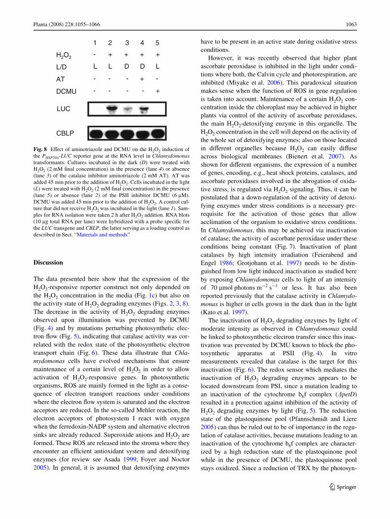
The activity of ascorbate peroxidases did not show anyinhibition during the time course of illumination of thecultures in the presence of 2 mM H2O2 (Fig. 7), indicatingthat indeed the redox-dependent inhibition of catalase isthe important factor leading to a loss of H2O2 degradationand subsequent expression of the reporter gene. To show acorrelation between the state of reduction of thioredoxinsand the activity of catalase, we measured the activity ofthe NADP-dependent malate dehydrogenase (MDH), oneof the classical enzymes activated by reduction of its disul-Wde bonds by reduced thioredoxin (e.g., Lemaire et al.2005, 2007). MDH activity increased upon incubation inthe light while catalase activity decreased (Fig. 7). Thesedata support the hypothesis that the activity of catalase is
Fig. 4 EVect of DCMU on the degradation of H2O2 in the medium byChlamydomonas cells. Cells were incubated with irradiation (whitelight, 70 �mol photons m¡2 s¡1) in the presence of 6 �M DCMU for45 min (open triangles) or its absence (open circles) prior to additionof 2 mM H2O2. Results from cells incubated in the absence of DCMUin the dark (closed circles) are also shown. Samples were taken at thetimes points indicated and the H2O2 concentration in the media wasdetermined as described in Sect. “Materials and methods”
0 1 2 3 4
0.0
0.5
1.0
1.5
2.0
Time (h)
H2O
2(m
M)
Fig. 5 Degradation of exogenously added H2O2 in the medium byphotosynthesis mutants. Time course of degradation of H2O2 addedto the medium of Chlamydomonas cultures in the light (white light,55 �mol photons m¡2 s¡1) (open symbol) or in the dark (closedsymbol). The rbcL and �petD mutants were grown in very dim light(<10 �mol photons m¡2 s¡1) for 5 days and then diluted to1 £ 106 cell ml¡1. The parental strain of the mutants (WTS34), named
WT, was incubated with irradiation (white light,55 �mol photons m¡2 s¡1) for 3 days and then diluted to1 £ 106cell ml¡1. Right after dilution, H2O2 (Wnal concentration2 mM) was added. Samples were taken at the time points indicated andthe H2O2 concentration in the media was determined as described inSect. “Materials and methods”
0 30 60 90 1200.0
0.5
1.0
1.5
2.0
0 30 60 90 1200 30 60 90 120
∆petDrbcL
H2O
2 (m
M)
Time (min)
wt
Time (min)Time (min)
123
1062 Planta (2008) 228:1055–1066
linked to the redox state of thioredoxin in the chloroplast.DCMU inhibited the activation of MDH as expected(Fig. 7).
It has to be mentioned that H2O2 inhibits CO2 Wxation,since the transketolase reactions of the Calvin cycle arevery sensitive to inhibition by H2O2 (Kaiser 1976). Underour conditions, photosynthetic oxygen evolution measuredin the presence of 1 mM NaHCO3 was inhibited by 50%upon addition of 2 mM H2O2 (data not shown) in accor-dance with data reported for a diVerent green algal species(Drabokova et al. 2007). This eVect, however, seems not tointerfere with our observations, neither with the inductionof the reporter gene nor with the light-dependent inactiva-tion of catalase.
Aminotriazole and DCMU aVect the expression of the reporter gene at the mRNA level
We have shown above that a down-regulation of catalaseactivity mediated either by photosynthetic electron Xowor the addition of AT appears to be responsible for aslower degradation of H2O2 in the light. This suggestedthat a down-regulation of catalase activity in the lightalso accounts for the light dependence of the LUC
reporter gene induction by H2O2. In order to conWrm thatthe activity of H2O2 degrading enzymes and photosyn-thetic electron Xow indeed are involved in the regulationof the reporter gene and to rule out the possibility thatDCMU and AT interfere with luciferase activity directly,we measured the eVect of AT and DCMU on H2O2 induc-tion of the reporter at the mRNA level (Fig. 8). In thelight, an increase of LUC mRNA level was observedafter H2O2 treatment. This induction was abolished bythe presence of DCMU, conWrming that photosyntheticelectron Xow is required for induction of the reportergene, as already observed at the activity level (Fig. 3). Inthe dark, a small increase in LUC mRNA levels wasobserved upon H2O2 addition, consistent with the lowlevel increase of LUC activity observed under these con-ditions (Fig. 1b). Addition of AT in the dark allowed astronger increase in LUC mRNA levels after H2O2 treat-ment, consistent with a lower degradation of this ROS(Fig. 2b). These results are in agreement with thoseobserved at the LUC activity level and indicate thatdown-regulation of the activity of H2O2 degradingenzymes is a prerequisite for an eYcient induction of thereporter gene by H2O2.
Fig. 6 EVect of light and DCMU on the activity of catalase measuredin vitro. Cells were incubated in the presence of 2 mM H2O2 in the light(white light, 70 �mol photons m¡2 s¡1) in the absence (circles) orpresence of 6 �M DCMU (open triangles) and in the dark withoutDCMU (closed circles) for the time indicated. H2O2 and DCMU wereadded at time point zero. Cells were harvested at the time indicated andopened up by freeze/thawing. H2O2 consumption by the crude extractswas measured polarographically. 100% activity corresponds to anactivity of 10 �mol O2 mg protein¡1 min¡1 produced when 1 mMH2O2 was added as substrate. The catalase activity in the crude extractof cells incubated in the light was strongly inhibited when 5 mM DTTwas added (stars). Also shown is the catalase activity of crude extractof dark-grown cells (diamonds) that were illuminated (white light,70 �mol photons m¡2 s¡1) for the time indicated
0 1 2 3 4 5 60
25
50
75
100
Time (h)
Cat
alas
e ac
tivity
(%
)
Fig. 7 EVect of light on the activity of ascorbate peroxidase and on theactivity of NADP-malate dehydrogenase (MDH) in vitro. Cells wereincubated in the presence of 2 mM H2O2 in the light (white light,70 �mol photons m¡2 s¡1) for the time indicated. H2O2 and DCMUwere added at time point zero. Cells were harvested at the timeindicated and opened up by freeze/thawing. Ascorbate peroxidase(squares) activity was measured photometrically at 290 nm in thecrude extract in the presence of ascorbate and H2O2 as outlinedin Sect. “Materials and methods”. 100% activity corresponds to anactivity of 18 �mol dehydroascorbate mg protein¡1 min¡1 producedwhen 1 mM H2O2 was added as substrate. The activity of MDH wasmeasured photometrically at 340 nm in the presence of NADPH andoxalacetate in the absence (inverted triangles) and in the presence of6 �M DCMU (diamonds). Maximal activity (100%) was achieved byincubation of samples with DTT and thioredoxin h (see Sect. “Materialsand methods” for details”). For comparison the catalase activity(circles) is shown (data taken from Fig. 6)
Time (h)
Enz
yme
activ
ity (
%)
0 1 2 3 4 5
25
50
75
100
123
Planta (2008) 228:1055–1066 1063
Discussion
The data presented here show that the expression of theH2O2-responsive reporter construct not only depended onthe H2O2 concentration in the media (Fig. 1c) but also onthe activity state of H2O2 degrading enzymes (Figs. 2, 3, 8).The decrease in the activity of H2O2 degrading enzymesobserved upon illumination was prevented by DCMU(Fig. 4) and by mutations perturbing photosynthetic elec-tron Xow (Fig. 5), indicating that catalase activity was cor-related with the redox state of the photosynthetic electrontransport chain (Fig. 6). These data illustrate that Chla-mydomonas cells have evolved mechanisms that ensuremaintenance of a certain level of H2O2 in order to allowactivation of H2O2-responsive genes. In photosyntheticorganisms, ROS are mainly formed in the light as a conse-quence of electron transport reactions under conditionswhere the electron Xow system is saturated and the electronacceptors are reduced. In the so-called Mehler reaction, theelectron acceptors of photosystem I react with oxygenwhen the ferredoxin-NADP system and alternative electronsinks are already reduced. Superoxide anions and H2O2 areformed. These ROS are released into the stroma where theyencounter an eYcient antioxidant system and detoxifyingenzymes (for review see Asada 1999; Foyer and Noctor2005). In general, it is assumed that detoxifying enzymes
have to be present in an active state during oxidative stressconditions.
However, it was recently observed that higher plantascorbate peroxidase is inhibited in the light under condi-tions where both, the Calvin cycle and photorespiration, areinhibited (Miyake et al. 2006). This paradoxical situationmakes sense when the function of ROS in gene regulationis taken into account. Maintenance of a certain H2O2 con-centration inside the chloroplast may be achieved in higherplants via control of the activity of ascorbate peroxidases,the main H2O2-detoxifying enzyme in this organelle. TheH2O2 concentration in the cell will depend on the activity ofthe whole set of detoxifying enzymes; also on those locatedin diVerent organelles because H2O2 can easily diVuseacross biological membranes (Bienert et al. 2007). Asshown for diVerent organisms, the expression of a numberof genes, encoding, e.g., heat shock proteins, catalases, andascorbate peroxidases involved in the abrogation of oxida-tive stress, is regulated via H2O2 signaling. Thus, it can bepostulated that a down-regulation of the activity of detoxi-fying enzymes under stress conditions is a necessary pre-requisite for the activation of those genes that allowacclimation of the organism to oxidative stress conditions.In Chlamydomonas, this may be achieved via inactivationof catalase; the activity of ascorbate peroxidase under theseconditions being constant (Fig. 7). Inactivation of plantcatalases by high intensity irradiation (Feierabend andEngel 1986; Grotjohann et al. 1997) needs to be distin-guished from low light induced inactivation as studied hereby exposing Chlamydomonas cells to light of an intensityof 70 �mol photons m¡2 s¡1 or less. It has also beenreported previously that the catalase activity in Chlamydo-monas is higher in cells grown in the dark than in the light(Kato et al. 1997).
The inactivation of H2O2 degrading enzymes by light ofmoderate intensity as observed in Chlamydomonas couldbe linked to photosynthetic electron transfer since this inac-tivation was prevented by DCMU known to block the pho-tosynthetic apparatus at PSII (Fig. 4). In vitromeasurements revealed that catalase is the target for thisinactivation (Fig. 6). The redox sensor which mediates theinactivation of H2O2 degrading enzymes appears to belocated downstream from PSI, since a mutation leading toan inactivation of the cytochrome b6f complex (�petD)resulted in a protection against inhibition of the activity ofH2O2 degrading enzymes by light (Fig. 5). The reductionstate of the plastoquinone pool (Pfannschmidt and Liere2005) can thus be ruled out to be of importance in the regu-lation of catalase activities, because mutations leading to aninactivation of the cytochrome b6f complex are character-ized by a high reduction state of the plastoquinone poolwhile in the presence of DCMU, the plastoquinone poolstays oxidized. Since a reduction of TRX by the photosyn-
Fig. 8 EVect of aminotriazole and DCMU on the H2O2 induction ofthe PHSP70A-LUC reporter gene at the RNA level in Chlamydomonastransformants. Cultures incubated in the dark (D) were treated withH2O2 (2 mM Wnal concentration) in the presence (lane 4) or absence(lane 3) of the catalase inhibitor aminotriazole (2 mM AT). AT wasadded 45 min prior to the addition of H2O2. Cells incubated in the light(L) were treated with H2O2 (2 mM Wnal concentration) in the presence(lane 5) or absence (lane 2) of the PSII inhibitor DCMU (6 �M).DCMU was added 45 min prior to the addition of H2O2. A control cul-ture that did not receive H2O2 was incubated in the light (lane 1). Sam-ples for RNA isolation were taken 2 h after H2O2 addition. RNA blots(10 �g total RNA per lane) were hybridized with a probe speciWc forthe LUC transgene and CBLP, the latter serving as a loading control asdescribed in Sect. “Materials and methods”
LUC
H2O2
L/D
AT
DCMU
-+---
+----
LDDLL
++++-
1 2 3 4 5
CBLP
123

1064 Planta (2008) 228:1055–1066
thetic electron transfer chain is prevented by DCMU and inthe petD mutant, these molecules are candidates for regula-tors of H2O2 degrading enzymes. Indeed, several H2O2
detoxifying enzymes have been shown to be inactivated byTRXs in vitro (Lemaire et al. 2004; Gelhaye et al. 2006).Proteomic studies revealed that also several ROS scaveng-ing enzymes which do not use TRXs as substrates such ascatalase, superoxide dismutase, or APX, are putative TRXtargets since they are bound to TRX aYnity columns(Balmer et al. 2004; Lemaire et al. 2004; Maeda et al. 2004;Wong et al. 2004; Michelet et al. 2006).
In the genomic sequence of Chlamydomonas (http://genome.jgi-psf.org/Chlre3/Chlre3.home.html), only a sin-gle gene encoding a typical catalase (gene model Chlre3/scaVold_30:1312932-1317626) was observed. A gene(gene model Chlre3/scaVold_1:4550894-4558523) encod-ing a putative catalase–peroxidase homologous to thosefound in prokaryotes (Jakopitsch et al. 2005) was alsofound. However, as judged on the basis of the number ofEST sequences, expression of the latter gene appears to bevery weak as compared to the expression of the typical cat-alase gene, even under oxidative stress conditions. Conse-quently, the catalase activities reported here are likely tocorrespond to the activity of the typical catalase, although itcannot be excluded that the putative catalase–peroxidasealso contributes to H2O2 degrading activity measured inextracts.
In higher plants, catalases were localized to peroxisomes(Heazlewood et al. 2005), while in Chlamydomonas theenzyme is most likely located in mitochondria (Kato et al.1997; Lemaire et al. 2004). This implies that a signal aboutthe redox state of the photosynthetic electron transfer chainhas to be transmitted to targets outside of the chloroplast.This signal could be initiated by a component whose redoxstate depends on the redox state of a functional photosyn-thetic electron transfer chain such as TRXs. The signal ispossibly transmitted from the chloroplast by redox media-tors such as glutathione or NADPH. Indeed, reducingequivalents of chloroplastic NADPH have been reported tobe transferred outside of the chloroplast in the form ofmalate (Scheibe 2004). This malate is formed by reductionof oxalacetate using NADPH as electron donor. Interest-ingly, the NADP-malate dehydrogenase of the chloroplastis activated by light by a mechanism dependent on theredox state of plastidic TRXs (Lemaire et al. 2005, 2007).Indeed, a reverse correlation between the activities of cata-lase and NADP-malate dehydrogenase was observed(Fig. 7). Once outside of the chloroplast, the signal may betransduced to a redox regulator of the catalase via diVerentredox mediators. Since the activity of Chlamydomonas cat-alase has been shown to be regulated by TRXs in vitro(Lemaire et al. 2004), the redox regulator of catalase islikely to be a TRX or involves TRX-related proteins like
glutaredoxins. There is some evidence that thioredoxinsbelonging to the TRX o and TRX h subtypes are present inthe mitochondria of Chlamydomonas (Lemaire et al. 2003).
In conclusion, the data presented provide support forthe idea that Chlamydomonas has evolved mechanismsthat allow maintenance of a certain level of H2O2. Weassume that these mechanisms ensure a regulation of geneexpression in response to ROS, allowing for an adaptationof the organisms to stress conditions. A fast inactivation ofcatalase activity is seen when cells are exposed to a 10-fold higher light intensity (data not shown). Regulation ofthe activity of ROS detoxifying enzymes appears to playan important role. A regulation via TRX or other compo-nents of the regulatory redox network like glutaredoxins,peroxiredoxins or glutathione provides an attractive strat-egy for the regulation of such enzyme activities. Thesecompounds may ensure a fast and reversible inactivationof the enzymes, thereby allowing for a rapid sensing ofchanges in environmental conditions. Most importantly,through thioredoxins or other redox compounds, the regu-lation of the activity of H2O2-consuming enzymes maydirectly be coupled to photosynthetic electron Xow, andthus to the main source for ROS production in photosyn-thetic organisms.
Acknowledgments The authors are grateful to Olivier Vallon (IB-PC, Paris) for many suggestions and the providing of strains, Myrosl-awa Miginiac-Maslow (IBP, Orsay) for valuable suggestions and toBill Rutherford, and Diana Kirilovsky (both at CEA, Saclay) for criti-cal reading of the manuscript. This work was supported in part bygrants of the Deutsche Forschungsgemeinschaft to C.F.B. (Be 903/13-2) and A.K.L. (Li883/10-1) and the Agence Nationale de la RechercheGrant JC05-45751 to S.D.L.
References
Apel K, Hirt H (2004) Reactive oxygen species: metabolism, oxida-tive stress, and signal transduction. Annu Rev Plant Biol55:373–399
Asada K (1999) The water–water cycle in chloroplasts: scavenging ofactive oxygens and dissipation of excess photons. Annu Rev PlantPhysiol Plant Mol Biol 50:601–639
Balmer Y, Vensel WH, Tanaka CK, Hurkman WJ, Gelhaye E, RouhierN, Jacquot JP, Manieri W, Schürmann P, Droux M, Buchanan BB(2004) Thioredoxin links redox to the regulation of fundamentalprocesses of plant mitochondria. Proc Natl Acad Sci USA101:2642–2647
Bienert GP, Møller AL, Kristiansen KA, Schulz A, Møller IM, Schjo-erring JK, Jahn TP (2007) SpeciWc aquaporins facilitate the diVu-sion of hydrogen peroxide across membranes. J Biol Chem282:1183–1192
Davletova S, Rizhsky L, Liang H, Shengqiang Z, Oliver DJ, Coutu J,Shulaev V, Schlauch K, Mittler R (2005) Cytosolic ascorbate per-oxidase 1 is a central component of the reactive oxygen gene net-work of Arabidopsis. Plant Cell 17:268–281
Dietz KJ, Jacob S, Oelze ML, Laxa M, Tognetti V, de Miranda SM,Baier M, Finkemeier I (2006) The function of peroxiredoxins inplant organelle redox metabolism. J Exp Bot 57:1697–1709
123
Planta (2008) 228:1055–1066 1065
Drabokova M, Admiraal W, Marsalek B (2007) Combined exposure tohydrogen peroxide and light-selective eVects on cyanobacteria,green algae, and diatoms. Environ Sci Technol 41:309–314
de Pinto MC, Paradiso A, Leonetti P, De Gara L (2006) Hydrogen per-oxide, nitric oxide and cytosolic ascorbate peroxidase at the cross-road between defence and cell death. Plant J 48:784–795
Engel N, Schmidt M, Lutz C, Feierabend J (2006) Molecular identiW-cation, heterologous expression and properties of light-insensitiveplant catalases. Plant Cell Environ 29:593–607
Feierabend J (2005) Catalases in plants: molecular and functionalproperties and role in stress defence. In: SmirnoV N (ed) Antiox-idants and reactive oxygen species in plants. Blackwell, Oxford,pp 101–140
Feierabend J, Engel S (1986) Photoinactivation of catalase in vitro andin leaves. Arch Biochem Biophys 251:567–576
Foyer CH, Noctor G (2005) Redox homeostasis and antioxidant signal-ing: a metabolic interface between stress perception and physio-logical responses. Plant Cell 17:1866–1875
Fuhrmann M, Hausherr A, Ferbitz L, Schodl T, Heitzer M, HegemannP (2004) Monitoring dynamic expression of nuclear genes inChlamydomonas reinhardtii by using a synthetic luciferase re-porter gene. Plant Mol Biol 55:869–881
Gadjev I, Vanderauwera S, Gechev TS, Laloi C, Minkov IN, ShulaevV, Apel K, Inze D, Mittler R, van Breusegem F (2006) Transcrip-tomic footprints disclose speciWcity of reactive oxygen speciessignaling in Arabidopsis. Plant Physiol 141:436–445
Gechev T, Gadjev I, van Breusegem F, Inze D, Dukiandjiev S, TonevaV, Minkov I (2002) Hydrogen peroxide protects tobacco fromoxidative stress by inducing a set of antioxidant enzymes. CellMol Life Sci 59:708–714
Gelhaye E, Navrot N, Macdonald IK, Rouhier N, Raven EL, JacquotJP (2006) Ascorbate peroxidase–thioredoxin interaction. Photo-synth Res 89:193–200
Grotjohann N, Janning A, Eising R (1997) In vitro photoinactivation ofcatalase isoforms from cotyledons of sunXower (Helianthus an-nuus L.). Arch Biochem Biophys 346:208–218
Gupta R, Luan S (2003) Redox control of protein tyrosine phospha-tases and mitogen-activated protein kinases in plants. Plant Phys-iol 132:1149–1152
Harris E (1989) The Chlamydomonas sourcebook: a comprehensiveguide to biology and laboratory use. Academic Press, SanDiego
Heazlewood JL, Tonti-Filippini J, Verboom RE, Millar AH (2005)Combining experimental and predicted datasets for determinationof the subcellular location of proteins in Arabidopsis. Plant Phys-iol 139:598–609
Hiner AN, Rodriguez-Lopez JN, Arnao MB, Lloyd Raven E, Garcia-Canovas F, Acosta M (2000) Kinetic study of the inactivation ofascorbate peroxidase by hydrogen peroxide. Biochem J 348:321–328
Jakopitsch C, Wanasinghe A, Jantschko W, Furtmuller PG, Obinger C(2005) Kinetics of interconversion of ferrous enzymes, compoundII and compound III, of wild-type synechocystis catalase–peroxi-dase and Y249F: proposal for the catalytic mechanism. J BiolChem 280:9037–9042
Joo JH, Wang S, Chen JG, Jones AM, FedoroV NV (2005) DiVerentsignaling and cell death roles of heterotrimeric G protein alphaand beta subunits in the Arabidopsis oxidative stress response toozone. Plant Cell 17:957–970
Kaiser W (1976) The eVect of hydrogen peroxide on CO2 Wxation ofisolated intact chloroplasts. Biochim Biophys Acta 440:476–482
Kato J, Yamahara T, Tanaka K, Takio S, Satoh T (1997) Characteriza-tion of catalase from green alga Chlamydomonas reinhardtii. JPlant Physiol 151:262–268
Kindle KL (1990) High-frequency nuclear transformation of Chla-mydomonas reinhardtii. Proc Natl Acad Sci USA 87:1228–1232
Kovtun Y, Chiu WL, Tena G, Sheen J (2000) Functional analysis ofoxidative stress-activated mitogen-activated protein kinase cas-cade in plants. Proc Natl Acad Sci USA 97:2940–2945
Kuras R, Wollman FA (1994) The assembly of cytochrome b6/f com-plexes: an approach using genetic transformation of the green algaChlamydomonas reinhardtii. EMBO J 13:1019–1027
Laloi C, Apel K, Danon A (2004) Reactive oxygen signalling: the lat-est news. Curr Opin Plant Biol 7:323–328
Lemaire SD, Collin V, Keryer E, Issakidis-Bourguet E, Lavergne D,Miginiac-Maslow M (2003) Chlamydomonas reinhardtii: a mod-el organism for the study of the thioredoxin family. Plant PhysiolBiochem 41:513–521
Lemaire SD, Guillon B, Le Maréchal P, Keryer E, Miginiac-MaslowM, Decottignies P (2004) New thioredoxin targets in the unicellu-lar photosynthetic eukaryote Chlamydomonas reinhardtii. ProcNatl Acad Sci USA 101:7475–7480
Lemaire SD, Quesada A, Merchan F, Corral JM, Igeno MI, Keryer E,Issakidis-Bourguet E, Hirasawa M, KnaV DB, Miginiac-MaslowM (2005) NADP-malate dehydrogenase from unicellular greenalga Chlamydomonas reinhardtii. A Wrst step toward redox regu-lation? Plant Physiol 137:514–521
Lemaire SD, Michelet L, ZaVagnini M, Massot V, Issakidis-BourguetE (2007) Thioredoxins in chloroplasts. Curr Genet 51:343–355
Maeda K, Finnie C, Svensson B (2004) Cy5 maleimide labelling forsensitive detection of free thiols in native protein extracts: identi-Wcation of seed proteins targeted by barley thioredoxin h iso-forms. Biochem J 378:497–507
Marchand C, Le Maréchal P, Meyer Y, Miginiac-Maslow M, Issakidis-Bourguet E, Decottignies P (2004) New targets of Arabidopsis thior-edoxins revealed by proteomic analysis. Proteomics 4:2696–2706
Michelet L, ZaVagnini M, Massot V, Keryer E, Vanacker H, Miginiac-Maslow M, Issakidis-Bourguet E, Lemaire SD (2006) Thioredox-ins, glutaredoxins, and glutathionylation: new crosstalks to ex-plore. Photosynth Res 89:225–245
Minko I, Holloway SP, Nikaido S, Carter M, Odom OW, Johnson CH,Herrin DL (1999) Renilla luciferase as a vital reporter for chloro-plast gene expression in Chlamydomonas. Mol Gen Genet262:421–425
Miyake C, Asada K (1996) Inactivation mechanism of ascorbate per-oxidase at low concentrations of ascorbate; hydrogen peroxidedecomposes compound I of ascorbate peroxidase. Plant CellPhysiol 37:423–430
Miyake C, Shinzaki Y, Nishioka M, Horiguchi S, Tomizawa K (2006)Photoinactivation of ascorbate peroxidase in isolated tobaccochloroplasts: Galdieria partita APX maintains the electron Xuxthrough the water–water cycle in transplastomic tobacco plants.Plant Cell Physiol 47:200–210
Panchuk II, Zentgraf U, Volkov RA (2005) Expression of the Apx genefamily during leaf senescence of Arabidopsis thaliana. Planta222:926–932
Pfannschmidt T, Liere K (2005) Redox regulation and modiWcation ofproteins controlling chloroplast gene expression. Antioxid RedoxSignal 7:607–618
Rentel MC, Lecourieux D, Ouaked F, Usher SL, Petersen L, OkamotoH, Knight H, Peck SC, Grierson CS, Hirt H, Knight MR (2004)OXI1 kinase is necessary for oxidative burst-mediated signallingin Arabidopsis. Nature 427:858–861
Scheibe R (2004) Malate valves to balance cellular energy supply.Physiol Plant 120:21–26
Shao N, Krieger-Liszkay A, Schroda M, Beck CF (2007) A reportersystem for the individual detection of hydrogen peroxide and sin-glet oxygen: its use for the assay of reactive oxygen species pro-duced in vivo. Plant J 50:475–487
Shigeoka S, Ishikawa T, Tamoi M, Miyagawa Y, Takeda T, Yabuta Y,Yoshimura K (2002) Regulation and function of ascorbate perox-idase isoenzymes. J Exp Bot 53:1305–1319
123

1066 Planta (2008) 228:1055–1066
Shikanai T, Takeda T, Yamauchi H, Sano S, Tomizawa KI, Yokota A,Shigeoka S (1998) Inhibition of ascorbate peroxidase under oxi-dative stress in tobacco having bacterial catalase in chloroplasts.FEBS Lett 428:47–51
Spreitzer RJ, Goldschmidt-Clermont M, Rahire M, Rochaix JD (1985)Nonsense mutations in the Chlamydomonas chloroplast gene thatcodes for the large subunit of ribulosebisphosphate carboxylase/oxygenase. Proc Natl Acad Sci USA 82:5460–5464
Teixeira FK, Menezes-Benavente L, Galvao VC, Margis R, Margis-Pinheiro M (2006) Rice ascorbate peroxidase gene family en-codes functionally diverse isoforms localized in diVerent subcel-lular compartments. Planta 224:300–314
Vandenabeele S, Vanderauwera S, Vuylsteke M, Rombauts S, Lange-bartels C, Seidlitz HK, Zabeau M, Van Montagu M, Inze D, vanBreusegem F (2004) Catalase deWciency drastically aVects geneexpression induced by high light in Arabidopsis thaliana. Plant J39:45–58
Vieira Dos Santos C, Rey P (2006) Plant thioredoxins are key actors inthe oxidative stress response. Trends Plant Sci 11:329–334
von GromoV ED, Treier U, Beck CF (1989) Three light-inducible heatshock genes of Chlamydomonas reinhardtii. Mol Cell Biol9:3911–3918
von Kampen J, Nieländer U, Wettern M (1993) Stress-dependent tran-scription of a gene encoding a G�-like polypeptide from Chla-mydomonas reinhardtii. J Plant Physiol 143:756–758
WaVenschmidt S, Woessner JP, Beer K, Goodenough UW (1993) Iso-dityrosine cross-linking mediates insolubilization of cell walls inChlamydomonas. Plant Cell 5:809–820
Wong JH, Cai N, Balmer Y, Tanaka CK, Vensel WH, Hurkman WJ,Buchanan BB (2004) Thioredoxin targets of developing wheatseeds identiWed by complementary proteomic approaches. Phyto-chemistry 65:1629–1640
Yabuta Y, Maruta T, Yoshimura K, Ishikawa T, Shigeoka S (2004) Twodistinct redox signaling pathways for cytosolic APX induction un-der photooxidative stress. Plant Cell Physiol 45:1586–1594
Yamazaki D, Motohashi K, Kasama T, Hara Y, Hisabori T (2004) Tar-get proteins of the cytosolic thioredoxins in Arabidopsis thaliana.Plant Cell Physiol 45:18–27
123