phylum arthropoda subphylum crustacea brünnich, 1772
TRANSCRIPT
9
SYSTEMATIC LIST OF INDIAN DIAPTOMID SPECIES
Phylum Arthropoda
Subphylum Crustacea Brünnich, 1772
Class Maxillopoda Dahl, 1956
Subclass Copepoda Milne Edwards, 1840
Order Calanoida Sars, 1903
Family Diaptomidae Sars, 1892
Subfamily Paradiaptominae Kiefer, 1932
I. Genus Paradiaptomus Sars, 1895
1. Paradiaptomus greeni (Gurney, 1906)
Subfamily Diaptominae Kiefer, 1932
VI. Genus Tropodiaptomus Kiefer, 1932
2. Tropodiaptomus defayeae n.sp.
3. Tropodiaptomus venkataramani n.sp.
4. Tropodiaptomus raoi n.sp.
5. Tropodiaptomus keralaensis n.sp.
6. Tropodiaptomus orientalis (Brady, 1886)
7. Tropodiaptomus doriai (Richard, 1897)
8. Tropodiaptomus hebereri Kiefer, 1930
9. Tropodiaptomus mutatus Kiefer, 1930
10. Tropodiaptomus vicinus Kiefer, 1930
11. Tropodiaptomus euchaetus Kiefer, 1936
II. Genus: Heliodiaptomus Kiefer, 1932
12. Heliodiaptomus viduus (Gurney, 1916)
13. Heliodiaptomus contortus (Gurney, 1907)

10
14. Heliodiaptomus cinctus (Gurney, 1907)
15. Heliodiaptomus pulcher (Gurney, 1907)
16. Heliodiaptomus kolleruensis Ranga Reddy & Radhakrishna, 1981
17. Heliodiaptomus elegans Kiefer, 1935
III. Genus Allodiaptomus Kiefer, 1936
18. Allodiaptomus (Allodiaptomus) mirabilipes Kiefer, 1936
19. Allodiaptomus (Allodiaptomus) intermedius Ranga Reddy, 1987
20. Allodiaptomus (Allodiaptomus) satanas (Brehm, 1952)
21. Allodiaptomus (Reductodiatpomus) raoi Kiefer, 1936
IV. Genus Neodiaptomus Kiefer, 1932
22. Neodiaptomus schmackeri (Poppe & Richard, 1892)
23. Neodiaptomus lindbergi Brehm, 1951
24. Neodiaptomus physalipus Kiefer, 1935
25. Neodiaptomus intermedius Flößner, 1984
26. Neodiaptomus meggitti Kiefer, 1932
V. Genus Phyllodiaptomus Kiefer, 1936
27. Phyllodiaptomus (Phyllodiaptomus) blanci (Guerne & Richard, 1896)
28. Phyllodiaptomus (Ctenodiaptomus) wellekensae Dumont & Ranga Reddy, 1993
29. Phyllodiaptomus (Ctenodiaptomus) annae (Apstein, 1907)
30. Phyllodiaptomus (Ctenodiaptomus) sasikumari Ranga Reddy & Venkateswarlu, 1989
VII. Genus Sinodiaptomus Kiefer, 1936
31. Sinodiaptomus (Rhinediaptomus) indicus Kiefer, 1936
32. Sinodiaptomus (Rhinediaptomus) mahanandiensis Ranga Reddy & Radhakrishna, 1980
VIII. Genus Megadiaptomus Kiefer, 1936
33. Megadiaptomus pseudohebes Ranga Reddy, 1987
34. Megadiaptomus hebes Kiefer, 1936

11
IX. Genus Eodiaptomus Kiefer, 1932
35. Eodiaptomus shihi Ranga Reddy, 1992
X. Genus Spicodiaptomus Rajendran, 1973
36. Spicodiaptomus chelospinus Rajendran, 1973
X1. Genus Keraladiaptomus Silva, Kakkassery, Maas & Dumont, 1994
37. Keraladiaptomus rangareddyi Silva, Kakkassery, Maas & Dumont, 1994
X11. Genus Acanthodiaptomus Kiefer, 1932
38. Acanthodiaptomus denticornis (Wierzejski, 1887)
XIII. Genus Arctodiaptomus Kiefer, 1932
39. Arctodiaptomus (Arctodiaptomus) similis similis (Baird, 1859)
40. Arctodiaptomus (Arctodiaptomus) eucanthus Kiefer, 1935
41. Arctodiaptomus (Rhabdodiaptomus) salinus (Daday, 1885)
42. Arctodiaptomus (Rhabdodiaptomus) michaeli Ranga Reddy, Balkhi& Yousuf, 1990
43. Arctodiaptomus (Stenodiaptomus) stewartianus (Brehm, 1924)
44. Arctodiaptomus (Stenodiaptomus) altissimus altissimus Kiefer, 1936
45. Arctodiaptomus (Haplodiaptomus) parvispineusKiefer, 1935

12
Subfamily Paradiaptominae Kiefer, 1932
Genus Paradiaptomus Sars, 1895
Broteas: Lovén, 1845 (partim)
Paradiaptomus: Sars, 1895
Broteas: Gurne & Richard, 1890: 200 (partim); Sars, 1899: 4.
Paradiaptomus: Giesbrecht & Schmiel, 1898: 95; Sars, 1907: 3; Tollinger, 1911: 187; van
Douwe, 1912a: 2; 1912b: 25; 1914: 96; Gurney, 1929: 572; Lowndes, 1936: 5; Kiefer, 1932a:
462; Kiefer, 1934: 123; Kiefer, 1978: 75; Defaye, 1988: 112; Dussart, 1989: 28; Rayner, 1992:
36-39; Dussart & Defaye, 1995: 124; Defaye et al., 200: 247-248; Dussart & Defaye, 2002: 64.
Lovenula: Grochmalicki, 1913: 525.
Generic diagnosis
Animals large (total length c. 2.0-2.5 mm). Antennule short, barely reaching the end of
prosome. Female: Fourth and fifth pedigers fused; urosome of 2 somites, genital double-somite
with asymmetrical lobes; P5 endopod with 2 spiniform setae. Male: Right antennule 21-
segmented, only 3 segments (exception: 4 segments in P. greeni) beyond geniculation, last
segment ending in a beak or rounded terminally; outermost seta of right caudal ramus enlarged;
right P5 with 1 disto-lateral spine; left P5 with sturdy lateral spiniform process on second
exopodal segment.
Type species: Paradiaptomus lamellatus Sars, 1895
Other valid species:
Paradiaptomus natalensis (Cooper, 1906)
Paradiaptomus greeni (Gurney, 1906)
Paradiaptomus schultzei van Douwe, 1912
13
Paradiaptomus similis van Douwe, 1912
Paradiaptomus rex Gauthier, 1951
Paradiaptomus hameri Rayner, 1999
Paradiaptomus peninsularis Rayner, 1999
Paradiaptomus warreni Rayner, 1999
Of the above species, P. greeni is the sole representative in India.
Paradiaptomus greeni (Gurney, 1906)
(Figs 1-4, Pl. 5)
Diaptomus greeni Gurney, 1906: 129-132, P1. 2, Figs 1-9; Tollinger, 1911: 119, Fig. F3.
Paradiaptomus similis van Douwe, 1912: 21-32, P1. IV, Figs 13-14.
Paradiaptomu greeni Gurney, 1931: 301-303, Figs 1-5; Kiefer, 1934:12, Figs 33-35; Kiefer,
1939: 92-95, Figs 1a-i; Brehm, 1950: 15; Brehm, 1953: 298-302, Figs 60-64; Rajendran,
1973:120-121, Figs 6 a-i; 141-161: Dussart & Defaye, 2002: 65; Ambedkar, 2005: 20-23, Figs 1-
8.
Type locality
Muddy stagnant pool, Cotton Experiment Station, Maha Ilupalama, North-Central
Province of Sri Lanka.
Material examined: as in Table 02.
Body size. Female 2.03-2.62 mm; male 1.86-2.13 mm.
Female
Body stout, fourth and fifth pedigers fused together, fusion indicated by indentation on
either side. Lateral wings of fifth pediger asymmetrical, large and somewhat ovate; left wing

14
smaller than right wing, posterolaterally directed; each wing armed with 1 apical and 1 inner
spine.
Urosome of 2 somites, second urosomite being fused to anal somite; sometimes
vague present septum separating the latte 2 segments; genital double-somite prominently
expanded in anterior half, with large bifid lobe on right side and small hook-like process on left
side. Second urosomite (urosomite 2 + anal somite) half as long as genital double-somite. Caudal
rami slightly asymmetrical, right ramus being somewhat narrower than left; inner margins hairy.
P5 (Fig. 1g-j, Pl. 5c). Coxal spine small. Seta on basis small. End claw (exopod 2)
moderately strong and blunt, carrying along inner margin a row of minute denticles; lateral spine
highly variable in size (Figs 1d, 1 e, Pl. 5 b). Exopod 3 small and with 2 unequal spines, outer
one being longer and stronger than inner one. Endopod unsegmented, somewhat shorter than
exopod1; apex with two unequal spines.
Male (Figs 2, 3). Metasomal wings greatly reduced, asymmetrical, right wing slightly
narrower than right one .
Caudal rami asymmetrical with hairy inner margins. All caudal setae of left ramus as well
as 4 inner setae of the right ramus normal, outer seta of the right ramus modified (Fig. 2a, Pl. 5d).
Antennule (Figs 2c, 3d-e). Left antennule as in female, right antennule modified and
geniculate; distal part beyond geniculation 4-segmented; with spine on segments 8 and 10-13;
length of spines in decreasing order as follows: 11>13>10>8>12; spine on segment 13 strong;
terminal segment beaked (Pl. 5e, f)
Right P5 (Figs 2d-i, 3f-m, Pl. 5g-i). Coxa with short, slender, acute spine; basis with
short sensory seta. Exopod 1 with dome-shaped blunt spine spine at distal outer angle; shape of
spine slightly variable (Figs 2e, 3h); distal inner corner with thumb-shaped hyaline lobe; exopod

15
2 very strong, almost rectangular, 1.8 times as long as wide; lateral spine inserted close to end
claw on posterior surface; end claw with serulations on inner margin. Endopod 2- segmented,
proximal segment larger; distal segment with a row of apical tiny spinules. Left P5: coxa with
slender spine; basis with small sensory seta; exopod 2 extended as a flange fringed with minute
spinules and armed laterally with a slender spine and slender seta.
Remarks
The following characters have been observed to show intraspecific variation: in the
female, the size of outer spine on left wing; lateral processes on genital double-somite, size of
lateral spine on second exopodal segment, and segmentation and apical spines of endopod of P5;
in the male right P5, the spinous process at distal outer corner and the hyaline projection at distal
inner corner of the first exopodal segment, the size and orientation of the lateral spine of second
exopodal segment, the form of end claw and segmentation of endopod.
P. greeni was reported by Gurney (1931) as being common to Sri Lanka, India and South
Africa. However, Rayner (1999), while revising the subfamily Paradiaptominae, reportedly
confirmed on the basis of material from India and Namibia that Gurney (1931) mistook P. greeni
for P. similis, an African species occurring in Kalahari, Namibia and other part of Africal.
Rayner (1999) also called attention to the subtle differences between these two species in the
male P5 and length and arrangement of spines on grasping antennule. Having examined the
permanent slide preparation of both sexes of P. similis from South Africa and compared this
species with several populations of P. greeni from India and Pakistan, I agree Rayners‘ (1999)
view that these two species are distinct from each other (Table 2).
Colour
16
The caudal setae as well as the terminal segments of antennule are brightly pink in both
sexes.
Ecology
P. greeni appears mainly during early monsoon period (June-August) in highly turbid
or moderately transparent, shallow, temporary, fishless water bodies such as seasonal ponds
and pools in which water temperature ranges from 19º to 29ºC and pH 7.0-7.5 and Secchi
transparency 39-40 cm. It often co-occurs with H. viduus, P. blanci and S. indicus. This species
is amenable to cultivation under laboratory conditions. According to Devi & Ranga Reddy
(1989a), who raised it from its eggs, it completes naupliar phase in 3 days and copepodid phase
in 6 days at a room temperature of 28º- 36º C.
Distribution
Previous records
This species has a widely distributed in Sri Lanka (Ceylon) and India. In India it has
been reported from Madurai, Nellore, Gooty, Guntakal, Dharamavaram, Nambur, Nellore,
Phandharpour, Soharva, and Kodaikanal.
Present records
Nallapadu, Vejendla, Akaveedu and Cumbum—all in the State of Andhra Pradesh.
Conservation Status: LR.

17
Subfamily Diaptominae Kiefer, 1932
Genus Tropodiaptomus Kiefer, 1932
This genus has two subgenera, viz. Tropodiaptomus s. str. and Anadiaptomus. The latter was
established by Brehm (1952) for only two Madagascan species whereas all other species belong
to the former. In India, the genus has been poorly studied.
Generic diagnosis
Female: urosome composed of 2 somites. Endopod with 1 subapical and 1 apical seta. Male:
right antennule with spine each on segments 8, 10, 11, 12, 13 and 15; segment 14 without spine;
spinous process on antepenultimate segment is always smooth. Left P5 exopod 1-segmented,
spatulate; apex rounded with spinulate seta (finger-thumb-process) and inner margin with
serulate or denticulate. Second exopodal segment of right P5 with hyaline lamella or spine near
the base of lateral spine.
Initially, Brehm (1953) provided a brief review of the Indian species along with an
identification key. The principal revisionary work on the Asiatic species of this genus is that of
Kiefer (1982). And according to Dussart & Defaye (2002), the genus contains t 64 species, most
of which are distributed in Africa and Asia. In India, the genus is represented by the following 14
species including three new species besides one new Nepalese species.
Type species: Tropodiaptomus orientalis (Brady, 1886)
Other valid species:
Tropodiaptomus doriai (Richard, 1897)
Tropodiaptomus hebereri (Kiefer, 1930)

18
Tropodiaptomus mutatus (Kiefer, 1930)
Tropodiaptomus vicinus (Kiefer, 1930)
Tropodiaptomus euchaetus Kiefer, 1936
Tropodiaptomus signatus Kiefer, 1982
Tropodiaptomus defayeae n. sp.
Tropodiaptomus keralaensis n. sp.
Tropodiaptomus raoi n. sp
Tropodiaptomus venkataramani n. sp.
Species incertae sedis:
Tropodiaptomus nielseni Brehm, 1953
Tropodiaptomus lakhimpurensis Reddiah, 1964
Tropodiaptomus chauhani Roy, 1984
Key to female Indian Tropodiaptomus spp.
1. Fourth seta (counted from outside) of third exopodal segment of P4 extraordinarily
long……………………………………………………………………………T. orientalis
The same normal…………………………………………………………………………..2
2. Genital double-somite with large swelling on subproximal left margin
………………………………………………………………………………..…T. signatus
No such structure…………………………………………………………………………….3
3. Fifth pediger with roundish hump mid-dorsally………………………..………T. hebereri

19
No such structures………………………………………………………………………..4
4. Genital double-somite dilated alike on both sides subproximally; setae on P5 endopod
always short………………………………………………………………………T. doriai
Genital double-somite not dilated on both sides sub proximally; setae on P5 endopod
either short or long……………………………..…………………………………………5
5. Genital double-somite produced into small lobe at right distal corner……………….....6
No such structure………………………………………………………………………….7
6. P5 endopod setae sturdy and spiniform; coxal spine large………………T. defayeae n. sp.
P5 endopod setae slender and setiform; coxal spine small……………………T. raoi n. sp.
7. On P5 endopod, apical seta as long as or longer than endopod……………………………8
On P5 endopod, apical seta shorter than endopod………………………………………10
8. P5 basis seta long; apical endopodal seta naked……………………….T. keralaensis n. sp.
P5 basis seta short; apical endopodal seta pinnate………………………………………….9
9. P5 endopod as long as inner margin of first exopodal segment……T. venkataramani n. sp.
P5 endopod shorter than first exopodal segment…………...………………….T. euchaetus
10. Female caudal rami with hairy outer margins; genital genital double-somite nearly
symmetrical ……………………………..………………………………………T. vicinus
Female caudal rami with hairless outer margins; genital double-somite decisively
asymmetrical…………………………………………………………………..T. mutatus
20
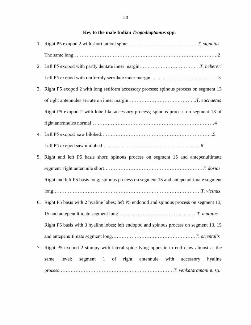
Key to the male Indian Tropodiaptomus spp.
1. Right P5 exopod 2 with short lateral spine…………………………………….T. signatus
The same long…………………………………………………………………………….2
2. Left P5 exopod with partly dentate inner margin……………………………….T. hebereri
Left P5 exopod with uniformly serrulate inner margin…………………………………...3
3. Right P5 exopod 2 with long setiform accessory process; spinous process on segment 13
of right antennules serrate on inner margin…………………………………...T. euchaetus
Right P5 exopod 2 with lobe-like accessory process; spinous process on segment 13 of
right antennules normal…………………………………………………………………4
4. Left P5 exopod saw bilobed…………………………………………………………...5
Left P5 exopod saw unilobed……………………………………………………6
5. Right and left P5 basis short; spinous process on segment 15 and antepenultimate
segment right antennule short……………………………………………………T. doriai
Right and left P5 basis long; spinous process on segment 15 and antepenultimate segment
long………………………………………………………………………………T. vicinus
6. Right P5 basis with 2 hyaline lobes; left P5 endopod and spinous process on segment 13,
15 and antepenultimate segment long…………………………………………T. mutatus
Right P5 basis with 3 hyaline lobes; left endopod and spinous process on segment 13, 15
and antepenultimate segment long……………………………………………T. orientalis
7. Right P5 exopod 2 stumpy with lateral spine lying opposite to end claw almost at the
same level; segment 1 of right antennule with accessory hyaline
process…………………………………………………………….T. venkataramani n. sp.

21
Right P5 exopod 2 elongate, with lateral spine lying distinctly proximal to end claw;
segment 13 of right antennules without accessory process………………………………8
8. Right P5 exopod 2 distinctly narrow beyond to level of lateral spine; exopod 1 with
prominent hyaline process at inner distal corner; left P5 exopod narrow
distally…………………………………………………………………T. keralaensis n. sp.
Right P5 exopod 2 normal beyond lateral spine; exopod 1 with or without short hyaline
process at inner distal corner; left P5 exopod broad distally…………………………….9
9. Right P5 asis stumpy; spinous process on segment 13 and antepenultimate segment short;
left P5 basis with hyaline lobe ………………………………………..…T. defayeae n. sp.
Right P5 basis elongate; the said spinous process long; left P5 basis without hyaline
lobe……………………………………………………………………………T. raoi n. sp.
Tropodiaptomus orientalis (Brady, 1886)
(Figs 5-7, Pl. 6)
Diaptomus orientalis: Brady, 1886.
Tropodiaptomus informis Kiefer, 1936e: 81-82, Figs 12-14; 1982: 246, Figs 12a-n; Brehm,
1953: 293-294 ; Devi & Ranga Reddy, 1990a: 55-75.
Tropodiaptomus australis, non Kiefer, Fernando, 1980.
Tropodiaptomus orientalis: Dussart & Fernando, 1985: 235, Figs 22-27; Dussart et al., 1984;
Dusart, 1989a, 1989b; Dussart & Defaye, 2002: 136.
?Tropodiaptomus sp. (prope orientalis (Sars) Dumont & Maas, 1988.
Material examined: as in Table 4.

22
Female (Fig. 5, Pl. 6a)
Total length exclusive of setae 1.3-2.0 mm, average length 1.6 mm (N = 7). Fourth and
fifth pedigers perfectly fused together without any lateral indentation; posterolateral wings of
fifth pediger large, nearly symmetrical and bilobed; outer apical spine larger than inner one.
Urosome of two somites. Genital double-somite 1.6 times longer than wide, with slight
dilation at midlength of right margin and armed with 2 tiny, equal dorsolateral spines lying
opposite to each other subproximally; in lateral view (Fig, 5b), incomplete septum discernible;
genital boss conspicuous, with genital opening facing posterolaterally. Caudal rami parallel,
symmetrical, 1.5 times as long as wide with hairy inner margins.
All cephalic appendages and P1-P4 as in T. defayeae n. sp. except fourth seta of third
exopodal segment of P4 being extraordinarily long (Fig. 5d, Pl. 6a).
P5 (Fig. 5f). Symmetrical. Coxa 1.2 times longer than wide and with a short acute
spinous process at inner distal corner. Basis wide with short, delicate sensory seta. First exopodal
segment 1.5 times longer than wide. Second exopodal segment tapering into a strong claw and
with spinulate outer and inner margins; lateral spine rudimentary. Third exopodal segment
partially fused to second segment, bearing short outer spinous process and long inner seta.
Endopod unisegmented, as long as first exopodal segment and with two strongly unequal setae;
apical seta about as long as endopod, with setules on outer distal margin; subapical seta less than
half as long as apical seta and naked; a pecten of spinules at the apex of endopod. No ovigerous
and spermatophore-bearing females were found in the present collections.

23
Male (Figs. 6, 7, Pl. 6b-f).
Total length exclusive of setae 1.5 -1.8 mm, average length 1.5 mm (N = 7) Rostral
spines as in Fig. 6c. Fifth pediger produced into small, almost symmetrical, postero-lateral
wings, each wing with two tiny unequal spines, apical spine relatively large.
Urosome of 5 somites, attenuating but little behind and gradually bending to right side.
Genital somite indented on left side; right distal corner with fine sensillum; sensillum not easily
visible in all specimens. Fourth urosomite asymmetrical, right distal corner being produced into
short roughly triangular projection. Caudal rami symmetrical, each ramus 1.5 times as long as
wide with hairy inner margins.
Antennules extending up to genital somite. Left antennule as in female. Right
antennule (Fig. 6 d-f, Pl. 6b, c) with spine on each of segments 8 and 10-13, 15; relative length of
spines in decreasing order as follows: 13>15>11>10>8>12. Spinous process on antepenultimate
segment as long as next segment, blunt or produced into beak-like structure apically.
P5 (Fig. 7, Pl. 6d-f). Right P5: coxa longer than wide, proximal inner corner produced in
to a small lobe; a slender spine arising from a lobe near mid-caudal border; basis twice as long
as broad, rectangular in outline, inner margin with 2 unequal hyaline lobes, proximal one
distinctly smaller than distal one; 1 additional small, hyaline lobe on caudal surface close to
inner margin. Sensory seta slender, inserted near outer distalcorner. First exopodal segment
produced into short spinous process at outer distal corner and inner distal corner, as illustrated;
also, one small triangular hyaline outgrowth occurring close to outer distal spinous process.
Second exopodal segment twice as long as wide, with inconspicuous hyaline lobe on mid-inner
margin; lateral spine arising from distal third of second exopodal segment, about as long as
segment and serrulate on inner distal margin; a hyaline outgrowth in the form of moderately

24
large spine lying near base of lateral spine on caudal surface; end claw 1.4 times as long as
second segment, distal half sharply bent inwards and finely serrulate. Endopod as long as first
exopodal segment, tapering towards apex; apex rounded with apical and subapical spinules. Left
P5 reaching up to first exopodal segment of right P5. Coxa with short hyaline spine arising from
a lobe-like structure. Basis roughly rectangular, nearly twice as long as long as wide with a
distinct hyaline lobe on inner distal margin and slender sensory seta at outer distal region.
Exopod nearly oval; distal half of inner distal margin finely serrulate; ending, as usual, in a
digitiform appendix paired with seta (‗finger-and-thumb‘); finger much slenderer than thumb
and beset with radiant, long spinular hairs. Endopod stout, vaguely 2-segmented and covering
proximal seta-cushion.
Remarks
One spectacular but non-conventional character that has hitherto been not documented
for T. orientalis is the presence of extraordinarily long seta (fourth one from outside) on the
third exopodal segment of P4; even the setules of this seta are quite large (Fig. 5d, Pl. 6a). All
the nine populations examined in the present study closely agree with Kifer‘s (1982) figures and
description of T. informis, now a synonym of T. orientalis, especially in various details of the
male: the hyaline outgrowths on the basis, the size and shape of the endopod and the second
exopodal segment of right P5; the presence of hyline lobe on the basis and the overall form and
the ornamentation of inner margin of the left P5; and the spinous process on antepenultimate
segment of grasping antennule. As pointed out by Kiefer (1982), the size and shape of the spines
on segments 10, 11, 13 and 15 of the grasping are variable. In the female, however, the genital
double-somite is somewhat shorter and the apical seta on P5 endopod longer. Compared with
Dussart & Fernando‘s (1985) account of this species, the Indian populations show some, perhaps

25
minor, differences: in the female, the genital-double somite is relatively short, but P5 endopod
and its outer seta long. In the male, the only difference is that the right P5 basis is somewhat
stouter. On the whole, T. orientalis appears to be a variable species.
According to Devi & Ranga Reddy, 1990, who studied morphology of the postembryonic
stages of this species under laboratory conditions, the duration of the naupliar and copepodid
phases is 15 days. And the shape and number of setae on the terminal segments of the antennules
in the late naupliar stages and shape and setae of P5 exo- and endopod of copepodids III-V are
most useful in the identification of instars.
Ecology
T. orientalis appears mailnly during early monsoon period in highly turbid or
moderately transparent, shallow temporary, fishless water bodies such as seasonal ponds and
pools in which water temperature ranges from 19-29ºC and pH 7.5-8.5 and Secchi transparency
25-68 cm. This species is amenable to cultivation under laboratory conditions. According to
Devi & Ranga Reddy (1990a) who raised this species from its eggs, it completes naupliar phase
in 4 days and copepodid phase in 11 days at a room temperature of 30- 36Cº. It often co-occurs
with N. Schmackeri and S. indicus.
Distribution
Previous records: Mysore, Karaikal, Gauhati, Barni Hat in Khasi Hills, Lake Kolleru at
Kolletikota and Manuguluru, Ponnur, Hyderabad, Warangal, Lake Kondakarla at
Visakhapatnam. Belur in Karnataka State.
Present records: see Table 4.
Conservation Status: Lower Risk, near threatned (LR nt)

26
Tropodiaptomus defayeae n. sp.
(Figs 8-14, Pls. 7, 8)
Type locality and Material examined
Temporary pond at Ankidha near Khatmandu (27° 42′ 0″ N, 85° 20′ 0″ E), Nepal. 2
females, 2 males.
Holotype adult male dissected on 4 slides, allotype adult female dissected on 6 slides,
paratype adult male mounted as whole specimen on a slide. Type material is kept in the
Department of Zoology, Acharya Nagarjuna Universiy, pending transfer to the Muséum national
d‘Histoire naturelle, Paris. Leg: M. K. Durga Prasad & U. Vasu.
Diagnosis
Male: on right antennnule, spinous process of 13th segment stout and without accessory
hyaline spine; spinous process of antepenultimate segment claw-like, shorter than next segment.
Right P5: basis somewhat oval, 1.4 times as long as wide with 2 small hyaline structures at
about the middle, proximal one, triangular, distal one membranous; a large thumb-like hyaline
lobe occurring close to subproximal inner margin on the anterior surface. First exopodal
segment produced into moderately long spinous process at outer distal corner; 1 accessory
hyaline lobe present near base of spinous process; inner distal corner produced into a small
triangular hyaline lobe. Second exopodal segment along with usual accessory spine and a small
hyaline button near mid-inner margin, lateral spine inserted at distal third, moderately strong, as
long as segment. Left P5: basis rectangular with one large, lamellate hyaline lobe on inner distal
margin. Exopod somewhat slender, saw with uniformly minute serrations with short radii.
Endopod unisegmented.

27
Female: distal outer corner of genital double-somite produced into small rounded lobe.
Left and right spines on genital double-somite tiny and directed posterolaterally. Inner margin of
caudal rami hairy. P5: endopod 0.6 times as long as first exopodal segment, setae transformed
into sturdy spines.
Description of adult female (Holotype) (Figs 8-11, Pls. 7a-d, 8a)
Total length exclusive of caudal setae 1.84 mm. Rostral spines as in Fig. 8b. Fourth and
fifth pedigers completely fused, fusion being indicated by indentation on each side. Fifth pediger
produced into asymmetrical posterolateral wings; left wing longer than right wing; each wing
bilobed and armed with 2 unequal spines, inner spine longer.
Urosome of 2 somites. Genital double-somite somewhat symmetrical, longer than anal
somite and caudal rami combined and produced into small rounded lobe at outer distal corner;
armed with 2 unequal dorsolateral spines (left spine slightly smaller than right spine) lying
opposite to each other subproximally. Anal somite slightly longer than caudal rami, proximal
third telescoped into genital double-somite, distally dilated and deeply cleft. Caudal rami
parallel, symmetrical, with hairy inner margins; each ramus 2.2 times as long as wide, lateral seta
much like other principal setae; dorsal jointed setae of both rami equal in length.
Antennule symmetrical (Fig. 8c), 25-segmented, extending beyond the tips of
caudal setae by last 2 or 3 segments. Armature of all cephalic appendages and swimming legs as
in T. defayeae n. sp.
P5 (Fig. 11, Pls. 7d, 8a). Coxa massive, roughly spherical in outer line, with
broadly triangular hyaline spine posteromedially and large hyaline spine posterolaterally. Basis
with short sensory seta. First exopodal segment 2.2 times as long as wide, with somewhat wavy,
convex outer margin and almost straight inner margin. Second exopodal segment with short
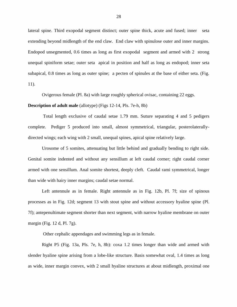
28
lateral spine. Third exopodal segment distinct; outer spine thick, acute and fused; inner seta
extending beyond midlength of the end claw. End claw with spinulose outer and inner margins.
Endopod unsegmented, 0.6 times as long as first exopodal segment and armed with 2 strong
unequal spiniform setae; outer seta apical in position and half as long as endopod; inner seta
subapical, 0.8 times as long as outer spine; a pecten of spinules at the base of either seta. (Fig.
11).
Ovigerous female (Pl. 8a) with large roughly spherical ovisac, containing 22 eggs.
Description of adult male (allotype) (Figs 12-14, Pls. 7e-h, 8b)
Total length exclusive of caudal setae 1.79 mm. Suture separating 4 and 5 pedigers
complete. Pediger 5 produced into small, almost symmetrical, triangular, posterolaterally-
directed wings; each wing with 2 small, unequal spines, apical spine relatively large.
Urosome of 5 somites, attenuating but little behind and gradually bending to right side.
Genital somite indented and without any sensillum at left caudal corner; right caudal corner
armed with one sensillum. Anal somite shortest, deeply cleft. Caudal rami symmetrical, longer
than wide with hairy inner margins; caudal setae normal.
Left antennule as in female. Right antennule as in Fig. 12b, Pl. 7f; size of spinous
processes as in Fig. 12d; segment 13 with stout spine and without accessory hyaline spine (Pl.
7f); antepenultimate segment shorter than next segment, with narrow hyaline membrane on outer
margin (Fig. 12 d, Pl. 7g).
Other cephalic appendages and swimming legs as in female.
Right P5 (Fig. 13a, Pls. 7e, h, 8b): coxa 1.2 times longer than wide and armed with
slender hyaline spine arising from a lobe-like structure. Basis somewhat oval, 1.4 times as long
as wide, inner margin convex, with 2 small hyaline structures at about midlength, proximal one

29
triangular, distal one membranous; a large thumb-like hyaline lobe occurring close to
subproximal inner margin on the frontal surface. First exopodal segment produced into spinous
process at outer distal corner; 1 accessory hyaline lobe present near base of spinous process;
inner distal corner produced into small triangular hyaline lobe. Second exopodal segment 1.7
times longer than wide (Pl. 8b), carrying small hyaline button near mid-inner margin; lateral
spine inserted on distal third, moderately strong, as long as segment, incurved, acutely pointed
with spinules on distal outer and inner margins. End claw 0.6 times as long as right P5, distal
third bent inwardly and bearing spinules on inner margin. Endopod longer than first exopodal
segment, incurved, with a row of apical spinules.
Left P5 reaching mid-margin of second exopodal segment of right P5. Coxa roughly
rectangular, with hyaline spine arising from a lobe-like strucrture. Basis rectangular, 1.7 times as
long as wide, with 1 large, lamellate hyaline lobe on inner distal margin and with 1 sensory seta
at outer distal corner. Exopod inner margin arc-like at midlength, with a series of serulations,
remarkably uniform in size and with usual 2 hairy pads; apex of exopod with usual combination
of digitiform appendix and spinulate seta. Endopod unisegmented and dilated proximally, with 1
row of long apical spinules.
Variation
Total body length of male (exclusive of caudal setae) varies from 1.79 mm-1.90 mm. On
male right P5, the shape and size of hyaline lobes on basis and second exopodal segment are
subject to some variation, as in Fig 13b, d.
Remarks
Morphologically T. defayeae n. sp. is somewhat more closely related to the Southeast
Asian T. ruttneri (Brehm, 1925) than to any of its eleven valid Indian congeners. The affinities

30
between these two species are apparent from the following characters: male left P5 exopod,
hyaline lobes on the basis and second exopodal segment of male right P5, habitus in both sexes,
and the nature of spines on the grasping antennules. However, a critical comparison reveals
several subtle but valid differences between these taxa. For example, as for the male, the frontal
hyaline lobe of right P5 is large, thumb-like vs. small, digitiform; the hyaline process at the inner
distal corner of first exopodal segment small vs. large; second exopodal segment without vs. with
hyaline lobe between lateral spine (aculeus) and end claw, and with vs. without tiny hyaline lobe
near inner margin on caudal face; left P5 basis with vs. without hyaline lobe; the spinous process
on 13th
segment short vs. long. As to the female, the caudal rami without vs. with hairs on outer
margins; the armature elements on endopod stout and spiniform vs. slender and setiform; and the
coxal spines of P5 large vs. small.
The morphological relationships of T. defayeae n. sp. with the Indian species are listed in
Table 5. Based on the proximal hyaline structure on basis and second exopodal segment of the
male right P5, Defayeae (2002) grouped T. ruttneri with the Asian T. doriai, T. vandouwei, T.
hebereri, T. oryzanus and T. foresti Defaye 2002. However T. defayeae n. sp. is distinctly
different not only from these species but also its other congeners by a unique combination of the
following characters: in the male the right P5, basis has a large, thumb-like frontal hyaline lobe;
first exopodal segment is produced into only a short hyaline lobe at inner distal corner; the left
P5 basis has distinct hyaline lobe on inner margin; the saw has uniformly minute serrations with
short radii. In the female, the genital double-somite is produced into a small chitinous lobe at
right distal corner; no hair on outer margins of caudal rami; and the P5 ednopod has sturdy, short
spiniform setae.

31
Etymology
The new species is named in honour Dr. Danielle Defaye, Senior Curator, Muséum
national d‘Histoire naturelle, Paris for her significant contributions to the study of freshwater
copepods.
Ecology and Distribution
T. defayeae n. sp. occurs in shallow, temporary waters (pH 7.0, temperature 29◦C in
Nepal. So far, it has not been known outside its type locality; it co-occurs with N. schmackeri.
Conservation Status: Vulnerable (VU D2).
Tropodiaptomus raoi n. sp.
(Figs 15-17, Pl. 9)
Type locality and material examined
A temporary shallow water pond in the forest near Siddavatam village (14°28′00″N
78°58′00″E), Kadapa district, Andhra Pradesh. The pond is rain-fed, seasonal, depth 5 m, air
temperature 32°C, water temperature 29 °C; pH 6.0, January 13, 2005, leg. D. Ambedkar.
Holotype adult male dissected on 4 slides, allotype female dissected on 4 slides each; 1
male and 1 female paratypes undissected, preserved in alcohol. The type material is kept in the
Department of Zoology, Acharya Nagarjuna University, pending transfer to the Muséum national
d‘Histoire naturelle, Paris. .
Diagnosis
Male: on right antennule, spine of 13th segment moderately strong and spinous process
on antepenultimate segment hook-like and longer than next segment. Right P5: inner margin of
basis with triangular hyaline lobe subproximally, in addition to a short hyaline growth close by.
First exopodal segment produced into a triangular spinous projection at outer distal corner.

32
Second exopodal segment with small, triangular knob (supplementary process) inserted on
dorsomedial surface near base of lateral spine; a small hyaline lobe present subproximally near
inner margin. Lateral spine short, as long as segment and inserted at distal third of the outer
margin of the segment End claw twice as long as the second segment. Endopod as long as first
exopodal segment. Left P5: basis rectangular, with a small hyaline lobe near outer distal corner
on anterior plane. Exopod roughly spatulate, twice as long as wide, inner distal margin nearly
straight with a series of fine serulations. Endopod pyriform, vaguely 2-segmented.
Female: Postero lateral wings asymmetrical, left being larger than right wing. Genital
somite with small bulge at about mid-outer margin and outer distal corner slightly produced into
rounded lobe, right genital spine directed laterally and left genital spine posterolaterally. P5:
Basis with short slender seta. First exopodal segment slender and subcylindrical. Endopod
shorter than first exopodal segment; setae moderately strong and naked.
Description of adult male (holotype) (Figs 15, 16, Pl. 9a-c)
Total length exclusive of caudal setae 1.77 -1.99 mm (n = 2). Suture separating pedigers
4 and 5 complete. Pediger 5 produced into small, almost symmetrical, triangular postero-laterally
directed wings; each wing with apical relatively large spine and subproximal, inner small spines.
Urosome of 5 somites, attenuating but little behind and nearly straight. Genital somite
with left side indentation and right caudal corner armed with fine sensillum. Fourth urosomite
asymmetrical, right distal corner being produced and rounded. Caudal rami symmetrical; each
ramus twice as long as wide, with hairy inner margins.
Left antennule as in female. Right antennule with spine on each of segments 8 and 10-13,
and 15; spine on segment 13 moderately strong. Relative length of spines in decreasing order as

33
follows: 13>15>11>10>12>8. Spinous process on antepenultimate segment lined with thin
hyaline lamella, longer than next segment, apical part hook-like (Fig. 15c, d, Pl. 9a).
Right P5 (Fig. 16a-b, Pl.9b,c). Coxa as long as wide, hyaline spine arsing from a lobe at
about the middle of the segment. Basis roughly rectangular 1.5 times longer than wide; inner
margin with triangular hyaline lobe subproximally, in addition to a short hyaline growth
occurring close by; usual sensory seta near outer distal margin. First exopodal segment produced
into a triangular spinous projection at outer distal corner. Second exopodal segment roughly
rectangular, twice as long as wide with triangular knob (supplementary process) inserted on
dorsomedial surface near base of lateral spine; a small hyaline lobe present subproximally near
inner margin. Lateral spine inserted at about distal third of the outer margin of the segment,
slightly shorter than the segment and with short spinules on distal outer and inner margins. End
claw twice as long as the second segment, doubly curvd, sickle-shaped and with a row of
serulations on inner margin barring proximal region. Endopod as long as first exopodal segment,
tapering towards apex, with two rows of apical spinules.
Left P5 overreaching first exopodal segment of right P5. Coxa subrectangular, armed
with small hyaline spine arising from a medial lobe-like structure on caudal surface. Basis
somewhat rectangular, 1.3 times longer than wide, with a small hyaline lobe near outer distal
corner on frontal plane (Fig. 16a) and with usual slender seta. Exopod roughly spatulate, twice
as long as wide, inner distal margin nearly straight, with a series of fine serulations, 2 usual
hairy pads in center, proximal one larger and set just adjacent to endopod; apex of exopod with
the usual finger-and-thumb-combination (Fig. 16a, b, Pl. 9c.); the ‗finger‘ rather slim, and
beset with spinules, about as long as the ‗thumb‘. Endopod pyriform, vaguely 2 segmented
with apical and subapical row of spinules.
34
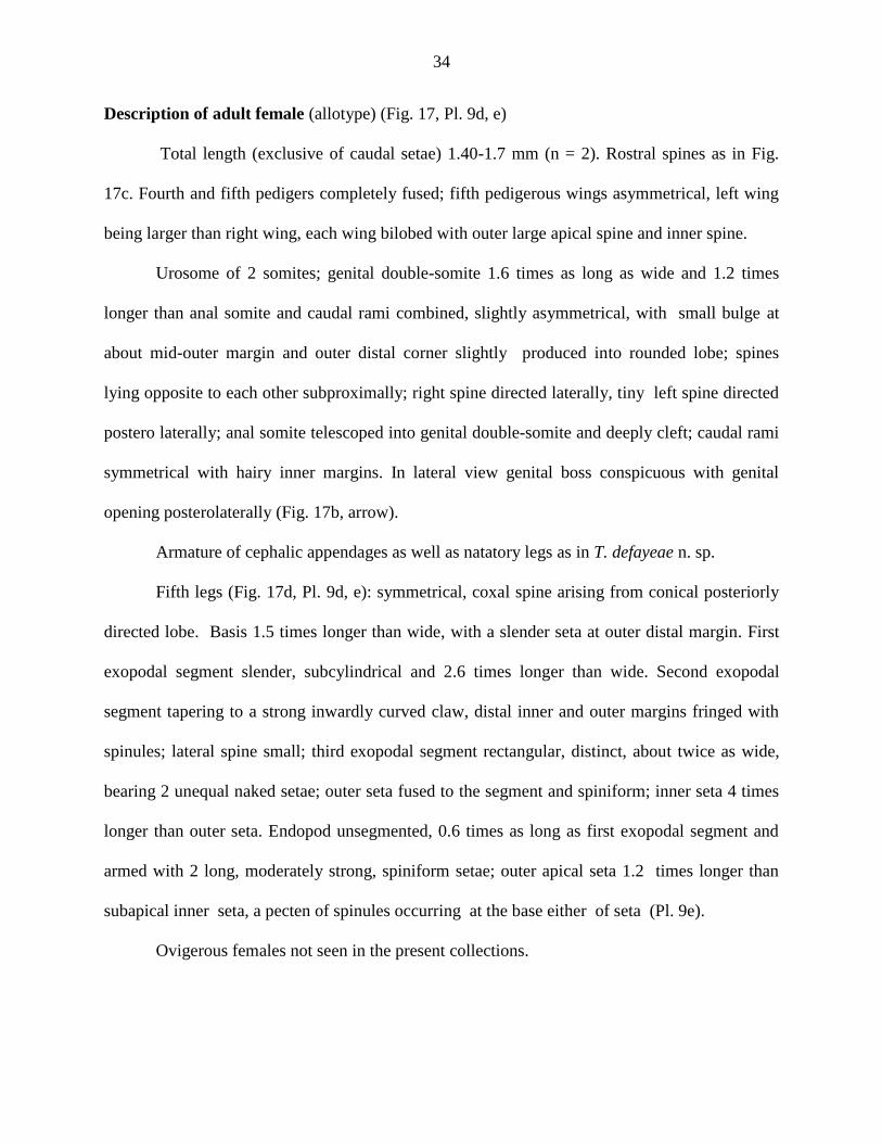
Description of adult female (allotype) (Fig. 17, Pl. 9d, e)
Total length (exclusive of caudal setae) 1.40-1.7 mm (n = 2). Rostral spines as in Fig.
17c. Fourth and fifth pedigers completely fused; fifth pedigerous wings asymmetrical, left wing
being larger than right wing, each wing bilobed with outer large apical spine and inner spine.
Urosome of 2 somites; genital double-somite 1.6 times as long as wide and 1.2 times
longer than anal somite and caudal rami combined, slightly asymmetrical, with small bulge at
about mid-outer margin and outer distal corner slightly produced into rounded lobe; spines
lying opposite to each other subproximally; right spine directed laterally, tiny left spine directed
postero laterally; anal somite telescoped into genital double-somite and deeply cleft; caudal rami
symmetrical with hairy inner margins. In lateral view genital boss conspicuous with genital
opening posterolaterally (Fig. 17b, arrow).
Armature of cephalic appendages as well as natatory legs as in T. defayeae n. sp.
Fifth legs (Fig. 17d, Pl. 9d, e): symmetrical, coxal spine arising from conical posteriorly
directed lobe. Basis 1.5 times longer than wide, with a slender seta at outer distal margin. First
exopodal segment slender, subcylindrical and 2.6 times longer than wide. Second exopodal
segment tapering to a strong inwardly curved claw, distal inner and outer margins fringed with
spinules; lateral spine small; third exopodal segment rectangular, distinct, about twice as wide,
bearing 2 unequal naked setae; outer seta fused to the segment and spiniform; inner seta 4 times
longer than outer seta. Endopod unsegmented, 0.6 times as long as first exopodal segment and
armed with 2 long, moderately strong, spiniform setae; outer apical seta 1.2 times longer than
subapical inner seta, a pecten of spinules occurring at the base either of seta (Pl. 9e).
Ovigerous females not seen in the present collections.

35
Etymology
The species is named in honor of Dr. T. R. Rao, Professor of Zoology (retired), Delhi
University, Delhi, for his significant contribution to zooplankton ecology.
Remarks
Among the Indian species, T. raoi n. sp. is somewhat close to T. defayeae n. sp. in the
following features. In the female, the nature of postero-lateral wings and the genital double-
somite with a proturberance at right distal corner; in male P5, the overall shape of the second
exopodal segment and its hyaline structures, and the general details of left P5 exopod. However,
the two species are distinctly different from each other in various other respects: the nature of
armature elements on the female P5 endopod, the relative size of spinous structures on the
grasping antennule, the shape of basis and its hyaline lobes on male right P5, the shape of left P5
basis with or without inner hyaline lobe, etc. The detailed morphological relationships of T. raoi
n. sp. with its other Indian congeners are as given in Table 4.
Ecology & Distribution
Tropodiaptomus raoi n. sp. is the only representative of Tropodiaptomus from Kadapa
district. Judging by the nature of its type locality, T. raoi n. sp. seems to prefer turbid shallow
waters. It is not know outside its type locality.
Conservation Status: Vulnerable (VU D2).
Tropodiaptomus keralaensis n. sp.
(Figs 18-21, Pls. 10, 11)
Type locality and Material examined
A turbid, turbid pond at Anzumurthymangalam village near Trichur (10° 31′ 12″ N,
76° 12′ 36″ E), Kerala; depth c. 1 m, air temperature 32°C, water temperature 29°C, pH 6.0;
36
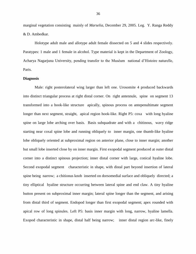
marginal vegetation consisting mainly of Marselia, December 29, 2005. Leg. Y. Ranga Reddy
& D. Ambedkar.
Holotype adult male and allotype adult female dissected on 5 and 4 slides respectively.
Paratypes: 1 male and 1 female in alcohol. Type material is kept in the Department of Zoology,
Acharya Nagarjuna University, pending transfer to the Muséum national d‘Histoire naturelle,
Paris.
Diagnosis
Male: right posterolateral wing larger than left one. Urosomite 4 produced backwards
into distinct triangular process at right distal corner. On right antennule, spine on segment 13
transformed into a hook-like structure apically, spinous process on antepenultimate segment
longer than next segment, straight, apical region hook-like. Right P5: coxa with long hyaline
spine on large lobe arching over basis. Basis subquadrate and with a chitinous, wavy ridge
starting near coxal spine lobe and running obliquely to inner margin, one thumb-like hyaline
lobe obliquely oriented at subproximal region on anterior plane, close to inner margin; another
but small lobe inserted close by on inner margin. First exopodal segment produced at outer distal
corner into a distinct spinous projection; inner distal corner with large, conical hyaline lobe.
Second exopodal segment characteristic in shape, with distal part beyond insertion of lateral
spine being narrow; a chitionus knob inserted on dorsomedial surface and obliquely directed; a
tiny elliptical hyaline structure occurring between lateral spine and end claw. A tiny hyaline
button present on subproximal inner margin; lateral spine longer than the segment, and arising
from distal third of segment. Endopod longer than first exopodal segment; apex rounded with
apical row of long spinules. Left P5: basis inner margin with long, narrow, hyaline lamella.
Exopod characteristic in shape, distal half being narrow; inner distal region arc-like, finely

37
spinulose with short radii; another spinular row running from top to subproximal region and
almost parallel to inner margin.
Female: caudal rami with hairy outer and inner margins. P5: basis with relatively long
sensory seta, extending up to proximal third of first exopodal segment. Inner seta on third
exopodal segment extending beyond mid length of end claw. Endopod about half as long as first
exopodal segment, bearing two moderately unequal naked spiniform setae.
Description of adult male (holotype) (Figs 18, 19, Pls. 10b-e, 11a-d)
Body size 1.30-1.57 mm (n = 5) exclusive caudal setae. Rostral spines as in Fig. 18b.
Fourth and fifth pedigers separated by a complete suture. Wings of fifth pediger asymmetrical,
right wing slenderer and longer than left wing; each wing with relatively large apical spine and
minute subprximal inner spine. Urosome of 5 somites, attenuating behind and gradually bending
to right side. Genital somite with tiny sensory spine on dorsal surface of inner distal corner.
Urosomite 4 asymmetrical, right distal corner produced backwards distinct triangular projection.
Caudal rami 1.8 times as long as maximum width, with hairy inner margins.
Left antennule as in female. Right antennule with spine on each of segments 8 and 10-13
and 15; spine on segment 13 transformed into a hook-like structure apically (Fig. 18c,
Pl..10b,11a). Relative length of spines in decreasing order as follows: 13>15>11>10>8>12.
Spinous process on antepenultimate segment longer than next segment, straight apical region
hook-like, slender and lined with narrow hyaline membrane (Fig. 18d, Pl. 10c).
Armature of appendages as well as natatory legs as in T. defayeae n. sp.
P5 asymmetrical, extremity of left leg5 reaches beyond first exopodal segment of right
P5. Right P5. (Fig. 19 Pls. 10d-e, 11b-d). Coxa as long as maximum width, with long hyaline
spine on large lobe projecting over basis. Basis subquadrate, 1.2 times longer than wide, 1

38
thumb-like hyaline lobe onliquely oriented at subproximal region on frontal plane, close to inner
margin; another but small lobe inserted close by on inner margin; sensory seta near outer distal
corner. Basis with thin, chitinous, wavy ridge starting near the lobe of coxal spine and running
obliquely to inner margin. First exopodal segment partly telescoped into basis, outer distal corner
produced into a distinct spinous projection; inner distal corner with large, conical, hyaline lobe
distinctly produced into a spinous projection. (Pl. 10e, arrow). Second exopodal segment about
twice as long as maximum width, with convex outer margin and concave inner margin; distal
part beyond insertion of lateral spine narrow; a chitionus knob inserted on dorsomedial surface
and obliquely directed; a tiny elliptical hyaline structure occurring between lateral spine and
end claw. A tiny hyaline button present on subproximal inner margin; lateral spine slightly
longer than its segment and arising from distal third of segment and with a row of spinules on
inner distal margin; end claw 0.7 times as long as the same leg, doubly curved; inner margin
except for proximal part beset with fine spinules. Endopod longer than first exopodal segment;
apex rounded with apical row of long spinules.
Left P5: extending beyond first exopodal segment of right P5; coxa subquadrate, 1.4
times longer than wide, with a delicate spine inserted near distal corner; basis elongately oval 1.6
times longer than wide, with a usual slender sensory seta; inner margin with long, narrow,
hyaline lamella on inner distal margin. Exopod characteristic in shape, distal half being narrow;
inner distal region arc-like, finely spinulose with short radii; another spinular row running from
top to subproximal refion and almost parallel to inner margin; usual hairy pads, proximal one set
just under endopod; apex of exopod with finger-and-thumb combination, the ‗finger‘ rather slim
and beset with radial spinules, about as long as ‗thumb‘, which is cylindrical and apically
rounded. Endopod vaguely 2- segmented, with apical row of spinules.

39
Description of adult female (allotype) (Figs 20, 21, Pl. 10a)
Total body length exclusive of caudal setae 1.76- 2.0 mm (n=5). Rostral spines (Fig.
20c) well-developed and acute. Pedigers 4 and 5 completely fused without lateral indentation;
posterolateral wings well developed, bilobed with 2 spines; apical one, as usual larger than inner
one.
Urosome of 2 somites; genital double-somite somewhat asymmetrical, left proximal
being dilated; 1.6 times as long as anal somite and caudal rami combined; 2 conspicuous,
unequal, dorsolateral spines, lying opposite to each other subproximally; right spine lying on
dorsal surface and posteriorly oriented; left spine laterally dirtected; caudal rami parallel,
symmetrical, 1.5 times as long as wide, with hairy outer and inner margins.
P5 (Fig. 21 Pl. 10a): coxa symmetrical, with stumpy subacute spine on a lobe at outer
distal corner. Basis with relatively long sensory seta, extending up to proximal third of first
exopodal segment; first exopodal segment roughly cylindrical and 2.4 times longer than wide
with convex outer and concave inner margin; end claw with spinulate outer and inner margins;
lateral spine represented by a short spine: third exopodal segment represented by a prominence,
bearing 2 unequal, naked spines; inner seta slender and 2.6 times longer than outer spine and
extending beyond midlength of end claw. Endopod unsegmented about half as long as first
exopodal segment, bearing two moderately unequal naked spines, inner subapical seta 0.6 times
longer than endopod, each seta with a pecten of spinules at base.
Etymology
The specific epithet alludes to the the state of Kerala in southwest India where the new
species was collected. The name with the Latin suffix ―-ensis‖ is an adjective for place.

40
Variation
Total body length of male (exclusive of caudal seta) 1.3-1.6 mm, female 1.5-1.8 mm. On
male right P5, the proximal hyaline lobe on basis, the accessory hyaline lobe on the second
exopodal segment, and endopod are subject to variation.
Remarks
T. keralaensis n. sp. closely resembles the Philippine T. lanoanus Kiefer, 1982. The
spectacular morphological morphological affinity between these two species is at once evident
from the form of the second exopodal segment and the position and size of the lateral spine
borne by it. The close kinship of these two species can also be noticed in the following
characters: in the female, the posterolateral wings and the armature elements of the endopod of
P5 (the other details of female are not given for T. lanoanus by Kiefer, 1982); in the male, the
spinous process of the right antennule including that of the antepenultimate segment and the
asymmetric fourth urosomite. A critical comparison, however, shows a series of differences
between the two species: in the male P5 of T. keralaensis n. sp., the exopod of left leg slender vs.
stout, distal inner margin as a single lobe vs. bilobed, right P5 basis with thumb-like vs.
digitiform proximal hyaline outgrowth, without vs. with hyaline lobe near inner distal corner;
first exopodal segment with large vs. small hyaline outgrowths; second exopodal segment with
concave vs. nearly straight inner margin, convex vs. straight outer margin, and accessory hyaline
structure large vs. small, conical vs. lobe-like, and inserted at about the middle vs. near inner
distal corner; 13th
segment of right antennule without vs. with accessory spine; in the female, the
outer margins of caudal rami naked vs. hairy. How the T. keralaensis n. sp. is related to its Indian
congeners can be understood from the tabulated data (Table 5). It is thus remarkable that the

41
close set sister species of T. keralaensis n. sp. is not any of its Indian congeners, but the
Southeast Asian T. lanoanus of the Phillippines.
Ecology and Distribution. T. keralaensis n. sp. was collected in a shallow temporary turbid
pond with marginal vegetation ( temperature is 29ºC, pH 7.5 and Secchi transparency 48 cm). It
is so far known only from its type locality.
Conservation Status: Vulnerable (VU D2)
Tropodiaptomus venkataramani n. sp.
(Figs. 22-25, Pls. 12, 13)
Type locality and Material examined. A small man-made temple pond in the
Tirupurankundram Kasi Viswanath temple in Madurai town (9° 52′ 12″ N, 78° 4′ 12″ E),
Madurai district, Tamilnadu State, 16 June 1971,leg. K. Venkataraman.
Holotype adult female and allotype adult male dissected on 6 and 4 slides, respectively,
paratype adult male mounted whole on a single slide. Type material is kept in the Department of
Zoology, Acharya Nagarjuna University, pending transfer to the Muséum national d‘Histoire
naturelle, Paris.
Diagnosis
Male: right wing shorter than left one. Urosome 4 asymmetrical, produced into a short
triangular process at outer distal corner. On right antennule, spine on segment 13 short and stout
with one accessory hyaline lobe; spinous process on antepenultimate segment slightly longer
than next segment and hook-like apically. Right P5: coxal lobe large, with articulate seta; basis
with 4 hyaline lobes including a large conical lobe on frontal face, second exopodal segment oval
in out line; lateral spine inserted at the same level as end claw. Endopod as long as first exopodal

42
segment. Left P5: basis with small distal hyaline lobe. Exopod oval in outline, with fine, similar
serrulations on inner margin.
Female: right posterolateral wing smaller than left one. Caudal rami with hairy inner
margins. P5: coxa with large spine arising from a lobe like structure. Basis with 1 small hyaline
outgrowth on caudal surface near distal margin. Endopod as long as first exopodal segment;
apical seta as long as endopod with stiff setules on distal outer margin.
Description of adult male (holotype) (Figs 22, Pl. 12b-e, 13a)
Body size exclusive caudal setae 1.90 mm. Rostral spines as in Fig. 22c. Body widest at
caudal border of first pediger. Lateral wings of fifth pediger strongly asymmetrical, left wing
being longer than right wing. Each wing with a pair of small hyaline spines.
Urosome of 5 somites, Genital somite with short spine at right distal corner. Fourth
urosomite asymmetrical, with outer distal corner being prominently produced into conical
spinous process, overreaching midlength of anal somite. Caudal rami symmetrical, 1.6 times as
long as wide with hairy inner margins; dorsal jointed seta of left ramus slightly longer than that
of right ramus; all setae slightly dilated proximally.
Left antennule as in female. Right antennule (Fig. 22d-h, Pl. 12c-e) with spine on each of
segments and 8 and 10-13 and 15; segment 13 with characteristically short and strong, and
with 1 accessory triangular lobe (Fig. 22d arrow, Pl. 12d arrow). Relative length of spines in
decreasing order as follows: 13>15>11>10>8>12. Antepenultimate (Fig. 22e-h) produced
somewhat hook-like spinous process, slightly longer than next segment, and lined with narrow
hyaline lamella.
P5 (Figs 23, Pls 12f, 13a). Right P5: coxa 0.7 times longer than wide and produced into
subtriangular lobe at distal inner angle. Basis roughly rectangular, 1.4 times longer than wide,

43
with 3 hyaline lobes on innermargin; proximal lobe squarish, relatively larger than other 2 lobes,
as illustrated; on frontal surface, 1 hyaline lobe modified into strong spinous projection reaching
inner margin; sensory seta short, arising from outer distalcorner; first exopodal segment about
twice as wide as long, outer distal corner produced into short spinous process; inner distal corner
with small hyaline process; a large broadly triangular hyaline lobe occurring on caudal plane at
about the middle of posterior border. Second exopodal segment subquadrate, 1.4 times as long as
wide, with convex outer and concave inner margins; 1 small hyaline lobe occurring on inner
margin; a large conical accessory structure inserted dorso-distally and oriented towards base of
lateral spine ; lateral spine strong, 1.2 times longer than its segment at about the same level as
end claw, inserted at outer distal corner and ornamented with a row of spinules on inner margin;
2 hyaline lobes present between lateral spine and end claw; end claw sickle-shaped, 2.8 times
longer than the exopod 2, lined with tiny spinules along inner margin barring some distance
proximally. Endopod subquadrate, as long as inner margin of first exopodal segment, apex
rounded with a row of fine spinules. Left P5: reaching beyond inner margin of right P5 basis.
Coxa somewhat quadrate, armed with slender spine arising from a lobe-like structure near distal
outer corner. Basis elongate, somewhat quadrate, 1.6 times longer than wide, a tiny hyaline lobe
on frontal surface close to subproximal inner margin, and another relatively large, somewhat
rectangular lobe on inner margin; usual slender seta near outer distal margin. Exopod distinctly
dilated at about the middle of arc-like inner margin; inner margin right from apex finely and
uniformly serrulated with short radii and hairy pads in center; apex of exopod with usual finger-
and-thumb combination. Endopod apparently unsegmented with apical row of somewhat large
spinules.
44
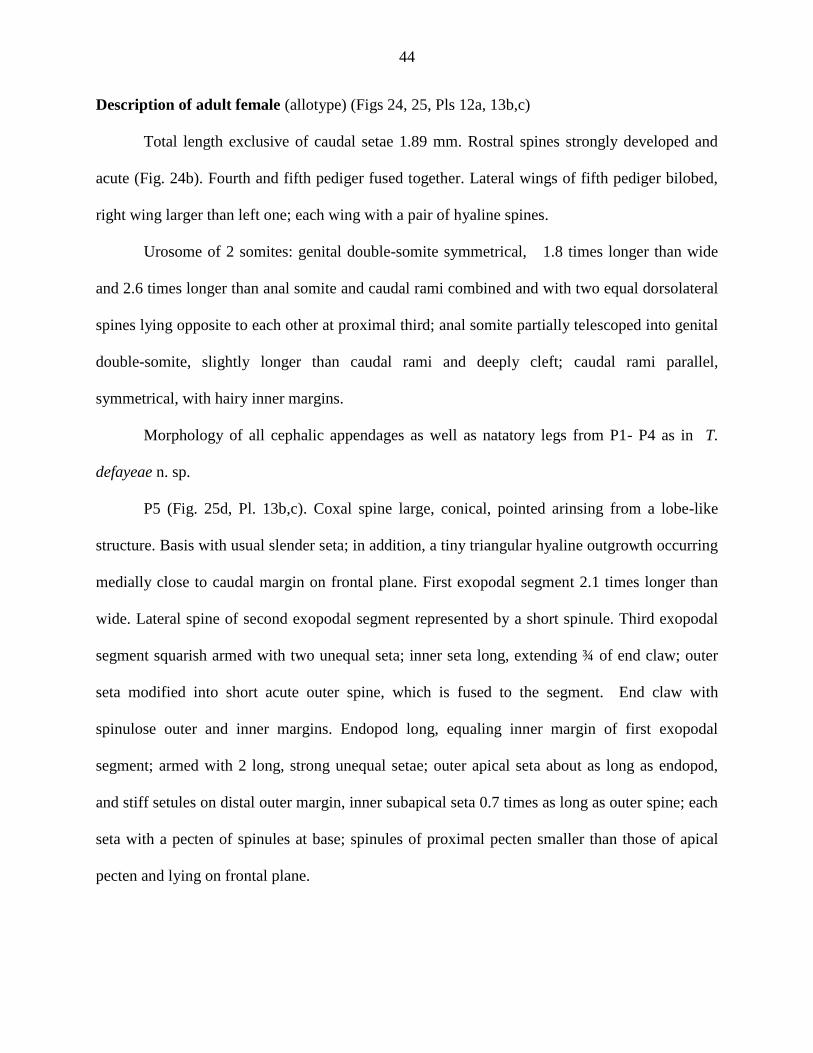
Description of adult female (allotype) (Figs 24, 25, Pls 12a, 13b,c)
Total length exclusive of caudal setae 1.89 mm. Rostral spines strongly developed and
acute (Fig. 24b). Fourth and fifth pediger fused together. Lateral wings of fifth pediger bilobed,
right wing larger than left one; each wing with a pair of hyaline spines.
Urosome of 2 somites: genital double-somite symmetrical, 1.8 times longer than wide
and 2.6 times longer than anal somite and caudal rami combined and with two equal dorsolateral
spines lying opposite to each other at proximal third; anal somite partially telescoped into genital
double-somite, slightly longer than caudal rami and deeply cleft; caudal rami parallel,
symmetrical, with hairy inner margins.
Morphology of all cephalic appendages as well as natatory legs from P1- P4 as in T.
defayeae n. sp.
P5 (Fig. 25d, Pl. 13b,c). Coxal spine large, conical, pointed arinsing from a lobe-like
structure. Basis with usual slender seta; in addition, a tiny triangular hyaline outgrowth occurring
medially close to caudal margin on frontal plane. First exopodal segment 2.1 times longer than
wide. Lateral spine of second exopodal segment represented by a short spinule. Third exopodal
segment squarish armed with two unequal seta; inner seta long, extending ¾ of end claw; outer
seta modified into short acute outer spine, which is fused to the segment. End claw with
spinulose outer and inner margins. Endopod long, equaling inner margin of first exopodal
segment; armed with 2 long, strong unequal setae; outer apical seta about as long as endopod,
and stiff setules on distal outer margin, inner subapical seta 0.7 times as long as outer spine; each
seta with a pecten of spinules at base; spinules of proximal pecten smaller than those of apical
pecten and lying on frontal plane.

45
Etymology
The new species is named for Dr. K. Venkat Raman, presently Director, Zoological
Survey of India, Kolkata, who collected the sample containing the new species.
Variation
Total body length of male (exclusive of caudal setae) 1.4-1.6 mm, female 1.5-1.8 mm.
The size and shape of the coxal spines on female P5 (Fig. 25e-f), as also the Schmeil‘s organ on
middle endopodal segment of P2 (Fig. 25a-c), the spinous process of the male right antennule
(Fig. 22e-h), and the hyaline structures on the basis, first and second exopodal segments, and the
spinous process of the first and second exopodal segments (Fig. 23b-e) show some
interpopulation variation.
Remarks
T. venkataramani n. sp. is unique in the genus Tropodiaptomus as a whole by possessing
an oval-shaped second exopodal segment on the male right P5 such that the lateral spine and the
end claw lie almost at the same level and separated by a nearly straight line. Further the setae on
the female P5 endopod are unusually long, a character shared with the Indian T. euchaetus and
also the Vietnamese T. foresti. In the latter, the setae are modified into simple, sturdy spiniform
structures. T. venkataramani n. sp. is entirely different from both these species in various other
details.
As for other Indian species, T. vicinus is somewhat close to T. venkataramani n. sp. in
regard to the shape of the second exopodal segment of the male right P5 and the alignment of
the lateral spine and end claw. However, the former is principally different from the latter by
having bilobed vs. unilobed saw, shorter vs. long setae on the female P5 endopod, slender vs.
stout male left P5 exopod, short vs. long, digitiform vs. claw-like spinous process on the

46
antepenultimate segment and so on. The morphological differences of T. venkataramani n. sp.
with other congeners are as listed in Table 5.
Ecology and Distribution. Ecology of this species is not known except that it was found in a
temple pond. It was not found in any other sample collected from Tamilnadu State.
Conservation Status: Vulnerable (VU D2).
Tropodiaptomus hebereri (Kiefer, 1930)
(Fig. 26)
Type locality: A pool in the marshy locality of Java, Indonesia.
Female
body size 1.5-1. 6. Rostral spines 40 µm in length. Last thoracic segment dorsally with
rounded hump that can be clearly seen in lateral view. Fifth pedigerous wings somewhat
asymmetrical, the left one longer than right. Genital double-somite slightly dilated proximally,
with a very small sensillum on either side, outer distal region enlarged and produced. Outer
margin of caudal rami without hair. Antennules extending up to the middle of the genital double-
somite. Middle endopod segment of leg 2 with Schmeils organ. P5 as illustrated. Endopod with
two terminal setae as in other Tropodiaptomus species.
Male
body size 1.35-1.4 mm. Fifth pedigerous wings symmetrical. Right antennule with small
spine on segment 10, 11. Spine on segment 13 long, slender and with 1 dorsal accessory spine.
Spine on segment 15 also somewhat long. Spinous process on antepenultimate segment
somewhat straight and with hook-like pointed tip. P5: coxa with long hyaline spine. Basis longer
than wide; inner margin lined carrying 2 hyaline outgrowths; also1 strong spiniform outgrowth
47
occurring on anterior surface close to proximal inner margin. First exopod with relatively long
spines at outer distal corner and a hyaline plug on distal inner corner. Second exopod
remarkably broad, with strong, slightly curved lateral spine with hyaline accessory spine near its
base. Terminal claw characteristically curved. Left P5: basis relatively short. Inner margin of
exopod (saw) with uniformly rounded serulations.
Ecology and Distribution
The ecology of this species not known though it was reportedly found in several
unspecified localities in India by Brehm (1953). Clearly, this is more common in South East
Asian region of Java, Sumatra and China (Yunnan) (see Dussart & Defaye, 2002) than in India.
Conservation Status: Lower Risk (LR nt)
Tropodiaptomus mutatus (Kiefer, 1930)
(Fig. 27)
Type locality: Khulna, Bangladesh, bordering the Indian State of West Bengal.
Female
Length 1. 5 mm. Right wing somewhat smaller than left wing. Genital double-somite
somewhat dilated proximally on right side and inner distal corner without any protuberance.
Caudal rami without hairs on outer margins. Antennules reaching up to the caudal end of genital
double-somite. P5 as in Fig. 27c.

48
Male
Somewhat smaller than female. Right antennule with remarkably slender spine on
segment 11 and it is twice as long as the spine on segment 10; spine on segment 13 strong and
without any accessory spine on dorsal surface. Spinous process on antepenultimate segment
claw-like and shorter than following segment. P5 as in Fig.27f; the details of the segment
proportions as illustrated; basis of right leg without spiniform-like hyaline lobe on frontal surface
near inner margin. Second exopod slender with relatively short and weakly curved terminal claw.
Endopod shorter. Left P5 with simple saw with finely denticulated inner margin and with short
endopod.
Remarks
T. mutatus has hitherto been reported only by Brehm (1953). Animals that he found in
different samples were said to be mainly having radial stripes on the saw of left exopod of male
P5, based on which Brehm concluded they are ‗mutatus‘. However, Kiefer‘s figures do not show
these stripes as distinctive of ‗mutatus‘. Unfortunately, Brehm did not depict the specimens that
he determined as ‗T. mutatus‘.
Distribution. Though this species has reportedly been found in India (Dussart & Defaye, 2002),
the exact localities of its occurrence are not known .
Conservation Status: Lower Risk (LR nt)

49
Tropodiaptomus vicinus (Kiefer, 1930)
(Fig. 28)
Type locality: Luckarac Lake, Sunda Islands.
Female: 1.3-1.5 mm. Rostral spines about 35-40 µm long. Metasomal wings nearly
symmetrical, outer corner (with one small hyaline spine) obliquely directed, caudal margin
deeply indented, lying close to the proximal bulge genital double-somite left with reaching the
spine of left genital double-somite, right wing somewhat shorter. Genital double-somite only
very slightly dilated proximally, right distal corner not produced. Caudal rami with hair on outer
and inner margin. Antennules reaching somewhat upto caudal rami. Middle endopod segment of
leg 2 with Schmeil‘sorgan. P5: Terminal setae of endopod of almost similar length with circle of
fine hairs.
Male
Length 1.2 -1.3 mm. Rostral spines as illustrated. Fifth pediger and urosome as in figure.
Right antennule spinous process on segments 8, 10, 11 small, spine on 13 segment long and
slender with accessory spine on dorsal surface. Segment 15 with strong spine. Spinous process
on antepenultimate segment straight, tip blunt. P5 drawings showing all important details of
segment proportions and their ornamentation. Especially the saw lobes which are fine and deeply
bilobed and ornamented with very fine denticles.
Ecology . T. vicinus occurs in littoral regions and swampy areas.
50
Distribution
Rice fields and pools of India; Toba Lake of Sumatra; rice fields of Malaysia, and
Mindanao Island. Brehm (1953) repoted T. vicinus from Yanaon (= Yanam) near Kakinad town.
According to Dussart & Defaye (2002), T. vicinus is also distributed in Thailand, Philippines and
East Kalimantan. In Thailand, this species is restricted to ponds and rivers (see Sanoamunag,
1999).
Conservation Status: Lower Risk (LR nt)
Tropodiaptomus signatus Kiefer, 1982
(Fig. 29)
Type locality: Unspecified locality in India.
Female
Body size. 1.45-1.5 mm. Rostral spines somewhat 40 µm long. Fifth pediger without
dorsal hump. Metasomal wings nearly symmetrical, outer corner of each wing with relatively
large spine. Genital double-somite with a swelling on left proximal region, carrying a small
sensory spine; distal right corner not produced. Outer and inner margins of caudal rami hairy.
Antennules extending up to the caudal margin of genital double-somite; seta longer and sturdier
than in other congeners . P2 with Schmeil‘s organ. Right P5: coxa with large hyaline spine on the
caudal surface. Second exopodal segment sharply bent inward (‗cramped‘) over the first exopod.
Third exopod small with well developed terminal setae. Endopod unisegmented with 2 almost
equal strong setae.

51
Male
Length about 1.35 mm. Fifth pediger and genital somite as in figure; right antennule with
well developed strong spines of different lengths on segments 10, 11, 13 and 15; segment 13
without accessory spine. Antepenultimate segment with claw-like spiniform, which is as long as
the next segment. Right P5: coxa with spherical chitinous plate at inner corner and with
relatively long hyaline spine on the caudal surface. Basis with 3 hyaline lobes on inner margin.
First exopod with 2 spherical hyaline plugs; outer distal corner produced into pointed process.
Second exopod with short thick lateral spine and long moderately curved end claw. Endopod
without any peculiarities. Left P5: the form of the saw and its denticles characteristic.
Remarks
Kiefer found these specimens together with T. orientalis. None of the Asiatic
Tropodiaptomus spp. has such a remarkable hyaline lobe on the left proximal part of genital
double-somite as does T. signatus. This character along the special of grasping antennule and
male P5 prompted Kiefer to describe this as a new species.
Ecology and Distribution
Ecology of this species is not known. Even regarding its distribution, it is known only to
be an Indian species (Dussart & Defaye, 2002).
Conservation Status: Lower Risk (LR nt)

52
Tropodiaptomus euchaetus Kiefer, 1936
(Fig. 30)
Type locality
Nilagiri Mountains of South India, small pool 2 × 1.5 m, about 60 cm depth, somewhat
2316 m from sea level. Habitat near Umbrella tree in Ootacamund, with Utricularia and
filamentous algae. Temperature at noon 22.1°C; pH 6.1.
Female
Length 1.67 mm. Fifth pedigerous wings symmetrical. Outer distalcorner of each wing
produced, obliquely directed, bearing a hyaline spine. Genital double-somite somewhat dilated
proximally bearing on either side strong hyaline spinule. Caudal rami without hairs on outer
margins. P2 with Schmeil‘s organ. Antennules extending almost to the caudal margin of genital
double-somite. Apical armature of the endopod as shown in figure: the apical seta (somewhat 80
μm), sturdy, about twice as long as subapical seta.
Male
According to Brehm (1953), the male is characterised by the following features: spinous
process on the antepenultimate segment of right antennule longer than succeeding segment and
outcurved; spinous process on segment 10 moderately long, that of segment 11 twice the length
of that of segment 10; segment 15 with moderately long spinous process. Caudal rami twice as
long as wide, with hary inner margins. Right P5: intercoxal plate semicircular; coxa with club-
like hyaline outgrowth. Basis as long as wide with rectangular hyaline membrane on proximal
inner margin.Second exopodal segment elongate, somewhat bent, lateral spine almost as long as

53
segment and inserted in the distal half of the segment; accessory chitinous structure in the form
of a very long, characteristic seta. Left P5: exopod large, inner margin bifid; proximal inner
margin smooth; distal margin furnished with fine, short striations
Remarks
Kiefer (1936) originally described T. euchaetus on the basis of females only. The large
setae on the endopod of the female P5 are highly characteristic of this species. In fact, the
specific epithet alludes to this feature. Brehm (1953) described and illustrated the male of this
species from Kodaikanal, but he was not sure whether it was indeed the male of this species. And
he also opined that what he described as male of T. euchaetus could well be a new species, for
which he even suggested the name setiger, attaching importance to the seta-like accessory
sturcute of the second exopodal segment of the male right P5. In this scenario, Seghal (1967)
reported T. ecuhaetus on the basis of three specimens found in a find fish pond in Narsinghpur of
Orissa. The endopod of female P5 have long apical setae. But other characters of the female do
not agree with the Kiefer‘s T. euchaetus. Seghal‘s (1967) descriptions and figures of male of T.
euchaetus are highly erroneous and hence not dependable. Similarly, his key for the Indian
species of the genus Tropodiaptomus is highly misleading. I have no access to the type
material/voucher specimens of this species not I could find it in my present survey. Hence it is
impossible at this stage to provide a reliable account of the male of T. euchaetus. However, for
the time being, Brehm‘s (1953) depiction of the male is treated as belonging to T. euchaetus.
Ecology and Dirtribution The species seems to be confined to ponds of the elevated,
somewhat cooler parts of Nilgiri Hills of South India: Otacamund and Kodaikanal.
Conservation Status: Lower Risk (LR nt).
54
Tropodiaptomus doriai (Richard, 1894)
(Fig. 31)
Type locality: Toba Lake, Sumatra.
Female
Length 1.12-124 mm. Rostral spines relatively short, somewhat 25-35 µm. Fifth pediger
almost symmetrical; fairly flat outgrowth on posterior margin. Genital somite generally
characteristically symmetrical. Proximal part slightly dilated with minute sensillum on each side.
Outer and inner margin of caudal ramus hairy. Antennules relatively short, barely reaching the
posterior margin of genital somite. Middle endopod of leg 2 with small Schmeil‘s organ. P5 both
the terminal setae of endopod provided with hairs.
Male
Length 1.0-1.5 mm. Rostral spines somewhat 30 µm long. Last thoracic segment slightly
asymmetrical. Genital somite without sensillum at anterodistal corner. Fourth urosomite is
produced at right distal corner. Right antennule segments 10 and 11 with very small each. On
segment 13 one long slender spine and one accessory spine near its base on posterior surface.
Spine on segment 15 distinctly short. The spinous process on the antepenultimate segment
straight and shorter than the next segment. P5 as in Fig. 31i. The saw of left exopod bilobed with
fine, somewhat similar denticles. A small chitinous knob present on the caudal surface of second
exopodal segment, sometimes opposite to the inner margin.

55
Ecology and Distribution
Ecology of this specis unknown. It is distributed in Sri Lanka, Thailand, Java, Sumatra,
and Sulawesi (Dussart & Defaye, 2002). Though this species reportedly occurs in India (Kiefer,
1982), the exact localities and habitats are not known.
Conservation Status: Lower Risk (LR nt)
Genus Heliodiaptomus Kiefer, 1932
Heliodiaptomus: Kiefer, 1932a: 473; Shen & Song, 1979: 113; Lai & Fernando,
1981:168; Ranga Reddy & Radhakrishna, 1981: 171; Dussart & Defaye, 1983: 96; 2002: 120.
Generic diagnosis
Kiefer‘s (1932) original definition of the genus Heliodiaptomus has been revised by
Ranga Reddy (1994) as follows: Animals moderate to somewhat large in size (females 1.0-2.0
mm and males 0.7-1.8 mm long). Female: lateral wings of fifth pediger generally moderately
developed; urosome of 3 somites; antennules extending slightly beyond, or sometimes only to
the base of, caudal setae; on P5, endopod without apical setae and end claw with hairy or
spinulose margins, and only exceptionally with denticulate margins. Male: right caudal ramus
without any ornamentation; on right P5, endopod generally cylindrical but varying in size; coxa
with or without lobe at distal inner corner; basis with 1 or 2 small hyaline lobes on inner margin;
lateral spine of second exopodal segment of the same leg usually proximal, sometimes even
articulated to its posterior face; on left P5, second exopodal segment with a short or,
occasionally, elongate, digitiform process and a well-developed seta apically.
Type species: *Heliodiaptomus viduus (Gurney, 1916)
Other valid species:
*Heliodiaptomus contortus (Gurney, 1907)
56
*Heliodiaptomus cinctus (Gurney, 1907)
*Heliodiaptomus pulcher (Gurney, 1907)
Heliodiaptomus nipponicus (Kokubo, 1914)
Heliodiaptomus kikuchii Kiefer, 1932
*Heliodiaptomus elegans Kiefer, 1935
Heliodiaptomus kieferi Brehm & Chappuis, 1935
Heliodiaptomus serratus Shen & Tai, 1962
Heliodiaptomus falxus Shen & Tai, 1964
Heliodiaptomus lamellatus Sung, Shen, Sung, Li & Chen, 1975
*Heliodiaptomus kolleruensis Ranga Reddy & Radhakrishna, 1981
Heliodiaptomus puthaiorum Sanoamuang, 2004
N. B. The above-listed, asterisked species are from India.
Keys to the female Indian Heliodiaptomus spp.
1. Posterior border of fourth pedigerous segment with a transverse row of spinules on the dorsal
surface; genital double-somite asymmetrical and longer than other 2 urosomites combined ...….2
Posterior border of fourth pedigerous segment without a transverse row of spinules on the dorsal
surface; genital double-somite symmetrical about as long as other 2 urosomites combined
……………………..…………………………………………………………………….H.elegans
2. End claw of the P5 with denticulate margins ……………………………………….H. viduus
End claw of the P5 with either hairy or finely spinulose margins ……………………………3
3. The spine of the left coxa is larger than that of the right in P5……………….……H. contortus

57
Both spines of coxa are similar in size………………………………………….…….H.cinctus
4. Metasomal wings symmetrical and bilobed……………..……………………...H. kollerurensis
Metasomal wings asymmetrical, the left wing being triangular while the right is relatively large
and somewhat bilobed………………………………………………………………...H. phlcher
Key to the males of Indian Heliodiaptomus spp.
1. The hyaline process of the antepenultimate segment of the grasping antennule
serrated……………………………………………………………………………..H. pulcher
The hyaline process of the antepenultimate segment of the grasping antennule simple……..2
2. End claw of the right P5 swollen and twisted at the base…………………………H. contortus
End claw of the right P5 normal……………………………………………………………….3
3. Right P5 basis with one hyaline lobe inner margin, and exopod 1 with a roughly
trapezoidal or triangular chitionus process on the distal prosterior aspect;… ……….H. viduus
Right P5 basis with two hyaline lobe inner margin: exopod 1 with shortblunt projection on
distal posterior aspect……………………………………………..…………………..H. elegans
4. Lateral spine mesially inserted on second exopodal segmentand; end claw sickle- shaped
…………………………………………………………………………………..H. kolleruensis
Lateral spine subproximally inserted on second exopod segment; end claw middle outcurved
endclaw……………………………………………………………………………….H. cinctus

58
Heliodiaptomus viduus (Gurney, 1916)
(Figs 33-35, Pl. 14)
Diaptomus viduus: Gurney, 1916: 338-339, P1. 2, Figs 11-14 (♂); Sewell, 1934: 75-78, Figs 3a-
d (♀).
Heliodiaptomus viduus: Kiefer, 1932a: 507, F ig. 82; Brehm, 1953: 242-244, Figs 1-2; Brehm,
1963: 159-163, Figs 1-10; Sehgal, 1967: 66-68, Figs 23-26; Rajendran, 1973: 116-117, Figs 3a-
n; Fernando, 1974: 60-61, Figs 238, 249-253;Lai & Fernando, 1981: 168, Figs 45-57; Ranga
Reddy & Radhakrishna, 1981: 162-163, PI.1, Figs 1-4; Dussart & Fernando, 1985: 237-238, Figs
33-38; Ambedkar, 2005: 24-30, Figs 1-13.
Neodiaptomus kamakhiae: Reddaiah, 1964:161-166, Figs 1a-h.
Helidiaptomus latifi: Das, 1974: 47-51, Figs 1-6.
Type locality: Snake‘s pool at Mahintele, Sri Lanka.
Material examined: as in Table 6.
Body size. Female 1.57-1.96 mm; male 1.13-1.15 mm.
Female
Third and fourth pedigers demarcated transverse row of spinules on dorsal surface.
Lateral wings of fifth pediger asymmetrical and armed with 2 unequal spines each wing. Genital
double- somite asymmetrical. Urosome of 3 somites, caudal rami with hairy outer and inner
margins. Leg 5: left coxal spine distinctly larger than right coxal spine, number of denticles
along the lateral margins of end claws slightly variable. Endopod somewhat exceeding midlength
of first exopodal segment.

59
Male
Body ornamentation as in female. Caudal rami 1.4 times as long as wide and with hirsute
inner margins. Right antennule with spines on each of segments 8 and 10-16; spinous process on
antepenultimate segment straight, slightly exceeding half the length of penultimate segment and
fringed with narrow hyaline membrane along outer margin. P5: exopod 1 of right P5 with
trapezoidal chitinous process on posterior surface. Distal outer corner of exopod 1 short and
pointed spinous process. Second exopodal segment elongately ovate; lateral spine inserted
proximally over posterior surface. End claw somewhat dilated at base with generally angular
outer margin. Endopod cylindrical and extending beyond origin of lateral spine on second
exopodal segment. Left P5: basis with two small hyaline lobes; endopod about as long as its
counterpart on right P5.
Remarks
H. viduus shows wide variation within and between populations in the number of
denticles on the end claw of female P5: 0-8 on outer margin and 8-13 on inner margin. The
spinous process at distal outer corner of first exopodal segment on male right P5 varies in shape
from short, blunt to long, pointed; the proximal part of end claw is sometimes narrow with
smoothly curved outer margin or rarely produced into a rounded prominence. The presence of
spinules between third and fourth pedigers in the male is being reported for the first time.
Abnormality: two proximal lateral spines were noticed on second exopodal segment of right P5
in one specimen. The interrelationships of the congeners can be understood from Table 9.

60
Ecology
H. viduus is successful in all kinds of freshwater habitats except in rivers. In the present
study it was found in 86 habitats of 2000 sampling sites. In Andhra Pradesh, H. viduus was found
in shallow, temporary, permanent aquatic habitats when the temperature was 25ºC- 35ºC. It was
noticed in a turbid pond in forest area at Siddavatam near Kadapa town of A. P. In Karnataka and
Tamilnadu H. viduus was found in large village tanks. Its co-occurrence with typical temporary
water forms like the fairy shrimps (Streptochephalus sp.) in river-fed hill-pool speaks of its wide
ecological amplitude. H. viduus co-occurs with P. blanci, N. schmackeri, H. contortous, P.
greeni. and N. lindbergi.
Distribution
Previous records
India, Sri Lanka, Pakistan, Bangladesh, Myanmar, and Thailand. While the species is
not common in its type locality, i.e. Sri Lanka (see Gurney, 1916; Fernando, 1974, 1980), it is
the most common diaptomid in South India (Ranga Reddy, 1994), and its abundance, according
to Hossain (1985), diminishes gradually towards the North. It was not recorded by Kiefer (1939)
in his scientific report on the Yale North India Expedition. According to Das (1974) and Hossain
(1985), it is the most abundant calanoid in Bangladesh. It is so far reported from Salt Lakes,
Calcutta; Indian Museum Tank, Calcutta; Kudra tank, Mirzapur; Portblair; P.W.D. tank,
Ghorwal; Bankipur; Cuttack, Portblaiir; Mysore; Madras; Delhi, and several places in Orissa:
Athmilike, Balasore, Berhampore, Chandwar, Whenkanal, Jenapore, Jaipore, Linghpur,
Narsinghapur, Puri and Sambalpur.Andhra Pradesh: Visakhapatnam, Anakapalli, Guntur,
Kondaveedu, Guntakal, Gooty, Dharmavaram, Tirupathi hills, and Warangal.
61
Present records
H. vuduus is recorded in the present study from Srikakulam, Vijayanagaram,
Visakhapatnam, East Godavari, West Godavari, Krishna, Guntur, Prakasm, Nellore, Hyderabad
and Warangal districts of Andhra Pradesh State and also from Chattisgarh, Tamilnadu and
Kerala States.
Conservation Status: Lower Risk (LR); Least Concern (lc).
Heliodiaptomus contortus (Gurney, 1907)
(Figs36, 37, Pl. 15)
Diaptomus contortus: Gurney, 1907: 28, Figs 9-10; Sewell, 1924: 788, P1. 45, Fig. 5.
Heliodiaptomus contortus: Kiefer, 1932a: 507, Figs 84-86; Brehm, 1950: 11-13, Figs 1a-d;
Brehm, 1953: 247-252, Figs 5-11; Brehm, 1963: 162-163; Ranga Reddy & Radhakrishna, 1981:
163-165, P1.2, Figs 1-10; Ranga Reddy & Devi, 1989: 129-130, Figs 41, 42, 50, 51, 62, 65;
Ambedkar, 2005: 30-33, Figs 1-9.
Type locality: Museum tank, Calcutta, India.
Material examined: as in Table 7.
Body size. Female 1.4 -1.6 mm; male 0.8 -1.3 mm.
Female
Fourth and fifth pedigers with a row of dorsal spinules. Lateral wings of fifth pediger
small, rounded, asymmetrical and armed with 2 unequal spines each; right wing with small
rounded prominence, whereas left wing larger than right wing. Genital double-somite relatively
short and asymmetrical, sub-proximal region produced on right side; left genital spine larger than

62
right spine. Caudal rami with hirsute outer margins. P5: left coxal spine distinctly larger than
right coxal spine, second exopodal segment with large, tooth-like lateral spine near the base;
third exopodal segment vestigial, being represented by a minute tubercle, bearing 2 unequal
setae. Endopod shorter than first exopodal segment.
Male
Fourth and fifth pedigers demarcated by a row of thin of spinules. Genital spine
extending slightly beyond midlength succeeding segment. Caudal rami asymmetrical, left caudal
ramus slightly wider than right one, with hairless outer and inner margins. Right P5: basis with
very large hyaline lobe on distal inner middle margin; second exopodal segment characteristic in
shape; lateral spine about as long as the segment; end claw very strong; basal region dilated and
generally twisted. Endopod uniformly narrow slightly bent halfway and much longer than first
exopodal segment, apex rounded with a thin row of spinules. Left P5: Basis without hyaline lobe,
second exopodal segment with well-developed pincers-like terminal processes. Endopod much
shorter than its counter part on right P5.
Remarks
The nature of lateral wings in female, the lateral spine of second exopodal segment agree
with earlier reports and the endopod of male right P5 and the arrangement of spinules between
fourth and fifth pedigers are somewhat variable. The interrelationships of the congeners can be
understood from Table 9. It is worthy of note that the closest sister species of H. contortus is the
Chinese H. falxus (see Shen & Tai, 1964).
63
Ecology
H. contortus has been recorded in roadside canals, ponds, reservoirs, and rice fields. It
was noticed in clear or turbid waters and canal-fed fish ponds and rice fields where the
temperature ranges from 26- 31ºC at pH 6.5-8.0 June - December. Its occurrence in seasonal
rain pools is unlikely. It was found by Ranga Reddy (1994) in a turbid pond (Secchi transparency
ca. 4 cm) on the Nagarjuna University campus from January to March 1984, when temperature
ranged from 24˚c to 30˚c and pH from 7.0-8.9. Pillai (1971) considered it mixohaline (salinity
0.5-30‰). According to Sewell (1934), this species, though usually found in freshwater, would
appear to be somewhat more adaptable to brackish water than Phyllodiaptomus blanci and
Heliodiaptomus cinctus. This species is amenable to cultivation under laboratory conditions.
According to Ranga Reddy & Devi (1989) who raised this species from its eggs, it completes
naupliar phase in 5 days and copepodid phase in 11 days at a room temperature of 24Cº - 28Cº.
It often co-occurs with H. viduus, A. raoi and S. indicus.
Distribution
Previous records
Indian Museum Tank, Calcutta, Chakradharpur, River Hoogly, Goa, Savantadadi, ‗Raie
Temnben‘, Chilka Lake, Chingrighatta, Linghipur, Vengalayapalem, Guntur environs, Lake
Kolleru at Kolletikota.
Present records
All the present distributional records of H.contortus point to its being endemic to India.
Though not common, it appears to be widely distributed in India. In the present study H.
contortus reprted recorded from Vengalayapalem pond near Guntur and Lake Kolleru at

64
Kolletikota in Krishna district, also found in Kaja village, tanks in Prakasam district and few
localities around Acharya Nagarjuna University campus. According to Green (pers. commun.),
this species occurs in Myanmar also. For distribution of localities in India see map.
Conservation Status: LR.
Heliodiaptomus cinctus (Gurney, 1907)
(Figs 38-40, Pl. 16)
Diaptomus cinctus: Gurney, 1907: 29, Figs 11-12; Sewell, 1924: 788, P1. 45, Fig. 4
Heliodiaptomus cinctus: Brehm 1953: 244-247, Figs 3-4; Sehgal, 1967: 57-61, Figs 4-9;
Abraham, 1972: 250-252, Figs 1-7, 11-17, 22-23; Ranga Reddy & RadhaKrishna, 1981: 165-
166, P1. 3, Figs 1-5; Ranga Reddy & Devi, 1990a: 62, Figs 41, 42, 65, 68; Ranga Reddy, 1994:
Figs 37-48.
Heliodiaptomus rangunensis: Kiefer, 1932b: 269-270, Figs 6-10.
Allodiaptomus cinctus: Kiefer, 1936c: 323-325, Figs11-15; Dussart & Fernando, 1985; 236-237,
Figs 28-32.
Allodiaptomus mirabilipes: Kiefer, 1936a: 139-140, Figs 7-8 (♀).
Allodiaptomus raoi: Rajendran, 1973: 126, Figs 7 a-j.
Arctodiaptomus shillongensis: Reddiah, 1965: 25, Figs 1 a-i.
Arctodiaptomus kieferi: Reddiah, 1965: 28, Figs 2a-i.
Eudiaptomus cinctus: Fernando, 1974: 60, Figs 224-247.
Allodiaptomus tiruttanii: Rajendran, 1979a: 5-8, Figs 1a-i.
65
Type locality: Swamp at Chakradharpur, India.
Material examined: as in Table 8.
Body size. Female 0.77-1.18 mm; male 0.7-1.6 mm.
Female
Fourth and fifth pedigers with transverse row of spinules (nearly about 45-50), extending
dorsolaterally. Lateral wings of fifth pediger small, rounded and asymmetrical; left wing
somewhat larger than right wing. Genital double-somite slightly dilated proximally, about as
long as following somites and caudal rami combined, asymmetrical, right side with laterally
directed slender spine and left side with chitinous projection, carrying thick, posteriorly directed
spine. Caudal rami with hair on outer and inner margins. P5: coxal spines are unequal in size,
lateral spine of second exopodal segment tooth-like. Third exopodal segment absent, its place
being occupied by a spine or seta. Other details as illustrated.
Male
Fourth and fifth pedigers with a row of dorsal spinules present (nearly 83-85). Genital
spine small. Second and third urosomites with ventral hairs. Right antennule with spine on each
of segments 8 and 10 -16; spinous process on antepenultimate segment nearly straight, tip
generally hooked. Right P5: coxa produced into large, bifid lobe at distal inner corner. Basis with
roughly crescentic hyaline lobe at midlength of inner margin. Second exopodal segment with 1
proximal lateral spine, about half as long as the segment: in some specimens from localities of
Kolimigundla in Andhra Pradesh and Dindigul near Madurai showing a spinule near distal outer
corner (Fig. 39m arrow). Endopod small, flask-shaped and longer than first exopodal segment.

66
Left P5: basis with 1 narrow hyaline lamella at the middle of inner margin. Endopod smaller than
its counterpart on right P5.
Remarks
Some degree of morphological variation has been observed in the following characters: in
females the number of spinules present between fourth and fifth pedigers, the size of the leftside
protuberance of genital somite and of its spine; in males the shape of distal outer spine on first
exopodal segment of right P5, presence of hyaline button on right P5 basis and as well as the
shape of end claw of right P5. The relationships of this species with its congeners can be
understood from Table 9.
Ecology
H. cinctus is eurytopic species occurring in such diverse habitats as lakes (Sewell, 1924,
1934; Ranga Reddy & RadhaKrishna, 1981), rivers (Ray et al. 1966), rice fields (Ranga Reddy
& Devi, 1990a), crystal clear river pools, irrigation canals, fishponds and slightly elevated,
permanent hill ponds (Ranga Reddy, 1977). Pillai (1971) recorded it in Cochin Backwaters when
salinity was 6.5‰. According to Sewell (1934): ―H.cinctus appears to be a true fresh-water
inhabitant and has but little power of adaptation to brackish water habitat‖. H. cinctus is
amenable to cultivation under laboratory conditions. According to Ranga Reddy & Devi (1990a)
who raised this speicies from its eggs, it completes naupliar phase in 6 days and copepodid phase
in 10 days at a room temperature of 26Cº-31Cº. It is often co-occurs with H.viduus, N. lindbergi
and A. intermedius.

67
Distribution
Previous records
Confined to India, Sri Lanka and Myanmar. In India, It is reported from: Calcutta, Chilka
lake, Chakradharpur, Calicut, Tellicherry, Cochin Backwaters Lake kolleru; Yard pond, Guntur;
and Rangaswami gundem, Akaveedu; a pond at Secunderabad (Ranga Reddy, 1977), It is fairly
widely distributed in India except in the subtropical Kashmir region. Ranga Reddy (1994)
noticed this species at Perintelmanna and Angadippuram (Kerala State), Tiruvegadam near
Madurai (Tamilnadu), Tungabhadra reservoir and a host of other places in Karnataka and Andhra
Pradesh in India.
Present records
In the present study it has been recorded in village tanks of Kerala and Tamilnadu States.
I found on November 23, 2003 only one male specimen in a fishpond at Sangam Jagarlamoodi
village. Dense populations of this species were found in at Bodigari Thota in Nellore,
Kolimigundla village in Kurnool district and Tiruvur in Krishna district of Andhra Pradesh State.
Conservation Status: Least concern (LR).
Heliodiaptomus pulcher (Gurney, 1907)
(Figs 41-43, Pl. 17)
Diaptomus pulcher: Gurney, 1907: 29-30, Figs 13-17.
Heliodiaptomus pulcher: Sehgal, 1967-64, Figs 16-18; Ranga Reddy & Radhakrishna, 1981:
166-168, P1.4, Figs 1-9; Ranga Reddy 1994: Figs 49-62.
Heliodiaptomus pulcher orissaensis: Sehgal, 1967: 67-68, Figs 19-22.
Type locality: Swamp at Chakradharpur, Jharkhand State, India.

68
Material examined
Sample collected from a village tank near Raipur (21° 13′ 48″ N, 81° 37′ 48″ E),
Chattisgarh State. October 24, 2005. Leg. Y. Ranga Reddy .
Body Size. Female 1.5-1.9 mm; male 1.4-1.8 mm.
Female
Dorsal spinules absent between fourth and fifth pedigers. Lateral wings of fifth pediger
small and asymmetrical, left wing produced, narrow and triangular, right wing relatively large
and bilobed. Each wing with 2 unequal spines. Genital double-somite exceeding combined
length of following 2 somites plus caudal rami; subproximal region only slightly dilated and
armed with a hyaline spine on each side. Second urosomite with sensillum on either side
proximally. Caudal rami about as long as anal somite. P5: Coxal spines equal in size. Lateral
spine of second exopodal segment slender. Third exopodal segment distinct, with 2 unequal
spines. Endopod about 2/3 as long as first exopodal segment.
Male
Lateral wings as in Fig. 42a. Genital spine slender and arising from distal outer corner.
Caudal rami 1.8 times as long as wide; on the ventral side of each ramus provided with a
triangular hyaline lobe near disto-medial region; both inner and outer margins hairy. Right
antennule with spine on each of segments 8 and 10-16; spine on segment 11 sharply bent
backwards; antepenultimate segment produced into short, comb-like process with 5-7 teeth. P5:
Coxa produced into large, roughly triangular lobe at distal inner corner. Basis with 1 somewhat
large, hyaline lamella on proximal inner margin. Second exopodal segment bent outwards, with
convex inner contour; 1 small hyaline lobe present at subproximal dilation region; lateral spine
proximal in position and slightly shorter than segment. Endopod barely as long as first exopodal

69
segment. Left P5: Basis with 2 hyaline lobes on inner margin. Endopod longer than its
counterpart on right P5.
Remarks
The number of teeth on the comb-like process of the grasping antennule, the shape of
inter coxal lobe, lateral spine on second exopodal segment on right P5 show intra- and
interpopulation variation. In males, the presence of small triangular hyaline lobe on ventral
surface of each rami is a consistent morphological character which has been overlooked by
earlier workers. For comparision of morphologic characters with other congeners see Table 9.
Ecology
This is the rarest species among the Indian heliodiaptomids. Its distributional records are
indicative of its stenotopic nature. Its presence in the open well at Mallenahalli is apparently due
to its having been washed in by the run-off from the surrounding rice field (Ranga Reddy, 1994).
H.pulcher is somewhat common in the State Chattishgarh when compare with other States of
India.
Distribution
H. pulcher is endemic to India. In the present study it has been recorded in good
numbers in Chattisgarsh State. Previously H. pulcher was known but from a few places, viz.
Chakradharpur (Gurney, 1907); fish ponds at Linghipur and Narsingpur in Orissa State ( Sehgal,
1967); Chilka Lake ( Sewell, 1924); Kondakarla Lake in Andhra Pradesh State ( Ranga Reddy &
Radha Krishna, 1981). Ranga Reddy (October 18, 1988) found this species in an open well in
rice field at a roadside hamlet called Mallenahalli, 16 km from Chickmagalur, in Karnataka
State.
Conservation Status. Vulnerable (VU B1).

70
Heliodiaptomus kolleurensis Ranga Reddy & Radhakrishna, 1981
(Figs 44, 45, Pl. 18)
Heliodiaptomus kolleruensis: Ranga Reddy & Radhakrishna, 1981: 169-171, Pl.5, Figs 1-13.
Type locality: Lake Kolleru, South India.
Material examined: 3 males, 3 females, Lake Kolleru, date?, leg. Y. Ranga Reddy.
Body size. Female. 1.4-1.7 mm; male 1.0-1.4 mm.
Female
Rostrum with 2 sturdy, pointed spines. Pedigers 4 and 5 perfectly fused; dorsal spinules
absent. Posterolateral wings well developed and symmetrical, each wing roughly bilobed, inner
lobe rounded and provided with 1 delicate spine whereas outer lobe narrow, triangular, laterally
directed and tipped with 1 minute spine. Genital double-somite barely as long as succeeding 2
somites and caudal rami combined; proximal part dilated and produced into stumpy arm on each
side; right arm slightly smaller than left arm with minute posteriorly-directed spine; left arm with
a spine directed towards the body axis. P5: coxal spines of both legs equal in size. Lateral spine
of second exopodal segment slender. Third segment distinct, with 2 strong, unequal spines.
Endopod 2/3 as long as first exopodal segment.
Male
Genital spine quite short. Caudal rami symmetrical, 1.5 times as long as wide, without
hairy outer and inner margins. Right antennule with spine on each of segments 8 and 10-15;
spinous process on antepenultimate segment slightly outcurved or, sometimes, straight and
extending somewhat beyond succeeding segment. Right P5: Coxal spine small like its
counterpart on left P5. Basis as long as its distal width, with 1 hyaline lobe at distal inner corner.
Second exopdal segment bent inward, characteristic in shape with proximal 2/3 being dilated;

71
inner margin with small hyaline lobe near subproximal region; lateral spine inserted at about the
middle of outer margin; end claw smoothly curved like and sickle-shaped. Endopod uniformly
narrow, slightly bent inward and longer than first exopodal segment. Left P5: basis about as long
as wide, with 1 hyaline lobe at about midlength of inner margin. Second exopodal segment with
thumb-like process and a modified seta. Endopod much stronger than in right P5.
Remarks
The assignment of this species to the genus Heliodiaptomus should be regarded as
tentative (cf. Ranga Reddy & Radhakrishna, 1981). However, this species shows the following
characters, which accord well with the genus Heliodiaptomus: in female; urosome of 3 somites;
on P5, endopod without apical setae and end claw with spinulate margins. Male: Right caudal
ramus without chitinous tooth on ventral side; on right P5, endopod generally cylindrical; coxa
without a lobe at distal inner margin. However, H. kolleruensis stands out in the genus
Heliodiaptomus by the following characters: well-developed, bilobed metasomal wings, and the
conspicuous arms of the genital segment in the female, and the shape of exopod and mesially
inserted lateral spine on second exopodal segment, and sickle-shaped end claw on the male right
P5.
Ecology
This is a rare species. It is found in large permanent, stagnant, turbid or moderately
transparent, freshwater bodies like ponds, lakes and reservoirs. Its occurrence in seasonal pools
and ponds, and also brackish habitats is hardly likely.
Distribution
H. kolleruensis appears to be endemic to South India, having been reported from Lake
Kolleru, and Bhadrakali tank at Warangal (Ranga Reddy & Radhakrishna, 1981). Subsequently,

72
Ranga Reddy (1994) collected this species in a turbid reservoir on the Nagarjuna University
campus, and in a pond at Jeedimetla.
Conservation Status: CR B1.
Heliodiaptomus elegans Kiefer, 1935
(Fig. 46)
Type locality: Pond near Rangoon, Myanmar.
Material examined: Not found in the present study.
Body size: Female 1.17-1.40 mm; male 1.08-1.14 mm.
Female
Fourth and fifth pedigers fused, but deeply indented laterally. Lateral wings of fifth
pediger well developed and asymmetrical. Urosome of 3 somites. Genital double-somite
symmetrical, about as long as succeeding 2 somites plus caudal rami. Caudal rami parallel with
hairy outer and inner margins. P5: right and left P5 more or less symmetrical. Coxal spine on
either P5 moderately large. First exopod with convex outer margin and almost straight inner
margin. Terminal claw nearly straight, inner margin lined with fine spinules. Endopod
unisegmented.
Male
Fourth and fifth pedigers fused except at lateral margins. Lateral wings of fifth pediger
small and rounded. Urosome of five somites. Second and third urosomites without ventral hairs.
Right antennules with spine on each of segments 8 and 10-16, spinous process on
antepenultimate segment generally longer than next segment and with outcurved apical region.
Right P5 of moderately build. Coxa more or less squarish and produced at distal inner corner into
small crescentic lobe. Coxal spine moderately strong and arising from large hyaline lobe. Basis

73
rectangular, with 2 small hyaline lobes on inner margin. First exopodal segment ending in short,
blunt projection at distal outer corner. Second exopodal segment slender, 2.5 times as long as
median width and with large triangular hyaline lobe proximally below the base of lateral spine,
lateral spine strong, somewhat longer than the segment. Terminal claw strong. Endopod
unisegmented. Left P5: coxa with a small hyaline spine at about mid-length of inner margin;
basis with elongate narrow hyaline lamella; first exopodal segment twice as long as wide,
incurved, with large hairy lobe on inner margin. Second exopodal segment inner margin fringed
with long, coarse hair on proximal half and with fine spinules on distal half. Apical process
short, articulate, conical or somewhat thumb-like. Seta sturdy, almost twice as long as apical
process, feathery and generally outcurved. Endopod flask-shaped, 2-segmented with subapical
row of close-set spinules and spine on each side.
Remarks
Morphologically, H. elegans is close to the Indian H. pulcher, as evident from the habitus
and P5 in both sexes. Other details as in Ranga Reddy & Dumont, 1999.
Distribution
H. elegans appears to be a rare species, restricted to few localities of Southeast Asia and China. It
has been reported from its type locality, a pond near Rangoon, Myanmar (erstwhile Burma) (Kiefer,
1935), from some lotic and lentic habitats of Cambodia (Brehm, 1951a), and from the middle and lower
Yangtse River: Guangdong (Shunde), Guangxi (Guellin), Hubei (Hunggang) and Jiangsu (Wuxi) (Shen &
Tai, 1962). During a 36-year survey of the Indian subcontinent by Ranga Reddy, H. elegans was found
only two occasions i.e., one at Dakha, Bangladesh (Ranga Reddy, 1994) and another in (2010) north-east
part of India. H. elegans inhabits lakes, ponds and streams. According to Shen & Tai (1962), it is ‗more
active in the limnetic than in the sublittorial regions‘.
Conservation Status: CR B1.
74
Genus Allodiaptomus Kiefer, 1936
Allodiaptomus: Kiefer, 1936a: 142; Shen & Song, 1979: 153; Dussart & Defaye, 1983: 98;
2001: 108, 102; 2002: 123; Ranga Reddy, 1987: 132; 1994: 28-30; Dussart & Defaye, 2002:
123.
Generic diagnosis
Ranga Reddy (1987) revised the original definition of the genus Allodiaptomus as
follows: Animals slender and of moderate size, fourth pediger with or without dorsal row of
spinules along posterior boarder. Female urosome of 3 somites. Antennules generally long,
extending beyond caudal setae. On male right P5, coxa produced into lobe-like structure at distal
inner corner; basis with elongate hyaline membrane on inner margin; second exopodal segment
with at least 2 outer lateral spines, a large proximal and a small distal, but devoid of median
lateral spine. Left P5 in male with finger- or thumb-shaped process and inner seta apically.
Ranga Reddy (1987) divided the genus Allodiaptomus into two subgenera and defined them
as follows:
1. Subgenus Allodiaptomus s. str. Kiefer, 1936
First exopodal segmentof legs 1-4 with outer marginal spine; third endopod-segment of legs 2-4
with 2 outer marginal setae (total 7 setae). Proximal lateral spine of second exopodal segmentof
male right P5 articulated to the margin.
2. Subgenus Reductodiaptomus Ranga Reddy, 1987
First exopodal segment of legs 1-4 without outer marginal spine; third endopod-segment
of legs 2-4 with 1 outer marginal seta (total 6 setae). Proximal lateral spine on second exopodal
segmentof male right P5 articulated to posterior face.

75
Type species: *Allodiaptomus (Allodiaptomus) mirabilipes Kiefer, 1936
Other valid species in the genus Allodiaptomus following Ranga Reddy et al., 2000
*Allodiaptomus (Allodiaptomus) intermedius Ranga Reddy, 1987
*Allodiaptomus (Allodiaptomus) satanas (Brehm, 1952)
Allodiaptomus (Allodiaptomus) rarus Ranga Reddy, Sanoamuang & Dumont, 1998
Sub genus: Reductodiaptomus Ranga Reddy, 1987
Type species: *Allodiaptomus (Reductodiaptomus) raoi Kiefer, 1936
In the present study, I came across the asterisked of the above species.
Key to the females of Indian Allodiaptomus spp.
1. First exopodal segmentof legs 1-4 with outer marginal spine; third Endopod-segment of
leg 2-4 with 7setae…………………………………………………Allodiaptomus s. str…..2
First exopodal segment of legs 1-4 without outer marginal spine; third Endopod-segment of
legs 2-4 with 6 setae……………………………….…..subgenus Reductodiaptomus A. (R) raoi
2. Posterior border of fifth pediger devoid of spinules; right metasomal wing rounded and much
smaller than left wing; endopods of fifth legs 1 segmented…………….…………………….3
Posterior border of fourth pediger devoid of spinules; right metasomal wing bilobed and
slightly smaller than left wing; endopods of fifth legs 2-segmented ………..…..A. (A) satanas
3. Left genital spine triangular and posteriorly directed ; second urosomite short, being 11% or
urosome length; right or left coxal spine in fifth legs about 0.7 times as long as endopod of
same appendage……………………..…………………………………….A. (A) intermedius

76
Left genital spine acicular with triangular apex and posterolaterally directed; second urosomite
long, being 2.5 times of Urosome length; right or left coxal spine in fifth legs not more than 0.3
times as long as endopod of that appendage………..……………………………...(A.) mirabilipes
Key to the males of Indian Allodiaptomus spp.
1. First exopod-segments of legs 1-4 with outer marginal spine; third endopod-segment of
legs 2-4 with 7 setae…………………………………………subgenus Allodiaptomus s. str.
First exopodsegment of leg 1-4 without outer marginal spine; third endopod segment of legs 2-4
with 6 setae…………………………………………subgenus Reductodiaptomus A. (R.) raoi
2. Antepenultimate segment of grasping antennule produced into short comb-like spinous
process; left P5 with 2 digitiform apical structures plus unjointed seta ………A. (A.) satanas
Antepenultimate segment of grasping antennule produced into short slender spinous process
that extends a little beyond mid-length of succeeding segment; left P5 with single digitiform
structure plus jointed seta…………………………………………………………………….3
3. Second exopodal segmentof right P5 with 3 outer lateral spines and rounded projection at
about mid-inner margin ……………………………………………………….A. (A.) mirabilipes
Second exopodal segmentof right P5 with 2 outer lateral spines and smooth inner
margin……………………………………………….……………..…………..A. (A) intermedius

77
Allodiaptomus (Allodiaptomus) mirabilipes Kiefer, 1936
(Figs 47-49, Pl. 19)
Allodiaptomus mirabilipes Kiefer, 1936a: 139-141, Figs 9-11. [♂ only; ♀ = Heliodiaptomus
cinctus (Gurney)]; Kiefer, 1936c: 323-325, Figs 16-17 (♀); Brehm, 1953: 253-256, Figs 12-16;
Abraham, 1972: 252-257, Figs 8-10, 24-25.
Allodiaptomus (Allodiaptomus) mirabilipes: Ranga Reddy, 1987: 119-122, Figs 31-41; Ranga
Reddy, 1994: Figs 123-132.
Type locality: An unspecified locality in Karnataka (formerly Mysore) State, South India.
Material examined: as in Table 10.
Body size. Female 1.13-1.48 mm; male 0.8-1.0 mm.
Female
Left metasomal wing ovate, extending right up to base of left genital spine. Genital
double-somite much dilated proximally; left genital spine acicular with triangular apex, arising
on short projection, and directed posterolaterally; right spine narrow, straight and set at right
angle to somite; somite indented laterally beyond genital aperture. Caudal rami symmetrical, 1.6
times as long as wide, and generally with hirsute lateral margins; in some populations, hair only
on outer margins, this showing inter population variation (Fig. 47g). P5: Coxal spine slender and
arising from a lobe-like structure. Basis with short sensory seta. Third exopodal segment
represented by 1spine and 1 seta; size of spine showing variation. Endopod over 2/3 as long as
first exopodal segment; apex obliquely truncate.
Male
Fourth and fifth pedigers demarcated by transverse row of delicate dorsal spinules. Caudal
rami nearly symmetrical, 1.7 times as long as wide and without hairs. Grasping antennule with

78
spine on each of segments 8 and 10-16; spine on segments 13 and 16 longer than depicted by
Kiefer (1936a); spinous process on antepenultimate segment 0.6 times as long as succeeding
segment, straight, with hooked tip or sometimes 2 small apical teeth. P5: right P5 with coxa
produced into obtriangular lobe at distal inner corner; basis with 1 elongate and proximally-
digitate hyaline lamella on inner margin; the shape lamella varying in some populations; first
exopodal segment produced into pointed spinous process at distal outer corner; second segment
highly unique, with its armature consisting of 2 principal lateral spines, 1 small accessory spine
at the base of outcurved proximal lateral spine and another accessory spine between distal lateral
spine and end claw; inner margin with characteristic chitinous plug at about midlength; length
and curvature of the proximal lateral spine showing inter- population variation; end claw strong,
greatly varying in shape. Endopod short, 1.4 times as long as maximum width at base and barely
reaching chitinous plug. Left P5: Basis with small hyaline lobe at mid-inner margin. Endopod
cylindrical, hardly reaching posterior border of first exopodal segment.
Remarks
Regarding the morphology of A. (A.) mirabilipes, it is noteworthy that the end claw of
the male right P5 is subject to extraordinary variation in size and shape. Another remarkable
feature is that whereas the caudal rami are hairy along outer and inner margin of female, they are
bare in male. On the whole this species is hightly variable.
Ecology
A. mirabilipes is the most common and endemic species in Kerala, occurring in rice
fields, ponds (including temple ponds) fed by surface run-off from nearby rice fields or irrigation
canals. Water temperature 26-30ºC, pH 6.5-8.0, Secchi transparency 17-89 cm. It was found in
rivers in Kerala: Bharathpuza, Muvattupuzha, Kundu and Periyar. It does not occur in isolated,
79
solely rain-fed, temporary or permanent water bodies. Abraham (1972) reported it from Cochin
Backwaters ―during monsoon period when the salinity of the water was very low bearing fresh
condition. In the present study it was found in good numbers in brackish habitats. It was found
co-occurring with H. viduus and P. sasikumari.
Distribution
A. mirabilipes is the sole representative of Allodiaptomus and endemic in Kerala;
its putative occurrence in Karnataka and Tamilnadu (see Ranga Reddy 1994) is questionable.
Previous records
It was reported from Quilon, Trivandrum and Ernakulam (Brehm, 1953) and
Cochin Backwaters (Abraham, 1972) in Kerala, an unspecified locality in Karnataka (Kiefer,
1936a,) and Madras in Tamilnadu.
Present records
In the present survey A. mirabilipes was met in good numbers in several habitats at
Trichur, Shornur, and Cherpelchery of Kerala State. For distribution, see map (Fig. 49).
Conservation Status: Risk (LR Lower).
Allodiaptomus (Allodiaptomus) intermedius Ranga Reddy, 1987
(Figs 50-52, Pl. 20)
Allodiaptomus (Allodiaptomus) intermedius: Ranga Reddy, 1987: 42-80; 1994: 133-147;
Ambedkar, 2005: 39-41, Figs 1-9.
Type locality: Pond at Dharmavaram, Anantapur district, South India.
Material examined: as in Table 11.
Body size. Female 0. 64-1.12 mm; male 0. 8-0.95 mm.

80
Female
Fourth and fifth pedigers separated by transverse row of spinules on dorsal surface.
Metasomal wings strongly asymmetrical as in A. mirabilipes; right wing small, evenly curved
with 2 almost similar spines, 1 dorsal and 1 lateral; left wing generally ovate with attenuated
apex, and extending up to base of left genital spine. Left wing showing inter-population
variation. Urosome composed of 3 somites; genital double-somite as in A. mirabilipes except
right genital spine being shorter and left spine undilated and posteriorly directed. Right caudal
ramus slightly wider than left ramus; each ramus 1.4 times as long as wide, with fine hairs along
both margins. P5: left coxal spine longer than its counterpart. Third exopodal segment
represented by 1 spine and 1 seta. Endopod cylindrical, slightly shorter than first exopodal
segment with spinules at the apex.
Male
Fourth and fifth pedigers, as in female, separated by transverse row of spinules.
Metasomal wings symmetrical, short, rounded, each wing with lateral spine; spine on right wing
slenderer. Posterior part of urosome slightly bent to right side. Genital somite with spine at right
posterior corner, and indented at left posterior corner. Margins of caudal rami without hairs.
Right antennule with spine on each of segments 8 and 10-16; spinous process on antepenultimate
segment as in Fig. 51f-i, Pl. 12d, e. P5: right P5 coxa produced into bifid lobe at inner distal
corner; basis with elongate, hyaline membrane on inner margin; first exopodal segment drawn
out in to short, pointed spinous process, second exopodal segment cylindrical, with 2 outer
lateral spines; proximal spine marginal in position; a broad, hyaline lobe lying at base of
proximal spine; distal lateral spine pointed, hardly reaching the base of end claw; sometimes a
small chitinous lobe, lying between distal spine and end claw; end claw sickle shaped, slightly
81
thickened at midlength, with finely spinulose inner margin. Endopod pyriform, apex rounded.
Left P5: coxa nearly rectangular with small hyaline spine towards distal inner corner; basis wider
than long, with somewhat elongate, hyaline lamella on inner margin; exopod indistinctly 2-
segmented. Endopod cylindrical, with incipient cross septum halfway.
Remarks
The female left metasomal wing, the orientation of spines on the genital double-somite,
coxal spines and end claw on P5 of female are somewhat variable. On male geniculate antennule,
segment 13 and spinous process on antepenultimate segment; coxal lobe as well as distal lateral
spine on right P5 of male are also variable. The unique features of A. intermedius include: the
left genital spine is posteriorly directed; on male right P5, coxa is produced into a bifid hyaline
lobe at the distal inner corner, the second exopodal segment cylindrical, 2.6 times as long as
wide, and of the two outer lateral spines borne by it, the proximal one is much stronger, dilated at
the base, fused to the segment and 3/5 the length of the same segment, whereas the distal spine
is small and blunt.
Ecology
A. intermedius was collected in tanks, ponds in rice fields and coffee plantations and open
wells, in which the water temperature range was 21ºC-30˚C, and pH 7.0. It was also reported
from rocky ponds at elevated places, i.e. Bababudangiri (alt. 1355 m) (Ranga Reddy, 1994). In
present study it has been collected in turbid village tanks and reservoirs in which the temperature
rane was 20-33ºC, pH 7.5-8.5, Secchi transparency 11-66 cm. A. Intermedius was accompanied
by H. viduus, N. lindbergi and H. cinctus on different occasions (see Material examined).

82
Distribution.
A. intermedius was originally reported from Dharmavaram and Cumbum tank in Andhra
Pradesh State (Ranga Reddy, 1987). Ranga Reddy (1994) found it in irrigation canals and canal-
fed ponds in Kurnool district of Andhra Pradesh and at the following localities in Karnataka
state: Kamalapuram tank, Gowramma pond and Ramsagar tank around Hospet, Kartikera and
Magadi tank on the way to Belur from Chickmagalur, small pond at Deverajpura, 4 km from
Belur, Bababudangiri (Dattathreyapeetha), a pond beside Vasanta Cool Estate, 3 km from
Chickmagalur, an open circular well near Agricultural Implements Industry, Chickmagalur. In
the present investigation, I have found this to be the most common allodiaptomid in Cumbum,
Nandyal, Banaganapalli, Avuku and Tadipatri in Andhra Pradesh. For distribution, see map (Fig.
52).
Conservation Status: Vulnerable (VU B2).
Allodiaptomus (A.) satanas (Brehm, 1952)
(Figs 53-54, Pl. 21)
Type locality: Soukna Lake, Panjab, India.
Body size. Female 1.5mm; male 1.3-1.6 mm.
Female
Fourth and fifth pedigers without dorsal spinular row between them. Lateral wings small
and asymmetrical, left wing narrow somewhat triangular while right wing relatively wide, round
and bilobed. Genital somite longer than rest of urosome including caudal rami, almost
symmetrical, proximal part dilated and armed with small hyaline spine on each side. Caudal rami
1.5 times as long as wide. Antennules extending to the tips of caudal setae. P5: coxal spines of

83
both legs small and equal. Third exopodal segment small but distinct, with 2 very unequal spines.
Endopod 2-segmented, surpassing 2/3 length of first exopodal segment; apex round.
Male
Spine at right distal corner of genital somite reaching midlength of succeedig somite;
second and third urosomites with ventral hairs. Caudal rami nearly twice as long as wide,
asymmetrical; right ramus with 2 chitinous small prominences on distinctly convex proximal
outer margin and 2 large stumpy teeth on ventral side; third seta of this ramus stouter than the
rest. Right antennule with spine on each of segments 8 and 10-15; spinous process on
antepenultimate segment short, comb-like with 3-5 teeth. P5: coxa produced into broad lobe at
distal inner corner. Basis with prominent horn-like outgrowth, lying perpendicular to the long
axis of the segment, at proximal inner margin. Second exopod segment with 2 lateral spines:
proximal spine long and S-shaped; distal spine short and unarticulate; inner margin with a ledge
opposite to origin of proximal lateral spine. Endopod roughly spindle-shaped and extending to
ledge of second exopodal segment. Left P5: Second exopodal segment with 2 digitiform
structures flanking 1 unjointed seta. Endopod strong and slightly longer than first exopodal
segment.
Ecology
A. satanas is rarest among the Indian allodiaptomids. It appears to be commonly restricted
to large, permanent water bodies like lakes.
Distribution
A. satanas is endemic to north and northeastern India. It was reported from Soukna
Lake in Panjab State (Brehm 1952), and Tasek Lake, Chitmareng Lake and nursery fish ponds in

84
Garo Hills of Meghalaya State of India (Ranga Reddy & Das, 1981). Also, see map for
distribution (Fig. 54).
Conservation Status: Vulnerable (VU B1).
Allodiaptomus (Reductodiaptomus) raoi Kiefer, 1936
(Figs 55-57, Pl. 22)
Allodiaptomus raoi Kiefer, 1936a: 137-139, Figs 1-6; Devi & Ranga Reddy, 1989b: 264- 265,
Figs 41-42, 80, 83.
Allodiaptomus raoi var. membranigera: Brehm, 1951: 100, Figs, 8-11.
Allodiaptomus specillodactylus: Shen & Tai, 1964: 234-235, Figs 26-33.
Allodiaptomus (Reductodiaptomus) raoi; Ranga Reddy, 1987: 114-119, Figs 1-30; Ambedkar,
2005: 42-45, Figs 1-7.
Type locality: Nellore, South India.
Body size. Female 0.8 -1.2 mm; male 0.8-1.0 mm.
Material examined: as in Table12.
Female
Body slender. Fourth and fifth pedigers demarcated by a transverse row of dorsal
spinules. Lateral wings of fifth pediger asymmetrical, left wing being longer that of right one.
Genital double-somite elongate but proximally less dilated than in A. mirabilipes and A.
intermedius; both spines drawn over dorsolateral margin of genital double-somite and directed
posterolaterally; right spine varying in size as illustrated in Fig. 55 b,c. Caudal rami 1.9 times as
long as wide. P1-P4: First exopodal segment of P1-P4 without outer marginl spine; third
endopod segment of P2-P4 with 6 setae. P5: coxal spine of left leg distinctly larger than that of
85
rightP5. Basis with elongate sensory seta extending beyond the midlength of first exopodal
segment. Third exopodal segment represented by 1 spine and 1 seta. Endopod narrow,
cylindrical, slightly shorter than first exopodal segment; apex abliquely truncate with a row of
fine spinules.
Male
Fourth and fifth pedigers separated by transverse row of delicate spinules. The structure
of right antennule as illustrated in the Fig. 56c-i, Pl. 22b, c. Right P5: Coxa produced at distal
inner corner into broadly triangular lobe. Basis with elongate hyaline lamella midway on inner
margin. Second exopodal segment with 2 lateral spines; proximal spine long, articulated to the
posterior face, distal lateral spine shorter than the proximal spine, typically marginal and
posteriorly bent. A flat hyaline lobe lying obliquely at the base of proximal spine. Endopod
proximally dilated and slightly more than half as long as second exopodal segment. Left P5:
Basis with 1 hyaline lobe at distal inner corner. Endopod indistinctly 2-segmented.
Remarks
The coxal spine on the left P5 in the female, the two lateral spines on the second
exopodal segment, and the endopod of the P5 in the male are variable.
A. raoi can be readily distinguished by the following characters: Body relatively more
slender than in other species. In female, the left metasomal wing is short with rounded apex,
barely reaching the left genital spine; the genital double-somite is less dilated proximally, with
its both spines lying on the dorsolateral surface; the caudal rami are longer; the natatory legs,
mandibles and maxillulae have reduced armature; in female, coxal spine on right P5 is much
smaller than on left P5. On male P5, the second exopodal segment of the right P5 is twice as long
86
as the basal width, with its proximal lateral spine almost straight and drawn over its posterior
face and generally over twice the length of the distal lateral spine; the endopod is tubular and
half as long as second exopodal segment. The coxa is produced into a broadly triangular, hyaline
lobe at its distal inner corner; the second exopodal segment of the left leg has a roughly
lanceolate outer process and unjointed inner seta.
Ecology
A. raoi is common in slow-flowing waters, fish ponds and reservoirs of Guntur and
Krishna Districts in Andhra Pradesh State, South India (surface water temperature 26-37ºC; pH
6.5 - 8.0). It is present almost throughout the year in River Krishna at Vijayawada, often co-
occurring with Pseudodiaptomus binghami (Ranga Reddy & Radhakrishna, 1982). A.raoi is the
most common diaptomid and it is well adapted to the lentic conditions of ricefields, ponds,
reservoirs and lakes (Lake Kolleru), which are directly fed by the river Krishna. It appears to be
most rheophil of all Indian diaptomids. A. raoi is amenable to cultivation under laboratory
conditions. According to Devi & Ranga Reddy (1989b) who raised this speicies from its eggs, it
completes naupliar phase in 7 days and copepodid phase in 8 days at a room temperature of
26ºC-31ºC. It co-occurs with H.viduus, N. lindbergi and H. contortus on different occasions.
Distribution
Previous records
A. raoi has so far been reported only from two places outside India viz., Tonle Sap in
Camboida (Brehm, 1951) and Chulapore reservoir in Thailand (Bricker et al., 1978) and South
China. In India it is known from Nellore (Kiefer, 1936a), River Tapti near Mandi (Brehm, 1953),

87
Madurai, Calicut, Bangalore, Balabadrapuram, and from unspecified localities in Gujarat and
Tripura States.
Present records
In present study, A. raoi was found in a host of diverse habitats in the deltaic region of
River Krishna. And also recorded as a common species in the habitats of Guntur: Uppalapadu,
Tadikonda (temple pond), Nidumukkala, Amaravati, Jonnalagadda, Gorantla, Chuttagunta, Kaja,
Kantheru, Nidamarru, Mangalagiri, Koppuravuru, Vadlamudi, Vaikuntapuram, Etukuru,
Prattipadu, Pedanandipadu, Bapatla; Prakasam district: Parchuru, Inkollu, Kolapudi,
Thativaripalem, Bollapalli, Konanki, Martur, Kotappa Konda, Vinukonda, Santhamaguluru,
Chirala, Ongole, Medaramitla, Mattigunta, Chinna Ganjam; Nellore district: Bodigrithota,
Kakupally, Krishnapatnam. For distribution, see map (Fig. 57).
Conservation Status: Lower Risk (LR).

88
Genus Neodiaptomus Kiefer, 1932
Neodiaptomus: Kiefer, 1932a: 474; Brehm, 1933: 135; Kiefer, 1939: 126; Shen & Song, 1979:
138; Lai & Fernando, 1981: 163; Ranga Reddy & Das, 1981: 8; Dussart & Defaye, 1983: 94;
2002; 117; Borutzky et al., 1991: 424; Ranga Reddy & Subba Reddy, 1992: 125; Ranga Reddy
1994: 43; Dussart, B.H. & D. Defaye, 2002: 117.
Generic diagnosis
Kiefer‘s (1932c) original definition of the genus Neodiaptomuss has been revised by
Ranga Reddy (1994) as follows: Animals of moderate size (1.1-1.5 mm). Female: antennules
long, extending beyond caudal setae. Fifth legs mostly asymmetrical. Coxal spines strong; end
claws generally with coarsely denticulate margins; third exopodal segment reduced or absent,
represented by 2 unequal spines; apex of endopod obliquely cut on inner margin, pointed and
without setae. Male: right antennule with spine on each of segments 10, 11, 13-15;
antepenultimate segment with long or sometimes short spinous process. On right P5, coxa
produced into somewhat triangular, pointed or bifid lobe (inter coxal plate) at distal inner corner;
endopod long, 1-segmented, dilated at base and attenuating apically (pyriform); lateral spine
inserted generally at the middle of outer margin of second exopodal segment. Right caudal ramus
with tooth-like chitinous structure at inner ventro-distal corner.
Type species: *Neodiaptomus schmackeri (Poppe & Richard, 1892)
Other valid species:
*Neodiaptomus physalipus Kiefer, 1935
* Neodiaptomus lindbergi Brehm, 1951
*Neodiaptomus intermedius Flößner, 1984

89
*Neodiaptomus meggitti Kiefer, 1932
Neodiaptomus lymphatus Brehm, 1933
Neodiaptomus yangtsekiangensis Mashiko, 1951
Neodiaptomus laii Kiefer, 1974
Neodiaptomus blachei Brehm, 1951
Neodiaptomus botulifer Kiefer, 1974
Neodiaptomus mesphistopheles Brehm, 1933
Neodiaptomus vietnamensis Dang & Ho, 1998
Neodiaptomus songkhramensis Sanoamuang & Athibai, 2002
Neodiaptomus siamensis Proongkiat & Sanoamuang, 2008
Of the species listed above, the asterisked ones are known from India.
Key to the females of Indian Neodiaptomus spp.
1. Second inner terminal seta of right caudal ramus sharply directed laterally, crossing over
third seta…………….…………………………………..………………………..N. intermedius
Second and third terminal setae of right ramus parallel to each other………....……………….2
2. Lateral wings of the fifth pediger distinctly wide; left wing only slightly longer than right wing
and with depressed posterior border; apex of endopod obliquely truncated...………N. lindbergi
Lateral wings of fifth pediger relatively narrow; left wing longer than the right and with
rounded or straight posterior border; apex of endopod nearly rounded……………N. physalipus

90
3. Urosome of 2 somites; genital double-somite nearly symmetrical, shorter than second
urosomite plus caudal rami; right proximal protrusion of the same somite inconspicuous and
with only 1 spine; 3rd
and 4th
caudal seta fused at the proxima portion……………...N. meggitti
Urosome of 3 somites; genital double-somite strongly asymmetrical, longer than other 2
urosomites plus caudal rami; right proximal protrusion very conspicuous and generally
with 2 spines; caudal setae proximally dilated and bent…………………………..N. schmackeri
Key to the males of Indian Neodiaptomus spp.
1. Ventral tooth-like process of right caudal ramus very large, half as long as ramus; on P5, inter
coxal plate wedge-shaped; subproximal enlargement of end claw on right P5 ―skull-shaped‖ or
broadly conical…………………………………………………………………..N. intermedius
Ventral tooth-like process less than half the length of ramus; on P5 inter coxal plate conical;
subproximal enlargement of end claw on right leg 5 bulbous………...………..………………2
2. Basis with crescentic hyaline lobe on inner margin and longitudinal chitinous fold on posterior
surface; on right P5, endopod less dilated at base and almost conical; on left P5, coxa with
large, chitinous outgrowth at distal outer region…………...………...………………N. meggitti
Basis with roundish hyaline lobe on inner margin, but without longitudinal chitinous fold on
posterior surface; on right P5, endopod much dilated at base and bottle-shaped; on left P5
coxa without any chitinous outgrowth at distal outer region…………………..N. physalipus
3. Right caudal ramus with moderate, tooth-like chitinous process and with 2 chitinous

91
projections near its base…………………………………………………………….N. lindbergi
Right caudal ramus with very small, triangular tooth-like chitinous process and with no
chitinous projections near its base………………………………………………..N. schmackeri
Neodiaptomus schmackeri (Poppe & Richard, 1892)
(Figs.58, 59, Pl. 23)
Diaptomus schmackeri: Poppe & Richard, 1892: 149-151, Figs 1-6; Rylov, 1925: 315; Kiefer,
1928: 106-109, Figs 20-22.
Neodiaptomus schmackeri: Kiefer, 1932a: 475, 509, Fig. 87; Kiefer, 1939:121-126, Figs 11-12;
Rajendran, 1971: 92-99, Figs 1-2; Shen & Song, 1979: 139-141, Figs 71a-i; Lai & Fernando,
1981: 165, Figs 24-35; Subba Reddy, 1989: 17-26, Figs 1-45; Bhattacharya et al., 1990: 73-78;
Borutzky et al., 1991: 426-427, Fig. 189; Ranga Reddy & Subba Reddy, 1992: 125, Fig. 1;
Ranga Reddy 1994: Figs 198-215; Dussart & Defaye, 2002: 117; Ambedkar, 2005: 46-48, Figs
1-10.
Diaptomus strigilipes: Gurney, 1907: 30-31, P1.2, Figs 18-20.
Neodiaptomus strigilipes: Brehm, 1953: 258-263, Figs 20-23; Dumont & Van de Velde, 1977:
62, Figs 410 g-k;Ranga Reddy & Radhakrishna, 1984: 28; Dussart & Fernando, 1985: 232-233,
Figs15-21.
Neodiaptomus strigilipes var. parvidens: Brehm 1953: 265.
Diaptomus handeli: Brehm, 1921: 1921-195; Rylov, 1925: 313-314, Figs 8-13.

92
Neodiaptomus handeli: Kiefer, 1932a: 475, 509; Mashiko, 1951: 144-145, Figs 3a-j; Uèno, 1966:
105-107, Figs 44-59; Lai & Fernando, 1978a: 113-115, Figs 1-8; Rajendran, 1979b: 49-52, Figs
1-2; Dussart & Fernando, 1985: 232-234, Figs 8-14.
Neodiaptomus bisegmantus: Hu, 1943: 115-118, Figs A, 1-7.
Type locality: Lake Tahoo, China.
Material examined: as in Table 14.
Body size: Female 0.9-1.7 mm; male 0.9-1.4 mm.
Female
Lateral wings well developed and asymmetrical. Genital double-somite asymmetrical,
almost as long as its maximum width; subproximal part dilated into a lobe on each side. Caudal
rami 1.4 times as long as wide, with hirsute inner margins. P5: coxal spines of both legs almost
equally large and arising directly from the segments. Right end claw with 6-8 denticles on inner
margin and 0-6 on outer margin. Left end claw with 8-11 denticles on inner margin and 0-4 on
outer margin. Third exopodal segment distinct. Endopod 1-segmented, shorter than first
exopodal segment.
Male
Caudal rami almost symmetrical; right ramus with small chitinous tooth at inner distal
corner on ventral surface; right antennule with spine on each of segments 8 and 10-15; spines on
segments 8, 12, and 15 very short; spinous process on antepenultimate segment straight, as long
as or slightly longer than succeeding segment, with generally hooked tip. Right P5: coxa with
roughly bifid hyaline lobe at distal inner corner. Lateral spine of second exopodal segment
generally shorter than the segment and lying at midlength of its outer margin. End claw sickle-

93
shaped and somewhat thickened subproximally. Endopod flask-shaped. Left P5: Basis with
roundish hyaline lamella on inner margin. Endopod vaguely two segmented.
Remarks
N. schamackeri as the type-species of the genus Neodiaptomus is the oldest known and
most confused taxon. Inaccurate depiction of this species from different parts of Asia by several
earlier workers has led to a great deal of taxonomic confusion. According to Ranga Reddy &
Subba Reddy (1992), the female caudal setae are highly diagnostic of this species, being dilated
proximally and distinctly curved. As for the males, the bilobed inter-coxal plate of right P5, the
subproximal thickening of the end claw on right P5 and the conspicuously small, tooth-like
process present at the ventro-distal inner corner of right caudal ramus can easily help separate N.
schmackeri from all the known congeners.
A close scrutiny of the material under examination has revealed that the species is subject
to inter- and intra-population variation in several characters such as the form of metasomal wings
and genital double-somite in female and its spines, and the spinous process on antepenultimate
segment of grasping antennule and inter-coxal plate on right P5 of male. The lateral spine on
second exopodal segment of the male right P5 is also somewhat variable: it is either shorter than
the segment as observed by Ranga Reddy or as long as the same segment (see Pl. 23d).
Distribution
Previous records
Among the various Neodiaptomus spp., N. schmckeri is most widely distributed India
and Southeast Asia including China, Thailand, Sri Lanka, Nepal, Bangladesh, Malaysia,
Singapore, Thailand, Philippines and east Siberia. It is distributed throughout India, having been

94
recorded from several northern and southern States: Assam, Bihar, (Gurney, 1907; Brehm,
1953), West Bengal (Sewell, 1924, 1934; Brehm, 1950; Rajendran, 1973), Madhya Pradesh and
Uttar Pradesh (Brehm, 1950), Orissa (Sehgal, 1967), Karnataka (Brehm, 1950), Tamilnadu
(Brehm, 1953; Rajendran, 1971, 1973), Kerala (Pillai, 1971), Andhra Pradesh
(Brehm, 1953; Ranga Reddy, 1977).
Present records
As in Material examined.
Ecology
N. schmackeri is dominant in hillpools (Guntur environs) in which the water
temperature ranged from 22 to 30 ºC, pH 6.5-7.5 and Secchi disc transparency 40-150 cm. In
India, N. schmackeri is mostly confined to ephemeral, turbid, fresh water pools and ponds of
plains rather than permanent habitats. Its occurrence in large water bodies like lakes is rare. It
was not found it in rivers or canals. It appears to be a predominantly freshwater, stenotopic
species. It often co-occurs with H. viduus, P. blanci or M. pseudohebes. Generally N.
schmackeri has not been accompanied by any of its congeners except on a single occasion when
it did it co-occur with N. intermedius.
Conservation status. Lower Risk (LR); Least Concern (lc).

95
Neodiaptomus physalipus Kiefer, 1935
(Figs 60-62, Pl.24)
Neodiaptomus physalipus: Kiefer, 1935: 117-119, Figs 11-16: Kiefer, 1939: 116-119, Figs 9a-f;
Brehm, 1953: 268-269, Figs 30-33; Ranga Reddy & Subba Reddy, 1992: 125-126, Figs 2-20;
Ranga Reddy, 1994: Figs 216-235; Dussart & Defaye, 2002: 118.
Neodiaptomus physalipus diaphorus: Kiefer, 1935: 119-120, Figs 17-18.
Neodiaptomus diaphorus: Kiefer, 1939: 119-121, Figs 10a-h; Rajendran, 1973: 127, 136, Figs
1a-1; Flößner, 1984: 262; Subba Reddy, 1989: 36-44, Figs 1-50.
Neodiaptomus tiwarii: Roy, 1984a: 101-108, Figs 1a-h.
Type locality: Small pond in swamp at Ootacamund, Nilgiri Hills, South India.
Material examined: 3 males, 3 females; pond at a Garden pit, Kodaikanal (9°48′ 0″ N,
78° 6′ 0″ E; altitude of 2225 m), 27 October, 2004, leg. Y. Ranga Reddy.
Body size. Female 1.0-1.5 mm; male 0.8-1.4 mm.
Female
Left wing of fifth pediger slightly larger than right wing and extending up to left genital
spine or nearly so. Genital double-somite asymmetrical, slightly longer than its own maximum
width and also longer than rest of urosome including caudal rami; subproximal part dilated with
2 unequal, variable spines on right side and 1 spine on left side. Caudal rami alike in dorsal view,
1.5 times as long as wide with hairy inner margins. Antennules reaching beyond caudal setae by
last 2 or 3 segments. P5: coxal spines almost equal in size. Sensory seta on basis reaching about
midlength of first exopodal segment. Inner and outer margins of end claw lined with denticles.
Third segment distinct. Endopod slightly shorter than first exopodal segment.

96
Male
Second and third urosomites with hairy ventral margin. Right caudal ramus larger than
left ramus, and ventro-distal inner corner with distinctly large tooth-like chitinous process, 0.4 as
long as its ramus. Right antennular segments 8 and 10-14 with a spine each; segment 15 with a
short spine; spinous process on antepenultimate segment almost staff-like, generally longer than
penultimate segment, with hook-like tip; sometimes tip normal or blunt. P5: intercoxal lobe
roughly conical with blunt tip. Basis with 1 spherical hyaline lobe on inner margin; lateral spine
of second exopodal segment lying at midlength of outer margin and as long as or slightly longer
than the segment; end claw sickle-shaped; subproximal outer region with bulbous enlargement,
with rounded or flat outer margin. Endopod bottle-shaped and stouter than second exopodal
segment. Left P5: Basis without hyaline lamella on inner margin. Other details as illustrated.
Remarks
The inter- and intra-population variation manifest in the wings of the fifth pediger of
female, and the spine of the 15th
segment and the spinous process on the antepenultimate
segment of the grasping antennules has led to the synonymisation of N. diaphorus with N.
physalipus (see Ranga Reddy & Subba Reddy, 1992). The large chitinous structure on the right
caudal ramus, the proximal enlargement of the end claw of right P5 and spinous process on the
right antennules in male are highly diagnostic of the species.
Ecology
Reported from Ooty Lake, Pykara dam, vegetation-infested pools and ponds near Pykara
in Nilgiri Hills at an altitude of 2225 m within a temperature range of 16- 19 ºC and pH of 6.6.
Distribution

97
N. physalipus is endemic to certain hills of South India. For distribution, see map (Fig.
62).
Conservation status: VU B1.
Neodiaptomus lindbergi Brehm, 1951
(Figs. 63-65, Pl. 25)
Neodiaptomus lindbergi: Brehm, 1951a: 158-160, Figs 1-6; Brehm, 1953: 264-268, Figs 24-29;
Ranga Reddy, 1977; 66-70, PI. 10, Figs 1-11; Subba Reddy, 1989: 51-59, Figs 1-52; Ranga
Reddy & Subba Reddy, 1992: 127-128, Figs 22-52; Ranga Reddy, 1994: Figs 236-268;
Ambedkar, 2005: 48-50, Figs 1-13.
Neodiaptomus sewelli: Roy, 1984a: 133-138, Figs 1a-h.
Type locality: Pondicherry, South India.
Material examined: as in Table 15.
Body size. Female 0.9-1.3 mm; male 0.7-1.1 mm.
Female
Genital double-somite longer than its own width as well as rest of the urosome including
caudal rami, asymmetrical and dilated subproximally; left side with 1 spine arising from narrow
lobe-like projection; rightside with 1 spinous projection and 1 short spine. Caudal rami with
hairy inner margins; basal part of right ramus expanded and lamellate in lateral view. P5: coxal
spine slightly dilated proximally; sensory seta of basis reaching beyond midlength of first
exopodal segment. End claw with denticulate margins. Endopod nearly as long as first exopodal
segment.

98
Male
Second and third urosomites fringed with ventral hairs. Right caudal ramus armed with
relatively large, tooth-like structure at ventro-distal inner corner and also with 2 small chitinous
projections near base of this structure. Right antennule with spine on each of segments 8 and 10-
16; spinous process on antepenultimate segment straight or somewhat curved and longer than
penultimate segment. Right P5: intercoxal hyaline plate nearly triangular; basis with crescentic
hyaline lobe on inner margin; lateral spine of second exopodal segment lying at about the middle
of outer margin and somewhat longer than, or just equal to, the segment; end claw elongate,
slender, sickle-shaped and without any proximal thickening. Endopod bottle-shaped and
extending beyond midlength of second exopodal segment. Left P5: Basis with 1 hyaline lobe
near inner distal corner.
Remarks
N. lindbergi displays a strong affinity to N. physalipus and N. intermedius especially in
regard to lamellate nature of the right caudal ramus. However, the shape of left metasomal wing
and the apex of endopod of P5 show subtle but valid differences between these species. The
males of those species, on the contrary, are quite distinct. The end claw of right P5 in male
appears to be alike in N. lindbergi and N. schmackeri superficially. A close observation of the
same, however, reveals a subtle but highly valid difference, i.e. the end claw invariably has a
slight thickening on the proximal inner margin in N. schmackeri and no such feature in N.
lindbergi.
The present study, as also the published reports (Ranga Reddy & Subba Reddy, 1992,
Ranga Reddy, 1994), clearly shows that the following characters are somewhat variable: the
wings on fifth pediger of female, the spines on the female genital double somite, the spines on

99
segments 15 and 16, and the spinous process on the antepenultimate segment of the grasping
antennule, the inter-coxal plate and spinous projection of first exopodal segment on right P5 of
male, the shape of the endopod of male right P5, and the form of the chitinous tooth on male
right caudal ramus.
Distribution
Previous records
N. lindbergi is endemic to peninsular India, where it is widely distributed. None of the
samples from North India have yielded this species. This is the sole neodiaptomid found in the
majority of the samples collected from Karnataka during this study. Previous records. Pondi,
Mourabas, Phandharpur, Kurdu and Dharwar Guntur, Acharya Nagarjuna University campus and
Timmasamudram (Ranga Reddy 1977).
Present records
Tadikonda, Yanamadala and Kaja in Guntur district; Prakasam: Ongole, Giddalur,
Akaveedu and Kolalapoodi village in Prakasam district of Andhra Pradesh.
Ecology
Like Neodiaptomus schmackeri, this species generally prefers ephemeral, turbid
freshwater ponds and pools of the plains. It was never found in large clear water bodies like lakes
and reservoirs. This speices was collected when the water temperature was 24ºC-36ºC, pH 7.0-
8.5 and Secchi disk transparency 11-65.5 cm. It often co-occurred with A. intermedius, H.
viduus, S. indicus and P.blanci.
Conservation Status: VU C2

100
Neodiaptomus intermedius Flößner, 1984
(Figs 66-68, Pl. 26)
Neodiaptomus diaphorus: Ranga Reddy, 1977: 58-63, P1.9, Figs 1-13.
Neodiaptomus intermedius: Flößner, 1984: 259-263, Figs 1-20; Subba Reddy, 1989: 67-76, Figs
1-40; Ranga Reddy & Subba Reddy, 1992: 129, Fig. 21; Ranga Reddy, 1994: Figs 269-
285.118.
Type locality: Reservoir in the adjacent lowland of the Niligiri Hills, South India.
Material examined: as in Table 16.
Body size. Female 0.8-1.8 mm; male 0. 8-1.4 smm.
Female
Metasomal wings well developed and asymmetrical; each wing armed with 2 small
unequal hyaline spines. Genital double-somite longer than rest of urosome. Genital structures as
in Pl. 26b. Caudal rami with hairy inner margins; 3rd
and 4th
caudal setae (counted from outer
side) of right ramus not parallel but crossing each other (Pl. 26c). P5 (Pl. 26e): coxal spine large,
conical, but relatively small on right P5; sensory seta of basis extending beyond the middle of
first exopodal segment; both margins of end claw with strongly developed denticles on inner and
outer margins (Pl. 26e); third exopodal segment distinct with 2 unequal spines. Endopod
unsegmented and slightly shorter than first exopodal segment.
Male
Metasomal wings asymmetrical; left wing more or less triangular with 2 unequal hyaline
spines; right wing larger than left wing with 2 unequal spines, outer spine being longer than inner
one and arising on small lobe-like structure. Second and third urosomites fringed with hairs on
ventral margin. Caudal rami strongly asymmetrical; right ramus being larger than left ramus and
armed at ventro-distal corner with massive tooth-like chitinous process, reaching almost half

101
the length of ramus (Pl. 26f). Right antennule with spine on each of segments 8 and 10-15;
spines on segments 8 and 12 unequal in size; spinous process on antepenualtimate segment
generally straight with outcurved tip and slightly longer than next segment (Pl. 26 h, i). P5 (Pl.
26h-j): intercoxal hyaline lamella large and wedge-shaped; inner margin of basis with spherical
hyaline lobe; lateral spine of second exopodal segment lying slightly below the middle of outer
margin; end claw sickle-shaped, subproximal part enlarged on outer side as illustrated (Pl. 26h,
i). Endopod bottle-shaped and slightly shorter than second exopodal segment. Left P5: basis with
small hyaline lobe on inner margin.
Remarks
The relative length of spines on genital double-somite and the number of denticles on the
lateral margins of end claws of female P5 are variable. On grasping antennule, the spines on
segments 14 and 15 are variable in size, and so also the form of the spinous process on the
antepenultimate segment varies. The subproximal enlargement of end claw also varies to some
extent.
Ecology
N. intermedius has been found in moderately transparent or turbid permanent or
temporary water bodies of either slightly elevated places or plains (water temperature 26 -30ºC;
pH 7-8.5 and Secchi transparency 26-60 cm). It co-occurred with either A. mirabilipes or R.
indicus.
Distribution
It is noteworthy that during this study N. intermedius was presently found in only in few
localities. However, it was said to be quite common in various water bodies of Acharya
Nagarjuna University campus and around during late 1980s (Ranga Reddy, pers.commun). This

102
shows that there is a shift in the distribution of this species. On the whole N. intermedius has a
rather restricted distribution in the Indian subcontinent. This is the sole neodiaptomd in Kerala.
Flößner, (1984) reported it from a medium-sized reservoir which is situated in the adjacent
lowland of the Nilgiri Hills, Tamilnadu, South India, at an altitude of 430 m.
Present records
Cumbum in A.P.; Irinjalakuda, Palakkad: Thrissur, Koyalamannam, Kundyangulam,
Shornur and Velacherry in Kerala State.
Conservation Status: Vulnerable (VU B1).
Neodiaptomus meggitti Kiefer, 1932
(Figs 69, 70)
Type locality: “Pools in Agri-Horticultural Gardens‖, Rangoon, Myanmar.
Body size. Female 1.3-1.4 mm; male 1.2-1.3 mm.
Female
Fourth and fifth pedigers completely fused together. Fifth pediger carrying delicate spine
or sensillum on either side of major articulation point. Spines on both wings almost equal in size.
Urosome of 2 somites. Genital double-somite only slightly longer than succeeding somite and
caudal rami combined, strong and asymmetrical. Caudal rami parallel, about as long as wide.
Caudal setae stout and curved; innermost seta with small bulge at base on outer side; the next
two 2 fused at base. P5: coxal spine massive, conical, pointed, but relatively small on right P5.
Basis with short, blunt prominent protuberance at about midlength of inner margin. Lateral spine
on second exopodal segment rather small, and lying over caudal surface of third exopodal
segment. Terminal claw incurved and with denticulate lateral margins. Endopod sturdy,
unisegmented with transverse row of close-set spinules subapically.
103
Male
Fourth and fifth pedigers separated by distinct septum. Lateral wings of fifth pediger
small and asymmetrical; right wing relatively short, somewhat rounded, and with 1 apical and 1
lateral spine, both of equal size. Urosome bent to right side. Second and third urosomites fringed
with ventral hairs; third urosomite with very minute denticles clustered around dorso-distal
corner. Fourth urosomite produced at right distal corner into triangular lobe; better part of
dorsum covered with denticles. Right caudal ramus with small chitinous tooth on ventral distal
corner. Right antennule with spines in decreasing order as follows: 13>11>10>15>12=8.
Spinous process on antepenultimate segment nearly straight, sturdy, apex produced in to a short
pointed beak. Right P5: coxa somewhat squarish, produced at distal inner corner into short,
conical, upright, blunt hyaline plate, barely reaching midlength of left P5 coxa; intercoxal plate
as in Fig. 69 i. Coxal spine small, setiform and mounted on massive lobe. Basis stout, dilating
distally with somewhat large, crescentic hyaline lobe on proximal inner margin. First exopodal
segment about twice as wide as long and produced into sturdy acute spinous process at distal
outer corner. Second exopodal segment somewhat obovate. Lateral spine strong, straight and as
long its segment and inserted just anterior to midlength of outer margin. Terminal claw sturdy,
nearly as long as remaining appendage, proximal third thickened and produced into somewhat
rounded triangular bulge at distal outer angle. Endopod unisegmented, apex obliquely truncate,
ending in acute point and with row of spinules flanked by minute subapical spine. Left P5: coxa
with denticulate thumb-like lobe at outer distal corner. Basis wider than long, inner margin
somewhat convex, with crescentic hyaline lobe at about midlength. First exopodal segment
slender, over twice as long as wide, with hairy lobe at distal inner corner. Second segment small,
obovate, outer margin straight, inner margin inflated and fringed with long, coarse, densely

104
arranged hair-like setae. Endopod cylindrical, overreaching midlength of second exopodal
segment, divided into 2 unequal segments by a faint suture.
Distribution
Outside its type locality in Myanmar, it was recorded only once, and, that too as strays at
Malacca in Malaysia. It was not found in the numerous samples collected from the main land of
India.
Ranga Reddy (2000) reported with detailed redescription the occurrence of N. meggitti
from the South Andaman Islands, which have similar geological history as Myanmar. On the
mainland of India, N. meggitti seems to have been replaced by its ecological equivalent, N.
intermedius Flössner, 1984. Both these congeners are confined to stagnant waters such as pools,
ponds, reservoirs, etc., and are generally rare in their occurrence. On the whole, N. meggitti is a
typical Oriental element.

105
Genus Phyllodiaptomus Kiefer, 1936
Phyllodiaptomus: Kiefer, 1936c: 323; Kiefer, 1978: 159; Shen & Song, 1979: 148; Lai &
Fernando, 1981: 165; Dussart & Defaye, 1983: 92; 2001: 90, 175; 2002: 114-116 Ranga Reddy
& Venkateswarlu, 1989: 142; Borutzky et al., 1991: 413; Dumont & Ranga Reddy, 1993: 65-93;
Ranga Reddy, 1994:70-85; Dussart & Defaye, 2002: 114.
Generic diagnosis
Animals of moderate body size (0.8-1.33 mm). Female: lateral wings of fifth pediger
moderately developed, left wing frequently longer than right wing; urosome of 3 somites, genital
double-somite longer than the rest of urosomites including caudal rami; antennule extending to
end of caudal setae, or slightly longer caudal setae. P5: interccoxal spine short and strong.
Endopod long, mostly 2-segmented, with a row fine spinules on rounded apex; end claw with
hairy or spinulose margins; third exopodal segment small but distinct. Male: Right antennule
with spine on each of segments 8, 10-16 and short comb-like serrate process on antepenultimate
segment. Right caudal ramus generally without chitinous tooth on ventral side; on P5, intercoxal
plated distinct; second exopodal segment of right P5 thin, hollowing out on posterior surface and
generally with 1 short, bent, digitiform, lateral spinous process, mostly distal in position. Second
exopodal segment of left P5 with thumb-like apical process, and with or without hyaline fan.
The genus Phyllodiaptomus has been divided into two subgenera: Phyllodiaptomus s. s. and
Ctenodiaptomus Dumont & Ranga Reddy& Sanoamuang, 1996.

106
Diagnosis of subgenus Phyllodiaptomus s. s.
Combshaped fan on made left P5 is transformed into a field of spinules; apical process of
the same leg large; male right P5 endopod spade-like. Female P5 endopod half or less than half
as long as first exopodal segment.
Type species: *Phyllodiaptomus blanci (Gurney & Richard, 1896)
Other valid species:
Phyllodiaptomus (P.) tunguidus Shen & Tai, 1964
Phyllodiaptomus (P.) longipes Kiefer, 1965
Phyllodiaptomus (P.) christinae Dumont, Ranga Reddy & Sanoamuang, 1996
Phyllodiaptomus (P.) surinensis Sanoamuang & Yindee, 2001
Phyllodiaptomu (P.) thailandicus Sanoamuang & Teeramaethee, 2006
Phyllodiaptomus (P.) irakiensis Khalaf, 2008
Diagnosis of subgenus Ctenodiaptomus
Comb-shaped fan on male left P5 prominent; apical process of the same leg small; male right P5
endopod pyriform. Female P5 endopods more than half the length of first exopodal segment.
Type species: *Phyllodiaptomus (C.) annae (Apstein, 1907)
Other valid species: *Phyllodiaptomus (C.) sasikumari Ranga Reddy & Venkateswarlu, 1989
*Phyllodiaptomus (C.) wellkensae Dumont & Ranga Reddy, 1993
Phyllodiaptomu (C.) praedictus Dumont & Ranga Reddy, 1994
Of the species listed above, the asterisked ones are known from India.

107
Keys to the females of Indian Phyllodiaptomus spp.
1. Left proximal region of genital double-somite distinctly dilated and armed with and
extraordinarily large, somewhat curved, laterally-directed spine………………P. wellekensae
Left proximal region of genital double-somite undilated and armed with normal, laterally or
posterolaterally-directed spine………………..……………………………………………….2
2. Inner spine on third exopod segment of P5 setiform and extending beyond tip of end
claw…………………………………………………………….……………………..P. annae
Inner spine on third exopod segment of P5 normal and never reaching tip of end claw…..…3
3. Left wing long and posteriorly directed; genital double-somite with slight bulge just below the
spine; genital spines postero-laterally directe……….…………………………………P. blanci
Left wing short and posterolaterally directed; genital double-somite without any bulge on right
margin; both genital spines laterally-directed …………….………………………P. sasikumari
Key to the males of Indian Phyllodiaptomus spp.
1. On left P5, second exopod segment without apically-serrate hyaline structure between apical
thumb-like process and inner seta; on right P5, tip of coxal hyaline plate produced; second
exopod segment with ledge at about midlength of inner margin, and hyaline lobe at distal outer
corner ………………………………………………….……………………………….P. blanci
On left P5, second exopod segment with apically serrate hyaline structure between apical

108
thumb-like process and inner seta; on right P5, tip of coxal hyaline lobe not produced; second
exopod segment without ledge on inner margin, and with or without hyaline lamella or
spinous projection………………………………………………………………………………2
2. on right P5, second exopod segment rectangular and with short, hyaline, spinous projection
between lateral spine and end claw; first exopod segment short and blunt at distal outer corner;
basis with long, wavy hyaline lamella……………….…………………………..P. wellekensae
On right P5, second exopod segment somewhat oval and without spinous projection between
lateral spine and end claw; first exopod-seegment with spinous process at distal outer corner;
basis with 2 hyaline lobes on inner margin…………………………………………………….3
3. On right P5, coxal hyaline plate short, roughly triangular; second exopodal segment
characteristic in shape as illustrated; basis stount……………………………………...P. annae
On right P5, coxal hyaline plate long, conical; second exopodal segment elongately oval; basis
slender……………………………………………………………………………...P. sasikumari
Phyllodiaptomus blanci (Guerne & Richard, 1896)
(Figs 71-73, Pls. 27, 28)
Diaptomus blanci: Guerne & Richard, 1896: 53-56, Figs 1-5; Meissner, 1904: 649; Gurney,
1907: 23, Fig. 29; Tollinger, 1911: 112, Fig. F; Rylov, 1930: 193, Figs 63, 1-5; Rylov 1936: 149.
Phyllodiaptomus blanci: Kiefer 1936c: 321-323, Figs 1-5; Brehm, 1953: 276-279, Figs 41-42;
Dumont & Van de Velde, 1977: 62, Figs 10 A-D; Kiefer, 1978; 159, PI. 71; Ranga Reddy &
Devi, 1990b: 168-170, Figs 57, 68, 71: Venkateswarlu, 1989: 12-20, Figs 1-56; Ranga Reddy &

109
Venkateswarlu, 1989: 142; Borutzky et al., 1991. 414-416, Fig. 184; Dumont & Ranga Reddy,
1993: 86, Figs 119-132; Ranga Reddy, 1994: Figs 376-406; Dussart & Defaye, 2001: 176. Fig.
37; 2002: 114; Ambedkar, 2005: 51-55, Figs 1-14.
Phyllodiaptomus peregrinator: Brehm, 1950: 2-4, Figs 1a-h.
Phyllodiaptomus subquadratus: Borutzky et al., 1991: 416, Fig. 185.
Phyllodiaptomus irakiensis: Khalaf, 2008: 257-269, Figs 1-5, Pl.1.
Type locality: Shallow water bodies in the vicinity of Bukhara, Uzbekistan Republic.
Body size. Female 0.8-1.3 mm; male 0.8-1.11 mm.
Material examined: as in Table 18.
Female (Fig. 71, Pl. 27)
Fourth and fifth pedigers fused. Metasomal wings asymmetrical; left wing generally
narrow, digitiform, posteriorly directed, and armed with a spine on the tip. Right wing bilobed.
Wings varying as in Fig. 71c-k. Urosome of 3 somites; genital double-somite longer than
succeeding 2 somites and caudal rami, asymmetrical and right side being more irregular than
left side, right lateral margin consistently dilated just below the right spine; spines unequal and
variable in size. Second urosomite smallest proximal part telescoped into genital double-somite.
Anal somite distinctly forked behind. Position, shape and orientation of lateral wings as well as
spines on genital double-somite are varying within and between populations (Fig. 71 c-k, Pl.
27a, c). Caudal rami parllel, symmetrical, and with fine hairs along both margins. P5:
symmetrical, coxal spines of both legs relatively small. Basis with elongate seta, extending
beyond the first exopodal segment. First exopodal segment twice as long as wide. Second

110
exopodal segment (end claw) armed with close-set hairs lateral margins. Third exopodal
segment small but distinct with 2 unequal spines. Endopod vaguely divided into 2 segments,
sturdy and slightly shorter than first exopodal segment.
Male (Fig. 72, Pl. 28)
Genital somite with a short spine at right posterior corner and indented at left posterior
corner. Caudal rami with hairy inner margin. Right antennule: spine on each of segments 8 and
10-16. Spine on segment 13 longest, spine on segment 16 shortest. Relative length of spines in
decreasing order as follows: 13>11>10>8>14>15>12>16; antepenultimate segment having
short, comb-like process with 3-9 teeth. Right P5: coxa with large, roughly triangular hyaline
plate at distal inner corner; basis with hyaline lobe on inner proximal margin; first exopodal
segment shorter than wide, with posterior outer corner expanded into a pointed lobe; second
exopodal segment much flattened, generally twisted around its axis and with a ledge on inner
margin and hyaline outgrowth on outer margin between lateral spine and end claw; lateral spine
digitiform, distal in position and generally outcurved. End claw slightly curved, stouter at base.
Endopod sturdy and variable in shape. Left P5: coxa roughly rectangular and with small spine at
distal inner corner: Basis with narrow hyaline lobe on distal inner margin (see below). Proximal
exopodal segment large and with hairy lobe on inner margin: distal segment produced in to
thumb-like process apically and carrying jointed seta on inner margin. Endopod unisegmented,
nearly as long as proximal segment of the exopod.

111
Remarks
This study brings out the morphological variation, within certain limits, of the following
features, which are otherwise quite diagnostic of P. blanci: in female, the shape of the
posterolateral wings as well as the spines on the first urosomite; in male right P5, the shape of
intercoxal plate, and the size of hyaline lobe on basis as well as second exopodal segment.rarely,
the hyaline lobe on the basis of the male left P5 is produced into a short digitiform process.
It is pertinent here to assess the validity of Iraqui Phyllodiaptomus irakiensis Khalaf,
2008. A close look at the description and figures of this species shows it is not distinct from the
most widespread and morphologically variable P. blanci. This is evident from the following
points:
(1) The left metasomal wing of the female is always produced and simple in
P.blanci. However, Khalaf (2008) mistook the transverse fold of the wing, an
artifact, for a separate lobe in P. irakiensis.
(2) The occurrence of conspicuous hyaline lobe on the dorsal side of second and
third urosomites of improbable in diaptomids.
(3) Both margins of caudal rami are having hairy in P. irakiensis as well.
Obviously, Khalaf has overlooked the important works of Dumont & Ranga
Reddy (1993), Ranga Reddy (1994) on P. blanci.
(4) The spines on each caudal ramus in both sexes are only imaginary.
(5) In female P5, the endopod always has a vague cross septum in P. blanci and
P. irakiensis.

112
Similarly, the other characters proposed by Khalaf as important in distinguishing P.
irakiensis from P. blanci are indeed variable. What is perplexing is that Khalaf depicts the
endopods of female P5 (Khalaf, 2008, Fig. 4 G, I) as very short, but takes no congisance of the
same while comparing the two species. In all probability, P. irakiensis is synonymous with P.
blanci.
Distribution
Previous records
Iran, Aral Sea, Iraq, India and Nepal. Though not common, this species is widely
distributed in India, especially in temporary waters.
In the present study P. blanci was noticed in various localities of Karnataka and
Andhra Pradesh, Orissa and Chattisgarh States of peninsular India. In Tamilnadu, this
species is replaced by either P. annae or P. wellekensae.
Ecology
P. blanci inhabits permanent, semi-permanent and temporary habitats in which the
water temperature range was 19-33ºC, pH 6.5-8 and Secchi transparency 15-40 cm. According to
the existing literature, this species is not only eurytopic but also euryhaline (Kiefer, 1978).
According to Sewell (1934), it is a true freshwater species and its presence in such brackish
habitats as Chilka Lake during the rainy season is only accidental. It possesses only a weak
adaptability to the increased chloride content. Its life cycle was studied by Kiselev (1930) in one
of the ponds around Bukhara. In large lakes, it is concentrated in the surface layer (0-2 M) of the
pelagic zone. P. blanci is amenable to cultivation under laboratory conditions. According to
Ranga Reddy & Devi (1990b) who raised this speicies from its eggs, it completes the naupliar
113
phase in 6 days and copepodid phase in 9 days at a room temperature of 26ºC-29ºC. In the
present study it co-occurred on different occasions with H. viduus, H. cinctus, S. indicus, M.
pseudohebes or N. schmackeri.
Conservation status: LR
Phyllodiaptomus wellkensae Dumont & Ranga Reddy, 1993
(Figs 74-76, Pl. 29, 30)
Phyllodiaptomus wellekensae: Dumont & Ranga Reddy, 1993: 66-80, Figs 1-68; Ranga Reddy,
1994: 406 Figs a-z.
Type locality
A small, man-made, temple pond, opposite the entrance road to Vivekanandapuram in
Kanyakumari, South India.
Body size. Female 1.14-1.36 mm; male 1.19- 1.27 mm.
Female (Fig. 74, Pl. 29)
Lateral wings of fifth pediger moderately developed and asymmetrical; left wing narrow,
somewhat triangular, more extensive than right wing and posterolaterally directed; right wing
wider than left wing and bilobed. Genital double-somite asymmetrical right side almost smooth
except at the middle where it slightly bulges; left side distinctly dilated below left wing and
armed with large, outcurved, laterally-directed spine, below which the lateral margin of the
somite quite smooth; right genital spine unusually smaller than left one, almost straight and lying
on dorso-lateral margin opposite to left spine. Caudal rami parallel, symmetrical, about 1.5 times
as long as wide with hairy outer and inner margins. Antennules extending beyond the tips of
caudal setae by last 2 segments. P 5: Coxal spine large, conical, pointed and arising from lobe-
like structure; sensory seta on basis long, extending beyond third exopodal segment; proximal

114
lateral spine on second exopodal segment as long as outer spine on third exopodal segment; both
spines with smooth lateral margins; end claw generally outcurved or, sometimes, straight with
about 4-9 spinules on outer margin and 12-15 on inner margin; third exopodal segment almost
squarish, well-defined at base; its inner setiform spine slightly dilated at base, with serrate lateral
margins and extending to about 2/3 length of end claw. Endopd strong, 2-segmented, about 0.7
as long as inner margin of first exopodal segment; apex rounded with transverse row close-set
spinules and slightly larger spine on each side.
Male
Lateral wings of fifth pediger small, postero-laterally directed; each wing with 1 small
apical spine and 1 inner sensillum; spine on right wing relatively large and placed on small
papilla. Genital somite with short, slender spine at right distal corner. Second and third
urosomites fringed with hairs on ventral margin. Caudal rami symmetrical, 1.6 times as long as
wide; lateral seta only slightly stouter than other setae.with spine on each of segments 8 and 10-
15; relative lengths of spines in decreasing order as follows; 13>11>10>14>15>8>12;
antepenultimate segment produced into short, comb-like serrate process with about 2-6 teeth. P5
: Coxa produced at distal inner corner into short, dilated, roughly triangular hyaline plate, always
pointing towards and arching over proximal inner region of basis of left leg; distal border of this
plate convex whereas its inner margin straight. Coxa also armed with strong, almost straight
spine, arising from lobe-like structure at mid-distal border of caudal surface. Basis almost
rectangular, 1.3 times as long as median width, distal inner region slightly dialted in lateral view;
inner margin wth long, wavy hyaline lamella; basis also with longitudinal chitinous ridge at the
middle of proximal half on posterior surface. First exopodal segment about 1.7 times wider than
long, with short, blunt outer corner. Second segment nearly rectangular, thin, hollowing out on

115
posterior surface and with short, pointed or rarely blunt, hyaline spinous projection at distal outer
corner between lateral spine and base of end claw; lateral spine digitiform, generally incurved,
closely adpressed to distal outer margin and extending beyond posterior border of the segment.
End claw 1.5 times as long as exopod, slightly curved, almost sickle-shaped, with blunt tip.
Endopod large, pyriform and reaching the proximal third of inner margin of second exopodal
segment. Left P5 reaching posterior border of first exopodal segment of right leg5. Basis with
small triangular hyaline lamella, folding over distal inner margin. Second exopodal segment
produced in to thumb-like apical process, lined with finely serrate hyaline lamella on inner
margin; inner seta large, modified, proximal third or so, dilated, jointed and studded with
numerous fine spinules; flanked by apical thumb-like process and inner seta is cock‘s comb-like,
apically dialted and serrate hyaline structure. Endopod short, 2-segmented, with obliquely
truncate apex.
Remarks
P. wellkensae belongs to the blanci -group, which consists of three species, viz. P.
blanci, P. annae and P. sasikumari (Ranga Reddy & Venkateswarlu, 1989). Within this group,
this species closely resembles P. blanci, but can be separated easily, inter alia, by considering
the following characters: in female, the left wings of fifth pediger and the genital double-somite
and its spiness; in male, the shape of coxal plate, basis and its inner hyaline lamella, second
exopodal segment and its spinous hyaline projection between lateral spine and end claw of right
P5, and the presence of a large cock‘s comb-like, serrate hyaline structure on the second
exopodal segment of left P5. The last mentioned charcter is shared only by two other
Phyllodiaptomus spp., viz. P. annae and P. sasikumari, from which this species can be readily
distinguished by the absence of spinous process at the distal outer corner of first exopodal

116
segment, by the characteristic shape of coxal plate and second exopodal segment of male right
P5, and also by the form of genital double-somite and its dissimilar spines in female.
Distribution and ecology
It appears to be endemic to the South Indian State of Tamilnadu. It probably prefers
shallow, seasonal or permanent water bodies such as ponds and pools.
Conservation Status: VU B2.
Phyllodiaptomus annae (Apstein, 1907)
(Figs 77-79, Pls. 31, 32)
Diaptomus annae: Apstein, 1907: 221-224, Fig. Q; Tollinger, 1911: 44, Fig. F.
Phyllodiaptomus annae: Kiefer, 1936c: 321-323, Figs 6-10; Brehm, 1953: 279-282; Rajendran,
1973: 120, Figs 5a-j; Fernando, 1974: 60, Figs 239-243; Lai & Fernando, 1981: 165-168, Figs
36-44; Dussart & Fernando, 1985: 238-239, Figs 40-47; Venkateswarlu, 1989: 29-37, Figs 1-48:
Ranga Reddy & Venkateswarlu, 1989: 142; Dumont & Ranga Reddy, 1993: 86-87, Figs 133-
145 ; Ranga Reddy, 1994 : Figs 407-343.
Type Locality: Colombo Lake, Sri Lanka.
Material examined: as in Table 19.
Body size. Female 1.4-1.8 mm; male 1.3-1.7 mm.
Female (Fig. 77, Pl. 31)
Rostral spines moderately developed and acute. Lateral wings of fifth pediger
asymmetrical, left wing narrow, roughly triangular and laterally directed; right wing wide and
bilobed. Genital double-somite slightly asymmetrical, longer than rest of urosome including
caudal rami; genital spines small, triangular and laterally directed. Lateral wings of fifth pediger

117
and genital doubl-somite varying as in Fig. 77g-l, Pl. 31a, b. Caudal rami 1.5 times as long as
wide. P5: coxal spines of both legs small, equal and pointed. Sensory seta of basis shorter than
first exopodal segment. Third exopodal segment with 2 unequal spines, inner spine long
setiform, extending beyond end claw. Endopod strong, 2-segmented, slightly shorter than first
exopodal segment.
Male (Fig. 78, Pl. 32)
Rostral spines as in female. Caudal rami symmetrical, 1.6 times as long as wide; lateral
caudal seta stoutest. Right antennule with spine on each of segments 8 and 10-16;
antepenultimate segment produced into short, comb-like structure with 2-8 teeth. Right P5: coxa
produced at distal inner corner into short, broadly triangular, apically-blunt, hyaline plate. Basis
longer than broad, with 2 hyaline lobes on inner margin. First exopodal segment produced at
distal outer corner into moderately large, pointed, spinous process. Second segment bent
outwards and characteristically dilated in the distal half of outer margin; lateral spine digitiform
and pressed large, more than half as long as second exopodal segment. Left P5: coxal spine very
small. Second exopodal segment with apically-serrate, membranous hyaline structure between
apical digitiform process and modified seta.
Remarks
The present study brings out the observed range of inter-population variation in the
metasomal wings and genital double-somite and its spines in female, the coxal plate and endopod
of right P5 and the antepenultimate process in the grasping antennule in male. Seta on the basis
of leg t in female is invariably shorter than the first exopodal segment, but the same is longer in
P. blanci. The inner spine on the third exopod segment is extending beyond the tip of end claw.

118
By contrast the seta is shorter in all other Phyllodiaptomus spp. All important morphological
differences among the six Phyllodiaptomus spp. are summarized in Table 21.
Distribution
South India (Brehm, 1953, Rajendran, 1973) and others. Srilanka, Thai Land.
Surprisingly, this species has not yet been known from any other State than Tamilnadu in India.
What was reported from Thailand as P. annae by Lai & Fernando (1981) and Boonsom (1984) is
synonymous with P. praedictus (see Dumont & Ranga Reddy, 1994).
Ecology
P. annae is found in temporary shallow, large permanent ponds, and village tanks in
which the water temperature ranged from 25-30ºC and pH 6.5-7.0 with Secchi transparency 10-
137cm. Overall, P. annae is rare as compared to P. blanci. According to Fernando (1980), it is
eurytopic in Sri Lanka. Lai & Fernando (1981) also recorded it in diverse habitats such as
reservoirs, fish ponds and rice fields. Boonsom (1984) reported it from a river. It was often
observed to co-occur with Spicodiaptomus chelospinus, P. greeni (Venkateswarlu, 1989),
Heliodiaptomus viduus, or Rhinediaptomus indicus.
Conservation status: VU B1.
Phyllodiaptomus sasikumari Ranga Reddy & Venkateswarlu, 1989
(Figs 80, 81, Pl. 33)
Phyllodiaptomus sasikumari: Ranga Reddy & Venkateswarlu, 1989: 133-142, Figs 1-49.
Dumont & Ranga Reddy, 1993: 91; Ranga Reddy, 1994: Figs 435-448; Dussart & Defaye,
2002: 115.

119
Type locality: Temporary pond at a hamlet called Kawalapara, 5km from Shornur town in the
Palghat district, South India.
Material examined: as in Table 20.
Body size. Female 1.4-1.8 mm; male 1.2-1.5 mm.
Female
Fourth and fifth pedigers fused, fusion being indicated by indentation on each side. Fifth
pediger with moderately-developed, symmetrical wings; left wing narrow, more or less
triangular and posterolaterally directed; right wing bilobed with outer part being better
developed; each wing with 2 unequal spines, apical spine being larger than subapical spine;
apical spine of left wing generally directed laterally. Urosome of 3 somites; genital double-
somite longer than succeeding 2 somites plus caudal rami, roughly symmetrical with
subproximal region dilated and lying 2 spines opposite to each other. Anal somite distinctly
forked behind. Caudal rami almost parallel, symmetrical, 1.5 times as long as wide, with fine
hairs along both margins; setae normal except for innermost seta of each ramus showing slight
bulge at base on outer side (Fig. 55 a). P5: right leg slightly stouter than left leg. Coxa with
relatively large, conical, posterolaterally-directed, blunt spine; basis much smaller than coxa;
lateral sensory seta extending beyond first exopodal segment; second exopod segment tapering
into a claw, both margins lined with close-set delicate spinules on nearly straight lateral margins,
as illustrated and with short outer spine near base of third segment; third exopod segment small
but well defined, with 2 unequal spines. Endopod divided into 2 segments; apex rounded with
transverse row of close-set spinules and small spine on each side.
Male

120
Urosome of 5 somites, attenuating but little behind and bent to rightside. Genital somite
with a fine sensillum at distal outer corner. Second urosomite fringed with ventral hairs. Anal
somite slightly forked. Caudal rami symmetrical with hairy inner margins. Right antennule with
spine on each of segments 8 and 10-16. Relative length of spines in decreasing order as follows:
13>11>10>8>15>14>12>16. Antepenultimate segment produced into short, comb-like process
with 3-5 teeth. P5: Coxa with a large, conical, hyaline plate lying over inner margin of basis of
left leg; basis nearly twice as long as median width and lined with 2 hyaline lobes on inner
margin; first exopodal segment with large, spinous process, reaching nearly the mid-outer margin
of second exopodal segment ; second segment elongately oval in outline; lateral spine digitiform
and adpressed to distal outer margin of the segment and extending beyond its posterior border;
first and second exopodal segments, especially the second one, frequently twisted around their
axis and bent inward; end claw smoothly curved and gradually attenuating into acuminate point.
Endopod relatively large, 0.5 as long as second exopodal segment, inner margin almost straight
or concave with 0-2 dentate projections subterminally. Left P5: coxal spine small; second exopod
segment with apically-serrate, membranous hyaline process, flanked by apical spinous process
and modified seta. Endopod bottle-shaped.
Ecology and Biogeography
Present survey indicates P. sasikumari prefers shallow, turbid, temporary waters such as
seasonal ponds and pools in which temperature range was 25-30ºC and pH 7.0-8.0 with Secchi
transparency 5-45 cm. In the present study, in all the samples collected from various habitats of
Kerala, it was often the sole calanoid, numerically dominating all other crustacean zooplankters.
On contrary, it was poorly represented in clear water pools and puddles in paddy fields (Ranga
Reddy & Venkateswarlu, 1989). It is endemic to Kerala State. For distribution, see Fig. 81.
Conservation Status: VU B1.

121
Genus Sinodiaptomus Kiefer, 1932
Sinodiaptomus; Kiefer, 1932a: 475; Kiefer, 1936a: 229; Kiefer, 1978: 157; Brehm, 1950: 17;
Shen & Song, 1979: 119; Dussart & Defaye, 1983: 100; 2002: 124; Borutzky et al., 1991: 418;
Ranga Reddy, 1994: 195-208.
Generic diagnosis:
Female. Endopod on P5 without apical setae, middle endopod segment on P2 without
Schmeli‘s organ. Male right antennule with comb on antepenultimate segment. Right P5: basis
with large chitinous projection on distal posterior surface, overlapping the first exopodal
segment. Endopod small. Left P5: entire posterior surface or its inner side of terminal thumb on
exopod with transverse, membranous folds, superimposing one another.
Kiefer (1936) divided the above genus into two subgenera, viz. Sinodiaptomus s. str. and
Rhinediaptomus.
1.Subgenus Sinodiaptomus s. str. Kiefer, 1932
Animals somewhat large (1.6-2.5 mm). Female fourth pediger with middrosal process. Male
right P5: Basis without chitinous process at proximal inner corner. Second exopodal segment
short, wide and concave on posterior surface; lateral spine small and distal.
Type species : Sinodiaptomus (sinodiaptomus) chaffanjoni (Richard, 1897)
Other valid species:
Sinodiaptomus (Sinodiaptomus) sarsi sarsi (Rylov, 1923)
Sinodiaptomus (Sinodiaptomus) sarsi valkanovi Kiefer, 1938

122
1.Subgenus Rhinediaptomus Kiefer, 1936
Animals moderate size (1.12-1.46 mm). Female fourth pediger without middorsal
process. Male right P5: Basis with chitinous process at proximal inner corner. Second exopodal
segment long slender and concave on the posterior surface; lateral spine proximal in position.
Type species: Sinodiaptomus (Rhinediaptomus) indicus Kiefer, 1936
Other valid species:
Sinodiaptomus (Rhinediaptomus) mahanandiensis Ranga Reddy & Radhakrishna, 1980
Both species of Rhinediaptomus were encountered in the present survey.
Key to the females of Sinodiaptomus (Rhinediaptomus) spp.
1. On P5, endopod shorter than first exopodal segment …………………………….S. (R.) indicus
The same as long as first exopod segment ……………………..……….S. (R.) mahanandiensis
Key to the males of Sinodiaptomus (Rhinediaptomus) spp.
1. On right P5, lateral spine on second exopod segment distinctly proximal and having a strong,
trapezoidal, chitinous structure at its base; on left P5, terminal thumb of exopod with
membranous folds extending from outer to inner margin ………………….……….S. (R.) indicus
On right P5, lateral spine arising from about the middle of second exopod segment and without
chitinous structure at its base; on left P5, terminal thumb of exopod having membranous folds on
inner side alone……………………………………………………...……..S. (R.) mahanandiensis
Sinodiaptomus (Rhinediaptomus) indicus Kiefer, 1936
(Figs 82, Pl. 34)
Sinodiaptomus (?) indicus, Kiefer1936a: 226-229, Figs 1-5.
Rhinediaptomus indicus: Brehm, 1953: 283-286, Figs 45-48; Brehm, 1954: 417; Brehm, 1963:
163, 165, Figs 20-25; Singh, 1972: 209-215; Rajendran, 1973: 117.
Figs 4a-k; Reddy, 1977: 79, PI. 12, Figs 1-8.
123
Sinodiaptomus (Rhinediaptomus) indicus: Dussart & Defaye, 1983: 100; 2002: 125; Borutzky et
al., 1991: 423- 424, Fig. 188; Ranga Reddy, 1994: Figs 1092-1102; Ambedkar, 2005: 1-10.
Type locality: ―Tumbha Kanta Seru‖ at Rajahmundry in Andhra Pradesh State (erstwhile
Madras Presidency), South India.
Material examined: as in Table 22.
Body Size. 1.5-1.8 mm; male 1.3-1.7 mm.
Female (Figs. 82a-d, Pl. 34a-d)
Rostral spines small. Fourth and fifth pedigers fused to each other. Lateral wings of fifth
pedigers small and asymmetrical; left wing somewhat larger and more extensive than right wing,
each wing with strong hyaline spines. Genital double-somite distinctly longer than the rest of the
urosome, including caudal rami; lateral margins uneven; mid-lateral region of right side dilated.
Caudal rami 1.3 times as long as wide, outer and inner margins hairy.
Male (Figs. 82e-l, Pl. 34e-h)
Fourth and fifth pedigers fused except at lateral margins. Right wing of fifth pediger
slightly narrower than left wing and carrying stronger apical spine with tubercle. Urosome 5-
segmented and bent towards right side; genital spine small, slender. Caudal rami as illustrated in
Fig. 81e. Right antennule with spine on segments 8 and 10-16; length of spines in decreasing
order as follows; 13>11>15>10>14>8>16>12; spine on segment 11 strong, bent backwards:
comb on antepenultimate segment with 6-8 teeth. Right P5. Coxa with large, crescentic
chitinous outgrowth at distal inner corner and strong hyaline spine mounted on lobe at mid-distal
region; basis wider than long, with massive chitinous outgrowth, ending in 2 unequal, spinous
process at proximal inner corner, 1 small, hyaline lobe at mid-inner margin, and 1 subproximal
and 1 distal chitinous projection near inner margin, on posterior surface; second exopod segment
2-3 times as long as wide, with minute chitinous knob; end claw strong, slightly variable in
124
shape. Endopod unsegmented. Left P5: Basis with small hyaline lobe on distal inner margin;
Terminal thumb on exopod with transverse, membranous folds (Pl. 32h, superimposing one
another. Endopod 2-segmented, reaching distal margin of first exopodal segment.
Distribution
R. indicus is a widely distributed and most common species in South India.
Ecology
Inhabits seasonal ponds, pools during the monsoon activity (June-November) in which
the was 24-31ºC, pH 6.5-8.0, Secchi transparency 15-58 cm. According to Singh (1972), it
appears to feed round the clock, but intensely at night; the females feed more than the males and
stage V copepodids. S. (R.) indicus co-exists with P. greeni, P. blanci and H. viduus.
Conservation status: Lower Risk (LR): Least Concern (lc).
Sinodiaptomus (Rhinediaptomus) mahanandiensis Ranga Reddy & Radhakrishna, 1989
(Fig. 83 Pl. 35)
Rhinediaptomus mahanandiensis: Ranga Reddy & RadhaKrishna, 1980: 194-198, Figs 1-10.
Sinodiaptomus (Rhinediaptomus) mahanandiensis: Dussart & Defaye, 1983: 100; 2002:125;
Ranga Reddy, 1994: Figs 1111-1120.
Type locality: A rain-pool at Mahanandi near Nandyal town in Andhra Pradesh State, South
India.
Body size. Female 1.87-1.72 mm; male 1.37- 1.90.
Female
Closely resembles S. (R). indicus but differs mainly in the following respects: a) body
slightly stouter, b) genital double-somite with almost straight left margin; median rightside bulge

125
less prominent; spines laterally directed, and c) P5: first exopodal segment only slightly longer
than wide; lateral margins of end claw generally straight; endopod as long as first exopodal
segment.
Male
Differs form S. (R.) indicus Kiefer in the following points: a) body somewhat stouter, b)
caudal rami 1.8 times as long as wide, c) right P5: lateral spine arising from about the middle of
the outer margin of second exopodal segment; no chitinous lobe at base of this spine, d) left P5:
terminal thumb having about 8 membranous folds on inner side.
Remarks
S. (R.) mahanandiensis is unique, inter alia, by the male right P5 having the lateral spine
at midlength of the second exopodal segment and without chitinous lobe at its base, and the
terminal thumb of left P5 with incomplete transverse striations. The larval morphology of this
species (Rao, 1990) vis-à-vis S. (R.) indicus and the allopatric distribution leave no doubt that it
is a distinct species.
Distribution
Not found any where outside Mahanandi, the type locality of the species, in South India.
Ecology
Occurs in small, seasonal, generally turbid water bodies, where it has replaced its
congener, S. (R.) indicus Kiefer. It appears soon after the monsoon showers refill these
ephemeral habitats around July, and lasts till about November. A dominant plankter in its type
locality, it co-occurs with Paradiaptomus greeni, a typical example of ephemeral waters, and
Neodiaptomus schmackeri.
Conservation status: Vulnerable (VU D2)

126
Genus Megadiaptomus Kiefer, 1936
Generic characters
Rather large animals, confined to certain temporary waters such as seasonal pools and
ponds in India and Sri Lanka. Body robust, long (2.5-3.0 mm). Female pediger 5 with poorly
developed, slightly asymmetrical wings. Genital double-somite and fifth pediger with tiny
sensory spines. Presence of lateally-spinulose papillae, instead of simple spines, on outer distal
margin of exopodal segments of P2-P4. Right male antennule with spiniform process on
segments 8 and 10 to 13; antepenultimate segment with digitiform process. Right male P5 with
long obtuse lobe situated on caudal side of basis. Exopod 2 with lateral spine inserted on frontal
surface in distal half. Left male P5 ending in large finger-like structure and strong spine.
Type species: Megadiaptomus hebes Kiefer 1936
Other valid species
Megadiaptomus pseudohebes Ranga Reddy, 1987
M. pseudohebes alone was encountered in the present study.
Key to the females of Megadiaptomus spp.
1. Genital double-somite twice as long as anal somite plus caudal rami; P5 coxa ornamented and
endopod shorter than first exopodal segment …………..............................................M. hebes
Genital double-somite only 1.4 times as long as anal somite plus caudal rami; P5 coxa
unornamented and endopod longer than first exopod………………. …………M. pseudohebes

127
Key to the males of Megadiaptomus spp.
1. Accessory spinous process on the posterior face of second exopod of right P5 distinctly
proximal; coxa on left P5 ornamented; coxa of left P5 ornamented……………………..M. hebes
Accessory spinous process on the posterior face of second exopod of right P5 present at
midlength; coxa of left P5 unornamented and end claw on right P5 as
illustrated……………………………………………………………………….…M. pseudohebes
Megadiaptomus pseudohebes Ranga Reddy, 1987
(Figs 85-87, Pl. 36)
Megadiaptomus pseudohebes: Ranga Reddy, 1987: 247-262, figs 1-59; Dussart & Defaye, 2002:
116; Ambedkar, 2005: 61-63: Figs 46-49, Figs 1-9.
Type locality: Rain-fed pond at Kondakarla village near Visakhapatnam.
Material examined: as in Table 23.
Body size. Female 2.65-3.13 mm; male 2.48-3.0 mm.
Female (Fig. 85, Pl. 36a-d)
Rostral spines short and blunt. Fourth and fifth pedigers fused. Metasomal wings
generally triangular, and almost symmetrical; each wing with 2 small, unequal, hyaline spines.
Urosome bimerous, genital double-somite nearly twice as long as anal somite. Caudal rami
symmetrical, each ramus about 1.5 times as long as wide, with coarse hairs on inner margins.
P1-P4 (Pl. 36 b, c) outer-edge spines of exopodal segments of of P2-P4 alone modified in
to spinose papillae; otherwise typical of subfamily Diaptominae.
128
Fifth legs strongly built. Coxa roughly rectangular. Basis subquadrate, with short, sensory
lateral seta. First exopodal segment about 1.5 times as long as wide. Endopod unsegmented, but
indented on proximal inner margin, invariably longer than first exopodal segment. First exopodal
segment about 1.5 times as long as wide. Second segment (end claw) armed with close-set
delicate spinules on lateral margins. Other details as in Fig. 84f.
Male (Figs 86, 87, Pl. 36e)
Fourth and fifth pedigers completely fused as in female. Metasomal wings small and
symmetrical. Urosome of 5 somites. Right antennule with spine on each of segments 8 and 10-
13; spines on segments 11 and 13 generally slender and unequal in length; antepenultimate
segment spinous process elongate, stout, somewhat dilated apically and highly variable as in Fig.
86 h-r. P5 (Fig. 87, Pl. 36e): Right leg strongly built. Coxa roughly rectangular, distal inner part
somewhat expanding and arching over coxa of left leg on posterior face and armed with a slender
hyaline spine. Basis inner margin with triangular hyaline lobe halfway on inner margin. First
exopodal segment much wider than long, with irregular hyaline membrane over disto-inner
border, and 2 hyaline lobes of varying nature. Second exopod with a lateral spine at disto-outer
margin and same segment carrying large, roughly triangular, plate-like accessory process,
obliquely lying at about midlength of posterior face and varying in shape as in Fig. 87 c, d. End
claw somewhat broad and straight in the proximal half whereas distal part bending at 60-70
degrees to proximal part. Endopod as long as first exopodal segment and its apex armed with
thick row of short spinules. Left P5: Basis longer than broad, carrying knob-like hyaline lobe on
inner margin. Second exopodal segment with 1 plate-like process, serrate on inner margin as well
as apex, and 1 strong, curved spinous process with finely serrate inner margin. Endopod
unisegmented, apex lined with minute spinules.

129
Remarks
The spinous process on antepenulatimate segment shows wide inter- and intra-
population variation in length and shape. Similarly, on male right P5, position of disto-lateral
spine on second exopodal segment also varies between populations (Fig. 87c, d, Pl. 36e). It has
already been clarified by Ranga Reddy (1988) that Heliodiaptomus alikunhii Sehgal, 1960,
rightly belongs to the genus Megadiaptiomus based on, inter alia, the large body size and the
presence of the characteristic papillae on P2-P4. However, for want of the type specimens, a
question mark still hangs on the identity of H. alikunhii. Given the fact that the type localities of
M. pseudohebes and H. alikunhii are close to each other geographically, it is most probable that
species are synonymous to each other, in which case M. pseudohebes would sink into the
synonymy of H. alikunhii according to the Principle of Priority (ICZN Art.23.3.3).
Distribution
Distribution: The species was first reported as Megadiaptomus hebes from seasonal
ponds on the outskirts of Guntur town by Ranga Reddy & Devi (1985). Ranga Reddy (1988)
found M. pseudohebes (= M. hebes) in a rain-fed pond at Kondakarla near Anakapalli town.
Subsequently it was found in five successive years (2003-2007) in Vejendla quarry pools near
Guntur (see Conservation Status of Indian diaptomids).
Ecology
Found but rarely in temporary water bodies where the temperature was from 22.5-26 ºC,
pH 7.0-8.0 and transparency 28-45cm; often co-occurring with N. schmackeri and P. blanci and
H. viduus.
Conservation status: CR B2.

130
Megadiaptomus hebes Kiefer, 1936
(Figs 88)
Megadiaptomus hebes Kiefer, 1936: 77-79, figs 1-6; Brehm, 1950: 17 (indistribution list);
Brehm, 1953: 302(in key); Brehm, 1954: 413 (in distribution list); Dussart & Fernando, 1985:
239, figs 48-53; Dussart & Defaye, 2002: 116.
Zeylanodiaptomus papillopedis Fernando & Hanek, 1976: 82-88, figs 1-8.
Type locality: Unspecified locality in Karnataka State, South India.
Female
Rostral spines stumpy and triangular. General body form as in M. pseudohebes. Fourth
and fifth pedigers fused. Metasomal wings lying close to proximal lateral margins of genital
double-somite, posteriorly directed and asymmetrical, left wing being narrower and somewhat
longer than right; each wing with 2 small hyaline spines. Urosome of 2 somites. Genital double-
somite only about 1.4 times as long as anal somite and caudal rami combined, slightly dilated
subproximally with 1 hyaline spine on either side. Caudal rami symmetrical, 1.3 times as long as
maximum width and with hirsute inner margins; setae as in M. pseudohebes Ranga Reddy. P1-P4
also similar to M. pseudohebes Ranga Reddy, but for segments of rami and spinose papillae on
P2-P4 being somewhat more elongate.
Fifth legs. Coxa ornamented with spinules on posterior face besides bearing small hyaline
spines halfway near lateral margin; Basis and end claw relatively slender; basis also provided
with some spinules near inner margin and also near base of endopod.
Male
Metasomalwings and their spines much reduced. Urosome straight, narrowing but little
behind. Grasping antennule with spine on each of segments 8 and 10-13. Spinous process on
antepenultimate segment stout, somewhat inflated subproximally and distally alone, and

131
slightly shorter than succeeding segment. Right P5: coxa almost as in M. pseudohebes. Basis
roughly rectangular, with 2 irregular longitudinal rows of spinules on and near proximal inner
margin, and 1 sensory seta near disto-outer corner; mid-distal region producing into hook-like
spinous process that is clearly visible in lateral view. First exopodal segment as in M.
pseudohebes, but for the 2 hyaline lobes being much reduced. Second segment almost oval in
outline and with strong, triangular, spinous process near proximal outer corner of posterior plane;
lateral spine short, acutely pointed and placed far distally. End claw sickle-shaped, smoothly
arched, finely serrate along inner margin, and with recurved and blunt tip. Endopod slightly
longer than basal width, reaching posterior border of first exopodal segment and bearing
transverse row on spinules of rounded tip. Left P5: coxa ornamented as figured, and with small
hyaline spine. Basis squarish, with hyaline lobe halfway on inner margin and sensory seta near
disto-outer region. First exopod narrowing distally, and fringed with short hair. Second segment
and apical processes borne by it as in M. pseudohebes except for the plate-like process being
slightly different in form and serrate along inner margin only, and spinous process being
relatively stout, carrying about 9 spinules, restricted mostly to disto-outer margin. Endopod as
long as first exopod, slightly bending inward and narrowing distally; apical region ornamented.
Remarks
M. hebes can be readily distinguished from its congener by the following characters: on
female P5, coxae are ornamented, and endopods are shorter than first exopod, on male P5, left
coxa is ornamented, and the triangular spinous process on the second exopod of right P5 is
distinctly proximal.
Conservation Status: DD

132
Genus Eodiaptomus Kiefer, 1932
Eodiaptomus : Kiefer, 1932a: 473: Bayly, 1966: 127; Shen & Song, 1979: 143; Dussart &
Defaye, 1983: 91; 2002: 111; Borutzky et al., 1991: 429; Ranga Reddy, 1994: 86 -98.
Generic diagnosis
Animals slender, about 1 mm long. Female: lateral wings of fifth pediger generally small;
urosome of 3 somites; antennules long, extending beyond the end of caudal setae; on P5, lateral
spine on exopod 2 absent, endopod 1- or, rarely, 2- segmented, apex pointed and without apical
setae, and end claw with smooth or spinulose margins. Male: right caudal ramus generally
without chitinous tooth on ventral side: spinous process on antepenultimate segment of right
antennule long and staff- like or, sometimes, short and claw-like; on right P5, endopod strongly
built and with, or sometimes, without plate-like structure at distal inner corner; lateral spine on
second exopod segment inserted characteristically near the base of end claw; on left P5 second
exopodal segment with digitiform process and modified seta apically.
Type species: Eodiaptomus lumholtzi (Sars, 1889)
Other valid species:
Eodiaptomus japonicus (Burckhardt, 1913)
Eodiaptomus sinensis (Burckhardt, 1913)
Eodiaptomus wolterecki Brehm, 1933
Eodiaptomus draconisignivomi Brehm, 1952
Eodiaptomus shihi Ranga Reddy, 1992
Eodiaptomus sanuamuangae Ranga Reddy & Dumont, 1998
Eodiaptomus phuphanensis Sanoamuang , 2001
Eodiaptomus indawgyi Dumont & Green, 2005
133
Eodiaptomus phuvongi Sanoamuang & Sivongxay, 2005.
E.shihi alone was encountered in the present survey
Eodiaptomus shihi Ranga Reddy, 1992
(Figs 90-95, Pl. 37)
Eodiaptomus shihi Ranga Reddy, 1992: 1-11, Figs 1-66; 1994: 86-98, Figs 518 -536; Dussart &
Defaye, 2002:113.
Type locality: Gandhisagar reservoir in the Mandsaur district of Madhya Pradesh State, central
India.
Body size. Female 0.9 - 1.33 mm; male .0.8 - 1.1 mm.
Female (Figs 96 -97, Pl. 37a, b)
Lateral wings of fifth pediger generally strongly asymmetrical: left wing longer than right
wing and highly variable in size and shape. Genital double-somite only slightly longer than
succeeding 2 somites plus caudal rami, symmetrical, proximal part dilated and with minute
dorsolateral spine on each side; in lateral view, the somite somewhat crenulate. P5: coxal spine
large, conical. End claw with bare, almost straight lateral margins and without proximal lateral
spine. Third segment small but distinct. Endopod sturdy, 2-segmented and slightly longer than
first exopodal segment.
Male (Figs 92-94, Pl. 37c-h)
Fourth and fifth pedigers separate. Genital somite armed at right posterior corner with
slender spine, reaching about midlength of succeeding somite. Caudal rami almost symmetrical,
1.6 times as long as wide; right ramus armed with small, tooth-like, chitionus structure near
ventro-distal region. Right antennule: spine present on each of segments 8 and 10 -16; spinous
process on antepenultimate segment generally slender, staff-like, extending to midlength or so of

134
terminal segment and highly variable in shape and size. Right P5. basis with tooth-like spinous
process or a crescentic hyaline lobe, near proximal inner corner and without any hyaline lobe on
inner margin. First exopodal segment produced in to massive spinous process at distal outer
corner. Second segment with strong lateral spine, located distally near the base of end claw.
Endopod large, generally obpyriform, 0.6 as long as second exopodal segment, inner margin
with fine spinules instead of serrations. Left P5: Endopod indistinctly 2-segmented, with
obliquely truncate apex.
Remarks
E. shihi is only the sole representative of Eodiaptomus from India and it is distinct from
all its congeners in the following characters: in female, the left metasomal wing relatively
strongly developed, though variable, and in P5, the endopod is 2-segmented and longer than first
exopodal segment; in male, the right caudal ramus has a tooth-like chitinous structure, perhaps
suggesting a close affinity of the genus Eodiaptomus to the genus Neodiaptomus Kiefer, 1932;
on right P5, basis without hyaline lobe on inner margin, and first exopodal segment is produced
into a remarkably large spinous process, and end claw is characteristic in shape. Another
noteworthy feature is the endopod of right P5 in male is always devoid of serrations on its inner
margin. Though Kiefer (1932a) mentioned the presence of these serrations as one of the generic
characters of Eodiaptomus, Bayly (1966) already raised apprehension about its reliability, having
found it wanting in a population of E. lumholtzi in Australia.
In E. shihi the third endopodal segment on P2-P4 has six setae instead of seven, exactly
as in E. lumholtzi, E. japonicus, and E. sinensis. Such reduced setal armature in all these species
is perhaps suggestive of their rheophil tendency as in Allodiaptomus raoi. In male right P5 the
basis is armed with a tooth-like structure. This feature together with the long spinous process on
135
the antepenultimate segment of grasping antennule, E.shihi closely resembles E.
draconisignivomi (Ranga Reddy, 1992). The spinous process on the antepenultimate segment of
the male right antennule shows a high degree of variation in shape and size. Similarly, the outer
spinous of the male P5 basis and also the shape of the end claw of the same appendage are also
subject to variation. What was depicted by Ranga Reddy (1992) as a tooth-like structure on the
same segment has also been observed to vary; sometimes it is in the form of a crescentic hyaline
lobe.
Distribution. E. shihi appears to be endemic to Madhya Pradesh State, Central India.
Ecology
E. shihi seems to prefer slow-flowing waters and river-fed ponds and reservoirs. Its
occurrence in completely isolated, rain-fed, standing waters is hardly likely, It may be treated as
an ecological equivalent of rheoplanktonic Allodiaptomus raoi (Ranga Reddy, 1992)
Conservation Status: VU B1.

136
Genus Spicodiaptomus Rajendran, 1979
Generic diagnosis:
Animals stout and long. Metasomal wings reduced in size and asymmetrical. Metasomal
wings, genital segments, and coxa of P5 in both sexes lacking prominent spines. Second
exopodal segment of right P5 in male with a single lateral spine midway, and an accessory spine
on posterior face opposite the lateral spine. Second exopodal segment of left P5 in male armed
apically with an outer plate-like structure and an inner modified seta.
Type and only species: Spicodiaptomus chelospinus Rajendran, 1979
Spicodiaptomus chelospinus Rajendran, 1979
(Figs 96-99, Pl. 38)
Female (Fig. 96, 97a-d, Pl. 38a-c)
Total length excluding caudal setae 2.3 mm. Fourth and fifth pedigers fused; metasomal
wings conspicuous, symmetrical; right wing only slightly wider than the left, each wing with a
pair of unequal hyaline spines, the outer spine relatively longer than the inner (Fig. 96a).
Urosome of 3 somites. Genital double-somite longer than succeeding 2 somites plus
caudal rami; symmetrical; proximal part wider and with a pair of minute, dorsolateral spines;
incompletely divided by a septum arising from about the mid-length of right margin and
traversing up to mid-dorsal region; postero-dorsal border characteristically crenate, with about
four roughly semicircular small lobes. Second urosomite smallest and partly telescoped into
genital double-somite. Caudal rami parallel, symmetrical, 1.5 times as long as wide, with coarse
hair along outer and inner margins.

137
Antennule 25 segmented, extending up to about posterior margin of genital double-
somite. Seta on segment 11 modified, being geniculated as illustrated. Other cephalic
appendages as well as natatory legs are typical of subfamily Diaptominae.
P5 symmetrical; coxa large, with a small, conical, hyaline spine on posterior surface near
outer distal margin. Basis much smaller than coxa, and with a short, delicate sensory lateral seta.
First exopodal segment twice as long as wide. Second exopodal segment with short spine at
disto-lateral corner; end claw moderately stout, both margins with close-set spinules. Third
exopodal segment defined at base, slightly longer than wide, and with 2 spines, inner spine
serrated and about 3 times as long as outerl spine. Endopod unsegmented, reaching 2/3 length of
first exopodal segment, and apex with a close-set row of spinules.
Male (Figs 97f-k, 98, Pl. 38d-j)
Total length excluding setae 1.98 mm. Suture between pedigers 4 and 5 complete,
Metasomal wings (Figs. 97a) short, narrow; right wing a little longer than left one, and directed
laterally; each wing with 2 small unequal spines. Urosome 5-merous, posterior part somewhat
curved to right side. Genital somite asymmetrical, slightly dilated postero-laterally, indented at
left posterior corner, and with no spine on right side. Caudal rami twice as long as wide; right
ramus somewhat longer than left ramus; inner margins of rami hirsute. Right antennule with
spines on segments 8, 10-16; the spine on segment 13 longest, relative lengths of spines in
decreasing order as follows; 13>11>14>10>15>16>8>12; spinous process on antepenultimate
segment strong, recurved, and shorter longer than succeeding segment.
P5: right leg strongly built. Coxa wider than long, with a small, conical, hyaline spine
near distal outer corner on posterior face; a large, acute, hyaline lobe arising from mid-inner
138
margin of posterior face and arching over coxa of left leg. Basis stout, longer than wide, with a
small hyaline lobe halfway on inner margin, and a seta near outer distal corner. First exopodal
segment over twice as wide as long, with a large, broadly triangular, hyaline lobe near inner
distal region, and a small lobe at broadly triangular, hyaline lobe near inner distal region, and a
small lobe at inner distal corner; outer distal corner produced into a short spinous process.
Second segment 1.7 times longer than maximum width at midlength; outer margin convex, inner
margin moderately concave, somewhat narrow distally, armed with a crescentic hyaline lobe
near inner proximal region on posterior face, and 2 spines: lateral spine slightly shorter than the
segment, arising form mid-outer margin, almost straight, tapered off to a point, and finely
serrated, and an accessory spine, about 1/3 as long as lateral spine, arising opposite the lateral
spine at midposterior face of the segment. End claw equaling basis and exopod in length, gently
arched with recurved terminal part ending in a sharp point, and with fine spinules along distal ¾
inner margin. Endopod 1-segmented, narrow, undefined at base, slightly less than half length of
first exopodal segment, apex rounded with a row of spinules, and minute spine on either side.
Left P5: coxa with a minute conical spine at about mid-distal region on posterior face.
Basis broader than long with a small hyaline lobe on inner margin. Exopod 2-segmented;
proximal segment over twice as long as distal segment; distal segment with 2 apical processes:
an outer process that is roughly triangular, plate-like, with serrated distal inner margin and apex,
and an inner process, longer than outer one, greatly dilated proximally but drawn out in to a
strong spiniform; Endopod large, 2-segmented, reaching midway of second exopodal segment.

139
Remarks
As observed by Ranga Reddy (1985), S. chelospinus has some kinship with Kiefer‘s
(1936) Megadiaptomus pseudohebes in the follwing characters: (i) large body size, (ii) reduced
size and asymmetry of metasomal wings, (iii) lack of prominent spines on metasomal wings,
female genital segments, and coxa P5 of both sexes, (iv) exopods of female P5, and (v) size of
endopods of male P5. However, the differences associated with the nature of the outer-edge
spines of the exopodal segments of legs 2-4, the armature of the grasping antennule, and
structure of male leg, particularly the size and position of the lateral spine, make them distinct at
the generic level.
The highly variable accessory spine on the second exopodal segment of the male right P5
is inserted somewhat close to the lateral margin (cf. Ranga Reddy, 1985).

140
Genus Keraladiaptomus Silva, Kakkassery, Maas & Dumont, 1994
Keraladiaptomus Silva, Kakkassery, Maas & Dumont, 1994: Dussart & Defaye, 1995: 182;
2001: 218; 2002: 113.
Generic diagnosis:
Right male P5: lateral spine of second exopodal segment inserted near end claw; endopod
with strongly tapering subapical innermargins. Male left P5: second exopodal segment with outer
blunt digitiform structure and inner modified seta, both of equal length; endopod large 2-
segmented and tapering distally. Female: slender with strongly pointed, well-developed wings on
fifth pediger; P5: first exopodal segment long and narrow; endopod 1-segmented with an oblique
row apical spinules; second exopodal segment with slender end claw, and short lateral spinous
process; third exopodal segment rounded and armed with inner characteristically long, naked
hook-like spine and outer short spinous process.
Type and only species: Keraladiaptomus rangareddyi Silva, et. al., 1994.
Keraladiaptomus rangareddyi Silva, Kakkassery, Maas & Dumont, 1994
(Figs 100, 101)
Keraladiaptomus rangareddyi: Silva et. al., 1994: 119-128; Figs 1-38; Dussart & Defaye 2001:
218; 2002: 113.
Type locality: temporary pond at Kuttappam Kulam pond at Mattan village, Trichur District,
Kerala State.
Male
First urosomite with slender spine on each side; fourth one with asymmetrical posterior
border. Caudal rami with hirsute inner margins. Right antennule with spine on segment 8, 10 11,
13 and 15. Largest spine on segment 13, small and thin ones on segments 10 and 11, still smaller

141
spines on segments 8, 12 and 15, digitiform process on antepenultimate segment shorter than
next segment.
P5: coxa of each leg with hyaline spine on conical outgrowth. Right P5 basis almost
twice as long as broad, with small pointed membrane on inner distal edge and sensory seta on
outer distal corner. First exopodal segment narrow, with series of triangular sclerotized
projections on distal margin; outer distal corner produced into moderate spinous process; second
segment almost rectangular, 2.5 times as long as broad; lateral spine slender, inserted close to
end claw, serrated along inner distal half; end claw long, slender, swollen at its base, slightly
incurved and serrated over most of its length; endopod strongly tapering towards apex, with
subapical row of spinules and small pointed outgrowth halfway.
Left P5 basis with rounded ear-like membrane on inner edge. Exopodite 2-segmented,
with long first and short, almost round, second segment, both lined with hairy cushions on inner
side. Second segment ending in 2 processes of similar length: external, blunt, finger-like
projection, lined with 1 inner row of small spines and internal, more pointed projection, lined
with 2 rows of small spines, tapering distally. Endopodite 2-segmented, first segment short, with
minute hyaline membrane at inner distal corner; second segment longer, slightly tapering,
apically ornamented with oblique row of spinules.
Female
Wings of fifth pediger developed and bilobed, outer lobes produced into a spinous
process. . Urosome of 3 somites; genital double-somite twice as long as broad, subproximal part
asymmetrically dilated; left side slightly rounded with small, distally-directed spine; right side
more protruding, with small, laterally directed spine. Furcal rami symmetrical, with setules on
external and interal margins. Antennules, almost reaching posterior end of second urosomite. P5:

142
coxa with laterodistal spinous projection on posterior side and group of long, latero-proximal
hair like spinules on anterior side; first exopodal segment long and narrow, cylindrical, 3.6 times
as long as broad; end claw spinulated lateral margins; second exopodal segment produced at
outer distal corner into pointed, strong, lateral spine of moderate length; third segment bearing
rather large rounded segment, tapering on outer distal corner into pointed spine and carrying at
midlength unusually long naked spine, with hook-like incurved tip. Endopod 1-segmented, but
with slight indentation on inner edge, overreaching midlength of first exopodal segment, with
rounded apex and subapical, oblique row of spinules.
Remarks
This species stands out in the family especially by the very long hook-like prehensile seta
on the third exopodal segment of the female P5. Even the first exopodal segment of the same
appendage also is unusually long and slender.
Distribution
Apart from type locality this species was found in Monastery pond on the Christ college
campus, Irinjalakuda town, Kerala.

143
Acanthodiaptomus Kiefer 1932
Generic diagnosis (Dussart & Defaye, 2001)
Maxilliped poorly developed. Right male antennule with very long spiniform extension
on segment 13 and with usual spines on segments 8, 10, 11, 15 and 16. Last segment with small
beak-like extension. Right male P5 with very small endopod; exopod 1 with rounded corners.
Left P5 male with exopod ending in spherical structure covered by papillae, produced into an
apical digitiform process and roughly spiniform, lateral process. Female P5 with endopod
bearing at its end 2 thick, unequal setae and a tuft of hair-setae; exopod 3 much reduced or even
fused to exopod 2 ; in the latter case, represented by the usual spine and seta, flanked by the
usual seta of exopod 2.
Acanthodiaptomus denticornis (Wierzejski, 1887)
(Figs 102,103)
Body Size : female 1.5-30 mm ; male : 1.43-2.5 mm.
Female
Rostrum with relatively large spine, postero-lateral wings of fifth pediger symmetrical
with small hyaline spines. Urosome of 2 somite. Genital double-somite very long, slightly
dilated proximally, right side somewhat more dilated than left side. Caudal rami symmetrical,
setae very long. Antennule reaching the end of caudal rami; seta on first segment short. P2 with
Schmeil‘s organ on the middle endopodal segment. P5 hyaline spine on coxa small. Seta on
basis very short. Lateral spine on second exopodal segment very small. Third exopodal segment
tiny and completely fused with second exopodal segment. Endopod almost as long as first
exopodal segment; apex with outer subapical row of hairy and 2 unequal strong setae.

144
Male
Rostrum as in female. Thorax and urosome alomost symmetrical. Left antennule as in
female. Right antennule with spine on segments 8, 10-16; the terminal segment ending in a claw-
like structure. Antepenulatimate segment with hyaline lamella. Right P5 : coxa with very
slender hyaline spine. Basis without hyaline lamella on inner margin. First exopodal segment
characteristically produced at distal outer and inner corners. Second exopodal egment with very
long lateral spines inserted posterior to the middle. End claw weakly or slightly curved. Endopod
very small. Left P5 : basis very stumpy, without hyaline lobe on inner margin. Second exopodal
segment spherical, with short conical terminal process and a short spiniform seta on outermargin.
Ecology and distribution
A. denticornis is a Palaearctic species, confined to high-altitude water bodies (Kiefer,
1978). According to Dussart & Defaye (2002), it is distributed in Europe; Asia; Baikal; Canada:
British Columbia, Alberta, Yukon Territories. In India, Kiefer (1939) reported it only from
certain swamps and lakes (Sundar Khun, altitude 1582 m ; Manasbal Lake, altitude 1584 m ;
temperature c. 19ºC; pH 8.5-9.3) of Kashmir in association with Arctodiaptomus euacanthus.
There is hardly any record of A. denticornis from any of the elevated localities of peninsular
India.

145
Genus Arctodiaptomus Kiefer, 1932
Arctodiaptomus: Kiefer, 1932a: 471; Rylov, 1935: 247; Damian-Georgescu, 1966: 80; Dussart,
1967: 130; Kiefer, 1971: 188; Kiefer, 1978: 118; Shen & Song, 1979: 123; Dussart & Defaye,
1983: 78; Borutzky et al., 1991: 255.
Generic diagnosis:
Kiefer (1978) revised the generic diagnosis of Arctodiaptomus as follows:
Male: exopod of left P5 with slender, digitiform terminal process, close to which arises a long
seta on anterior side. Inner margin of basis of the same leg with a hyaline membrane, extending
mostly beyond the end of the segment. Basis of right P5 with differently-shaped hyaline lamella
on inner margin and often with some-what plung-like chitinous outgrowth on proximal posterior
face. Sensory spine on segment 8 of grasping antennule mostly in the form of a typical spine.
Rostrum with 2 relatively short to very strong, pointed, triangular processes of specific
significance.
Female: rostrum as in male, but without the right side hook of specific significance. Fifth pediger
with small to very large lateral wings. Setation of antennules in three different types, but always
with 2 setae on segment11. Middle endopod segment of P2 with Schmeils‘s lobe. Endopod of P5
without apical setae, only with a subapical row of hairs on the anterior aspect.
Based on the characters of the grasping antennule and male P5, Kiefer (1932a, 1932c,
1935) divided the genus into four subgenera: Arctodiaptomus s. str. Kiefer, 1932,
Rhabdodiaptomus Kiefer, 1932, Stenodiaptomus Kiefer 1932, and Haplodiaptomus Kiefer, 1935.
Subsequently, Borutzky et al. (1991) added a fifth subgenus, Mesodiaptomus Borutzky et al.
1991.

146
Arctodiaptomus (Arctodiaptomus) similis similis (Baird, 1859)
(Fig. 104)
Type locality: A pond ― Briket Mamillah‖. Jerusalem.
Body size. 1. 36-1.80mm; male 1.21-1.40mm.
Female
Lateral wings of fifth pediger relatively small, each with 2 similar, large hyaline spines.
Urosome mostly 2-merous. Genital double-somite generally very long; proximal part moderately
dilated, with strong spine on each side. Antennules extending up to middle or end of genital
double-somite; segments 11, 13, 14 and 17 with 2 setae, segments 12, 14 and 16 with 1 seta; seta
on first segment fairly long ( 200-300µm); first seta longer than other 2 setae on second segment.
Male
Fourth urosomite produced backwards on right side and armed with rows of fine spinules.
Middle part of left antennule normal; segments 9 and 11 alone with 2 setae. Right antennule:
segment 15 sometimes with smaller tooth on proximal margin; spinous process of
antepenultimate segment almost straight, slender, with hook-like apex and long hyaline
membrane. Right P5: basis relatively short, with larger hyaline lamella on inner margin. First
exopodal segment produced at distal outer corner into long spine and with 2 chitinous humps of
variable form and size on posterior face. Lateral spine of second segment long and proximal in
position. End claw sturdy and deeply bent. Left P5: Coxal spine very minute.
Variability
According to Kiefer (1979), the number of setae on segments 13, 15 and 17 of male left
antennule varies from 1 to 2 (forma saetosior).

147
Distribution
Israel, Lebanon, Jordan, Syria, Iran, Bangladesh, west coast of Caspian Sea, west
Antolia, southeast Bulgaria, Sardinia (Italy), and (?) India.
Ecology
This is the most common diaptomid in Israel. According to Dimentaman & Por (1985):
―Though it is a typical inhabitant of temporary rainpools, it also appears in permanent waters as
result of the winter floods. Salinity in these habitats is low, i.e., below 2 m S cm-1
at 20°C. Water
temperature ranges between 4◦ to 30°C.‖ The specimens show high concentration of red
carotenoids during cold season, but become almost colorless during late spring owing to decrease
in caraotenoid pigment. The above workers further observed: ―The main adaptation of A. s.
similis for living in temporary waters is the production embreyonated resting eggs, which are
able to withstand desiccation, high temperatures, intense irradiation and other unfavorable
condition of the dry season. A relatively rapid rate of maturation, with gravid females appearing
4-5 weeks after the filling of the water basin (in the Coastal Plain), is another important
adaptation to the short-lived habitats‖.
Arctodiaptomus (Arctodiaptomus) euacanthus Kiefer, 1935
(Fig. 108)
Type locality
Temporary pond near Sonamarg, alt. 2620, Kashmir, India.
Body size. Female 1.8 mm; male 1.55 mm.
Female
Lateral wings of fifth pediger quite large and asymmetrical, left wing wider and more
extensive than right wing. Urosome 3-merous; proximal part of genital asymmetrically dilated
and armed with short, thick spine on each side. Caudal rami almost twice as long as wide.

148
Antennules extending almost to end of genital double-somite. P5: coxal spine relatively short,
but very thick. End claw slender and distinctly bent. Third exopod segment strongly built.
Endopod extending up to midlength of first exopodal segment.
Male
Fourth and fifth pedigers with lateral notch as in female. Third and fourth urosomites
obliquely bent backwards to the right and ornamented with numerous fine spineless on right
dorsolateral surface. Caudal rami symmetrical and thrice as long as wide. Right antennule: spines
present on segments 10, 11 and 13-15; spines on segments 10, 11 and 13 equally longs, but that
on segment 13 stronger; spinous process on antepenultimate segment almost straight, nearly as
longs as penultimate segment and lined with smooth or serrulate hyaline membrane. Right P5:
basis quadratic with hyaline membrane, reaching slightly beyond its distal border. First exopod
segment distinctly slender, thrice as long as median width and with crescentic hyaline lobe at
about the middle of posterior surface; lateral spine extraordinarily long and strong and lying in
the proximal fourth of outer margin. End claw long, relatively thin and smoothly bent. Endopod
long, overreaching midlength of second exopodal segment. Left P5: basis with 2 contiguous
hyaline lamellae on inner margin; distal one finger-like and reaching middle of endopod.
Distribution. Sonamarg in Kashmir, North India.
Ecology
Known from temporary ponds and large swamps rich in algae and/or macrophytes line
Potamogeton crispus and Limnanthemum nymphaeoides. Altitude: 2620 m; water temperatue 10-
19.1°C; pH 7.0-9.6; season April-May.
Conservation Status: Data Deficient (DD).

149
Arctodiaptomus (Rhabdodiaptomus) michaeli Reddy, Balkhi & Yousuf, 1990
(Fig. 105)
Type locality
Alapathar Lake, Kilanmarg, alt. 3200 m, Kashmir, India.
Body length. Female 1.4-1.7mm; male 1.2-1.3 mm.
Female
Fourth and fifth pedigers notched laterally. Lateral wings of fifth pediger moderate in
size and asymmetrical; left wing distinctly larger, posteriorly directed, attenuating apically and
extending almost up to left genital spine; right wing triangular and posterolaterally directed.
Urosome of 3 somites; genital double-somite incompletely divided at midlength; proximal part
asymmetrically dilated and armed with small spine on each side. Caudal rami 2.7 times as long
as wide and with hairs along both margins; setae slightly dilated proximally. Antennules
extending nearly up to midlength of genital double-somite; segments 9, 11, 13, 15-17 and 22-24
with 2 setae; seta on first segment 300-360µm long. P1: spine on first exopodal segment setiform
and unusually long, equaling combined length of second and third segments. P5: as in Fig. 105h.
Male
Fourth and fifth pedigers separate. Genital double-somite with minute sensory spine on
each side. Caudal rami nearly symmetrical, somewhat dilated apically, 4 times as long as median
width and smooth. Right antennule: segments 8 and 10- 13 with spine each; spine on segment 10
longest followed in decreasing order by spines on segments 11, 8, 13 and 12; spinous process on
antepenultimate segment nearly straight, staff-like and shorter than penultimate segment. P1:
spine on first exopod segment long, as in female. Right P5: basis produced into short, thumb-like
process at proximal inner corner and with large hyaline lobe on inner margin. Second exopodal
segment with relatively large, triangular lobe just below lateral spine; lateral spine slightly

150
shorter than the segment. End claw with recurved tip. Left P5: basis with long, narrow hyaline
lamella, extending beyond its distal border.
Distribution. Kashmir, India.
Ecology
Ranga Reddy et al. (1990) recorded this species in two Kashmir lakes: Alapathar Lake,
Kilanmarg; (altitude 3200 m; temperature: air 8°C, water 12°C; pH 6.93; dissolved oxygen 6.8
mg1-1). Marsar Lake, upper Dacigam (altitude 3600 m; temperature: air -2°C, water 9°C; pH
7.54; dissolved oxygen 7.2 mg1-1). There are no subsequent records of this species.
Arctodiaptomus (Rhabdodiaptomus) salinus (Daday, 1885)
(Fig. 111)
Type locality
Turda, Cluj Province, Romania.
Body size
Female 1.0-1.8 mm; male 1.0-1.7 mm.
Female
Rostral spines well developed, slender. Lateral wings of fifth pediger small, slightly
asymmetrical. Urosome of 3 somites. Genital double-somite slightly dilated subproximally and
somewhat asymmetrical; right spine perpendicular to the somite, left spine postero-laterally
directed. Caudal rami longer than anal somite; setae normal. Antennules long, extending
sometimes up to end of caudal setae; segment 11 with 2 setae, segments 12-17 with 1 seta each;
seta of first segment as long as first 3 to 5 segments. P5: coxal spine short and strong. First
exopodal segment slender. Inner spine of third exopod segment 2/3 as long as end claw. End
claw almost straight.

151
Male
Rostral spines somewhat smaller than in female. Right wing larger than left wing. Third
and fourth urosomites with rows of dorsal spinules. Left antennule as in female. Right one with
slender spines on segments 8, 10, 11 and 13; segments 14-16 unarmed; spinous process on
antepenultimate segment staff-like, longer than penultimate segment, but often reaching even
antennular tip. Right P5: coxal spine quite large. Basis with highly distinctive chitinous process
on proximal posterior surface; hyaline membrane on inner margin hardly reaching end of
segment. First exopodal segment produced at distal outer corner into long and sturdy spinous
process. Second segment with 1 large hyaline process near the base of lateral spine; lateral spine
roughly as long as the segment and lying proximal to the mid-outer margin. End claw distinctly
sickle-shaped. Endopod with undulated inner margin left P5: coxal spine very minute. Basis with
small hyaline membrane at about the mid-inner margin.
Ecology and distribution.
A. salinus is a halophilous species that seldom occurs in freshwater. It is both
eurythermal (6-34°C) and euryhaline (2 to 15 g of chloride, or upto 14 g of sulphate per liter),
occurring in water bodies of all dimensions. It is a characteristic species of plankton of saline
waters of semi-desert region (steppes) and brackish water. The geographic range extends to
countries around the Mediterranean and also steppes and deserts up to central Asia: North Africa
(Egypt; Algeria; Tunisia; Morocco, Tiselit Lake, above 2000 m); Central Asia (Caspian region,
Tibet, Turkey, Syria, Palestine, Iran, Pakistan, India, Mongolia, China); Europe (Spain, France,
Germany, Romania, Bulgaria, Yugoslavia, Hungary, European Russia, Italy).
Ecology and Biology
Conservation Status: Data Deficient (DD)

152
Arctodiaptomus (Stenodiaptomus) stewartianus (Brehm, 1924)
(Fig. 106)
Type locality
Lake Raham Tso and a water body in the Gyantse valley, Tibet.
Body size. Female 1.3 -1.5 mm; male 1.2 mm.
Female
Lateral wings of fifth pediger relatively small but strikingly asymmetrical; left wing
wider and more extensive than right wing and directed backwards; right wing narrow and
directed laterally. Genital double-somite nearly as long as rest of urosome including caudal rami;
subproximal part almost symmetrically dilated, armed with long, slender spine on each side;
right distal corner with slight swelling. Caudal rami almost twice as long as wide. Antennules
extending to about distal border of genital double-somite. P5: coxal spine distinctly long, slender,
extending beyond distal border of basis. First exopod segment 2.5 times as long as wide; distal
inner corner somewhat dilated. Second segment with very small lateral spine. Endopod quite
small, extending to midlength of first exopodal segment.
Male
Right antennule with spine on each of segments 10, 11 and 13; spine on segment 13
largest, obliquely bent and reaching beyond midlength of 14th segment; antepenultimate segment
without spinous process, but with long, very narrow, smooth or finely serrate hyaline membrane,
or the hyaline membrane may be absent. Right P5: basis expanded distally, with unusually large,
spherical hyaline outgrowth at mid-inner margin, a chitinous lobe near proximal inner corner and
another large, hyaline lobe at distal inner corner. Second exopodal segment proximally dilated
and distally much attenuated; outer margin of attenuated part with 2 round, chitinous projections,
and large hyaline lobe near the base of lateral spine; lateral spine strong, lying proximal to the

153
middle of outer margin and with coarsely denticulate inner margin. End claw extraordinarily
strong, sharply bent beyond midlength, and nearly of the same width throughout. Endopod 1-
segmented, very elongate, almost reaching end of second exopodal segment. Left P5: basis with
small hyaline outgrowth on distal inner margin.
Distribution
North India: several lakes in the Tibetan region, viz. Tsar Tso, Pangur Tso, Mitpal Tso,
Yaye Tso and Tso Moriri; China: north-western and southern Tibet.
Ecology
Known from small to very large lakes (alt. 4252-4875 m), often infested with littoral
plants such as Potamogeton pectinatus. Water temperature 11.7 -15.1°C; pH 8.7-9.6. Collected
in the months of June and August.
Conservation Status: Data Deficient (DD).
Arctodiaptomus (Stenodiaptomus) altissimus altissimus Kiefer, 1936
(Fig. 109)
Type locality
Ice-covered Lake Ororotse Tso, just south of Chang-chenmo River, altitude 5297 m,
Ladak, India.
Body size. Female 1.5 mm; male 1.4 mm.
Female
Lateral wings of fifth pediger strongly asymmetrical: left wing almost oval in outline,
right wing narrow with distal half being elongate and bent outwards. Genital double-somite
almost as long as rest of urosome including caudal rami and asymmetrical, left side only slightly
dilated at the middle, provided with small hyaline spine; right distal corner of the somite also

154
produced into finely ornamented, small lobe. Caudal rami about twice as long as wide.
Antennules extending beyond caudal setae. P5: coxal spine extraordinarily long, reaching
beyond posterior border of basis and outcurved. First exopod segment slender, straight, nearly
thrice as long as wide. Second segment with characteristic minute spine at distal outer corner.
Third segment small. Edopodite 1-segmented, almost cylindrical, about 0.4 as long as first
exopodal segment.
Male
Fourth urosomite produced at right distal corner into small lobe. Caudal rami
symmetrical. Right antennule with spines on segments 10, 11 and 13; spine on segment 13 very
long and lying perpendicular to the long axis of the appendage; antepenultimate segment with
extremely narrow, distal hyaline membrane. Right P5: basis with large, almost spherical hyaline
lone at mid-inner margin, a strong, somewhat triangular chitinous outgrowth near proximal inner
corner and a third long, curved (in lateral view), chitinous outgrowth at about mid-posterior
surface of the segment. Second exopod segment elongate and characteristic in shape and
chitinous ornamentation; lateral spine very long, slender, doubly curved and lying in the
proximal fourth of outer margin. End claw strong, almost of equal width all along, with minute
tubercle at base on outer margin. Endopod very long, reaching base of end claw. Left P5: basis
with somewhat large hyaline outgrowth at distal inner corner.
Distribution and ecology
North India. Known from Ororotse Tso, and ice-covered eutrophic lake (alt. 5297 m), just
south of Chang- chenmo river in the Tibetan region.
Conservation Status: Data Deficient (DD).

155
Arctodiaptomus (Haplodiaptomus) parvispineus Kiefer, 1935
(Fig. 107)
Type locality
A pond at Chushol, south of Panggong Tso, altitude 4491 m, Ladak, India.
Body length. Female 2. 06 mm, male 1.70-1.75 mm.
Female
Lateral wings of fifth pediger relatively large, asymmetrical right wing distinctly smaller
than left wing and laterally directed. Genital double-somite shorter than rest of urosome
including caudal rami and asymmetrically dialted, with larger lobe on right side; genital spines
minute. Caudal rami elongate, about thrice as long as wide; left ramus slightly longer than right
one. Antennules extending to about the end of middle urosomite (details of armature no known).
P5: coxal spine short, thick and blunt. First exopod segment twice as longas wide. Third segment
small. Endopod about half as long as first exopodal segment.
Male
Fourth and fifth pedigers distinct. Caudal rami somewhat slenderer than in female. Right
antennule: segments 10, 11 and 13 with spine each; spine on segment 13 strikingly short;
antepenultimate segment without spinous process. Right P5: basis with spherical chitinous hump
near proximal inner margin and large hyaline lamella on distal margin. First exopodal segment
produced at distal outer corner into short, blunt spinous process. Second segment plump, 1.5
times as long as wide, with large chitinous outgrowth on posterior surface, visible more clearly
in lateral view; lateral spine slender, shorter than segment and distal to middle of outer margin.
Endopod cylindrical, extending to midlength of second exopodal segment. Left P5: basis with
narrow hyaline membrane. Endopod 1-segmented and plump.

156
Distribution and ecology
Indian Tibet: ‗Cushol, south of Panggong Tso, 4491m above level‘. It occurs in pools and
ponds with large algal colonies.
Conservation Status: Data Deficient (DD).