physical therapy diagnosis and management of a patient with
TRANSCRIPT
E88 / The Journal of Manual & Manipulative Therapy, 2006The Journal of Manual & Manipulative TherapyVol. 14 No. 4 (2006), E88 - E123
Physical Therapy Diagnosis and Management of a Patient with Chronic Daily Headache: A Case Report
Address all correspondence and request for reprints to:Tamer S. IssaIssa Physical Therapy Randolph Medical Center 4701 Randolph Rd Suite G1 Rockville, MD 20852 [email protected]
Abstract: Chronic headaches are a significant health problem for patients and often a clinical enigma for the medical professionals who treat such patients. The purpose of this case report is to describe the physical therapy diagnosis and management of a patient with chronic daily headache. The patient was a 48-year-old woman with a medical diagnosis of combined common migraine headache and chronic tension-type headache. An exacerbation of these long-standing headache complaints had resulted in a chronic daily headache for the preceding eight months. Symptoms included bilateral headache, neck pain, left facial pain, and tinnitus. Outcome measures used included the Henry Ford Hospital Headache Disabil-ity Inventory (HDI) and the Neck Disability Index (NDI). Examination revealed myofascial, articular, postural, and neuromuscular impairments of the head and neck region. Treatment incorporated myofascial trigger point dry needling, orthopaedic manual physical therapy, exercise therapy, and patient education. On the final visit, the patient reported no headaches during the preceding month. There was a 31% improvement in the HDI emotional score, a 42% improvement in the functional score, and a 36% improvement in the total score for the HDI, the latter exceeding the minimal detectable change for the total score on this measure. The NDI at discharge showed an 18% improvement with a maximal improvement during the course of treatment of 26%. Both improvements exceeded the minimal clinically important difference for the NDI. This case report indicates that physical therapy diagnosis and management as described may be indicated for the conservative care of patients with chronic headaches.
Key Words: Chronic Daily Headache, Physical Therapy, Diagnosis, Management, Orthopaedic Manual Physical Therapy, Dry Needling, Myofascial Trigger Points
Tamer S. Issa, PT, BSc, DPTPeter A. Huijbregts, PT, DPT, OCS, FAAOMPT, FCAMT
Headaches are one of the most common reasons why people seek medical attention. They constitute
the leading cause for neurology visits, accounting for one-third of outpatient visits1. No data are available discussing the prevalence of headache as a cause for orthopaedic physical therapy visits; however, Boisson-nault2 reported headache as a co-morbidity in 22% of patients presenting for outpatient physical and occupa-tional therapy services. Most relevant to the physical therapist are those headaches that to some extent have
(or may have) a neuromusculoskeletal etiology, as those are the headache types that could logically be expected to benefit from physical therapy (PT) diagnosis and management. The International Headache Society (IHS) has long aimed to improve upon the understanding, diagnosis, and management of headache disorders. The IHS published the first internationally accepted and clinically useful headache classification system in 1988 with the first edition of the International Classification of Headache Disorders (ICHD); a second edition (ICHD-II) was published in 20043. The ICHD-II has classified hundreds of different types of headaches within two categories: primary headaches and secondary headaches. Primary headaches are the most common headache types and have no other underlying cause. They include migraine headache (MH), tension-type headache (TTH), cluster headache and additional trigeminal autonomic cephalalgias, and other primary headaches. Secondary headaches are classified according to their causes and
Physical Therapy Diagnosis and Management of a Patient with Chronic Daily Headache: A Case Report / E89
are classified in 10 separate categories. Of the primary headaches, there is mounting evidence in the scientific literature that TTH and—to a lesser extent MH—may have an underlying neuromusculoskeletal contribution. Secondary headaches with a neuromusculoskeletal etiology include cervicogenic headache (CGH), occipital neuralgia (ON), and headache associated with temporomandibular disorder (TMD).
TTH is the most common yet least studied of the primary headaches4,5. It was once thought to be primarily psychogenic, but now there is evidence of a neurobiologi-cal component. Recent studies aimed at understanding the etiology and mechanism of TTH have looked at the role of muscle contraction, the significance of pericra-nial muscle tenderness, and the combined influence of these peripheral inputs with central etiologic features6,7. The most well-documented abnormality found in TTH is pericranial muscle tenderness6-8. It has been proposed that in patients with chronic TTH, prolonged nociceptive stimuli from pericranial myofascial tissue contribute to supraspinal facilitation leading to central sensitization, which in turn results in an increased general pain sensi-tivity6,7,9. Central sensitization arises from the amplifica-tion of receptiveness of central pain-signaling neurons to input from low-threshold mechanoreceptors and is clinically characterized by the presence of hyperalgesia and/or allodynia10,11. Table 1 lists the ICHD-II diagnostic criteria for some of the TTH forms.
It has been hypothesized that part of the continued peripheral nociceptive input leading to central sensitiza-tion in patients with TTH originates in myofascial trigger points (MFTrPs). Referred pain originating in these MFTrPs may also contribute to the clinical presentation of patients with TTH12-15. A MFTrP is defined as a hyper-sensitive nodule within a taut band in skeletal muscle, which is painful on compression and which may cause characteristic referred pain, tenderness, or autonomic phenomena12-14,16-18. Myofascial trigger points can be found in a specific muscle or group of muscles and can limit the flexibility of the affected muscles12. Active MFTrPs cause clinical symptoms of pain and restricted motion, whereas latent trigger points may not contribute to pain but still influence muscle fatigue and mobility12-14,16-19. Several muscles of the head and neck have referral pain patterns into the head that can cause or contribute to pain distribution patterns commonly associated not only with TTH but also with MH and secondary headaches such as CGH, occipital neuralgia, and TMD. Other trigger point–related symptoms may include tinnitus, eye symptoms, and torticollis12-21.
MH is a common disabling headache with a strong genetic basis. This headache type can be divided in two categories: migraine with or without aura (Table 1). The pathophysiology of MH is believed to be a neurovascular disorder of the trigeminovascular system in which a dysfunctional vasodilation in the brainstem mechani-
cally irritates sensory fibers of the trigeminal nerve resulting in the release of inflammatory substances and the activation of meningeal nociceptors. Release of substance P and calcitonin gene-related peptide further contributes to vasodilation and neurogenic inflamma-tion leading to an increased activation of neurons in the trigeminal ganglion and subsequent transmission of pain signals to the brain. During the progression of an MH episode, the spinal and supraspinal nervous centers become sensitized resulting in increased pain and sensitivity to stimuli22.
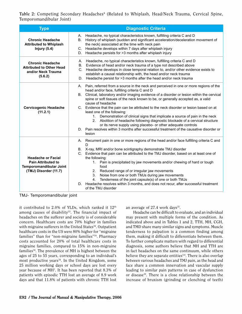
The proposed etiology of CGH is based on the conver-gence of afferent sensory input into the cervicotrigeminal nucleus from structures that are innervated by the first three spinal nerves or the trigeminal nerve. A subsequent “misinterpretation” of nociceptive signals originating in the cervical somatosensory structures as coming from the structures in the head innervated by the trigeminal nerve is thought to be responsible for this type of head-ache23-27. Musculoskeletal structures in the neck that are innervated by the first three spinal nerves that may refer pain into the head include the atlanto-occipital joints, joints and ligaments of the atlanto-axial joint, the C2-C4 zygapophyseal joints, the C2-C3 intervertebral disk, and muscles innervated by C1-C323-29. Table 2 lists the diagnostic criteria for CGH.
Temporomandibular disorder describes a variety of conditions affecting the temporomandibular joint (TMJ) and the muscles of mastication30. Symptoms include jaw and facial pain, limited TMJ mobility, joint sounds, tinnitus, and—most relevant to this case report—head-aches15,16,30,31. A classification of TMD into two subtypes provides a better understanding of the disorder and possible treatment options30. Arthralgia encompasses impairments related to the joint biomechanics, internal derangements, degenerative changes, developmental defects, and other pathologies related to the TMJ30. Myalgia is related to impairments and pain in the musculature surrounding the TMJ30. Table 2 lists the diagnostic criteria for TMD-related headache.
Data on the epidemiology of headache further un-derscore the need for knowledge related to headache. We noted that headaches are one of the most common reasons for people to seek medical attention. Headaches are more prevalent in women than in men but preva-lence tends to decrease with age1,32,33. Up to one adult in twenty has a headache every day or nearly every day1. Most of the population studies and research have focused on MH: European and American studies have showed the prevalence of MH as 6-8% in males and 15-18% of females each year1. One in four American households has a migraine sufferer, totaling approximately 29.5 million people32. TTH is even more prevalent: it affects two-thirds of males and over 80% of females in developed countries1. Episodic TTH is the most common headache type reported in over 70% of some populations; chronic

E90 / The Journal of Manual & Manipulative Therapy, 2006
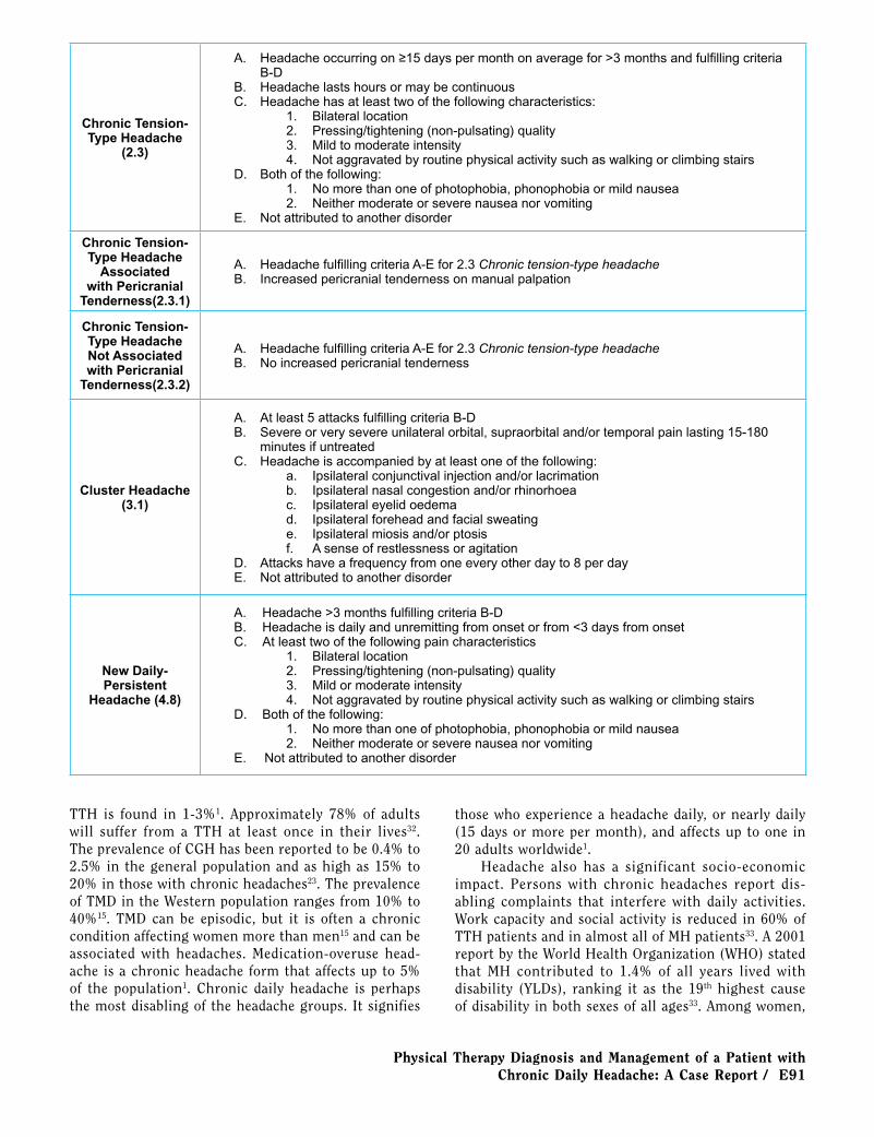
Table 1: Competing Primary Headaches4 (Migraine, Tension-Type, Cluster, New Daily-Persistent Headache)
Type Diagnostic Criteria
Migraine without Aura (1.1)
A. At least 5 attacks fulfilling criteria B-DB. Headache attacks lasting 4-72 hours (untreated or unsuccessfully treated)C. Headache has at least two of the following characteristics:
1. Unilateral location2. Pulsating quality3. Moderate or severe pain intensity4. Aggravation by or causing avoidance of routine physical activity (e.g., walking or
climbing stairs)D. During headache at least one of the following:
1. Nausea and/or vomiting2. Photophobia and phonophobia
E. Not attributed to another disorder
Typical Migraine with Aura (1.2.1)
A. At least 2 attacks fulfilling criteria B-DB. Aura consisting of at least one of the following, but no motor weakness:
1. Fully reversible visual symptoms including positive features (e.g., flickering lights, spots or lines) and/or negative features (i.e., loss of vision)
2. Fully reversible sensory symptoms including positive features (i.e., pins and needles) and/or negative features (i.e., numbness)
3. Fully reversible dysphasic speech disturbanceC. At least two of the following:
1. Homonymous visual symptoms and/or unilateral sensory symptoms2. At least one aura symptom develops gradually over ≥5 minutes and/or different aura
symptoms occur in succession over ≥5 minutes3. Each symptom lasts ≥5 and ≤60 minutes
D. Headache fulfilling criteria B-D for 1.1 Migraine without aura begins during the aura or follow aura within 60 minutes
E. Not attributed to another disorder
Chronic Migraine (1.5.1)
A. Headache fulfilling criteria C and D for 1.1 Migraine without aura on ≥15 days/month for >3 months
B. Not attributed to another disorder
Probable Migraine without Aura
(1.6.1)
A. Attacks fulfilling all but one of criteria A-D for 1.1 Migraine without auraB. Not attributed to another disorder
Infrequent Episodic Tension-
Type Headache (2.1)
A. At least 10 episodes occurring on <1 day per month on average (<12 days per year) and fulfilling criteria B-D
B. Headache lasting from 30 minutes to 7 daysC. Headache has at least two of the following characteristics:
1. Bilateral location2. Pressing/tightening (non-pulsating) quality3. Mild or moderate intensity4. Not aggravated by routine physical activity such as walking or climbing stairs
D. Both of the following:1. No nausea or vomiting (anorexia may occur)2. No more than one of photophobia or phonophobia
E. Not attributed to another disorder
Frequent Episodic Tension-Type
Headache (2.2)
A. At least 10 episodes of occurring on ≥1 but <15 days per month for at least 3 months and fulfilling criteria B-D
B. Headache lasting from 30 minutes to 7 daysC. Headache has at least two of the following characteristics:
1. Bilateral location2. Pressing/tightening (non-pulsating) quality3. Mild to moderate intensity4. Not aggravated by routine physical activity such as walking or climbing stairs
D. Both of the following:1. No nausea and/or vomiting (anorexia may occur)2. No more than one of photophobia and phonophobia
E. Not attributed to another disorder
Physical Therapy Diagnosis and Management of a Patient with Chronic Daily Headache: A Case Report / E91
Chronic Tension-Type Headache
(2.3)
A. Headache occurring on ≥15 days per month on average for >3 months and fulfilling criteria B-D
B. Headache lasts hours or may be continuousC. Headache has at least two of the following characteristics:
1. Bilateral location2. Pressing/tightening (non-pulsating) quality3. Mild to moderate intensity4. Not aggravated by routine physical activity such as walking or climbing stairs
D. Both of the following:1. No more than one of photophobia, phonophobia or mild nausea2. Neither moderate or severe nausea nor vomiting
E. Not attributed to another disorder
Chronic Tension-Type Headache
Associated with Pericranial
Tenderness(2.3.1)
A. Headache fulfilling criteria A-E for 2.3 Chronic tension-type headacheB. Increased pericranial tenderness on manual palpation
Chronic Tension-Type Headache Not Associated with Pericranial
Tenderness(2.3.2)
A. Headache fulfilling criteria A-E for 2.3 Chronic tension-type headacheB. No increased pericranial tenderness
Cluster Headache (3.1)
A. At least 5 attacks fulfilling criteria B-DB. Severe or very severe unilateral orbital, supraorbital and/or temporal pain lasting 15-180
minutes if untreatedC. Headache is accompanied by at least one of the following:
a. Ipsilateral conjunctival injection and/or lacrimationb. Ipsilateral nasal congestion and/or rhinorhoeac. Ipsilateral eyelid oedemad. Ipsilateral forehead and facial sweatinge. Ipsilateral miosis and/or ptosisf. A sense of restlessness or agitation
D. Attacks have a frequency from one every other day to 8 per dayE. Not attributed to another disorder
New Daily-Persistent
Headache (4.8)
A. Headache >3 months fulfilling criteria B-DB. Headache is daily and unremitting from onset or from <3 days from onsetC. At least two of the following pain characteristics
1. Bilateral location2. Pressing/tightening (non-pulsating) quality3. Mild or moderate intensity4. Not aggravated by routine physical activity such as walking or climbing stairs
D. Both of the following:1. No more than one of photophobia, phonophobia or mild nausea2. Neither moderate or severe nausea nor vomiting
E. Not attributed to another disorder
TTH is found in 1-3%1. Approximately 78% of adults will suffer from a TTH at least once in their lives32. The prevalence of CGH has been reported to be 0.4% to 2.5% in the general population and as high as 15% to 20% in those with chronic headaches23. The prevalence of TMD in the Western population ranges from 10% to 40%15. TMD can be episodic, but it is often a chronic condition affecting women more than men15 and can be associated with headaches. Medication-overuse head-ache is a chronic headache form that affects up to 5% of the population1. Chronic daily headache is perhaps the most disabling of the headache groups. It signifies
those who experience a headache daily, or nearly daily (15 days or more per month), and affects up to one in 20 adults worldwide1.
Headache also has a significant socio-economic impact. Persons with chronic headaches report dis-abling complaints that interfere with daily activities. Work capacity and social activity is reduced in 60% of TTH patients and in almost all of MH patients33. A 2001 report by the World Health Organization (WHO) stated that MH contributed to 1.4% of all years lived with disability (YLDs), ranking it as the 19th highest cause of disability in both sexes of all ages33. Among women,
E92 / The Journal of Manual & Manipulative Therapy, 2006
it contributed to 2.0% of YLDs, which ranked it 12th
among causes of disability33. The financial impact of headaches on the sufferer and society is of considerable concern. Healthcare costs are 70% higher in families with migraine sufferers in the United States34. Outpatient healthcare costs in the US were 80% higher for “migraine families” than for “non-migraine families”34. Pharmacy costs accounted for 20% of total healthcare costs in migraine families, compared to 15% in non-migraine families34. The prevalence of MH is highest between the ages of 25 to 55 years, corresponding to an individual’s most productive years34. In the United Kingdom, some 25 million working days or school days are lost every year because of MH1. It has been reported that 8.3% of patients with episodic TTH lost an average of 8.9 work days and that 11.8% of patients with chronic TTH lost
an average of 27.4 work days35. Headache can be difficult to evaluate, and an individual
may present with multiple forms of the condition. As indicated above and in Tables 1 and 2, TTH, MH, CGH, and TMD share many similar signs and symptoms. Muscle tenderness to palpation is a common finding among them, making it difficult to differentiate between them. To further complicate matters with regard to differential diagnosis, some authors believe that MH and TTH are in fact headaches on the same continuum, while others believe they are separate entities3,6. There is also overlap between various headaches and TMJ pain, as the head and face share a common innervation and vascular supply leading to similar pain patterns in case of dysfunction or disease30. There is a close relationship between the increase of bruxism (grinding or clenching of teeth)
Table 2: Competing Secondary Headaches4 (Related to Whiplash, Head/Neck Trauma, Cervical Spine, Temporomandibular Joint)
Type Diagnostic Criteria
Chronic Headache Attributed to Whiplash
Injury (5.4)
A. Headache, no typical characteristics known, fulfilling criteria C and DB. History of whiplash (sudden and significant acceleration/deceleration movement of
the neck) associated at the time with neck painC. Headache develops within 7 days after whiplash injuryD. Headache persists for >3 months after whiplash injury
Chronic Headache Attributed to Other Head
and/or Neck Trauma (5.6.2)
A. Headache, no typical characteristics known, fulfilling criteria C and DB. Evidence of head and/or neck trauma of a type not described aboveC. Headache develops in close temporal relation to, and/or other evidence exists to
establish a causal relationship with, the head and/or neck traumaD. Headache persist for >3 months after the head and/or neck trauma
Cervicogenic Headache (11.2.1)
A. Pain, referred from a source in the neck and perceived in one or more regions of the head and/or face, fulfilling criteria C and D
B. Clinical, laboratory and/or imaging evidence of a disorder or lesion within the cervical spine or soft tissues of the neck known to be, or generally accepted as, a valid cause of headache
C. Evidence that the pain can be attributed to the neck disorder or lesion based on at least one of the following:
1. Demonstration of clinical signs that implicate a source of pain in the neck2. Abolition of headache following diagnostic blockade of a cervical structure
or its nerve supply using placebo- or other adequate controlsD. Pain resolves within 3 months after successful treatment of the causative disorder or
lesion
Headache or Facial Pain Attributed to
Temporomandibular Joint (TMJ) Disorder (11.7)
A. Recurrent pain in one or more regions of the head and/or face fulfilling criteria C and D
B. X-ray, MRI and/or bone scintigraphy demonstrate TMJ disorderC. Evidence that pain can be attributed to the TMJ disorder, based on at least one of
the following:1. Pain is precipitated by jaw movements and/or chewing of hard or tough
food2. Reduced range of or irregular jaw movements3. Noise from one or both TMJs during jaw movements4. Tenderness of the joint capsule(s) of one or both TMJs
D. Headache resolves within 3 months, and does not recur, after successful treatment of the TMJ disorder
TMJ- Temporomandibular joint
Physical Therapy Diagnosis and Management of a Patient with Chronic Daily Headache: A Case Report / E93
and parafunction (excessive or unnecessary function related to the jaw) found in TMD and an increase in TTH frequency30. One review looked at the CGH diag-nostic criteria and concluded that there was insufficient specificity to separate CGH from MH patients36. Another study looked at the association between MH and TMD and concluded that they were two clearly differentiated diagnostic entities30. Various authors agree that there are neuromusculoskeletal abnormalities that play a role in the pathogenesis and presentation of TTH6-8,12-
16,20,24-26,37, 38, MH26,37,38, CGH16,24-26,28-30,37,38, TMD-related headaches15,16,30,31,38, and occipital neuralgia headaches21 further exacerbating the difficulty faced by the clinician with regard to differential diagnosis16,21.
Despite the high prevalence of headache disorders and their socio-economic and personal impact, headache disorders continue to be underestimated in scale, poorly diagnosed, and undertreated by the medical community1,33. The patient described in this case report presented with a medical diagnosis of MH and chronic TTH with an onset of a new type of chronic daily headache potentially related to a history of motor vehicle accident (MVA) and/or possibly caused by TMD. The etiology of various headaches is often hard to determine with potential combined influences of neurological, musculoskeletal, neurovascular, psychological, and nutritional factors and chemical imbalances in the brain. Some headaches are indicative of an underlying disease process; some of these are life-threatening and others benign. Thus, a thorough medical evaluation is necessary with any new onset or ongoing headache. Likewise, a thorough PT examination should aim to rule out serious pathology by way of a systems review approach, to determine the type of headache, and to define the neuromusculoskeletal factors that may be contributing to the headache. An accurate differential diagnosis is imperative in deter-mining whether a headache is neuromusculoskeletal in origin, which is treatable, or whether it is another type of headache that requires medical consultation and (co) management. The purposes of this case report describing a patient with chronic daily headache are to:
1. Describe the PT differential diagnosis and deci-sion-making process
2. Provide a treatment rationale and description of subsequent PT management using a combination of myofascial trigger point dry needling, orthopaedic manual physical therapy (OMPT), exercise therapy, and patient education
Case Description
HistoryThe patient described in this case report was a married
48-year-old-female with four teenage children, two dogs, one cat, and a horse, which made for a busy home life. She was referred to PT within a multi-disciplinary pain
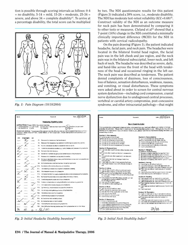
management practice with a medical diagnosis of common MH, chronic TTH, and TMD. The patient worked as a full-time general counsel attorney and had been at her current job for 6 months. Work environment was sedentary with physical demands related to sitting deskwork with some time spent using the computer and telephone. She had not lost any work time because of her headaches. The patient was a non-smoker and drank two glasses of wine per week and one cup of caffeinated coffee per day. The wine and coffee were not reported as triggers for her headaches. Recreational activities included yoga once a week, aerobic and resistance training three times a week, and reading. Prior to the initial evaluation, the patient was asked to complete a pain drawing (Figure 1) and two outcome assessment tools: the Henry Ford Hospital Headache Disability Inventory [HDI] (Figure 2) and the Neck Disability Index [NDI] (Figure 3). The HDI and NDI were chosen as outcome measures to assess the response to treatment on the patient’s headache and neck-related self-perceived disability.
The HDI is a 25-item questionnaire that aims to measure the self-perceived disabling effects of headache on daily life. The questionnaire contains two subgroups of questions, thereby creating emotional and functional subscale scores and a total score. Two additional items on the questionnaire ask the patient to rate the severity of their headache as: 1) mild, 2) moderate, or 3) severe, and the frequency as 1) less than or equal to one per month, 2) more than one but less than four per month, or 3) four or more times per month. The results of the HDI for this patient (Figure 2) indicated severe head-ache intensity, headache frequency greater than one per week, and a total score of 56/100 (emotional 26/52, functional 30/48). The HDI has good internal consis-tency reliability; correlations between the emotional and functional subscale scores and the total score were both excellent (r = 0.89)39. It also has good short-term (1-week) (r=0.93-0.95)40 and generally good long-term (2-month) test-retest reliability (r=0.83)39 for the total scores. The HDI also exhibits good internal construct validity (P<0.001)39. A minimal detectable change (MDC95) score at 1-week retest is 16 points; this value for the MDC95 indicates that a clinician can be 95% confident that a true change has occurred with a change in the HDI score ≥ 16 points40. Similarly, a 29-point score improvement constitutes the MDC95 over a 2-month time period39. The HDI test is simple to administer and takes little time to complete. This self-reporting outcome measure is useful in periodically and reliably assessing the effects of treatment intervention in patients with disabling headaches39,40.
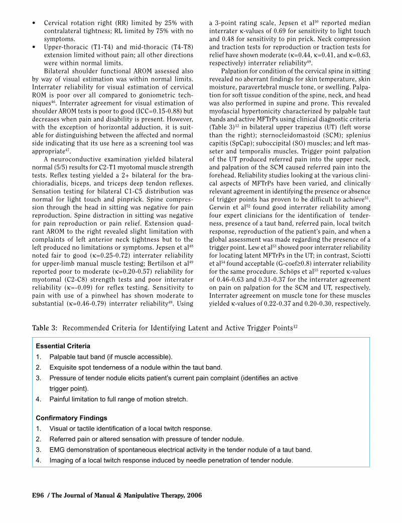
The NDI is a 10-item questionnaire that aims to measure the self-perceived disabling effects of neck pain on daily life. It is a modification of the Oswestry Low Back Pain Index, which has been used as a self-reporting outcome measure for low-back pain disability. Interpreta-
E94 / The Journal of Manual & Manipulative Therapy, 2006
tion is possible through scoring intervals as follows: 0-4 = no disability, 5-14 = mild, 15-24 = moderate, 25-34 = severe, and above 34 = complete disability41. To arrive at a percentage disability, the total score can be multiplied
by two. The NDI questionnaire results for this patient (Figure 3) indicated a 38% score, i.e., moderate disability. The NDI has moderate test-retest reliability (ICC=0.68)42. Construct validity of the NDI as an outcome measure for neck pain has been demonstrated by comparing it to other tests or measures. Cleland et al42 showed that a 7-point (14%) change in the NDI constituted a minimally clinically important difference (MCID) for the NDI in patients with cervical radiculopathy.
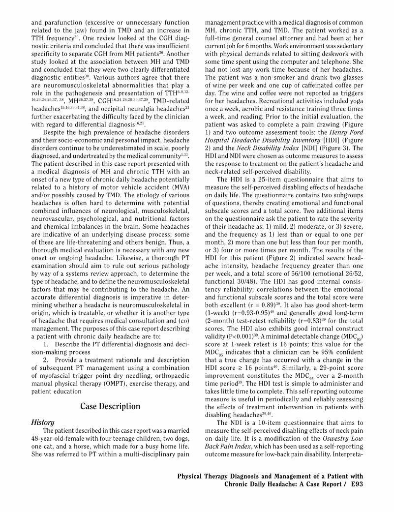
On the pain drawing (Figure 1), the patient indicated headache, facial pain, and neck pain. The headaches were located in the bilateral frontal head region, the facial pain was in the left cheek and jaw region, and the neck pain was in the bilateral suboccipital, lower neck, and left back of neck. The headache was described as severe, daily, and band-like across the front of the head with tender-ness of the head and occasional ringing in the left ear. The neck pain was described as tenderness. The patient denied complaints of dizziness, loss of consciousness, loss of balance, sensation disturbances, weakness, nausea, and vomiting, or visual disturbances. These symptoms were asked about in order to screen for central nervous system dysfunction—including cord compression, cranial nerve dysfunction due to undiagnosed central processes, vertebral or carotid artery compromise, post-concussive syndrome, and other intracranial pathology—that might Fig. 1: Pain Diagram (10/18/2004)
Fig. 2: Initial Headache Disability Inventory39 Fig. 3: Initial Neck Disability Index41

Physical Therapy Diagnosis and Management of a Patient with Chronic Daily Headache: A Case Report / E95
be causing the current complaints of headache43. The diagnostic accuracy of these symptoms for implicating the mentioned pathologies has not been validated.
The patient reported that symptoms were improved by local application of heat, stretching, sometimes doing nothing, and Imitrex (a Triptan-class MH drug) if it was a migraine-type headache. The patient identified this migraine-type headache as the headache that caused pain behind her left eye; this identification was confirmed by the positive response to medication specific for an MH (i.e., Imitrex). However, the patient noted that the use of Imitrex did not always relieve the present headache, which would seem to indicate the presence of more than one type of headache. Symptoms were aggravated by bright light, certain smells, hunger, hot weather, exercise, and change in barometric pressure. No diurnal pattern of symptoms was noted. Sleep was undisturbed in a habitual left and/or right sidelying position with use of a cervical pillow.
A review of the available physician medical records and radiological reports indicated a history of MH since age 17. The onset time and cause of her neck pain was unknown. Onset of the newly described headache was 3 years before, and a neurologist who specialized in headache management supervised its diagnosis and management. Follow-up with the physician had occurred approximately 1 year prior because of the onset of left tinnitus. The patient was then referred to a dentist due to suspicion of TMD. The dentist prescribed a night splint, which the patient wore on and off. She continued to see her dentist regularly until her mother died in February of 2004. Headaches had become more intense in March of 2004 and continued to become progressively worse over the next 6 months. The patient was unable to relate possible reasons contributing to the onset or worsening of the complaints.
Her neurologist then referred the patient to a pain management outpatient practice in August 2004, where she was seen by a physician who was a neurologist and pain management specialist. This physician reported increased and abnormal tone of the left arm and pro-nounced slowness of finger tapping of the left hand. He was concerned with the facts of increasing severity of headaches, worsening of symptoms lying down compared to being upright, and motor dysfunction with the left arm when raised. The physician ordered a magnetic resonance imaging (MRI) study; findings showed no focal signal abnormality or mass lesions in the brain. MRI and magnetic resonance angiography (MRA) studies of the brain done approximately 2 years earlier were again evaluated and found normal. The patient had a follow-up visit with the same physician one month later with continued complaints of headache more than 50% of the time. At the time of the initial physical therapy evalua-tion, the headache was daily during some weeks but at other times the patient could go several days without a
headache. At times she took the Imitrex daily or even twice daily but the effect varied from none to satisfactory headache relief. The TMJ remained uncomfortable, but the dentist told the patient that improvement as a result of wearing the splint would take time. The neurologist had recommended Botulinum Toxin injections for selected neck, shoulder, and facial muscles in combination with PT but the patient elected against these injections.
The medical history for this patient included MH, asthma, depression, and a fractured pelvis and nose as a result of an MVA 5 years before. Her surgical history included tubal ligation, laser surgery for cervix dysplagia, and tonsillectomy. Current medications included Celexa 20mg once a day (QD) (anti-depressant), Imitrex 50mg as needed (PRN), Zanaflex PRN (short-acting muscle relaxant), Advair Diskus QD (asthma treatment), and Yasmin (birth control). A screening examination using a systems approach revealed that the patient was receiv-ing psychological counseling once a week. The patient’s family history included the father alive at 69 with high blood pressure and diabetes and the mother deceased at age 69 from an overdose. The patient provided no further details on her mother’s death. There was no indication in the family history of headaches, including MH. First-degree relatives of persons who never had MH are at no increased risk of MH without aura (relative risk= 1.11 [95% confidence interval (CI) 0.83-1.39]) or with aura (relative risk= 0.65 [95% CI: 0.36-0.94])44.
Physical ExaminationThe patient stood 5’7” at 155 lbs with a mesomor-
phic body type. Postural observation of this patient from the side using a 3-point grading system (increased, normal, decreased) revealed decreased lumbar lordosis, increased thoracic kyphosis, and increased cranio-cervi-cal extension resulting in a forward head posture (FHP). Observation from the back revealed symmetrical iliac crest and shoulder heights; the head was side-bent to the right. Fedorak et al45 noted fair intra- (κ=0.50) and poor interrater reliability (κ=0.16) for visual assessment of cervical and lumbar lordosis using a similar 3-point rating system.
Cranio-cervical, cervical, and upper thoracic spine active range of motion (AROM) testing in a sitting position assessed quality of motion, range, and pain provocation; limitations were estimated visually with the following findings:• Cranio-cervical flexion limited by 50%; extension
not limited.• C1-C2 rotation right limited by 50%.• Cervical flexion limited by 25% with tightness re-
ported in the upper back; extension hypermobility with an apex of the curve observed at C5-C6.
• Cervical side-bending right (SBR) limited by 75% with tightness in the contralateral neck; SBL limited by 25% with restriction noted ipsilateral.
E96 / The Journal of Manual & Manipulative Therapy, 2006
• Cervical rotation right (RR) limited by 25% with contralateral tightness; RL limited by 75% with no symptoms.
• Upper-thoracic (T1-T4) and mid-thoracic (T4-T8) extension limited without pain; all other directions were within normal limits.Bilateral shoulder functional AROM assessed also
by way of visual estimation was within normal limits. Interrater reliability for visual estimation of cervical ROM is poor over all compared to goniometric tech-niques46. Interrater agreement for visual estimation of shoulder AROM tests is poor to good (ICC=0.15-0.88) but decreases when pain and disability is present. However, with the exception of horizontal adduction, it is suit-able for distinguishing between the affected and normal side indicating that its use here as a screening tool was appropriate47.
A neuroconductive examination yielded bilateral normal (5/5) results for C2-T1 myotomal muscle strength tests. Reflex testing yielded a 2+ bilateral for the bra-chioradialis, biceps, and triceps deep tendon reflexes. Sensation testing for bilateral C1-C5 distribution was normal for light touch and pinprick. Spine compres-sion through the head in sitting was negative for pain reproduction. Spine distraction in sitting was negative for pain reproduction or pain relief. Extension quad-rant AROM to the right revealed slight limitation with complaints of left anterior neck tightness but to the left produced no limitations or symptoms. Jepsen et al48 noted fair to good (κ=0.25-0.72) interrater reliability for upper-limb manual muscle testing; Bertilson et al49 reported poor to moderate (κ=0.20-0.57) reliability for myotomal (C2-C8) strength tests and poor interrater reliability (κ=-0.09) for reflex testing. Sensitivity to pain with use of a pinwheel has shown moderate to substantial (κ=0.46-0.79) interrater reliability49. Using
a 3-point rating scale, Jepsen et al50 reported median interrater κ-values of 0.69 for sensitivity to light touch and 0.48 for sensitivity to pin prick. Neck compression and traction tests for reproduction or traction tests for relief have shown moderate (κ=0.44, κ=0.41, and κ=0.63, respectively) interrater reliability49.
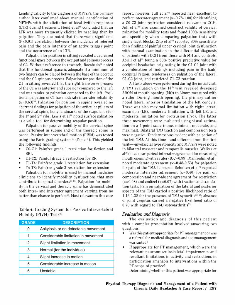
Palpation for condition of the cervical spine in sitting revealed no aberrant findings for skin temperature, skin moisture, paravertebral muscle tone, or swelling. Palpa-tion for soft tissue condition of the spine, neck, and head was also performed in supine and prone. This revealed myofascial hypertonicity characterized by palpable taut bands and active MFTrPs using clinical diagnostic criteria (Table 3)12 in bilateral upper trapezius (UT) (left worse than the right); sternocleidomastoid (SCM); splenius capitis (SpCap); suboccipital (SO) muscles; and left mas-seter and temporalis muscles. Trigger point palpation of the UT produced referred pain into the upper neck, and palpation of the SCM caused referred pain into the forehead. Reliability studies looking at the various clini-cal aspects of MFTrPs have been varied, and clinically relevant agreement in identifying the presence or absence of trigger points has proven to be difficult to achieve51. Gerwin et al52 found good interrater reliability among four expert clinicians for the identification of tender-ness, presence of a taut band, referred pain, local twitch response, reproduction of the patient’s pain, and when a global assessment was made regarding the presence of a trigger point. Lew et al53 showed poor interrater reliability for locating latent MFTrPs in the UT; in contrast, Sciotti et al54 found acceptable (G-coef≥0.8) interrater reliability for the same procedure. Schöps et al55 reported κ-values of 0.46-0.63 and 0.31-0.37 for the interrater agreement on pain on palpation for the SCM and UT, respectively. Interrater agreement on muscle tone for these muscles yielded κ-values of 0.22-0.37 and 0.20-0.30, respectively.
Table 3: Recommended Criteria for Identifying Latent and Active Trigger Points12
Essential Criteria
1. Palpable taut band (if muscle accessible).
2. Exquisite spot tenderness of a nodule within the taut band.
3. Pressure of tender nodule elicits patient’s current pain complaint (identifies an active
trigger point).
4. Painful limitation to full range of motion stretch.
Confirmatory Findings
1. Visual or tactile identification of a local twitch response.
2. Referred pain or altered sensation with pressure of tender nodule.
3. EMG demonstration of spontaneous electrical activity in the tender nodule of a taut band.
4. Imaging of a local twitch response induced by needle penetration of tender nodule.
Physical Therapy Diagnosis and Management of a Patient with Chronic Daily Headache: A Case Report / E97
Lending validity to the diagnosis of MFTrPs, the primary author later confirmed above manual identification of MFTrPs with the elicitation of local twitch responses (LTR) during treatment; Hong et al56 concluded that an LTR was more frequently elicited by needling than by palpation. They also noted that there was a significant (P<0.01) correlation between the incidence of referred pain and the pain intensity of an active trigger point and the occurrence of an LTR.
Palpation for position in sitting revealed a decreased functional space between the occiput and spinous process of C2. Without reference to research, Rocabado57 noted that this functional space is adequate if a minimum of two fingers can be placed between the base of the occiput and the C2 spinous process. Palpation for position of the C1 in sitting revealed that the right transverse process of the C1 was anterior and superior compared to the left and was tender to palpation compared to the left. Posi-tional palpation of C1 has moderate interrater reliability (κ=0.63)58. Palpation for position in supine revealed no aberrant findings for palpation of the articular pillars of the cervical spine, bony landmarks of the scapula, or for the 1st and 2nd ribs. Lewis et al59 noted surface palpation as a valid tool for determining scapular position.
Palpation for passive mobility of the cervical spine was performed in supine and of the thoracic spine in prone. Passive inter-vertebral motion (PIVM) was tested using the Paris grading system60 (Table 4). This yielded the following findings:• C0-C1: Painfree grade 1 restriction for flexion and
SBL• C1-C2: Painful grade 1 restriction for RR• T1-T4: Painfree grade 1 restriction for extension• T4-T8: Painfree grade 2 restriction for extension
Palpation for mobility is used by manual medicine clinicians to identify mobility dysfunctions that may contribute to spinal disorders61-66. Palpation for mobil-ity in the cervical and thoracic spine has demonstrated both intra- and interrater agreement varying from no better than chance to perfect65. Most relevant to this case
report, however, Jull et al61 reported near excellent to perfect interrater agreement (κ=0.78-1.00) for identifying a C0-C3 joint restriction considered relevant to CGH. Jull et al67 also examined construct validity of cervical palpation for mobility tests and found 100% sensitivity and specificity when comparing palpation tests with single facet blocks. Zito et al68 reported 80% sensitivity for a finding of painful upper cervical joint dysfunction with manual examination in the differential diagnosis of patients with CGH from those with MH and controls. Aprill et al69 found a 60% positive predictive value for occipital headaches originating in the C1-C2 joint with a combination of findings including pain in the (sub) occipital region, tenderness on palpation of the lateral C1-C2 joint, and restricted C1-C2 rotation.
All tests above were performed during the initial visit. A TMJ evaluation on the 14th visit revealed decreased AROM of mouth opening (MO) to 30mm measured with a ruler. During mouth opening, the primary author noted lateral anterior translation of the left condyle. There was also maximal limitation with right lateral excursion (LE), moderate limitation with left LE, and moderate limitation for protrusion (Pro). The latter three movements were evaluated using visual estima-tion on a 4-point scale (none, minimal, moderate, and maximal). Bilateral TMJ traction and compression tests were negative. Tenderness was evident with palpation of the left TMJ. At this time—and different from the first visit—-myofascial hypertonicity and MFTrPs were noted in bilateral masseter and temporalis muscles. Walker et al70 noted near-perfect interrater agreement for measuring mouth opening with a ruler (ICC=0.99). Manfredini et al71 noted moderate agreement (κ=0.48-0.53) for palpation for pain of the TMJ. Lobbezoo-Scholten et al72 reported moderate interrater agreement (κ=0.40) for pain on compression and near-absent agreement for restriction (κ=0.08) and endfeel (κ=0.07) with traction and transla-tion tests. Pain on palpation of the lateral and posterior aspects of the TMJ carried a positive likelihood ratio of 1.16-1.38 for the presence of TMJ synovitis73, 74; absence of joint crepitus carried a negative likelihood ratio of 0.70 with regard to TMJ osteoarthritis73.
Evaluation and DiagnosisThe evaluation and diagnosis of this patient
with a complex presentation involved answering two questions:• Was this patient appropriate for PT management or was
a referral for medical diagnosis and (co)management warranted?
• If appropriate for PT management, which were the relevant neuromusculoskeletal impairments and resultant limitations in activity and restrictions in participation amenable to interventions within the PT scope of practice?Determining whether this patient was appropriate for
Table 4: Grading System for Passive Intervertebral Mobility (PIVM) Tests60
GRADE DESCRIPTION
0 Ankylosis or no detectable movement
1 Considerable limitation in movement
2 Slight limitation in movement
3 Normal (for the individual)
4 Slight increase in motion
5 Considerable increase in motion
6 Unstable
E98 / The Journal of Manual & Manipulative Therapy, 2006
PT management required the therapist to both exclude with a sufficient degree of diagnostic confidence potential serious pathology responsible for the current presenta-tion and to ascertain that the provided medical headache diagnoses fit with the signs and symptoms noted during the history and physical examination.
In the authors’ clinical opinion, serious pathology was ruled out sufficiently by the comprehensive examina-tion of the referring neurologist and the findings from the history and examination noted above. However, it should be noted that data on the diagnostic accuracy of history items and physical examination as discussed above is either absent or insufficient to confidently exclude central nervous system pathology potentially capable of producing similar signs and symptoms. Therefore, this decision was based mainly on clinician experience and interpretation of the tests based on a pathophysiologic rather than research-based rationale.
This patient came with medical diagnoses of chronic TTH, MH, and TMD. As discussed above these but also many other headache types within the ICHD-II could potentially present with the same signs and symptoms as collected during the history and physical examination. Although it is not the role of the physical therapist to make a medical diagnosis, it is his or her responsibil-ity to ascertain that the provided medical diagnosis fits with the history and physical examination findings. Discrepancies between the diagnosis provided and the signs and symptoms observed should lead to medical referral. Only when the signs and symptoms observed fit with the diagnosis provided will a PT examina-tion and diagnosis indicate whether the patient might benefit from PT intervention. A clinical decision-making process was performed to confirm or cast doubt on the provided medical diagnosis. In this case, key differential diagnostic data were derived from the headache’s onset, nature, severity, chronicity, characteristics, associated symptoms, and physical examination findings.
Of the primary headache groups noted in the ICDH-II, only MH and TTH required further diagnostic consid-eration (Table 1)4. With the given patient presentation, the diagnostic criteria for migraine without aura (1.1) were not met entirely met. The patient had at least five attacks (criterion A), the headaches lasted 4-72 hours (criterion B), and the headaches were of severe intensity (criterion 4C). However, she did not fulfill a second characteristic out of the four in criterion C: She did describe a unilateral location (behind the left eye), but this was not part of her primary headache. The patient described aggravation by exercise, but not aggravation by or avoidance of routine physical activity (e.g., walking or climbing stairs). She also did not describe a pulsating quality to her headaches. With regard to criterion D, the patient described aggravation by bright light (photopho-bia), but she did not mention phonophobia, nausea, or vomiting (see Table 1). Typical aura with migraine (1.2.1)
was not a consideration mainly because her symptoms were not accompanied by any aura. She did not meet the frequency and chronic nature of chronic migraine (1.5.1), as outlined in criterion A. However, with a report of symptomatic relief of her unilateral headache with a Triptan-class medication, a diagnosis of MH without aura was considered likely despite the patient not meeting all diagnostic criteria.
Episodic (2.1) and frequent episodic TTH (2.2) could be eliminated because the frequency per month of her headaches exceeded criteria for both leaving chronic TTH (2.3) and new daily-persistent headache (4.8). Their criteria are very similar and the patient’s headache fulfilled criteria for both types; however, new daily-persistent headache (4.8) is daily and unremitting since or very close to a time of onset that is clearly re-called and unambiguous4. This was not evident with the onset of daily headache for this patient being described as insidious and vague. Chronic TTH (2.3) exists in two forms: associated (2.3.1) and not associated with peri-cranial tenderness (2.3.2) described as local tenderness to manual palpation by the second and third finger on muscles of the head and neck (i.e., frontalis, tempora-lis, masseter, pterygoid, SCM, splenius, and trapezius muscles)4. Palpation of the neck and head musculature in this patient revealed tenderness, characterized by pal-pable taut bands and active MFTrPs, making a diagnosis of chronic TTH associated with pericranial tenderness (2.3.1) very plausible.
A medical history of suspected TMD and a motor vehicle accident (MVA) five years prior during which the patient sustained a fractured nose and neuromus-culoskeletal impairments found during the examination warranted further inquiry of the secondary headache groups. Whether to classify a secondary headache depends on a few factors. If a headache is a new headache that presents with another disorder known to be capable of causing it, then it is described as a secondary headache4. If a primary headache already exists, factors that support adding a secondary headache diagnosis include a close temporal relation to a causative disorder, a discernible worsening of the primary headache, good evidence that the causative disorder can exacerbate the primary headache, and improvement or resolution of the headache after relief of the presumed causative disorder4. In respect to the improvement or resolution of the headache, in many cases there is insufficient follow-up time or a diagnosis needs to be made prior to the end of expected time for remission. In these cases, it is recommended to describe the headache as a headache probably attributed to [the disorder]; a definitive diagnosis can only be made once the time-sensitive outcome criterion D is fulfilled4.
Of the secondary headache groups, headache at-tributed to head and/or neck trauma and headache or facial pain attributed to a disorder of cranium, neck, eyes, ears, nose, sinuses, teeth, mouth, or other facial or
cranial structures required further investigation for this patient (Table 2)4. The presentation did not fulfill chronic headache attributed to whiplash injury (5.4), because the patient did not describe a discernable whiplash injury after her MVA and the headache did not develop within 7 days after a possible or suspected whiplash injury. A fractured nose might constitute possible head trauma, but there was no evidence that the headache developed in close temporal relation to the trauma thereby making chronic headache attributed to other head and/or neck trauma (5.6.2) unlikely. Although the primary author suspected headache due to TMD and this suspicion was to some degree substantiated later based on the exami-nation findings for the TMJ noted above, this case did not meet the established criteria for headache or facial pain attributed to temporomandibular joint disorder (11.7); evidence of TMD established by way of X-ray, MRI, and/or bone scintigraphy was not available (criterion B). Also the time-dependent outcome criterion D could not be met. Cervicogenic headache (11.2.1) was a possible secondary headache diagnosis because the examination findings met criteria A, B, and C1. Again, the time-sensitive outcome criterion D could not be confirmed. Clinical findings that supported the diagnosis of CGH included FHP, suboccipital tenderness, and upper cervical positional abnormalities and limited mobility.
In summary, after the initial evaluation, the relevant signs and symptoms associated with the patient’s head-aches seemed to be consistent with and fulfill ICDH-II diagnostic criteria4 for:
1. Chronic TTH associated with pericranial tenderness2. Probable MH without aura3. Probable cervicogenic headacheThe ICHD-II is an update of the original 1988 clas-
sification and includes expanded definitions and clari-fications3,4. Few studies have examined the reliability and validity of this new edition. Relevant to this patient is the fact that there is considerable symptom overlap between the diagnostic criteria for TTH and CGH23, yet some evidence shows that they are distinct disorders75,76. It should be noted that the absence of data on diagnostic accuracy of the ICHD-II does and should affect the level of diagnostic confidence with regard to the established headache diagnoses.
After excluding serious underlying undiagnosed pathology and establishing the seeming appropriateness of the headache diagnoses provided by the referring physician, the next step in the diagnostic process was to ascertain whether neuromusculoskeletal impairments caused or contributed to the patient’s headaches and neck pain. The patient presented with several physical examination findings of the musculoskeletal system of the head and neck that have been shown to contribute to various headache types. Myofascial trigger points have been noted to cause referred pain to the head, neck, and
face contributing to TTH, MH, and CGH12-21. Cervical spine joint dysfunction has been noted to contribute to CGH due to referred pain from the facet joints and influence of neural and vascular structures of the head and neck23-
29,77-79. FHP with posterior rotation of the cranium may lead to adverse affects on the structure and function of the cervical spine and TMJ, increasing the incidence of neck, interscapular, and headache pain18,31,37,78,80,81.
In light of this complex patient presentation, the primary author decided to assess for a suspected TMD at a later date due to a lack of time and a lower as-signed priority. TMD constitutes a variety of conditions involving the TMJ, muscles of mastication, and other associated structures. The diagnosis of TMD is varied, and agreement has not been met on the pathophysi-ologic mechanisms involved15. At some point during the course of treatment, the patient mentioned the onset of jaw pain. It was at that time that a TMJ evaluation was performed. The American Academy of Orofacial Pain (AAOFP)’s diagnostic criteria for TMD classify two major subgroups82:
1. Temporomandibular joint articular disorders including congenital and developmental disorders, disc derangement disorders, dislocation, inflammatory condi-tions, arthritides, ankylosis, and fracture
2. Masticatory muscle disorders divided into myofas-cial pain, myositis, myospasm, myofibrotic contracture, local myalgia (unclassified), myofibrotic contracture, and neoplasia
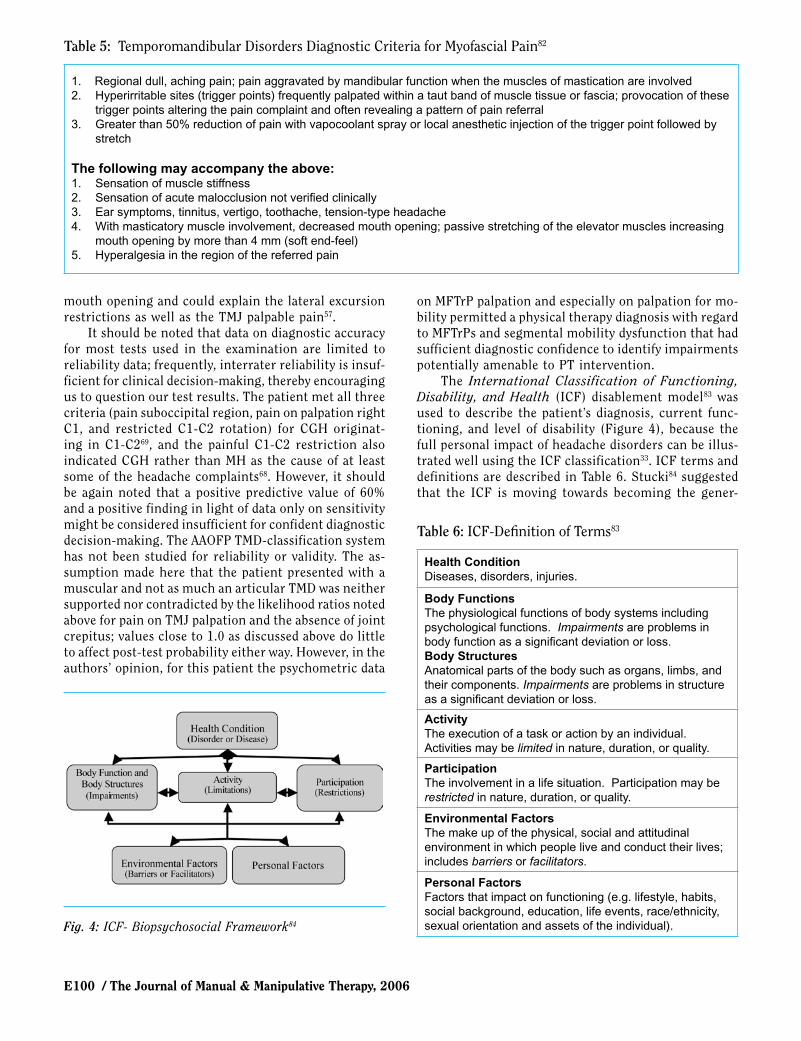
The TMJ evaluation indicated diagnoses of myofascial pain and left condylar hypermobility based on the history and on active and passive movement and palpation find-ings. The patient reported being under high stress and complained of jaw pain, stiffness, and pain with chewing. Limitations were present during mouth opening with anterior-lateral translation of the left condyle, bilateral lateral excursion (right worse than left), and protrusion. Palpation revealed myofascial hypertonicity and pain in the muscles of mastication and over the left TMJ. No joint sounds were noted. Therefore, the patient clearly met the diagnostic inclusion criteria for TMJ myofascial pain (Table 5)82. But the myofascial pain diagnosis did not explain the anterior-lateral translation of the left condyle, the discrepancy between left and right lateral excursion, and the pain with palpation of the left TMJ. Further investigation of the TMJ articular disorders did not show any plausible diagnosis for which all inclusion criteria were met. With the absence of joint sounds and without radiographic imaging, disc displacement disorders, inflammatory, and osteoarthritic disorders could not be excluded nor included82. The diagnosis of left condylar hypermobility is not a classified disorder named by the AAOFP, but it has been used to describe an articular condition that is likely to precede disc de-rangement disorders of the TMJ57. It is characterized by excessive condylar rotation (anterior translation) with
E100 / The Journal of Manual & Manipulative Therapy, 2006
mouth opening and could explain the lateral excursion restrictions as well as the TMJ palpable pain57.
It should be noted that data on diagnostic accuracy for most tests used in the examination are limited to reliability data; frequently, interrater reliability is insuf-ficient for clinical decision-making, thereby encouraging us to question our test results. The patient met all three criteria (pain suboccipital region, pain on palpation right C1, and restricted C1-C2 rotation) for CGH originat-ing in C1-C269, and the painful C1-C2 restriction also indicated CGH rather than MH as the cause of at least some of the headache complaints68. However, it should be again noted that a positive predictive value of 60% and a positive finding in light of data only on sensitivity might be considered insufficient for confident diagnostic decision-making. The AAOFP TMD-classification system has not been studied for reliability or validity. The as-sumption made here that the patient presented with a muscular and not as much an articular TMD was neither supported nor contradicted by the likelihood ratios noted above for pain on TMJ palpation and the absence of joint crepitus; values close to 1.0 as discussed above do little to affect post-test probability either way. However, in the authors’ opinion, for this patient the psychometric data
on MFTrP palpation and especially on palpation for mo-bility permitted a physical therapy diagnosis with regard to MFTrPs and segmental mobility dysfunction that had sufficient diagnostic confidence to identify impairments potentially amenable to PT intervention.
The International Classification of Functioning, Disability, and Health (ICF) disablement model83 was used to describe the patient’s diagnosis, current func-tioning, and level of disability (Figure 4), because the full personal impact of headache disorders can be illus-trated well using the ICF classification33. ICF terms and definitions are described in Table 6. Stucki84 suggested that the ICF is moving towards becoming the gener-
Table 5: Temporomandibular Disorders Diagnostic Criteria for Myofascial Pain82
1. Regional dull, aching pain; pain aggravated by mandibular function when the muscles of mastication are involved2. Hyperirritable sites (trigger points) frequently palpated within a taut band of muscle tissue or fascia; provocation of these
trigger points altering the pain complaint and often revealing a pattern of pain referral3. Greater than 50% reduction of pain with vapocoolant spray or local anesthetic injection of the trigger point followed by
stretch
The following may accompany the above:1. Sensation of muscle stiffness2. Sensation of acute malocclusion not verified clinically3. Ear symptoms, tinnitus, vertigo, toothache, tension-type headache4. With masticatory muscle involvement, decreased mouth opening; passive stretching of the elevator muscles increasing
mouth opening by more than 4 mm (soft end-feel)5. Hyperalgesia in the region of the referred pain
Table 6: ICF-Definition of Terms83
Health ConditionDiseases, disorders, injuries.
Body FunctionsThe physiological functions of body systems including psychological functions. Impairments are problems in body function as a significant deviation or loss.Body StructuresAnatomical parts of the body such as organs, limbs, and their components. Impairments are problems in structure as a significant deviation or loss.
Activity The execution of a task or action by an individual. Activities may be limited in nature, duration, or quality.
ParticipationThe involvement in a life situation. Participation may be restricted in nature, duration, or quality.
Environmental FactorsThe make up of the physical, social and attitudinal environment in which people live and conduct their lives; includes barriers or facilitators.
Personal FactorsFactors that impact on functioning (e.g. lifestyle, habits, social background, education, life events, race/ethnicity, sexual orientation and assets of the individual).Fig. 4: ICF- Biopsychosocial Framework84
Physical Therapy Diagnosis and Management of a Patient with Chronic Daily Headache: A Case Report / E101
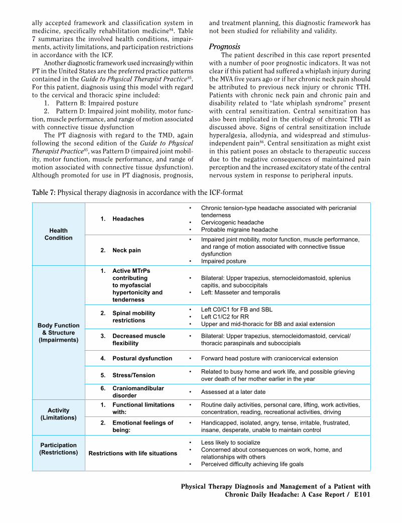
ally accepted framework and classification system in medicine, specifically rehabilitation medicine84. Table 7 summarizes the involved health conditions, impair-ments, activity limitations, and participation restrictions in accordance with the ICF.
Another diagnostic framework used increasingly within PT in the United States are the preferred practice patterns contained in the Guide to Physical Therapist Practice85. For this patient, diagnosis using this model with regard to the cervical and thoracic spine included:
1. Pattern B: Impaired posture2. Pattern D: Impaired joint mobility, motor func-
tion, muscle performance, and range of motion associated with connective tissue dysfunction
The PT diagnosis with regard to the TMD, again following the second edition of the Guide to Physical Therapist Practice85, was Pattern D (impaired joint mobil-ity, motor function, muscle performance, and range of motion associated with connective tissue dysfunction). Although promoted for use in PT diagnosis, prognosis,
and treatment planning, this diagnostic framework has not been studied for reliability and validity.
PrognosisThe patient described in this case report presented
with a number of poor prognostic indicators. It was not clear if this patient had suffered a whiplash injury during the MVA five years ago or if her chronic neck pain should be attributed to previous neck injury or chronic TTH. Patients with chronic neck pain and chronic pain and disability related to “late whiplash syndrome” present with central sensitization. Central sensitization has also been implicated in the etiology of chronic TTH as discussed above. Signs of central sensitization include hyperalgesia, allodynia, and widespread and stimulus-independent pain86. Central sensitization as might exist in this patient poses an obstacle to therapeutic success due to the negative consequences of maintained pain perception and the increased excitatory state of the central nervous system in response to peripheral inputs.
Table 7: Physical therapy diagnosis in accordance with the ICF-format
Health Condition
1. Headaches
• Chronic tension-type headache associated with pericranial tenderness
• Cervicogenic headache• Probable migraine headache
2. Neck pain
• Impaired joint mobility, motor function, muscle performance, and range of motion associated with connective tissue dysfunction
• Impaired posture
Body Function & Structure
(Impairments)
1. Active MTrPs contributing to myofascial hypertonicity and tenderness
• Bilateral: Upper trapezius, sternocleidomastoid, splenius capitis, and suboccipitals
• Left: Masseter and temporalis
2. Spinal mobility restrictions
• Left C0/C1 for FB and SBL• Left C1/C2 for RR• Upper and mid-thoracic for BB and axial extension
3. Decreased muscle flexibility
• Bilateral: Upper trapezius, sternocleidomastoid, cervical/thoracic paraspinals and suboccipials
4. Postural dysfunction • Forward head posture with craniocervical extension
5. Stress/Tension• Related to busy home and work life, and possible grieving
over death of her mother earlier in the year
6. Craniomandibular disorder
• Assessed at a later date
Activity (Limitations)
1. Functional limitations with:
• Routine daily activities, personal care, lifting, work activities, concentration, reading, recreational activities, driving
2. Emotional feelings of being:
• Handicapped, isolated, angry, tense, irritable, frustrated, insane, desperate, unable to maintain control
Participation(Restrictions) Restrictions with life situations
• Less likely to socialize• Concerned about consequences on work, home, and
relationships with others• Perceived difficulty achieving life goals

E102 / The Journal of Manual & Manipulative Therapy, 2006
Emotional stress and depression are relevant psy-chological impairments that serve as poor prognostic indicators for this patient diagnosed with both TTH and MH. High levels of depression and anxiety are common in patients with chronic TTH87,88. Significant functional and well-being impairments have been noted in chronic TTH patients, including adversely affected sleep, energy levels, emotional well-being, and performance in daily responsibilities. In contrast, work and social functioning are generally only severely impaired in a small minority88. Leistad et al89 showed the deleterious effect of cognitive stress on electromyography (EMG) muscle activity and reported on pain noted in patients with MH and TTH and in healthy controls. Although EMG peak activity revealed no between-group differences, the TTH patients recorded higher pain responses in the temporalis and frontalis muscles, a higher increase of pain during the cognitive test, and delayed pain recovery in all muscle regions when compared to controls. They also had delayed EMG recovery in the trapezius compared with controls and MH patients. The MH patients developed more pain in the splenius and temporalis than did the controls; pain responses were higher in the neck and trapezius compared to patients with TTH with delayed pain recovery in the trapezius and temporalis muscles89. In this patient, the history revealed both multiple emotional stressors and a history of depression. First, her mother died earlier in the year from a medication overdose: headache symptoms became worse a month after her death. The patient had a 30-year history of MH as a physical stressor. Also, the patient had a history of depression and was seeing a therapist and used anti-depressive medication. She noted work and home-related stress to her physicians and physical therapist, yet she maintained a successful career as an attorney and managed a household of four teenage children and three pets. The responses to her perceived emotional and functional disability on the HDI and NDI questionnaires were revealing with regard to perceived stress levels (Table 7). The patient reported feeling handicapped, isolated, angry, tense, irritable, frustrated, insane, desperate, and unable to maintain control. From a functional standpoint, she reported limitations with routine daily activities, personal care, lifting, work activities, concentration, reading, recre-ational activities, and driving. All of these findings were significant in that they most likely contributed to pain through a stress-related increase of muscular tension and pain perception.
The prolonged nature of complaints and the wors-ening of the condition over time despite the medical management by various health care providers seemed to also indicate an unfavorable prognosis. The patient had increased her headache medication intake to daily use and sometimes twice daily. One had to surmise that to expect this patient’s chronic pain condition to improve with time on its current course and without specific
therapeutic intervention would be unrealistic.On the other hand, this patient presented with a
number of musculoskeletal impairments that might indi-cate the potential for successful treatment of the chronic headache and neck pain by way of an OMPT approach. Manual therapy techniques to address spine dysfunction and soft tissue/myofascial restrictions, combined with exercise therapy to address postural imbalances and poor cervical muscle activation/endurance, have been noted to be effective treatment approaches both individually and collectively in the treatment of headaches38,77-79,90-93. Studies have shown that trigger point dry needling re-lieved symptoms related to myofascial pain51 and that it improved headache indices, tenderness, and neck mobility in TTH patients93. In some studies, spinal manipulation has been shown to be efficacious in the treatment of chronic TTH, MH, and CGH92. In this patient, the noted moderate to high headache intensity and chronic nature of headaches were not predictors of a negative outcome in the treatment of CGH using therapeutic exercise and manipulative therapy94.
Another positive prognostic indicator—although not known at the time of the evaluation—were the significant within-session improvements of pain and neck mobil-ity observed early in the intervention period. Tuttle95 reported that positive within-session changes in cervical mobility and pain could predict between-session changes for PT treatment of the cervical spine: odds ratios (OR) for within-session changes to predict between-session changes using an improved/not improved categorization for cervical mobility ranged from 2.5 (95% CI: 0.6-4.3) to 21.3 (95% CI: 10.1-96.1); for pain intensity, the OR was 4.5 (95% CI: 1.2-14.4). The positive likelihood ratio for cervical mobility improvements ranged from 2.1 (95% CI: 0.7-6.2) to 5.0 (95% CI: 2.6-9.9); for pain intensity improvements, it was 2.5 (95% CI: 1.3-4.6).
InterventionFollowing the initial evaluation, the patient was
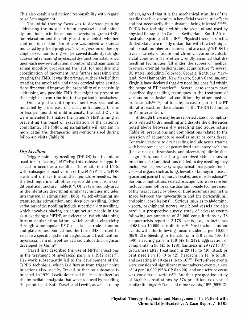
initially seen twice a week for approximately 6 weeks for a total of 11 visits, after which period she was out of town for almost 3 weeks. After this absence from therapy, she was seen 9 times over the next 3 months and finally one month later for a total of 21 visits. As noted above, specific assessment and treatment of the TMD began on the 14th visit. The patient was reassessed at each visit, and treatment on that visit was dependent on subjective reporting and objective reassessments.
The treatment progression was based on the ther-apist’s clinical experience. After the initial evaluation, the findings, recommended treatment plan, and expected outcome were outlined to the patient using charts and other skeletal aids. In the authors’ opinion, educating a patient on her problem and how it will be treated may be extremely important for optimal success and patient compliance with exercise and self-management concepts.
Physical Therapy Diagnosis and Management of a Patient with Chronic Daily Headache: A Case Report / E103
This also established patient responsibility with regard to self-management.
The initial therapy focus was to decrease pain by addressing the most pertinent myofascial and spinal dysfunctions, to initiate a home exercise program (HEP) for relaxation and flexibility, and to establish whether continuation of the plan of care was indeed warranted indicated by patient progress. The progression of therapy emphasized monitoring self-perceived disability ratings, addressing remaining myofascial dysfunctions established upon each new re-evaluation, monitoring and maintaining spinal mobility, progressing the HEP for mobility and coordination of movement, and further assessing and treating the TMD. It was the primary author’s belief that treating the myofascial and upper cervical spine restric-tions first would improve the probability of successfully addressing any possible TMD that might be present or that might be contributing to the patient’s headaches.
Once a plateau of improvement was reached as indicated by a decrease of headache frequency to one or less per month of mild intensity, the last 1-2 visits were intended to finalize the patient’s HEP, aiming at preventing the onset or exacerbation of the patient’s complaints. The following paragraphs will explain in more detail the therapeutic interventions used during follow-up visits (Table 8).
Dry NeedlingTrigger point dry needling (TrPDN) is a technique
used for “releasing” MFTrPs; this release is hypoth-esized to occur as a result of the elicitation of LTRs with subsequent inactivation of the MFTrP. The TrPDN treatment utilizes fine solid acupuncture needles, but the technique is in all other aspects different from tra-ditional acupuncture (Table 9)96. Other terminology used in the literature describing similar techniques includes intramuscular stimulation (IMS), twitch-obtaining in-tramuscular stimulation, and deep dry needling. Other variations of dry needling include superficial dry needling, which involves placing an acupuncture needle in the skin overlying a MFTrP, and electrical twitch-obtaining intramuscular stimulation, which applies electricity through a monopolar EMG needle electrode at motor end-plate zones. Sometimes the term IMS is used to refer to a specific system of diagnosis and treatment for myofascial pain of hypothesized radiculopathic origin as developed by Gunn96.
Travell first described the use of MFTrP injections in the treatment of myofascial pain in a 1942 paper97. Her work subsequently led to the development of the TrPDN technique, which is different from trigger point injections also used by Travell in that no substance is injected. In 1979, Lewitt described the “needle effect” as the immediate analgesia that was produced by needling the painful spot. Both Travell and Lewitt, as well as many
others, agreed that it is the mechanical stimulus of the needle that likely results in beneficial therapeutic effects and not necessarily the substance being injected12,98-100. TrPDN is a technique within the scope of and used by physical therapists in Canada, Switzerland, South Africa, Australia, Spain, and the UK101. Physical therapists in the United States are mostly unfamiliar with the technique, but a small number are trained and are using TrPDN to treat a variety of acute and chronic neuromusculosk-eletal conditions. It is often wrongly assumed that dry needling techniques fall under the scopes of medical practice, oriental medicine, and acupuncture101; several US states, including Colorado, Georgia, Kentucky, Mary-land, New Hampshire, New Mexico, South Carolina, and Virginia have declared that dry needling does fall within the scope of PT practice101. Several case reports have described dry needling techniques in the treatment of various musculoskeletal conditions by other medical professionals102-109, but to date, no case report in the PT literature exists on the inclusion of the TrPDN technique in PT intervention.
Although there may be no reported cases of complica-tions related to dry needling and despite the differences noted above between dry needling and acupuncture (Table 9), precautions and complications related to the insertion of acupuncture needles must be considered. Contraindications to dry needling include acute trauma with hematoma, local or generalized circulatory problems (i.e., varicosis, thrombosis, and ulceration), diminished coagulation, and local or generalized skin lesions or infections110. Complications related to dry needling may include vasodepressive syncope; hematoma; penetration of visceral organs such as lung, bowel, or kidney; increased spasm and pain of the muscle treated; and muscle edema110. Serious complications related to acupuncture are rare but include pneumothorax, cardiac tamponade (compression of the heart caused by blood or fluid accumulation in the space between the myocardium and the pericardium), and spinal cord lesions111. Serious injuries to abdominal viscera, perhipheral nerves, and blood vessels are also rare111. A prospective survey study of adverse events following acupuncture of 32,000 consultations by 78 acupucturists reported 2,178 events, i.e., an incidence of 684 per 10,000 consultations112. Most included minor events with the following mean incidence per 10,000 (95% CI): bleeding or hematoma in 310 cases (160 to 590), needling pain in 110 (48 to 247), aggravation of complaints in 96 (43 to 178), faintness in 29 (22 to 37), drowsiness after treatment in 29 (16 to 49), stuck or bent needle in 13 (0 to 42), headache in 11 (6 to 18), and sweating in 10 cases (6 to 16)112. Forty-three events were considered significant minor adverse events, a rate of 14 per 10,000 (95% CI: 8 to 20), and one seizure event was considered serious112. Another prospective study of 34,000 consultations by 574 practitioners revealed similar findings113: Transient minor events, 15% (95% CI:

E104 / The Journal of Manual & Manipulative Therapy, 2006
Table 8: Physical Therapy Visits and Treatment Interventions
Treatment DateMyofascial OMPT
TechniquesArticular OMPT
TechniquesExercise Therapy
Education
10/18/04 (Initial eval)NDI: 38/100 HDI: 56/100
(Trial Treatment)1. TrPDN: Lt UT and
SCM2. STM & PIR
1. Self-Stretch: UT and SCM for HEP
1. Eval findings & recommended Tx plan
2. Postural instruction in sitting
10/25/04Comments:Significant initial improvement of HA complaints.
1. TrPDN: Rt UT and SCM
2. STM & PIR
1. Reverse NAGS: T1-4
2. LAD: C0-23. Rt lateral glide (Gr.
IV): C0/14. SNAG: C1/2 RR
3x
1. Self-stretch: SOs & SpCap for HEP
2. Review of HEP
1. Education for stress reduction through breathing and relaxation of head, neck, and jaw.
10/28/04Comments: MTrPs reproduced HA pain.
1. TrPDN: Bil UT and Lt SCM
2. STM & PIR
1. Reverse NAGS: T1-4
2. PA (Prog. Osc.): T4-8
3. Rt lateral glide (Gr. IV): C0/1
4. SNAG: C1/2 RR 5x
1. Review of HEP
11/01/2004Comments: Onset of mid-back pain. Pain in mid-back with axial extension. Patient brought in her cervical pillow from home.
1. TrPDN: Lt LT2. STM & PIR: Bil
UT, SCM, SpCap, SOs
3. SID
1. Reverse NAGS: T1-4
2. PA (Prog. Osc.): T4-8
3. LAD: C0-24. Rt lateral glide (Gr.
IV): C0/15. SNAG: C1/2 RR 5x
1. Self-stretch: Lower Trapezius for HEP
1. Education for proper neck and head positioning during sleep with use of towel roll.
2. Education for suboccipital release self-treatment
11/04/04 1. TrPDN: Rt SCM2. STM & PIR: Bil
UT, SCM, SpCap, SOs
3. SID
1. PA (Prog. Osc.): T1-8
1. Review of Self-Tx for SO release
11/08/04 1. STM & PIR: Bil UT, SCM, SOs
2. SID
1. PA (Prog. Osc.): T1-8
1. Neck clocks2. AROM
AR of the head/axial extension
3. Shoulder clocks
11/11/04NDI: 28/100 HDI: 42/100Comments:Notes overall improvement
1. TrPDN: Bil UT, Lt SCM
2. STM & PIR3. SID
1. PA (Prog. Osc.): T1-8
1. Review of exercises issued last visit
11/18/04 1. TrPDN: Lt UT & LT
2. STM & PIR3. SID
1. Traction Mob Gr. V: T4-8
11/29/04 1. TrPDN: Lt UT & LT
2. STM & PIR3. SID
1. Traction Mob Gr. V: T4-8
12/02/04 1. TrPDN: Bil temporalis, masseter, SCM
2. STM & PIR (mouth opening)
1. Proper tongue position, resting jaw position, and nasal diaphragmatic breathing
2. Self STM of temporalis and masseter for HEP
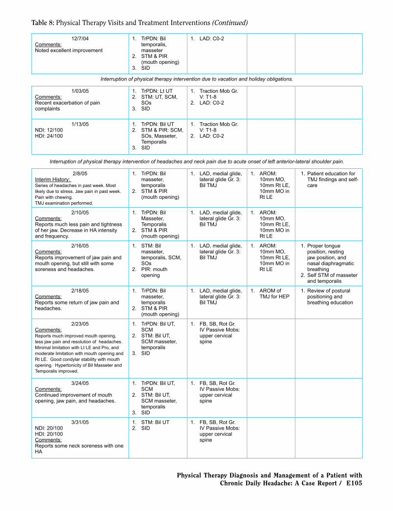
Physical Therapy Diagnosis and Management of a Patient with Chronic Daily Headache: A Case Report / E105
12/7/04Comments:Noted excellent improvement
1. TrPDN: Bil temporalis, masseter
2. STM & PIR (mouth opening)
3. SID
1. LAD: C0-2
Interruption of physical therapy intervention due to vacation and holiday obligations.
1/03/05Comments:Recent exacerbation of pain complaints
1. TrPDN: Lt UT2. STM: UT, SCM,
SOs3. SID
1. Traction Mob Gr. V: T1-8
2. LAD: C0-2
1/13/05NDI: 12/100 HDI: 24/100
1. TrPDN: Bil UT2. STM & PIR: SCM,
SOs, Masseter, Temporalis
3. SID
1. Traction Mob Gr. V: T1-8
2. LAD: C0-2
Interruption of physical therapy intervention of headaches and neck pain due to acute onset of left anterior-lateral shoulder pain.
2/8/05Interim History: Series of headaches in past week. Most likely due to stress. Jaw pain in past week. Pain with chewing. TMJ examination performed.
1. TrPDN: Bil masseter, temporalis
2. STM & PIR (mouth opening)
1. LAD, medial glide, lateral glide Gr. 3: Bil TMJ
1. AROM: 10mm MO, 10mm Rt LE, 10mm MO in Rt LE
1. Patient education for TMJ findings and self-care
2/10/05Comments:Reports much less pain and tightness of her jaw. Decrease in HA intensity and frequency.
1. TrPDN: Bil Masseter, Temporalis
2. STM & PIR (mouth opening)
1. LAD, medial glide, lateral glide Gr. 3: Bil TMJ
1. AROM: 10mm MO, 10mm Rt LE, 10mm MO in Rt LE
2/16/05Comments:Reports improvement of jaw pain and mouth opening, but still with some soreness and headaches.
1. STM: Bil masseter, temporalis, SCM, SOs
2. PIR: mouth opening
1. LAD, medial glide, lateral glide Gr. 3: Bil TMJ
1. AROM: 10mm MO, 10mm Rt LE, 10mm MO in Rt LE
1. Proper tongue position, resting jaw position, and nasal diaphragmatic breathing
2. Self STM of masseter and temporalis
2/18/05Comments:Reports some return of jaw pain and headaches.
1. TrPDN: Bil masseter, temporalis
2. STM & PIR (mouth opening)
1. LAD, medial glide, lateral glide Gr. 3: Bil TMJ
1. AROM of TMJ for HEP
1. Review of postural positioning and breathing education
2/23/05Comments:Reports much improved mouth opening, less jaw pain and resolution of headaches. Minimal limitation with Lt LE and Pro, and moderate limitation with mouth opening and Rt LE. Good condylar stability with mouth opening. Hypertonicity of Bil Masseter and Temporalis improved.
1. TrPDN: Bil UT, SCM
2. STM: Bil UT, SCM masseter, temporalis
3. SID
1. FB, SB, Rot Gr. IV Passive Mobs: upper cervical spine
3/24/05Comments:Continued improvement of mouth opening, jaw pain, and headaches.
1. TrPDN: Bil UT, SCM
2. STM: Bil UT, SCM masseter, temporalis
3. SID
1. FB, SB, Rot Gr. IV Passive Mobs: upper cervical spine
3/31/05NDI: 20/100 HDI: 20/100Comments:Reports some neck soreness with one HA
1. STM: Bil UT2. SID
1. FB, SB, Rot Gr. IV Passive Mobs: upper cervical spine
Table 8: Physical Therapy Visits and Treatment Interventions (Continued)
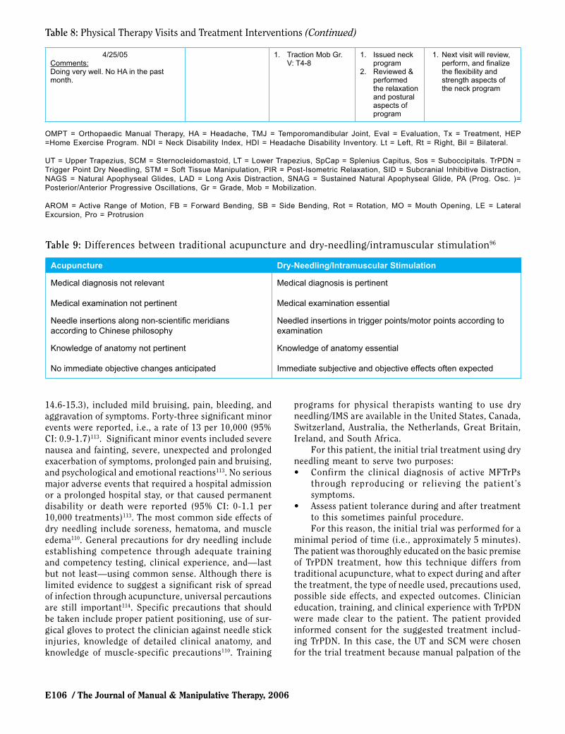
E106 / The Journal of Manual & Manipulative Therapy, 2006
4/25/05Comments:Doing very well. No HA in the past month.
1. Traction Mob Gr. V: T4-8
1. Issued neck program
2. Reviewed & performed the relaxation and postural aspects of program
1. Next visit will review, perform, and finalize the flexibility and strength aspects of the neck program
OMPT = Orthopaedic Manual Therapy, HA = Headache, TMJ = Temporomandibular Joint, Eval = Evaluation, Tx = Treatment, HEP =Home Exercise Program. NDI = Neck Disability Index, HDI = Headache Disability Inventory. Lt = Left, Rt = Right, Bil = Bilateral.
UT = Upper Trapezius, SCM = Sternocleidomastoid, LT = Lower Trapezius, SpCap = Splenius Capitus, Sos = Suboccipitals. TrPDN = Trigger Point Dry Needling, STM = Soft Tissue Manipulation, PIR = Post-Isometric Relaxation, SID = Subcranial Inhibitive Distraction, NAGS = Natural Apophyseal Glides, LAD = Long Axis Distraction, SNAG = Sustained Natural Apophyseal Glide, PA (Prog. Osc. )= Posterior/Anterior Progressive Oscillations, Gr = Grade, Mob = Mobilization.
AROM = Active Range of Motion, FB = Forward Bending, SB = Side Bending, Rot = Rotation, MO = Mouth Opening, LE = Lateral Excursion, Pro = Protrusion
Table 8: Physical Therapy Visits and Treatment Interventions (Continued)
14.6-15.3), included mild bruising, pain, bleeding, and aggravation of symptoms. Forty-three significant minor events were reported, i.e., a rate of 13 per 10,000 (95% CI: 0.9-1.7)113. Significant minor events included severe nausea and fainting, severe, unexpected and prolonged exacerbation of symptoms, prolonged pain and bruising, and psychological and emotional reactions113. No serious major adverse events that required a hospital admission or a prolonged hospital stay, or that caused permanent disability or death were reported (95% CI: 0-1.1 per 10,000 treatments)113. The most common side effects of dry needling include soreness, hematoma, and muscle edema110. General precautions for dry needling include establishing competence through adequate training and competency testing, clinical experience, and—last but not least—using common sense. Although there is limited evidence to suggest a significant risk of spread of infection through acupuncture, universal percautions are still important114. Specific precautions that should be taken include proper patient positioning, use of sur-gical gloves to protect the clinician against needle stick injuries, knowledge of detailed clinical anatomy, and knowledge of muscle-specific precautions110. Training
programs for physical therapists wanting to use dry needling/IMS are available in the United States, Canada, Switzerland, Australia, the Netherlands, Great Britain, Ireland, and South Africa.
For this patient, the initial trial treatment using dry needling meant to serve two purposes:• Confirm the clinical diagnosis of active MFTrPs
through reproducing or relieving the patient’s symptoms.
• Assess patient tolerance during and after treatment to this sometimes painful procedure. For this reason, the initial trial was performed for a
minimal period of time (i.e., approximately 5 minutes). The patient was thoroughly educated on the basic premise of TrPDN treatment, how this technique differs from traditional acupuncture, what to expect during and after the treatment, the type of needle used, precautions used, possible side effects, and expected outcomes. Clinician education, training, and clinical experience with TrPDN were made clear to the patient. The patient provided informed consent for the suggested treatment includ-ing TrPDN. In this case, the UT and SCM were chosen for the trial treatment because manual palpation of the
Table 9: Differences between traditional acupuncture and dry-needling/intramuscular stimulation96
Acupuncture Dry-Needling/Intramuscular Stimulation
Medical diagnosis not relevant Medical diagnosis is pertinent
Medical examination not pertinent Medical examination essential
Needle insertions along non-scientific meridians according to Chinese philosophy
Needled insertions in trigger points/motor points according to examination
Knowledge of anatomy not pertinent Knowledge of anatomy essential
No immediate objective changes anticipated Immediate subjective and objective effects often expected
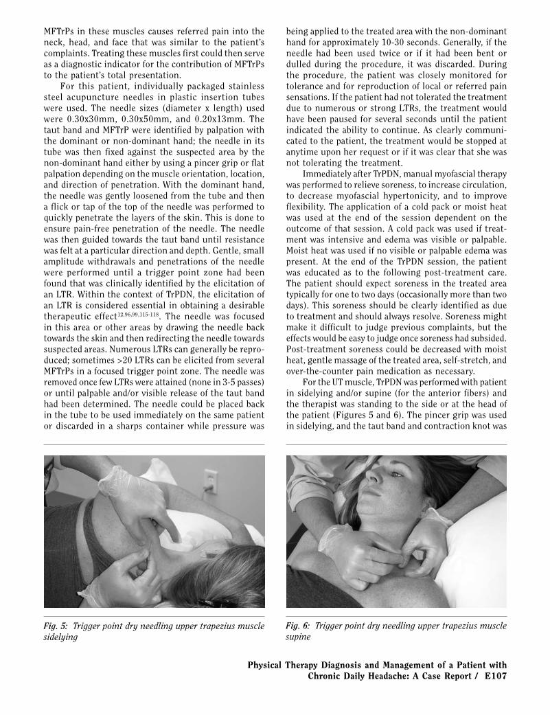
Physical Therapy Diagnosis and Management of a Patient with Chronic Daily Headache: A Case Report / E107
MFTrPs in these muscles causes referred pain into the neck, head, and face that was similar to the patient’s complaints. Treating these muscles first could then serve as a diagnostic indicator for the contribution of MFTrPs to the patient’s total presentation.
For this patient, individually packaged stainless steel acupuncture needles in plastic insertion tubes were used. The needle sizes (diameter x length) used were 0.30x30mm, 0.30x50mm, and 0.20x13mm. The taut band and MFTrP were identified by palpation with the dominant or non-dominant hand; the needle in its tube was then fixed against the suspected area by the non-dominant hand either by using a pincer grip or flat palpation depending on the muscle orientation, location, and direction of penetration. With the dominant hand, the needle was gently loosened from the tube and then a flick or tap of the top of the needle was performed to quickly penetrate the layers of the skin. This is done to ensure pain-free penetration of the needle. The needle was then guided towards the taut band until resistance was felt at a particular direction and depth. Gentle, small amplitude withdrawals and penetrations of the needle were performed until a trigger point zone had been found that was clinically identified by the elicitation of an LTR. Within the context of TrPDN, the elicitation of an LTR is considered essential in obtaining a desirable therapeutic effect12,96,99,115-118. The needle was focused in this area or other areas by drawing the needle back towards the skin and then redirecting the needle towards suspected areas. Numerous LTRs can generally be repro-duced; sometimes >20 LTRs can be elicited from several MFTrPs in a focused trigger point zone. The needle was removed once few LTRs were attained (none in 3-5 passes) or until palpable and/or visible release of the taut band had been determined. The needle could be placed back in the tube to be used immediately on the same patient or discarded in a sharps container while pressure was
being applied to the treated area with the non-dominant hand for approximately 10-30 seconds. Generally, if the needle had been used twice or if it had been bent or dulled during the procedure, it was discarded. During the procedure, the patient was closely monitored for tolerance and for reproduction of local or referred pain sensations. If the patient had not tolerated the treatment due to numerous or strong LTRs, the treatment would have been paused for several seconds until the patient indicated the ability to continue. As clearly communi-cated to the patient, the treatment would be stopped at anytime upon her request or if it was clear that she was not tolerating the treatment.
Immediately after TrPDN, manual myofascial therapy was performed to relieve soreness, to increase circulation, to decrease myofascial hypertonicity, and to improve flexibility. The application of a cold pack or moist heat was used at the end of the session dependent on the outcome of that session. A cold pack was used if treat-ment was intensive and edema was visible or palpable. Moist heat was used if no visible or palpable edema was present. At the end of the TrPDN session, the patient was educated as to the following post-treatment care. The patient should expect soreness in the treated area typically for one to two days (occasionally more than two days). This soreness should be clearly identified as due to treatment and should always resolve. Soreness might make it difficult to judge previous complaints, but the effects would be easy to judge once soreness had subsided. Post-treatment soreness could be decreased with moist heat, gentle massage of the treated area, self-stretch, and over-the-counter pain medication as necessary.
For the UT muscle, TrPDN was performed with patient in sidelying and/or supine (for the anterior fibers) and the therapist was standing to the side or at the head of the patient (Figures 5 and 6). The pincer grip was used in sidelying, and the taut band and contraction knot was
Fig. 5: Trigger point dry needling upper trapezius muscle sidelying
Fig. 6: Trigger point dry needling upper trapezius muscle supine

E108 / The Journal of Manual & Manipulative Therapy, 2006
grasped with the non-dominant hand while the dominant hand performed the needling procedure in the inferior-lateral, anterior-superior, or anterior-lateral direction. The therapist took care only to needle the muscle fibers accessible between the thumb and index finger. Using flat palpation, the UT taut band and contraction knot were fixed in between the index and middle finger against the superior portion of the scapula. Using the pincer grip in supine, the muscle was needled in the posterior direc-tion towards the index finger. For this technique, the 0.30x30mm and 0.30x50mm needle are used depending on patient physical make-up, location and, direction of treated area. Precautions included that the needle should never be directed in the anterior-medial or inferior direc-tion to avoid puncturing the apex of the lung.
For the SCM muscle, TrPDN was performed in supine or sidelying with the neck and head adequately supported and with the therapist standing to the side or at the head of the patient (Figure 7). In supine, the pincer grip was used to grasp both heads of the muscle while the needle was directed from the medial side to the posterior-lateral and/or anterior to posterior di-
against the rib or laterally at a shallow angle tangential to the chest wall. For this muscle the 0.30x30mm needle was again used. Precautions for needling this muscle included avoiding penetration of the lung by needling over a rib or tangentially to the chest wall.
For the masseter muscle, TrPDN was performed in sidelying or supine with the neck and head supported by a pillow and with the therapist standing to the side or at the head of patient (Figure 9). The taut bands and sensitive knots in the superficial and deep masseter muscle were identified by pincer grip or flat palpation. The treated area was fixed between the index and middle fingers and the needle was directed between the fingers. For this technique, the 0.20x13mm needle was used as precautions included avoiding needle stick injury to the facial nerve.
For the temporalis muscle, TrPDN was performed in sidelying or supine with the neck and head supported by a pillow and with the therapist again standing to the side or at the head of the patient (Figure 10). The tem-poral artery was first palpated and then the taut bands
Fig. 7: Trigger point dry needling sternocleidomastoid muscle
Fig. 8: Trigger point dry needling lower trapezius muscle
rection between the thumb and index finger. For this muscle, the 0.30x30mm needle was used. Precautions for dry needling of the SCM included that the carotid artery should be identified medial to the SCM and that the direction of the needle should not be in the medial direction to avoid puncturing said artery.
For the lower trapezius (LT) muscle, TrPDN was per-formed in sidelying or prone with the therapist standing to the treated side (Figure 8). The diagonally oriented taut band was fixed between the index and middle finger with the contraction knot firmly over a rib. The index finger and middle finger were subsequently located in the intercostal space. The needle was directed anteriorly
and sensitive knot were identified and fixed against the temporal bone between the index and middle finger. The needle was then directed towards the contraction knot. The 0.20x13mm needle was used, as the precau-tion for this technique was avoidance of the superficial temporal artery.
Orthopaedic Manual Physical TherapyWell integrated into physical therapy, OMPT in-
cludes manipulation techniques with purported effects on either soft tissue or joints. Soft tissue manipulation uses manual techniques aimed at relaxing muscles, increasing circulation, breaking up adhesions or scar

Physical Therapy Diagnosis and Management of a Patient with Chronic Daily Headache: A Case Report / E109
tissue, and easing pain in the soft tissues. Soft tissue manipulation techniques include manual trigger point therapy, strain-counterstrain, muscle energy technique, neuromuscular technique, myofascial release, and other therapeutic massage techniques. Paris119 defined joint manipulation as “the skilled passive movement to a joint.” Joint manipulation techniques are aimed at restoring motion at a joint and modulating pain. Joint manipula-tion techniques include non-thrust, thrust, and traction forces applied at various grades and directions119.
Soft tissue manipulation is used to describe therapeu-tic massage and manual techniques for mobilizing soft tissue (e.g., muscles, connective tissue, fascia, tendons, ligaments) to improve the function of the muscular, circulatory, lymphatic, and nervous systems120. Some techniques are general for treating large areas using the palm of the hand and the forearm, for example, while other techniques are specific like deep stroking, transverse friction, and trigger point compression release using the thumb and fingertips. Post-isometric relaxation (PIR) was a technique used for this patient with the goal of achieving muscle relaxation and elongation12,121. With the patient relaxed and the body supported, the therapist passively lengthened the muscle to its first barrier. The patient then performed a minimal isometric contraction of the muscle (10-25%) for 5 sec during an inhalation while the therapist stabilized the muscle to be stretched. Then the patient relaxed completely and exhaled, while the therapist passively stretched the muscle to the new barrier. The technique was then repeated 3-5 times. This technique is very similar to contract/relax and hold/relax techniques. Specific muscles treated with this technique are indicated in Table 8 with the technique illustrated for the upper trapezius muscle in Figure 11. Subcranial inhibitive distraction (SID) is a myofascial technique described by Paris60 that is aimed at releas-
ing tension in suboccipital soft tissue and suboccipital musculature (Figure 12). The patient was lying supine with head supported. The therapist placed the three middle fingers just caudal to the nuchal line, lifted the fingertips upwards resting the hands on the treatment table, and then applied a gentle cranial pull, causing a long axis extension. The procedure was performed with this patient for 2-5 minutes as indicated in Table 8.
Mulligan122,123 described reverse natural apophyseal glides as a mid- to end-range oscillatory mobilization indicated from the C7 vertebra down that were intended to aid in treatment of end-range loss of neck mobility, postural dysfunction (FHP with UT pain), and degenera-tive lower cervical or upper thoracic spine segments. The patient was seated and the therapist stood to the side and cradled the head to the body with forearm maintaining
Fig. 9: Trigger point dry needling masseter muscle Fig. 10: Trigger point dry needling temporalis muscle
Fig. 11: Post-isometric relaxation upper trapezius muscle
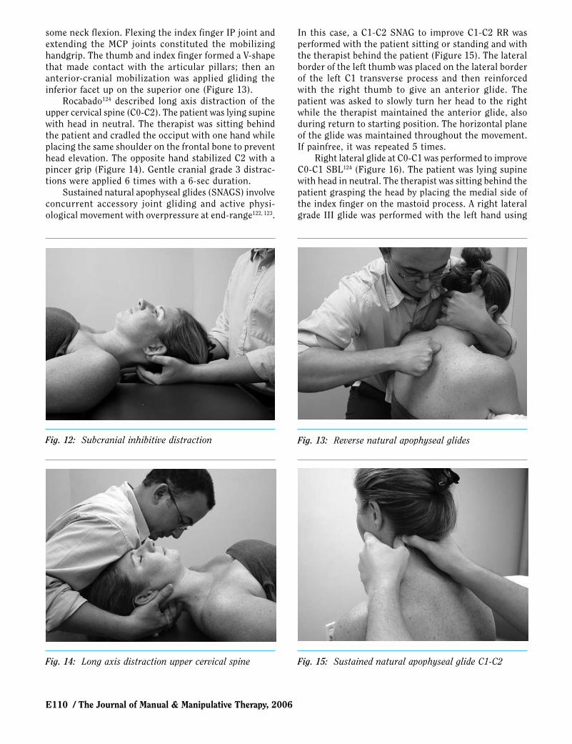
E110 / The Journal of Manual & Manipulative Therapy, 2006
some neck flexion. Flexing the index finger IP joint and extending the MCP joints constituted the mobilizing handgrip. The thumb and index finger formed a V-shape that made contact with the articular pillars; then an anterior-cranial mobilization was applied gliding the inferior facet up on the superior one (Figure 13).
Rocabado124 described long axis distraction of the upper cervical spine (C0-C2). The patient was lying supine with head in neutral. The therapist was sitting behind the patient and cradled the occiput with one hand while placing the same shoulder on the frontal bone to prevent head elevation. The opposite hand stabilized C2 with a pincer grip (Figure 14). Gentle cranial grade 3 distrac-tions were applied 6 times with a 6-sec duration.
Sustained natural apophyseal glides (SNAGS) involve concurrent accessory joint gliding and active physi-ological movement with overpressure at end-range122, 123.
In this case, a C1-C2 SNAG to improve C1-C2 RR was performed with the patient sitting or standing and with the therapist behind the patient (Figure 15). The lateral border of the left thumb was placed on the lateral border of the left C1 transverse process and then reinforced with the right thumb to give an anterior glide. The patient was asked to slowly turn her head to the right while the therapist maintained the anterior glide, also during return to starting position. The horizontal plane of the glide was maintained throughout the movement. If painfree, it was repeated 5 times.
Right lateral glide at C0-C1 was performed to improve C0-C1 SBL124 (Figure 16). The patient was lying supine with head in neutral. The therapist was sitting behind the patient grasping the head by placing the medial side of the index finger on the mastoid process. A right lateral grade III glide was performed with the left hand using
Fig. 12: Subcranial inhibitive distraction Fig. 13: Reverse natural apophyseal glides
Fig. 14: Long axis distraction upper cervical spine Fig. 15: Sustained natural apophyseal glide C1-C2
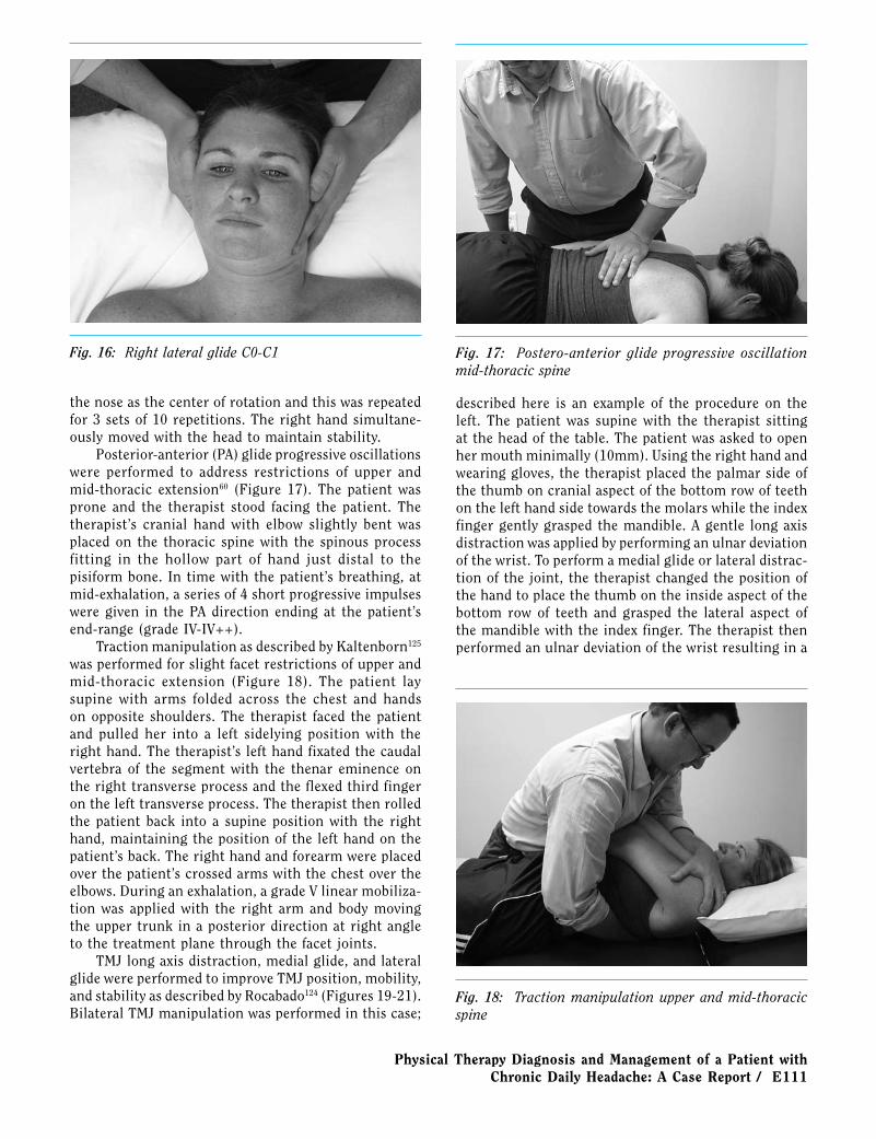
Physical Therapy Diagnosis and Management of a Patient with Chronic Daily Headache: A Case Report / E111
the nose as the center of rotation and this was repeated for 3 sets of 10 repetitions. The right hand simultane-ously moved with the head to maintain stability.
Posterior-anterior (PA) glide progressive oscillations were performed to address restrictions of upper and mid-thoracic extension60 (Figure 17). The patient was prone and the therapist stood facing the patient. The therapist’s cranial hand with elbow slightly bent was placed on the thoracic spine with the spinous process fitting in the hollow part of hand just distal to the pisiform bone. In time with the patient’s breathing, at mid-exhalation, a series of 4 short progressive impulses were given in the PA direction ending at the patient’s end-range (grade IV-IV++).
Traction manipulation as described by Kaltenborn125 was performed for slight facet restrictions of upper and mid-thoracic extension (Figure 18). The patient lay supine with arms folded across the chest and hands on opposite shoulders. The therapist faced the patient and pulled her into a left sidelying position with the right hand. The therapist’s left hand fixated the caudal vertebra of the segment with the thenar eminence on the right transverse process and the flexed third finger on the left transverse process. The therapist then rolled the patient back into a supine position with the right hand, maintaining the position of the left hand on the patient’s back. The right hand and forearm were placed over the patient’s crossed arms with the chest over the elbows. During an exhalation, a grade V linear mobiliza-tion was applied with the right arm and body moving the upper trunk in a posterior direction at right angle to the treatment plane through the facet joints.
TMJ long axis distraction, medial glide, and lateral glide were performed to improve TMJ position, mobility, and stability as described by Rocabado124 (Figures 19-21). Bilateral TMJ manipulation was performed in this case;
described here is an example of the procedure on the left. The patient was supine with the therapist sitting at the head of the table. The patient was asked to open her mouth minimally (10mm). Using the right hand and wearing gloves, the therapist placed the palmar side of the thumb on cranial aspect of the bottom row of teeth on the left hand side towards the molars while the index finger gently grasped the mandible. A gentle long axis distraction was applied by performing an ulnar deviation of the wrist. To perform a medial glide or lateral distrac-tion of the joint, the therapist changed the position of the hand to place the thumb on the inside aspect of the bottom row of teeth and grasped the lateral aspect of the mandible with the index finger. The therapist then performed an ulnar deviation of the wrist resulting in a
Fig. 16: Right lateral glide C0-C1 Fig. 17: Postero-anterior glide progressive oscillation mid-thoracic spine
Fig. 18: Traction manipulation upper and mid-thoracic spine
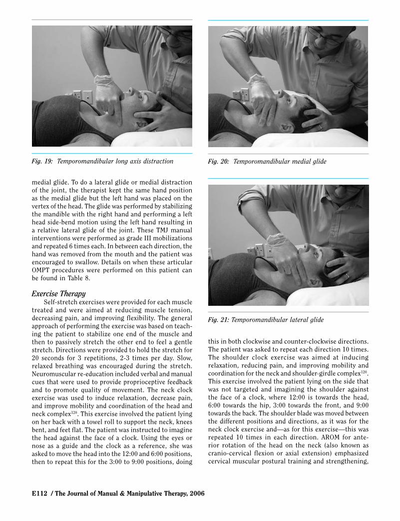
E112 / The Journal of Manual & Manipulative Therapy, 2006
medial glide. To do a lateral glide or medial distraction of the joint, the therapist kept the same hand position as the medial glide but the left hand was placed on the vertex of the head. The glide was performed by stabilizing the mandible with the right hand and performing a left head side-bend motion using the left hand resulting in a relative lateral glide of the joint. These TMJ manual interventions were performed as grade III mobilizations and repeated 6 times each. In between each direction, the hand was removed from the mouth and the patient was encouraged to swallow. Details on when these articular OMPT procedures were performed on this patient can be found in Table 8.
Exercise TherapySelf-stretch exercises were provided for each muscle
treated and were aimed at reducing muscle tension, decreasing pain, and improving flexibility. The general approach of performing the exercise was based on teach-ing the patient to stabilize one end of the muscle and then to passively stretch the other end to feel a gentle stretch. Directions were provided to hold the stretch for 20 seconds for 3 repetitions, 2-3 times per day. Slow, relaxed breathing was encouraged during the stretch. Neuromuscular re-education included verbal and manual cues that were used to provide proprioceptive feedback and to promote quality of movement. The neck clock exercise was used to induce relaxation, decrease pain, and improve mobility and coordination of the head and neck complex120. This exercise involved the patient lying on her back with a towel roll to support the neck, knees bent, and feet flat. The patient was instructed to imagine the head against the face of a clock. Using the eyes or nose as a guide and the clock as a reference, she was asked to move the head into the 12:00 and 6:00 positions, then to repeat this for the 3:00 to 9:00 positions, doing
this in both clockwise and counter-clockwise directions. The patient was asked to repeat each direction 10 times. The shoulder clock exercise was aimed at inducing relaxation, reducing pain, and improving mobility and coordination for the neck and shoulder-girdle complex120. This exercise involved the patient lying on the side that was not targeted and imagining the shoulder against the face of a clock, where 12:00 is towards the head, 6:00 towards the hip, 3:00 towards the front, and 9:00 towards the back. The shoulder blade was moved between the different positions and directions, as it was for the neck clock exercise and—as for this exercise—this was repeated 10 times in each direction. AROM for ante-rior rotation of the head on the neck (also known as cranio-cervical flexion or axial extension) emphasized cervical muscular postural training and strengthening,
Fig. 19: Temporomandibular long axis distraction Fig. 20: Temporomandibular medial glide
Fig. 21: Temporomandibular lateral glide
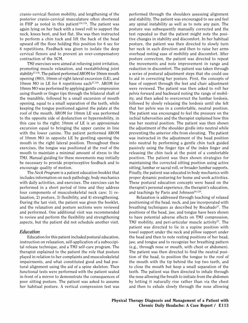
Physical Therapy Diagnosis and Management of a Patient with Chronic Daily Headache: A Case Report / E113
cranio-cervical flexion mobility, and lengthening of the posterior cranio-cervical musculature often shortened in FHP as noted in this patient120,124. The patient was again lying on her back with a towel roll to support the neck, knees bent, and feet flat. She was then instructed to perform a chin tuck and lift the back of the head upward off the floor holding this position for 6 sec for 6 repetitions. Feedback was given to isolate the deep cervical flexors and to prevent an over-compensatory contraction of the SCM.
TMJ exercises were aimed at relieving joint irritation, promoting muscle relaxation, and reestablishing joint stability57,126. The patient performed AROM for 10mm mouth opening (MO), 10mm of right lateral excursion (LE), and 10mm MO in LE for 6 repetitions each57,126. AROM for 10mm MO was performed by applying gentle compression using thumb or finger tips through the bilateral shaft of the mandible, followed by a small excursion of mouth opening, equal to a small separation of the teeth, while keeping the tongue positioned against the palate at the roof of the mouth. AROM for 10mm LE was performed to the opposite side of dysfunction or hypermobility, in this case to the right; 10mm of LE is an approximate excursion equal to bringing the upper canine in line with the lower canine. The patient performed AROM of 10mm MO in opposite LE by gentling opening the mouth in the right lateral position. Throughout these exercises, the tongue was positioned at the roof of the mouth to maintain a minimal amount of stress to the TMJ. Manual guiding for these movements may initially be necessary to provide proprioceptive feedback and to encourage quality of motion.
The Neck Program is a patient education booklet that includes information on neck pathology, body mechanics with daily activities, and exercises. The exercises can be performed in a short period of time and they address four components of musculoskeletal neck care: 1) re-laxation, 2) posture, 3) flexibility, and 4) strengthening. During the last visit, the patient was given the booklet, and the relaxation and posture sections were reviewed and performed. One additional visit was recommended to review and perform the flexibility and strengthening aspects, but the patient did not schedule another visit.
EducationEducation for this patient included postural education,
instruction on relaxation, self-application of a suboccipi-tal release technique, and a TMJ self-care program. The therapist explained to the patient the role that posture played in relation to her complaints and musculoskeletal impairments, and what constituted good and bad pos-tural alignment using the aid of a spine skeleton. Then functional tests were performed with the patient seated in front of a mirror to demonstrate the consequences of poor sitting posture. The patient was asked to assume her habitual posture. A vertical compression test was
performed through the shoulders assessing alignment and stability. The patient was encouraged to see and feel any spinal instability as well as to note any pain. The posture was subsequently manually corrected and the test repeated so that the patient might note the posi-tive changes in stability and discomfort. In her habitual posture, the patient was then directed to slowly turn her neck in each direction and then to raise her arms overhead noting ease of mobility and discomfort. After posture correction, the patient was directed to repeat the movements and note improvement in range and reduction in discomfort. The patient was taken through a series of postural adjustment steps that she could use to aid in correcting her posture. First, the concepts of base of support through the feet and chair adjustment were reviewed. The patient was then asked to roll her pelvis forward and backward noting the range of mobil-ity, and then asked to overcorrect the lumbar lordosis, followed by slowly releasing the lordosis until she felt that her pelvis was in a comfortable, neutral position. The patient was encouraged to feel the pressure on the ischial tuberosities and the therapist explained how this was her neutral position. The patient was then shown the adjustment of the shoulder girdle into neutral while preventing the anterior ribs from elevating. The patient was instructed in the adjustment of the head position into neutral by performing a gentle chin tuck guided passively using the finger tips of the index finger and releasing the chin tuck at the point of a comfortable position. The patient was then shown strategies for maintaining the corrected sitting position using active sitting, lumbar or sacral roll, or broader lumbar supports. Finally, the patient was educated in body mechanics with proper dynamic posturing for home and work activities. These postural education concepts were based on the therapist’s personal experience, the therapist’s education, and teachings by Paris and Johnson60,120.
Relaxation is addressed through teaching of relaxed positioning of the head, neck, and jaw incorporated with breathing techniques as described by Rocabado57. The positions of the head, jaw, and tongue have been shown to have potential adverse effects on TMJ compression, TMJ mobility, and peri-articular muscle activity57. The patient was directed to lie in a supine position with towel support under the neck and pillow support under the head and then to note resting positions of her head, jaw, and tongue and to recognize her breathing pattern (e.g., through nose or mouth, with chest or abdomen). The patient was then directed to find the neutral posi-tion of the head, to position the tongue to the roof of the mouth with the tip behind the top two teeth, and to close the mouth but keep a small separation of the teeth. The patient was then directed to inhale through the nose allowing the breath to initiate from the abdomen by letting it naturally rise rather than via the chest and then to exhale slowly through the nose allowing

E114 / The Journal of Manual & Manipulative Therapy, 2006
the abdomen to naturally fall. This relaxation exercise was also instructed in a corrected sitting or standing posture and recommended as needed to relieve stress and tension. Conscious correcting of her posture and performing these relaxation exercises was encouraged once every hour for any duration.
A suboccipital release self-treatment technique was taught to address tension at the base of the head as well as for relieving headaches. The patient was educated on placing two tennis balls in a thin sock and then tying the sock to maintain stability of the balls. The technique was to be performed in a quiet dark room lying supine with the use of a neck roll to support the neck and a pillow to support the head with knees supported. The tennis balls were to be placed above the neck roll at the base of the head with additional support as needed under the head to prevent cranio-cervical extension. Lying in this position for 5 minutes while focusing on relaxation and breathing exercises, patients have described good success with the release of tension and pain as well as reducing or warding off headaches.
A TMJ self-care program was instructed for reducing pain, relaxing muscles, relieving intra-joint irritation, and maintaining gains achieved from therapeutic inter-vention57. Advice for self-care included a soft non-chewy diet, no wide opening of mouth (maximum of 2 fingers width), no biting, no gum chewing, prevention of direct pressure on mandible or sleeping on problematic side, yawning with tongue against the palate, tongue against the palate at rest, nasal breathing maintaining free airway space, and maintaining good posture.
OutcomesOn the patient’s last visit, she reported doing very
well with no headache in the preceding month. The primary author recommended that the patient follow up for one additional visit to perform the flexibility and
strengthening portions of the issued Neck Program, and review the entire program prior to discontinuation of therapy. However, the patient did not schedule another appointment for an unknown reason, so unfortunately objective data and outcome measures could not be reassessed.
Over all, the patient noted a significant decrease in headache frequency with progressive improvement since the start of therapy from more than once per week (daily) to 1-4 per month and finally to none in the month preceding her last therapy visit. Headache intensity also progressively improved since therapy onset from a reported severe to a mild intensity. Throughout the duration of PT treatment, the patient also reported cessation of tinnitus, less neck tenderness, and improved jaw and neck mobility and function. The patient reported improvement with the following activities of daily living: she was now able to do as much work as she wanted, able to drive longer distances with only slight neck pain, able to read as much as she wanted with only slight neck pain, and experienced some neck pain related only to a few recreational activities.
The last set of outcome measures completed by the patient was on the second to last visit. The results of the four HDI (Table 10) and NDI (Table 11) outcome measures completed throughout the treatment period are illustrated in Figures 22 and 23. The results of the HDI outcome measure showed a 31% improvement for the emotional score, a 42% improvement for the functional score, and a 36% improvement in the total score between the time of the initial evaluation and the second to last visit. During the same time period, the results of the NDI outcome measure showed an 18% improvement and—at one time—a 26% improvement for an earlier assessment date. Although specific subjective and objective signs of improvement were not assessed on the last visit date, which was approximately 1 month
Table 10: HDI - Outcome Measures
10/18/04 11/11/04 1/13/05 3/31/05
Frequency >1/wk >1/wk 1-4/month 1-4/month
Intensity Severe Moderate Mild Not completed
Emotional Score (max 52) 26 18 12 10
Functional Score (max 48) 30 24 12 10
Total Score (max 100) 56 42 24 20
Table 11: NDI - Outcome Measures
10/18/04 11/11/04 1/13/04 3/31/05
Total Score (max 50) 19 14 6 10
Disability Score 38% 28% 12% 20%
Disability Moderate Moderate Mild Mild

Physical Therapy Diagnosis and Management of a Patient with Chronic Daily Headache: A Case Report / E115
after the previous follow-up, there was noted significant improvement through the assessment of self-perceived outcome measures used in this case. A 29% change in the HDI total score constitutes the MDC95 for this measure39. A 7-point or 14% change in the NDI score constitutes the MCID for the NDI42. The change in the total score of the HDI outcome measure in this case was a decrease of 36%, which indicates that the MDC was exceeded and that a true change had indeed occurred in pain and disability due to the headache complaints. Similarly, the change in NDI score, which exceeded the established MCID for this measure, indicated that a true and meaningful change in neck-related disability had occurred during the course of treatment.
DiscussionThe patient described in this case report had a very
complex presentation. Medical diagnoses included chronic TTH associated with pericranial tenderness, probable MH without aura, probable CGH, and TMD. Impairments identified during the PT examination included postural deviations (FHP), MFTrPs and decreased length in the neck and jaw muscles, hypomobility in the C0-C2 and T1-T8 segments, and hypermobility of the left TMJ. Poor prognostic indicators, including the presence of possible central sensitization, emotional stress, depres-sion, and the worsening nature of a chronic condition, further complicated the patient’s presentation. Despite this complexity, the patient clearly improved over the course of treatment. She exceeded the MDC on the HDI indicating that a true improvement had occurred with regard to headache-related disability while exceeding the MCID on the NDI implied that a true and clinically meaningful improvement also occurred for disability due to neck pain. In addition, the patient reported on the last
visit after one month of no treatment that she had not experienced a single headache in the preceding month. This is especially significant considering that the patient initially described daily headaches. Although a case report does not allow us to infer a cause-and-effect relationship between intervention and outcome, true and meaningful changes in a previously worsening, chronic condition do imply that the PT management described was at least contributory to the positive changes noted.
At this point, we should mention and discuss two assumptions that are at the basis of the choices made in the PT diagnosis and management of this patient:
1. The impairments found during the PT examina-tion as noted above can contribute to or even cause the medical headache diagnoses relevant to this patient.
2. By addressing these neuromusculoskeletal impairments, PT management can affect limitations in activities and restrictions in participation attributed to these medical headache diagnoses.
So what is the evidence linking the neuromusculo-skeletal impairments noted in this patient to the medical diagnoses of chronic TTH associated with pericranial tenderness, probable MH without aura, probable CGH, and TMD?
Myofascial trigger points were among the main neuro-musculoskeletal impairments identified and treated in this patient. Active MFTrPs were diagnosed by way of subjective history, neck mobility tests, and manual palpation. The patient described her headache pain as a moderate to severe band-like pain across her forehead; she reported her other head and neck pain as tenderness. Myofascial pain is often described as a dull, aching, tightening, or pressure-like pain of mild to moderate intensity12. The patient also complained of tinnitus in the left ear. Unilat-eral tinnitus may be associated with MFTrPs in the deep masseter muscle. It is thought to be due to a referred
Fig. 22: HDI outcome measure scores Fig. 23: NDI outcome measure scores
E116 / The Journal of Manual & Manipulative Therapy, 2006
sensory phenomenon or motor unit facilitation of the tensor tympani and/or stapedius muscles of the middle ear12. Various authors have claimed that head, neck, and shoulder girdle muscles are capable of referring pain into the head experienced as headaches12,20. Knowledge of myofascial referred pain patterns when interpreting the patient’s pain diagram (Figure 1) indicated a pos-sible involvement of the UT, SCM, SO, temporalis, and masseter muscles12. In addition, referred pain patterns for the masseter and temporalis were consistent with the reported facial pain; referral patterns for the upper, middle, and lower portions of the trapezius muscles were consistent with the patient’s report of upper and lower neck pain12. Matching established referral patterns to a patient report of pain distribution then guided further examination: Referred pain recognized by the patient as part of her headache pain was elicited with compression of tender nodules within the left SCM and left UT. A trial of TrPDN on the first visit further confirmed the existence of active MFTrPs by the elicitation of numerous LTRs with local and referred pain phenomena. In addi-tion, the patient’s cervical AROM test findings indicated myofascial tightness. Muscles with MFTrPs are known to exhibit decreased flexibility and painful limitation to full stretch.
The primary author inferred in this case not only that MFTrPs contributed to myofascial hypertonicity, myofascial tenderness, and referred pain into the head, neck, and face but also that these trigger points played a major etiologic role in the chronic TTH diagnosed in this patient. Studies have shown that prolonged pe-ripheral nociceptive input from pericranial myofascial tissue can sensitize the second-order neurons at the spinal and trigeminal level leading to impaired central nervous system (CNS) modulation of this nociceptive activity, thereby resulting in an increased general pain sensitivity7. In patients with chronic TTH with pericra-nial muscle tenderness, there appears to be just such a disruption of the balance between peripheral nociceptive input and CNS pain modulation7. The convergent afferent input in this scenario includes nociceptive input from muscles innervated by C1-C3, among which are the UT, SCM, and suboccipital muscles, and of those innervated by the trigeminal nerve, which includes the temporalis and the masseter muscles. In addition, we need to take into consideration the nociceptive input from possible segmental dysfunction of the upper cervical spine (C0-C3), thereby providing a potential explanation for the observed clinical link between and diagnostic confusion with regard to TTH and CGH. The resultant temporal and spatial summation is the CNS misinterpretation of the peripheral input leading to central sensitization.
The neuro-anatomical explanation of how structures innervated by cervical nerve roots can refer pain into the head or face starts with the trigeminal nucleus caudalis. This nucleus descends as low as the C3 or C4 segments of
the spinal cord. The trigeminocervical nucleus, a column of gray matter that is located adjoining to the trigeminal nucleus caudalis, exchanges sensory information from the upper cervical spinal nerve roots to the trigeminal nerve via interneurons, thereby explaining how cervi-cal nociceptive input may be referred to the sensory receptive fields of the trigeminal nerve supplying the head and face, most commonly affecting the ophthalmic division, which in turn may lead to the perception of referred symptoms to the forehead, temple, or orbit. Because afferent sensory signals ascend or descend up to three spinal cord segments, nociceptive signals for spinal segments C6 or C7 may even interact with the trigeminocervical nucleus, and ultimately this may result in referral of pain into the head or face from structures as far distant as the lower neck region25.
Fernandez-de-las-Penas et al13 provided clinical support for this proposed etiologic role for MFTrPs in TTH. They found that there was a significant difference between a group of patients with chronic TTH and healthy controls with regard to the presence of active MFTrPs (P<0.001), but not for latent MFTrPs (P>0.05); they also noted that active MFTrPs in the UT, SCM, and temporalis muscles were associated with chronic TTH13. In another study, Fernandez-de-las-Penas et al127 also demonstrated significant (P<0.001) between-group dif-ferences for active MFTrPs in the SO muscles in patients with chronic TTH as compared to healthy controls; the between-group difference with regard to latent MFTrPs was again not significant (P>0.05). In addition, they found that chronic TTH subjects with active suboccipital MFTrPs described greater headache intensity, duration, and frequency compared to those with latent MFTrPs (P<0.05). Furthermore, the characteristics of TTH noted in various studies13,20 were very similarly described as pressure and/or tightness or increased tenderness of neck and shoulder muscles, and the similarity of this description with the documented nature of pain due to MFTrPs further increases the plausibility of referred pain from MFTrPs as contributory to TTH.
Assessing pericranial muscle tenderness and pressure pain thresholds are ways of evaluating diagnostic criteria with regard to the contribution of muscular disorders. Studies have shown that subjects with episodic and chronic TTH have increased muscle tenderness compared to subjects with MH and those without headaches128. In a random sample of 735 adults, 87% of those with chronic TTH and 66% of those with episodic TTH were found to also have signs of muscular disorders including increased tenderness recorded by either manual palpation or pres-sure algometry and/or increased EMG levels8. Relevant to the clinical examination of this patient, this same study also found that manual palpation for tenderness was more specific and sensitive than EMG and algometry8. This degree of pericranial muscle tenderness has also been shown to be strongly correlated to the frequency
Physical Therapy Diagnosis and Management of a Patient with Chronic Daily Headache: A Case Report / E117
and intensity of TTH128. Myofascial palpation revealed latent and active MFTrPs
using the recommended diagnostic criteria (Table 3). Although not specifically mentioned as a diagnostic cri-terion for MFTrPs, in this patient myofascial hypertonic-ity was also noted during the palpatory examination of the muscles affected. Myofascial hypertonicity has been shown to be increased in patients with chronic TTH as compared to normal control subjects, irrespective of whether they had a headache that day129. Research129 supports the clinical observation that hypertonic muscles are more tender than muscles with normal tone. In this patient, myofascial hypertonicity was indeed associated with tenderness.
Postural deviations as found in this patient have also been associated with TTH: Fernandez-de-las-Penas et al130 demonstrated that subjects with chronic TTH had a higher prevalence of FHP compared to a healthy control group (P<0.001). They also noted a significant negative correlation (r=-0.5; P<0.04) between FHP and headache frequency, thereby linking a more pronounced FHP to a higher headache frequency. These same researchers again confirmed this significant negative correlation between FHP and headache frequency but also duration in a related study127. However, FHP is not limited to patients with TTH: Marcus et al37 found spinal postural abnormalities in 90% of those with chronic headache of various types versus 46% of controls37.
Postural abnormalities of the cervical spine can theoretically also contribute to the activation of trigger points in head, neck, and shoulder musculature131. Shortening of the SO, semispinalis, splenii, UT, and SCM muscles is associated with FHP12. Fernandez-de-las-Penas et al127 noted that patients with chronic TTH and active MFTrPs had a greater FHP than patients with latent trigger points, although this difference was non-significant. The study also showed a positive correlation between the degree of FHP and the presence of active suboccipital MFTrPs127.
Above we provided the neuro-anatomical explanation for CGH; interconnections between the trigeminocervi-cal nucleus and the trigeminal nucleus caudalis allow pain from cervical sources to be perceived as headache and facial pain. The postural deviation in this patient (i.e., FHP) is often a contributing factor to the onset and/or the maintenance of headaches, neck pain, and facial pain, because it may result in suboccipital com-pression with consequences on the trigeminocervical complex and vertebral artery, excessive compression of the facet joints and posterior surfaces of the vertebral bodies, alteration of cervical spine biomechanics, and altered proprioceptive input31. There has been notable study on the effects of poor activation, motor control, and endurance of the deep and postural tonic muscles of the neck and shoulder girdle as it relates to postural abnormalities, neck pain, and CGH91. A specific test has
been used to test the tonic function of the deep cervical flexors91, but this test was not used in this case; rather, the primary author simply assumed that this impairment existed given the identified FHP resulting in a cranio-cervical extension position, suboccipital hypertonicity, and decreased cranio-cervical flexion mobility. In addi-tion, the identification of C0-C2 segmental restrictions with—per the authors of this paper—a sufficient degree of diagnostic confidence further strengthened the likeli-hood of a diagnosis of CGH.
Forward head posture has also been associated with TMD, as it too leads to increased muscle tension of the muscles acting on the mandible, abnormal tongue posi-tion, tongue thrust and anterior open bite, and increased muscle activity of the muscles of mastication31.
The connection between MH and neuromusculoskeletal impairments as identified in this patient is less well sup-ported in the research literature. Fernandez-de-las-Penas et al132 reported a significantly (P<0.001) greater number of active but not latent MFTrPs in patients with MH as compared to healthy controls; trigger points were located mostly ipsilateral to the headache. Migraine subjects also had a significantly (P<0.001) greater FHP than controls and more limited mobility in neck extension (P=0.02) and flexion-extension combined (P=0.01), leading those authors to hypothesize a contributory role for trigger points in the initiation or perpetuation of MH.
Our second assumption concerned the effect that PT management might have on limitations in activities and restrictions in participation related to headache complaints by addressing the identified neuromusculo-skeletal impairments hypothesized to be related to the various headache diagnoses. Many headache sufferers seek numerous different treatment approaches, become dependent on medications to relieve their symptoms, and eventually accept their headaches as a fact of life. Treatment of chronic headache is varied and has included pharmacologic, non-pharmacologic, anesthetic, and surgi-cal interventions. Pharmacologic treatment of headaches has included over-the-counter analgesics6,133, non-steroidal anti-inflammatories6,14,17,25,29,133,134, tricyclic antidepres-sants6,14,17,25,133,134, anticonvulsants14,17,25,134, and muscle relax-ants6,14, 25. Non-pharmacologic treatment may include spinal manipulation24,25,29,38, so-called “conventional” PT6,14,16,24,25,29,38, transcutaneous electrical nerve stimulation (TENS)6,25,29, biofeedback/relaxation therapy6,14,17,25,29,133, intraoral dental devices14,30, acupuncture135, wellness education6,14,16,38, and psychotherapy6,25,29,133. Anesthetic intervention for the treatment of headaches has included blocks of spinal roots, spinal nerves25,29, and zygapophyseal joints25; MFTrP injections (using analgesics and Botulinum Toxin)6,16,17,24,25; and neurolytic procedures (radiofrequency thermal neurolysis and cryoneurolysis)24,25,29. Surgical interventions have included neurectomy, dorsal rhizotomy, microsurgical vascular and nerve decompression, and cervical spine fusion25,29. The PT plan of care for the
E118 / The Journal of Manual & Manipulative Therapy, 2006
patient described in this case report consisted of dry needling, myofascial and articular OMPT, exercise, and education as discussed above.
The efficacy of TrPDN has been studied generally for the treatment of myofascial pain or trigger point inactivation51,98,99,136-138 and specifically for use in patients with low back pain (LBP)116,139 140, jaw pain141, hemiparetic shoulder pain142, and cervical radiculopathy118. A Cochrane review suggested that dry needling might be useful in combination with other therapies in the treatment of LBP139. A systematic review of 23 randomized controlled trials (RCT) of needling therapies in the treatment of myofascial pain found that direct trigger point needling was an effective treatment in decreasing symptoms, but efficacy compared to placebo could not be established51. A randomized controlled trial (RCT) that looked at the immediate effects of dry needling and acupuncture in the treatment of chronic neck pain found that dry nee-dling was not effective in decreasing pain or improving mobility as compared to acupuncture143. However, this study did not take into account that dry needling often causes soreness immediately after treatment and pain relief is often reported two to three days later and may, therefore, have provided an inaccurate indication of the effectiveness of dry needling for chronic neck pain. Another RCT suggested that deep needling of MFTrPs might be more effective than traditional acupuncture or superficial trigger point needling in the treatment of chronic LBP in the elderly140. However, only two studies have looked at the effect of TrPDN on various headache forms93,127. One RCT that compared dry needling of MFTrPs in the neck and metoprolol in migraine prophylaxis for the treatment of MH found a significant reduction in headache frequency (P<0.01) for both groups and no between-group difference in frequency or duration136. The second RCT study—albeit one with a small sample size—looked at the effect of dry needling on TTH and found a significant improvement in headache indices, tenderness score, and neck mobility limitation93.
With regard to the research basis on the efficacy of OMPT, massage therapy has been deemed effective in the treatment of LBP due to MFTrPs144. One study looked at the efficacy of ischemic compression technique and transverse friction massage for the treatment of active and latent MFTrPs; both techniques produced significant improvement in pressure pain threshold and in visual analog pain scale scores without significant between-group differences145. Manual pressure release of latent MFTrPs in the UT has shown a reduction in perceived pain and a significant increase in pressure tolerance (p<0.001)146. Lewit121 treated 351 muscle groups in 244 patients with myofascial pain with the PIR technique and found immediate pain relief in 94%, lasting pain relief in 63%, and lasting relief of point tenderness in 23% of the sites treated147. Blanco et al147 demonstrated the improvement of active mouth opening following a
single treatment of PIR as used for the patient in this case report in subjects with latent MFTrPs in the mas-seter muscle.
A systematic review of 9 RCTs involving 683 patients into the efficacy of spinal manipulative therapy for chronic headaches that included TTH, MH, and CGH concluded that spinal manipulative therapy was more efficacious than massage for CGH and that it was of comparable efficacy to the use of prophylactic medication (Amitrip-tyline) in the short-term treatment of tension-type and MH92. Another systematic review suggested that spinal manipulation has proven to be effective in CGH, but it has not been shown to be consistently effective in the treatment of TTH148. An RCT of 127 subjects with MH treated by chiropractic thrust manipulation for a maximum of 16 treatments over a 2-month period showed statisti-cally significant improvement in favor of the treatment group with regard to headache frequency (P<0.005) and duration (P<0.01), headache-related disability (P<0.05), and medication use (P<0.001)149. A systematic review of 20 RCTs that looked at efficacy of manual therapy for the treatment of mechanical neck disorders concluded that manual therapies combined with exercise therapy were more effective in improving pain and patient satisfaction in mechanical neck disorders with and without headaches than just manual therapy alone150. Although cervical SNAG techniques are widely used for the treatment of neck pain and mobility restrictions, little evidence has been provided on the biomechanical basis or on the ef-ficacy of the technique151.
Jull et al77 studied the effectiveness of various com-binations of OMPT and an exercise program consisting of deep cervical flexor endurance training, scapular retraction exercises, postural education, and low-load cervical flexion and extension resistive exercises in 200 patients with cervicogenic headache. The three active treatments (OMPT, exercise therapy, and OMPT com-bined with exercise) reduced headache frequency and intensity more than the control therapy immediately
post-intervention and after 12 months. The combined OMPT and exercise treatment showed clinically but not statistically relevant increased effect sizes over the other two treatment groups at 12 months.
A systematic review of studies that looked at the effectiveness of PT interventions for the treatment of TMD supported the effectiveness of the use of active and passive oral exercises and postural training exercises in reducing symptoms. However, most of the studies included only small sample sizes and were of poor methodology; only one study included OMPT treatments152.
We acknowledge that this case report has a number of limitations. As discussed earlier, the format of a case report does not allow us to infer a cause-and-effect relationship. Also, the lack of long-term follow-up and the lack of outcome data collected on the final visit negatively affect our careful assertion that there was

Physical Therapy Diagnosis and Management of a Patient with Chronic Daily Headache: A Case Report / E119
a positive effect over the course of treatment. We also acknowledge that for many of the tests and measures and also with regard to the classification systems used to diagnose the headache (ICHD-II) and TMD (AAOFP diagnostic criteria), insufficient data are available on diagnostic accuracy to reach a diagnosis with research-based confidence. Despite plausible neuro-anatomical explanations as provided above, the bulk of the clinical research linking the neuromusculoskeletal impairments theoretically amenable to PT management to the head-ache diagnoses relevant to this patient is correlational research; we need to remember that correlation does not imply causation. It is relevant to consider that neck pain accompanies 60-70% of all headache types153-155. In addition, recent research has established bidirectional interactions between afferents from the three upper cervical nerves and trigeminal afferents in the trigeminocervi-cal nucleus156-158. In clinical terms, nociceptive afferent information originating in a structure innervated by the trigeminal nerve can, therefore, be perceived as neck pain. As also pointed out recently by Jull159, this bidirectional relationship might explain the high prevalence of neck pain (and perhaps of associated neuromusculoskeletal impairments in the cervical structures evident upon a physical examination as described in this case report) as one possible symptom (and associated signs) of headache types where such cervical musculoskeletal impairments truly do not have an etiologic role. This case report also has highlighted the fact that the research basis for the PT management of patients with headache—and specifically for the interventions used for this particular patient—is still too limited for confident research-supported design of a plan of care for a patient as described in this case report. Instead, the plan of care remains based to a large extent on a pathophysiologic rather than a research-based rationale.
ConclusionThis case provides a detailed account of the PT
diagnosis and management of a patient with chronic headaches, facial pain, and neck pain. After ascertaining that there were no contra-indications to PT examination, the therapist used the ICHD-II and AAOFP classification systems to classify the headache and TMJ complaints. Physical examination findings were evaluated based on
a pathophysiologic and research-based rationale, and relevant impairments amenable to PT management were identified. The ICF disablement model was used to describe the patient’s diagnosis, impairments, current functioning, and level of disability. The preferred practice patterns from the Guide to Physical Therapist Practice85 were used as broad consensus-based guidelines for the development of a plan of care. Treatment incorporated TrPDN, OMPT, exercise therapy, and education. Over the course of treat-ment, true and meaningful changes were documented with regard to headache and neck pain-related disability. The patient also reported a noted decrease in headache frequency from daily to none over the month preceding the last visit. Despite the fact that a case report does not allow us to infer a cause-and-effect relationship between intervention and outcome, true and meaningful changes in a previously worsening, chronic condition do imply that the PT management described was at least contributory to the positive changes noted.
This case report has also shown a great many areas in need of further research. Data is lacking on the diag-nostic accuracy of the clinical tests and measures and also on the diagnostic classification systems used for this patient. Research is also limited with regard to providing a causal or at least contributory link between the various headache types discussed in this case report and the neu-romusculoskeletal impairments that might be amenable to PT management. Finally, research is lacking in the area of efficacy of the PT interventions used for patients with various headache types that might pose an indication for PT management, i.e., TTH, MH, CGH, TMD-related headaches, and occipital neuralgia. We hope that this case report not only serves to make the clinical reasoning process related to PT diagnosis and management of patients with various headache types more transparent to the various health care professionals involved in the care of such patients but also that the identified areas in need of further study will lead to additional much needed research.
AcknowledgmentThis article is based on a Capstone Project paper
written in partial fulfillment of the Doctor of Physi-cal Therapy degree requirements at the University of St. Augustine for Health Sciences in St. Augustine, FL, USA.
RefeReNCeS1. Headache Fact Sheet. Lifting The Burden. Available at: http://
www.l-t-b.org/pages/18/index.htm. Accessed May 20, 2006.2. Boissonnault WG. Prevalence of comorbid conditions, surgeries,
and medication use in a physical therapy outpatient population: A multi-centered study. J Orthop Sports Phys Ther1999;29:506-519; discussion 520-525.
3. Olesen J. The International Classification of Headache Disorders.
2nd ed. Application to practice. Funct Neurol 2005;20(2):61-68.4. Olesen J. The International Classification of Headache Disorders,
2nd ed. Cephalalgia 2004;24:1-150.5. Schoenen J, MacGregor E, Holroyd K, et al. Tension-type
headache. In: Olesen J, ed. The International Classification of Headache Disorders. 2nd ed. Oxford, UK: Blackwell, 2003.
6. Schulman EA. Overview of tension-type headache. Curr Pain
E120 / The Journal of Manual & Manipulative Therapy, 2006
Headache Rep 2001;5:454-462.7. Jensen R, Bendtsen L, Olesen J. Muscular factors are of impor-
tance in tension-type headache. Headache 1998;38:10-17.8. Jensen R, Rasmussen BK. Muscular disorders in tension-type
headache. Cephalalgia 1996;16:97-103.9. Bendtsen L. Central sensitization in tension-type headache: Pos-
sible pathophysiological mechanisms. Cephalalgia 2000;20:486-508.
10. Meyer RA, Campbell JN, Raja SN. Peripheral neural mechanisms of nociception. In: Wall PD, Melzack R, eds. Textbook of Pain. 3rd ed. New York, NY: Churchill Livingstone, 1994.
11. Dommerholt J. Persistent myalgia following whiplash. Curr Pain Headache Rep 2005;9:326-330.
12. Simons DG, Travell JG, Simons LS. Travell & Simons’ Myofascial Pain and Dysfunction: The Trigger Point Manual. Vol. 1. Upper half of body. 2nd ed. Philadelphia, PA: Lippincott Williams & Wilkins, 1999.
13. Fernandez-de-las-Penas C, Alonso-Blanco C, Cuadrado ML, Gerwin RD, Pareja JA. Myofascial trigger points and their relationship to headache clinical parameters in chronic tension-type headache. Headache 2006;46:1264-1272.
14. Graff-Radford SB. Regional myofascial pain syndrome and headache: Principles of diagnosis and management. Curr Pain Headache Rep 2001;5:376-381.
15. Freund B, Schwartz M. Post-traumatic myofascial pain of the head and neck. Curr Pain Headache Rep 2002;6:361-369.
16. Davidoff RA. Trigger points and myofascial pain: Toward under-standing how they affect headaches. Cephalalgia 1998;18:436-448.
17. Borg-Stein J. Cervical myofascial pain and headache. Curr Pain Headache Rep 2002;6:324-330.
18. Alvarez DJ, Rockwell PG. Trigger points: Diagnosis and manage-ment. Am Fam Physician 2002;65:653-660.
19. Han SC, Harrison P. Myofascial pain syndrome and trigger-point management. Reg Anesth 1997;22:89-101.
20. Fernandez-de-las-Penas C, Cuadrado ML, Gerwin RD, Pareja JA. Referred pain from the trochlear region in tension-type headache: A myofascial trigger point from the superior oblique muscle. Headache 2005;45:731-737.
21. Graff-Radford SB, Jaeger B, Reeves JL. Myofascial pain may present clinically as occipital neuralgia. Neurosurg 1986;19:610-613.
22. Borsook D, Burstein R, Moulton E, Becerra L. Functional imaging of the trigeminal system: Applications to migraine pathophysiology. Headache 2006;46:S32-S38.
23. Haldeman S, Dagenais S. Cervicogenic headaches: A critical review. Spine J 2001;1:31-46.
24. Jensen S. Neck related causes of headache. Aust Fam Physician 2005;34:635-639.
25. Biondi DM. Cervicogenic headache: Mechanisms, evaluation, and treatment strategies. J Am Osteopath Assoc 2000;100:S7-14.
26. Alix ME, Bates DK. A proposed etiology of cervicogenic headache: The neurophysiologic basis and anatomic relationship between the dura mater and the rectus posterior capitis minor muscle. J Manipulative Physiol Ther 1999;22:534-539.
27. Martelletti P, van Suijlekom H. Cervicogenic headache: Practical approaches to therapy. CNS Drugs 2004;18:793-805.
28. Petersen SM. Articular and muscular impairments in cervi-cogenic headache: A case report. J Orthop Sports Phys Ther 2003;33:21-30; discussion 30-22.
29. Pollmann W, Keidel M, Pfaffenrath V. Headache and the cervical
spine: A critical review. Cephalalgia 1997;17:801-816.30. Benoliel R, Sharav Y. Craniofacial pain of myofascial origin:
Temporomandibular pain and tension-type headache. Compend Contin Educ Dent 1998;19:701-704, 706, 708-710 passim; quiz 722.
31. Mannheimer JS, Rosenthal RM. Acute and chronic postural abnormalities as related to craniofacial pain and temporoman-dibular disorders. Dent Clin North Am 1991;35:185-208.
32. NHF Headache Fact Sheet. National Headache Foundation. Available at: http://www.headaches.org/consumer/presskit/NHAW04/Categories_of_Headache.pdf. Accessed May 20, 2006.
33. Leonardi M, Steiner TJ, Scher AT, Lipton RB. The global burden of migraine: Measuring disability in headache disorders with WHO’s Classification of Functioning, Disability and Health (ICF). J Headache Pain 2005;6:429-440.
34. Stang PE, Crown WH, Bizier R, Chatterton ML, White R. The family impact and costs of migraine. Am J Manag Care 2004;10:313-320.
35. Schwartz B, Stewart W, Simon D, R. L. Epidemiology of ten-sion-type headache. JAMA 1998;279:381-383.
36. Fishbain DA, Lewis J, Cole B, Cutler RB, Rosomoff RS, Rosomoff HL. Do the proposed cervicogenic headache diagnostic criteria demonstrate specificity in terms of separating cervicogenic headache from migraine? Curr Pain Headache Rep 2003;7:387-394.
37. Marcus DA, Scharff L, Mercer S, Turk DC. Musculoskeletal abnormalities in chronic headache: A controlled comparison of headache diagnostic groups. Headache 1999;39:21-27.
38. Mills Roth J. Physical therapy in the treatment of chronic headache. Curr Pain Headache Rep 2003;7:482-489.
39. Jacobson GP, Ramadan NM, Aggarwal SK, Newman CW. The Henry Ford Hospital Headache Disability Inventory (HDI). Neurol 1994;44:837-842.
40. Jacobson GP, Ramadan NM, Norris L, Newman CW. Headache disability inventory (HDI): Short-term test-retest reliability and spouse perceptions. Headache 1995;35:534-539.
41. Vernon H, Mior S. The Neck Disability Index: A study of reliability and validity. J Manipulative Physiol Ther 1991;14:409-415.
42. Cleland JA, Fritz JM, Whitman JM, Palmer JA. The reliability and construct validity of the Neck Disability Index and patient-specific functional scale in patients with cervical radiculopathy. Spine 2006;31:598-602.
43. Roy M. Level I. Differential Diagnosis Part-A: Course Manual. Fairfax, VA: North American Institute of Orthopaedic Manual Therapy, Inc., 2002.
44. Gardner KL. Genetics of migraine: An update. Headache 2006;46:S19-24.
45. Fedorak C, Ashworth N, Marshall J, Paull H. Reliability of the visual assessment of cervical and lumbar lordosis: How good are we? Spine 2003;28:1857-1859.
46. Youdas JW, Carey JR, Garrett TR. Reliability of measurements of cervical spine range of motion: Comparison of three methods. Phys Ther 1991;71:98-104; discussion 105-106.
47. Terwee CB, de Winter AF, Scholten RJ, et al. Interobserver reproducibility of the visual estimation of range of motion of the shoulder. Arch Phys Med Rehabil 2005;86:1356-1361.
48. Jepsen J, Laursen L, Larsen A, Hagert CG. Manual strength test-ing in 14 upper limb muscles: A study of inter-rater reliability. Acta Orthop Scand 2004;75:442-448.
49. Bertilson BC, Grunnesjo M, Strender LE. Reliability of clinical tests in the assessment of patients with neck/shoulder problems:
Physical Therapy Diagnosis and Management of a Patient with Chronic Daily Headache: A Case Report / E121
Impact of history. Spine 2003;28:2222-2231.50. Jepsen JR, Laursen LH, Hagert CG, Kreiner S, Larsen AI. Di-
agnostic accuracy of the neurological upper limb examination. I. Inter-rater reproducibility of selected findings and patterns. BMC Neurol 2006;6:8.
51. Cummings TM, White AR. Needling therapies in the management of myofascial trigger point pain: A systematic review. Arch Phys Med Rehabil 2001;82:986-992.
52. Gerwin RD, Shannon S, Hong CZ, Hubbard D, Gevirtz R. Inter-rater reliability in myofascial trigger point examination. Pain 1997;69(1-2):65-73.
53. Lew PC, Lewis J, Story I. Inter-therapist reliability in locating latent myofascial trigger points using palpation. Man Ther 1997;2:87-90.
54. Sciotti VM, Mittak VL, DiMarco L, et al. Clinical precision of myofascial trigger point location in the trapezius muscle. Pain 2001;93:259-266.
55. Schops P, Pfingsten M, Siebert U. [Reliability of manual medical examination techniques of the cervical spine. Study of quality assurance in manual diagnosis.] (German) Z Orthop Ihre Grenzgeb 2000;138:2-7.
56. Hong CZ, Kuan TS, Chen JT, Chen SM. Referred pain elicited by palpation and by needling of myofascial trigger points: A comparison. Arch Phys Med Rehabil 1997;78(9):957-960.
57. Rocabado M. TMJ Evaluation and Treatment of the Cranioman-dibular System. Course notes. St. Augustine, FL: University of St. Augustine, 2001.
58. Metcalfe S, Reese H, B S. The effect of high-velocity low-amplitude manipulation on cervical spine muscle strength: A randomized clinical trial. J Manual Manipulative Ther 2006;14:152-157.
59. Lewis J, Green A, Reichard Z, Wright C. Scapular position: The validity of skin surface palpation. Man Ther 2002;7:26-30.
60. Paris SV. S1: Introduction to Spinal Evaluation and Manipula-tion. Seminar manual. 3rd ed. St. Augustine, FL: Institute of Physical Therapy University of St. Augustine for Health Sciences, 1999.
61. Jull G, Zito G, Trott P, Potter H, Shirley D. Inter-examiner reli-ability to detect painful upper cervical joint dysfunction. Aust J Physiother 1997;43:125-129.
62. McGregor AH, Wragg P, Gedroyc WM. Can interventional MRI provide an insight into the mechanics of a posterior-anterior mobilisation? Clin Biomech 2001;16:926-929.
63. Pool JJ, Hoving JL, de Vet HC, van Mameren H, Bouter LM. The interexaminer reproducibility of physical examination of the cervical spine. J Manipulative Physiol Ther 2004;27:84-90.
64. Fjellner A, Bexander C, Faleij R, Strender LE. Interexaminer reliability in physical examination of the cervical spine. J Ma-nipulative Physiol Ther 1999;22:511-516.
65. Huijbregts PA. Spinal motion palpation: A review of reliability studies. J Manual Manipulative Ther 2002;10:24-39.
66. Humphreys BK, Delahaye M, Peterson CK. An investigation into the validity of cervical spine motion palpation using subjects with congenital block vertebrae as a ‘gold standard’. BMC Musculoskelet Disord 2004;5:19.
67. Jull G, Bogduk N, Marsland A. The accuracy of manual diagnosis for cervical zygapophysial joint pain syndromes. Med J Aust 1988;148:233-236.
68. Zito G, Jull G, Story I. Clinical tests of musculoskeletal dys-function in the diagnosis of cervicogenic headache. Man Ther 2006;11:118-129.
69. Aprill C, Axinn MJ, Bogduk N. Occipital headaches stemming from
the lateral atlanto-axial (C1-2) joint. Cephalalgia 2002;22:15-22.
70. Walker N, Bohannon RW, Cameron D. Discriminate validity of temporomandibular joint range of motion measurements obtained with a ruler. J Orthop Sports Phys Ther 2000;30:484-492.
71. Manfredini D, Tognini F, Zampa V, Bosco M. Predictive value of clinical findings for temporomandibular joint effusion. Oral Surg Oral Med Oral Pathol Oral Radiol Endod 2003;96:521-526.
72. Lobbezoo-Scholte AM, De Wijer A, Steenks MH, Bosman F. Interexaminer reliability of six orthopaedic tests in diagnos-tic subgroups of craniomandibular disorders. J Oral Rehabil 1994;21:273-285.
73. Israel HA, Diamond B, Saed-Nejad F, Ratcliffe A. Osteoarthritis and synovitis as major pathoses of the temporomandibular joint: Comparison of clinical diagnosis with arthroscopic morphology. J Oral Maxillofac Surg 1998;56:1023-1027; discussion 1028.
74. Holmlund AB, Axelsson S. Temporomandibular arthropathy: Cor-relation between clinical signs and symptoms and arthroscopic findings. Int J Oral Maxillofac Surg 1996;25:178-181.
75. Nilsson N, Bove G. Evidence that tension-type headache and cervicogenic headache are distinct disorders. J Manipulative Physiol Ther 2000;23:288-289.
76. Fernandez-de-las-Penas C, Alonso-Blanco C, San-Roman J, Miangolarra-Page JC. Methodological quality of randomized controlled trials of spinal manipulation and mobilization in tension-type headache, migraine, and cervicogenic headache. J Orthop Sports Phys Ther 2006;36:160-169.
77. Jull G, Trott P, Potter H, et al. A randomized controlled trial of exercise and manipulative therapy for cervicogenic headache. Spine 2002;27:1835-1843; discussion 1843.
78. Moore MK. Upper crossed syndrome and its relationship to cervicogenic headache. J Manipulative Physiol Ther 2004;27:414-420.
79. Niere K, Robinson P. Determination of manipulative physio-therapy treatment outcome in headache patients. Man Ther 1997;2:199-205.
80. Griegel-Morris P, Larson K, Mueller-Klaus K, Oatis CA. Incidence of common postural abnormalities in the cervical, shoulder, and thoracic regions and their association with pain in two age groups of healthy subjects. Phys Ther 1992;72:425-431.
81. Agustsson H. Evaluation and Treatment of the Craniomandibular System. Course manual. St. Augustine, FL: University of St. Augustine, 2004.
82. Okeson JP. Orofacial Pain: Guidelines for Assessment, Clas-sification, and Management. Hanover Park, IL: Quintessence Publishing Co, Inc, 1996.
83. The International Classification of Functioning, Disability, and Health. Available at: http://www.who.int/classifications/icf/en/. Accessed May 6, 2006.
84. Stucki G. International Classification of Functioning, Disability, and Health (ICF): A promising framework and classification for rehabilitation medicine. Am J Phys Med Rehabil 2005;84:733-740.
85. Guide to Physical Therapist Practice. 2nd ed. American Physical Therapy Association. Phys Ther 2001;81:9-746.
86. Moog M, Quintner J, Hall T, Zusman M. The late whiplash syn-drome: A psychophysical study. Eur J Pain 2002;6:283-294.
87. Choi YC, Kim WJ, Kim CH, Lee MS. A clinical study of chronic headaches: Clinical characteristics and depressive trends in migraine and tension-type headaches. Yonsei Med J 1995;36:508-514.
E122 / The Journal of Manual & Manipulative Therapy, 2006
88. Holroyd KA, Stensland M, Lipchik GL, Hill KR, O’Donnell FS, Cordingley G. Psychosocial correlates and impact of chronic tension-type headaches. Headache 2000;40:3-16.
89. Leistad RB, Sand T, Westgaard RH, Nilsen KB, Stovner LJ. Stress-induced pain and muscle activity in patients with migraine and tension-type headache. Cephalalgia 2006;26:64-73.
90. Hanten WP, Olson SL, Lindsay WA, Lounsberry KA, Stewart JK. The effect of manual therapy and a home exercise program on cervicogenic headaches: A case report. J Manual Manipulative Ther 2005;13:35-43.
91. Jull G. Management of cervical headache. Man Ther 1997;2:182-190.
92. Bronfort G, Assendelft WJ, Evans R, Haas M, Bouter L. Efficacy of spinal manipulation for chronic headache: A systematic review. J Manipulative Physiol Ther 2001;24:457-466.
93. Karakurum B, Karaalin O, Coskun O, Dora B, Ucler S, Inan L. The ‘dry-needle technique’: Intramuscular stimulation in tension-type headache. Cephalalgia 2001;21:813-817.
94. Jull GA, Stanton WR. Predictors of responsiveness to physio-therapy management of cervicogenic headache. Cephalalgia 2005;25:101-108.
95. Tuttle N. Do changes within a manual therapy treatment session predict between-session changes for patients with cervical spine pain? Aust J Physiother 2005;51:43-48.
96. Gunn CC. The Gunn Approach to the Treatment of Chronic Pain: Intramuscular Stimulation for Myofascial Pain of Radiculopathic Origin. 2nd ed. New York, NY: Churchill Livingstone, 1996.
97. Travell J, Rinzler S, Herman M. Pain and disability of the shoulder and arm: Treatment by intramuscular infiltration with procaine hydrochloride. JAMA 1942;120:417–422.
98. Lewit K. The needle effect in the relief of myofascial pain. Pain 1979;6:83-90.
99. Hong CZ. Lidocaine injection versus dry needling to myofascial trigger point: The importance of the local twitch response. Am J Phys Med Rehabil 1994;73:256-263.
100. Chen JT, Chung KC, Hou CR, Kuan TS, Chen SM, Hong CZ. Inhibitory effect of dry needling on the spontaneous electrical activity recorded from myofascial trigger spots of rabbit skeletal muscle. Am J Phys Med Rehabil 2001;80:729-735.
101. Dommerholt J. Dry needling in orthopaedic physical therapy practice. Orthop Phys Ther Pract 2004;16(3):11-16.
102. Cummings M. Myofascial pain from pectoralis major following trans-axillary surgery. Acupunct Med 2003;21:105-107.
103. Cummings M. Referred knee pain treated with electroacupuncture to iliopsoas. Acupunct Med 2003;21:32-35.
104. Ingber RS. Shoulder impingement in tennis/racquetball players treated with subscapularis myofascial treatments. Arch Phys Med Rehabil 2000;81:679-682.
105. Ingber RS. Iliopsoas myofascial dysfunction: A treatable cause of “failed” low back syndrome. Arch Phys Med Rehabil 1989;70:382-386.
106. Kaye MJ. Evaluation and treatment of a patient with upper quarter myofascial pain syndrome. J Sports Chiro Rehabil 2001;15(1):26-33.
107. Gozon B, Chu J, Schwartz I. Lumbosacral radiculopathic pain presenting as groin and scrotal pain: Pain management with twitch-obtaining intramuscular stimulation. A case report and review of literature. Electromyogr Clin Neurophysiol 2001;41:315-318.
108. Chu J, Takehara I, Li TC, Schwartz I. Electrical twitch obtain-ing intramuscular stimulation (ETOIMS) for myofascial pain
syndrome in a football player. Br J Sports Med 2004;38:E25.109. Gunn CC, Byrne D, Goldberger M, et al. Treating whiplash-associ-
ated disorders with intramuscular stimulation: A retrospective review of 43 patients with long-term follow-up. J Musculoskel Pain 2001;9:69-89.
110. Gerwin RD, Dommerholt J. Trigger Point Needling. Seminar manual. Bethesda, MD: Janet G. Travell, MD, Seminar Series, 2001.
111. Peuker E, Gronemeyer D. Rare but serious complications of acupuncture: Traumatic lesions. Acupunct Med 2001;19:103-108.
112. White A, Hayhoe S, Hart A, Ernst E. Survey of adverse events following acupuncture (SAFA): A prospective study of 32,000 consultations. Acupunct Med 2001;19:84-92.
113. MacPherson H, Thomas K, Walters S, Fitter M. A prospective survey of adverse events and treatment reactions following 34,000 consultations with professional acupuncturists. Acupunct Med 2001;19:93-102.
114. Walsh B. Control of infection in acupuncture. Acupunct Med 2001;19:109-111.
115. Chu J. The local mechanism of acupuncture. Zhonghua Yi Xue Za Zhi (Taipei). 2002;65:299-302.
116. Chu J, Yuen KF, Wang BH, Chan RC, Schwartz I, Neuhauser D. Electrical twitch-obtaining intramuscular stimulation in lower back pain: A pilot study. Am J Phys Med Rehabil 2004;83:104-111.
117. Chu J, Schwartz I. The muscle twitch in myofascial pain relief: Effects of acupuncture and other needling methods. Electromyogr Clin Neurophysiol 2002;42:307-311.
118. Chu J. Twitch-obtaining intramuscular stimulation (TOIMS): Long-term observations in the management of chronic par-tial cervical radiculopathy. Electromyogr Clin Neurophysiol 2000;40:503-510.
119. Paris SV, Loubert PV. Foundations of Clinical Orthopaedics. St. Augustine, FL: Institute Press, 1999.
120. Johnson GS, Johnson VS. Functional Orthopedics I: Soft Tis-sue Mobilization, PNF and Joint Mobilization. Course manual. Baltimore, MD: The Institute of Physical Art, 2001.
121. Lewit K, Simons DG. Myofascial pain: Relief by post-isometric relaxation. Arch Phys Med Rehabil 1984;65:452-456.
122. Mulligan B. The Mulligan Concept: Spinal and Peripheral Manual Therapy Treatment Techniques. Course manual. Baltimore, MD: Northeast Seminars, 2000.
123. Mulligan B. Manual Therapy “NAGS”, SNAGS”, “MWMS” etc. 4th ed. Wellington, New Zealand: Plane View Services, 1999.
124. Rocabado M. CF3- Advanced Cranio-Facial. Course notes. St. Augustine, FL: University of St. Augustine for Health Sciences, 2002.
125. Kaltenborn FM, Evjenth O, Kaltenborn TB, Morgan D, Vollowitz E, Kaltenborn F. Manual Mobilization of the Joints: The Spine. 4th ed. Minneapolis, MN: OPTP, 2003.
126. Rocabado M. CF2- Intermediate Cranio-Facial. Course notes. St. Augustine, FL: University of St. Augustine for Health Sciences, 2002.
127. Fernandez-de-las-Penas C, Alonso-Blanco C, Cuadrado ML, Ger-win RD, Pareja JA. Trigger points in the suboccipital muscles and forward head posture in tension-type headache. Headache 2006;46:454-460.
128. Jensen R, Rasmussen BK, Pedersen B, Olesen J. Muscle tender-ness and pressure pain thresholds in headache: A population study. Pain 1993;52:193-199.
Physical Therapy Diagnosis and Management of a Patient with Chronic Daily Headache: A Case Report / E123
129. Ashina M, Bendtsen L, Jensen R, Sakai F, Olesen J. Muscle hard-ness in patients with chronic tension-type headache: Relation to actual headache state. Pain 1999;79(2-3):201-205.
130. Fernandez-de-las-Penas C, Alonso-Blanco C, Cuadrado ML, Pareja JA. Forward head posture and neck mobility in chronic tension-type headache: A blinded, controlled study. Cephalalgia 2006;26:314-319.
131. Gerwin RD, Dommerholt J. Treatment of myofascial pain syndromes. In: Weiner RS, Ed. Pain Management: A Practical Guide for Clinicians, 6th ed. Boca Raton, FL: CRC Press, 2002:235-249.
132. Fernandez-de-Las-Penas C, Cuadrado ML, Pareja JA. Myofascial trigger points, neck mobility and forward head posture in uni-lateral migraine. Cephalalgia 2006;26(9):1061-1070.
133. Millea PJ, Brodie JJ. Tension-type headache. Am Fam Physician 2002;66:797-804.
134. Goadsby PJ, Boes C. Chronic daily headache. J Neurol Neurosurg Psychiatry 2002;72 (Suppl 2):ii2-ii5.
135. Audette JF, Blinder RA. Acupuncture in the management of myo-fascial pain and headache. Curr Pain Headache Rep 2003;7:395-401.
136. Hesse J, Mogelvang B, Simonsen H. Acupuncture versus meto-prolol in migraine prophylaxis: A randomized trial of trigger point inactivation. J Intern Med 1994;235:451-456.
137. Kamanli A, Kaya A, Ardicoglu O, Ozgocmen S, Zengin FO, Bayik Y. Comparison of lidocaine injection, botulinum toxin injection, and dry needling to trigger points in myofascial pain syndrome. Rheumatol Int 2005;25:604-611.
138. Ilbuldu E, Cakmak A, Disci R, Aydin R. Comparison of laser, dry needling, and placebo laser treatments in myofascial pain syndrome. Photomed Laser Surg 2004;22:306-311.
139. Furlan AD, van Tulder MW, Cherkin DC, et al. Acupuncture and dry-needling for low back pain. Cochrane Database Syst Rev 2005(1):CD001351.
140. Itoh K, Katsumi Y, Kitakoji H. Trigger point acupuncture treat-ment of chronic low back pain in elderly patients: A blinded RCT. Acupunct Med 2004;22:170-177.
141. McMillan AS, Nolan A, Kelly PJ. The efficacy of dry needling and procaine in the treatment of myofascial pain in the jaw muscles. J Orofac Pain 1997;11(4):307-314.
142. DiLorenzo L, Traballesi M, Morelli D, et al. Hemiparetic shoulder pain syndrome treated with deep dry needling during early reha-bilitation: A prospective, open-label, randomized investigation. Journal Musculoskel Pain 2004;12(2):25-34.
143. Irnich D, Behrens N, Gleditsch JM, et al. Immediate effects of dry needling and acupuncture at distant points in chronic neck pain: Results of a randomized, double-blind, sham-controlled crossover trial. Pain 2002;99(1-2):83-89.
144. Chatchawan U, Thinkhamrop B, Kharmwan S, Knowles J, Eungpinichpong W. Effectiveness of traditional Thai massage versus Swedish massage among patients with back pain as-
scociated with myofascial trigger points. J Bodywork Mov Ther 2005;9:298-309.
145. Fernandez-de-las-Penas C, Blanco CA, Carnero JF, Page JCM. The immediate effect of ischemic compression technique and transverse friction massage on tenderness of active and latent myofascial trigger points: A pilot study. J Bodywork Mov Ther 2006;10:3-9.
146. Fryer G, Hodgson L. The effect of manual pressure release on myofascial trigger points in the upper trapezius muscle. J Bodywork Mov Ther 2005;9:248-255.
147. Blanco CR, Fernandez-de-las-Penas C, Xumet JEH, Algaba CP, Rabadan MF, Quintana MCLdl. Changes in active mouth opening following a single treatment of latent myofascial trigger points in the masseter muscle involving post-isometric relaxation or strain/counterstrain. J Bodywork Mov Ther 2006;10:197-205.
148. Fernandez-de-Las-Penas C, Alonso-Blanco C, Cuadrado ML, Miangolarra JC, Barriga FJ, Pareja JA. Are manual therapies ef-fective in reducing pain from tension-type headache? A systematic review. Clin J Pain 2006;22:278-285.
149. Tuchin PJ, Pollard H, Bonello R. A randomized controlled trial of chiropractic spinal manipulative therapy for migraine. J Manipulative Physiol Ther 2000;23:91-95.
150. Gross AR, Kay T, Hondras M, et al. Manual therapy for mechani-cal neck disorders: A systematic review. Man Ther 2002;7:131-149.
151. Hearn A, Rivett DA. Cervical SNAGs: A biomechanical analysis. Man Ther 2002;7:71-79.
152. McNeely ML, Armijo Olivo S, Magee DJ. A systematic review of the effectiveness of physical therapy interventions for temporo-mandibular disorders. Phys Ther 2006;86:710-725.
153. Fishbain DA, Cutler R, Cole B, Rosomoff HL, Rosomoff RS. International Headache Society headache diagnostic patterns in pain facility patients. Clin J Pain 2001;17:78-93.
154. Hagen K, Einarsen C, Zwart JA, Svebak S, Bovim G. The co-occur-rence of headache and musculoskeletal symptoms amongst 51,050 adults in Norway. Eur J Neurol. Sep 2002;9(5):527-533.
155. Henry P, Dartigues J, Puymirat C, Peytour T, Lucas J. The association of cervicalgia-headaches: An epidemiologic study. Cephalalgia 1987;7:189-190.
156. Bartsch T, Goadsby PJ. Stimulation of the greater occipital nerve induces increased central excitability of dural afferent input. Brain 2002;125:1496-1509.
157. Bartsch T, Goadsby PJ. The trigeminocervical complex and migraine: Current concepts and synthesis. Curr Pain Headache Rep 2003;7:371-376.
158. Bartsch T, Goadsby PJ. Increased responses in trigeminocervical nociceptive neurons to cervical input after stimulation of the dura mater. Brain 2003;126:1801-1813.
159. Jull G. Diagnosis of cervicogenic headache. J Manual Manipula-tive Ther 2006;14:136-138.