physics of microswimmers { single particle motion and ... · physics of microswimmers { single...
TRANSCRIPT
Physics of Microswimmers – Single Particle Motion and Collective Behavior
Jens Elgeti, Roland G. Winkler, Gerhard Gompper∗
(Dated: December 9, 2014)
Locomotion and transport of microorganisms in fluids is an essential aspect of life.Search for food, orientation toward light, spreading of off-spring, and the formationof colonies are only possible due to locomotion. Swimming at the microscale occursat low Reynolds numbers, where fluid friction and viscosity dominates over inertia.Here, evolution achieved propulsion mechanisms, which overcome and even exploit drag.Prominent propulsion mechanisms are rotating helical flagella, exploited by many bac-teria, and snake-like or whip-like motion of eukaryotic flagella, utilized by sperm andalgae. For artificial microswimmers, alternative concepts to convert chemical energy orheat into directed motion can be employed, which are potentially more efficient. Thedynamics of microswimmers comprises many facets, which are all required to achievelocomotion. In this article, we review the physics of locomotion of biological and syn-thetic microswimmers, and the collective behavior of their assemblies. Starting fromindividual microswimmers, we describe the various propulsion mechanism of biologi-cal and synthetic systems and address the hydrodynamic aspects of swimming. Thiscomprises synchronization and the concerted beating of flagella and cilia. In addition,the swimming behavior next to surfaces is examined. Finally, collective and cooperatephenomena of various types of isotropic and anisotropic swimmers with and withouthydrodynamic interactions are discussed.
CONTENTS
I. Introduction 1A. Biological Microswimmers 2B. Synthetic Microswimmers 7C. Theoretical Model Microswimmers 8
II. Life at Low Reynolds Numbers 10A. Hydrodynamics 10B. Solution of Stokes Equation 10C. Dipole Swimmers 11D. Fluctuations and Noise 11
III. Swimming due to Flagellar Motion 13A. Anisotropic Hydrodynamic Friction of Slender
Bodies 13B. Swimming Velocity of Beating Flagella and Sperm 13C. Propulsion by Helical Flagella 14
IV. Swimming near Surfaces 16A. Hydrodynamics of Surface Capturing 17B. Propulsion-Induced Surfaces Accumulation 18C. Sperm Hydrodynamics near Surfaces 20D. Bacteria Swimming at Surfaces 22
V. Synchronization 23A. Basic Concepts 24B. Experimental Results: Microrotors and Colloidal
Oscillators 26C. Synchronization of Chlamydomonas Beating 27D. Synchronization of Rotating Bacterial Flagella and
Bundle Formation 28E. Synchronization of Sperm and Flagella 30F. Cilia Synchronization 31
VI. Collective and Cooperative Motion 32
∗ Theoretical Soft Matter and Biophysics, Institute of ComplexSystems and Institute for Advanced Simulation, Forschungszen-trum Julich, D-52425 Julich, Germany
A. Hydrodynamic Interactions betweenMicroswimmers 34
B. Generic Model of Flocking 34
C. Self-Propelled Rods 35
D. Active Brownian Spheres 37
E. Spermatozoa and Flagella 40
F. Sperm Vortices 41
G. Swarming of Bacteria 42
VII. Other Forms of Active Matter 42
A. Mixtures of Filaments and Motor Proteins — ActiveGels 42
B. Cellular Tissues 43
VIII. Summary and Conclusions 43
References 45
I. INTRODUCTION
Cell motility is a major achievement of biological evo-lution and is essential for a wide spectrum of cellularactivities. Microorganisms, such as spermatozoa, bac-teria, protozoa, and algae, use flagella—whip-like struc-tures protruding from their bodies—for their propulsion.Swimming of uni- and multi-cellular organisms is essen-tial for their search for food (chemotaxis), the reactionto light (phototaxis), and the orientation in the grav-itation field (gravitaxis). Furthermore, flagellar motionplays a major role in higher organisms, where they trans-port fluid in the respiratory system in form of cilia, areinvolved in cellular communications, and even determinethe morphological left-right asymmetry in the embryo.
Unicellular swimmers, e.g., bacteria like Escherichiacoli, spermatozoa, and Paramecia are typically of a fewto several ten micrometers in size. The physics ruling
arX
iv:1
412.
2692
v1 [
phys
ics.
bio-
ph]
8 D
ec 2
014
2
the swimming on this micrometer scale is very differ-ent from that applying to swimming in the macro-world.Swimming at the micrometer scale is swimming at lowReynolds numbers (Purcell, 1977), where viscous damp-ing by far dominates over inertia. Hence, swimming con-cepts of the high Reynolds-number macro-world are inef-fective on small scales. In the evolutionary process, mi-croorganisms acquired propulsion strategies, which suc-cessfully overcome and even exploit viscous drag.
The design of artificial nano- and microswimmers ishighly desirable to perform a multitude of tasks in techni-cal and medical applications. Two general design strate-gies are currently followed, each posing particular chal-lenges. First, successful concepts realized in nature canbe adopted or underlying principles and mechanisms canbe exploited. Second, novel construction principles canbe invented, which are simpler but potentially more effi-cient, or plainly more practical from an engineering per-spective. Major obstacles in such an endeavor are theavailability of sustainable energy sources for artificial mi-croswimmers, and physical concepts for efficient energyconversion into a propulsive force. Another issue is theactive control of artificial microswimmers, such that theyperform tasks or respond to external stimuli. The designand fabrication of a synthetic swimmer with such fea-tures would be extremely valuable in a diversity of fieldslike medicine, biology, material science, and environmen-tal science. Such machines might transport cargo, e.g.,in medicine or microfluidic chips, conduct operations incells, remove toxic materials from human bodies or toxicwater streams, or actively control material behavior as,e.g., viscoelastic properties.
Microswimmers hardly ever swim alone. Sperm cellsare released by the millions to compete in the run for theegg. Bacteria grow by dividing and invading their sur-roundings together. Artificial microswimmers will onlybe able to deliver useful quantities of pharmaceuticals ormodify material properties when present in large num-bers. Indeed, in assemblies of motile microorganisms, co-operativity reaches a new level of complexity as they ex-hibit highly organized movements with remarkable large-scale patterns such as networks, complex vortices, orswarms.
In this article, we review the physics of locomotion ofbiological and synthetic microswimmers, and the emer-gent collective behavior of their assemblies. Several pre-vious review articles concerning microswimmers have ad-dressed different aspects of their motility and collectivebehavior. Generic aspects of the emergent large-scalebehavior of self-propelled particles and active soft mat-ter have been reviewed by Toner et al. (2005a), Ra-maswamy (2010), Vicsek and Zafeiris (2012), Marchettiet al. (2013), and Saintillan and Shelley (2013). The hy-drodynamics of swimming has been reviewed by Laugaand Powers (2009), Ishikawa (2009), Koch and Subrama-nian (2011), and Golestanian et al. (2011). Aspects of
bacterial motility have been discussed by Harshey (2003)and Cates (2012). Sperm motility and chemotaxis hasbeen reviewed by Alvarez et al. (2014). The dynamicalproperties of active Brownian particles have been dis-cussed by Romanczuk et al. (2012), with emphasize onthe stochastic dynamics in the framework of statisticalphysics. The propulsion of synthetic swimmer on thenanoscale and the development of nanomachines havebeen addressed by Ozin et al. (2005), Sengupta et al.(2012), and Ebbens and Howse (2010).
A. Biological Microswimmers
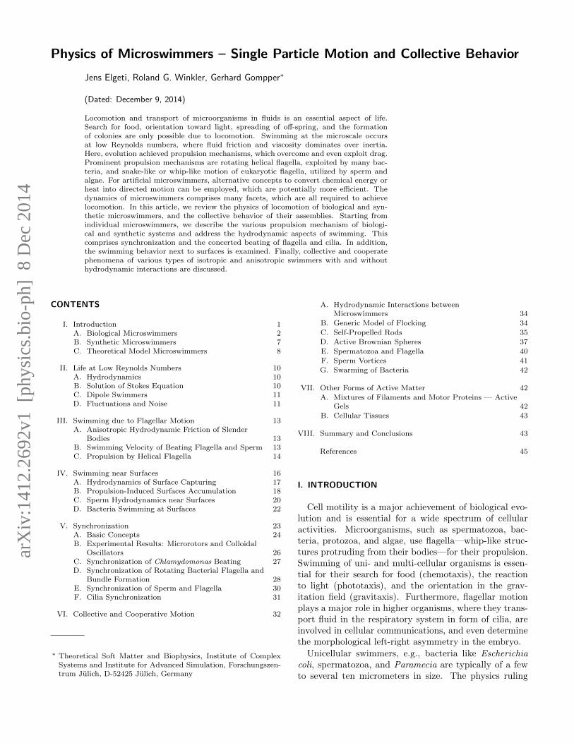
Flagellated Bacteria. A wide variety of bacteria ex-ploit helical filaments, called flagella, for their propulsion.Different species possess various numbers and differentarrangements of flagella. According to the arrangement,flagellated bacteria are classified as monotrichous bacte-ria which possess a single flagellum only, lophotrichous,bacteria with multiple flagella located at a particular spoton their surface, amphitrichous bacteria grow a singleflagellum on each of the two opposite ends, and peritri-chous bacteria are covered by multiple flagella pointingin all directions (Janssen and Graham, 2011). Promi-nent examples of peritrichous bacteria are Escherichiacoli (Berg, 2004), Salmonella typhimurium, see Fig. 1,Rhizobium lupini, or Proteus mirabilis bacteria to namejust a few. A flagellum is rotated by a motor complex,which consists of several proteins, and is anchored in thebacterial cell wall (Berg, 2003, 2004; Brennen and Winet,1977), see Fig. 2. The motor itself is connected to theflagellum by a flexible hook.
FIG. 1 Salmonella swim by a bundle of rotating helical flag-ella. From Brennen and Winet (1977).
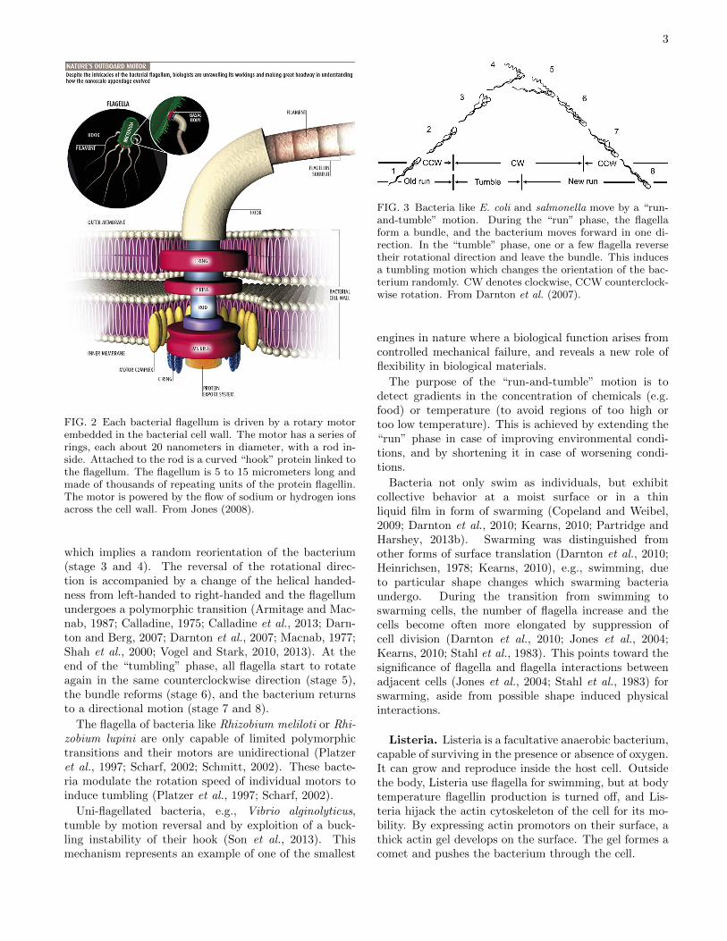
Bacteria like E. coli and Salmonella swim in a “run-and-tumble” motion illustrated in Fig. 3 (Berg, 2004;Hyon et al., 2012; Macnab, 1977; Turner et al., 2000). Inthe “run” phase (stage 1 in Fig. 3), the helical winding ofall flagella is left-handed, and they rotate counterclock-wise. The flagella form a bundle (see also Fig. 1), andthe bacterium moves forward in a direction determinedby its long axis. At the beginning of the “tumble” phase,one flagellum reverses its rotational direction to clock-wise (stage 2 in Fig. 3). The flagellum leaves the bundle
3
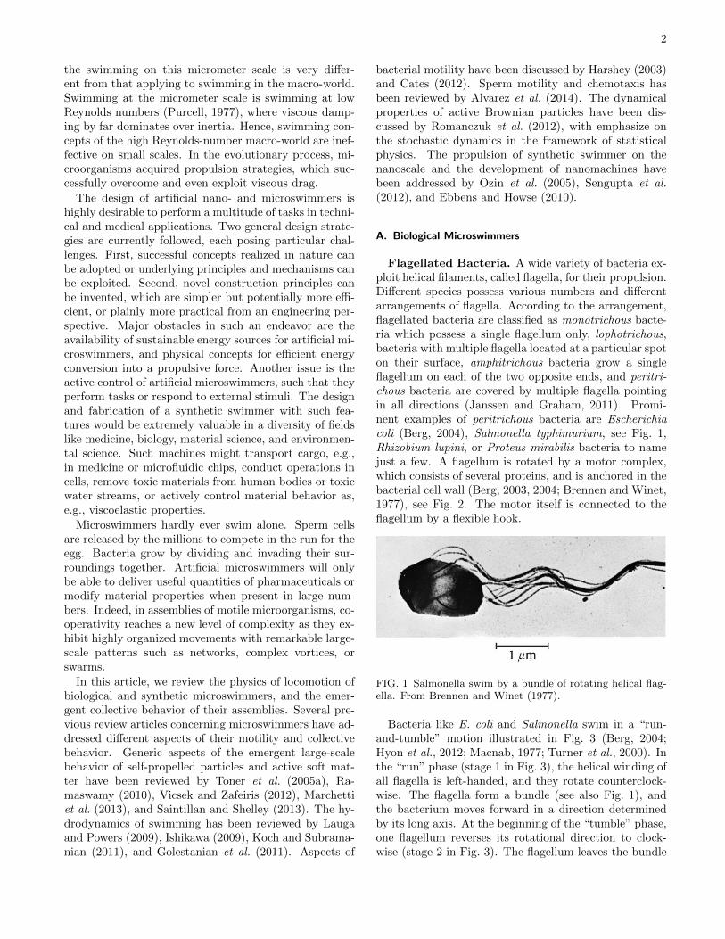
FIG. 2 Each bacterial flagellum is driven by a rotary motorembedded in the bacterial cell wall. The motor has a series ofrings, each about 20 nanometers in diameter, with a rod in-side. Attached to the rod is a curved “hook” protein linked tothe flagellum. The flagellum is 5 to 15 micrometers long andmade of thousands of repeating units of the protein flagellin.The motor is powered by the flow of sodium or hydrogen ionsacross the cell wall. From Jones (2008).
which implies a random reorientation of the bacterium(stage 3 and 4). The reversal of the rotational direc-tion is accompanied by a change of the helical handed-ness from left-handed to right-handed and the flagellumundergoes a polymorphic transition (Armitage and Mac-nab, 1987; Calladine, 1975; Calladine et al., 2013; Darn-ton and Berg, 2007; Darnton et al., 2007; Macnab, 1977;Shah et al., 2000; Vogel and Stark, 2010, 2013). At theend of the “tumbling” phase, all flagella start to rotateagain in the same counterclockwise direction (stage 5),the bundle reforms (stage 6), and the bacterium returnsto a directional motion (stage 7 and 8).
The flagella of bacteria like Rhizobium meliloti or Rhi-zobium lupini are only capable of limited polymorphictransitions and their motors are unidirectional (Platzeret al., 1997; Scharf, 2002; Schmitt, 2002). These bacte-ria modulate the rotation speed of individual motors toinduce tumbling (Platzer et al., 1997; Scharf, 2002).
Uni-flagellated bacteria, e.g., Vibrio alginolyticus,tumble by motion reversal and by exploition of a buck-ling instability of their hook (Son et al., 2013). Thismechanism represents an example of one of the smallest
FIG. 3 Bacteria like E. coli and salmonella move by a “run-and-tumble” motion. During the “run” phase, the flagellaform a bundle, and the bacterium moves forward in one di-rection. In the “tumble” phase, one or a few flagella reversetheir rotational direction and leave the bundle. This inducesa tumbling motion which changes the orientation of the bac-terium randomly. CW denotes clockwise, CCW counterclock-wise rotation. From Darnton et al. (2007).
engines in nature where a biological function arises fromcontrolled mechanical failure, and reveals a new role offlexibility in biological materials.
The purpose of the “run-and-tumble” motion is todetect gradients in the concentration of chemicals (e.g.food) or temperature (to avoid regions of too high ortoo low temperature). This is achieved by extending the“run” phase in case of improving environmental condi-tions, and by shortening it in case of worsening condi-tions.
Bacteria not only swim as individuals, but exhibitcollective behavior at a moist surface or in a thinliquid film in form of swarming (Copeland and Weibel,2009; Darnton et al., 2010; Kearns, 2010; Partridge andHarshey, 2013b). Swarming was distinguished fromother forms of surface translation (Darnton et al., 2010;Heinrichsen, 1978; Kearns, 2010), e.g., swimming, dueto particular shape changes which swarming bacteriaundergo. During the transition from swimming toswarming cells, the number of flagella increase and thecells become often more elongated by suppression ofcell division (Darnton et al., 2010; Jones et al., 2004;Kearns, 2010; Stahl et al., 1983). This points toward thesignificance of flagella and flagella interactions betweenadjacent cells (Jones et al., 2004; Stahl et al., 1983) forswarming, aside from possible shape induced physicalinteractions.
Listeria. Listeria is a facultative anaerobic bacterium,capable of surviving in the presence or absence of oxygen.It can grow and reproduce inside the host cell. Outsidethe body, Listeria use flagella for swimming, but at bodytemperature flagellin production is turned off, and Lis-teria hijack the actin cytoskeleton of the cell for its mo-bility. By expressing actin promotors on their surface, athick actin gel develops on the surface. The gel formes acomet and pushes the bacterium through the cell.
4
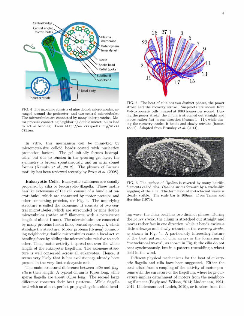
FIG. 4 The axoneme consists of nine double microtubules, ar-ranged around the perimeter, and two central microtubules.The microtubules are connected by many linker proteins. Mo-tor proteins connecting neighboring double microtubules leadto active bending. From http://en.wikipedia.org/wiki/
Cilium.
In vitro, this mechanism can be mimicked bymicrometer-size colloid beads coated with nucleationpromotion factors. The gel initially formes isotropi-cally, but due to tension in the growing gel layer, thesymmetry is broken spontaneously, and an actin cometformes (Kawska et al., 2012). The physics of Listeriamotility has been reviewed recently by Prost et al. (2008).
Eukaryotic Cells. Eucaryotic swimmers are usuallypropelled by cilia or (eucaryotic-)flagella. These motilehairlike extensions of the cell consist of a bundle of mi-crotubules, which are connected by motor proteins andother connecting proteins, see Fig. 4. The underlyingstructure is called the axoneme. It consists of two cen-tral microtubules, which are surrounded by nine doublemicrotubules (rather stiff filaments with a persistencelength of about 1 mm). The microtubules are connectedby many proteins (nexin links, central spokes, ...), whichstabilize the structure. Motor proteins (dynein) connect-ing neighboring double microtubules cause a local activebending force by sliding the microtubules relative to eachother. Thus, motor activity is spread out over the wholelength of the eukaryotic flagellum. The axoneme struc-ture is well conserved across all eukaryotes. Hence, itseems very likely that it has evolutionary already beenpresent in the very first eukaryotic cells.
The main structural difference between cilia and flag-ella is their length. A typical cilium is 10µm long, whilesperm flagella are about 50µm long. The second largedifference concerns their beat patterns. While flagellabeat with an almost perfect propagating sinusoidal bend-
FIG. 5 The beat of cilia has two distinct phases, the powerstroke and the recovery stroke. Snapshots are shown fromVolvox somatic cells, imaged at 1000 frames per second. Dur-ing the power stroke, the cilium is stretched out straight andmoves rather fast in one direction (frames 1 - 11), while dur-ing the recovery stroke, it bends and slowly retracts (frames13-27). Adapted from Brumley et al. (2014).
FIG. 6 The surface of Opalina is covered by many hairlikefilaments called cilia. Opalina swims forward by a stroke-likewiggling of the cilia. The formation of metachronal waves isclearly visible. The scale bar is 100µm. From Tamm andHorridge (1970).
ing wave, the ciliar beat has two distinct phases. Duringthe power stroke, the cilium is stretched out straight andmoves rather fast in one direction, while it bends, twists alittle sideways and slowly retracts in the recovery stroke,as shown in Fig. 5. A particularly interesting featureof the beat pattern of cilia arrays is the formation of“metachronal waves”, as shown in Fig. 6; the cilia do notbeat synchronously, but in a pattern resembling a wheatfield in the wind.
Different physical mechanisms for the beat of eukary-otic flagella and cilia have been suggested. Either thebeat arises from a coupling of the activity of motor pro-teins with the curvature of the flagellum, where large cur-vature implies detachment of motors from the neighbor-ing filament (Bayly and Wilson, 2014; Lindemann, 1994,2014; Lindemann and Lesich, 2010), or it arises from the
5
cooperative behavior of several motors pulling in oppositedirections, with the “winners” pulling along the “losers”for a while. This tug-of-war of the molecular motors re-sults in a negative force-velocity relation at zero velocity.The system is thus intrinsically unstable, and starts mov-ing in one direction. As the filaments move relative toeach other and deform, elastic forces build up, eventu-ally causing stalling and motion reversal. The systemthus starts to oscillate in time. This oscillation explainshow the axoneme’s internal machinery self-organizes togenerate the flagella beat (Camalet et al., 1999; Hilfingeret al., 2009; Julicher and Prost, 1997; Riedel-Kruse et al.,2007).
The domain of eukaryotes is home to an extraordinarynumber of different microswimmers. While all employaxonemes as motors, they use them in all kinds of waysand arrangements to propel themselves. In the following,we present a few examples of eukaryotic microswimmers,which have received particular attention in the biophys-ical community.
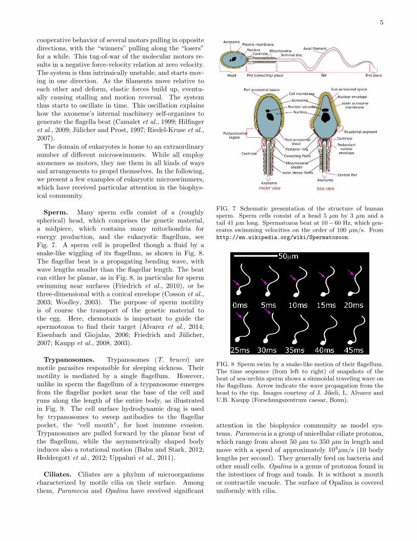
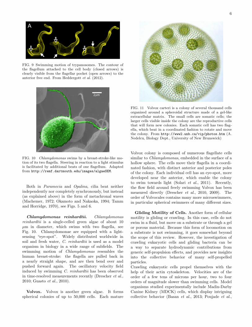
Sperm. Many sperm cells consist of a (roughlyspherical) head, which comprises the genetic material,a midpiece, which contains many mitochondria forenergy production, and the eukaryotic flagellum, seeFig. 7. A sperm cell is propelled though a fluid by asnake-like wiggling of its flagellum, as shown in Fig. 8.The flagellar beat is a propagating bending wave, withwave lengths smaller than the flagellar length. The beatcan either be planar, as in Fig. 8, in particular for spermswimming near surfaces (Friedrich et al., 2010), or bethree-dimensional with a conical envelope (Cosson et al.,2003; Woolley, 2003). The purpose of sperm motilityis of course the transport of the genetic material tothe egg. Here, chemotaxis is important to guide thespermotozoa to find their target (Alvarez et al., 2014;Eisenbach and Giojalas, 2006; Friedrich and Julicher,2007; Kaupp et al., 2008, 2003).
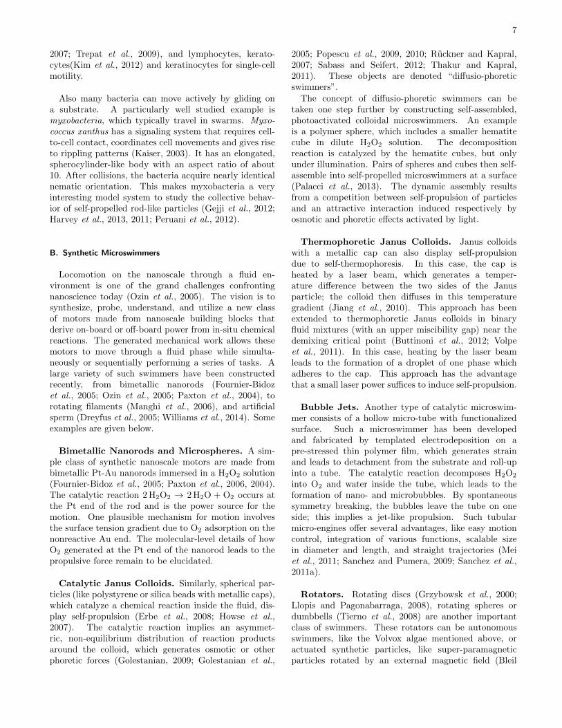
Trypanosomes. Trypanosomes (T. brucei) aremotile parasites responsible for sleeping sickness. Theirmotility is mediated by a single flagellum. However,unlike in sperm the flagellum of a trypanosome emergesfrom the flagellar pocket near the base of the cell andruns along the length of the entire body, as illustratedin Fig. 9. The cell surface hydrodynamic drag is usedby trypanosomes to sweep antibodies to the flagellarpocket, the “cell mouth”, for host immune evasion.Trypanosomes are pulled forward by the planar beat ofthe flagellum, while the asymmetrically shaped bodyinduces also a rotational motion (Babu and Stark, 2012;Heddergott et al., 2012; Uppaluri et al., 2011).
Ciliates. Ciliates are a phylum of microorganismscharacterized by motile cilia on their surface. Amongthem, Paramecia and Opalina have received significant
FIG. 7 Schematic presentation of the structure of humansperm. Sperm cells consist of a head 5 µm by 3 µm and atail 41 µm long. Spermatozoa beat at 10− 60 Hz, which gen-erates swimming velocities on the order of 100 µm/s. Fromhttp://en.wikipedia.org/wiki/Spermatozoon.
FIG. 8 Sperm swim by a snake-like motion of their flagellum.The time sequence (from left to right) of snapshots of thebeat of sea-urchin sperm shows a sinusoidal traveling wave onthe flagellum. Arrow indicate the wave propagation from thehead to the tip. Images courtesy of J. Jikeli, L. Alvarez andU.B. Kaupp (Forschungszentrum caesar, Bonn).
attention in the biophysics community as model sys-tems. Paramecia is a group of unicellular ciliate protozoa,which range from about 50 µm to 350 µm in length andmove with a speed of approximately 103µm/s (10 bodylengths per second). They generally feed on bacteria andother small cells. Opalina is a genus of protozoa found inthe intestines of frogs and toads. It is without a mouthor contractile vacuole. The surface of Opalina is covereduniformly with cilia.
6
FIG. 9 Swimming motion of trypanosomes. The contour ofthe flagellum attached to the cell body (closed arrows) isclearly visible from the flagellar pocket (open arrows) to theanterior free end. From Heddergott et al. (2012).
FIG. 10 Chlamydomonas swims by a breast-stroke-like mo-tion of its two flagella. Steering in reaction to a light stimulusis facilitated by additional beats of one flagellum. Adaptedfrom http://remf.dartmouth.edu/images/algaeSEM.
Both in Paramecia and Opalina, cilia beat neitherindependently nor completely synchronously, but instead(as explained above) in the form of metachronal waves(Machemer, 1972; Okamoto and Nakaoka, 1994; Tammand Horridge, 1970), see Figs. 5 and 6.
Chlamydomonas reinhardtii. Chlamydomonasreinhardtii is a single-celled green algae of about 10µm in diameter, which swims with two flagella, seeFig. 10. Chlamydomonae are equipped with a light-sensing “eye-spot”. Widely distributed worldwide insoil and fresh water, C. reinhardtii is used as a modelorganism in biology in a wide range of subfields. Theswimming motion of Chlamydomonas resembles thehuman breast-stroke: the flagella are pulled back ina nearly straight shape, and are then bend over andpushed forward again. The oscillatory velocity fieldinduced by swimming C. reinhardtii has been observedin time-resolved measurements recently (Drescher et al.,2010; Guasto et al., 2010).
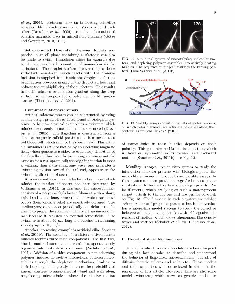
Volvox. Volvox is another green algae. It formsspherical colonies of up to 50,000 cells. Each mature
FIG. 11 Volvox carteri is a colony of several thousand cellsorganized around a spheroidal structure made of a gel-likeextracellular matrix. The small cells are somatic cells; thelarger cells visible inside the colony are the reproductive cellsthat will form new colonies. Each somatic cell has two flag-ella, which beat in a coordinated fashion to rotate and movethe colony. From http://www2.unb.ca/vip/photos.htm (A.Nedelcu, Biology Dept., University of New Brunswick)
Volvox colony is composed of numerous flagellate cellssimilar to Chlamydomonas, embedded in the surface of ahollow sphere. The cells move their flagella in a coordi-nated fashion, with distinct anterior and posterior polesof the colony. Each individual cell has an eye-spot, moredeveloped near the anterior, which enable the colonyto swim towards light (Solari et al., 2011). Recently,the flow field around freely swimming Volvox has beenmeasured directly (Drescher et al., 2010, 2009). Theorder of Volvocales contains many more microswimmers,in particular spherical swimmers of many different sizes.
Gliding Motility of Cells. Another form of cellularmotility is gliding or crawling. In this case, cells do notswim in a fluid, but move on a substrate or through a gelor porous material. Because this form of locomotion ona substrate is not swimming, it goes somewhat beyondthe scope of this review. However, the investigation ofcrawling eukaryotic cells and gliding bacteria can bea way to separate hydrodynamic contributions fromgeneric self-propulsion effects, and provides new insightsinto the collective behavior of many self-propelledparticles.Crawling eukaryotic cells propel themselves with thehelp of their actin cytoskeleton. Velocities are of theorder of a few tens of microns per hour, two to fourorders of magnitude slower than swimming cells. Modelorganisms studied experimentally include Madin-DarbyCanine Kidney (MDCK) cells, which display intriguingcollective behavior (Basan et al., 2013; Poujade et al.,
7
2007; Trepat et al., 2009), and lymphocytes, kerato-cytes(Kim et al., 2012) and keratinocytes for single-cellmotility.
Also many bacteria can move actively by gliding ona substrate. A particularly well studied example ismyxobacteria, which typically travel in swarms. Myxo-coccus xanthus has a signaling system that requires cell-to-cell contact, coordinates cell movements and gives riseto rippling patterns (Kaiser, 2003). It has an elongated,spherocylinder-like body with an aspect ratio of about10. After collisions, the bacteria acquire nearly identicalnematic orientation. This makes myxobacteria a veryinteresting model system to study the collective behav-ior of self-propelled rod-like particles (Gejji et al., 2012;Harvey et al., 2013, 2011; Peruani et al., 2012).
B. Synthetic Microswimmers
Locomotion on the nanoscale through a fluid en-vironment is one of the grand challenges confrontingnanoscience today (Ozin et al., 2005). The vision is tosynthesize, probe, understand, and utilize a new classof motors made from nanoscale building blocks thatderive on-board or off-board power from in-situ chemicalreactions. The generated mechanical work allows thesemotors to move through a fluid phase while simulta-neously or sequentially performing a series of tasks. Alarge variety of such swimmers have been constructedrecently, from bimetallic nanorods (Fournier-Bidozet al., 2005; Ozin et al., 2005; Paxton et al., 2004), torotating filaments (Manghi et al., 2006), and artificialsperm (Dreyfus et al., 2005; Williams et al., 2014). Someexamples are given below.
Bimetallic Nanorods and Microspheres. A sim-ple class of synthetic nanoscale motors are made frombimetallic Pt-Au nanorods immersed in a H2O2 solution(Fournier-Bidoz et al., 2005; Paxton et al., 2006, 2004).The catalytic reaction 2 H2O2 → 2 H2O + O2 occurs atthe Pt end of the rod and is the power source for themotion. One plausible mechanism for motion involvesthe surface tension gradient due to O2 adsorption on thenonreactive Au end. The molecular-level details of howO2 generated at the Pt end of the nanorod leads to thepropulsive force remain to be elucidated.
Catalytic Janus Colloids. Similarly, spherical par-ticles (like polystyrene or silica beads with metallic caps),which catalyze a chemical reaction inside the fluid, dis-play self-propulsion (Erbe et al., 2008; Howse et al.,2007). The catalytic reaction implies an asymmet-ric, non-equilibrium distribution of reaction productsaround the colloid, which generates osmotic or otherphoretic forces (Golestanian, 2009; Golestanian et al.,
2005; Popescu et al., 2009, 2010; Ruckner and Kapral,2007; Sabass and Seifert, 2012; Thakur and Kapral,2011). These objects are denoted “diffusio-phoreticswimmers”.
The concept of diffusio-phoretic swimmers can betaken one step further by constructing self-assembled,photoactivated colloidal microswimmers. An exampleis a polymer sphere, which includes a smaller hematitecube in dilute H2O2 solution. The decompositionreaction is catalyzed by the hematite cubes, but onlyunder illumination. Pairs of spheres and cubes then self-assemble into self-propelled microswimmers at a surface(Palacci et al., 2013). The dynamic assembly resultsfrom a competition between self-propulsion of particlesand an attractive interaction induced respectively byosmotic and phoretic effects activated by light.
Thermophoretic Janus Colloids. Janus colloidswith a metallic cap can also display self-propulsiondue to self-thermophoresis. In this case, the cap isheated by a laser beam, which generates a temper-ature difference between the two sides of the Janusparticle; the colloid then diffuses in this temperaturegradient (Jiang et al., 2010). This approach has beenextended to thermophoretic Janus colloids in binaryfluid mixtures (with an upper miscibility gap) near thedemixing critical point (Buttinoni et al., 2012; Volpeet al., 2011). In this case, heating by the laser beamleads to the formation of a droplet of one phase whichadheres to the cap. This approach has the advantagethat a small laser power suffices to induce self-propulsion.
Bubble Jets. Another type of catalytic microswim-mer consists of a hollow micro-tube with functionalizedsurface. Such a microswimmer has been developedand fabricated by templated electrodeposition on apre-stressed thin polymer film, which generates strainand leads to detachment from the substrate and roll-upinto a tube. The catalytic reaction decomposes H2O2
into O2 and water inside the tube, which leads to theformation of nano- and microbubbles. By spontaneoussymmetry breaking, the bubbles leave the tube on oneside; this implies a jet-like propulsion. Such tubularmicro-engines offer several advantages, like easy motioncontrol, integration of various functions, scalable sizein diameter and length, and straight trajectories (Meiet al., 2011; Sanchez and Pumera, 2009; Sanchez et al.,2011a).
Rotators. Rotating discs (Grzybowsk et al., 2000;Llopis and Pagonabarraga, 2008), rotating spheres ordumbbells (Tierno et al., 2008) are another importantclass of swimmers. These rotators can be autonomousswimmers, like the Volvox algae mentioned above, oractuated synthetic particles, like super-paramagneticparticles rotated by an external magnetic field (Bleil
8
et al., 2006). Rotators show an interesting collectivebehavior, like a circling motion of Volvox around eachother (Drescher et al., 2009), or a lane formation ofrotating magnetic discs in microfluidic channels (Gotzeand Gompper, 2010, 2011).
Self-propelled Droplets. Aqueous droplets sus-pended in an oil phase containing surfactants can alsobe made to swim. Propulsion arises for example dueto the spontaneous bromination of mono-olein as thesurfactant. The droplet surface is covered by a densesurfactant monolayer, which reacts with the brominefuel that is supplied from inside the droplet, such thatbromination proceeds mainly at the droplet surface, andreduces the amphiphilicity of the surfactant. This resultsin a self-sustained bromination gradient along the dropsurface, which propels the droplet due to Marangonistresses (Thutupalli et al., 2011).
Biomimetic Microswimmers.Artifical microswimmers can be constructed by using
similar design principles as those found in biological sys-tems. A by now classical example is a swimmer whichmimics the propulsion mechanism of a sperm cell (Drey-fus et al., 2005). The flagellum is constructed from achain of magnetic colloid particles and is attached to ared blood cell, which mimics the sperm head. This artifi-cial swimmer is set into motion by an alterating magneticfield, which generates a sidewise oscillatory deflection ofthe flagellum. However, the swimming motion is not thesame as for a real sperm cell; the wiggling motion is morea wagging than a travelling sine wave, and generates aswimming motion toward the tail end, opposite to theswimming direction of sperm.
A more recent example of a biohybrid swimmer whichmimics the motion of sperm has been presented byWilliams et al. (2014). In this case, the microswimmerconsists of a polydimethylsiloxane filament with a short,rigid head and a long, slender tail on which cardiomy-ocytes (heart-muscle cells) are selectively cultured. Thecardiomyocytes contract periodically and deform the fil-ament to propel the swimmer. This is a true microswim-mer because it requires no external force fields. Theswimmer is about 50 µm long and reaches a swimmingvelocity up to 10 µm/s.
Another interesting example is artificial cilia (Sanchezet al., 2011b). The assembly of oscillatory active filamentbundles requires three main components. The first two,kinesin motor clusters and microtubules, spontaneouslyorganize into aster-like structures (Nedelec et al.,1997). Addition of a third component, a non-adsorbingpolymer, induces attractive interactions between micro-tubules through the depletion mechanism, leading totheir bundling. This greatly increases the probability ofkinesin clusters to simultaneously bind and walk alongneighboring microtubules, where the relative motion
FIG. 12 A minimal system of microtubules, molecular mo-tors, and depleting polymer assembles into actively beatingbundles. The sequence of images illustrates the beating pat-tern. From Sanchez et al. (2011b).
FIG. 13 Motility assays consist of carpets of motor proteins,on which polar filaments like actin are propelled along theircontour. From Schaller et al. (2010).
of microtubules in these bundles depends on theirpolarity. This generates a cilia-like beat pattern, whichis, however, symmetric in its forward and backwardmotions (Sanchez et al., 2011b), see Fig. 12.
Motility Assays. An in-vitro system to study theinteraction of motor proteins with biological polar fila-ments like actin and microtubules are motility assays. Inthese systems, motor proteins are grafted onto a planarsubstrate with their active heads pointing upwards. Po-lar filaments, which are lying on such a motor-proteincarpet, attach to the motors and are pushed forward,see Fig. 13. The filaments in such a system are neitherswimmers nor self-propelled particles, but it is neverthe-less a interesting model systems to study the collectivebehavior of many moving particles with self-organized di-rections of motion, which shows phenomena like densitywaves and vortices (Schaller et al., 2010; Sumino et al.,2012).
C. Theoretical Model Microswimmers
Several detailed theoretical models have been designedduring the last decades to describe and understandthe behavior of flagellated microswimmers, but also ofdiffusio-phoretic spheres and rods, etc. These modelsand their properties will be reviewed in detail in theremainder of this article. However, there are also somemodel swimmers, which serve as generic models to
9
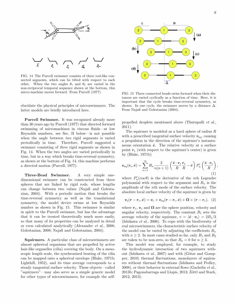
FIG. 14 The Purcell swimmer consists of three rod-like con-nected segments, which can be tilted with respect to eachother. When the two angles θ1 and θ2 are varied in thenon-reciprocal temporal sequence shown at the bottom, thismicro-machine moves forward. From Purcell (1977).
elucidate the physical principles of microswimmers. Thelatter models are briefly introduced here.
Purcell Swimmer. It was recognized already morethan 30 years ago by Purcell (1977) that directed forwardswimming of micromachines in viscous fluids—at lowReynolds numbers, see Sec. II below—is not possiblewhen the angle between two rigid segments is variedperiodically in time. Therefore, Purcell suggested aswimmer consisting of three rigid segments as shown inFig. 14. When the two angles are varied periodically intime, but in a way which breaks time-reversal symmetry,as shown at the bottom of Fig. 14, this machine performsa directed motion (Purcell, 1977).
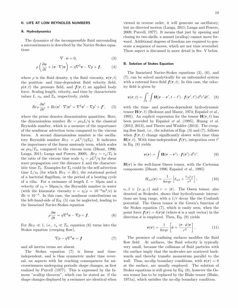
Three-Bead Swimmer. A very simple one-dimensional swimmer can be constructed from threespheres that are linked by rigid rods, whose lengthscan change between two values (Najafi and Golesta-nian, 2004). With a periodic motion that breaks thetime-reversal symmetry as well as the translationalsymmetry, the model device swims at low Reynoldsnumber as shown in Fig. 15. This swimmer is similarin spirit to the Purcell swimmer, but has the advantagethat it can be treated theoretically much more easily,so that many of its properties can be analyzed in detailor even calculated analytically (Alexander et al., 2008;Golestanian, 2008; Najafi and Golestanian, 2004).
Squirmers. A particular class of microswimmers arealmost spherical organisms that are propelled by activehair-like organelles (cilia) covering the body. On a meso-scopic length scale, the synchronized beating of the ciliacan be mapped onto a spherical envelope (Blake, 1971b;Lighthill, 1952), and its time average corresponds to asteady tangential surface velocity. These objects—called”squirmers”—may also serve as a simple generic modelfor other types of microswimmers, for example the self-
FIG. 15 Three connected beads swim forward when their dis-tances are varied cyclically as a function of time. Here, it isimportant that the cycle breaks time-reversal symmetry, asshown. In one cycle, the swimmer moves by a distance ∆.From Najafi and Golestanian (2004).
propelled droplets mentioned above (Thutupalli et al.,2011).
The squirmer is modeled as a hard sphere of radius Rwith a prescribed tangential surface velocity vsq, causinga propulsion in the direction of the squirmer’s instanta-neous orientation e. The relative velocity at a surfacepoint rs (with respect to the squirmer’s center) is givenby (Blake, 1971b)
vsq(rs, e) =
∞∑n=1
Bn2
n(n+ 1)
(e · rsR
rsR− e)P ′n
(e · rsR
),
(1)where P ′n(cos θ) is the derivative of the nth Legendrepolynomial with respect to the argument and Bn is theamplitude of the nth mode of the surface velocity. Theabsolute local surface velocity of the squirmer is given by
vs(r − rc, e) = vc + vsq(r − rc, e) + Ω× (r − rc), (2)
where rc, vc and Ω are the sphere position, velocity andangular velocity, respectively. The constant B1 sets theaverage velocity of the squirmer, v = 〈e · vs〉 = 2B1/3(Ishikawa et al., 2006). To cope with the biodiversity ofreal microswimmers, the characteristic surface velocity ofthe model can be varied by adjusting the coefficients Bnwith n ≥ 2. In most cases studied so far, only B1 and B2
are taken to be non-zero, so that Bn = 0 for n ≥ 3.This model was employed, for example, to study
the hydrodynamic interaction of two squirmers with-out (Ishikawa et al., 2007) and with (Gotze and Gomp-per, 2010) thermal fluctuations, monolayers of squirm-ers without thermal fluctuations (Ishikawa and Pedley,2008), or their behavior in external flows (Gachelin et al.,2013b; Pagonabarraga and Llopis, 2013; Zottl and Stark,2012, 2013).
10
II. LIFE AT LOW REYNOLDS NUMBERS
A. Hydrodynamics
The dynamics of the incompressible fluid surroundinga microswimmers is described by the Navier-Stokes equa-tions
∇ · v = 0, (3)
ρ
(∂v
∂t+ (v · ∇)v
)= η∇2v −∇p+ f , (4)
where ρ is the fluid density, η the fluid viscosity, v(r, t)the position- and time-dependent fluid velocity field,p(r, t) the pressure field, and f(r, t) an applied bodyforce. Scaling length, velocity, and time by characteristicvalues L, v0, and T0, respectively, yields
ReT∂v′
∂t′+Re(v′ · ∇)v′ = ∇2v′ −∇p′ + f ′, (5)
where the prime denotes dimensionless quantities. Here,the dimensionless number Re = ρv0L/η is the classicalReynolds number, which is a measure of the importanceof the nonlinear advection term compared to the viscousforces. A second dimensionless number is the oscilla-tory Reynolds number ReT = ρL2/(ηT0). It indicatesthe importance of the linear unsteady term, which scalesas ρv0/T0, compared to the viscous term (Dhont, 1996;Lauga, 2011; Lauga and Powers, 2009). ReT = τη/T0 isthe ratio of the viscous time scale τη = ρL2/η for shearwave propagation over the distance L and the character-istic time T0. Examples for T0 could be the self advectiontime L/v0 (for which ReT = Re), the rotational periodof a bacterial flagellum, or the period of a beating cycleof a cilia. For a swimmer of length L = 10µm and avelocity of v0 = 50µm/s, the Reynolds number in water(with the kinematic viscosity ν = η/ρ = 10−6m2/s) isRe ≈ 10−3. In this case, the nonlinear contributions onthe left-hand-side of Eq. (5) can be neglected, leading tothe linearized Navier-Stokes equation
ρ∂v
∂t= η∇2v −∇p+ f . (6)
For ReT 1, i.e., τη T0, equation (6) turns into theStokes equation (creeping flow),
∇p− η∇2v = f (7)
and all inertia terms are absent.The Stokes equation (7) is linear and time-
independent, and is thus symmetric under time rever-sal, an aspects with far reaching consequences for mi-croswimmers undergoing periodic shape changes, as firstrealized by Purcell (1977). This is expressed by the fa-mous ”scallop theorem”, which can be stated as: if theshape changes displayed by a swimmer are identical when
viewed in reverse order, it will generate an oscillatory,but no directed motion (Lauga, 2011; Lauga and Powers,2009; Purcell, 1977). It means that just by opening andclosing its two shells, a mussel (scallop) cannot move for-ward. Additional degrees of freedom are required to gen-erate a sequence of moves, which are not time reversibel.These aspect is discussed in more detail in Sec. V below.
B. Solution of Stokes Equation
The linearized Navier-Stokes equations (3), (6), and(7), can be solved analytically for an unbounded systemwith a external force field f(r, t). In this case, the veloc-ity field is given by
v(r, t) =
∫ t
0
∫H(r − r′, t− t′) · f(r′, t′) d3r′dt′, (8)
with the time- and position-dependent hydrodynamictensor H(r, t) (Bedeaux and Mazur, 1974; Espanol et al.,1995). An explicit expression for the tensor H(r, t) hasbeen provided by Espanol et al. (1995), Huang et al.(2012, 2013), and Theers and Winkler (2013). The creep-ing flow limit, i.e., the solution of Eqs. (3) and (7), followswhen f(r, t) change significantly slower with time thanH(r, t). With time-independent f(r), integration over t′
in Eq. (8) yields
v(r) =
∫H(r − r′) · f(r′) d3r′. (9)
H(r) is the well-know Oseen tensor, with the Cartesiancomponents (Dhont, 1996; Espanol et al., 1995)
Hαβ(r) =1
8πηr
[δαβ +
rαrβr2
]; (10)
α, β ∈ x, y, z and r = |r|. The Oseen tensor, alsodenoted as Stokeslet, shows that hydrodynamic interac-tions are long range, with a 1/r decay like the Coulombpotential. The Oseen tensor is the Green’s function ofthe Stokes equation (7), which is easily seen, when thepoint force f(r) = δ(r)e (where e is a unit vector) in thedirection e is employed. Then, Eq. (9) yields
v(r) =1
8πηr
[e+
(r · e)r
r2
]. (11)
The presence of confining surfaces modifies the fluidflow field. At surfaces, the fluid velocity is typicallyvery small, because the collisions of fluid particles withthe surface imply that the molecules are scattered back-wards and thereby transfer momentum parallel to thewall. Thus, no-slip boundary conditions, with v(r) = 0at the surface, are usually employed. The solution ofStokes equations is still given by Eq. (9), however the Os-sen tensor has to be replaced by the Blake tensor (Blake,1971a), which satisfies the no-slip boundary condition.
11
FIG. 16 Schematics of the flow field dipole swimmers, (a)pusher and (b) puller.
C. Dipole Swimmers
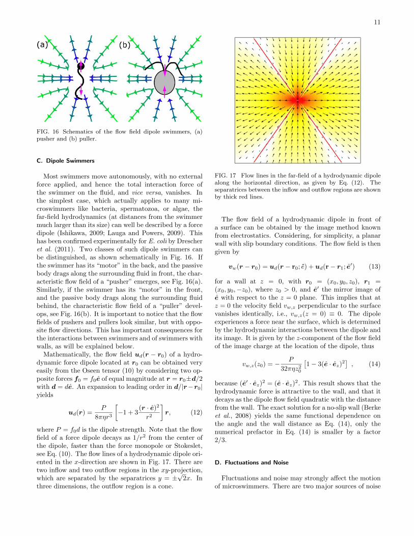
Most swimmers move autonomously, with no externalforce applied, and hence the total interaction force ofthe swimmer on the fluid, and vice versa, vanishes. Inthe simplest case, which actually applies to many mi-croswimmers like bacteria, spermatozoa, or algae, thefar-field hydrodynamics (at distances from the swimmermuch larger than its size) can well be described by a forcedipole (Ishikawa, 2009; Lauga and Powers, 2009). Thishas been confirmed experimentally for E. coli by Drescheret al. (2011). Two classes of such dipole swimmers canbe distinguished, as shown schematically in Fig. 16. Ifthe swimmer has its “motor” in the back, and the passivebody drags along the surrounding fluid in front, the char-acteristic flow field of a “pusher” emerges, see Fig. 16(a).Similarly, if the swimmer has its “motor” in the front,and the passive body drags along the surrounding fluidbehind, the characteristic flow field of a “puller” devel-ops, see Fig. 16(b). It is important to notice that the flowfields of pushers and pullers look similar, but with oppo-site flow directions. This has important consequences forthe interactions between swimmers and of swimmers withwalls, as will be explained below.
Mathematically, the flow field ud(r − r0) of a hydro-dynamic force dipole located at r0 can be obtained veryeasily from the Oseen tensor (10) by considering two op-posite forces f0 = f0e of equal magnitude at r = r0±d/2with d = de. An expansion to leading order in d/|r−r0|yields
ud(r) =P
8πηr3
[−1 + 3
(r · e)2
r2
]r, (12)
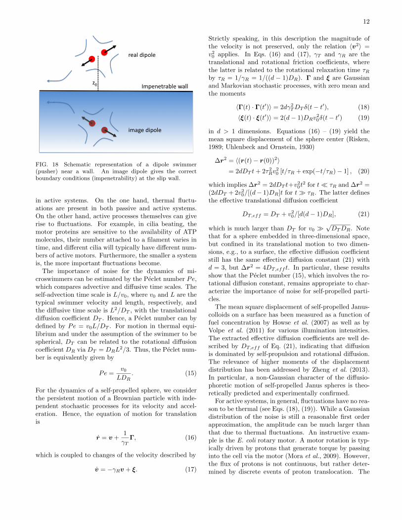
where P = f0d is the dipole strength. Note that the flowfield of a force dipole decays as 1/r2 from the center ofthe dipole, faster than the force monopole or Stokeslet,see Eq. (10). The flow lines of a hydrodynamic dipole ori-ented in the x-direction are shown in Fig. 17. There aretwo inflow and two outflow regions in the xy-projection,which are separated by the separatrices y = ±
√2x. In
three dimensions, the outflow region is a cone.
FIG. 17 Flow lines in the far-field of a hydrodynamic dipolealong the horizontal direction, as given by Eq. (12). Theseparatrices between the inflow and outflow regions are shownby thick red lines.
The flow field of a hydrodynamic dipole in front ofa surface can be obtained by the image method knownfrom electrostatics. Considering, for simplicity, a planarwall with slip boundary conditions. The flow field is thengiven by
vw(r − r0) = ud(r − r0; e) + ud(r− r1; e′) (13)
for a wall at z = 0, with r0 = (x0, y0, z0), r1 =(x0, y0,−z0), where z0 > 0, and e′ the mirror image ofe with respect to the z = 0 plane. This implies that atz = 0 the velocity field vw,z perpendicular to the surfacevanishes identically, i.e., vw,z(z = 0) ≡ 0. The dipoleexperiences a force near the surface, which is determinedby the hydrodynamic interactions between the dipole andits image. It is given by the z-component of the flow fieldof the image charge at the location of the dipole, thus
vw,z(z0) = − P
32πηz20
[1− 3(e · ez)2
], (14)
because (e′ · ez)2 = (e · ez)2. This result shows that thehydrodynamic force is attractive to the wall, and that itdecays as the dipole flow field quadratic with the distancefrom the wall. The exact solution for a no-slip wall (Berkeet al., 2008) yields the same functional dependence onthe angle and the wall distance as Eq. (14), only thenumerical prefactor in Eq. (14) is smaller by a factor2/3.
D. Fluctuations and Noise
Fluctuations and noise may strongly affect the motionof microswimmers. There are two major sources of noise
12
FIG. 18 Schematic representation of a dipole swimmer(pusher) near a wall. An image dipole gives the correctboundary conditions (impenetrability) at the slip wall.
in active systems. On the one hand, thermal fluctu-ations are present in both passive and active systems.On the other hand, active processes themselves can giverise to fluctuations. For example, in cilia beating, themotor proteins are sensitive to the availability of ATPmolecules, their number attached to a filament varies intime, and different cilia will typically have different num-bers of active motors. Furthermore, the smaller a systemis, the more important fluctuations become.
The importance of noise for the dynamics of mi-croswimmers can be estimated by the Peclet number Pe,which compares advective and diffusive time scales. Theself-advection time scale is L/v0, where v0 and L are thetypical swimmer velocity and length, respectively, andthe diffusive time scale is L2/DT , with the translationaldiffusion coefficient DT . Hence, a Peclet number can bydefined by Pe = v0L/DT . For motion in thermal equi-librium and under the assumption of the swimmer to bespherical, DT can be related to the rotational diffusioncoefficient DR via DT = DRL
2/3. Thus, the Peclet num-ber is equivalently given by
Pe =v0LDR
. (15)
For the dynamics of a self-propelled sphere, we considerthe persistent motion of a Brownian particle with inde-pendent stochastic processes for its velocity and accel-eration. Hence, the equation of motion for translationis
r = v +1
γTΓ, (16)
which is coupled to changes of the velocity described by
v = −γRv + ξ. (17)
Strictly speaking, in this description the magnitude ofthe velocity is not preserved, only the relation 〈v2〉 =v20 applies. In Eqs. (16) and (17), γT and γR are thetranslational and rotational friction coefficients, wherethe latter is related to the rotational relaxation time τRby τR = 1/γR = 1/((d − 1)DR). Γ and ξ are Gaussianand Markovian stochastic processes, with zero mean andthe moments
〈Γ(t) · Γ(t′)〉 = 2dγ2TDT δ(t− t′), (18)
〈ξ(t) · ξ(t′)〉 = 2(d− 1)DRv20δ(t− t′) (19)
in d > 1 dimensions. Equations (16) – (19) yield themean square displacement of the sphere center (Risken,1989; Uhlenbeck and Ornstein, 1930)
∆r2 = 〈(r(t)− r(0))2〉= 2dDT t+ 2τ2Rv
20 [t/τR + exp(−t/τR)− 1] , (20)
which implies ∆r2 = 2dDT t+v20t
2 for t τR and ∆r2 =(2dDT + 2v20/[(d− 1)DR]t for t τR. The latter definesthe effective translational diffusion coefficient
DT,eff = DT + v20/[d(d− 1)DR], (21)
which is much larger than DT for v0 √DTDR. Note
that for a sphere embedded in three-dimensional space,but confined in its translational motion to two dimen-sions, e.g., to a surface, the effective diffusion coefficientstill has the same effective diffusion constant (21) withd = 3, but ∆r2 = 4DT,eff t. In particular, these resultsshow that the Peclet number (15), which involves the ro-tational diffusion constant, remains appropriate to char-acterize the importance of noise for self-propelled parti-cles.
The mean square displacement of self-propelled Janus-colloids on a surface has been measured as a function offuel concentration by Howse et al. (2007) as well as byVolpe et al. (2011) for various illumination intensities.The extracted effective diffusion coefficients are well de-scribed by DT,eff of Eq. (21), indicating that diffusionis dominated by self-propulsion and rotational diffusion.The relevance of higher moments of the displacementdistribution has been addressed by Zheng et al. (2013).In particular, a non-Gaussian character of the diffusio-phoretic motion of self-propelled Janus spheres is theo-retically predicted and experimentally confirmed.
For active systems, in general, fluctuations have no rea-son to be thermal (see Eqs. (18), (19)). While a Gaussiandistribution of the noise is still a reasonable first orderapproximation, the amplitude can be much larger thanthat due to thermal fluctuations. An instructive exam-ple is the E. coli rotary motor. A motor rotation is typ-ically driven by protons that generate torque by passinginto the cell via the motor (Mora et al., 2009). However,the flux of protons is not continuous, but rather deter-mined by discrete events of proton translocation. The
13
stochasticity of this process creates a “shot noise”. Mod-elling the motor in some detail, Mora et al. (2009) derivean expression for the effective diffusion coefficient of theflagellar rotation. At high load, the effective diffusion co-efficient is thermal, but at low loads, diffusion increasesand becomes dominated by the shot noise.
Measurements of the rotational diffusion coefficient ofChlamydomonas yield DR = 0.4rad2/s (Drescher et al.,2011). This value can be compared with that of apassive sphere of radius R = 5µm, which is DR =kBT/(8πηR
3) ≈ 3× 10⁻³rad2/s, i.e., it is about two or-ders of magnitude smaller than the value of the activemicroorganism.
Similarly, for E. coli the values DR = 0.057rad2/s(Drescher et al., 2011) and DR ≈ 2rad2/s (Saragostiet al., 2012) have been reported for non-tumbling andtumbling cells, respectively. A study of paralyzed E. coli(Tavaddod et al., 2011) yields the rotational diffusion co-efficient DR = 0.032rad2/s, which is only a factor twosmaller than the value of swimming but non-tumblingcells. Thus, tumbling events evidently increase the ro-tational diffusion coefficient by more than an order ofmagnitude.
We conclude that noise and fluctuations in microswim-mer motion can be much pronounced compared to thatof the dynamics of respective passive objects, and thatactivity-based noise can be the dominant contribution toobserved phenomena.
III. SWIMMING DUE TO FLAGELLAR MOTION
A. Anisotropic Hydrodynamic Friction of Slender Bodies
A microorganism is able to swim forward in a fluid bywiggling or rotating a flagellum, because the hydrody-namic friction of a long, slender body in a viscous envi-ronment is anisotropic. This can be demonstrated easilyfor a long and thin rod of radius a and length L: itexperiences less friction when pulled along its axis thanperpendicular to it.
We approximate the rod as a sequence of touchingbeads of radius a. In general, the equation of motionof the i-th bead is given by (Bird et al., 1987; Winkler,2007)
ζ (ri − v(ri)) = Fi, (22)
i.e., the frictional force is equal to the applied force. Here,ζ = 6πηa is Stokes friction coefficient for a sphere withno-slip boundary conditions moving in a viscous fluid.The fluid velocity v(ri) is determined by the motion ofall other beads j 6= i and follows from Eq. (9). Theforce density on the fluid originates from the forces ofthe various beads
f(r) =∑i
Fiδ(r − ri(t)). (23)
Thus, we obtain (Doi and Edwards, 1986)
ri(t) =1
ζFi +
∑j 6=i
H(ri(t)− rj(t)) · Fj , (24)
which turns into (Harnau et al., 1996)
r(s, t) =1
3πηf(s) +
∫H(r(s, t)− r(s′, t)) · f(s′) ds′
(25)
in the continuum limit, where s (with −L/2 < s < L/2)is the contour coordinate along the centerline of the rod,and f(s) the linear force density.
The anisotropic friction coefficients of a rod are definedby the force-velocity relation
F = ζ‖v‖ + ζ⊥v⊥ (26)
for the motion parallel (v‖) and perpendicular (v⊥) to therod axis. Calculations based on Eq. (26), with constantfriction coefficients ζ‖ and ζ⊥, are denoted “resistive-forcetheory” (Gray and Hancock, 1955; Lighthill, 1976).
To calculate ζ‖ and ζ⊥, it is easiest to consider thespecial cases of a rod aligned along the x-axis of thereference frame and pulled parallel and perpendicularto its long axis, respectively, with the constant forceF = F e. Since, we consider a rigid body, the forcedensity is f(s) = F e/L, and the average rod velocityvrod =
∫r(s)ds/L becomes
vrod = F
[e
3πηL+
(e+ (ex · e)ex)
4πηL2
∫ L
2a
L− ss
ds
].
(27)
The lower cutoff of the integral excludes a region of thethickness of the rod and prevents self-interactions. Be-cause (ex · e)ex = 1 and 0 for parallel and perpendicularorientation of the force, respectively, evaluation of theintegral yields
ζ⊥ = 2ζ‖ , ζ⊥ =4πηL
ln(L/2a)(28)
in the asymptotic limit of a long rod (Doi and Edwards,1986). It is therefore easier to pull a long rod along itsaxis than perpendicular to it by a factor two. The loga-rithmic divergence is a result of the long-range nature ofhydrodynamic interactions of different parts of the rod,which reduce the friction coefficient compared to that ofa rod of non-interacting beads (∼ L). Corrections of thefriction coefficients for a more precise hydrodynamic cal-culation for a cylinder are provided in Tirado et al. (1984)and Howard (2001).
B. Swimming Velocity of Beating Flagella and Sperm
The result (28) together with Eq. (26) can now be usedto calculate the swimming velocity of a sinusoidally beat-ing flagellum. In this case, the time-dependent shape is
14
given by
y(x, t) = A sin(kx− ωt), (29)
where A is the beating amplitude, ω the frequency, andk = 2π/λ the wave number with the wave length λ. Thevelocity of a segment of the flagellum at x is then
vy(x, t) =∂y
∂t= −Aω cos(kx− ωt), (30)
where geometric nonlinearities are neglected. With thelocal tangent vector (not normalized)
t(x, t) = (1, Ak cos(kx− ωt), 0)T , (31)
the velocity v(x, t) = (0, vy(x, t), 0) can be decomposedinto v‖ = (v · t)t/t2 and v⊥ = v − v‖, with
v‖ = − A2ωk cos2(kx− ωt)1 +A2k2 cos2(kx− ωt)
t. (32)
According to Eq. (26), this generates the force
Fx = (ζ‖ − ζ⊥)1
L
∫A2ωk cos2(kx− ωt)
1 +A2k2 cos2(kx− ωt)dx (33)
in the swimming direction, while the force in the perpen-dicular direction vanishes when averaged over the wholeflagellum. For small beating amplitudes, Eq. (33) caneasily be integrated, which yields the average propulsionforce
Fx =1
2(ζ‖ − ζ⊥)A2ωk. (34)
The swimming velocity then follows from vx ' Fx/ζ‖ as
vflag = −1
2
(ζ⊥ζ‖− 1
)A2ωk . (35)
This simplified calculation shows several important as-pects of flagellar propulsion. First, swimming is only pos-sible due to the frictional anisotropy, i.e. ζ‖ 6= ζ⊥. Sec-ond, for a travelling wave in the positive x-direction, theflagellum moves in the negative x-direction, i.e., move-ment is opposite to the direction of the travelling wave.Third, the swimming velocity increases linearly with thebeating frequency ω and the wave vector k, but quadrat-ically with the beating amplitude A. And finally, theswimming velocity is independent of the fluid viscosity.
A more refined calculation has been performed by Grayand Hancock (1955), also employing resistive force the-ory, to determine the swimming velocity of sperm. Forthe sinusoidal beating pattern (29) and ζ⊥/ζ‖ = 2, theyfind
vsperm =1
2A2ωk
[1 +A2k2 (36)
+
√1 +
1
2A2k2
3RhL
(ln
(kd
4π
)− 1
2
)]−1. (37)
Here, L is the length of the flagellum and Rh is the ra-dius of the head. The general conclusions wit respectto Eq. (35) remain valid, but additional effects appear.The second term in the brackets of Eq. (36)—its originis already recognizable in Eq. (32)—arises from the finitebeating amplitude and implies a saturation of the veloc-ity for large beating amplitudes. The last term in thebrackets describes the reduction of velocity due the dragof the passive head.
Friedrich et al. (2010) employed a wave form with in-creasing amplitude of the flagellar beat with increasingdistance from the head to describe the beat geometryof bull sperm, and use direct experimental input for thebeat amplitude and frequency. In this way, experimentaltrajectories can be reproduced quite accurately by re-sistive force theory, when the friction anisotropy is cho-sen appropriately. This yields the friction anisotropyζ⊥/ζ‖ = 1.81± 0.07.
The swimming of sperm has also been analyzedby slender-body theory (Hancock, 1953; Johnson andBrokaw, 1979; Lighthill, 1976) (taking into account thehydrodynamic interactions of different parts of the de-formed flagellum as in Sec.III.A for slender rods) by Hig-don (1979). Results agree with the resistive-force ap-proach by Gray and Hancock (1955) within about 10%.
A higher order solution, taking into account the fullhydrodynamics, is possible for an infinitely long flagel-lum in two spatial dimensions (where hydrodynamics isof longer range than in three dimensions) — correspond-ing to an infinite sheet with a laterally propagating waveand transverse oscillations in three dimensions. Here, theswimming velocity
vsperm =1
2A2ωk
(1− 19
16A2k2
)(38)
has already been obtained by Taylor (1951) in his pio-neering work. This result confirms all qualitative featuresdiscussed above, but shows somewhat different numericalcoefficients (which is in part due to the different dimen-sionality).
The sperm structure or beating pattern is typicallynot completely symmetric, but has some chirality. Inthis case, sperm swim on helical trajectories (Crenshaw,1989; Elgeti and Gompper, 2008). In particular, the he-licity of the swimming trajectories is very pronounced forsea urchin sperm (Bohmer et al., 2005; Crenshaw, 1996;Kaupp et al., 2003).
C. Propulsion by Helical Flagella
Resistive force theory (Gray and Hancock, 1955;Lighthill, 1976) as well as slender body theory (Hancock,1953; Johnson and Brokaw, 1979; Lighthill, 1976) havebeen applied to describe propulsion of rotating helicalflagella. Various aspect of the approaches have been
15
summarized by Lauga and Powers (2009) in their re-view. Here, we briefly address slender-body results inthe light of recent experiments on macroscopic helices atlow Reynolds numbers (Rodenborn et al., 2013).
As shown by Lighthill (1976), the velocity of a pointat s along the contour of a flagellum of finite thicknesscan be described as
v(s) =1
4πηf⊥(s) (39)
+
∫H(r(s)− r(s′)) · f(s′) Θ(|r(s)− r(s′)| − δ) ds′
within the far-field approximation (as in Eq. (25)). Here,δ = a
√e/2 is the cutoff to avoid self-interactions, Θ(x)
is Heaviside’s step function, and f⊥ is the normal com-ponent of the Stokeslet strength (Eq. 10), i.e.,
f⊥ =(I− tt
)· f , (40)
where t is the local tangent vector to the filament andI the unit matrix (Lauga and Powers, 2009; Rodenbornet al., 2013).
A helix oriented along the z-axis can be parameterizedas
r(s) = (Rh cosϕ,Rh sinϕ,ϕP/(2π))T . (41)
Here, ϕ = 2πs cosϑ/P is the helical phase, P the pitchof the helix, ϑ the pitch angle, and Rh the helix radius.From Eq. (41), we obtain the tangent vector and theforce (40). Assuming a very long helix L/P 1, wecan neglect end effects and approximate the local forcedensity by
f(s) = (−fϕ sinϕ, fϕ cosϕ, fz)T
(42)
and the local velocity by
v(s) = (−ΩRh sinϕ,ΩRh cosϕ, vz)T. (43)
Inserting Eqs. (41), (42), and (43) in Eqs. (39) and (40),respectively, the translational velocity vz and rotationalfrequency Ω can be represented as(
vzΩ
)=
(A11 A12
A12 A22
)(fzmz
)(44)
by the pulling force density fz and the moment densitymz = Rhfϕ. Note that we assume the helix to remainaligned along the z-axis, i.e., other torques are compen-sated by additional external forces. Neglecting end ef-fects, the matrix elements are given by
A11 =1
4πη
(sin2 ϑ+
1
sinϑ
∫ ϕL
ϕ0
(1
Φ+ϕ2 cot2 ϑ
Φ3
)dϕ
),
A12 =1
4πηRh
(−1
2sin 2ϑ+
1
sinϑ
∫ ϕL
ϕ0
ϕ sinϕ cotϑ
Φ3dϕ
),
A22 =1
4πηR2h
(cos2 ϑ+
1
sinϑ
∫ ϕL
ϕ0
(cosϕ
Φ+
sin2 ϕ
Φ3
)dϕ
),
(45)
0
5
10
15
20
25
30
0 2 4 6 8 10 12
F T/(ηΩ
r2 )
Lcos(ϑ) /P
h
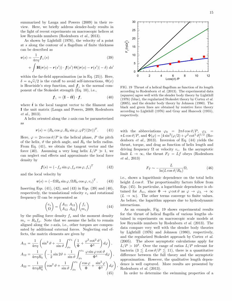
FIG. 19 Thrust of a helical flagellum as function of its lengthaccording to Rodenborn et al. (2013). The experimental data(squares) agree well with the slender body theory by Lighthill(1976) (blue), the regularized Stokeslet theory by Cortez et al.(2005), and the slender body theory by Johnson (1980). Theblack and green lines are obtained by resistive force theoryaccording to Lighthill (1976) and Gray and Hancock (1955),respectively.
with the abbreviations ϕ0 = 2πδ cosϑ/P , ϕL =πL cosϑ/P , and Φ(ϕ) = [4 sin2(ϕ/2) +ϕ2 cot2 ϑ]1/2 (Ro-denborn et al., 2013). Inversion of Eq. (44) yields thethrust, torque, and drag as function of helix length anddriving frequency Ω or velocity vz. In the asymptoticlimit L → ∞, the thrust FT = Lf obeys (Rodenbornet al., 2013)
FT ∼ −L
ln(L cosϑ/Rh)Ω, (46)
i.e., shows a logarithmic dependence on the total helixheight L cosϑ. The proportionality factors follow fromEqs. (45). In particular, a logarithmic dependence is ob-tained for A11, since Φ → ϕ cotϑ as ϕ → ϕL → ∞(L → ∞). The other terms converge to finite values.As before, the logarithm appears due to hydrodynamicinteractions.
As an example, Fig. 19 shows experimental resultsfor the thrust of helical flagella of various lengths ob-tained in experiments on macroscopic scale models atlow Reynolds numbers by Rodenborn et al. (2013). Thedata compare very well with the slender body theoriesby Lighthill (1976) and Johnson (1980), respectively,and the regularized Stokeslet approach by Cortez et al.(2005). The above asymptotic calculations apply forL/P > 103. Over the range of ratios L/P relevant forbacteria (3 . L cosϑ/P . 11), there is a quantitativedifference between the full theory and the asymptoticapproximation. However, the qualitative length depen-dence is well captured. More results are presented byRodenborn et al. (2013).
In order to determine the swimming properties of a
16
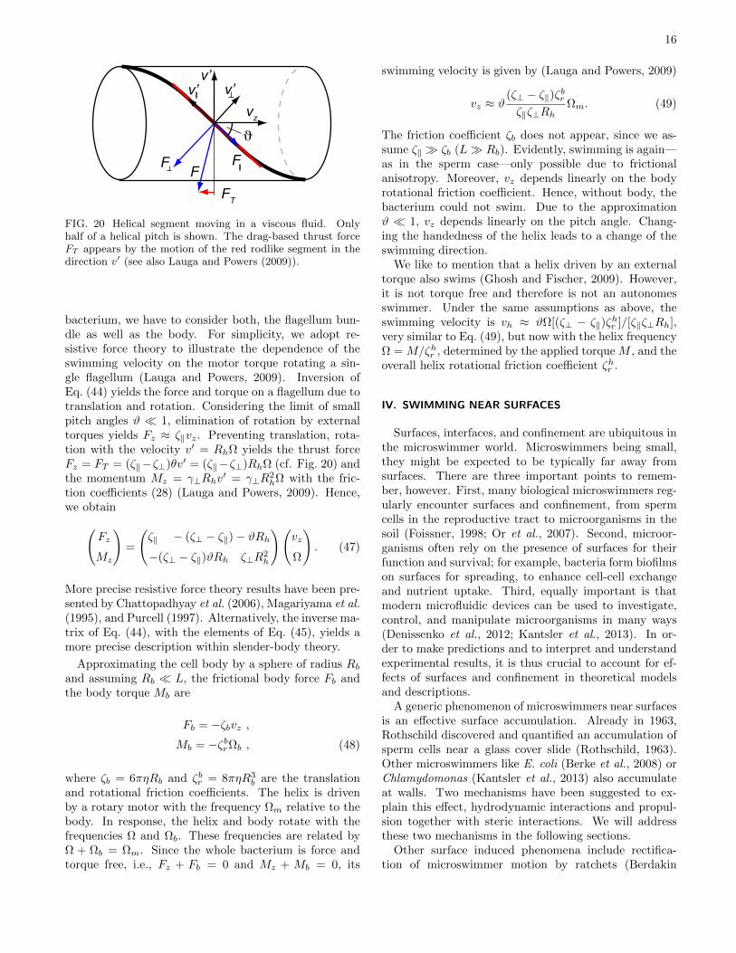
v’v’ v’
FF F
FT
vz
FIG. 20 Helical segment moving in a viscous fluid. Onlyhalf of a helical pitch is shown. The drag-based thrust forceFT appears by the motion of the red rodlike segment in thedirection v′ (see also Lauga and Powers (2009)).
bacterium, we have to consider both, the flagellum bun-dle as well as the body. For simplicity, we adopt re-sistive force theory to illustrate the dependence of theswimming velocity on the motor torque rotating a sin-gle flagellum (Lauga and Powers, 2009). Inversion ofEq. (44) yields the force and torque on a flagellum due totranslation and rotation. Considering the limit of smallpitch angles ϑ 1, elimination of rotation by externaltorques yields Fz ≈ ζ‖vz. Preventing translation, rota-tion with the velocity v′ = RhΩ yields the thrust forceFz = FT = (ζ‖−ζ⊥)θv′ = (ζ‖−ζ⊥)RhΩ (cf. Fig. 20) andthe momentum Mz = γ⊥Rhv
′ = γ⊥R2hΩ with the fric-
tion coefficients (28) (Lauga and Powers, 2009). Hence,we obtain(
Fz
Mz
)=
(ζ‖ − (ζ⊥ − ζ‖)− ϑRh−(ζ⊥ − ζ‖)ϑRh ζ⊥R
2h
)(vz
Ω
). (47)
More precise resistive force theory results have been pre-sented by Chattopadhyay et al. (2006), Magariyama et al.(1995), and Purcell (1997). Alternatively, the inverse ma-trix of Eq. (44), with the elements of Eq. (45), yields amore precise description within slender-body theory.
Approximating the cell body by a sphere of radius Rband assuming Rb L, the frictional body force Fb andthe body torque Mb are
Fb = −ζbvz ,Mb = −ζbrΩb , (48)
where ζb = 6πηRb and ζbr = 8πηR3b are the translation
and rotational friction coefficients. The helix is drivenby a rotary motor with the frequency Ωm relative to thebody. In response, the helix and body rotate with thefrequencies Ω and Ωb. These frequencies are related byΩ + Ωb = Ωm. Since the whole bacterium is force andtorque free, i.e., Fz + Fb = 0 and Mz + Mb = 0, its
swimming velocity is given by (Lauga and Powers, 2009)
vz ≈ ϑ(ζ⊥ − ζ‖)ζbrζ‖ζ⊥Rh
Ωm. (49)
The friction coefficient ζb does not appear, since we as-sume ζ‖ ζb (L Rb). Evidently, swimming is again—as in the sperm case—only possible due to frictionalanisotropy. Moreover, vz depends linearly on the bodyrotational friction coefficient. Hence, without body, thebacterium could not swim. Due to the approximationϑ 1, vz depends linearly on the pitch angle. Chang-ing the handedness of the helix leads to a change of theswimming direction.
We like to mention that a helix driven by an externaltorque also swims (Ghosh and Fischer, 2009). However,it is not torque free and therefore is not an autonomesswimmer. Under the same assumptions as above, theswimming velocity is vh ≈ ϑΩ[(ζ⊥ − ζ‖)ζ
hr ]/[ζ‖ζ⊥Rh],
very similar to Eq. (49), but now with the helix frequencyΩ = M/ζhr , determined by the applied torque M , and theoverall helix rotational friction coefficient ζhr .
IV. SWIMMING NEAR SURFACES
Surfaces, interfaces, and confinement are ubiquitous inthe microswimmer world. Microswimmers being small,they might be expected to be typically far away fromsurfaces. There are three important points to remem-ber, however. First, many biological microswimmers reg-ularly encounter surfaces and confinement, from spermcells in the reproductive tract to microorganisms in thesoil (Foissner, 1998; Or et al., 2007). Second, microor-ganisms often rely on the presence of surfaces for theirfunction and survival; for example, bacteria form biofilmson surfaces for spreading, to enhance cell-cell exchangeand nutrient uptake. Third, equally important is thatmodern microfluidic devices can be used to investigate,control, and manipulate microorganisms in many ways(Denissenko et al., 2012; Kantsler et al., 2013). In or-der to make predictions and to interpret and understandexperimental results, it is thus crucial to account for ef-fects of surfaces and confinement in theoretical modelsand descriptions.
A generic phenomenon of microswimmers near surfacesis an effective surface accumulation. Already in 1963,Rothschild discovered and quantified an accumulation ofsperm cells near a glass cover slide (Rothschild, 1963).Other microswimmers like E. coli (Berke et al., 2008) orChlamydomonas (Kantsler et al., 2013) also accumulateat walls. Two mechanisms have been suggested to ex-plain this effect, hydrodynamic interactions and propul-sion together with steric interactions. We will addressthese two mechanisms in the following sections.
Other surface induced phenomena include rectifica-tion of microswimmer motion by ratchets (Berdakin
17
et al., 2013; Kantsler et al., 2013; Tailleur and Cates,2009), rotation of microgears in bacterial suspensions (DiLeonardo et al., 2010), collective surface adhesion in clus-ters (Wensink and Lowen, 2008) or geometric traps formicroswimmers (Kaiser et al., 2012).
A. Hydrodynamics of Surface Capturing
The far-field interactions of microswimmers can be un-derstood in terms of a multipole expansion. For a forcefree swimmer, the dominant term is the dipole term,which distinguishes pushers from pullers, see Sec. II.C. Amicroswimmer at a distance z from a no-slip wall, withan orientation angle θ between the swimming directionand the surface normal vector (pointing into the fluid),experiences a angular velocity (Berke et al., 2008)
Ωr(θ, z) = −3P cos θ sin θ
64πηz3
[1 +
(γ2 − 1)
2(γ2 + 1)(1 + cos2 θ)
](50)
and a drift velcoity (Berke et al., 2008)
uz(θ, z) = − 3P
64πηz2(1− 3 cos2 θ) . (51)
where P is the dipole strength and γ the aspect ratio ofthe swimmer shape.
Equation (51) allows several interesting predictions.First, the hydrodynamic interactions decay slowly as1/z2 with increasing distance from the surface, as al-ready explained in Sec. II.C. Second, for pushers (likesperm), the hydrodynamic interaction is attractive fororientations nearly parallel to the wall (with θ near 90),but repulsive for orientations nearly perpendicular to thewall (with θ near 0); for pullers (like Chlamydomonas),the hydrodynamic interaction is repulsive when they areswimming parallel to the surface. However, in these con-siderations, the rotation of the swimmer orientation dueto hydrodynamics interactions has not yet been takeninto account. Equation (50) shows that for the pusher,swimming parallel to the surface and being slowly at-tracted to it is indeed the stable state. On the otherhand, for pushers the parallel orientation is unstable,a reorientation toward the surface occurs, and the mi-croswimmer moves to the surface head-on (Berke et al.,2008; Spagnolie and Lauga, 2012).
As both pushers and pullers come closer to the sur-face, higher orders in the multipole expansion becomeimportant. For spherical or ellipsoidal squirmers, the im-portance of higher order terms has been studied (Spag-nolie and Lauga, 2012). The swimmers are driven byimposing a surface velocity on part of the particle, like inthe squirmer model (see Sec. I.C). A boundary-integralformulation of the Stokes equation is used to generatenumerically “exact” results for comparison. In the mul-tipole expansion, a general axisymmetric swimmer is de-scribed as a linear combination of fundamental solutions
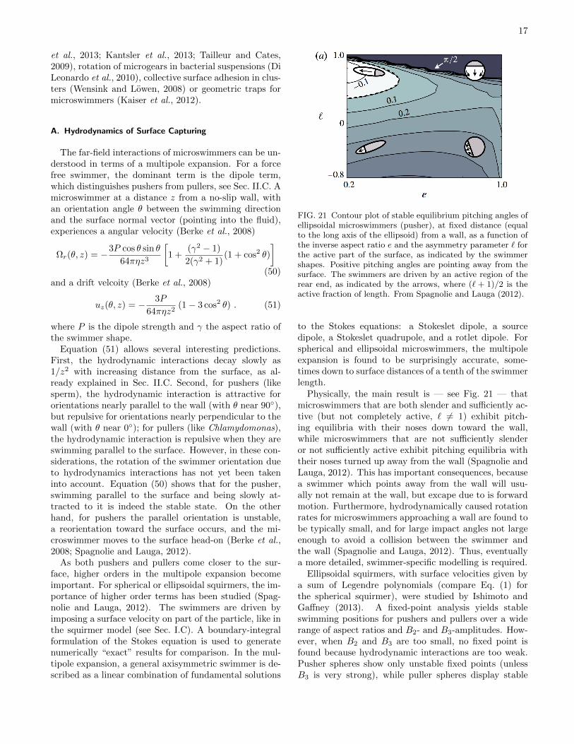
FIG. 21 Contour plot of stable equilibrium pitching angles ofellipsoidal microswimmers (pusher), at fixed distance (equalto the long axis of the ellipsoid) from a wall, as a function ofthe inverse aspect ratio e and the asymmetry parameter ` forthe active part of the surface, as indicated by the swimmershapes. Positive pitching angles are pointing away from thesurface. The swimmers are driven by an active region of therear end, as indicated by the arrows, where (` + 1)/2 is theactive fraction of length. From Spagnolie and Lauga (2012).
to the Stokes equations: a Stokeslet dipole, a sourcedipole, a Stokeslet quadrupole, and a rotlet dipole. Forspherical and ellipsoidal microswimmers, the multipoleexpansion is found to be surprisingly accurate, some-times down to surface distances of a tenth of the swimmerlength.
Physically, the main result is — see Fig. 21 — thatmicroswimmers that are both slender and sufficiently ac-tive (but not completely active, ` 6= 1) exhibit pitch-ing equilibria with their noses down toward the wall,while microswimmers that are not sufficiently slenderor not sufficiently active exhibit pitching equilibria withtheir noses turned up away from the wall (Spagnolie andLauga, 2012). This has important consequences, becausea swimmer which points away from the wall will usu-ally not remain at the wall, but excape due to is forwardmotion. Furthermore, hydrodynamically caused rotationrates for microswimmers approaching a wall are found tobe typically small, and for large impact angles not largeenough to avoid a collision between the swimmer andthe wall (Spagnolie and Lauga, 2012). Thus, eventuallya more detailed, swimmer-specific modelling is required.
Ellipsoidal squirmers, with surface velocities given bya sum of Legendre polynomials (compare Eq. (1) forthe spherical squirmer), were studied by Ishimoto andGaffney (2013). A fixed-point analysis yields stableswimming positions for pushers and pullers over a widerange of aspect ratios and B2- and B3-amplitudes. How-ever, when B2 and B3 are too small, no fixed point isfound because hydrodynamic interactions are too weak.Pusher spheres show only unstable fixed points (unlessB3 is very strong), while puller spheres display stable
18
fixed points with a swimmer orientation toward the wall.As the aspect ratio increases, puller trajectories at a fixeddistance from the wall become more unstable, and con-versely pushers become stable. Pushers at a fixed pointhave an orientation away from the wall. Some of the un-stable fixed points are found to be surrounded by stablelimit cycles, which correspond to swimmers which changetheir distance from the wall periodically. For an elon-gated puller with aspect ratio a = 2, the distance fromthe wall is predicted to vary between its size and threetimes its size Ishimoto and Gaffney (2013). Furthermore,it is predicted that a change of boundary conditions fromno-slip to slip significantly changes the location and char-acteristics of fixed points, and thereby of the swimmingbehavior near surfaces.
Another important aspect is the competition of hydro-dynamic interactions with rotational diffusion (Drescheret al., 2011; Elgeti and Gompper, 2009). When a pusheris deviating by a (small) angle δφ from parallel align-ment with the wall due to rotational diffusion, then —in the far-field approximation — it takes a time of ordertr = δφ/Ωr (see Eq. (50)) to become aligned again dueto hydrodynamic interactions. During this time, it canswim a distance ∆z1 = v0 sin(δφ)tr away from the walldue to self-propulsion, but drifts toward the wall by a dis-tance ∆z1 = uztr due to hydrodynamic interactions, seeEq. (51). This implies that for δφ & (r0/z)
2, the effectiveswimmer velocity points away from the wall, where r0 isthe swimmer size and z is the distance from the wall, andin the time interval tr travels a distance ∆z ∼ (z3/r20)δφ.For distances z a few times r0, this implies ∆z ∼ z. Thus,for small swimming velocities and large angular fluctua-tions, a microswimmer is expected to exhibit also largefluctuations in its distance from a wall. Here, the impor-tance of orientational fluctuations can be quantified bythe orientational Peclet number Peφ(z) = Ωr(z)/DR. Ofcourse, very close to a wall, hydrodynamic interactionsbecome more important, but also the dipole approxima-tion breaks down.
A similar conclusion was reached by Drescher et al.(2011), who considered cell-cell scattering. By estimatingthe mean-square angular change of orientation in cell-cellencounters, both due to hydrodynamic interactions andto rotational diffusion, Drescher et al. (2011) estimated ahydrodynamic horizon rH , beyond which hydrodynamicinteractions become irrelevant. For non-tumbling E. coli,rH is found to be comparable to the length of the cellbody, about 3µm. However, other effects like flagellarinteractions would become important at such short dis-tances. Of course, rotational diffusion becomes less im-portant with increasing microswimmer size.
B. Propulsion-Induced Surfaces Accumulation
Hydrodynamics is not the only mechanism that can ex-plain accumulation of swimmers at surfaces. Indeed, ithas to be realized that any self-propelled particle in con-finement will eventually encounter a surface. Without areorientation, the particle will just stay there. Therefore,rotational diffusion is required to induce a detachmentfrom the wall. In order to elucidate this adhesion mecha-nism, and how noise drives a self-propelled particle awayfrom a wall, it is interesting to consider the behavior of“Brownian” rods — in the absence of any hydrodynamicinteractions. In this case, excluded-volume interactionsfavor parallel orientation near the wall, while the noiseleads to fluctuations of the rod orientation and therebyan effective repulsion from the wall. The competitionof these two effects gives rise to an interesting adsorptionbehavior (Elgeti and Gompper, 2009; Li and Tang, 2009).
Results of Brownian dynamics simulations (Elgeti andGompper, 2009) are shown in Fig. 22. While passive rodsare depleted from the surface (because their entropy isreduced near the surface due to restricted orientationalfluctuations), active rods show an increased probabilitydensity near the surface, which grows with increasingpropulsion force ft, see Fig. 22(left). In addition, thesurface accumulation of the rods strongly depends onthe rod length l. The surface excess — the integratedprobability density to find a rod near the surface rela-tive to a uniform bulk density distribution — is shown inFig. 22(right). The results show (i) that short rods showlittle or no surface aggregation for any propelling force,and (ii) that the surface excess initially increases with in-creasing ft and L, but then saturates and becomes nearlyindependent of ft and L for large propulsion forces androd lengths (Elgeti and Gompper, 2009).
The physical mechanism behind this behavior is as fol-lows. A rod hits the surface at some point in time, be-cause swimming directions in the bulk are randomly dis-tributed. After contact with the wall, it gets reorientedparallel to the wall. Then it moves parallel to the wall,slowly wiggling its trajectory from the wall again, untilit is sufficiently far from the wall that frequent contactsno longer occur.
The surface accumulation of self-propelled rods can beunderstood more quantitatively by exploiting an anal-ogy of the trajectories of rod-like microswimmers withthe conformations of semi-flexible polymers. The self-propulsion of the rod, combined with rotational diffusionleads to a trajectory with a persistence length
ξp ∼ v/DR ∼ ηvl3/kBT (52)
similar to a fluctuating polymer. The key difference ishowever, that the path is directed, and thus not forward-backward symmetric like a polymer. The trajectory ofa rod colliding with the wall displays a sharp kink, im-possible for a semi-flexible polymer. Leaving the wall, on
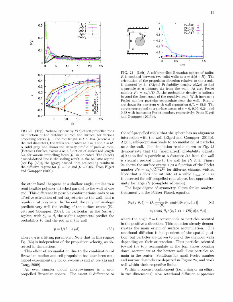
19
FIG. 22 (Top) Probability density P (z) of self-propelled rodsas function of the distance z from the surface, for variouspropelling forces ft. The rod length is l ' 10a (where a isthe rod diameter), the walls are located at z = 0 and z = 5l.A solid gray line shows the density profile of passive rods.(Bottom) Surface excess s as a function of scaled rod lengthl/a, for various propelling forces ft, as indicated. The (black)dashed-dotted line is the scaling result in the ballistic regime(see Eq. (53)), the (gray) dashed lines are scaling results inthe diffusive regime for ft = 0.5 and ft = 0.05. From Elgetiand Gompper (2009).
the other hand, happens at a shallow angle, similar to asemi-flexible polymer attached parallel to the wall at oneend. This difference in possible conformations leads to aneffective attraction of rod-trajectories to the wall, and arepulsion of polymers. In the end, the polymer analogypredicts very well the scaling of the surface excess (El-geti and Gompper, 2009). In particular, in the ballisticregime, with ξp d, the scaling arguments predict theprobability to find the rod near the wall
p = l/(l + aBd), (53)
where aB is a fitting parameter. Note that in this regimeEq. (53) is independent of the propulsion velocity, as ob-served in simulations.
This effect of accumulation due to the combination ofBrownian motion and self-propulsion has later been con-firmed experimentally for C. crecentus and E. coli (Li andTang, 2009).
An even simpler model microswimmer is a self-propelled Brownian sphere. The essential difference to
0
0.2
0.4
0.6
0.8
1
1.2
0 0.5 1 1.5
ρ
∆z/λ
Pe=0.00
Pe=1.46
Pe=7.30
Pe=14.6
R0
z
−d
+dR
R
θ
FIG. 23 (Left) A self-propelled Brownian sphere of radiusR is confined between two solid walls at z = ±(d + R). Theorientation of the propulsion direction relative to the z-axis,is denoted by θ. (Right) Probability density ρ(∆z) to finda particle at a distance ∆z from the wall. At zero Pecletnumber Pe = v0/
√DrD, the probability density is uniform
beyond the short range of the repulsive wall. With increasingPeclet number particles accumulate near the wall. Resultsare shown for a system with wall separation d/λ = 15.6. Thecurves correspond to a surface excess of s = 0, 0.05, 0.24, and0.38 with increasing Peclet number, respectively. From Elgetiand Gompper (2013b).
the self-propelled rod is that the sphere has no alignmentinteraction with the wall (Elgeti and Gompper, 2013b).Again, self-propulsion leads to accumulation of particlesnear the wall. The simulation results shown in Fig. 23demonstrate that the (normalized) probability densityρ(∆z) to find a particle at a distance ∆z from the wallis strongly peaked close to the wall for Pe & 5. Figure24 shows the surface excess s as a function of the Pecletnumber Pe = v0/
√DRDT for different channel widths.
Note that s does not saturate at a value smax < 1 asis observed for self-propelled rods above, but approachesunity for large Pe (complete adhesion).
The large degree of symmetry allows for an analytictreatment via the Fokker-Planck equation
∂tρ(z, θ, t) = Dr1
sin(θ)∂θ [sin(θ)∂θρ(z, θ, t)] (54)
− v0 cos(θ)∂zρ(z, θ, t) +D∂2zρ(z, θ, t),
where the angle θ = 0 corresponds to particles orientedin the positive z-direction. This equation already demon-strates the main origin of surface accumulation. Therotational diffusion is independent of the spatial posi-tion, but particles are driven to one of the chamber wallsdepending on their orientation. Thus particles orientedtoward the top, accumulate at the top, those pointingdown, accumulate at the bottom wall. Less particles re-main in the center. Solutions for small Peclet numberand narrow channels are depicted in Figure 24, and workwell within their respective limits.
Within a concave confinement (i.e. a ring or an ellipsein two dimensions), slow rotational diffusion suppresses
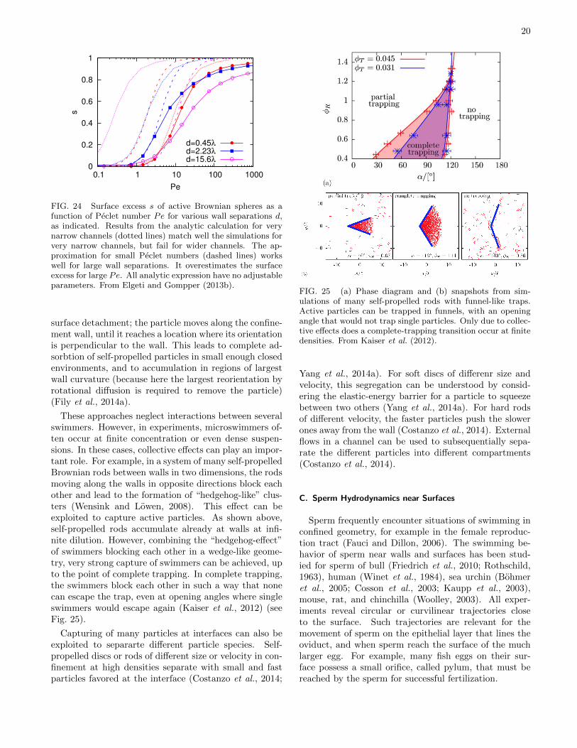
20
0
0.2
0.4
0.6
0.8
1
0.1 1 10 100 1000
s
Pe
d=0.45λ
d=2.23λ
d=15.6λ
FIG. 24 Surface excess s of active Brownian spheres as afunction of Peclet number Pe for various wall separations d,as indicated. Results from the analytic calculation for verynarrow channels (dotted lines) match well the simulations forvery narrow channels, but fail for wider channels. The ap-proximation for small Peclet numbers (dashed lines) workswell for large wall separations. It overestimates the surfaceexcess for large Pe. All analytic expression have no adjustableparameters. From Elgeti and Gompper (2013b).
surface detachment; the particle moves along the confine-ment wall, until it reaches a location where its orientationis perpendicular to the wall. This leads to complete ad-sorbtion of self-propelled particles in small enough closedenvironments, and to accumulation in regions of largestwall curvature (because here the largest reorientation byrotational diffusion is required to remove the particle)(Fily et al., 2014a).
These approaches neglect interactions between severalswimmers. However, in experiments, microswimmers of-ten occur at finite concentration or even dense suspen-sions. In these cases, collective effects can play an impor-tant role. For example, in a system of many self-propelledBrownian rods between walls in two dimensions, the rodsmoving along the walls in opposite directions block eachother and lead to the formation of “hedgehog-like” clus-ters (Wensink and Lowen, 2008). This effect can beexploited to capture active particles. As shown above,self-propelled rods accumulate already at walls at infi-nite dilution. However, combining the “hedgehog-effect”of swimmers blocking each other in a wedge-like geome-try, very strong capture of swimmers can be achieved, upto the point of complete trapping. In complete trapping,the swimmers block each other in such a way that nonecan escape the trap, even at opening angles where singleswimmers would escape again (Kaiser et al., 2012) (seeFig. 25).
Capturing of many particles at interfaces can also beexploited to separarte different particle species. Self-propelled discs or rods of different size or velocity in con-finement at high densities separate with small and fastparticles favored at the interface (Costanzo et al., 2014;
FIG. 25 (a) Phase diagram and (b) snapshots from sim-ulations of many self-propelled rods with funnel-like traps.Active particles can be trapped in funnels, with an openingangle that would not trap single particles. Only due to collec-tive effects does a complete-trapping transition occur at finitedensities. From Kaiser et al. (2012).
Yang et al., 2014a). For soft discs of differenr size andvelocity, this segregation can be understood by consid-ering the elastic-energy barrier for a particle to squeezebetween two others (Yang et al., 2014a). For hard rodsof different velocity, the faster particles push the slowerones away from the wall (Costanzo et al., 2014). Externalflows in a channel can be used to subsequentially sepa-rate the different particles into different compartments(Costanzo et al., 2014).
C. Sperm Hydrodynamics near Surfaces
Sperm frequently encounter situations of swimming inconfined geometry, for example in the female reproduc-tion tract (Fauci and Dillon, 2006). The swimming be-havior of sperm near walls and surfaces has been stud-ied for sperm of bull (Friedrich et al., 2010; Rothschild,1963), human (Winet et al., 1984), sea urchin (Bohmeret al., 2005; Cosson et al., 2003; Kaupp et al., 2003),mouse, rat, and chinchilla (Woolley, 2003). All exper-iments reveal circular or curvilinear trajectories closeto the surface. Such trajectories are relevant for themovement of sperm on the epithelial layer that lines theoviduct, and when sperm reach the surface of the muchlarger egg. For example, many fish eggs on their sur-face possess a small orifice, called pylum, that must bereached by the sperm for successful fertilization.
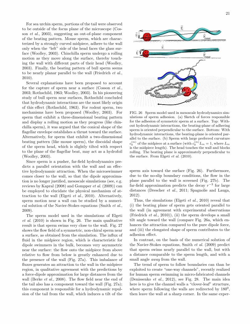
21
For sea urchin sperm, portions of the tail were observedto be outside of the focus plane of the microscope (Cos-son et al., 2003), suggesting an out-of-plane componentof the beating pattern. Mouse sperm, which are charac-terized by a strongly curved midpiece, adhere to the wallonly when the “left” side of the head faces the glass sur-face (Woolley, 2003). Chinchilla sperm undergo a rollingmotion as they move along the surface, thereby touch-ing the wall with different parts of their head (Woolley,2003). Finally, the beating pattern of bull sperm seemsto be nearly planar parallel to the wall (Friedrich et al.,2010).
Several explanations have been proposed to accountfor the capture of sperm near a surface (Cosson et al.,2003; Rothschild, 1963; Woolley, 2003). In his pioneeringstudy of bull sperm near surfaces, Rothschild concludedthat hydrodynamic interactions are the most likely originof this effect (Rothschild, 1963). For rodent sperm, twomechanisms have been proposed (Woolley, 2003). Forsperm that exhibit a three-dimensional beating patternand display a rolling motion as they progress (like chin-chilla sperm), it was argued that the conical shape of theflagellar envelope establishes a thrust toward the surface.Alternatively, for sperm that exhibit a two-dimensionalbeating pattern (like mouse sperm), the discoidal shapeof the sperm head, which is slightly tilted with respectto the plane of the flagellar beat, may act as a hydrofoil(Woolley, 2003).
Since sperm is a pusher, far-field hydrodynamics pre-dicts a parallel orientation with the wall and an effec-tive hydrodynamic attraction. When the microswimmercomes closer to the wall, so that the dipole approxima-tion is no longer justified, mesoscale simulations (see, e.g.reviews by Kapral (2008) and Gompper et al. (2009)) canbe employed to elucidate the physical mechanism of at-traction to the wall (Elgeti et al., 2010). Alternatively,sperm motion near a wall can be studied by a numeri-cal solution of the Navier-Stokes equations (Smith et al.,2009).
The sperm model used in the simulations of Elgetiet al. (2010) is shown in Fig. 26. The main qualitativeresult is that sperm swims very close to the wall. Fig. 27shows the flow field of a symmetric, non-chiral sperm neara surface, as obtained from the simulation. The influx offluid in the midpiece region, which is characteristic fordipole swimmers in the bulk, becomes very asymmetricnear the surface: the flow onto the midpiece from aboverelative to flow from below is greatly enhanced due tothe presence of the wall (Fig. 27a). This imbalance offluxes generates an attraction to the wall in the midpieceregion, in qualitative agreement with the predictions bya force-dipole approximation for large distances from thewall (Berke et al., 2008). The flow field near the end ofthe tail also has a component toward the wall (Fig. 27a);this component is responsible for a hydrodynamic repul-sion of the tail from the wall, which induces a tilt of the
FIG. 26 Sperm model used in mesoscale hydrodynamics sim-ulations of sperm adhesion. (a) Sketch of forces responsiblefor the adhesion of symmetric sperm at a surface. Top: With-out hydrodynamic interactions, the beating-plane of adheringsperm is oriented perpendicular to the surface. Bottom: Withhydrodynamic interactions, the beating-plane is oriented par-allel to the surface. (b) Sperm with large preferred curvature
c(m)0 of the midpiece at a surface (with c
(m)0 Lm = 1, where Lm
is the midpiece length). The head touches the wall and blocksrolling. The beating plane is approximately perpendicular tothe surface. From Elgeti et al. (2010).
sperm axis toward the surface (Fig. 26). Furthermore,due to the no-slip boundary conditions, the flow in theplane parallel to the wall is screened (Fig. 27b). Thefar-field approximation predicts the decay r−3 for largedistances (Drescher et al., 2011; Spagnolie and Lauga,2012).
Thus, the simulations (Elgeti et al., 2010) reveal that(i) the beating plane of sperm gets oriented parallel tothe wall (in agreement with experimental observations(Friedrich et al., 2010)), (ii) the sperm develops a smalltilt angle toward the wall (compare Fig. 26a, which en-hances the attraction compared to the pure dipole force,and (iii) the elongated shape of sperm contributes to theadhesion effect.
In contrast, on the basis of the numerical solution ofthe Navier-Stokes equations, Smith et al. (2009) predictthat sperm swims nearly parallel to the wall, but witha distance comparable to the sperm length, and with asmall angle away from the wall.
The trend of sperm to follow boundaries can than beexploited to create “one-way channels”, recently realizedfor human sperm swimming in micro-fabricated channels(Denissenko et al., 2012), see Fig. 28. The main ideahere is to give the channel walls a “clover-leaf” structure,where sperm following the walls are redirected by 1800,then leave the wall at a sharp corner. In the same exper-
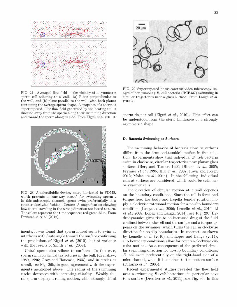
22
FIG. 27 Averaged flow field in the vicinity of a symmetricsperm cell adhering to a wall. (a) Plane perpendicular tothe wall, and (b) plane parallel to the wall, with both planescontaining the average sperm shape. A snapshot of a sperm issuperimposed. The flow field generated by the beating tail isdirected away from the sperm along their swimming directionand toward the sperm along its side. From Elgeti et al. (2010).
FIG. 28 A microfluidic device, micro-fabricated in PDMS,which presents a “one-way street” for swimming sperm .In this anisotropic channels sperm swim preferentially in acounter-clockwise fashion. Center: A magnification showinghow sperm traveling in the wrong direction are forced to turn.The colors represent the time sequences red-green-blue. FromDenissenko et al. (2012).
iments, it was found that sperm indeed seem to swim atinterfaces with finite angle toward the surface confirmingthe predictions of Elgeti et al. (2010), but at variancewith the results of Smith et al. (2009).
Chiral sperm also adhere to surfaces. In this case,sperm swim on helical trajectories in the bulk (Crenshaw,1989, 1996; Gray and Hancock, 1955), and in circles ata wall, see Fig. 26b, in good agreement with the exper-iments mentioned above. The radius of the swimmingcircles decreases with increasing chirality. Weakly chi-ral sperm display a rolling motion, while strongly chiral
FIG. 29 Superimposed phase-contrast video microscopy im-ages of non-tumbling E. coli bacteria (HCB437) swimming incircular trajectories near a glass surface. From Lauga et al.(2006).
sperm do not roll (Elgeti et al., 2010). This effect canbe understood from the steric hindrance of a stronglyasymmetric shape.
D. Bacteria Swimming at Surfaces
The swimming behavior of bacteria close to surfacesdiffers from the “run-and-tumble” motion in free solu-tion. Experiments show that individual E. coli bacteriaswim in clockwise, circular trajectories near planar glasssurfaces (Berg and Turner, 1990; DiLuzio et al., 2005;Frymier et al., 1995; Hill et al., 2007; Kaya and Koser,2012; Molaei et al., 2014). In the following, individualcells at surfaces are considered, which could be swimmeror swarmer cells.
The direction of circular motion at a wall dependson the boundary conditions. Since the cell is force andtorque free, the body and flagella bundle rotation im-ply a clockwise rotational motion for a no-slip boundarycondition (Lauga et al., 2006; Lemelle et al., 2010; Liet al., 2008; Lopez and Lauga, 2014), see Fig. 29. Hy-drodynamics gives rise to an increased drag of the fluidconfined between the cell and the surface and a torque ap-pears on the swimmer, which turns the cell in clockwisedirection for no-slip boundaries. In contrast, as shownby Lemelle et al. (2010) and Lopez and Lauga (2014),slip boundary conditions allow for counter-clockwise cir-cular motion. As a consequence of the preferred circu-lar swimming direction for no-slip boundary conditions,E. coli swim preferentially on the right-hand side of amicrochannel, when it is confined to the bottom surface(DiLuzio et al., 2005).
Recent experimental studies revealed the flow fieldnear a swimming E. coli bacterium, in particular nextto a surface (Drescher et al., 2011), see Fig. 30. In this
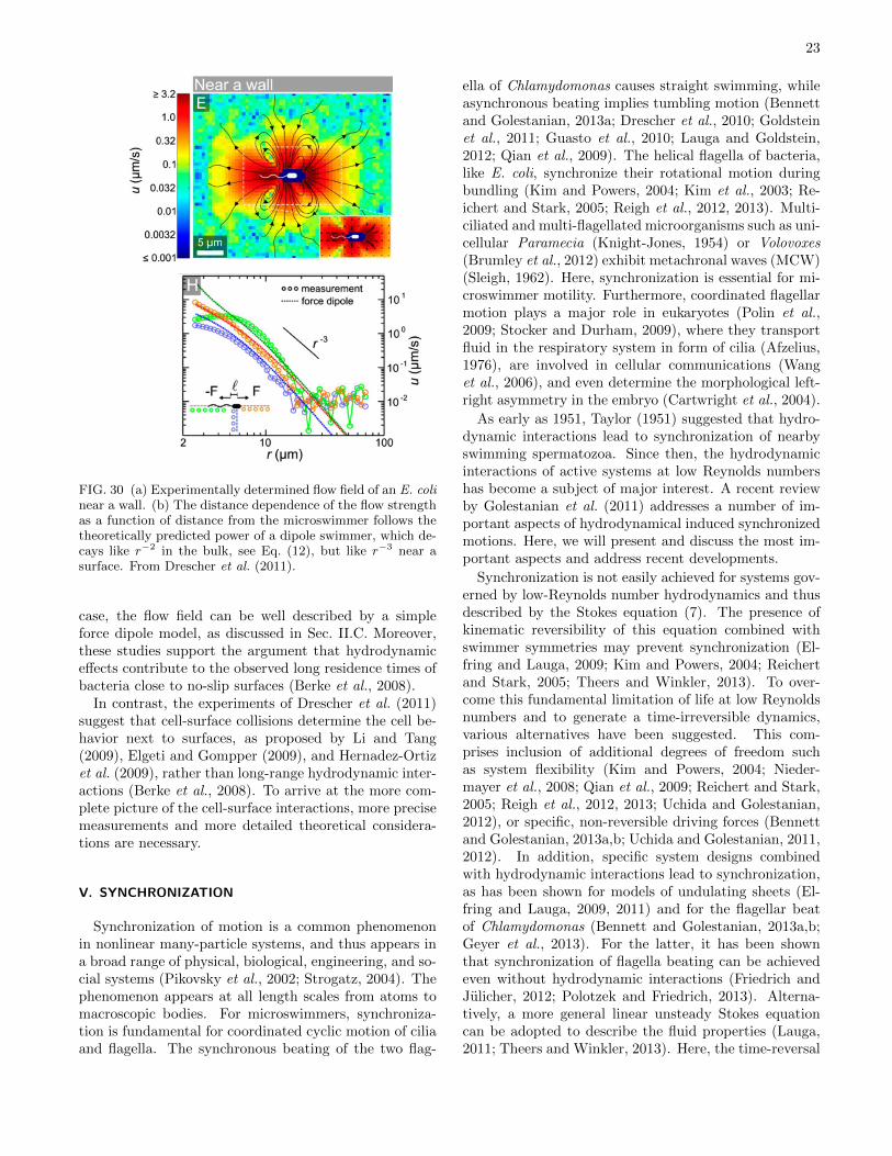
23
FIG. 30 (a) Experimentally determined flow field of an E. colinear a wall. (b) The distance dependence of the flow strengthas a function of distance from the microswimmer follows thetheoretically predicted power of a dipole swimmer, which de-cays like r−2 in the bulk, see Eq. (12), but like r−3 near asurface. From Drescher et al. (2011).
case, the flow field can be well described by a simpleforce dipole model, as discussed in Sec. II.C. Moreover,these studies support the argument that hydrodynamiceffects contribute to the observed long residence times ofbacteria close to no-slip surfaces (Berke et al., 2008).
In contrast, the experiments of Drescher et al. (2011)suggest that cell-surface collisions determine the cell be-havior next to surfaces, as proposed by Li and Tang(2009), Elgeti and Gompper (2009), and Hernadez-Ortizet al. (2009), rather than long-range hydrodynamic inter-actions (Berke et al., 2008). To arrive at the more com-plete picture of the cell-surface interactions, more precisemeasurements and more detailed theoretical considera-tions are necessary.
V. SYNCHRONIZATION
Synchronization of motion is a common phenomenonin nonlinear many-particle systems, and thus appears ina broad range of physical, biological, engineering, and so-cial systems (Pikovsky et al., 2002; Strogatz, 2004). Thephenomenon appears at all length scales from atoms tomacroscopic bodies. For microswimmers, synchroniza-tion is fundamental for coordinated cyclic motion of ciliaand flagella. The synchronous beating of the two flag-
ella of Chlamydomonas causes straight swimming, whileasynchronous beating implies tumbling motion (Bennettand Golestanian, 2013a; Drescher et al., 2010; Goldsteinet al., 2011; Guasto et al., 2010; Lauga and Goldstein,2012; Qian et al., 2009). The helical flagella of bacteria,like E. coli, synchronize their rotational motion duringbundling (Kim and Powers, 2004; Kim et al., 2003; Re-ichert and Stark, 2005; Reigh et al., 2012, 2013). Multi-ciliated and multi-flagellated microorganisms such as uni-cellular Paramecia (Knight-Jones, 1954) or Volovoxes(Brumley et al., 2012) exhibit metachronal waves (MCW)(Sleigh, 1962). Here, synchronization is essential for mi-croswimmer motility. Furthermore, coordinated flagellarmotion plays a major role in eukaryotes (Polin et al.,2009; Stocker and Durham, 2009), where they transportfluid in the respiratory system in form of cilia (Afzelius,1976), are involved in cellular communications (Wanget al., 2006), and even determine the morphological left-right asymmetry in the embryo (Cartwright et al., 2004).
As early as 1951, Taylor (1951) suggested that hydro-dynamic interactions lead to synchronization of nearbyswimming spermatozoa. Since then, the hydrodynamicinteractions of active systems at low Reynolds numbershas become a subject of major interest. A recent reviewby Golestanian et al. (2011) addresses a number of im-portant aspects of hydrodynamical induced synchronizedmotions. Here, we will present and discuss the most im-portant aspects and address recent developments.
Synchronization is not easily achieved for systems gov-erned by low-Reynolds number hydrodynamics and thusdescribed by the Stokes equation (7). The presence ofkinematic reversibility of this equation combined withswimmer symmetries may prevent synchronization (El-fring and Lauga, 2009; Kim and Powers, 2004; Reichertand Stark, 2005; Theers and Winkler, 2013). To over-come this fundamental limitation of life at low Reynoldsnumbers and to generate a time-irreversible dynamics,various alternatives have been suggested. This com-prises inclusion of additional degrees of freedom suchas system flexibility (Kim and Powers, 2004; Nieder-mayer et al., 2008; Qian et al., 2009; Reichert and Stark,2005; Reigh et al., 2012, 2013; Uchida and Golestanian,2012), or specific, non-reversible driving forces (Bennettand Golestanian, 2013a,b; Uchida and Golestanian, 2011,2012). In addition, specific system designs combinedwith hydrodynamic interactions lead to synchronization,as has been shown for models of undulating sheets (El-fring and Lauga, 2009, 2011) and for the flagellar beatof Chlamydomonas (Bennett and Golestanian, 2013a,b;Geyer et al., 2013). For the latter, it has been shownthat synchronization of flagella beating can be achievedeven without hydrodynamic interactions (Friedrich andJulicher, 2012; Polotzek and Friedrich, 2013). Alterna-tively, a more general linear unsteady Stokes equationcan be adopted to describe the fluid properties (Lauga,2011; Theers and Winkler, 2013). Here, the time-reversal
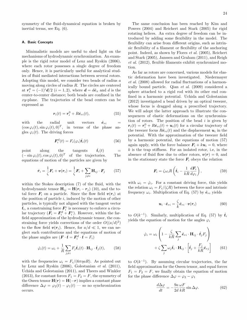
24
symmetry of the fluid-dynamical equation is broken byinertial terms, see Eq. (6).
A. Basic Concepts
Minimalistic models are useful to shed light on themechanisms of hydrodynamic synchronization. An exam-ple is the rigid rotor model of Lenz and Ryskin (2006),where each rotor possesses a single degree of freedomonly. Hence, it is particularly useful for analytical stud-ies of fluid mediated interactions between several rotors.Adopting this model, we consider two beads of radius amoving along circles of radius R. The circles are centeredat r0i = (−1)id/2 (i = 1, 2), where d = dex and d is thecenter-to-center distance; both beads are confined in thexy-plane. The trajectories of the bead centers can beexpressed as
ri(t) = r0i +Reri(t), (55)
with the radial unit vectors eri =(cosϕi(t), sinϕi(t), 0)T , in terms of the phase an-gles ϕi(t). The driving forces
F di (t) = Fi(ϕi)ti(t) (56)
point along the tangents ti(t) =(− sinϕi(t), cosϕi(t), 0)T of the trajectories. Theequations of motion of the particles are given by
ri =1
ζFi + v(ri) =
1
ζFi +
∑j 6=i
Hij · Fj (57)
within the Stokes description (7) of the fluid, with thehydrodynamic tensor Hij = H(ri− rj) (10), and the to-tal force Fi on a particle. Since the flow field v(ri) atthe position of particle i, induced by the motion of otherparticles, is typically not aligned with the tangent vectorti, a constraining force F ci is necessary to enforce a circu-lar trajectory (Fi = F di + F ci ). However, within the far-field approximation of the hydrodynamic tensor, the con-straining force yields corrections of the order O((a/d)2)to the flow field v(ri). Hence, for a/d 1, we can ne-glect such contributions and the equations of motion ofthe phase angles are (F · t = F di · t = Fi)
ϕi(t) = ωi +1
R
∑j 6=i
Fj ti(t) ·Hij · tj(t), (58)
with the frequencies ωi = Fi/(6πηaR). As pointed outby Lenz and Ryskin (2006), Golestanian et al. (2011),Uchida and Golestanian (2011), and Theers and Winkler(2013), for constant forces F1 = F2 = F , the symmetry ofthe Oseen tensor H(r) = H(−r) implies a constant phasedifference ∆ϕ = ϕ2(t) − ϕ1(t) — so no synchronizationoccurs.
The same conclusion has been reached by Kim andPowers (2004) and Reichert and Stark (2005) for rigidrotating helices. An extra degree of freedom can be in-troduced by adding some flexibility in the model. Theflexibility can arise from different origins, such as intrin-sic flexibility of a filament or flexibility of the anchoringpoint. Indeed, as shown by Flores et al. (2005), Reichertand Stark (2005), Janssen and Graham (2011), and Reighet al. (2012), flexible filaments exhibit synchronized mo-tion.
As far as rotors are concerned, various models for elas-tic deformation have been investigated. Niedermayeret al. (2008) allowed for radial fluctuations of a harmon-ically bound particle. Qian et al. (2009) considered asphere attached to a rigid rod with its other end con-fined in a harmonic potential. Uchida and Golestanian(2012) investigated a bead driven by an optical tweezer,whose focus is dragged along a prescribed trajectory.We will adopt the latter approach to illustrate the con-sequences of elastic deformations on the synchroniza-tion of rotors. The position of the bead i is given byri(t) = r0i + Reri(t) + ui(t) for a circular trajectory ofthe tweezer focus Reri(t) and the displacement ui in thepotential. With the approximation of the tweezer fieldby a harmonic potential, the equations of motion (57)again apply, with the force balance Fi + kui = 0, wherek is the trap stiffness. For an isolated rotor, i.e, in theabsence of fluid flow due to other rotors, v(r) = 0, andin the stationary state the force Fi obeys the relation
Fi = ζωiR
(ti −
1
kR
dFidϕi
), (59)
with ωi = ϕi. For a constant driving force, this yieldsthe relation ωi = Fi/(ζR) between the force and intrinsicfrequency ωi. Multiplication of Eq. (57) by eri yields
ui · eri =ζ
keri · v(ri) (60)
to O(k−1). Similarly, multiplication of Eq. (57) by tiyields the equation of motion for the angles ϕi
ϕi = ωi
1− ζ
kR
∑j
eri ·Hij · tjFj
+ ζ
∑j
ωj ti ·Hij ·[tj +
FjkRerj
](61)
to O(k−1). By assuming circular trajectories, the farfield approximation for the Oseen tensor, and equal forcesF1 = F2 = F , we finally obtain the equation of motionfor the phase difference ∆ϕ = ϕ2 − ϕ1
d∆ϕ
dt= −9a
2d
ωF
kRsin ∆ϕ. (62)
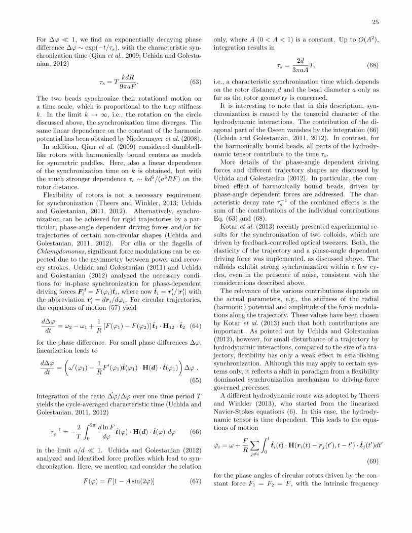
25
For ∆ϕ 1, we find an exponentially decaying phasedifference ∆ϕ ∼ exp(−t/τs), with the characteristic syn-chronization time (Qian et al., 2009; Uchida and Golesta-nian, 2012)
τs = TkdR
9πaF. (63)
The two beads synchronize their rotational motion ona time scale, which is proportional to the trap stiffnessk. In the limit k → ∞, i.e., the rotation on the circlediscussed above, the synchronization time diverges. Thesame linear dependence on the constant of the harmonicpotential has been obtained by Niedermayer et al. (2008).
In addition, Qian et al. (2009) considered dumbbell-like rotors with harmonically bound centers as modelsfor symmetric paddles. Here, also a linear dependenceof the synchronization time on k is obtained, but withthe much stronger dependence τs ∼ kd5/(a3RF ) on therotor distance.
Flexibility of rotors is not a necessary requirementfor synchronization (Theers and Winkler, 2013; Uchidaand Golestanian, 2011, 2012). Alternatively, synchro-nization can be achieved for rigid trajectories by a par-ticular, phase-angle dependent driving forces and/or fortrajectories of certain non-circular shapes (Uchida andGolestanian, 2011, 2012). For cilia or the flagella ofChlamydomonas, significant force modulations can be ex-pected due to the asymmetry between power and recov-ery strokes. Uchida and Golestanian (2011) and Uchidaand Golestanian (2012) analyzed the necessary condi-tions for in-phase synchronization for phase-dependentdriving forces F di = F (ϕi)ti, where now ti = r′i/|r′i| withthe abbreviation r′i = dri/dϕi. For circular trajectories,the equations of motion (57) yield
d∆ϕ
dt= ω2 − ω1 +
1
R[F (ϕ1)− F (ϕ2)] t1 ·H12 · t2 (64)
for the phase difference. For small phase differences ∆ϕ,linearization leads to
d∆ϕ
dt=
(ω′(ϕ1)− 1
RF ′(ϕ1)t(ϕ1) ·H(d) · t(ϕ1)
)∆ϕ .
(65)
Integration of the ratio ∆ϕ/∆ϕ over one time period Tyields the cycle-averaged characteristic time (Uchida andGolestanian, 2011, 2012)
τ−1s = − 2
T
∫ 2π
0
d lnF
dϕt(ϕ) ·H(d) · t(ϕ) dϕ (66)
in the limit a/d 1. Uchida and Golestanian (2012)analyzed and identified force profiles which lead to syn-chronization. Here, we mention and consider the relation
F (ϕ) = F [1−A sin(2ϕ)] (67)
only, where A (0 < A < 1) is a constant. Up to O(A2),integration results in
τs =2d
3πaAT, (68)
i.e., a characteristic synchronization time which dependson the rotor distance d and the bead diameter a only asfar as the rotor geometry is concerned.
It is interesting to note that in this description, syn-chronization is caused by the tensorial character of thehydrodynamic interactions. The contribution of the di-agonal part of the Oseen vanishes by the integration (66)(Uchida and Golestanian, 2011, 2012). In contrast, forthe harmonically bound beads, all parts of the hydrody-namic tensor contribute to the time τs.
More details of the phase-angle dependent drivingforces and different trajectory shapes are discussed byUchida and Golestanian (2012). In particular, the com-bined effect of harmonically bound beads, driven byphase-angle dependent forces are addressed. The char-acteristic decay rate τ−1s of the combined effects is thesum of the contributions of the individual contributionsEq. (63) and (68).
Kotar et al. (2013) recently presented experimental re-sults for the synchronization of two colloids, which aredriven by feedback-controlled optical tweezers. Both, theelasticity of the trajectory and a phase-angle dependentdriving force was implemented, as discussed above. Thecolloids exhibit strong synchronization within a few cy-cles, even in the presence of noise, consistent with theconsiderations described above.
The relevance of the various contributions depends onthe actual parameters, e.g., the stiffness of the radial(harmonic) potential and amplitude of the force modula-tions along the trajectory. These values have been chosenby Kotar et al. (2013) such that both contributions areimportant. As pointed out by Uchida and Golestanian(2012), however, for small disturbance of a trajectory byhydrodynamic interactions, compared to the size of a tra-jectory, flexibility has only a weak effect in establishingsynchronization. Although this may apply to certain sys-tems only, it reflects a shift in paradigm from a flexibilitydominated synchronization mechanism to driving-forcegoverned processes.
A different hydrodynamic route was adopted by Theersand Winkler (2013), who started from the linearizedNavier-Stokes equations (6). In this case, the hydrody-namic tensor is time dependent. This leads to the equa-tions of motion
ϕi = ω +F
R
∑j 6=i
∫ t
0
ti(t) ·H(ri(t)− rj(t′), t− t′) · tj(t′)dt′
(69)
for the phase angles of circular rotors driven by the con-stant force F1 = F2 = F , with the intrinsic frequency
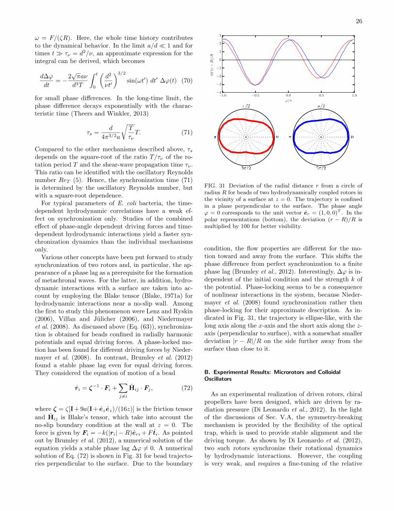
26
ω = F/(ζR). Here, the whole time history contributesto the dynamical behavior. In the limit a/d 1 and fortimes t τν = d2/ν, an approximate expression for theintegral can be derived, which becomes
d∆ϕ
dt= −2
√πaν
d3T
∫ t
0
(d2
νt′
)3/2
sin(ωt′) dt′ ∆ϕ(t) (70)
for small phase differences. In the long-time limit, thephase difference decays exponentially with the charac-teristic time (Theers and Winkler, 2013)
τs =d
4π3/2a
√T
τνT. (71)
Compared to the other mechanisms described above, τsdepends on the square-root of the ratio T/τν of the ro-tation period T and the shear-wave propagation time τν .This ratio can be identified with the oscillatory Reynoldsnumber ReT (5). Hence, the synchronization time (71)is determined by the oscillatory Reynolds number, butwith a square-root dependence.
For typical parameters of E. coli bacteria, the time-dependent hydrodynamic correlations have a weak ef-fect on synchronization only. Studies of the combinedeffect of phase-angle dependent driving forces and time-dependent hydrodynamic interactions yield a faster syn-chronization dynamics than the individual mechanismsonly.
Various other concepts have been put forward to studysynchronization of two rotors and, in particular, the ap-pearance of a phase lag as a prerequisite for the formationof metachronal waves. For the latter, in addition, hydro-dynamic interactions with a surface are taken into ac-count by employing the Blake tensor (Blake, 1971a) forhydrodynamic interactions near a no-slip wall. Amongthe first to study this phenomenon were Lenz and Ryskin(2006), Vilfan and Julicher (2006), and Niedermayeret al. (2008). As discussed above (Eq. (63)), synchroniza-tion is obtained for beads confined in radially harmonicpotentials and equal driving forces. A phase-locked mo-tion has been found for different driving forces by Nieder-mayer et al. (2008). In contrast, Brumley et al. (2012)found a stable phase lag even for equal driving forces.They considered the equation of motion of a bead
ri = ζ−1 · Fi +∑j 6=i
Hij · Fj , (72)
where ζ = ζ[I + 9a(I + ezez)/(16z)] is the friction tensor
and Hij is Blake’s tensor, which take into account theno-slip boundary condition at the wall at z = 0. Theforce is given by Fi = −k(|ri| −R)eri +F ti. As pointedout by Brumley et al. (2012), a numerical solution of theequation yields a stable phase lag ∆ϕ 6= 0. A numericalsolution of Eq. (72) is shown in Fig. 31 for bead trajecto-ries perpendicular to the surface. Due to the boundary
FIG. 31 Deviation of the radial distance r from a circle ofradius R for beads of two hydrodynamically coupled rotors inthe vicinity of a surface at z = 0. The trajectory is confinedin a plane perpendicular to the surface. The phase angleϕ = 0 corresponds to the unit vector er = (1, 0, 0)T . In thepolar representations (bottom), the deviation (r − R)/R ismultiplied by 100 for better visibility.
condition, the flow properties are different for the mo-tion toward and away from the surface. This shifts thephase difference from perfect synchronization to a finitephase lag (Brumley et al., 2012). Interestingly, ∆ϕ is in-dependent of the initial condition and the strength k ofthe potential. Phase-locking seems to be a consequenceof nonlinear interactions in the system, because Nieder-mayer et al. (2008) found synchronization rather thenphase-locking for their approximate description. As in-dicated in Fig. 31, the trajectory is ellipse-like, with thelong axis along the x-axis and the short axis along the z-axis (perpendicular to surface), with a somewhat smallerdeviation |r − R|/R on the side further away from thesurface than close to it.
B. Experimental Results: Microrotors and ColloidalOscillators
As an experimental realization of driven rotors, chiralpropellers have been designed, which are driven by ra-diation pressure (Di Leonardo et al., 2012). In the lightof the discussions of Sec. V.A, the symmetry-breakingmechanism is provided by the flexibility of the opticaltrap, which is used to provide stable alignment and thedriving torque. As shown by Di Leonardo et al. (2012),two such rotors synchronize their rotational dynamicsby hydrodynamic interactions. However, the couplingis very weak, and requires a fine-tuning of the relative
27
torque within a resolution of 10−3. Even then, phaseslips occur, where one of the rotors moves faster thanthe other until a stable phase-locked state may appearagain.
In contrast, strong static correlations are obtainedthrough hydrodynamic interactions between colloidalparticles in rotating energy landscapes (Koumakis andDi Leonardo, 2013). Thereby, the particles are driven pe-riodically around a circle by rotating energy landscapeswith a variable number of minima. In the co-moving, ro-tating frame, a colloid experiences a tilted periodic poten-tial due to the hydrodynamic interaction with the othercolloids. This enhances the probability to overcome thebarriers and allows the different colloids to synchronizetheir rotational motion.
Another strategy to synchronize the dynamics of beadshas been proposed by Gueron and Levit-Gurevich (1998);Gueron et al. (1997). It is based on the difference betweenthe power stroke and the recovery stroke of cilia, or, moregenerally, the presence of two phases with broken sym-metry. The latter concept has been applied to colloidalsystems by Lagomarsino et al. (2003), Kotar et al. (2010),Bruot et al. (2011), Damet et al. (2012), and Lhermer-out et al. (2012). Here, the periodic motion of colloidsis controlled by a configuration-dependent external in-put. Thereby, various properties can be switched fromone state to another upon a particular geometry beingassumed by the colloids. Examples are different effectivedrags (Lagomarsino et al., 2003), or harmonic potentialswith different equilibrium positions (Kotar et al., 2010).The latter can be realized by optical traps. These strate-gies lead to a synchronized motion by hydrodynamic in-teractions (Bruot et al., 2011; Damet et al., 2012; Kotaret al., 2010; Lagomarsino et al., 2003; Lhermerout et al.,2012; Wollin and Stark, 2011).
C. Synchronization of Chlamydomonas Beating
Chlamydomonas with its two beating flagella (seeSec. I.A) has become a model system for experimentalstudies of synchronization (Polotzek and Friedrich, 2013).When the flagella beat synchronously, the alga swimsalong a straight path, while dephasing leads to reorien-tation in a run-and-tumble-like manner (Goldstein et al.,2009; Lauga and Goldstein, 2012; Polin et al., 2009).
The measured flow field of a swimming Chlamy-domonas (Drescher et al., 2010; Guasto et al., 2010) iswell described by three Stokeslets (see Fig. 32). Thisfinding has stimulated a more detailed theoretical mod-eling of Chlamydomonas by three spheres — two spheresdriven on circular orbits, which are mimicking the flagel-lar beat (see Sec. V.A), are linked by a frictionless scaf-fold to a third sphere representing the cell body (Bennettand Golestanian, 2013a,b; Friedrich and Julicher, 2012;Polotzek and Friedrich, 2013).
FIG. 32 Time- and azimuthally-averaged flow field ofChlamydomonas. (a) Streamlines computed from the experi-mentally measured velocity vectors (blue). (b) Streamlines ofthe azimuthally-averaged flow of an assumed three-Stokesletmodel: flagellar thrust is distributed between two Stokesletslocated at the approximate flagellar position (green arrows),whose sum balances drag on the cell body (central red arrow).From Drescher et al. (2010).
The extra translational and overall rotational degreesof freedom, compared to the two degrees of freedom ofthe rotors of the systems of Sec. V.A, combined with non-linearities, give rise to additional features. As shown byBennett and Golestanian (2013b); Friedrich and Julicher(2012); Polotzek and Friedrich (2013), force and momen-tum balance predominantly couple the phases of the ro-tors via translation and rotation of the cell body. Thiscoupling suggest the possibility of flagella synchroniza-tion by local hydrodynamic friction even in the absenceof hydrodynamic interactions. The efficiency of synchro-nization naturally depends on the geometry. It is weakerthan hydrodynamic coupling, when the cell body is muchlarger than the distance between the neighboring flagellaor cilia (Uchida and Golestanian, 2012).
The three-sphere model exhibits a very rich dynami-cal swimming behavior, when a phase-angle dependentdriving force and noise is added. Without hydrodynamicinteractions, the cell just moves forward and backwardand there is no net motion. However, in the presence ofhydrodynamic interactions, which vary in strength dur-ing a beating cycle, the broken symmetry between thepower and recovery stroke leads to a net propulsion (Ben-nett and Golestanian, 2013b). Interestingly, a run-and-tumbling motion is obtained in the presence of noise. Thethreshold-like run-and-tumble behavior displayed by bac-teria like E. coli and believed to be controlled by a sophis-ticated biochemical feedback mechanism, emerges herenaturally from nonlinearities in the propulsion mecha-nism and could be triggered even by white noise. More-over, the same model could describe the experimentallyobserved antiphase beating pattern of a Chlamydomonasmutant (Bennett and Golestanian, 2013b; Leptos et al.,2013).
An experimental and theoretical study of the effect ofthe hydrodynamic coupling on the synchronization of theflagella beating is presented by Geyer et al. (2013). The
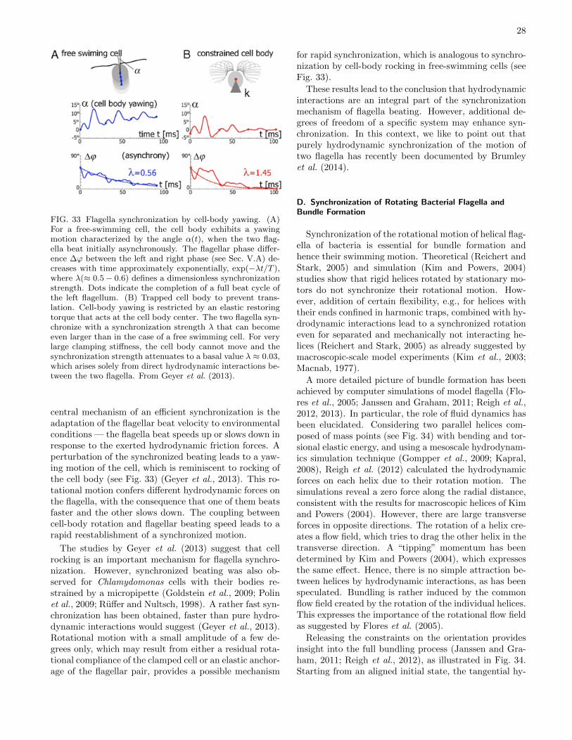
28
FIG. 33 Flagella synchronization by cell-body yawing. (A)For a free-swimming cell, the cell body exhibits a yawingmotion characterized by the angle α(t), when the two flag-ella beat initially asynchronously. The flagellar phase differ-ence ∆ϕ between the left and right phase (see Sec. V.A) de-creases with time approximately exponentially, exp(−λt/T ),where λ(≈ 0.5− 0.6) defines a dimensionless synchronizationstrength. Dots indicate the completion of a full beat cycle ofthe left flagellum. (B) Trapped cell body to prevent trans-lation. Cell-body yawing is restricted by an elastic restoringtorque that acts at the cell body center. The two flagella syn-chronize with a synchronization strength λ that can becomeeven larger than in the case of a free swimming cell. For verylarge clamping stiffness, the cell body cannot move and thesynchronization strength attenuates to a basal value λ ≈ 0.03,which arises solely from direct hydrodynamic interactions be-tween the two flagella. From Geyer et al. (2013).
central mechanism of an efficient synchronization is theadaptation of the flagellar beat velocity to environmentalconditions — the flagella beat speeds up or slows down inresponse to the exerted hydrodynamic friction forces. Aperturbation of the synchronized beating leads to a yaw-ing motion of the cell, which is reminiscent to rocking ofthe cell body (see Fig. 33) (Geyer et al., 2013). This ro-tational motion confers different hydrodynamic forces onthe flagella, with the consequence that one of them beatsfaster and the other slows down. The coupling betweencell-body rotation and flagellar beating speed leads to arapid reestablishment of a synchronized motion.
The studies by Geyer et al. (2013) suggest that cellrocking is an important mechanism for flagella synchro-nization. However, synchronized beating was also ob-served for Chlamydomonas cells with their bodies re-strained by a micropipette (Goldstein et al., 2009; Polinet al., 2009; Ruffer and Nultsch, 1998). A rather fast syn-chronization has been obtained, faster than pure hydro-dynamic interactions would suggest (Geyer et al., 2013).Rotational motion with a small amplitude of a few de-grees only, which may result from either a residual rota-tional compliance of the clamped cell or an elastic anchor-age of the flagellar pair, provides a possible mechanism
for rapid synchronization, which is analogous to synchro-nization by cell-body rocking in free-swimming cells (seeFig. 33).
These results lead to the conclusion that hydrodynamicinteractions are an integral part of the synchronizationmechanism of flagella beating. However, additional de-grees of freedom of a specific system may enhance syn-chronization. In this context, we like to point out thatpurely hydrodynamic synchronization of the motion oftwo flagella has recently been documented by Brumleyet al. (2014).
D. Synchronization of Rotating Bacterial Flagella andBundle Formation
Synchronization of the rotational motion of helical flag-ella of bacteria is essential for bundle formation andhence their swimming motion. Theoretical (Reichert andStark, 2005) and simulation (Kim and Powers, 2004)studies show that rigid helices rotated by stationary mo-tors do not synchronize their rotational motion. How-ever, addition of certain flexibility, e.g., for helices withtheir ends confined in harmonic traps, combined with hy-drodynamic interactions lead to a synchronized rotationeven for separated and mechanically not interacting he-lices (Reichert and Stark, 2005) as already suggested bymacroscopic-scale model experiments (Kim et al., 2003;Macnab, 1977).
A more detailed picture of bundle formation has beenachieved by computer simulations of model flagella (Flo-res et al., 2005; Janssen and Graham, 2011; Reigh et al.,2012, 2013). In particular, the role of fluid dynamics hasbeen elucidated. Considering two parallel helices com-posed of mass points (see Fig. 34) with bending and tor-sional elastic energy, and using a mesoscale hydrodynam-ics simulation technique (Gompper et al., 2009; Kapral,2008), Reigh et al. (2012) calculated the hydrodynamicforces on each helix due to their rotation motion. Thesimulations reveal a zero force along the radial distance,consistent with the results for macroscopic helices of Kimand Powers (2004). However, there are large transverseforces in opposite directions. The rotation of a helix cre-ates a flow field, which tries to drag the other helix in thetransverse direction. A “tipping” momentum has beendetermined by Kim and Powers (2004), which expressesthe same effect. Hence, there is no simple attraction be-tween helices by hydrodynamic interactions, as has beenspeculated. Bundling is rather induced by the commonflow field created by the rotation of the individual helices.This expresses the importance of the rotational flow fieldas suggested by Flores et al. (2005).
Releasing the constraints on the orientation providesinsight into the full bundling process (Janssen and Gra-ham, 2011; Reigh et al., 2012), as illustrated in Fig. 34.Starting from an aligned initial state, the tangential hy-
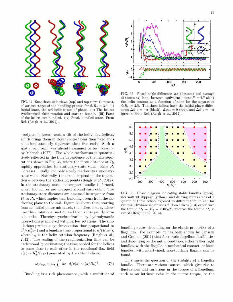
29
FIG. 34 Snapshots, side views (top) and top views (bottom),of various stages of the bundling process for d/Rh = 3.5. (i)Initial state, the red helix is out of phase. (ii) The helicessynchronized their rotation and start to bundle. (iii) Partsof the helices are bundled. (iv) Final, bundled state. FromRef. (Reigh et al., 2012).
drodynamic forces cause a tilt of the individual helices,which brings them in closer contact near their fixed endsand simultaneously separates their free ends. Such aspatial approach was already assumed to be necessaryby Macnab (1977). The whole mechanism is quantita-tively reflected in the time dependence of the helix sepa-rations shown in Fig. 35, where the mean distance at P2
rapidly approaches its stationary-state value, while P5
increases initially and only slowly reaches its stationary-state value. Naturally, the details depend on the separa-tion d between the anchoring points (Reigh et al., 2012).In the stationary state, a compact bundle is formed,where the helices are wrapped around each other. Thestationary-state distances are assumed in sequence fromP1 to P5, which implies that bundling occurs from the an-choring plane to the tail. Figure 35 shows that, startingfrom an initial phase mismatch, the helices first synchro-nize their rotational motion and then subsequently forma bundle. Thereby, synchronization by hydrodynamicinteractions is achieved within a few rotations. The sim-ulations predict a synchronization time proportional tod2/(R2
hω0) and a bunding time proportional to d/(Rhω0),where ω0 is the helix rotation frequency (Reigh et al.,2012). The scaling of the synchronization time can beunderstood by estimating the time needed for the helicesto come close to each other in the rotational flow fieldv(r) ∼ R2
h/(ω0r) generated by the other helices,
ω0tsyn ∼ ω0
∫ π
0
dφ d/v(d) ∼ (d/Rh)2. (73)
Bundling is a rich phenomenon, with a multitude of
FIG. 35 Phase angle difference ∆φ (bottom) and averagedistances 〈d〉 (top) between equivalent points Pi = iP alongthe helix contour as a function of time for the separationd/Rh = 2.5. The three helices have the initial phase differ-ences ∆φ12 = −π (black), ∆φ13 = 0 (red), and ∆φ12 = −π(green). From Ref. (Reigh et al., 2012).
200 300 400 500 600 700 8002.0
2.5
3.0
3.5
4.0
4.5
5.0
5.5
6.0
d/R
h
M2 /kBT
(c)
FIG. 36 Phase diagram indicating stable bundles (green),intermittent slippage (yellow), and drifting states (red) of asystem of three helices exposed to different torques and forvarious helix-base separations d. Two helices (1, 3) experiencethe torque M1 = M3 = 400kBT , whereas the torque M2 isvaried (Reigh et al., 2013).
bundling states depending on the elastic properties of aflagellum. For example, it has been shown by Janssenand Graham (2011) that for certain flagellum flexibilitiesand depending on the initial condition, either rather tightbundles, with the flagella in mechanical contact, or loosebundles, with intertwined, non-touching flagella can befound.
This raises the question of the stability of a flagellarbundle. There are various sources, which give rise tofluctuations and variations in the torque of a flagellum,such as an intrinsic noise in the motor torque, or the
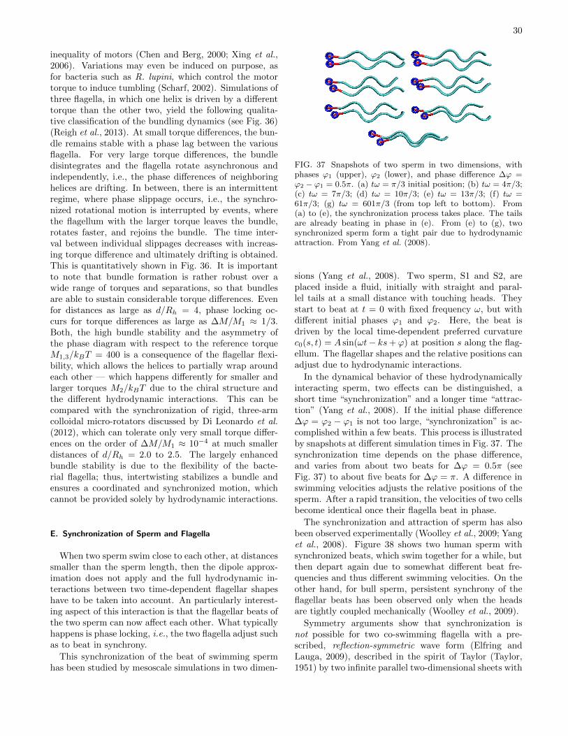
30
inequality of motors (Chen and Berg, 2000; Xing et al.,2006). Variations may even be induced on purpose, asfor bacteria such as R. lupini, which control the motortorque to induce tumbling (Scharf, 2002). Simulations ofthree flagella, in which one helix is driven by a differenttorque than the other two, yield the following qualita-tive classification of the bundling dynamics (see Fig. 36)(Reigh et al., 2013). At small torque differences, the bun-dle remains stable with a phase lag between the variousflagella. For very large torque differences, the bundledisintegrates and the flagella rotate asynchronous andindependently, i.e., the phase differences of neighboringhelices are drifting. In between, there is an intermittentregime, where phase slippage occurs, i.e., the synchro-nized rotational motion is interrupted by events, wherethe flagellum with the larger torque leaves the bundle,rotates faster, and rejoins the bundle. The time inter-val between individual slippages decreases with increas-ing torque difference and ultimately drifting is obtained.This is quantitatively shown in Fig. 36. It is importantto note that bundle formation is rather robust over awide range of torques and separations, so that bundlesare able to sustain considerable torque differences. Evenfor distances as large as d/Rh = 4, phase locking oc-curs for torque differences as large as ∆M/M1 ≈ 1/3.Both, the high bundle stability and the asymmetry ofthe phase diagram with respect to the reference torqueM1,3/kBT = 400 is a consequence of the flagellar flexi-bility, which allows the helices to partially wrap aroundeach other — which happens differently for smaller andlarger torques M2/kBT due to the chiral structure andthe different hydrodynamic interactions. This can becompared with the synchronization of rigid, three-armcolloidal micro-rotators discussed by Di Leonardo et al.(2012), which can tolerate only very small torque differ-ences on the order of ∆M/M1 ≈ 10−4 at much smallerdistances of d/Rh = 2.0 to 2.5. The largely enhancedbundle stability is due to the flexibility of the bacte-rial flagella; thus, intertwisting stabilizes a bundle andensures a coordinated and synchronized motion, whichcannot be provided solely by hydrodynamic interactions.
E. Synchronization of Sperm and Flagella
When two sperm swim close to each other, at distancessmaller than the sperm length, then the dipole approx-imation does not apply and the full hydrodynamic in-teractions between two time-dependent flagellar shapeshave to be taken into account. An particularly interest-ing aspect of this interaction is that the flagellar beats ofthe two sperm can now affect each other. What typicallyhappens is phase locking, i.e., the two flagella adjust suchas to beat in synchrony.
This synchronization of the beat of swimming spermhas been studied by mesoscale simulations in two dimen-
FIG. 37 Snapshots of two sperm in two dimensions, withphases ϕ1 (upper), ϕ2 (lower), and phase difference ∆ϕ =ϕ2 − ϕ1 = 0.5π. (a) tω = π/3 initial position; (b) tω = 4π/3;(c) tω = 7π/3; (d) tω = 10π/3; (e) tω = 13π/3; (f) tω =61π/3; (g) tω = 601π/3 (from top left to bottom). From(a) to (e), the synchronization process takes place. The tailsare already beating in phase in (e). From (e) to (g), twosynchronized sperm form a tight pair due to hydrodynamicattraction. From Yang et al. (2008).
sions (Yang et al., 2008). Two sperm, S1 and S2, areplaced inside a fluid, initially with straight and paral-lel tails at a small distance with touching heads. Theystart to beat at t = 0 with fixed frequency ω, but withdifferent initial phases ϕ1 and ϕ2. Here, the beat isdriven by the local time-dependent preferred curvaturec0(s, t) = A sin(ωt− ks+ ϕ) at position s along the flag-ellum. The flagellar shapes and the relative positions canadjust due to hydrodynamic interactions.
In the dynamical behavior of these hydrodynamicallyinteracting sperm, two effects can be distinguished, ashort time “synchronization” and a longer time “attrac-tion” (Yang et al., 2008). If the initial phase difference∆ϕ = ϕ2 − ϕ1 is not too large, “synchronization” is ac-complished within a few beats. This process is illustratedby snapshots at different simulation times in Fig. 37. Thesynchronization time depends on the phase difference,and varies from about two beats for ∆ϕ = 0.5π (seeFig. 37) to about five beats for ∆ϕ = π. A difference inswimming velocities adjusts the relative positions of thesperm. After a rapid transition, the velocities of two cellsbecome identical once their flagella beat in phase.
The synchronization and attraction of sperm has alsobeen observed experimentally (Woolley et al., 2009; Yanget al., 2008). Figure 38 shows two human sperm withsynchronized beats, which swim together for a while, butthen depart again due to somewhat different beat fre-quencies and thus different swimming velocities. On theother hand, for bull sperm, persistent synchrony of theflagellar beats has been observed only when the headsare tightly coupled mechanically (Woolley et al., 2009).
Symmetry arguments show that synchronization isnot possible for two co-swimming flagella with a pre-scribed, reflection-symmetric wave form (Elfring andLauga, 2009), described in the spirit of Taylor (Taylor,1951) by two infinite parallel two-dimensional sheets with
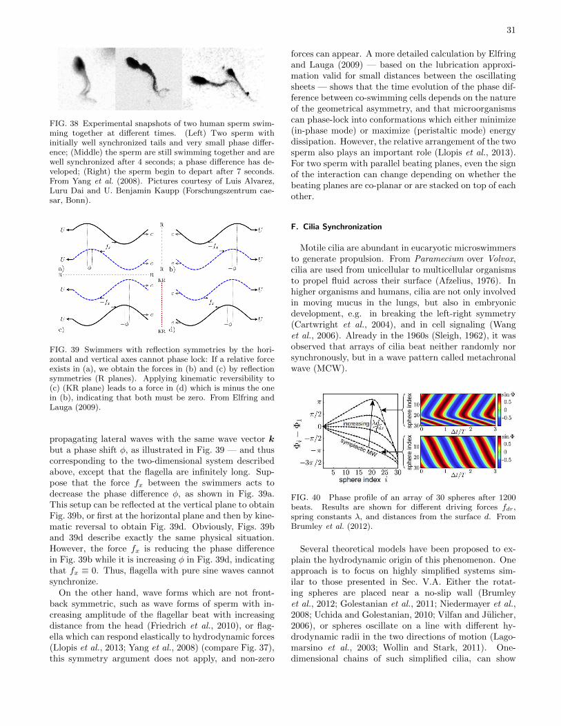
31
FIG. 38 Experimental snapshots of two human sperm swim-ming together at different times. (Left) Two sperm withinitially well synchronized tails and very small phase differ-ence; (Middle) the sperm are still swimming together and arewell synchronized after 4 seconds; a phase difference has de-veloped; (Right) the sperm begin to depart after 7 seconds.From Yang et al. (2008). Pictures courtesy of Luis Alvarez,Luru Dai and U. Benjamin Kaupp (Forschungszentrum cae-sar, Bonn).
FIG. 39 Swimmers with reflection symmetries by the hori-zontal and vertical axes cannot phase lock: If a relative forceexists in (a), we obtain the forces in (b) and (c) by reflectionsymmetries (R planes). Applying kinematic reversibility to(c) (KR plane) leads to a force in (d) which is minus the onein (b), indicating that both must be zero. From Elfring andLauga (2009).
propagating lateral waves with the same wave vector kbut a phase shift φ, as illustrated in Fig. 39 — and thuscorresponding to the two-dimensional system describedabove, except that the flagella are infinitely long. Sup-pose that the force fx between the swimmers acts todecrease the phase difference φ, as shown in Fig. 39a.This setup can be reflected at the vertical plane to obtainFig. 39b, or first at the horizontal plane and then by kine-matic reversal to obtain Fig. 39d. Obviously, Figs. 39band 39d describe exactly the same physical situation.However, the force fx is reducing the phase differencein Fig. 39b while it is increasing φ in Fig. 39d, indicatingthat fx ≡ 0. Thus, flagella with pure sine waves cannotsynchronize.
On the other hand, wave forms which are not front-back symmetric, such as wave forms of sperm with in-creasing amplitude of the flagellar beat with increasingdistance from the head (Friedrich et al., 2010), or flag-ella which can respond elastically to hydrodynamic forces(Llopis et al., 2013; Yang et al., 2008) (compare Fig. 37),this symmetry argument does not apply, and non-zero
forces can appear. A more detailed calculation by Elfringand Lauga (2009) — based on the lubrication approxi-mation valid for small distances between the oscillatingsheets — shows that the time evolution of the phase dif-ference between co-swimming cells depends on the natureof the geometrical asymmetry, and that microorganismscan phase-lock into conformations which either minimize(in-phase mode) or maximize (peristaltic mode) energydissipation. However, the relative arrangement of the twosperm also plays an important role (Llopis et al., 2013).For two sperm with parallel beating planes, even the signof the interaction can change depending on whether thebeating planes are co-planar or are stacked on top of eachother.
F. Cilia Synchronization
Motile cilia are abundant in eucaryotic microswimmersto generate propulsion. From Paramecium over Volvox,cilia are used from unicellular to multicellular organismsto propel fluid across their surface (Afzelius, 1976). Inhigher organisms and humans, cilia are not only involvedin moving mucus in the lungs, but also in embryonicdevelopment, e.g. in breaking the left-right symmetry(Cartwright et al., 2004), and in cell signaling (Wanget al., 2006). Already in the 1960s (Sleigh, 1962), it wasobserved that arrays of cilia beat neither randomly norsynchronously, but in a wave pattern called metachronalwave (MCW).
FIG. 40 Phase profile of an array of 30 spheres after 1200beats. Results are shown for different driving forces fdr,spring constants λ, and distances from the surface d. FromBrumley et al. (2012).
Several theoretical models have been proposed to ex-plain the hydrodynamic origin of this phenomenon. Oneapproach is to focus on highly simplified systems sim-ilar to those presented in Sec. V.A. Either the rotat-ing spheres are placed near a no-slip wall (Brumleyet al., 2012; Golestanian et al., 2011; Niedermayer et al.,2008; Uchida and Golestanian, 2010; Vilfan and Julicher,2006), or spheres oscillate on a line with different hy-drodynamic radii in the two directions of motion (Lago-marsino et al., 2003; Wollin and Stark, 2011). One-dimensional chains of such simplified cilia, can show
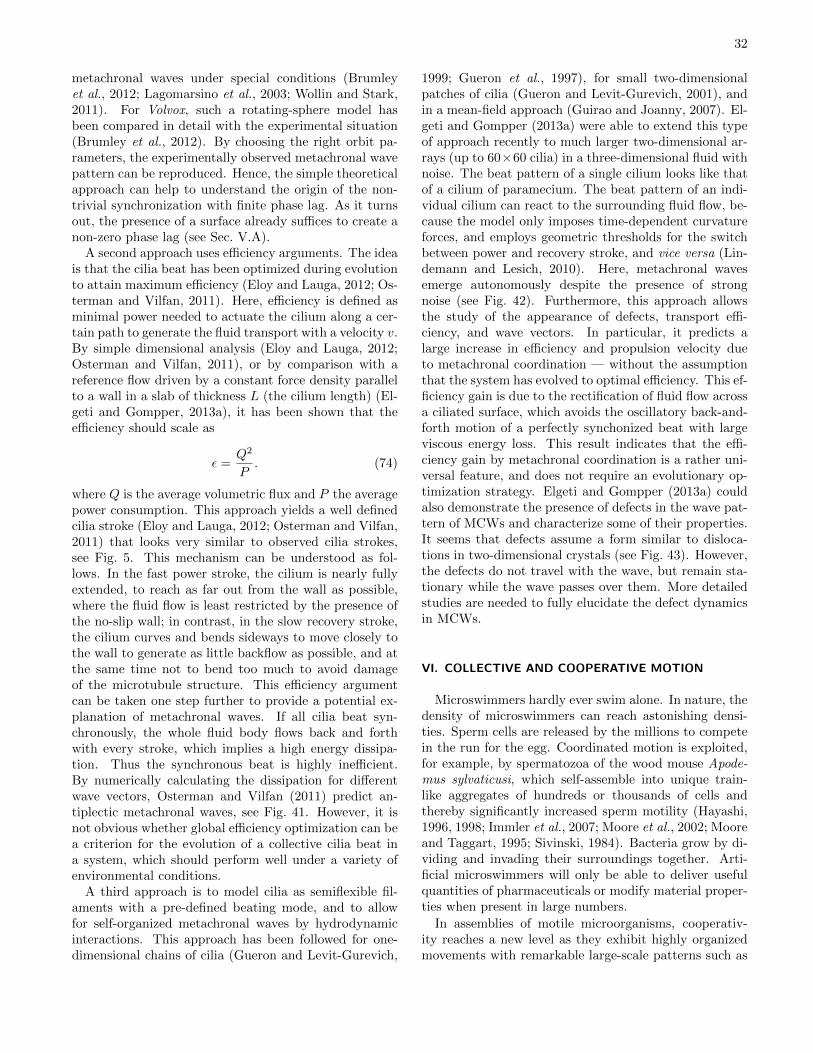
32
metachronal waves under special conditions (Brumleyet al., 2012; Lagomarsino et al., 2003; Wollin and Stark,2011). For Volvox, such a rotating-sphere model hasbeen compared in detail with the experimental situation(Brumley et al., 2012). By choosing the right orbit pa-rameters, the experimentally observed metachronal wavepattern can be reproduced. Hence, the simple theoreticalapproach can help to understand the origin of the non-trivial synchronization with finite phase lag. As it turnsout, the presence of a surface already suffices to create anon-zero phase lag (see Sec. V.A).
A second approach uses efficiency arguments. The ideais that the cilia beat has been optimized during evolutionto attain maximum efficiency (Eloy and Lauga, 2012; Os-terman and Vilfan, 2011). Here, efficiency is defined asminimal power needed to actuate the cilium along a cer-tain path to generate the fluid transport with a velocity v.By simple dimensional analysis (Eloy and Lauga, 2012;Osterman and Vilfan, 2011), or by comparison with areference flow driven by a constant force density parallelto a wall in a slab of thickness L (the cilium length) (El-geti and Gompper, 2013a), it has been shown that theefficiency should scale as
ε =Q2
P. (74)
where Q is the average volumetric flux and P the averagepower consumption. This approach yields a well definedcilia stroke (Eloy and Lauga, 2012; Osterman and Vilfan,2011) that looks very similar to observed cilia strokes,see Fig. 5. This mechanism can be understood as fol-lows. In the fast power stroke, the cilium is nearly fullyextended, to reach as far out from the wall as possible,where the fluid flow is least restricted by the presence ofthe no-slip wall; in contrast, in the slow recovery stroke,the cilium curves and bends sideways to move closely tothe wall to generate as little backflow as possible, and atthe same time not to bend too much to avoid damageof the microtubule structure. This efficiency argumentcan be taken one step further to provide a potential ex-planation of metachronal waves. If all cilia beat syn-chronously, the whole fluid body flows back and forthwith every stroke, which implies a high energy dissipa-tion. Thus the synchronous beat is highly inefficient.By numerically calculating the dissipation for differentwave vectors, Osterman and Vilfan (2011) predict an-tiplectic metachronal waves, see Fig. 41. However, it isnot obvious whether global efficiency optimization can bea criterion for the evolution of a collective cilia beat ina system, which should perform well under a variety ofenvironmental conditions.
A third approach is to model cilia as semiflexible fil-aments with a pre-defined beating mode, and to allowfor self-organized metachronal waves by hydrodynamicinteractions. This approach has been followed for one-dimensional chains of cilia (Gueron and Levit-Gurevich,
1999; Gueron et al., 1997), for small two-dimensionalpatches of cilia (Gueron and Levit-Gurevich, 2001), andin a mean-field approach (Guirao and Joanny, 2007). El-geti and Gompper (2013a) were able to extend this typeof approach recently to much larger two-dimensional ar-rays (up to 60×60 cilia) in a three-dimensional fluid withnoise. The beat pattern of a single cilium looks like thatof a cilium of paramecium. The beat pattern of an indi-vidual cilium can react to the surrounding fluid flow, be-cause the model only imposes time-dependent curvatureforces, and employs geometric thresholds for the switchbetween power and recovery stroke, and vice versa (Lin-demann and Lesich, 2010). Here, metachronal wavesemerge autonomously despite the presence of strongnoise (see Fig. 42). Furthermore, this approach allowsthe study of the appearance of defects, transport effi-ciency, and wave vectors. In particular, it predicts alarge increase in efficiency and propulsion velocity dueto metachronal coordination — without the assumptionthat the system has evolved to optimal efficiency. This ef-ficiency gain is due to the rectification of fluid flow acrossa ciliated surface, which avoids the oscillatory back-and-forth motion of a perfectly synchonized beat with largeviscous energy loss. This result indicates that the effi-ciency gain by metachronal coordination is a rather uni-versal feature, and does not require an evolutionary op-timization strategy. Elgeti and Gompper (2013a) couldalso demonstrate the presence of defects in the wave pat-tern of MCWs and characterize some of their properties.It seems that defects assume a form similar to disloca-tions in two-dimensional crystals (see Fig. 43). However,the defects do not travel with the wave, but remain sta-tionary while the wave passes over them. More detailedstudies are needed to fully elucidate the defect dynamicsin MCWs.
VI. COLLECTIVE AND COOPERATIVE MOTION
Microswimmers hardly ever swim alone. In nature, thedensity of microswimmers can reach astonishing densi-ties. Sperm cells are released by the millions to competein the run for the egg. Coordinated motion is exploited,for example, by spermatozoa of the wood mouse Apode-mus sylvaticusi, which self-assemble into unique train-like aggregates of hundreds or thousands of cells andthereby significantly increased sperm motility (Hayashi,1996, 1998; Immler et al., 2007; Moore et al., 2002; Mooreand Taggart, 1995; Sivinski, 1984). Bacteria grow by di-viding and invading their surroundings together. Arti-ficial microswimmers will only be able to deliver usefulquantities of pharmaceuticals or modify material proper-ties when present in large numbers.
In assemblies of motile microorganisms, cooperativ-ity reaches a new level as they exhibit highly organizedmovements with remarkable large-scale patterns such as
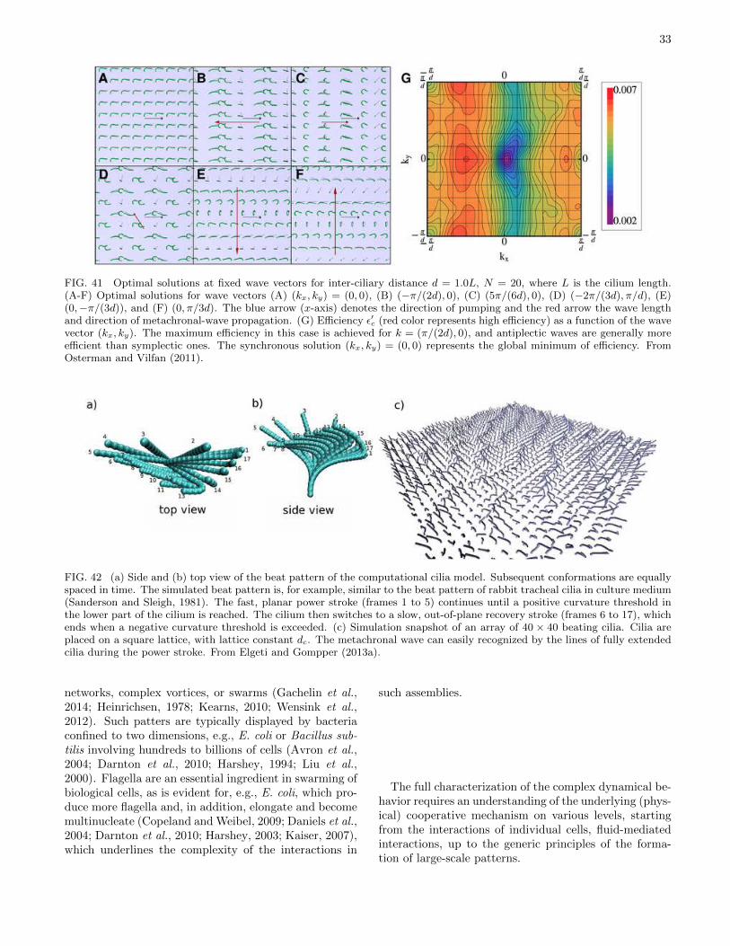
33
FIG. 41 Optimal solutions at fixed wave vectors for inter-ciliary distance d = 1.0L, N = 20, where L is the cilium length.(A-F) Optimal solutions for wave vectors (A) (kx, ky) = (0, 0), (B) (−π/(2d), 0), (C) (5π/(6d), 0), (D) (−2π/(3d), π/d), (E)(0,−π/(3d)), and (F) (0, π/3d). The blue arrow (x-axis) denotes the direction of pumping and the red arrow the wave lengthand direction of metachronal-wave propagation. (G) Efficiency ε′c (red color represents high efficiency) as a function of the wavevector (kx, ky). The maximum efficiency in this case is achieved for k = (π/(2d), 0), and antiplectic waves are generally moreefficient than symplectic ones. The synchronous solution (kx, ky) = (0, 0) represents the global minimum of efficiency. FromOsterman and Vilfan (2011).
FIG. 42 (a) Side and (b) top view of the beat pattern of the computational cilia model. Subsequent conformations are equallyspaced in time. The simulated beat pattern is, for example, similar to the beat pattern of rabbit tracheal cilia in culture medium(Sanderson and Sleigh, 1981). The fast, planar power stroke (frames 1 to 5) continues until a positive curvature threshold inthe lower part of the cilium is reached. The cilium then switches to a slow, out-of-plane recovery stroke (frames 6 to 17), whichends when a negative curvature threshold is exceeded. (c) Simulation snapshot of an array of 40 × 40 beating cilia. Cilia areplaced on a square lattice, with lattice constant dc. The metachronal wave can easily recognized by the lines of fully extendedcilia during the power stroke. From Elgeti and Gompper (2013a).
networks, complex vortices, or swarms (Gachelin et al.,2014; Heinrichsen, 1978; Kearns, 2010; Wensink et al.,2012). Such patters are typically displayed by bacteriaconfined to two dimensions, e.g., E. coli or Bacillus sub-tilis involving hundreds to billions of cells (Avron et al.,2004; Darnton et al., 2010; Harshey, 1994; Liu et al.,2000). Flagella are an essential ingredient in swarming ofbiological cells, as is evident for, e.g., E. coli, which pro-duce more flagella and, in addition, elongate and becomemultinucleate (Copeland and Weibel, 2009; Daniels et al.,2004; Darnton et al., 2010; Harshey, 2003; Kaiser, 2007),which underlines the complexity of the interactions in
such assemblies.
The full characterization of the complex dynamical be-havior requires an understanding of the underlying (phys-ical) cooperative mechanism on various levels, startingfrom the interactions of individual cells, fluid-mediatedinteractions, up to the generic principles of the forma-tion of large-scale patterns.
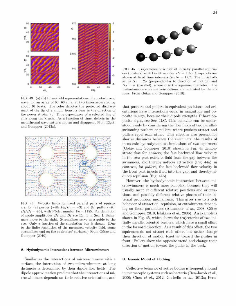
34
FIG. 43 (a),(b) Phase-field representations of a metachronalwave, for an array of 60 60 cilia, at two times separated byabout 40 beats. The color denotes the projected displace-ment of the tip of a cilium from its base in the direction ofthe power stroke. (c) Time dependence of a selected line ofcilia along the x axis. As a function of time, defects in themetachronal wave pattern appear and disappear. From Elgetiand Gompper (2013a).
FIG. 44 Velocity fields for fixed parallel pairs of squirm-ers, for (a) pusher (with B2/B1 = −3) and (b) puller (withB2/B1 = +3), with Peclet number Pe = 1155. For definitionof mode amplitudes B1 and B2 see Eq. 1 in Sec. I. Swim-mers move to the right. Streamlines serve as a guide to theeye. Only a fraction of the simulation box is shown. (Dueto the finite resolution of the measured velocity field, somestreamlines end on the squirmers’ surfaces.) From Gotze andGompper (2010).
A. Hydrodynamic Interactions between Microswimmers
Similar as the interactions of microswimmers with asurface, the interaction of two microswimmers at longdistances is determined by their dipole flow fields. Thedipole approximation predicts that the interactions of mi-croswimmers depends on their relative orientation, and
FIG. 45 Trajectories of a pair of initially parallel squirm-ers (pushers) with Peclet number Pe = 1155. Snapshots areshown at fixed time intervals ∆tv/σ = 1.67. The initial off-set is ∆z = 2σ (perpendicular to direction of motion) and∆x = σ (parallel), where σ is the squirmer diameter. Theinstantaneous squirmer orientations are indicated by the ar-rows. From Gotze and Gompper (2010).
that pushers and pullers in equivalent positions and ori-entations have interactions equal in magnitude and op-posite in sign, because their dipole strengths P have op-posite signs, see Sec. II.C. This behavior can be under-stood easily by considering the flow fields of two parallel-swimming pushers or pullers, where pushers attract andpullers repel each other. This effect is also present forshorter distances between the swimmers; the results ofmesoscale hydrodynamics simulations of two squirmers(Gotze and Gompper, 2010) shown in Fig. 44 demon-strate that for pushers, the fast backward flow velocityin the rear part extracts fluid from the gap between theswimmers, and thereby induces attraction (Fig. 44a); incontrast, for pullers, the fast backward flow velocity inthe front part injects fluid into the gap, and thereby in-duces repulsion (Fig. 44b).
However, the hydrodynamic interaction between mi-croswimmers is much more complex, because they willusually meet at different relative positions and orienta-tions, and possibly different relative phases of their in-ternal propulsion mechanisms. This gives rise to a richbehavior of attraction, repulsion, or entrainment depend-ing on these parameters (Alexander et al., 2008; Gotzeand Gompper, 2010; Ishikawa et al., 2006). An example isshown in Fig. 45, which shows the trajectories of two ini-tially parallel oriented pushers, which have a small offsetin the forward direction. As a result of this offset, the twosquirmers do not attract each other, but rather changetheir direction of motion together toward the pusher infront. Pullers show the opposite trend and change theirdirection of motion toward the puller in the back.
B. Generic Model of Flocking
Collective behavior of active bodies is frequently foundin microscopic systems such as bacteria (Ben-Jacob et al.,2000; Chen et al., 2012; Gachelin et al., 2013a; Peru-
35
ani et al., 2012; Sokolov et al., 2007) and synthetic mi-croswimmers (Buttinoni et al., 2013; Ibele et al., 2009;Palacci et al., 2013; Theurkauff et al., 2012; Wang et al.,2013), but also in macroscopic systems such as flocks ofbirds and schools of fish (Cavagna et al., 2010; Vicsekand Zafeiris, 2012). Despite the very different propulsionmechanisms and interactions in these systems, they allfavor alignment of neighboring bodies, thus leading tosimilar forms of collective behavior.
Therefore, it is natural to look for a model, which isable to capture the generic collective properties of all thevarious systems of self-propelled particles and organisms.Such a model was proposed in a pioneering work by Vic-sek et al. (1995b). In this model, now often called the“Vicsek model”, N polar point particles move in spacewith constant magnitude of velocity v0. The dynamicsproceeds in two steps, a streaming step of duration ∆t, inwhich particles move ballistically, and a interaction step,in which particles align their velocity direction with theaverage direction of motion of their neighbors. In twospatial dimensions, this implies the dynamics for the po-sition ri and velocity vi of particle i,
ri(t+ ∆t) = ri(t) + vi(t)∆t, (75)
θi(t+ ∆t) = 〈θ(t)〉σ + ∆θ, (76)
where θi is the angle between vi and the x-axis of a Carte-sian coordinate system, 〈θ(t)〉σ is the average orientationof all particles within a circle of diameter σ. and ∆θis a random noise uniformly distributed in the interval[−η/2,+η/2]. The essential control parameters of theVicsek model are the particle density ρ, the noise strengthη, and the propulsion velocity v0 in units of σ/∆t.
A numerical investigation of this model by Vicsek et al.(1995b) shows a phase transiton with increasing den-sity or decreasing noise strength from a random isotropicphase to an aligned phase, in which particles move col-lectively in a spontaneously selected direction. This ob-servation of a non-equilibrium phase transition in a sys-tem of self-propelled point particles has led to numer-ous analytical (Baskaran and Marchetti, 2009; Bertinet al., 2009; Golestanian, 2009; Peruani et al., 2008; Ra-maswamy et al., 2003; Simha and Ramaswamy, 2002;Toner and Tu, 1995; Toner et al., 2005b) as well ascomputational (Aldana et al., 2007; Chate et al., 2008;D’Orsogna et al., 2006; Ginelli et al., 2010; Gregoire andChate, 2004; Huepe and Aldana, 2004; Redner et al.,2013; Szabo et al., 2006) studies. A review is providedby Vicsek and Zafeiris (2012).
C. Self-Propelled Rods
When we consider a system of self-propelled particlesbeyond the phenomenological description of interactionsas in the Vicsek model (Vicsek et al., 1995b), the simplestphysical interaction which leads to alignment is volume
exclusion of rod-like microswimmers and self-propelledrods. Here polar interactions, with alignment in theswimming directions, have to be distinguished from ne-matic interactions, with aligment independent of the di-rection of motion. Of particular interest are experimentswith elongated self-propelled particles on the microscopicscale in two dimensions, such as motility assays whereactin filaments are propelled on a carpet of myosin mo-tor proteins (Harada et al., 1987; Schaller et al., 2010),microtubules propelled by surface-bound dyneins (Sum-ino et al., 2012), and microswimmers that are attractedto surfaces (as described in Sec. IV).
Self-propelled rods (SPRs) in two dimensions are oftenmodelled as linear chains of (overlapping) beads (Abke-nar et al., 2013; Wensink and Lowen, 2008; Yang et al.,2010). Both, models with strict excluded-volume inter-actions and with a finite overlap energy, which allowsrods to cross, have been employed. In the latter case,rod crossing is included to mimic in a two-dimensionalsimulation the possible escape of rods into the third di-mension when two rods collide (like actin filaments ormicrotubules in a motility assay) (Abkenar et al., 2013).
In such models, self-propulsion of rods leads to en-hanced aggregation and cluster formation, as well as tovarious kinds of ordered phases in Brownian Dynamicssimulations (Abkenar et al., 2013; Kraikivski et al., 2006;McCandlish et al., 2012; Peruani et al., 2006; Wensinket al., 2012; Wensink and Lowen, 2008; Yang et al., 2010),as well as in theoretical approaches based on the Smolu-chowski equation (Baskaran and Marchetti, 2008a,b; Pe-ruani et al., 2010) and on continuum models in the “hy-drodynamic limit” (Baskaran and Marchetti, 2008a,b).
A few simulation snapshots of a two-dimensional sys-tem, with different rod number densities ρ, propulsionvelocities v0, and noise levels are shown in Fig. 46, anda corresponding phase diagram in Fig. 47. Here, the im-portance of propulsion compared to noise is characterizedby the Peclet number Pe = Lrodv0/D‖, where Lrod is therod length, v0 the swimming velocity of an isolated swim-mer, and D‖ the diffusion coefficient in the direction ofthe instantaneous rod orientation. The phase diagram ofFig. 47 describes a system in which rods interact with a“soft” interaction potential, so that the rods are penetra-ble at high Peclet numbers (Abkenar et al., 2013). Dis-ordered states, motile clusters, nematic phases, and laneformation are observed. In particular, Fig. 46 displays aphase with a single polar cluster, which is formed at in-termediate rod density and not too large Peclet numbers,and a phase with both nematic order and a lane struc-ture where rods moving in opposite directions, which isformed at high rod densities (Abkenar et al., 2013; Mc-Candlish et al., 2012). Near the transition from a disor-dered state of small- and intermediate-size clusters to anordered state, the cluster-size distribution obeys a power-law decay, P (n) ∼ n−γ , with an exponent γ ' 2 (Abke-nar et al., 2013; Huepe and Aldana, 2004, 2008; Peruani
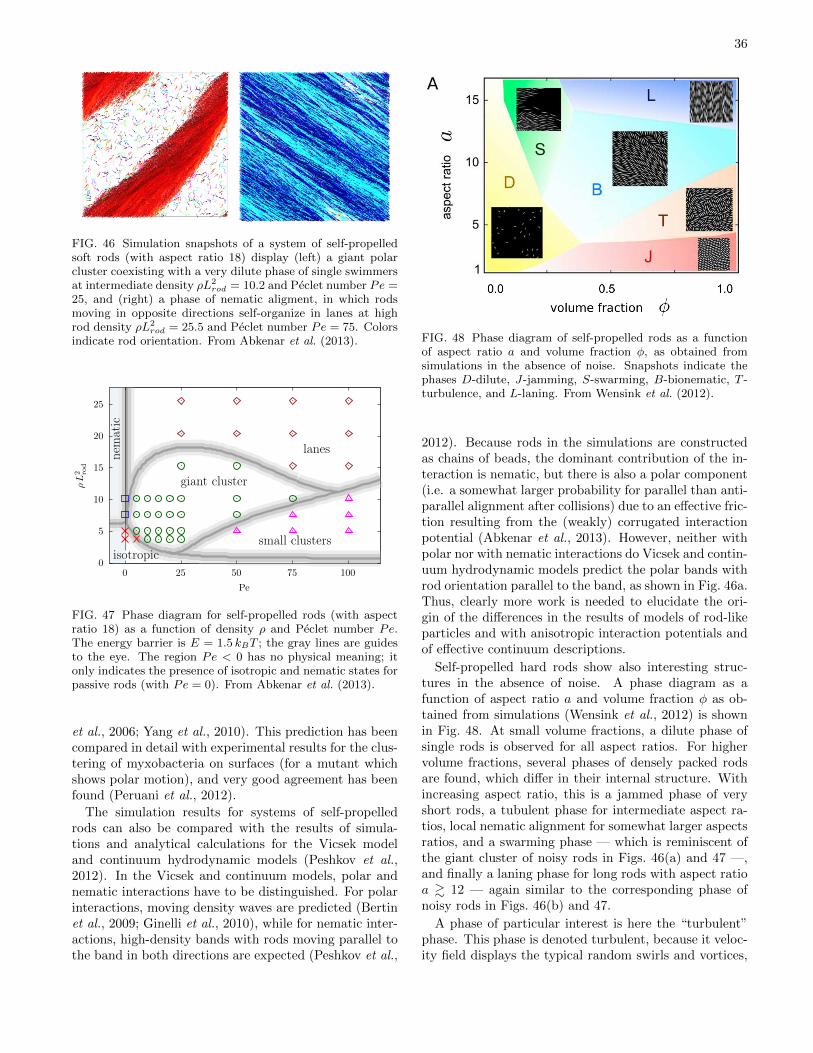
36
FIG. 46 Simulation snapshots of a system of self-propelledsoft rods (with aspect ratio 18) display (left) a giant polarcluster coexisting with a very dilute phase of single swimmersat intermediate density ρL2
rod = 10.2 and Peclet number Pe =25, and (right) a phase of nematic aligment, in which rodsmoving in opposite directions self-organize in lanes at highrod density ρL2
rod = 25.5 and Peclet number Pe = 75. Colorsindicate rod orientation. From Abkenar et al. (2013).
ρL
2 rod
Pe
giant cluster
small clusters
lanes
0
5
10
15
20
25
0 25 50 75 100
isotropic
nem
atic
FIG. 47 Phase diagram for self-propelled rods (with aspectratio 18) as a function of density ρ and Peclet number Pe.The energy barrier is E = 1.5 kBT ; the gray lines are guidesto the eye. The region Pe < 0 has no physical meaning; itonly indicates the presence of isotropic and nematic states forpassive rods (with Pe = 0). From Abkenar et al. (2013).
et al., 2006; Yang et al., 2010). This prediction has beencompared in detail with experimental results for the clus-tering of myxobacteria on surfaces (for a mutant whichshows polar motion), and very good agreement has beenfound (Peruani et al., 2012).
The simulation results for systems of self-propelledrods can also be compared with the results of simula-tions and analytical calculations for the Vicsek modeland continuum hydrodynamic models (Peshkov et al.,2012). In the Vicsek and continuum models, polar andnematic interactions have to be distinguished. For polarinteractions, moving density waves are predicted (Bertinet al., 2009; Ginelli et al., 2010), while for nematic inter-actions, high-density bands with rods moving parallel tothe band in both directions are expected (Peshkov et al.,
FIG. 48 Phase diagram of self-propelled rods as a functionof aspect ratio a and volume fraction φ, as obtained fromsimulations in the absence of noise. Snapshots indicate thephases D-dilute, J-jamming, S-swarming, B-bionematic, T -turbulence, and L-laning. From Wensink et al. (2012).
2012). Because rods in the simulations are constructedas chains of beads, the dominant contribution of the in-teraction is nematic, but there is also a polar component(i.e. a somewhat larger probability for parallel than anti-parallel alignment after collisions) due to an effective fric-tion resulting from the (weakly) corrugated interactionpotential (Abkenar et al., 2013). However, neither withpolar nor with nematic interactions do Vicsek and contin-uum hydrodynamic models predict the polar bands withrod orientation parallel to the band, as shown in Fig. 46a.Thus, clearly more work is needed to elucidate the ori-gin of the differences in the results of models of rod-likeparticles and with anisotropic interaction potentials andof effective continuum descriptions.
Self-propelled hard rods show also interesting struc-tures in the absence of noise. A phase diagram as afunction of aspect ratio a and volume fraction φ as ob-tained from simulations (Wensink et al., 2012) is shownin Fig. 48. At small volume fractions, a dilute phase ofsingle rods is observed for all aspect ratios. For highervolume fractions, several phases of densely packed rodsare found, which differ in their internal structure. Withincreasing aspect ratio, this is a jammed phase of veryshort rods, a tubulent phase for intermediate aspect ra-tios, local nematic alignment for somewhat larger aspectsratios, and a swarming phase — which is reminiscent ofthe giant cluster of noisy rods in Figs. 46(a) and 47 —,and finally a laning phase for long rods with aspect ratioa & 12 — again similar to the corresponding phase ofnoisy rods in Figs. 46(b) and 47.
A phase of particular interest is here the “turbulent”phase. This phase is denoted turbulent, because it veloc-ity field displays the typical random swirls and vortices,
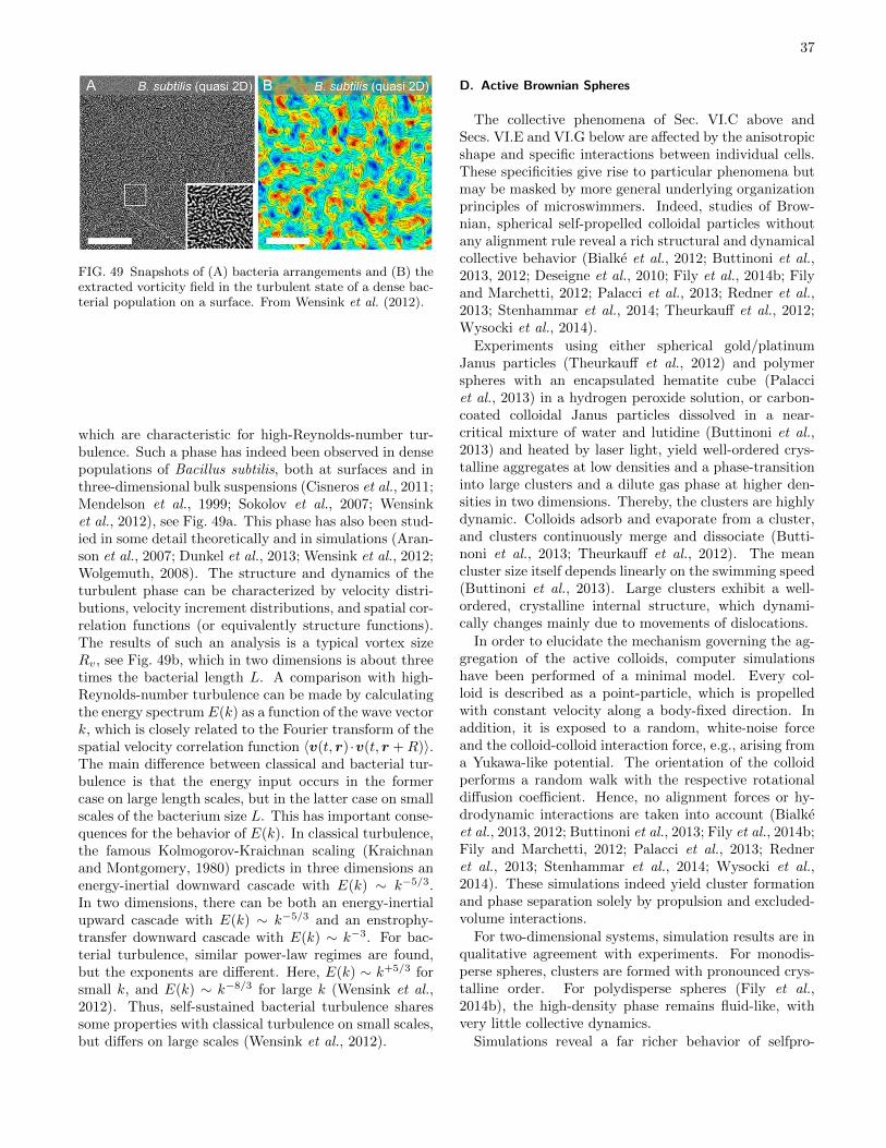
37
FIG. 49 Snapshots of (A) bacteria arrangements and (B) theextracted vorticity field in the turbulent state of a dense bac-terial population on a surface. From Wensink et al. (2012).
which are characteristic for high-Reynolds-number tur-bulence. Such a phase has indeed been observed in densepopulations of Bacillus subtilis, both at surfaces and inthree-dimensional bulk suspensions (Cisneros et al., 2011;Mendelson et al., 1999; Sokolov et al., 2007; Wensinket al., 2012), see Fig. 49a. This phase has also been stud-ied in some detail theoretically and in simulations (Aran-son et al., 2007; Dunkel et al., 2013; Wensink et al., 2012;Wolgemuth, 2008). The structure and dynamics of theturbulent phase can be characterized by velocity distri-butions, velocity increment distributions, and spatial cor-relation functions (or equivalently structure functions).The results of such an analysis is a typical vortex sizeRv, see Fig. 49b, which in two dimensions is about threetimes the bacterial length L. A comparison with high-Reynolds-number turbulence can be made by calculatingthe energy spectrum E(k) as a function of the wave vectork, which is closely related to the Fourier transform of thespatial velocity correlation function 〈v(t, r) ·v(t, r +R)〉.The main difference between classical and bacterial tur-bulence is that the energy input occurs in the formercase on large length scales, but in the latter case on smallscales of the bacterium size L. This has important conse-quences for the behavior of E(k). In classical turbulence,the famous Kolmogorov-Kraichnan scaling (Kraichnanand Montgomery, 1980) predicts in three dimensions anenergy-inertial downward cascade with E(k) ∼ k−5/3.In two dimensions, there can be both an energy-inertialupward cascade with E(k) ∼ k−5/3 and an enstrophy-transfer downward cascade with E(k) ∼ k−3. For bac-terial turbulence, similar power-law regimes are found,but the exponents are different. Here, E(k) ∼ k+5/3 forsmall k, and E(k) ∼ k−8/3 for large k (Wensink et al.,2012). Thus, self-sustained bacterial turbulence sharessome properties with classical turbulence on small scales,but differs on large scales (Wensink et al., 2012).
D. Active Brownian Spheres
The collective phenomena of Sec. VI.C above andSecs. VI.E and VI.G below are affected by the anisotropicshape and specific interactions between individual cells.These specificities give rise to particular phenomena butmay be masked by more general underlying organizationprinciples of microswimmers. Indeed, studies of Brow-nian, spherical self-propelled colloidal particles withoutany alignment rule reveal a rich structural and dynamicalcollective behavior (Bialke et al., 2012; Buttinoni et al.,2013, 2012; Deseigne et al., 2010; Fily et al., 2014b; Filyand Marchetti, 2012; Palacci et al., 2013; Redner et al.,2013; Stenhammar et al., 2014; Theurkauff et al., 2012;Wysocki et al., 2014).
Experiments using either spherical gold/platinumJanus particles (Theurkauff et al., 2012) and polymerspheres with an encapsulated hematite cube (Palacciet al., 2013) in a hydrogen peroxide solution, or carbon-coated colloidal Janus particles dissolved in a near-critical mixture of water and lutidine (Buttinoni et al.,2013) and heated by laser light, yield well-ordered crys-talline aggregates at low densities and a phase-transitioninto large clusters and a dilute gas phase at higher den-sities in two dimensions. Thereby, the clusters are highlydynamic. Colloids adsorb and evaporate from a cluster,and clusters continuously merge and dissociate (Butti-noni et al., 2013; Theurkauff et al., 2012). The meancluster size itself depends linearly on the swimming speed(Buttinoni et al., 2013). Large clusters exhibit a well-ordered, crystalline internal structure, which dynami-cally changes mainly due to movements of dislocations.
In order to elucidate the mechanism governing the ag-gregation of the active colloids, computer simulationshave been performed of a minimal model. Every col-loid is described as a point-particle, which is propelledwith constant velocity along a body-fixed direction. Inaddition, it is exposed to a random, white-noise forceand the colloid-colloid interaction force, e.g., arising froma Yukawa-like potential. The orientation of the colloidperforms a random walk with the respective rotationaldiffusion coefficient. Hence, no alignment forces or hy-drodynamic interactions are taken into account (Bialkeet al., 2013, 2012; Buttinoni et al., 2013; Fily et al., 2014b;Fily and Marchetti, 2012; Palacci et al., 2013; Redneret al., 2013; Stenhammar et al., 2014; Wysocki et al.,2014). These simulations indeed yield cluster formationand phase separation solely by propulsion and excluded-volume interactions.
For two-dimensional systems, simulation results are inqualitative agreement with experiments. For monodis-perse spheres, clusters are formed with pronounced crys-talline order. For polydisperse spheres (Fily et al.,2014b), the high-density phase remains fluid-like, withvery little collective dynamics.
Simulations reveal a far richer behavior of selfpro-
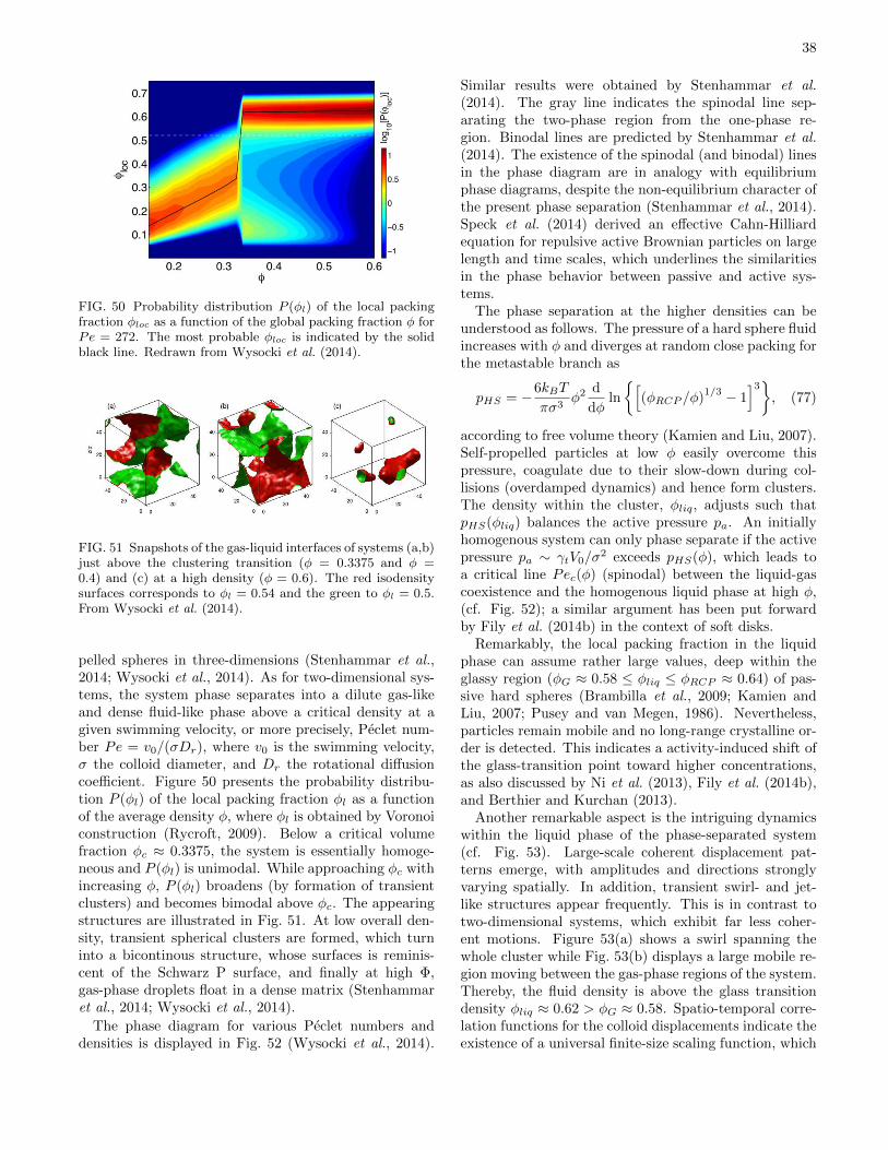
38
FIG. 50 Probability distribution P (φl) of the local packingfraction φloc as a function of the global packing fraction φ forPe = 272. The most probable φloc is indicated by the solidblack line. Redrawn from Wysocki et al. (2014).
FIG. 51 Snapshots of the gas-liquid interfaces of systems (a,b)just above the clustering transition (φ = 0.3375 and φ =0.4) and (c) at a high density (φ = 0.6). The red isodensitysurfaces corresponds to φl = 0.54 and the green to φl = 0.5.From Wysocki et al. (2014).
pelled spheres in three-dimensions (Stenhammar et al.,2014; Wysocki et al., 2014). As for two-dimensional sys-tems, the system phase separates into a dilute gas-likeand dense fluid-like phase above a critical density at agiven swimming velocity, or more precisely, Peclet num-ber Pe = v0/(σDr), where v0 is the swimming velocity,σ the colloid diameter, and Dr the rotational diffusioncoefficient. Figure 50 presents the probability distribu-tion P (φl) of the local packing fraction φl as a functionof the average density φ, where φl is obtained by Voronoiconstruction (Rycroft, 2009). Below a critical volumefraction φc ≈ 0.3375, the system is essentially homoge-neous and P (φl) is unimodal. While approaching φc withincreasing φ, P (φl) broadens (by formation of transientclusters) and becomes bimodal above φc. The appearingstructures are illustrated in Fig. 51. At low overall den-sity, transient spherical clusters are formed, which turninto a bicontinous structure, whose surfaces is reminis-cent of the Schwarz P surface, and finally at high Φ,gas-phase droplets float in a dense matrix (Stenhammaret al., 2014; Wysocki et al., 2014).
The phase diagram for various Peclet numbers anddensities is displayed in Fig. 52 (Wysocki et al., 2014).
Similar results were obtained by Stenhammar et al.(2014). The gray line indicates the spinodal line sep-arating the two-phase region from the one-phase re-gion. Binodal lines are predicted by Stenhammar et al.(2014). The existence of the spinodal (and binodal) linesin the phase diagram are in analogy with equilibriumphase diagrams, despite the non-equilibrium character ofthe present phase separation (Stenhammar et al., 2014).Speck et al. (2014) derived an effective Cahn-Hilliardequation for repulsive active Brownian particles on largelength and time scales, which underlines the similaritiesin the phase behavior between passive and active sys-tems.
The phase separation at the higher densities can beunderstood as follows. The pressure of a hard sphere fluidincreases with φ and diverges at random close packing forthe metastable branch as
pHS = −6kBT
πσ3φ2
d
dφln
[(φRCP /φ)
1/3 − 1]3
, (77)
according to free volume theory (Kamien and Liu, 2007).Self-propelled particles at low φ easily overcome thispressure, coagulate due to their slow-down during col-lisions (overdamped dynamics) and hence form clusters.The density within the cluster, φliq, adjusts such thatpHS(φliq) balances the active pressure pa. An initiallyhomogenous system can only phase separate if the activepressure pa ∼ γtV0/σ
2 exceeds pHS(φ), which leads toa critical line Pec(φ) (spinodal) between the liquid-gascoexistence and the homogenous liquid phase at high φ,(cf. Fig. 52); a similar argument has been put forwardby Fily et al. (2014b) in the context of soft disks.
Remarkably, the local packing fraction in the liquidphase can assume rather large values, deep within theglassy region (φG ≈ 0.58 ≤ φliq ≤ φRCP ≈ 0.64) of pas-sive hard spheres (Brambilla et al., 2009; Kamien andLiu, 2007; Pusey and van Megen, 1986). Nevertheless,particles remain mobile and no long-range crystalline or-der is detected. This indicates a activity-induced shift ofthe glass-transition point toward higher concentrations,as also discussed by Ni et al. (2013), Fily et al. (2014b),and Berthier and Kurchan (2013).
Another remarkable aspect is the intriguing dynamicswithin the liquid phase of the phase-separated system(cf. Fig. 53). Large-scale coherent displacement pat-terns emerge, with amplitudes and directions stronglyvarying spatially. In addition, transient swirl- and jet-like structures appear frequently. This is in contrast totwo-dimensional systems, which exhibit far less coher-ent motions. Figure 53(a) shows a swirl spanning thewhole cluster while Fig. 53(b) displays a large mobile re-gion moving between the gas-phase regions of the system.Thereby, the fluid density is above the glass transitiondensity φliq ≈ 0.62 > φG ≈ 0.58. Spatio-temporal corre-lation functions for the colloid displacements indicate theexistence of a universal finite-size scaling function, which
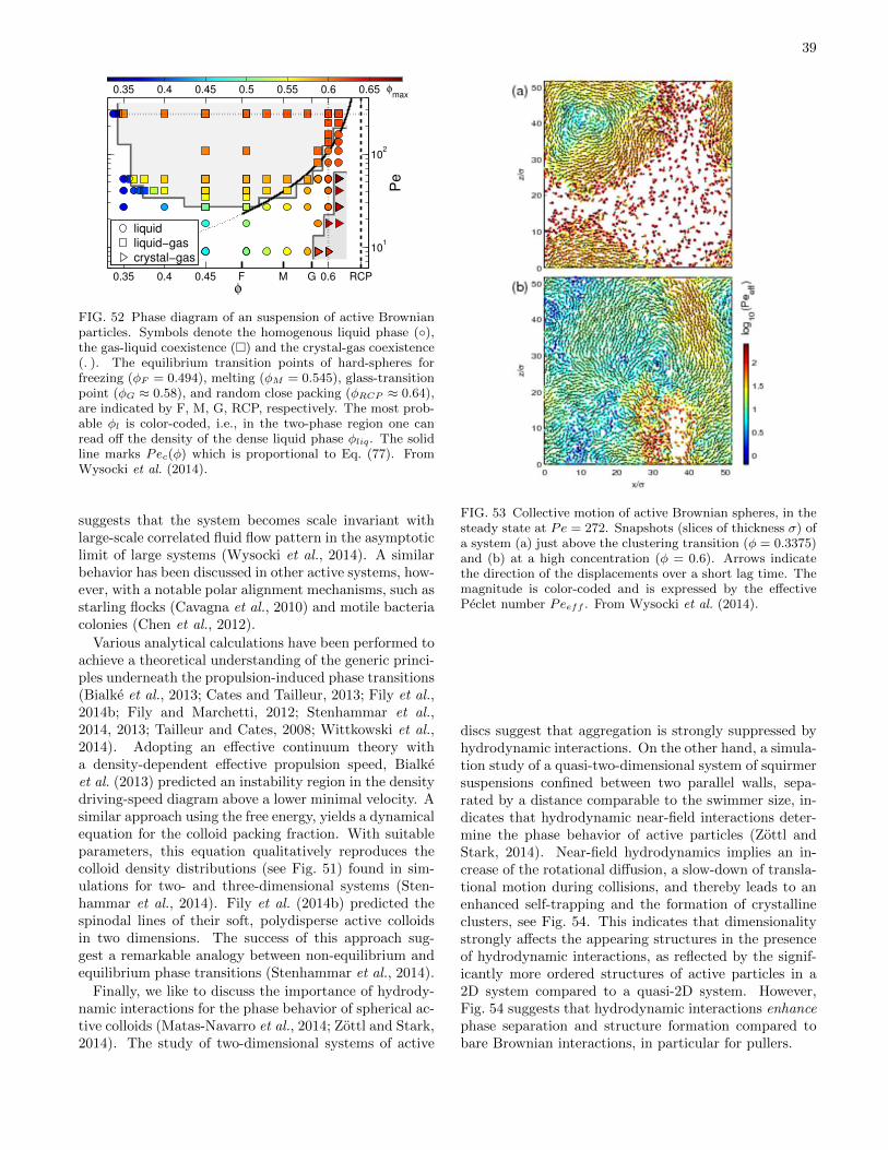
39
0.35 0.4 0.45 F M G 0.6 RCP
101
102
φ
Pe
φmax
0.35 0.4 0.45 0.5 0.55 0.6 0.65
liquid
liquid−gas
crystal−gas
FIG. 52 Phase diagram of an suspension of active Brownianparticles. Symbols denote the homogenous liquid phase (),the gas-liquid coexistence () and the crystal-gas coexistence(.). The equilibrium transition points of hard-spheres forfreezing (φF = 0.494), melting (φM = 0.545), glass-transitionpoint (φG ≈ 0.58), and random close packing (φRCP ≈ 0.64),are indicated by F, M, G, RCP, respectively. The most prob-able φl is color-coded, i.e., in the two-phase region one canread off the density of the dense liquid phase φliq. The solidline marks Pec(φ) which is proportional to Eq. (77). FromWysocki et al. (2014).
suggests that the system becomes scale invariant withlarge-scale correlated fluid flow pattern in the asymptoticlimit of large systems (Wysocki et al., 2014). A similarbehavior has been discussed in other active systems, how-ever, with a notable polar alignment mechanisms, such asstarling flocks (Cavagna et al., 2010) and motile bacteriacolonies (Chen et al., 2012).
Various analytical calculations have been performed toachieve a theoretical understanding of the generic princi-ples underneath the propulsion-induced phase transitions(Bialke et al., 2013; Cates and Tailleur, 2013; Fily et al.,2014b; Fily and Marchetti, 2012; Stenhammar et al.,2014, 2013; Tailleur and Cates, 2008; Wittkowski et al.,2014). Adopting an effective continuum theory witha density-dependent effective propulsion speed, Bialkeet al. (2013) predicted an instability region in the densitydriving-speed diagram above a lower minimal velocity. Asimilar approach using the free energy, yields a dynamicalequation for the colloid packing fraction. With suitableparameters, this equation qualitatively reproduces thecolloid density distributions (see Fig. 51) found in sim-ulations for two- and three-dimensional systems (Sten-hammar et al., 2014). Fily et al. (2014b) predicted thespinodal lines of their soft, polydisperse active colloidsin two dimensions. The success of this approach sug-gest a remarkable analogy between non-equilibrium andequilibrium phase transitions (Stenhammar et al., 2014).
Finally, we like to discuss the importance of hydrody-namic interactions for the phase behavior of spherical ac-tive colloids (Matas-Navarro et al., 2014; Zottl and Stark,2014). The study of two-dimensional systems of active
FIG. 53 Collective motion of active Brownian spheres, in thesteady state at Pe = 272. Snapshots (slices of thickness σ) ofa system (a) just above the clustering transition (φ = 0.3375)and (b) at a high concentration (φ = 0.6). Arrows indicatethe direction of the displacements over a short lag time. Themagnitude is color-coded and is expressed by the effectivePeclet number Peeff . From Wysocki et al. (2014).
discs suggest that aggregation is strongly suppressed byhydrodynamic interactions. On the other hand, a simula-tion study of a quasi-two-dimensional system of squirmersuspensions confined between two parallel walls, sepa-rated by a distance comparable to the swimmer size, in-dicates that hydrodynamic near-field interactions deter-mine the phase behavior of active particles (Zottl andStark, 2014). Near-field hydrodynamics implies an in-crease of the rotational diffusion, a slow-down of transla-tional motion during collisions, and thereby leads to anenhanced self-trapping and the formation of crystallineclusters, see Fig. 54. This indicates that dimensionalitystrongly affects the appearing structures in the presenceof hydrodynamic interactions, as reflected by the signif-icantly more ordered structures of active particles in a2D system compared to a quasi-2D system. However,Fig. 54 suggests that hydrodynamic interactions enhancephase separation and structure formation compared tobare Brownian interactions, in particular for pullers.
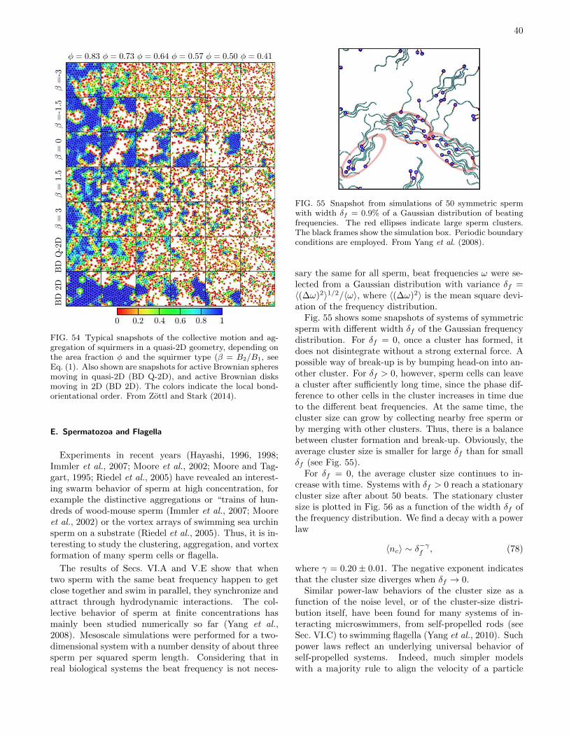
40
FIG. 54 Typical snapshots of the collective motion and ag-gregation of squirmers in a quasi-2D geometry, depending onthe area fraction φ and the squirmer type (β = B2/B1, seeEq. (1). Also shown are snapshots for active Brownian spheresmoving in quasi-2D (BD Q-2D), and active Brownian disksmoving in 2D (BD 2D). The colors indicate the local bond-orientational order. From Zottl and Stark (2014).
E. Spermatozoa and Flagella
Experiments in recent years (Hayashi, 1996, 1998;Immler et al., 2007; Moore et al., 2002; Moore and Tag-gart, 1995; Riedel et al., 2005) have revealed an interest-ing swarm behavior of sperm at high concentration, forexample the distinctive aggregations or “trains of hun-dreds of wood-mouse sperm (Immler et al., 2007; Mooreet al., 2002) or the vortex arrays of swimming sea urchinsperm on a substrate (Riedel et al., 2005). Thus, it is in-teresting to study the clustering, aggregation, and vortexformation of many sperm cells or flagella.
The results of Secs. VI.A and V.E show that whentwo sperm with the same beat frequency happen to getclose together and swim in parallel, they synchronize andattract through hydrodynamic interactions. The col-lective behavior of sperm at finite concentrations hasmainly been studied numerically so far (Yang et al.,2008). Mesoscale simulations were performed for a two-dimensional system with a number density of about threesperm per squared sperm length. Considering that inreal biological systems the beat frequency is not neces-
FIG. 55 Snapshot from simulations of 50 symmetric spermwith width δf = 0.9% of a Gaussian distribution of beatingfrequencies. The red ellipses indicate large sperm clusters.The black frames show the simulation box. Periodic boundaryconditions are employed. From Yang et al. (2008).
sary the same for all sperm, beat frequencies ω were se-lected from a Gaussian distribution with variance δf =〈(∆ω)2〉1/2/〈ω〉, where 〈(∆ω)2〉 is the mean square devi-ation of the frequency distribution.
Fig. 55 shows some snapshots of systems of symmetricsperm with different width δf of the Gaussian frequencydistribution. For δf = 0, once a cluster has formed, itdoes not disintegrate without a strong external force. Apossible way of break-up is by bumping head-on into an-other cluster. For δf > 0, however, sperm cells can leavea cluster after sufficiently long time, since the phase dif-ference to other cells in the cluster increases in time dueto the different beat frequencies. At the same time, thecluster size can grow by collecting nearby free sperm orby merging with other clusters. Thus, there is a balancebetween cluster formation and break-up. Obviously, theaverage cluster size is smaller for large δf than for smallδf (see Fig. 55).
For δf = 0, the average cluster size continues to in-crease with time. Systems with δf > 0 reach a stationarycluster size after about 50 beats. The stationary clustersize is plotted in Fig. 56 as a function of the width δf ofthe frequency distribution. We find a decay with a powerlaw
〈nc〉 ∼ δ−γf , (78)
where γ = 0.20± 0.01. The negative exponent indicatesthat the cluster size diverges when δf → 0.
Similar power-law behaviors of the cluster size as afunction of the noise level, or of the cluster-size distri-bution itself, have been found for many systems of in-teracting microswimmers, from self-propelled rods (seeSec. VI.C) to swimming flagella (Yang et al., 2010). Suchpower laws reflect an underlying universal behavior ofself-propelled systems. Indeed, much simpler modelswith a majority rule to align the velocity of a particle
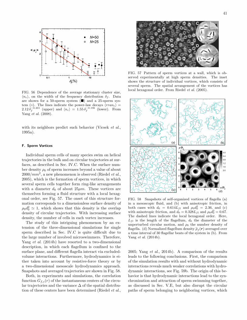
41
FIG. 56 Dependence of the average stationary cluster size,〈nc〉, on the width of the frequency distribution δf . Dataare shown for a 50-sperm system () and a 25-sperm sys-tem (). The lines indicate the power-law decays 〈rranc〉 =2.12 δ−0.201
f (upper) and 〈nc〉 = 1.55 δ−0.196f (lower). From
Yang et al. (2008).
with its neighbors predict such behavior (Vicsek et al.,1995a).
F. Sperm Vortices
Individual sperm cells of many species swim on helicaltrajectories in the bulk and on circular trajectories at sur-faces, as described in Sec. IV.C. When the surface num-ber density ρ0 of sperm increases beyond a value of about2000/mm2, a new phenomenon is observed (Riedel et al.,2005), which is the formation of sperm vortices, in whichseveral sperm cells together form ring-like arrangementswith a diameter d0 of about 25µm. These vortices arethemselves forming a fluid structure with a local hexag-onal order, see Fig. 57. The onset of this structure for-mation corresponds to a dimensionless surface density ofρ0d
20 ' 1, which shows that this density is the overlap
density of circular trajectories. With increasing surfacedensity, the number of cells in each vortex increases.
The study of this intriguing phenomenon by an ex-tension of the three-dimensional simulations for singlesperm described in Sec. IV.C is quite difficult due tothe large number of involved microswimmers. Therefore,Yang et al. (2014b) have resorted to a two-dimensionaldescription, in which each flagellum is confined to thesurface plane, and different flagella interact via excluded-volume interactions. Furthermore, hydrodynamics is ei-ther taken into account by resistive-force theory or bya two-dimensional mesoscale hydrodynamics approach.Snapshots and averaged trajectories are shown in Fig. 58.
Both, in experiments and simulations, the correlationfunction Gf,c(r) of the instantaneous centers of the circu-lar trajectories and the variance ∆ of the spatial distribu-tion of these centers have been determined (Riedel et al.,
FIG. 57 Pattern of sperm vortices at a wall, which is ob-served experimentally at high sperm densities. The insetshows the structure of individual vortices, which consists ofseveral sperm. The spatial arrangement of the vortices haslocal hexagonal order. From Riedel et al. (2005).
0 1 2 3 4 5 60
1
2
3
4
5
6
(d)x/D
0
y/D0
0.000
0.4000
0.8000
1.200
1.600
2.000
FIG. 58 Snapshots of self-organized vortices of flagella (a)in a mesoscopic fluid, and (b) with anisotropic friction, inboth cases with d0 = 0.614Lfl and ρ0d
20 = 2.36, and (c)
with anisotropic friction, and d0 = 0.328Lfl and ρ0d20 = 0.67.
The dashed lines indicate the local hexagonal order. Here,Lfl is the length of the flagellum, d0 the diameter of theunperturbed circular motion, and ρ0 the number density offlagella. (d) Normalized flagellum density ρf (r) averaged overa time interval of 30 flagellar beats of the system in (b). FromYang et al. (2014b).
2005; Yang et al., 2014b). A comparison of the resultsleads to the following conclusions. First, the comparisonof the simulation results with and without hydrodynamicinteractions reveals much weaker correlations with hydro-dynamic interactions, see Fig. 59b. The origin of this be-havior is that hydrodynamic interactions lead to the syn-chronization and attraction of sperm swimming together,as discussed in Sec. V.E, but also disrupt the circularpaths of sperm belonging to neighboring vortices, which
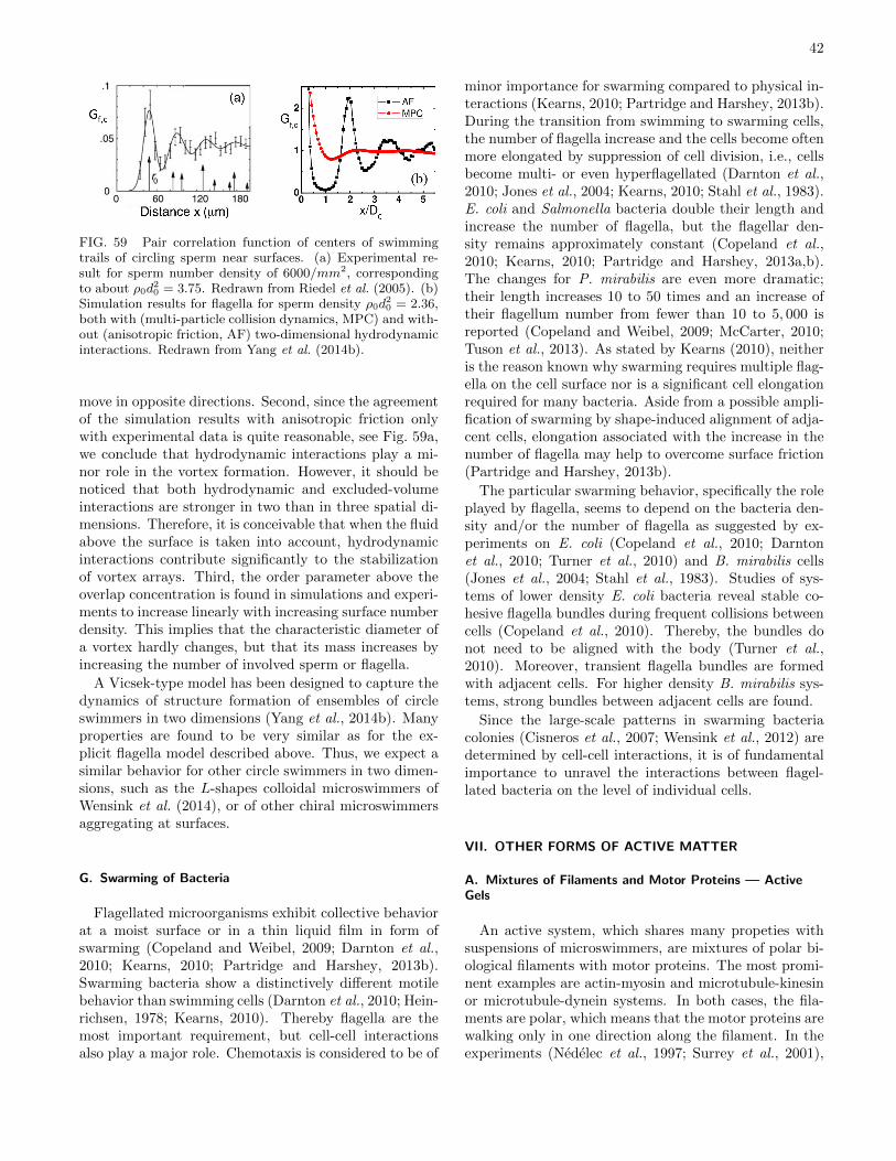
42
FIG. 59 Pair correlation function of centers of swimmingtrails of circling sperm near surfaces. (a) Experimental re-sult for sperm number density of 6000/mm2, correspondingto about ρ0d
20 = 3.75. Redrawn from Riedel et al. (2005). (b)
Simulation results for flagella for sperm density ρ0d20 = 2.36,
both with (multi-particle collision dynamics, MPC) and with-out (anisotropic friction, AF) two-dimensional hydrodynamicinteractions. Redrawn from Yang et al. (2014b).
move in opposite directions. Second, since the agreementof the simulation results with anisotropic friction onlywith experimental data is quite reasonable, see Fig. 59a,we conclude that hydrodynamic interactions play a mi-nor role in the vortex formation. However, it should benoticed that both hydrodynamic and excluded-volumeinteractions are stronger in two than in three spatial di-mensions. Therefore, it is conceivable that when the fluidabove the surface is taken into account, hydrodynamicinteractions contribute significantly to the stabilizationof vortex arrays. Third, the order parameter above theoverlap concentration is found in simulations and experi-ments to increase linearly with increasing surface numberdensity. This implies that the characteristic diameter ofa vortex hardly changes, but that its mass increases byincreasing the number of involved sperm or flagella.
A Vicsek-type model has been designed to capture thedynamics of structure formation of ensembles of circleswimmers in two dimensions (Yang et al., 2014b). Manyproperties are found to be very similar as for the ex-plicit flagella model described above. Thus, we expect asimilar behavior for other circle swimmers in two dimen-sions, such as the L-shapes colloidal microswimmers ofWensink et al. (2014), or of other chiral microswimmersaggregating at surfaces.
G. Swarming of Bacteria
Flagellated microorganisms exhibit collective behaviorat a moist surface or in a thin liquid film in form ofswarming (Copeland and Weibel, 2009; Darnton et al.,2010; Kearns, 2010; Partridge and Harshey, 2013b).Swarming bacteria show a distinctively different motilebehavior than swimming cells (Darnton et al., 2010; Hein-richsen, 1978; Kearns, 2010). Thereby flagella are themost important requirement, but cell-cell interactionsalso play a major role. Chemotaxis is considered to be of
minor importance for swarming compared to physical in-teractions (Kearns, 2010; Partridge and Harshey, 2013b).During the transition from swimming to swarming cells,the number of flagella increase and the cells become oftenmore elongated by suppression of cell division, i.e., cellsbecome multi- or even hyperflagellated (Darnton et al.,2010; Jones et al., 2004; Kearns, 2010; Stahl et al., 1983).E. coli and Salmonella bacteria double their length andincrease the number of flagella, but the flagellar den-sity remains approximately constant (Copeland et al.,2010; Kearns, 2010; Partridge and Harshey, 2013a,b).The changes for P. mirabilis are even more dramatic;their length increases 10 to 50 times and an increase oftheir flagellum number from fewer than 10 to 5, 000 isreported (Copeland and Weibel, 2009; McCarter, 2010;Tuson et al., 2013). As stated by Kearns (2010), neitheris the reason known why swarming requires multiple flag-ella on the cell surface nor is a significant cell elongationrequired for many bacteria. Aside from a possible ampli-fication of swarming by shape-induced alignment of adja-cent cells, elongation associated with the increase in thenumber of flagella may help to overcome surface friction(Partridge and Harshey, 2013b).
The particular swarming behavior, specifically the roleplayed by flagella, seems to depend on the bacteria den-sity and/or the number of flagella as suggested by ex-periments on E. coli (Copeland et al., 2010; Darntonet al., 2010; Turner et al., 2010) and B. mirabilis cells(Jones et al., 2004; Stahl et al., 1983). Studies of sys-tems of lower density E. coli bacteria reveal stable co-hesive flagella bundles during frequent collisions betweencells (Copeland et al., 2010). Thereby, the bundles donot need to be aligned with the body (Turner et al.,2010). Moreover, transient flagella bundles are formedwith adjacent cells. For higher density B. mirabilis sys-tems, strong bundles between adjacent cells are found.
Since the large-scale patterns in swarming bacteriacolonies (Cisneros et al., 2007; Wensink et al., 2012) aredetermined by cell-cell interactions, it is of fundamentalimportance to unravel the interactions between flagel-lated bacteria on the level of individual cells.
VII. OTHER FORMS OF ACTIVE MATTER
A. Mixtures of Filaments and Motor Proteins — ActiveGels
An active system, which shares many propeties withsuspensions of microswimmers, are mixtures of polar bi-ological filaments with motor proteins. The most promi-nent examples are actin-myosin and microtubule-kinesinor microtubule-dynein systems. In both cases, the fila-ments are polar, which means that the motor proteins arewalking only in one direction along the filament. In theexperiments (Nedelec et al., 1997; Surrey et al., 2001),
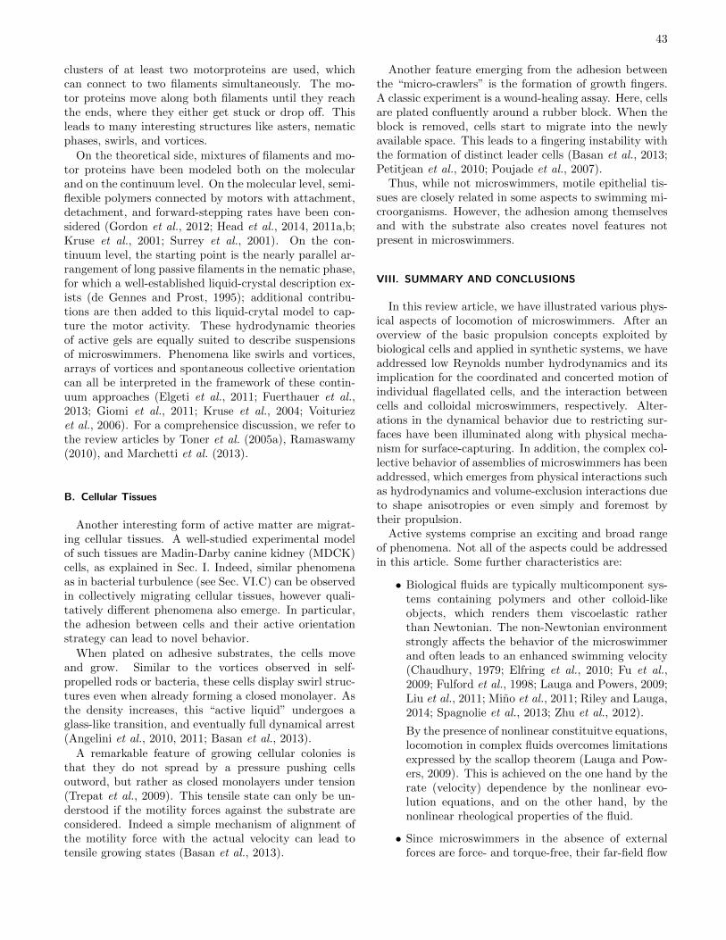
43
clusters of at least two motorproteins are used, whichcan connect to two filaments simultaneously. The mo-tor proteins move along both filaments until they reachthe ends, where they either get stuck or drop off. Thisleads to many interesting structures like asters, nematicphases, swirls, and vortices.
On the theoretical side, mixtures of filaments and mo-tor proteins have been modeled both on the molecularand on the continuum level. On the molecular level, semi-flexible polymers connected by motors with attachment,detachment, and forward-stepping rates have been con-sidered (Gordon et al., 2012; Head et al., 2014, 2011a,b;Kruse et al., 2001; Surrey et al., 2001). On the con-tinuum level, the starting point is the nearly parallel ar-rangement of long passive filaments in the nematic phase,for which a well-established liquid-crystal description ex-ists (de Gennes and Prost, 1995); additional contribu-tions are then added to this liquid-crytal model to cap-ture the motor activity. These hydrodynamic theoriesof active gels are equally suited to describe suspensionsof microswimmers. Phenomena like swirls and vortices,arrays of vortices and spontaneous collective orientationcan all be interpreted in the framework of these contin-uum approaches (Elgeti et al., 2011; Fuerthauer et al.,2013; Giomi et al., 2011; Kruse et al., 2004; Voituriezet al., 2006). For a comprehensice discussion, we refer tothe review articles by Toner et al. (2005a), Ramaswamy(2010), and Marchetti et al. (2013).
B. Cellular Tissues
Another interesting form of active matter are migrat-ing cellular tissues. A well-studied experimental modelof such tissues are Madin-Darby canine kidney (MDCK)cells, as explained in Sec. I. Indeed, similar phenomenaas in bacterial turbulence (see Sec. VI.C) can be observedin collectively migrating cellular tissues, however quali-tatively different phenomena also emerge. In particular,the adhesion between cells and their active orientationstrategy can lead to novel behavior.
When plated on adhesive substrates, the cells moveand grow. Similar to the vortices observed in self-propelled rods or bacteria, these cells display swirl struc-tures even when already forming a closed monolayer. Asthe density increases, this “active liquid” undergoes aglass-like transition, and eventually full dynamical arrest(Angelini et al., 2010, 2011; Basan et al., 2013).
A remarkable feature of growing cellular colonies isthat they do not spread by a pressure pushing cellsoutword, but rather as closed monolayers under tension(Trepat et al., 2009). This tensile state can only be un-derstood if the motility forces against the substrate areconsidered. Indeed a simple mechanism of alignment ofthe motility force with the actual velocity can lead totensile growing states (Basan et al., 2013).
Another feature emerging from the adhesion betweenthe “micro-crawlers” is the formation of growth fingers.A classic experiment is a wound-healing assay. Here, cellsare plated confluently around a rubber block. When theblock is removed, cells start to migrate into the newlyavailable space. This leads to a fingering instability withthe formation of distinct leader cells (Basan et al., 2013;Petitjean et al., 2010; Poujade et al., 2007).
Thus, while not microswimmers, motile epithelial tis-sues are closely related in some aspects to swimming mi-croorganisms. However, the adhesion among themselvesand with the substrate also creates novel features notpresent in microswimmers.
VIII. SUMMARY AND CONCLUSIONS
In this review article, we have illustrated various phys-ical aspects of locomotion of microswimmers. After anoverview of the basic propulsion concepts exploited bybiological cells and applied in synthetic systems, we haveaddressed low Reynolds number hydrodynamics and itsimplication for the coordinated and concerted motion ofindividual flagellated cells, and the interaction betweencells and colloidal microswimmers, respectively. Alter-ations in the dynamical behavior due to restricting sur-faces have been illuminated along with physical mecha-nism for surface-capturing. In addition, the complex col-lective behavior of assemblies of microswimmers has beenaddressed, which emerges from physical interactions suchas hydrodynamics and volume-exclusion interactions dueto shape anisotropies or even simply and foremost bytheir propulsion.
Active systems comprise an exciting and broad rangeof phenomena. Not all of the aspects could be addressedin this article. Some further characteristics are:
• Biological fluids are typically multicomponent sys-tems containing polymers and other colloid-likeobjects, which renders them viscoelastic ratherthan Newtonian. The non-Newtonian environmentstrongly affects the behavior of the microswimmerand often leads to an enhanced swimming velocity(Chaudhury, 1979; Elfring et al., 2010; Fu et al.,2009; Fulford et al., 1998; Lauga and Powers, 2009;Liu et al., 2011; Mino et al., 2011; Riley and Lauga,2014; Spagnolie et al., 2013; Zhu et al., 2012).
By the presence of nonlinear constituitve equations,locomotion in complex fluids overcomes limitationsexpressed by the scallop theorem (Lauga and Pow-ers, 2009). This is achieved on the one hand by therate (velocity) dependence by the nonlinear evo-lution equations, and on the other hand, by thenonlinear rheological properties of the fluid.
• Since microswimmers in the absence of externalforces are force- and torque-free, their far-field flow
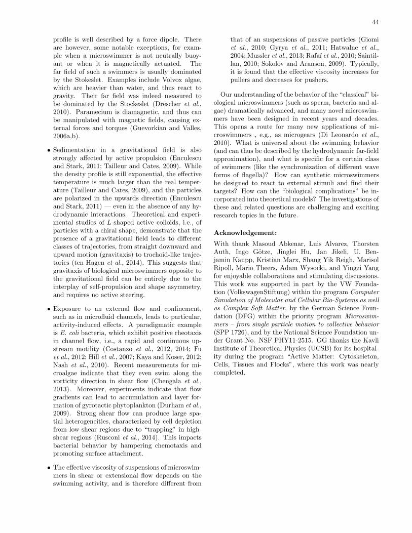
44
profile is well described by a force dipole. Thereare however, some notable exceptions, for exam-ple when a microswimmer is not neutrally buoy-ant or when it is magnetically actuated. Thefar field of such a swimmers is usually dominatedby the Stokeslet. Examples include Volvox algae,which are heavier than water, and thus react togravity. Their far field was indeed measured tobe dominated by the Stockeslet (Drescher et al.,2010). Paramecium is diamagnetic, and thus canbe manipulated with magnetic fields, causing ex-ternal forces and torques (Guevorkian and Valles,2006a,b).
• Sedimentation in a gravitational field is alsostrongly affected by active propulsion (Enculescuand Stark, 2011; Tailleur and Cates, 2009). Whilethe density profile is still exponential, the effectivetemperature is much larger than the real temper-ature (Tailleur and Cates, 2009), and the particlesare polarized in the upwards direction (Enculescuand Stark, 2011) — even in the absence of any hy-drodynamic interactions. Theoretical and experi-mental studies of L-shaped active colloids, i.e., ofparticles with a chiral shape, demonstrate that thepresence of a gravitational field leads to differentclasses of trajectories, from straight downward andupward motion (gravitaxis) to trochoid-like trajec-tories (ten Hagen et al., 2014). This suggests thatgravitaxis of biological microswimmers opposite tothe gravitational field can be entirely due to theinterplay of self-propulsion and shape asymmetry,and requires no active steering.
• Exposure to an external flow and confinement,such as in microfluid channels, leads to particular,activity-induced effects. A paradigmatic exampleis E. coli bacteria, which exhibit positive rheotaxisin channel flow, i.e., a rapid and continuous up-stream motility (Costanzo et al., 2012, 2014; Fuet al., 2012; Hill et al., 2007; Kaya and Koser, 2012;Nash et al., 2010). Recent measurements for mi-croalgae indicate that they even swim along thevorticity direction in shear flow (Chengala et al.,2013). Moreover, experiments indicate that flowgradients can lead to accumulation and layer for-mation of gyrotactic phytoplankton (Durham et al.,2009). Strong shear flow can produce large spa-tial heterogeneities, characterized by cell depletionfrom low-shear regions due to “trapping” in high-shear regions (Rusconi et al., 2014). This impactsbacterial behavior by hampering chemotaxis andpromoting surface attachment.
• The effective viscosity of suspensions of microswim-mers in shear or extensional flow depends on theswimming activity, and is therefore different from
that of an suspensions of passive particles (Giomiet al., 2010; Gyrya et al., 2011; Hatwalne et al.,2004; Mussler et al., 2013; Rafaı et al., 2010; Saintil-lan, 2010; Sokolov and Aranson, 2009). Typically,it is found that the effective viscosity increases forpullers and decreases for pushers.
Our understanding of the behavior of the “classical” bi-ological microswimmers (such as sperm, bacteria and al-gae) dramatically advanced, and many novel microswim-mers have been designed in recent years and decades.This opens a route for many new applications of mi-croswimmers , e.g., as microgears (Di Leonardo et al.,2010). What is universal about the swimming behavior(and can thus be described by the hydrodynamic far-fieldapproximation), and what is specific for a certain classof swimmers (like the synchronization of different waveforms of flagella)? How can synthetic microswimmersbe designed to react to external stimuli and find theirtargets? How can the “biological complications” be in-corporated into theoretical models? The investigations ofthese and related questions are challenging and excitingresearch topics in the future.
Acknowledgement:
With thank Masoud Abkenar, Luis Alvarez, ThorstenAuth, Ingo Gotze, Jinglei Hu, Jan Jikeli, U. Ben-jamin Kaupp, Kristian Marx, Shang Yik Reigh, MarisolRipoll, Mario Theers, Adam Wysocki, and Yingzi Yangfor enjoyable collaborations and stimulating discussions.This work was supported in part by the VW Founda-tion (VolkswagenStiftung) within the program ComputerSimulation of Molecular and Cellular Bio-Systems as wellas Complex Soft Matter, by the German Science Foun-dation (DFG) within the priority program Microswim-mers – from single particle motion to collective behavior(SPP 1726), and by the National Science Foundation un-der Grant No. NSF PHY11-2515. GG thanks the KavliInstitute of Theoretical Physics (UCSB) for its hospital-ity during the program “Active Matter: Cytoskeleton,Cells, Tissues and Flocks”, where this work was nearlycompleted.
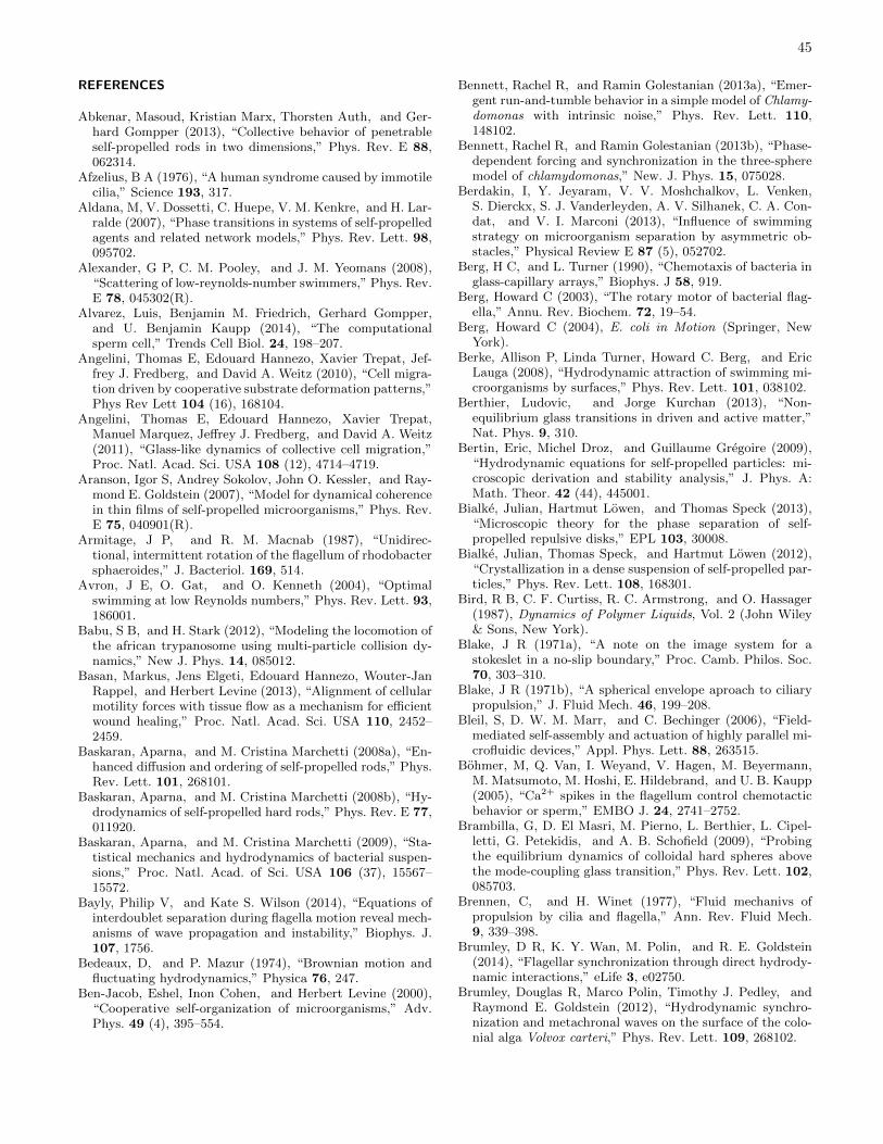
45
REFERENCES
Abkenar, Masoud, Kristian Marx, Thorsten Auth, and Ger-hard Gompper (2013), “Collective behavior of penetrableself-propelled rods in two dimensions,” Phys. Rev. E 88,062314.
Afzelius, B A (1976), “A human syndrome caused by immotilecilia,” Science 193, 317.
Aldana, M, V. Dossetti, C. Huepe, V. M. Kenkre, and H. Lar-ralde (2007), “Phase transitions in systems of self-propelledagents and related network models,” Phys. Rev. Lett. 98,095702.
Alexander, G P, C. M. Pooley, and J. M. Yeomans (2008),“Scattering of low-reynolds-number swimmers,” Phys. Rev.E 78, 045302(R).
Alvarez, Luis, Benjamin M. Friedrich, Gerhard Gompper,and U. Benjamin Kaupp (2014), “The computationalsperm cell,” Trends Cell Biol. 24, 198–207.
Angelini, Thomas E, Edouard Hannezo, Xavier Trepat, Jef-frey J. Fredberg, and David A. Weitz (2010), “Cell migra-tion driven by cooperative substrate deformation patterns,”Phys Rev Lett 104 (16), 168104.
Angelini, Thomas E, Edouard Hannezo, Xavier Trepat,Manuel Marquez, Jeffrey J. Fredberg, and David A. Weitz(2011), “Glass-like dynamics of collective cell migration,”Proc. Natl. Acad. Sci. USA 108 (12), 4714–4719.
Aranson, Igor S, Andrey Sokolov, John O. Kessler, and Ray-mond E. Goldstein (2007), “Model for dynamical coherencein thin films of self-propelled microorganisms,” Phys. Rev.E 75, 040901(R).
Armitage, J P, and R. M. Macnab (1987), “Unidirec-tional, intermittent rotation of the flagellum of rhodobactersphaeroides,” J. Bacteriol. 169, 514.
Avron, J E, O. Gat, and O. Kenneth (2004), “Optimalswimming at low Reynolds numbers,” Phys. Rev. Lett. 93,186001.
Babu, S B, and H. Stark (2012), “Modeling the locomotion ofthe african trypanosome using multi-particle collision dy-namics,” New J. Phys. 14, 085012.
Basan, Markus, Jens Elgeti, Edouard Hannezo, Wouter-JanRappel, and Herbert Levine (2013), “Alignment of cellularmotility forces with tissue flow as a mechanism for efficientwound healing,” Proc. Natl. Acad. Sci. USA 110, 2452–2459.
Baskaran, Aparna, and M. Cristina Marchetti (2008a), “En-hanced diffusion and ordering of self-propelled rods,” Phys.Rev. Lett. 101, 268101.
Baskaran, Aparna, and M. Cristina Marchetti (2008b), “Hy-drodynamics of self-propelled hard rods,” Phys. Rev. E 77,011920.
Baskaran, Aparna, and M. Cristina Marchetti (2009), “Sta-tistical mechanics and hydrodynamics of bacterial suspen-sions,” Proc. Natl. Acad. of Sci. USA 106 (37), 15567–15572.
Bayly, Philip V, and Kate S. Wilson (2014), “Equations ofinterdoublet separation during flagella motion reveal mech-anisms of wave propagation and instability,” Biophys. J.107, 1756.
Bedeaux, D, and P. Mazur (1974), “Brownian motion andfluctuating hydrodynamics,” Physica 76, 247.
Ben-Jacob, Eshel, Inon Cohen, and Herbert Levine (2000),“Cooperative self-organization of microorganisms,” Adv.Phys. 49 (4), 395–554.
Bennett, Rachel R, and Ramin Golestanian (2013a), “Emer-gent run-and-tumble behavior in a simple model of Chlamy-domonas with intrinsic noise,” Phys. Rev. Lett. 110,148102.
Bennett, Rachel R, and Ramin Golestanian (2013b), “Phase-dependent forcing and synchronization in the three-spheremodel of chlamydomonas,” New. J. Phys. 15, 075028.
Berdakin, I, Y. Jeyaram, V. V. Moshchalkov, L. Venken,S. Dierckx, S. J. Vanderleyden, A. V. Silhanek, C. A. Con-dat, and V. I. Marconi (2013), “Influence of swimmingstrategy on microorganism separation by asymmetric ob-stacles,” Physical Review E 87 (5), 052702.
Berg, H C, and L. Turner (1990), “Chemotaxis of bacteria inglass-capillary arrays,” Biophys. J 58, 919.
Berg, Howard C (2003), “The rotary motor of bacterial flag-ella,” Annu. Rev. Biochem. 72, 19–54.
Berg, Howard C (2004), E. coli in Motion (Springer, NewYork).
Berke, Allison P, Linda Turner, Howard C. Berg, and EricLauga (2008), “Hydrodynamic attraction of swimming mi-croorganisms by surfaces,” Phys. Rev. Lett. 101, 038102.
Berthier, Ludovic, and Jorge Kurchan (2013), “Non-equilibrium glass transitions in driven and active matter,”Nat. Phys. 9, 310.
Bertin, Eric, Michel Droz, and Guillaume Gregoire (2009),“Hydrodynamic equations for self-propelled particles: mi-croscopic derivation and stability analysis,” J. Phys. A:Math. Theor. 42 (44), 445001.
Bialke, Julian, Hartmut Lowen, and Thomas Speck (2013),“Microscopic theory for the phase separation of self-propelled repulsive disks,” EPL 103, 30008.
Bialke, Julian, Thomas Speck, and Hartmut Lowen (2012),“Crystallization in a dense suspension of self-propelled par-ticles,” Phys. Rev. Lett. 108, 168301.
Bird, R B, C. F. Curtiss, R. C. Armstrong, and O. Hassager(1987), Dynamics of Polymer Liquids, Vol. 2 (John Wiley& Sons, New York).
Blake, J R (1971a), “A note on the image system for astokeslet in a no-slip boundary,” Proc. Camb. Philos. Soc.70, 303–310.
Blake, J R (1971b), “A spherical envelope aproach to ciliarypropulsion,” J. Fluid Mech. 46, 199–208.
Bleil, S, D. W. M. Marr, and C. Bechinger (2006), “Field-mediated self-assembly and actuation of highly parallel mi-crofluidic devices,” Appl. Phys. Lett. 88, 263515.
Bohmer, M, Q. Van, I. Weyand, V. Hagen, M. Beyermann,M. Matsumoto, M. Hoshi, E. Hildebrand, and U. B. Kaupp(2005), “Ca2+ spikes in the flagellum control chemotacticbehavior or sperm,” EMBO J. 24, 2741–2752.
Brambilla, G, D. El Masri, M. Pierno, L. Berthier, L. Cipel-letti, G. Petekidis, and A. B. Schofield (2009), “Probingthe equilibrium dynamics of colloidal hard spheres abovethe mode-coupling glass transition,” Phys. Rev. Lett. 102,085703.
Brennen, C, and H. Winet (1977), “Fluid mechanivs ofpropulsion by cilia and flagella,” Ann. Rev. Fluid Mech.9, 339–398.
Brumley, D R, K. Y. Wan, M. Polin, and R. E. Goldstein(2014), “Flagellar synchronization through direct hydrody-namic interactions,” eLife 3, e02750.
Brumley, Douglas R, Marco Polin, Timothy J. Pedley, andRaymond E. Goldstein (2012), “Hydrodynamic synchro-nization and metachronal waves on the surface of the colo-nial alga Volvox carteri,” Phys. Rev. Lett. 109, 268102.
46
Bruot, Nicolas, Loıc Damet, Jurij Kotar, Pietro Cicuta, andMarco Cosentino Lagomarsino (2011), “Noise and synchro-nization of a single active colloid,” Phys. Rev. Lett. 107,094101.
Buttinoni, Ivo, Julian Bialke, Felix Kummel, Hartmut Lowen,Clemens Bechinger, and Thomas Speck (2013), “Dynam-ical clustering and phase separation in suspensions of self-propelled colloidal particles,” Phys. Rev. Lett. 110, 238301.
Buttinoni, Ivo, Giovanni Volpe, Felix Kummel, Giorgio Volpe,and Clemens Bechinger (2012), “Active brownian motiontunable by light,” J. Phys.: Condens. Matter 24, 284129.
Calladine, C R (1975), “Construction of bacterial flagella,”Nature 255, 121.
Calladine, C R, B. F. Luisi, and J. V. Pratap (2013), “A”mechanistic” explanation of the multiple helical formsadopted by bacterial flagellar filaments,” J. Mol. Biol. 425,914.
Camalet, S, F. Julicher, and J. Prost (1999), “Self-organizedbeating and swimming of internally driven filaments,”Phys. Rev. Lett. 82, 1590–1593.
Cartwright, J H E, O. Piro, and I. Tuval (2004), “Fluid-dynamical basis of the embryonic development of left-rightasymmetry in vertebrates,” Proc. Natl. Acad. Sci. USA101, 7234.
Cates, M E (2012), “Diffusive transport without detailed bal-ance in motile bacteria: does microbiology need statisticalphysics?” Rep. Prog. Phys. 75, 042601.
Cates, M E, and J. Tailleur (2013), “When are active Brow-nian particles and run-and-tumble particles equivalent?consequences for motility-induced phase separation,” EPL101, 20010.
Cavagna, Andrea, Alessio Cimarelli, Irene Giardina, Gior-gio Parisi, Raffaele Santagati, Fabio Stefanini, and Mas-similiano Viale (2010), “Scale-free correlations in starlingflocks,” Proc. Natl. Acad. Sci. USA 107, 11865–11870.
Chate, Hugues, Francesco Ginelli, Guillaume Gregoire,and Franck Raynaud (2008), “Collective motion of self-propelled particles interacting without cohesion,” Phys.Rev. E 77, 046113.
Chattopadhyay, Suddhashil, Radu Moldovan, Chuck Yeung,and X. L. Wu (2006), “Swimming efficiency of bacteriumescherichia coli,” Proc. Natl. Acad. Sci. USA 103, 13712.
Chaudhury, T K (1979), “On swimming in a visco-elastic liq-uid,” J. Fluid Mech. 95, 189.
Chen, Xiao, Xu Dong, Avraham Be’er, Harry L. Swinney,and H. P. Zhang (2012), “Scale-invariant correlations indynamic bacterial clusters,” Phys. Rev. Lett. 108, 148101.
Chen, Xiaobing, and Howard C. Berg (2000), “Torque-speedrelationship of the flagellar rotary motor of escherichiacoli,” Biophys. J. 78, 1036.
Chengala, Anwar, Miki Hondzo, and Jian Sheng (2013), “Mi-croalga propels along vorticity direction in a shear flow,”Phys. Rev. E 87, 052704.
Cisneros, L H, R. Cortez, C. Dombrowski, R. E. Goldstein,and J. O. Kessler (2007), “Fliud dynamics of self-propelledmicroorganisms, from individuals to concentrated popula-tions,” Exp. Fluids 73, 737.
Cisneros, Luis H, John O. Kessler, Sujoy Ganguly, and Ray-mond E. Goldstein (2011), “Dynamics of swimming bacte-ria: Transition to directional order at high concentration,”Phys. Rev. E 83, 061907.
Copeland, M F, S. T. Flickinger, H. H. Tucson, and D. B.Weibel (2010), “Studying the dynamics of flagella in multi-cellular communities of escherichia coli by using biarsenical
dyes,” Appl. Environ. Microbiol. 76, 1241.Copeland, M F, and D. B. Weibel (2009), “Bacterial swarm-
ing: a model system for studying dynamic self-assembly,”Soft Matter 5, 1174.
Cortez, R, L. Fauci, and A. Medovikov (2005), “The methodof regularized Stokeslets in three dimensions: Analysis, val-idation, and application to helical swimming,” J. FliudMech. 17, 031504.
Cosson, J, P. Huitorel, and C. Gagnon (2003), “How sper-matozoa come to be confined to surfaces,” Cell Motil. Cy-toskel. 54, 56–63.
Costanzo, A, R. Di Leonardo, G. Ruocco, and L. Ange-lani (2012), “Transport of self-propelling bacteria in micro-channel flow,” J. Phys.: Condens. Matter 24, 065101.
Costanzo, Andrea, Jens Elgeti, Thorsten Auth, GerhardGompper, and Marisol Ripoll (2014), “Motility-sorting ofself-propelled particles in microchannels,” EPL 107, 36003[1–6].
Crenshaw, H C (1989), “Kinematics of helical motion of mi-croorganisms capable of motion with four degrees of free-dom,” Biophys. J. 56, 1029–1035.
Crenshaw, H C (1996), “A new look at locomotion in mi-croorganisms: Rotating and translating,” Am. Zool. 36,608–618.
Damet, Loic, Giovanni M. Cicuta, Jurij Kotar,Marco Cosentino Lagomarsino, and Pietro Cicuta(2012), “Hydrodynamically synchronized states in activecolloidal arrays,” Soft Matter 8, 8672.
Daniels, R, J. Vanderleyden, and J. Michiels (2004), “Quo-rum sensing and swarming migration in bacteria,” FEMSMicrobiol. Rev. 28, 262.
Darnton, N C, and H. C. Berg (2007), “Force-extension mea-surements on bacterial flagella: Triggering polymorphictransformations,” Biophys. J. 92, 2230.
Darnton, N C, L. Turner, S. Rojevsky, and H. C. Berg (2010),“Dynamics of bacterial swarming,” Biophys. J. 98, 2082.
Darnton, Nicholas C, Linda Turner, Svetlana Rojevsky, andHoward C. Berg (2007), “On torque and tumbling in swim-ming escherichia coli,” J. Bacteriol. 189, 1756–1764.
de Gennes, P-G, and J. Prost (1995), The Physics of LiquidCrystals (Clarendon Press, Oxford).
Denissenko, Petr, Vasily Kantsler, David J. Smith, and Jack-son Kirkman-Brown (2012), “Human spermatozoa migra-tion in microchannels reveals boundary-following naviga-tion,” Proc. Natl. Acad. Sci. USA 109, 8007.
Deseigne, Julien, Olivier Dauchot, and Hugues Chate (2010),“Collective motion of vibrated polar disks,” Phys. Rev.Lett. 105, 098001.
Dhont, J K G (1996), An Introduction to Dynamics of Col-loids (Elsevier, Amsterdam).
Di Leonardo, R, L. Angelani, D. Dell’arciprete, G. Ruocco,V. Iebba, S. Schippa, M. P. Conte, F. Mecarini, F. De An-gelis, and E. Di Fabrizio (2010), “Bacterial ratchet mo-tors,” Proc. Natl. Acad. Sci. USA 107, 9541–9545.
Di Leonardo, R, A. Buzas, L. Kelemen, G. Vizsnyiczai,L. Oroszi, and P. Ormos (2012), “Hydrodynamic synchro-nization of light driven microrotors,” Phys. Rev. Lett. 109,034104.
DiLuzio, W R, L. Turner, M. Mayer, P. Garstecki, D. B.Weibel, H. C. Berg, and G. M. Whitesides (2005), “Es-cherichia coli swim on the right-hand side,” Nature 435,1271.
Doi, M, and S. F. Edwards (1986), The Theory of PolymerDynamics (Clarendon Press, Oxford).
47
D’Orsogna, M R, Y. L. Chuang, A. L. Bertozzi, and L. S.Chayes (2006), “Self-propelled particles with soft-core in-teractions: Patterns, stability, and collapse,” Phys. Rev.Lett. 96, 104302.
Drescher, K, J. Dunkel, L. H. Cisneros, S. Ganguly, and R. E.Goldstein (2011), “Fluid dynamics and noise in bacterialcell-cell and cell-surface scattering,” Proc. Natl. Acad. Sci.USA 108, 10940.
Drescher, Knut, Raymond E. Goldstein, Nicolas Michel,Marco Polin, and Idan Tuval (2010), “Direct measurementof the flow field around swimming microorganisms,” Phys.Rev. Lett. 105, 168101.
Drescher, Knut, Kyriacos C. Leptos, Idan Tuval, TakujiIshikawa, Timothy J. Pedley, and Raymond E. Goldstein(2009), “Dancing volvox: Hydrodynamic bound states ofswimming algae,” Phys. Rev. Lett. 102, 168101.
Dreyfus, R, J. Baudry, M. L. Roper, M. Fermigier, H. A.Stone, and J. Bibette (2005), “Microscopic artificial swim-mers,” Nature 437, 862–865.
Dunkel, Jorn, Sebastian Heidenreich, Knut Drescher, Henri-cus H. Wensink, Markus Bar, and Raymond E. Goldstein(2013), “Fluid dynamics of bacterial turbulence,” Phys.Rev. Lett. 110, 228102.
Durham, William M, John O. Kessler, and Roman Stocker(2009), “Disruption of vertical motility by shear triggersformation of thin phytoplankton layers,” Science 323, 1067.
Ebbens, Stephen J, and Jonathan R. Howse (2010), “In pur-suit of propulsion at the nanoscale,” Soft Matter 6, 726–738.
Eisenbach, Michael, and Laura C. Giojalas (2006), “Spermguidance in mammals – an unpaved road to the egg,” Nat.Rev. Mol. Cell. Biol. 7, 276–285.
Elfring, G J, and E. Lauga (2009), “Hydrodynamic phaselocking of swimming microorganisms,” Phys. Rev. Lett.103, 088101.
Elfring, Gwynn J, and Eric Lauga (2011), “Passive hydrody-namic synchronization of two-dimensional swimming cells,”Phys. Fluids 23, 011902.
Elfring, Gwynn J, Shun Pak, and Eric Lauga (2010), “Two-dimensional flagellar synchronization in viscoelastic fluids,”J. Fluid Mech. 646, 505–515.
Elgeti, J, M. E. Cates, and D. Marenduzzo (2011), “Defecthydrodynamics in 2d polar active fluids,” Soft Matter 7,3177–3185.
Elgeti, J, and G. Gompper (2008), “Hydrodynamics of activemesoscopic systems,” in NIC Symposium 2008 , NIC series,Vol. 39, edited by G. Munster, D. Wolf, and M. Kremer(Neumann Institute for Computing, Julich) p. 53.
Elgeti, J, and G. Gompper (2009), “Self-propelled rods nearsurfaces,” EPL 85, 38002 [1–6].
Elgeti, Jens, and Gerhard Gompper (2013a), “Emergence ofmetachronal waves in cilia arrays,” Proc. Natl. Acad. Sci.USA 110, 4470–4475.
Elgeti, Jens, and Gerhard Gompper (2013b), “Wall accumu-lation of self-propelled spheres,” EPL 101, 48003 [1–6].
Elgeti, Jens, U. Benjamin Kaupp, and Gerhard Gompper(2010), “Hydrodynamics of sperm cells near surfaces,” Bio-phys. J. 99, 1018–1026.
Eloy, Christophe, and Eric Lauga (2012), “Kinematics of themost efficient cilium,” Phys Rev Lett 109, 038101.
Enculescu, Mihaela, and Holger Stark (2011), “Active col-loidal suspensions exhibit polar order under gravity,” Phys.Rev. Lett. 107, 058301.
Erbe, A, M. Zientara, L. Baraban, C. Kreidler, and P. Lei-
derer (2008), “Various driving mechanisms for generatingmotion of colloidal particles,” J. Phys. Condens. Matter 20,404215.
Espanol, Pep, Miguel A. Rubio, and Ignacio Zuniga (1995),“Scaling of the time-dependent self-diffusion coefficient andthe propagation of hydrodynamic interactions,” Phys. Rev.E 51, 803.
Fauci, Lisa J, and Robert Dillon (2006), “Biofluidmechanicsof reproduction,” Annu. Rev. Fluid Mech. 38, 371–394.
Fily, Yaouen, Aparna Baskaran, and Michael F. Hagan(2014a), “Dynamics of self-propelled particles under strongconfinement,” Soft Matter 10, 5609.
Fily, Yaouen, Silke Henkes, and M. Cristina Marchetti(2014b), “Freezing and phase separation of self-propelleddisks,” Soft Matter 10, 2132.
Fily, Yaouen, and M. Cristina Marchetti (2012), “Athermalphase separation of self-Propelled particles with no align-ment,” Phys. Rev. Lett. 108, 235702.
Flores, H, E. Lobaton, S. Mendez-Diez, S. Tlupova, andR. Cortez (2005), “A study of bacterial flagellar bundling,”Bull. Math. Biol. 67, 137–168.
Foissner, Wilhelm (1998), “An updated compilation of worldsoil ciliates (protozoa, ciliophora), with ecological notes,new records, and descriptions of new species,” EuropeanJournal of Protistology 34 (2), 195–235.
Fournier-Bidoz, A, A. C. Arsenault, I. Manners, and G. A.Ozin (2005), “Synthetic self-propelled nanorotors,” Chem.Commun. , 441–443.
Friedrich, B M, and F. Julicher (2012), “Flagellar synchro-nization independent of hydrodynamic interactions,” PhysRev. Lett. 109, 138102.
Friedrich, B M, I. H. Riedel-Kruse, J. Howard, and F. Julicher(2010), “High-precision tracking of sperm swimming finestructure provides strong test of resistive force theory,” J.Exp. Biol. 213, 1226–1234.
Friedrich, Benjamin M, and Frank Julicher (2007), “Chemo-taxis of sperm cells,” Proc. Natl. Acad. Sci. 104, 13256–13261.
Frymier, P D, R. M. Ford, H. C. Berg, and P. T. Cummings(1995), “Three-dimensional tracking of motile bacteria neara solid planar surface,” Proc. Nat. Acad. Sci. USA 92, 6195.
Fu, HC, T.R. Powers, and R. Stocker (2012), “Bacterial rheo-taxis,” Proc. Natl. Acad. Sci. USA 109, 4780–4785.
Fu, Henry C, Charles W. Wolgemuth, and Thomas R. Pow-ers (2009), “Swimming speeds of filaments in nonlinearlyviscoelastic fluids,” Phys. Fluids 21, 033102.
Fuerthauer, S, M. Strempel, S. W. Grill, and F. Julicher(2013), “Active Chiral Processes in Thin Films,” Phys.Rev. Lett. 110 (4).
Fulford, Glenn R, David F. Katz, and Robert L. Powell(1998), “Swimming of spermatozoa in a linear viscoelasticfluid,” Biorheology 35, 295.
Gachelin, J, A Rousselet, A Lindner, and E Clement (2014),“Collective motion in an active suspension of escherichiacoli bacteria,” New J. Phys. 16, 025003.
Gachelin, Jeremie, Gaston Mino, Helene Berthet, Anke Lind-ner, Annie Rousselet, and Eric Clement (2013a), “Non-newtonian viscosity of Escherichia coli suspensions,” Phys.Rev. Lett. 110, 268103.
Gachelin, Jeremie, Gaston Mino, Helene Berthet, Anke Lind-ner, Annie Rousselet, and Eric Clement (2013b), “Non-newtonian viscosity of escherichia coli suspensions,” Phys.Rev. Lett. 110, 268103.
Gejji, Richard, Pavel M. Lushnikov, and Mark Alber (2012),
48
“Macroscopic model of self-propelled bacteria swarmingwith regular reversals,” Phys. Rev. E 85, 021903.
Geyer, Veikko F, Frank Julicher, Jonathon Howard, and Ben-jamin M. Friedrich (2013), “Cell-body rocking is a domi-nant mechanism for flagellar synchronization in a swim-ming alga,” Proc. Natl. Acad. Sci. USA 110, 18058–18063.
Ghosh, A, and P. Fischer (2009), “Controlled propulsion ofartificial magnetic nanostructured propellers,” Nano Lett.9, 2243.
Ginelli, Francesco, Fernando Peruani, Markus Bar, andHugues Chate (2010), “Large-scale collective properties ofself-propelled rods,” Phys. Rev. Lett. 104, 184502.
Giomi, L, L. Mahadevan, B. Chakraborty, and M. F. Hagan(2011), “Excitable patterns in active nematics,” Phys. Rev.Lett. 106, 218101.
Giomi, Luca, Tanniemola B. Liverpool, and M. CristinaMarchetti (2010), “Sheared active fluids: Thickening, thin-ning, and vanishing viscosity,” Phys. Rev. E 81, 051908.
Goldstein, Raymond E, Marco Polin, and Idan Tuval (2009),“Noise and synchronization in pairs of beating eukaryoticflagella,” Phys. Rev. Lett. 103, 168103.
Goldstein, Raymond E, Marco Polin, and Idan Tuval (2011),“Emergence of synchronized beating during the regrowthof eukaryotic flagella,” Phys. Rev. Lett. 107, 148103.
Golestanian, R (2008), “Three-sphere low-Reynolds-numberswimmer with a cargo container,” Eur. Phys. J. E 25, 1.
Golestanian, R (2009), “Anomalous diffusion of symmetricand asymmetric active colloids,” Phys. Rev. Lett. 102,188305.
Golestanian, R, T. B. Liverpool, and A. Ajdari (2005),“Propulsion of a molecular machine by asymmetric distri-bution of reaction products,” Phys. Rev. Lett. 94, 220801.
Golestanian, Ramin, Julia M. Yeomans, and Nariya Uchida(2011), “Hydrodynamic synchronization at low reynoldsnumber,” Soft Matter 7, 3074–3082.
Gompper, G, T. Ihle, D. M. Kroll, and R. G. Winkler(2009), “Multi-particle collision dynamics – a particle-based mesoscale simulation approach to the hydrodynamicsof complex fluids,” Adv. Polym. Sci. 221, 1–87.
Gordon, Daniel, Anne Bernheim-Groswasser, Chen Keasar,and Oded Farago (2012), “Hierarchical self-organization ofcytoskeletal active networks,” Phys. Biol. 9, 026005.
Gotze, I O, and G. Gompper (2010), “Flow generation byrotating colloids in planar microchannels,” EPL 92, 64003.
Gotze, I O, and G. Gompper (2011), “Dynamic self-assemblyand directed flow of rotating colloids in microchannels,”Phys. Rev. E 84, 041921.
Gotze, Ingo O, and Gerhard Gompper (2010), “Mesoscalesimulations of hydrodynamic squirmer interactions,” Phys.Rev. E 82, 041921 [1–9].
Gray, J, and G. J. Hancock (1955), “The propulsion of sea-urchin spermatozoa,” J. Exp. Biol. 32, 802–814.
Gregoire, Guillaume, and Hugues Chate (2004), “Onsetof collective and cohesive motion,” Phys. Rev. Lett. 92,025702.
Grzybowsk, Bartosz A, Howard A. Stone, and George M.Whitesides (2000), “Dynamic self-assembly of magnetized,millimetre-sized objects rotating at a liquid-air interface,”Nature 405, 1033–1036.
Guasto, Jeffrey S, Karl A. Johnson, and J. P. Gollub (2010),“Oscillatory flows induced by microorganisms swimming intwo dimensions,” Phys. Rev. Lett. 105, 168102.
Gueron, S, and K. Levit-Gurevich (1998), “Computation ofthe internal forces in cilia: application to ciliary motion,
the effects of viscosity, and cilia interactions,” Biophys J.74, 1658.
Gueron, S, and K. Levit-Gurevich (1999), “Energeticconsiderations of ciliary beating and the advantage ofmetachronal coordination,” Proc. Nat. Acad. Sci. USA96 (22), 12240–12245.
Gueron, S, and K. Levit-Gurevich (2001), “A three-dimensional model for ciliary motion based on the internal9+2 structure,” Proc. Biol. Sci 268 (1467), 599–607.
Gueron, Shay, Konstantin Levit-Gurevich, Nadav Liron, andJacob J. Blum (1997), “Cilia internal mechanism andmetachronal coordination as the result of hydrodynamicalcoupling,” Proc. Natl. Acad. Sci. USA 94 (12), 6001.
Guevorkian, Karine, and James M Valles, Jr (2006a), “Align-ing paramecium caudatum with static magnetic fields.”Biophys J 90, 3004–3011.
Guevorkian, Karine, and James M Valles, Jr (2006b), “Swim-ming paramecium in magnetically simulated enhanced, re-duced, and inverted gravity environments.” Proc Natl AcadSci U S A 103 (35), 13051–13056.
Guirao, B, and J.F. Joanny (2007), “Spontaneous creationof macroscopic flow and metachronal waves in an array ofcilia,” Biophys. J. 92, 1900–1917.
Gyrya, V, K. Lipnikov, I. S. Aranson, and L. Berlyand (2011),“Effective shear viscosity and dynamics of suspensions ofmicro-swimmers from small to moderate concentrations,”J. Math. Biol. 62, 707–740.
ten Hagen, Borge, Felix Kummel, Raphael Wittkowski,Daisuke Takagi, Hartmut Lowen, and Clemens Bechinger(2014), “Gravitaxis of asymmetric self-propelled colloidalparticles,” Nat. Commun. 5, 4829.
Hancock, G J (1953), “The self-propulsion of microscopic or-ganisms through liquids,” Proc. R. Soc. Lond. A 217, 96.
Harada, Yoshie, Akira Noguchi, Akiyoshi Kishino, andToshio Yanagida (1987), “Sliding movement of single actinfilaments on one-headed myosin filaments,” Nature 326,805–808.
Harnau, L, R. G. Winkler, and P. Reineker (1996), “Dynamicstructure factor of semiflexible macromolecules in dilute so-lution,” J. Chem. Phys. 104, 6355.
Harshey, R M (1994), “Bees aren’t the only ones: swarmingin gram-negative bacteria,” Mol. Microbiol. 13, 389.
Harshey, R M (2003), “Bacterial motility on a surface: manyways to a common goal,” Annu. Rev. Microbiol. 57, 249.
Harvey, Cameron W, Mark Alber, Lev S. Tsimring, andIgor S. Aranson (2013), “Continuum modeling of myxobac-teria clustering,” New J. Phys. 15, 035029.
Harvey, Cameron W, Faruck Morcos, Christopher R. Sweet,Dale Kaiser, Santanu Chatterjee, Xiaomin Liu, Danny Z.Chen, and Mark Alber (2011), “Study of elastic collisionsof myxococcus xanthus in swarms,” Phys. Biol. 8, 026016.
Hatwalne, Y, S. Ramaswamy, M. Rao, and R. A. Simha(2004), “Rheology of active-particle suspensions,” Phys.Rev. Lett. 92, 118101.
Hayashi, F (1996), “Insemination through an externally at-tached spermatophore: bundled sperm and post-copulatorymate guarding by male fishflies,” J. Insect Physiol. 42, 859–866.
Hayashi, F (1998), “Sperm co-operation in the fishfly,” Funct.Ecol. 12, 347–350.
Head, D A, W. J. Briels, and Gerhard Gompper (2014),“Non-equilibrium structure and dynamics in a microscopicmodel of thin-film active gels,” Phys. Rev. E 89, 032705[1–11].
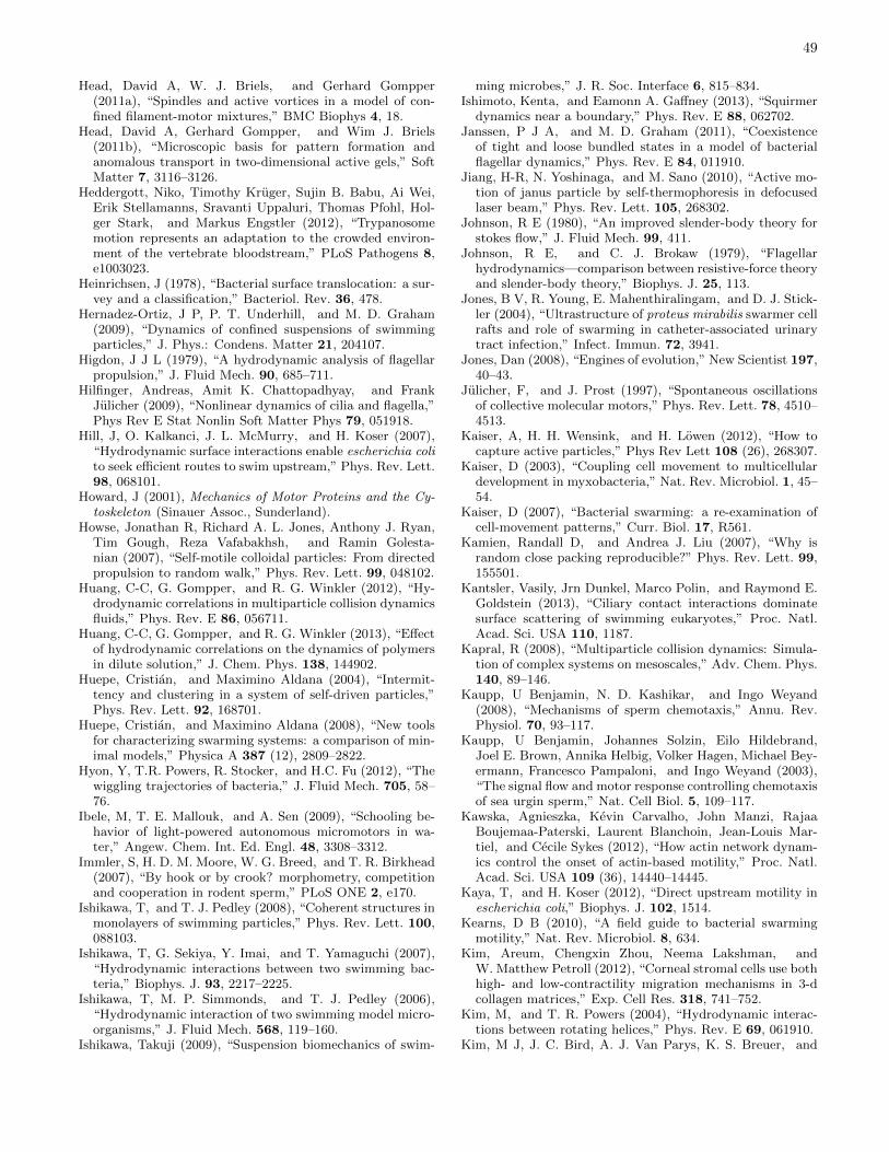
49
Head, David A, W. J. Briels, and Gerhard Gompper(2011a), “Spindles and active vortices in a model of con-fined filament-motor mixtures,” BMC Biophys 4, 18.
Head, David A, Gerhard Gompper, and Wim J. Briels(2011b), “Microscopic basis for pattern formation andanomalous transport in two-dimensional active gels,” SoftMatter 7, 3116–3126.
Heddergott, Niko, Timothy Kruger, Sujin B. Babu, Ai Wei,Erik Stellamanns, Sravanti Uppaluri, Thomas Pfohl, Hol-ger Stark, and Markus Engstler (2012), “Trypanosomemotion represents an adaptation to the crowded environ-ment of the vertebrate bloodstream,” PLoS Pathogens 8,e1003023.
Heinrichsen, J (1978), “Bacterial surface translocation: a sur-vey and a classification,” Bacteriol. Rev. 36, 478.
Hernadez-Ortiz, J P, P. T. Underhill, and M. D. Graham(2009), “Dynamics of confined suspensions of swimmingparticles,” J. Phys.: Condens. Matter 21, 204107.
Higdon, J J L (1979), “A hydrodynamic analysis of flagellarpropulsion,” J. Fluid Mech. 90, 685–711.
Hilfinger, Andreas, Amit K. Chattopadhyay, and FrankJulicher (2009), “Nonlinear dynamics of cilia and flagella,”Phys Rev E Stat Nonlin Soft Matter Phys 79, 051918.
Hill, J, O. Kalkanci, J. L. McMurry, and H. Koser (2007),“Hydrodynamic surface interactions enable escherichia colito seek efficient routes to swim upstream,” Phys. Rev. Lett.98, 068101.
Howard, J (2001), Mechanics of Motor Proteins and the Cy-toskeleton (Sinauer Assoc., Sunderland).
Howse, Jonathan R, Richard A. L. Jones, Anthony J. Ryan,Tim Gough, Reza Vafabakhsh, and Ramin Golesta-nian (2007), “Self-motile colloidal particles: From directedpropulsion to random walk,” Phys. Rev. Lett. 99, 048102.
Huang, C-C, G. Gompper, and R. G. Winkler (2012), “Hy-drodynamic correlations in multiparticle collision dynamicsfluids,” Phys. Rev. E 86, 056711.
Huang, C-C, G. Gompper, and R. G. Winkler (2013), “Effectof hydrodynamic correlations on the dynamics of polymersin dilute solution,” J. Chem. Phys. 138, 144902.
Huepe, Cristian, and Maximino Aldana (2004), “Intermit-tency and clustering in a system of self-driven particles,”Phys. Rev. Lett. 92, 168701.
Huepe, Cristian, and Maximino Aldana (2008), “New toolsfor characterizing swarming systems: a comparison of min-imal models,” Physica A 387 (12), 2809–2822.
Hyon, Y, T.R. Powers, R. Stocker, and H.C. Fu (2012), “Thewiggling trajectories of bacteria,” J. Fluid Mech. 705, 58–76.
Ibele, M, T. E. Mallouk, and A. Sen (2009), “Schooling be-havior of light-powered autonomous micromotors in wa-ter,” Angew. Chem. Int. Ed. Engl. 48, 3308–3312.
Immler, S, H. D. M. Moore, W. G. Breed, and T. R. Birkhead(2007), “By hook or by crook? morphometry, competitionand cooperation in rodent sperm,” PLoS ONE 2, e170.
Ishikawa, T, and T. J. Pedley (2008), “Coherent structures inmonolayers of swimming particles,” Phys. Rev. Lett. 100,088103.
Ishikawa, T, G. Sekiya, Y. Imai, and T. Yamaguchi (2007),“Hydrodynamic interactions between two swimming bac-teria,” Biophys. J. 93, 2217–2225.
Ishikawa, T, M. P. Simmonds, and T. J. Pedley (2006),“Hydrodynamic interaction of two swimming model micro-organisms,” J. Fluid Mech. 568, 119–160.
Ishikawa, Takuji (2009), “Suspension biomechanics of swim-
ming microbes,” J. R. Soc. Interface 6, 815–834.Ishimoto, Kenta, and Eamonn A. Gaffney (2013), “Squirmer
dynamics near a boundary,” Phys. Rev. E 88, 062702.Janssen, P J A, and M. D. Graham (2011), “Coexistence
of tight and loose bundled states in a model of bacterialflagellar dynamics,” Phys. Rev. E 84, 011910.
Jiang, H-R, N. Yoshinaga, and M. Sano (2010), “Active mo-tion of janus particle by self-thermophoresis in defocusedlaser beam,” Phys. Rev. Lett. 105, 268302.
Johnson, R E (1980), “An improved slender-body theory forstokes flow,” J. Fluid Mech. 99, 411.
Johnson, R E, and C. J. Brokaw (1979), “Flagellarhydrodynamics—comparison between resistive-force theoryand slender-body theory,” Biophys. J. 25, 113.
Jones, B V, R. Young, E. Mahenthiralingam, and D. J. Stick-ler (2004), “Ultrastructure of proteus mirabilis swarmer cellrafts and role of swarming in catheter-associated urinarytract infection,” Infect. Immun. 72, 3941.
Jones, Dan (2008), “Engines of evolution,” New Scientist 197,40–43.
Julicher, F, and J. Prost (1997), “Spontaneous oscillationsof collective molecular motors,” Phys. Rev. Lett. 78, 4510–4513.
Kaiser, A, H. H. Wensink, and H. Lowen (2012), “How tocapture active particles,” Phys Rev Lett 108 (26), 268307.
Kaiser, D (2003), “Coupling cell movement to multicellulardevelopment in myxobacteria,” Nat. Rev. Microbiol. 1, 45–54.
Kaiser, D (2007), “Bacterial swarming: a re-examination ofcell-movement patterns,” Curr. Biol. 17, R561.
Kamien, Randall D, and Andrea J. Liu (2007), “Why israndom close packing reproducible?” Phys. Rev. Lett. 99,155501.
Kantsler, Vasily, Jrn Dunkel, Marco Polin, and Raymond E.Goldstein (2013), “Ciliary contact interactions dominatesurface scattering of swimming eukaryotes,” Proc. Natl.Acad. Sci. USA 110, 1187.
Kapral, R (2008), “Multiparticle collision dynamics: Simula-tion of complex systems on mesoscales,” Adv. Chem. Phys.140, 89–146.
Kaupp, U Benjamin, N. D. Kashikar, and Ingo Weyand(2008), “Mechanisms of sperm chemotaxis,” Annu. Rev.Physiol. 70, 93–117.
Kaupp, U Benjamin, Johannes Solzin, Eilo Hildebrand,Joel E. Brown, Annika Helbig, Volker Hagen, Michael Bey-ermann, Francesco Pampaloni, and Ingo Weyand (2003),“The signal flow and motor response controlling chemotaxisof sea urgin sperm,” Nat. Cell Biol. 5, 109–117.
Kawska, Agnieszka, Kevin Carvalho, John Manzi, RajaaBoujemaa-Paterski, Laurent Blanchoin, Jean-Louis Mar-tiel, and Cecile Sykes (2012), “How actin network dynam-ics control the onset of actin-based motility,” Proc. Natl.Acad. Sci. USA 109 (36), 14440–14445.
Kaya, T, and H. Koser (2012), “Direct upstream motility inescherichia coli,” Biophys. J. 102, 1514.
Kearns, D B (2010), “A field guide to bacterial swarmingmotility,” Nat. Rev. Microbiol. 8, 634.
Kim, Areum, Chengxin Zhou, Neema Lakshman, andW. Matthew Petroll (2012), “Corneal stromal cells use bothhigh- and low-contractility migration mechanisms in 3-dcollagen matrices,” Exp. Cell Res. 318, 741–752.
Kim, M, and T. R. Powers (2004), “Hydrodynamic interac-tions between rotating helices,” Phys. Rev. E 69, 061910.
Kim, M J, J. C. Bird, A. J. Van Parys, K. S. Breuer, and
50
T. R. Powers (2003), “A macroscopic scale model of bac-terial flagellar bundling,” Proc. Natl. Acad. Sci. USA 100,15481.
Knight-Jones, E W (1954), “Relations between metachronismand the direction of the ciliary beat,” Q. J. Microsc. Sci.95, 503.
Koch, Donald L, and Ganesh Subramanian (2011), “Collec-tive hydrodynamics of swimming microorganisms: Livingfluids,” Ann. Rev. Fluid Mech. 43, 637.
Kotar, Jurij, Luke Debono, Nicolas Bruot, Stuart Box, DavidPhillips, Stephen Simpson, Simon Hanna, and Pietro Ci-cuta (2013), “Optimal hydrodynamic synchronization ofcolloidal rotors,” Phys. Rev. Lett. 111, 228103.
Kotar, Jurij, Marco Leoni, Bruno Bassetti, Marco CosentinoLagomarsino, and Pietro Cicuta (2010), “Hydrodynamicsynchronization of colloidal oscillators,” Proc. Natl. Acad.Sci. USA 107, 7669–7673.
Koumakis, N, and R. Di Leonardo (2013), “Stochastic hydro-dynamic synchronization in rotating energy landscapes,”Phys. Rev. Lett. 110, 174103.
Kraichnan, R H, and D. Montgomery (1980), “Two-dimensional turbulence,” Rep. Prog. Phys. 43, 547–619.
Kraikivski, Pavel, Reinhard Lipowsky, and Jan Kierfeld(2006), “Enhanced ordering of interacting filaments bymolecular motors,” Phys. Rev. Lett. 96, 258103.
Kruse, K, S. Camalet, and F. Julicher (2001), “Self-propagating patterns in active filament bundles,” Phys.Rev. Lett. 87, 138101.
Kruse, K, J. F. Joanny, F. Julicher, J. Prost, and K. Sekimoto(2004), “Asters, vortices, and rotating spirals in active gelsof polar filaments,” Phys. Rev. Lett. 92 (7), 078101.
Lagomarsino, M Cosentino, P. Jona, and B. Bassetti (2003),“Metachronal waves for deterministic switching two-stateoscillators with hydrodynamic interaction,” Phys. Rev. E68, 021908.
Lauga, E (2011), “Life around the scallop theorem,” Soft Mat-ter 7, 3060.
Lauga, E, W. R. DiLuzio, G. M. Whitesides, and H. A. Stone(2006), “Swimming in circles: motion of bacteria near solidboundaries,” Biophys. J. 90, 400.
Lauga, E, and R. E. Goldstein (2012), “Dancing of the mi-croswimmers,” Phys. Today 65, 30.
Lauga, Eric, and Thomas R. Powers (2009), “The hydro-dynamics of swimming microorganisms,” Rep. Prog. Phys.72, 096601.
Lemelle, L, J.-F. Palierne, E. Chatre, and C. Place (2010),“Counterclockwise circular motion of bacteria swimming atthe air-liquid interface,” J. Bacteriol. 192, 6307.
Lenz, Peter, and Andrey Ryskin (2006), “Collective effectsin ciliar arrays,” Phys. Biol. 3 (4), 285.
Leptos, Kyriacos C, Kirsty Y. Wan, Marco Polin, Idan Tuval,Adriana I. Pesci, and Raymond E. Goldstein (2013), “An-tiphase synchronization in a flagellar-dominance mutant ofChlamydomonas,” Phys. Rev. Lett. 111, 158101.
Lhermerout, Romain, Nicolas Bruot, Giovanni M Cicuta, Ju-rij Kotar, and Pietro Cicuta (2012), “Collective synchro-nization states in arrays of driven colloidal oscillators,” NewJ. Phys. 14, 105023.
Li, Guanglai, Lick-Kong Tam, and Jay X. Tang (2008), “Am-plified effect of Brownian motion in bacterial near-surfaceswimming,” Proc. Natl Acad. Sci. USA 105 (47), 18355.
Li, Guanglai, and Jay X. Tang (2009), “Accumulation ofmicroswimmers near a surface mediated by collision androtational brownian motion,” Phys. Rev. Lett. 103, 078101.
Lighthill, J (1976), “Flagellar hydrodynamics—The John vonNeumann Lecture 1975,” SIAM Rev. 18, 161.
Lighthill, M J (1952), “On the squirming motion of nearlyspherical deformable bodies through liquids at very smallreynolds numbers,” Commun. Pure Appl. Math. 5, 109.
Lindemann, C B (1994), “A geometric clutch hypothesis toexplain oscillations of the axoneme of cilia and flagella,” J.Theor. Biol. 168, 175–189.
Lindemann, Charles B (2014), “Engaging the “clutch” tomove forward,” Biophys. J. 107, 1487–1488.
Lindemann, Charles B, and Kathleen A. Lesich (2010), “Flag-ellar and ciliary beating: the proven and the possible,” J.Cell Sci. 123, 519–528.
Liu, Bin, Thomas R. Powers, and Kenneth S. Breuer (2011),“Force-free swimming of a model helical flagellum in vis-coelastic fluids,” Proc. Natl. Acad. Sci. USA 108, 19516.
Liu, J-K, G.-H. Deng, Z.-T. Yuan, and D. J. Liu (2000),“Turbulence under the microscope,” J. Biol. Phys. 26, 77.
Llopis, I, and I. Pagonabarraga (2008), “Hydrodynamicregimes of active rotators at fluid interfaces,” Eur. Phys.J. E 26, 103–113.
Llopis, I, I. Pagonabarraga, M. Cosentino Lagomarsino, andC. P. Lowe (2013), “Cooperative motion of intrinsic andactuated semiflexible swimmers,” Phys. Rev. E 87, 032720.
Lopez, Diego, and Eric Lauga (2014), “Dynamics of swim-ming bacteria at complex interfaces,” Phys. Fluids 26,071902.
Machemer, H (1972), “Ciliary activity and the origin ofmetachrony in paramecium: Effects of increased viscosity,”J. Exp. Biol. 57, 239–259.
Macnab, R M (1977), “Bacterial flagella rotating in bundles:A study in helical geometry,” Proc. Natl. Acad. Sci. USA74, 221.
Magariyama, Y, S. Sugiyama, K. Muramoto, I. Kawagishi,Y. Imae, and S. Kudo (1995), “Simultaneous measurementof bacterial flagellar rotation rate and swimming speed,”Biophys. J. 69, 2154.
Manghi, Manoel, Xaver Schlagberger, and Roland R. Netz(2006), “Propulsion with a rotating elastic nanorod,” Phys.Rev. Lett. 96, 068101.
Marchetti, M C, J. F. Joanny, S. Ramaswamy, T. B. Liver-pool, J. Prost, Madan Rao, and R. Aditi Simha (2013),“Hydrodynamics of soft active matter,” Rev. Mod. Phys.85, 1143.
Matas-Navarro, Ricard, Ramin Golestanian, Tanniemola B.Liverpool, and Suzanne M. Fielding (2014), “Hydrody-namic suppression of phase separation in active suspen-sions,” Phys. Rev. E 90, 032304.
McCandlish, Samuel R, Aparna Baskaran, and Michael F.Hagan (2012), “Spontaneous segregation of self-propelledparticles with different motilities,” Soft Matter 8, 2527–2534.
McCarter, L L (2010), “Bacterial acrobatics on a surface:Swirling packs, collisions, and reversals during swarming,”J. Bacteriol. 192, 3246.
Mei, Y F, A. A. Solovev, S. Sanchez, and O. G. Schmidt(2011), “Rolled-up nanotech on polymers: from basic per-ception to self-propelled catalytic microengines,” Chem.Soc. Rev. 40, 2109.
Mendelson, NH, A Bourque, K Wilkening, KR Anderson, andJC Watkins (1999), “Organized cell swimming motions inBacillus subtilis colonies: Patterns of short-lived whirls andjets,” J. Bacteriol. 181 (2), 600–609.
Mino, Gaston, Thomas E. Mallouk, Thierry Darnige, Mauri-
51
cio Hoyos, Jeremi Dauchet, Jocelyn Dunstan, RodrigoSoto, Yang Wang, Annie Rousselet, and Eric Clement(2011), “Enhanced diffusion due to active swimmers at asolid surface,” Phys. Rev. Lett. 106, 048102.
Molaei, Mehdi, Michael Barry, Roman Stocker, and JianSheng (2014), “Failed escape: Solid surfaces prevent tum-bling of escherichia coli,” Phys. Rev. Lett. 113, 068103.
Moore, H D M, K. Dvorakova, N. Jenkins, and W. G.Breed (2002), “Exceptional sperm cooperation in the woodmouse,” Nature 418, 174–177.
Moore, H D M, and D. A. Taggart (1995), “Sperm pairingin the opossum increases the efficiency of sperm movementin a viscous environment,” Biol. Reprod. 52, 947–953.
Mora, Thierry, Howard Yu, and Ned S. Wingreen (2009),“Modeling torque versus speed, shot noise, and rotationaldiffusion of the bacterial flagellar motor,” Phys. Rev. Lett.103, 248102.
Mussler, Matthias, Salima Rafaı, Philippe Peyla, and Chris-tian Wagner (2013), “Effective viscosity of non-gravitacticchlamydomonas reinhardtii microswimmer suspensions,”EPL 101, 54004.
Najafi, Ali, and Ramin Golestanian (2004), “Simple swimmerat low reynolds number: Three linked spheres,” Phys. Rev.E 69, 062901.
Nash, R W, R. Adhikari, J. Tailleur, and M. E. Cates (2010),“Run-and-tumble particles with hydrodynamics: Sedimen-tation, trapping, and upstream swimming,” Phys. Rev.Lett. 104, 258101.
Nedelec, F J, T. Surrey, A. C. Maggs, and S. Leibler (1997),“Self-organization of microtubules and motors,” Nature389, 305–308.
Ni, Ran, Martien A. Cohen Stuart, and Marjolein Dijkstra(2013), “Pushing the glass transition towards random closepacking using self-propelled hard spheres,” Nat. Commun.4, 2704.
Niedermayer, Thomas, Bruno Eckhardt, and Peter Lenz(2008), “Synchronization, phase locking, and metachronalwave formation in ciliary chains,” Chaos 18 (3), 037128.
Okamoto, Ken-Ichi, and Yasuo Nakaoka (1994), “Reconsti-tution of metachronal waves in ciliated cortical sheets ofparamecium ii. asymmetry of the ciliary movements,” J.Exp. Biol. 192, 73–81.
Or, D, B.F. Smets, J.M. Wraith, A. Dechesne, and S.P. Fried-man (2007), “Physical constraints affecting bacterial habi-tats and activity in unsaturated porous media – a review,”Advances in Water Resources 30, 1505–1527.
Osterman, Natan, and Andrej Vilfan (2011), “Finding theciliary beating pattern with optimal efficiency,” Proc. Natl.Acad. Sci. USA 108, 15727–15732.
Ozin, Geoffrey A, Ian Manners, Sebastien Fournier-Bidoz,and Andre Arsenault (2005), “Dream nanomachines,” Adv.Mater. 17, 3011–3018.
Pagonabarraga, Ignacio, and Isaac Llopis (2013), “The struc-ture and rheology of sheared model swimmer suspensions,”Soft Matter 9, 7174–7184.
Palacci, J, S. Sacanna, A. P. Steinberg, D. J. Pine, and P. M.Chaikin (2013), “Living crystals of light-activated colloidalsurfers,” Science 339, 936–940.
Partridge, J D, and R. M. Harshey (2013a), “More thanmotility: Salmonella flagella contribution to overriding fric-tion and facilitating colony hydration during swarming,” J.Bacteriol. 195, 919.
Partridge, J D, and R. M. Harshey (2013b), “Swarmming:flexible roaming plans,” J. Bacteriol. 195, 909.
Paxton, W, S. Sundararajan, T. Mallouk, and A. Sen (2006),“Chemical locomotion,” Angew. Chem. Int. Ed. 45, 5420.
Paxton, W F, K. C. Kistler, C. C. Olmeda, A. Sen, S. K.St. Angelo, Y. Cao, T. E. Mallouk, P. E. Lammert, andV. H. Crespi (2004), “Catalytic nanomotors: autonomousmovement of striped nanorods,” J. Am. Chem. Soc. 126,13424.
Peruani, F, L Schimansky-Geier, and M Bar (2010), “Clusterdynamics and cluster size distributions in systems of self-propelled particles,” Eur. Phys. J. Special Topics 191 (1),173–185.
Peruani, Fernando, Andreas Deutsch, and Markus Bar(2006), “Nonequilibrium clustering of self-propelled rods,”Phys. Rev. E 74, 030904(R).
Peruani, Fernando, Andreas Deutsch, and Markus Bar(2008), “A mean-field theory for self-propelled particles in-teracting by velocity alignment mechanisms,” Eur. Phys.J. Special Topics 157 (1), 111–122.
Peruani, Fernando, Jorn Starruss, Vladimir Jakovljevic, LotteSøgaard-Andersen, Andreas Deutsch, and Markus Bar(2012), “Collective motion and nonequilibrium cluster for-mation in colonies of gliding bacteria,” Phys. Rev. Lett.108, 098102.
Peshkov, Anton, Igor S. Aranson, Eric Bertin, Hugues Chate,and Francesco Ginelli (2012), “Nonlinear field equations foraligning self-propelled rods,” Phys. Rev. Lett. 109, 268701.
Petitjean, L, M Reffay, E Grasland-Mongrain, M Poujade,B Ladoux, A Buguin, and P Silberzan (2010), “Velocityfields in a collectively migrating epithelium,” Biophys. J.98 (9), 1790–800.
Pikovsky, A, M. Rosenblum, and J. Kurths (2002), Synchro-nization: A Universal Concept in Nonlinear Science (Cam-bridge University Press).
Platzer, J, W. Sterr, M. Hausmann, and R. Schmitt (1997),“Three genes of a motility operon and their role in flagellarrotary speed variation in Rhizobium meliloti,” J. Bacteriol.179, 6391–6399.
Polin, M, Tuval I, K. Drescher, J. P. Gollub, and R. E. Gold-stein (2009), “Chlamydomonas swims with two ’gears’ in aeukaryotic version of run-and-tumble locomotion,” Science325, 487.
Polotzek, K, and B. M. Friedrich (2013), “A three-sphereswimmer for flagellar synchronization,” New. J. Phys. 15,045005.
Popescu, M N, S. Dietrich, and G. Oshanin (2009), “Confine-ment effects on diffusiophoretic self-propellers,” J. Chem.Phys. 130, 194702.
Popescu, M N, S. Dietrich, M. Tasinkevych, and J. Ralston(2010), “Phoretic motion of spheroidal particles due to self-generated solute gradients,” Eur. Phys. J. E 31, 351–367.
Poujade, M, E. Grasland-Mongrain, A. Hertzog, J. Jouan-neau, P. Chavrier, B. Ladoux, A. Buguin, and P. Silberzan(2007), “Collective migration of an epithelial monolayer inresponse to a model wound,” Proc. Natl. Acad. Sci. USA104, 15988–15993.
Prost, Jacques, Jean-Francois Joanny, Peter Lenz, and CecileSykes (2008), “Cell motility,” Chap. The physics of Listeriapropulsion (Springer, New York) p. 1.
Purcell, E M (1977), “Life at low reynolds numbers,” Am. J.Phys. 45, 3.
Purcell, E M (1997), “The efficiency of propulsion by a rotat-ing flagellum,” Proc. Natl. Acad. Sci. USA 94, 11307.
Pusey, P N, and W. van Megen (1986), “Phase behaviour ofconcentrated suspensions of nearly hard colloidal spheres,”
52
Nature 320, 340.Qian, Bian, Hongyuan Jiang, David A. Gagnon, Kenneth S.
Breuer, and Thomas R. Powers (2009), “Minimal model forsynchronization induced by hydrodynamic interactions,”Phys. Rev. E 80, 061919.
Rafaı, Salima, Levan Jibuti, and Philippe Peyla (2010), “Ef-fective viscosity of microswimmer suspensions,” Phys. Rev.Lett. 104, 098102.
Ramaswamy, Sriram (2010), “The mechanics and statisticsof active matter,” Annual Review of Condensed MatterPhysics 1 (1), 323–345.
Ramaswamy, Sriram, R Aditi Simha, and John Toner (2003),“Active nematics on a substrate: Giant number fluctua-tions and long-time tails,” Europhys. Lett. 62 (2), 196.
Redner, Gabriel S, Michael F. Hagan, and Aparna Baskaran(2013), “Structure and dynamics of a phase-separating ac-tive colloidal fluid,” Phys. Rev. Lett. 110, 055701.
Reichert, M, and H. Stark (2005), “Synchronization of rotat-ing helices by hydrodynamic interactions,” Eur. Phys. J. E17, 493.
Reigh, S Y, R. G. Winkler, and G. Gompper (2012), “Syn-chronization and bundling of anchored bacterial flagella,”Soft Matter 8, 4363.
Reigh, S Y, R. G. Winkler, and G. Gompper (2013), “Syn-chronization, slippage, and unbundling of driven helicalflagella,” PLoS ONE 8, e70868.
Riedel, Ingmar H, Karsten Kruse, and Jonathon Howard(2005), “A self-organized vortex array of hydrodynamicallyentrained sperm cells,” Science 309, 300–303.
Riedel-Kruse, IH, A. Hilfinger, and F. Julicher (2007), “Howmolecular motors shape the flagellar beat,” HFSP 1, 192–208.
Riley, E E, and E. Lauga (2014), “Enhanced active swimmingin viscoelastic fluids,” EPL 108, 34003.
Risken, H (1989), The Fokker-Planck Equation (Springer,Berlin).
Rodenborn, Bruce, Chih-Hung Chen, Harry L. Swinney, BinLiu, and H. P. Zhang (2013), “Propulsion of microorgan-isms by a helical flagellum,” Proc. Natl. Acad. Sci. USA110, E338.
Romanczuk, P, M. Bar, W. Ebeling, B. Lindner, andL. Schimansky-Geier (2012), “Active brownian particles,”Eur. Phys. J. Special Topics 1, 202.
Rothschild, L (1963), “Non-random distribution of bull sper-matozoa in a drop of sperm suspension,” Nature 198, 1221–1222.
Ruckner, Gunnar, and Raymond Kapral (2007), “Chemicallypowered nanodimers,” Phys. Rev. Lett. 98, 150603.
Ruffer, U, and W. Nultsch (1998), “Flagellar coordination inChlamydomonas cells held on micropipettes,” Cell Motil.Cytoskeleton 41, 297.
Rusconi, Roberto, Jeffrey S. Guasto, and Roman Stocker(2014), “Bacterial transport suppressed by fluid shear,”Nat. Phys. 10, 212.
Rycroft, Chris H (2009), “VORO++: A three-dimensionalvoronoi cell library in C++,” Chaos 19, 041111.
Sabass, Benedikt, and Udo Seifert (2012), “Dynamics andefficiency of a self-propelled, diffusiophoretic swimmer,” J.Chem. Phys. 136, 064508.
Saintillan, David (2010), “Extensional rheology of active sus-pensions,” Phys. Rev. E 81, 056307.
Saintillan, David, and Michael J. Shelley (2013), “Activesuspensions and their nonlinear models,” C. R. Physique14, 497–517.
Sanchez, Samuel, and Martin Pumera (2009), “Nanorobots:The ultimate wireless self-propelled sensing and actuatingdevices,” Chem. Asian J. 4, 1402–1410.
Sanchez, Samuel, Alexander A. Solovev, Stefan M. Harazim,Christoph Deneke, Yong Feng Mei, and Oliver G. Schmidt(2011a), “The smallest man-made jet engine,” The Chem-ical Record 11, 367–370.
Sanchez, Timothy, David Welch, Daniela Nicastro, and Zvon-imir Dogic (2011b), “Cilia-like beating of active micro-tubule bundles,” Science 333, 456–459.
Sanderson, M J, and M. A. Sleigh (1981), “Ciliary activityof cultured rabbit tracheal epithelium: beat pattern andmetachrony,” J. Cell Sci. 47, 331–347.
Saragosti, J, P. Silberzan, and A. Buguin (2012), “Model-ing e. coli tumbles by rotational diffusion. implications forchemotaxis,” PLoS ONE 7, e35412.
Schaller, Volker, Christoph Weber, Christine Semmrich, Er-win Frey, and Andreas R Bausch (2010), “Polar patternsof driven filaments,” Nature 467 (7311), 73–77.
Scharf, B (2002), “Real-time imaging of fluorescent flagellarfilaments of Rhizobium lupini H13-3: flagellar rotation andpH-induced polymorphic transitions,” J. Bacteriol. 184,5979–5986.
Schmitt, R (2002), “Sinorhizobial chemotaxis: a departurefrom the enterobacterial paradigm,” Microbiology 148,627.
Sengupta, Samudra, Michael E. Ibele, and AyusmanSen (2012), “Fantastic voyage: Designing self-powerednanorobots,” Angew. Chem. Int. Ed. 51, 8434–8445.
Shah, Deepan S H, Tania Perehinec, Susan M. Stevens, Shin-Ichi Aizawa, and R. Elizabeth Sockett (2000), “The flagel-lar filament of rhodobacter sphaeroides: ph-induced poly-morphic transitions and analysis of the flic gene,” J. Bac-teriol. 182, 5218.
Simha, R Aditi, and Sriram Ramaswamy (2002), “Hydrody-namic fluctuations and instabilities in ordered suspensionsof self-propelled particles,” Phys. Rev. Lett. 89, 058101.
Sivinski, J (1984), “Sperm in competition,” in Sperm Compe-tition and the Evolution of Animal Mating Systems, editedby R. L. Smith (Academic, Orlando) p. 174.
Sleigh, MA (1962), “2.the biology of cilia and flagella,” Chap.The Structure of Cilia (Pergamon Press, Oxford) pp. 11–75.
Smith, D J, E. A. Gaffney, J. R. Blake, and J. C. Kirkman-Brown (2009), “Human sperm accumulation near surfaces:a simulation study,” J. Fluid Mech. 621, 289–320.
Sokolov, A, I. S. Aranson, J. O. Kessler, and R. E. Gold-stein (2007), “Concentration dependence of the collectivedynamics of swimming bacteria,” Phys. Rev. Lett. 98 (15),158102.
Sokolov, Andrey, and Igor S. Aranson (2009), “Reduction ofviscosity in suspension of swimming bacteria,” Phys. Rev.Lett. 103, 148101.
Solari, C A, K. Drescher, and R. E. Goldstein (2011), “Theflagellar photoresponse in volvox species,” J. Phycol. 47,580–583.
Son, Kwangmin, Jeffrey S. Guasto, and Roman Stocker(2013), “Bacteria can exploit a flagellar buckling instabilityto change direction,” Nat. Phys. 9, 494.
Spagnolie, S E, and E. Lauga (2012), “Hydrodynamics of self-propulsion near a boundary: predictions and accuracy offar-field approximations,” Journal of Fluid Mechanics 700,105–147.
Spagnolie, S E, B. Liu, and T. R. Powers (2013), “Locomotionof helical bodies in viscoelastic fluids: enhanced swimming
53
at large helical amplitudes,” Phys. Rev. Lett. 111, 068101.Speck, Thomas, Julian Bialke, Andreas M. Menzel, and Hart-
mut Lowen (2014), “Effective cahn-hilliard equation for thephase separation of active brownian particles,” Phys. Rev.Lett. 112, 218304.
Stahl, S J, K. R. Stewart, and F. D. Williams (1983), “Ex-tracellular slime associated with proteus mirabilis duringswarming,” J. Bacteriol. 154, 930.
Stenhammar, Joakim, Davide Marenduzzo, Rosalind J. Allen,and Michael E. Cates (2014), “Phase behaviour of activeBrownian particles: the role of dimensionality,” Soft Matter10, 1489–1499.
Stenhammar, Joakim, Adriano Tiribocchi, Rosalind J. Allen,Davide Marenduzzo, and Michael E. Cates (2013), “Con-tinuum theory of phase separation kinetics for active Brow-nian particles,” Phys. Rev. Lett. 111, 145702.
Stocker, R, and W. M. Durham (2009), “Tumbling forstealth,” Science 325, 400.
Strogatz, S H (2004), Sync: How Order Emerges From ChaosIn the Universe, Nature, and Daily Life (Hyperion).
Sumino, Yutaka, Ken H Nagai, Yuji Shitaka, Dan Tanaka,Kenichi Yoshikawa, Hugues Chate, and Kazuhiro Oiwa(2012), “Large-scale vortex lattice emerging from collec-tively moving microtubules,” Nature 483 (7390), 448–452.
Surrey, Thomas, Francois Nedelec, Stanislas Leibler, andEric Karsenti (2001), “Physical properties determining self-organization of motors and microtubules,” Science 292,1167–1171.
Szabo, B, G. J. Szollosi, B. Gonci, Zs. Juranyi, D. Selmeczi,and Tamas Vicsek (2006), “Phase transition in the col-lective migration of tissue cells: Experiment and model,”Phys. Rev. E 74, 061908.
Tailleur, J, and M. E. Cates (2008), “Statistical mechanicsof interacting run-and-tumble bacteria,” Phys. Rev. Lett.100, 218103.
Tailleur, J, and M. E. Cates (2009), “Sedimentation, trap-ping, and rectification of dilute bacteria,” Epl 86 (6),60002.
Tamm, S L, and G. A. Horridge (1970), “Relation betweenorientation of central fibrils and direction of beat in cilia ofopalina,” Proc. R. Soc. Lond. B 175, 219.
Tavaddod, S, M. A. Charsooghi, F. Abdi, H. R. Khalesifard,and R. Golestanian (2011), “Probing passive diffusion offlagellated and deflagellated escherichia coli,” Eur. Phys. J.E 34 (2), 1–7.
Taylor, G I (1951), “Analysis of the swimming of microscopicorganisms,” Proc. Roy. Soc. Lond. A 209, 447–461.
Thakur, Snigdha, and Raymond Kapral (2011), “Dynamicsof self-propelled nanomotors in chemically active media,”J. Chem. Phys. 135, 024509.
Theers, Mario, and Roland G. Winkler (2013), “Synchroniza-tion of rigid microrotors by time-dependent hydrodynamicinteractions,” Phys. Rev. E 88, 023012.
Theurkauff, I, C. Cottin-Bizonne, J. Palacci, C. Ybert, andL. Bocquet (2012), “Dynamic clustering in active colloidalsuspensions with chemical signaling,” Phys. Rev. Lett. 108,268303.
Thutupalli, S, R. Seemann, and S. Herminghaus (2011),“Swarming behavior of simple model squirmers,” New. J.Phys. 13, 073021.
Tierno, P, R. Golestanian, I. Pagonabarraga, and F. Sagues(2008), “Controlled swimming in confined fluids of mag-netically actuated colloidal rotors,” Phys. Rev. Lett. 101,218304.
Tirado, M M, C. Lopez Martınez, and J. G. de la Torre(1984), “Comparison of theories for the translational androtational diffusion coefficients of rod-like macromolecules.application to short DNA fragments,” J. Chem. Phys. 81,2047.
Toner, J, Y. Tu, and S. Ramaswamy (2005a), “Hydrodynam-ics and phases of flocks,” Ann. Phys. (N.Y.) 318, 170.
Toner, John, and Yuhai Tu (1995), “Long-range order in atwo-dimensional dynamical XY model: How birds fly to-gether,” Phys. Rev. Lett. 75, 4326–4329.
Toner, John, Yuhai Tu, and Sriram Ramaswamy (2005b),“Hydrodynamics and phases of flocks,” Ann. Phys. 318 (1),170–244.
Trepat, X, M. R. Wasserman, T. E. Angelini, E. Millet, D. A.Weitz, J. P. Butler, and J. J. Fredberg (2009), “Physicalforces during collective cell migration,” Nature Physics 5,426–430.
Turner, L, R. Zhang, N. C. Darnton, and H. C. Berg (2010),“Visualization of flagella during bacterial swarming,” J.Bacteriol. 192, 3259.
Turner, Linda, William S. Ryu, and Howard C. Berg (2000),“Real-Time Imaging of Fluorescent Flagellar Filaments,”J. Bacteriol. 182 (10), 2793.
Tuson, H H, M. F. Copeland, S. Carey, R. Sacotte, andD. B. Weible (2013), “Flagellum density regulates proteusmirabilis swarmer cell motility in viscous environments,” J.Bacteriol. 195, 368.
Uchida, Nariya, and Ramin Golestanian (2010), “Synchro-nization and collective dynamics in a carpet of microfluidicrotors,” Phys. Rev. Lett. 104, 178103.
Uchida, Nariya, and Ramin Golestanian (2011), “Genericconditions for hydrodynamic synchronization,” Phys. Rev.Lett. 106, 058104.
Uchida, Nariya, and Ramin Golestanian (2012), “Hydrody-namic synchronization between objects with cyclic rigidtrajectories,” Eur. Phys. J. E 35, 1.
Uhlenbeck, G E, and L. S. Ornstein (1930), “On the theoryof the Brownian motion,” Phys. Rev. 36, 823.
Uppaluri, Sravanti, Jan Nagler, Eric Stellamanns, Niko Hed-dergott, Stephan Herminghaus, Markus Engstler, andThomas Pfohl (2011), “Impact of microscopic motilityon the swimming behavior of parasites: Straighter try-panosomes are more directional,” PLoS Comp. Biol. 7,e1002058.
Vicsek, T, A. Czirok, E. Ben-Jacob, I. Cohen, and O. Shochet(1995a), “Novel type of phase transition in a system of self-driven particles,” Phys. Rev. Lett. 75, 1226.
Vicsek, Tamas, Andras Czirok, Eshel Ben-Jacob, Inon Cohen,and Ofer Shochet (1995b), “Novel type of phase transitionin a system of self-driven particles,” Phys. Rev. Lett. 75,1226–1229.
Vicsek, Tamas, and Anna Zafeiris (2012), “Collective mo-tion,” Phys. Rep. 517, 71–140.
Vilfan, Andrej, and Frank Julicher (2006), “Hydrodynamicflow patterns and synchronization of beating cilia,” Phys.Rev. Lett. 96, 058102.
Vogel, Reinhard, and Holger Stark (2010), “Force-extensioncurves of bacterial flagella,” Eur. Phys. J. E 33, 259.
Vogel, Reinhard, and Holger Stark (2013), “Rotation-inducedpolymorphic transitions in bacterial flagella,” Phys. Rev.Lett. 110, 158104.
Voituriez, R, J. F. Joanny, and J. Prost (2006), “Genericphase diagram of active polar films,” Phys. Rev. Lett. 96,028102.
54
Volpe, G, I. Buttinoni, D. Vogt, H. J. Kummerer, andC. Bechinger (2011), “Microswimmers in patterned envi-ronments,” Soft Matter 7, 8810.
Wang, Q, J. Pan, and W. J. Snell (2006), “Intraflagellartransport particles participate directly in cilium-generatedsignaling in Chlamydomonas,” Cell 125, 549.
Wang, Wei, Wentao Duan, Ayusman Sen, and Thomas E.Mallouk (2013), “Catalytically powered dynamic assemblyof rod-shaped nanomotors and passive tracer particles,”Proc. Natl. Acad. Sci. USA 110, 17744–17749.
Wensink, H H, J. Dunkel, S. Heidenreich, K. Drescher, R. E.Goldstein, H. Lowen, and J. M. Yeomans (2012), “Meso-scale turbulence in living fluids,” Proc. Natl. Acad. Sci.USA 109, 14308.
Wensink, H H, V. Kantsler, R. E. Goldstein, and J. Dunkel(2014), “Controlling active self-assembly through brokenparticle-shape symmetry,” Phys. Rev. E 89, 010302(R).
Wensink, H H, and H. Lowen (2008), “Aggregation of self-propelled colloidal rods near confining walls,” Phys. Rev.E 78, 031409.
Williams, Brian J, Sandeep V. Anand, Jagannathan Ra-jagopalan, and M. Taher A. Saif (2014), “A self-propelledbiohybrid swimmer at low reynolds number,” Nat. Com-mun. 5, 3081.
Winet, H, G. S. Bernstein, and J. Head (1984), “Observa-tions on the response of human spermatozoa to gravity,boundaries and fluid shear,” J. Reprod. Fert. 70, 511–523.
Winkler, R G (2007), “Diffusion and segmental dynamics ofrodlike molecules by fluorescence correlation spectroscopy,”J. Chem. Phys. 127, 054904.
Wittkowski, Raphael, Adriano Tiribocchi, Joakim Sten-hammar, Rosalind J. Allen, Davide Marenduzzo, andMichael E. Cates (2014), “Scalar φ4 field theory for active-particle phase separation,” Nat. Commun. 5, 4351.
Wolgemuth, Charles W (2008), “Collective swimming and thedynamics of bacterial turbulence,” Biophys. J. 95, 1564–1574.
Wollin, C, and H. Stark (2011), “Metachronal waves ina chain of rowers with hydrodynamic interactions,” Eur.Phys. J. E 34, 42.
Woolley, D M (2003), “Motility of spermatozoa at surfaces,”Reprod. 126, 259–270.
Woolley, David M, Rachel F. Crockett, William D. I. Groom,and Stuart G. Revell (2009), “A study of synchronizationbetween the flagella of bull spermatozoa, with related ob-servations,” J. Exp. Biol. 212, 2215–2223.
Wysocki, A, R. G. Winkler, and G. Gompper (2014), “Co-operative motion of active Brownian spheres in three-dimensional dense suspensions,” EPL 105, 48004.
Xing, Jianhua, Fan Bai, Richard Berry, and George Oster(2006), “Torque-speed relationship of the bacterial flagellarmotor,” Proc. Natl. Acad. Sci. USA 103, 1260.
Yang, Xingbo, M. Lisa Manning, and M. Cristina Marchetti(2014a), “Aggregation and segregation of confined activeparticles,” Soft Matter 10, 6477.
Yang, Yingzi, Jens Elgeti, and Gerhard Gompper (2008),“Cooperation of sperm in two dimensions: Synchroniza-tion, attraction and aggregation through hydrodynamic in-teractions,” Phys. Rev. E 78, 061903 [1–9].
Yang, Yingzi, Vincent Marceau, and Gerhard Gompper(2010), “Swarm behavior of self-propelled rods and swim-ming flagella,” Phys. Rev. E 82, 031904 [1–13].
Yang, Yingzi, Feng Qiu, and Gerhard Gompper (2014b),“Self-organized vortices of circling self-propelled particles
and curved active flagella,” Phys. Rev. E 89, 012720 [1–14].
Zheng, Xu, Borge ten Hagen, Andreas Kaiser, Meiling Wu,Haihang Cui, Zhanhua Silber-Li, and Hartmut Lowen(2013), “Non-gaussian statistics for the motion of self-propelled janus particles: Experiment versus theory,” Phys.Rev. E 88, 032304.
Zhu, L, E. Lauga, and L. Brandt (2012), “Self-propulsion inviscoelastic fluids: pushers vs. pullers,” Phys. Fluids 24,051902.
Zottl, Andreas, and Holger Stark (2012), “Nonlinear dynam-ics of a microswimmer in Poiseuille flow,” Phys. Rev. Lett.108, 218104.
Zottl, Andreas, and Holger Stark (2013), “Periodic andquasiperiodic motion of an elongated microswimmer inpoiseuille flow,” Eur. Phys. J. E 36, 1.
Zottl, Andreas, and Holger Stark (2014), “Hydrodynamicsdetermines collective motion and phase behavior of activecolloids in quasi-two-dimensional confinement,” Phys. Rev.Lett. 112, 118101.