physiologic measures physiologic tests in audiology otoacoustic emissions evoked potentials...
TRANSCRIPT
Physiologic Measures
Physiologic Tests in AudiologyOtoacoustic EmissionsEvoked PotentialsImmittance Measures
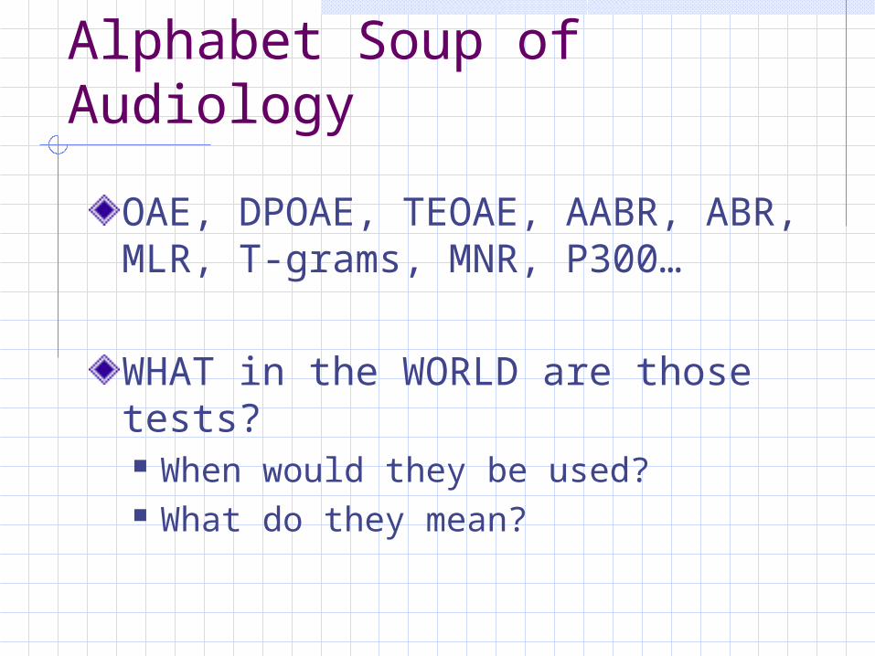
Alphabet Soup of Audiology
OAE, DPOAE, TEOAE, AABR, ABR, MLR, T-grams, MNR, P300…
WHAT in the WORLD are those tests? When would they be used? What do they mean?
Otoacoustic Emissions
Just About Everything You Want To Know About OAE
OriginTypes of TestsInterpretation
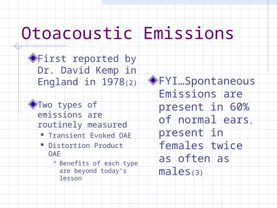
Otoacoustic EmissionsFirst reported by Dr. David Kemp in England in 1978(2)
Two types of emissions are routinely measured
Transient Evoked OAE Distortion Product OAE
Benefits of each type are beyond today’s lesson
FYI…Spontaneous Emissions are present in 60% of normal ears,
present in females twice as often as males(3)
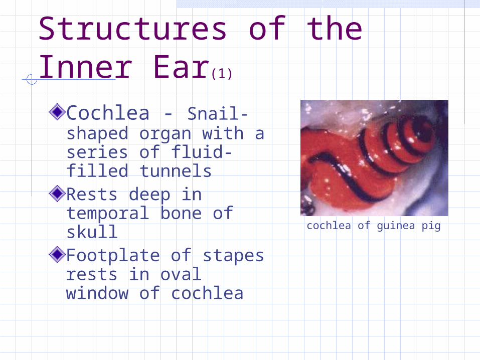
Structures of the Inner Ear(1)
Cochlea - Snail-shaped organ with a series of fluid-filled tunnelsRests deep in temporal bone of skullFootplate of stapes rests in oval window of cochlea
cochlea of guinea pig
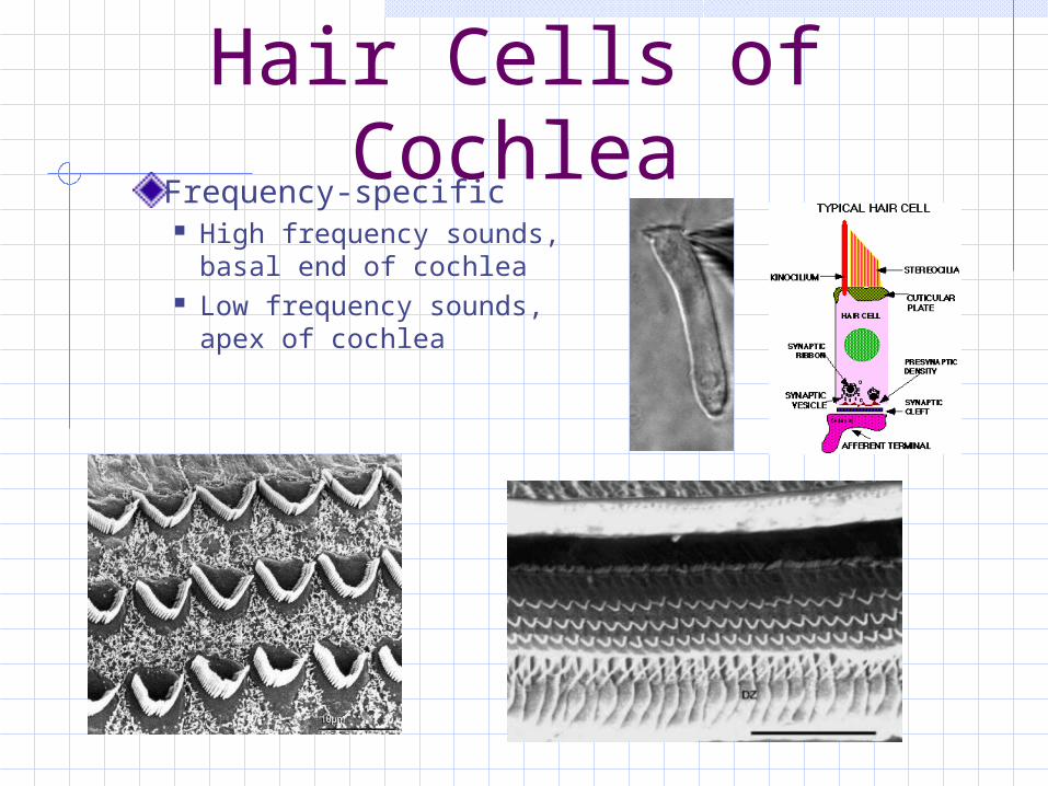
Hair Cells of CochleaFrequency-specific High frequency sounds,
basal end of cochlea Low frequency sounds,
apex of cochlea
So What IS an OAE? (4)
OAEs are actually soft sounds generated by the movement of the structures (outer hair cells) in the cochleaStimulation is sent in through the middle ear, emission occurs within the cochlea, sound then must travel BACK OUT through the middle ear, external ear and be recorded by the microphone of the device
What IS an OAE?
• OAE will likely be observed if auditory threshold is between 0dB and 30dB/40dB HL(5)
• Variables influence OAE Middle ear state Noise in room Noise of subject Debris in EAC
Response is calculated above the noise floor 1-2dB, up to 20dB
Measured across frequency range
TEOAE
Transient Evoked Otoacoustic EmissionAbrupt Click or Tone Burst activates the cochlea across a wide frequency region, if outer hair cells are normal, TEOAEs are produced(4) TEOAE amplitude/noise floor difference calculated at individual frequencies, usually 1K Hz to 5K HzStimulation usually presented at 80dB SPL
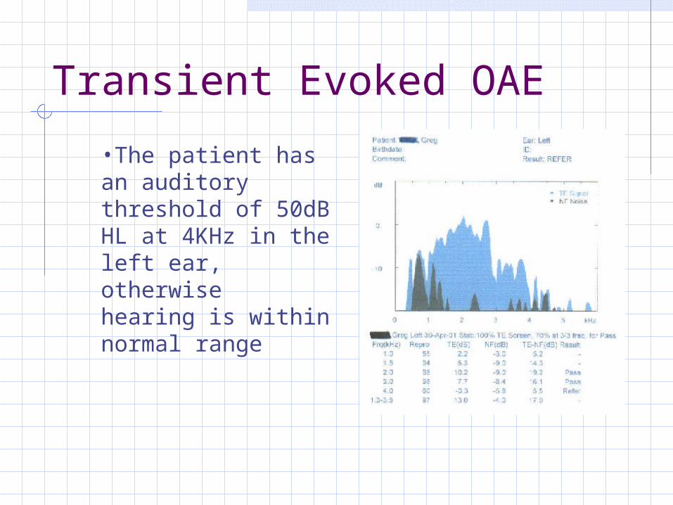
Transient Evoked OAE
•The patient has an auditory threshold of 50dB HL at 4KHz in the left ear, otherwisehearing is within normal range
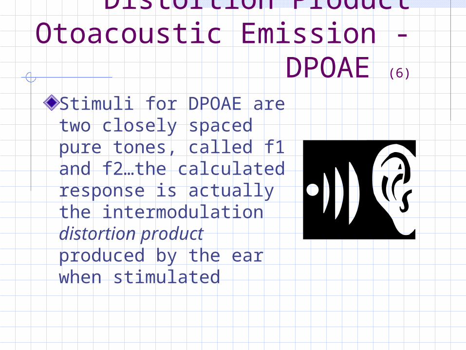
Distortion Product Otoacoustic Emission -
DPOAE (6)
Stimuli for DPOAE are two closely spaced pure tones, called f1 and f2…the calculated response is actually the intermodulation distortion product produced by the ear when stimulated
DPOAE Usual frequency range for stimulation is 500 to 10,000 HzDue to noise floor, difficult to obtain results below 1500 HzVariable stimulation, generally 55dB SPL and 65dB SPL for f2 and f1, respectivelyTones across frequencies presented
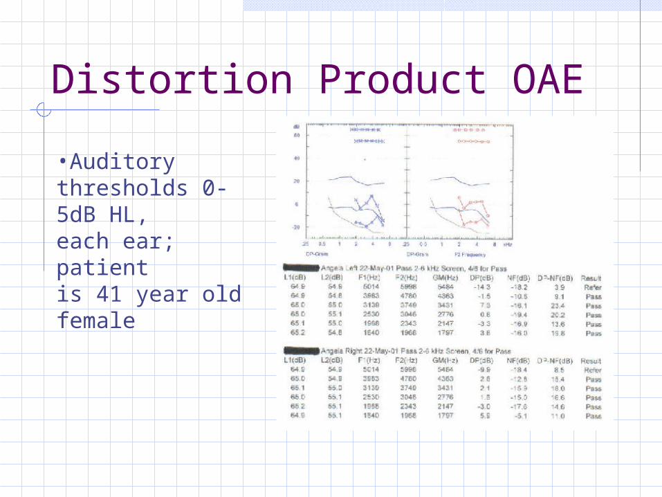
Distortion Product OAE
•Auditorythresholds 0-5dB HL,each ear; patientis 41 year old female
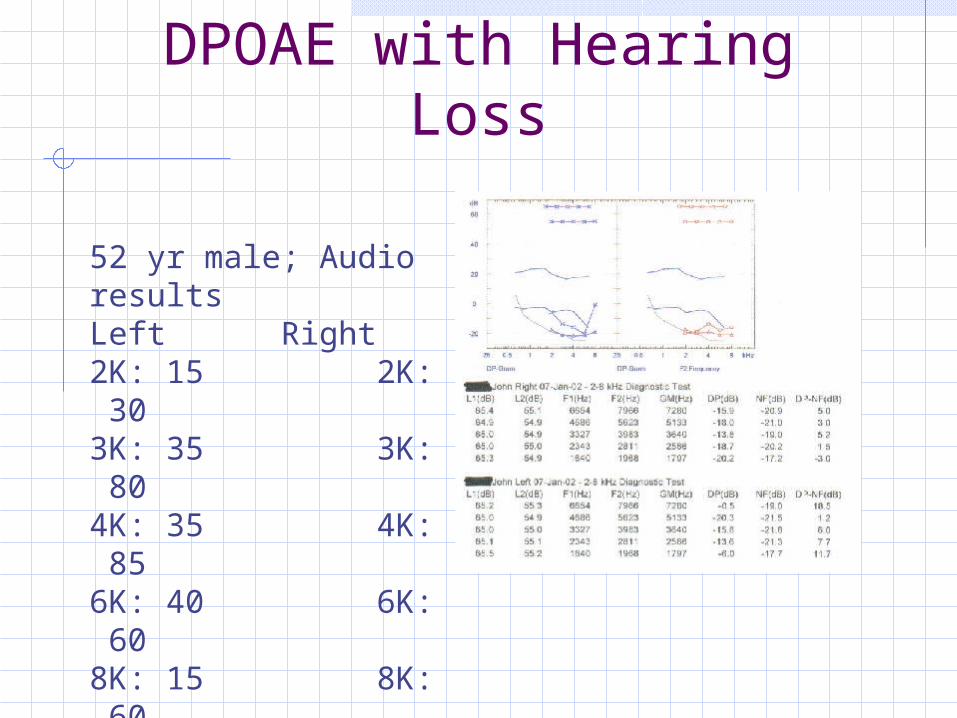
DPOAE with Hearing Loss
52 yr male; Audio resultsLeft Right2K: 15 2K: 303K: 35 3K: 804K: 35 4K: 856K: 40 6K: 608K: 15 8K: 60
Use for Otoacoustic Emissions
Sensitive measure of outer hair cell functionImportant for early identification and diagnosis of auditory dysfunction in pediatric and adult populationsUseful for screenings in newborn nurseriesCan confirm soundfield results in toddlersCan substantiate results that are “questionable” in adult patients who attempt to feign a hearing loss
How to Interpret OAE?NOT A TEST OF HEARING…RATHER, A TEST OF OUTER HAIR CELL INTEGRITY
Results provided by frequency ranges, found to correlate with hearing in normal range
Report summary will state at which frequencies the responses were obtained
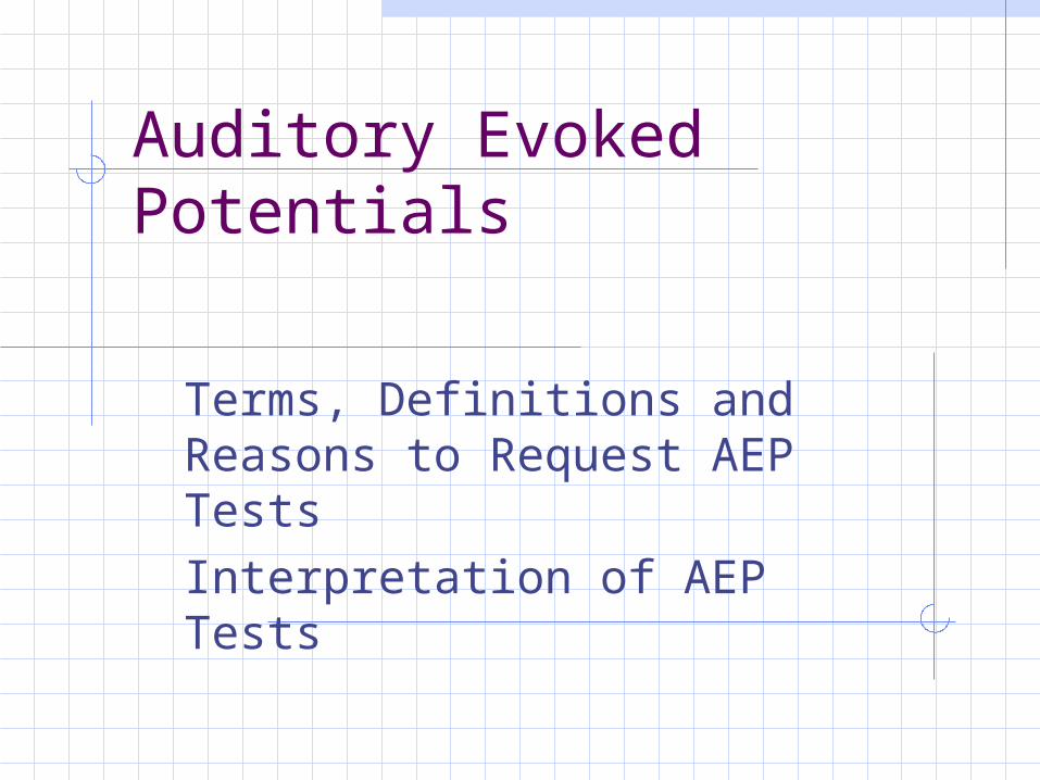
Auditory Evoked Potentials
Terms, Definitions and Reasons to Request AEP TestsInterpretation of AEP Tests
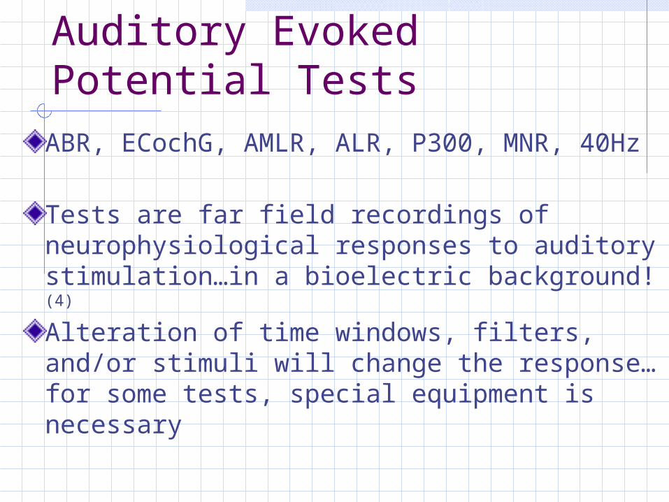
Auditory Evoked Potential TestsABR, ECochG, AMLR, ALR, P300, MNR, 40Hz
Tests are far field recordings of neurophysiological responses to auditory stimulation…in a bioelectric background!(4)
Alteration of time windows, filters, and/or stimuli will change the response…for some tests, special equipment is necessary
Auditory Brainstem Response (ABR) Tests
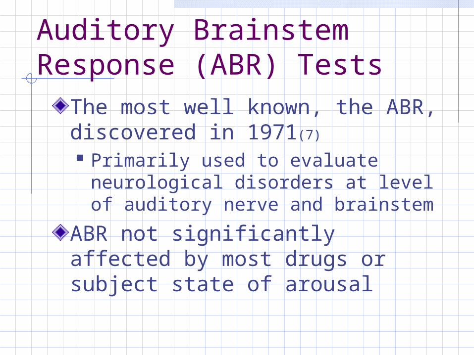
The most well known, the ABR, discovered in 1971(7) Primarily used to evaluate
neurological disorders at level of auditory nerve and brainstem
ABR not significantly affected by most drugs or subject state of arousal
Auditory Evoked Potential Tests
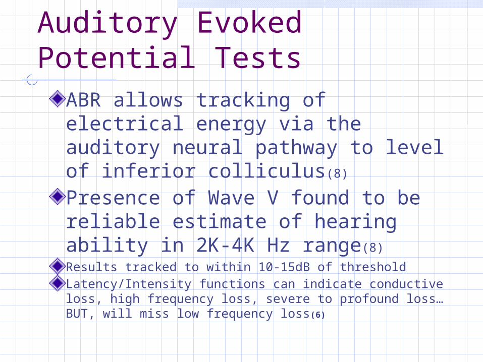
ABR allows tracking of electrical energy via the auditory neural pathway to level of inferior colliculus(8)
Presence of Wave V found to be reliable estimate of hearing ability in 2K-4K Hz range(8)
Results tracked to within 10-15dB of thresholdLatency/Intensity functions can indicate conductive loss, high frequency loss, severe to profound loss…BUT, will miss low frequency loss(6)

Auditory Evoked PotentialsCan be used as auto screen method, AABR for Pass/ReferPatient must be quiet, relaxed; infants asleep or sedatedClick stimuli provides information about 2K to 4K Hz region of cochleaCan use bone oscillator to perform bone conducted ABR
Auditory Evoked Potentials
Possible to construct an “audiogram” based on ABR results obtained with 500Hz, 1000 Hz, 2000 Hz tone burstsUsed to identify auditory dys-synchrony (auditory neuropathy), a dysfunction of neural pathways(9,10)
Auditory Evoked PotentialsLikely abnormal in Patients with Multiple Sclerosis and other demyelinating processes Hyperbilirubinemia at levels requiring
exchange transfusion Patients with severe high frequency loss
ABRs, like OAE, NOT a test of hearing, but of neural function, neural synchrony
Auditory Evoked Potential Test
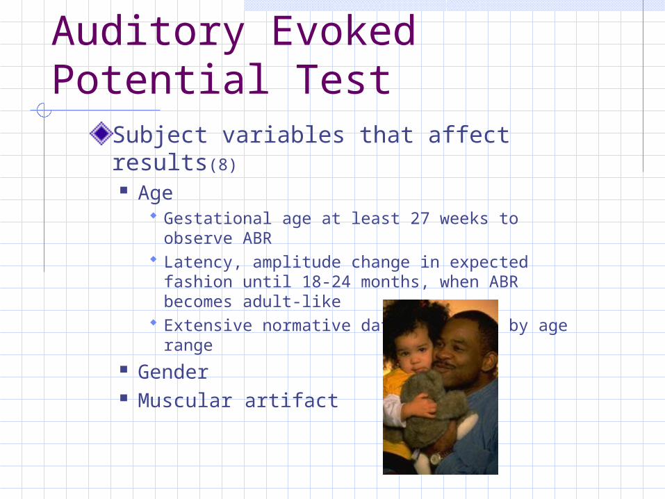
Subject variables that affect results(8)
Age Gestational age at least 27 weeks to observe
ABR Latency, amplitude change in expected
fashion until 18-24 months, when ABR becomes adult-like
Extensive normative data available by age range
Gender Muscular artifact
Other AEP TestsAMLR Auditory Middle Latency ResponseALR Auditory Late ResponseP300 Event Related Response40Hz Variation of MLR
On-going studies regarding clinical utility of these tests continue
Most recorded since 1960s(6)
Not in widespread use outside of research sites
Why Request an Evoked Potential Test?
Can be used to construct an audiogram in patients incapable of voluntary responses (infant, mentally handicapped)In adults, rule out retrocochlear or demyelinating process
More Reasons to Request AEP
To construct an audiogram in non-cooperative adults (malingering)To identify auditory dys-synchrony (auditory neuropathy) (10)
To assess aided thresholds when behavioral testing not possible (13)
Auditory Evoked Potential Tests
Not necessarily first line of testing for Audiologists!!
When referring children/infants for auditory evaluation, evoked potential tests may be the last needed, following soundfield, OAE, BOA, VRA, etc.Sedated ABRs can often be avoided, use other methods first
Immittance Measures
TympanogramAcoustic Stapedial Reflex
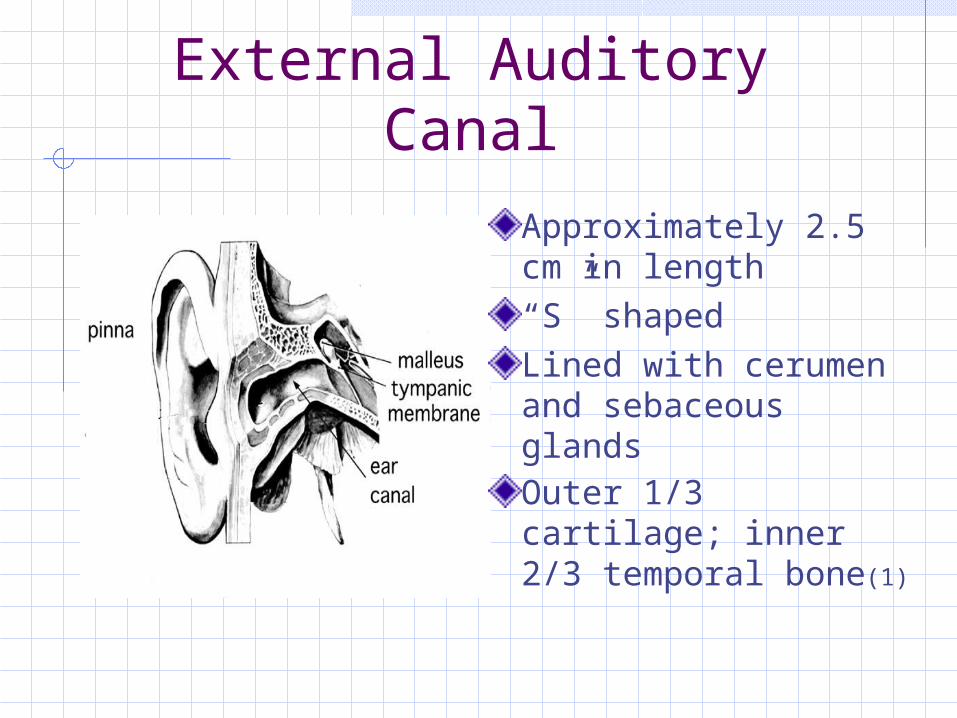
External Auditory Canal
Approximately 2.5 cm in length“S” shapedLined with cerumen and sebaceous glandsOuter 1/3 cartilage; inner 2/3 temporal bone(1)
What IS Immittance? (6)
Combination of two words Admittance is the reciprocal of Impedance Acoustic admittance is a measure of the flow of
energy through middle ear and impedance is the opposition to this flow
No better, quicker or less expensive single audiologic procedure exists to assess status of middle ear, cochlea, eighth nerve and lower brainstem than a complete Immittance Battery
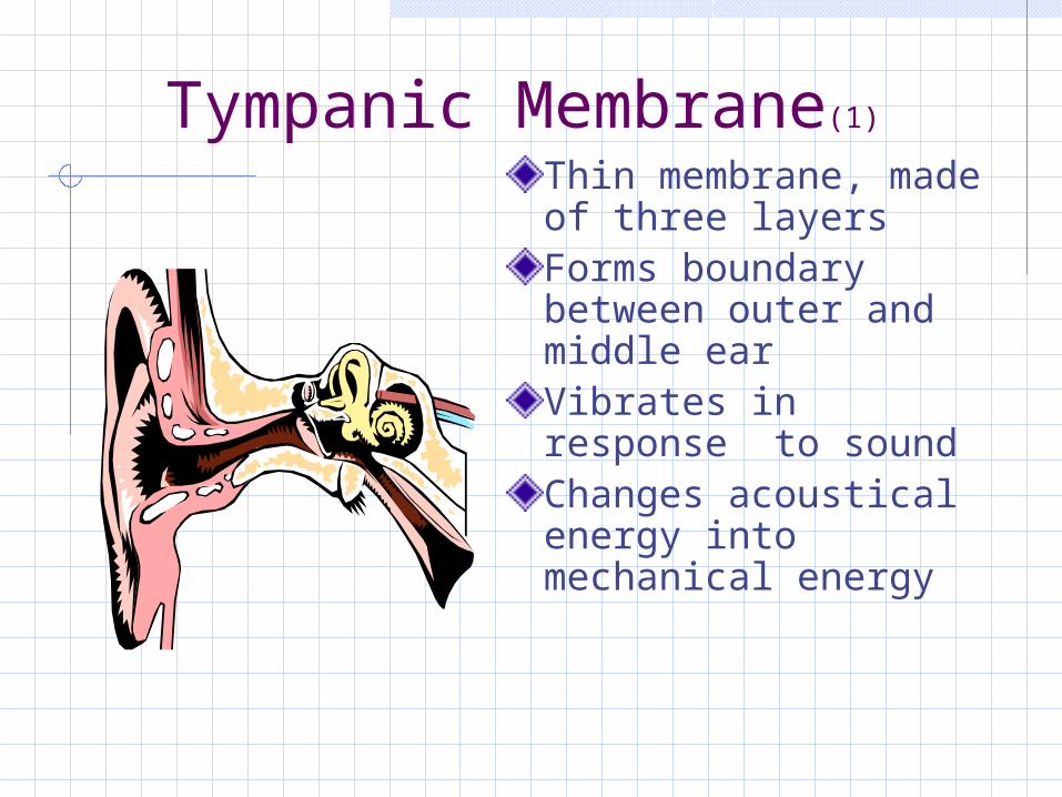
Tympanic Membrane(1)
Thin membrane, made of three layersForms boundary between outer and middle earVibrates in response to soundChanges acoustical energy into mechanical energy
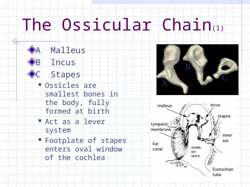
The Ossicular Chain(1)
A MalleusB IncusC Stapes Ossicles are
smallest bones in the body, fully formed at birth
Act as a lever system
Footplate of stapes enters oval window of the cochlea
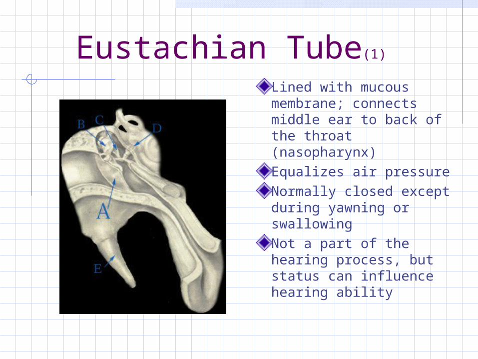
Eustachian Tube(1) Lined with mucous membrane; connects middle ear to back of the throat (nasopharynx)Equalizes air pressureNormally closed except during yawning or swallowingNot a part of the hearing process, but status can influence hearing ability
Stapedial Muscle (11)
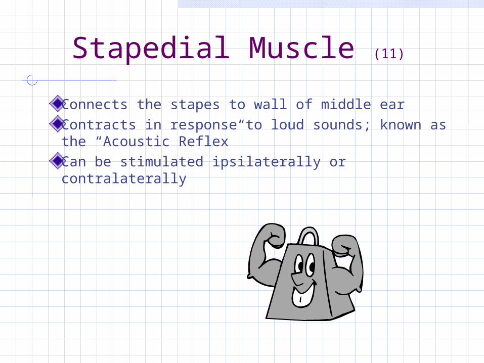
Connects the stapes to wall of middle ear Contracts in response to loud sounds; known as the “Acoustic Reflex”Can be stimulated ipsilaterally or contralaterally
Valuable Information/Simple Test
Test results reveal Ear canal volume(6)
Children 0.42ml to 0.97mlAdults 0.63ml to 1.46ml
Peak amplitude of tympanogram Pressure point of peak
Normal values +/- 100mm H2O
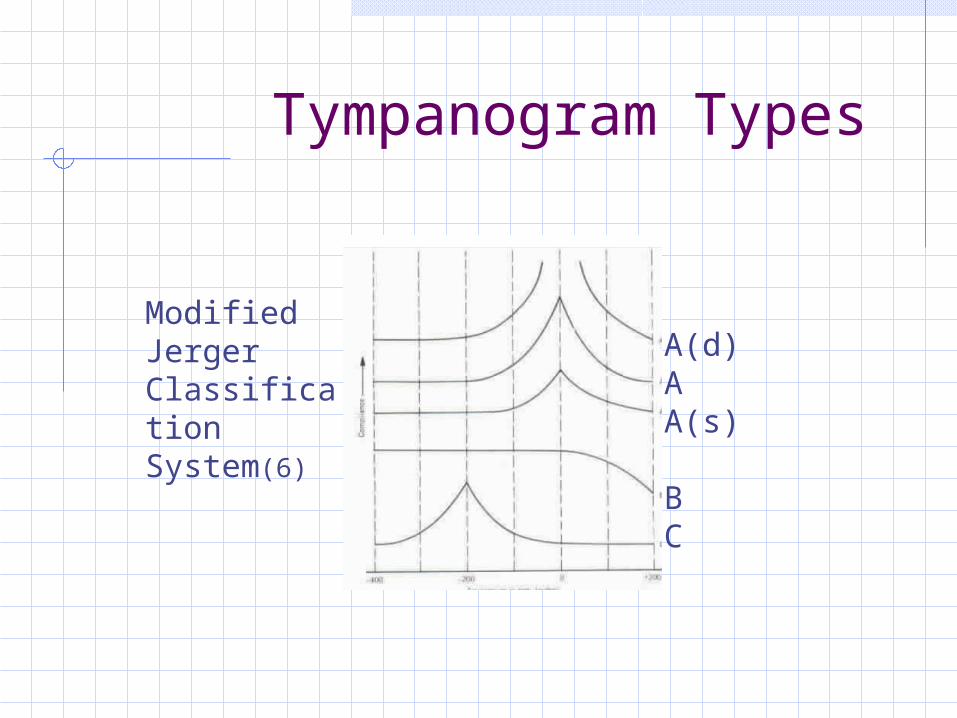
Tympanogram Types
Modified Jerger Classification System(6)
A(d)AA(s)
BC
Some Thoughts in Closing…
•Physiologic test measures in Audiologymay be used on patients of any age and provide valuable information about auditory-neural functions•While these tests are NOT direct tests of hearing, information about theauditory system is provided
•Otoacoustic Emissions•Auditory Evoked Potentials•Immittance Measures