physiological degeneration in opalina. - journal of cell science
TRANSCRIPT
PHYSIOLOGICAL DEGENERATION IN OPALINA. 633
Physiological Degeneration in Opalina.
By
C. Clifford Doltcll, B.A.,Scholar of Trinity College, Cambridge.
With Plate 38 and 2 Text-figures.
INTRODUCTION.
In many respects Opa l ina r a n a r u m , Purk. and Val., isone of the most interesting of the ciliated Protozoa. Althoughits adult appearance has been familiar to all zoologists for along time, it is only in the course of the last year that itsinteresting life-cycle has become fully known. We owe thisknowledge to Neresheinier, who has, however, only given usa preliminary account of his important researches. His fullpaper will be awaited with much interest.
It will be unnecessary for me to give a detailed descriptionof this very common species. But I would remind the readerthat it is a large, multinucleate, holotrichous form found inthe large intestine of our common frog and toad (Ranatempora r i a , L., and Bufo vulgar i s , L.). The nuclei maybe regarded as consisting of meganucleus and micronucleusfused to form a synkaryon. For an account of the life-historythe reader is referred to accounts already published, butmore especially to the memoirs of Zeller and Neresheimer.It will be necessary for me to give a brief account of theearlier part of the life-cycle, however; that is to say, of thepart prior to encystment, which takes place in the adult host.

634 . 0. CLIFFORD DOBELL.
Neresheimer has accurately, though briefly, described thisphase, and I have been able to confirm fully his observations.
In the spring, at the frog's breeding season, Opal inaencysts, and is cast out into the water in the excreta. Thechanges which take place before encystation are as follows :—The adult animal divides in an oblique manner, giving rise totwo daughter individuals. Each of these then divides intotwo, and these again divide until small Opalines containingseveral nuclei are formed. During these divisions importantchanges occur in the nuclear apparatus. The nuclei are seento become less distinct, this being due to the fact that thechromatin is cast out into the cytoplasm in the form of smallstrands and particles, or chromidia. Sometimes I haveobserved these united to form a network through thecreature. Finally, the original nuclei vanish, and we are leftwith only distributed chromatic material. The chromidiasoon become aggregated at certain centres, and thus synthe-sise new nuclei, which are from two to ten in number.These nuclei are seen to be composed of large chromatingranules, arranged irregularly. They change, however, withthe approaching encystment of the animal, which soon takesplace. The chromatin travels to the periphery of the nucleuswhere it becomes arranged in a thin layer with two to fourlarge cap-like thickenings.1 In optical section these nucleiappear as rings. The cap-like projections are soon cast offinto the cytoplasm, where they degenerate, this constitutingthe first nuclear reduction. Encystment follows, and in thenormal course of events the cyst is cast into the water, wherea second reduction of chromatin occurs. I will leave thedescription of the life-cycle at this point, merely noting thateach nucleus is now a reduced gamete nucleus, and takes partin the formation of a single ciliated gamete whose destiny isto conjugate in the tadpole's gut. The essential points in theprocess just described are (1) formation of chromidia,(2) synthesis of fresh nuclei from these chromidia, (3) reduc-tion of chromatin, and (4) encystment.
1 First described by Loewcnthsl as " microfiucleus-like structures,"

PHYSIOLOGICAL DEGENERATION IN OPALINA. C35
DESCRIPTION or DEGENERATION IN OPALINA.
Having cleared the ground by describing the ordinarycourse of events preceding gamete formation, I will nowpass on to a description of the physiological degeneration ofthe forms whose usual destiny is to encyst.
Two different kinds of degenerative changes may be distin-guished—the one caused by removal from the host, the otherwithin the host. The former is much the more rapid, and iseasily induced at any time. Degeneration results from drying,from increase in the number of bacteria, and also doubtlessfrom lack o£ food, and increase of metabolites. The entireorganism simply decomposes, often after first throwing outits nuclei in fragments. It is the second kind of degene-ration—that within the host—with which I am here con-cerned.
In nature, as we have seen already, the encystation ofOpal ina is contemporary with the sexual activity of its host.The set of degenerative changes which I am about to describetook place when the ordinary activities of the host animalswere modified by captivity and starvation. Frogs, as is wellknown, can endure starvation for many weeks. But the con-tained Opalinte, appareutly, cannot do so—at all eventsat their encystation period. Starvation is, I believe, thedetermining factor in their degeneration. Other causes,which materially influence degeneration in the organismswhen removed from their host—such as change in reaction ofthe medium, drying, increase in the number of bacteria, etc.—do not appear to come into play. For after lengthy starva-tion the rectal contents of the frogs and toads examined con-sisted of ouly a small quantity of a clear, mucous fluid,alkaline in reaction, and containing but few bacteria. It wasin cases such as this—where starvation of the host had some-times lasted for at least two months—that the most advancedstages in the degeneration of Opal ina were encountered.My observations extend over a period from the middle of

63C U. CLIFFORD DOBKLL.
January to the beginning of April, and are based upon thecareful examination of the rectal contents of over fifty frogsand toads. The original object of the research was to studythe life-histories of the small Protozoa (flagellates andamoeba)) which occur in this situation. My attention wasattracted by the curious degeneration forms, although theirtrue nature was not, for some time, understood. However,after a careful study of fresh material, the complete series ofdegeneration changes here described became apparent.
TEXT-FIGURE 1.
One of the earliest changes which the OpalinEe undergois a change of shape. Instead of remaining of a flattened,ovate form, they become modified into all sorts of indefiniteshapes. Some of these are shown in text-fig. 1, but a greatnumber of other forms may be seen. The drawings areschematic, and merely indicate the kind of thing which oneencounters. These forms do not divide in the normal manner,but simply constrict off pieces, apparently at random, of allshapes and sizes. These again divide until small, irregularlydiscoidal, or ovoid forms are produced which have a lengthof about 10 n to 30/i. These usually contain from one to

PHYSIOLOGICAL DEGENERATION IN OPAMNA. 637
four nuclei, but much larger individuals—up to 50 /x, withnine or ten nuclei—are also found in the same condition.Such forms undergo two remarkable changes—(1) they com-pletely lose all cilia, and (2) they give rise to globules of asubstance of high refractivity in their cytoplasm. The natureof these globules I am unable to determine. I may mention,however, that they have the following properties :—
In the fresh state they are somewhat greenish, and veryhighly refringent. They are coloured a bright pink witheosin, and a bright greeuish-yellow with picric acid. Withiodine they appear to become slightly more greenish, but thereaction is not well marked. They are insoluble in water,alcohol, and weak acids and alkalies. Heidenhain's ironhasmatoxylin colours them a dark greyish- or brownish-black—not so dark, however, as the chromatin. Delafield's haama-toxylin does not colour them—neither does borax-carmine.
From their remarkably vivid coloration with eosin I havetermed these globules " eosinophile " bodies, in ignorance oftheir chemical constitution. Although they first appear asseparate globules of small size, they ultimately run together,forming large masses lying in the cells. They do not appearto have any connection with the nucleus.
If these degenerate forms be obtained at the right stage,the loss of cilia may be observed in the living animals. Ittakes some days for all to be completely lost, and all do notseem to disappear in the same way. Apparently some ofthem actually dissolve, for they become gradually fainterand fainter, and finally disappear. Others are thrown offentirely, and after moving spontaneously for a short timeafter detachment, they become motionless and fade away.Still others undergo fusion with one another, and ultimatelywith the cytoplasm. In this manner many individuals arisewhich are completely divested of ciliary covering, and con-tain refringent eosinophile bodies. The nucleus also under-goes remarkable modifications. I term these forms the•atrichous forms.
The nuclear changes which the atrichous forms undergo are

038 C. CLIFFORD DOBELL.
as follows, and may be easily seen in the living animal:—The chromatiii which was at first evenly distributed throughthe nucleus becomes massed in granules at the periphery,whilst the nucleus itself increases in size until it becomessometimes double its original diameter (text-fig. 2, a, I).
<*•• I- c d, e .TEXT-FIGUHE 2 (from permanent preparations, stained wilh Heideukain's
iron-litematoxylin).
The chromatin becomes evenly disposed in a single layer,so that in optical section the nucleus has a very character-istic annular appearance (c), the ring being thickened atvarious points (see also Plate, figs. 8, 12). A typical atri-chous form is therefore distinguished by having no cilia, bypossessing " eosiuophile globules" and a large ring-likenucleus (or nuclei). When first seen they have a remai'kableiippearance, and their connection with the ordinary Opal inawould hardly be suspected. These forms are quite motion-less.
In many of the larger atrichous forms division of thenucleus takes place, followed frequently by division of tliocytoplasm. Division may be equal, by a constriction appear-ing in the middle (text-fig. 2, d), or unequal. In this latterprocess a blister-like elevation of the chromatin appears, andis finally constricted off. This is shown in text-fig. 2, e.Above is a cap-like outgrowth of the chromatiu, whilst belowa later stage is seen in optical section (cf. also Plate, figs. 10,
11)-When cytoplasmic division follows the result is either
equal bipartition or budding (cf. Plate, figs. 9, 10). Thebuds so produced are sometimes very small, not reaching agreater diameter than 4—5 fx. Occasionally buds are pro-duced in which no nuclear material whatsoever can be

PHYSIOLOGICAL DEGENERATION IN OPALINA. 639
detected, and very commonly small buds are given off whichcontain an eosinophile globule, but no nucleus. All theseenucleate buds appear to die and disintegrate. Finally, anumber of uninucleate atrichous forms result, which are ofan average diameter of about 20 ju. At this stage they show amarked tendency to attach themselves to one another, thusforming small colonies (cf. Plate, fig. 12). No fusion, as arule, appears to take place.
The chromatin of the nucleus, which is of a very variablesize, but on an average about 8—10 JU in diameter, is seen tobe arranged in lumps peripherally. It soon leaves thenucleus, however, and fills the cytoplasm, where it takes theform of irregular granules and masses of different shapesand sizes. These chromidia, as they may be called, appearto be sometimes in the form of minute hollow spheres, ring-like in optical section. By their formation the originalnucleus dwindles away, and finally disappears (see Plate,figs. 1, 2, 7). As a rule most of this chromatin is cast out ofthe organism, which then dies and breaks up. But occa-sionally a remarkable thing happens. Only a part of thechromatin is cast out and perishes. The remaining granulesrun together again, very much as drops of oil might runtogether in a watery medium. All the irregular chromidia]masses may become aggregated at a single centre, but atother times two such centres are formed, so that finally twonuclei, consisting of solid chromatin, are synthesised. Thesetwo solid lumps then approach one another and fuse (seePlate, figs. 3—6). During the chromidial stages a soft cyst-wall is sometimes formed.
I have been unable to obtain any further stages after this,except such as are disintegrative. Kept under a waxedcoverslip or in a hanging drop they always perish bydischarging their nuclei in fragments and then breaking up.This also appears to happen in the frog's gut. It would beexceedingly interesting to know whether a recovery could bemade under suitable conditions or not. I have endeavouredto restore some of these degenerate fragments by transfer-
VOL. 5 1 , PAET 4.—NEW SERIES. 47
640 C. CLIFFORD DOBELL.
ring them into the boiled rectal contents of a normal frog,but without success. I have not been able to obtain sufficientmaterial for more extended experiments in this direction.
The final result then is death ; and with this I finish mydescription of degenerative changes in Opalina ranarumso far as I have observed them. Before leaving the subject,however, I must draw attention to the extraordinary parallelwhich exists between these changes and certain so-called" sexual" processes. In many Protozoa gamete nuclei areformed from the original compound nuclei by resynthesisfrom chrornidia, very much in the same way as the solidchromatiu nuclei which I have just described in Opalina.In certain autogamic processes the nuclei ai-e formed andfase in the same cell. Compare, for example, the autogamyof Bodo lacertse, Grassi, as described by Prowazek. Theanimal encysts, and inside the cyst the nucleus gives offchromidia into the cytoplasm. From these chroinidia a newnucleus is built up, and this divides into two. Bach daughternucleus forms two " polar bodies," and the reduced nuclei(the gamete nuclei) approach one another and fuse. IuBodo lacertse heterogamy also occurs, but in Tricho-mastix lacertas, Blochmann, and in Entamceba coli,Losch, only autogamy is known—no other " sexual " act.
It is possible that the process which 1 have just describedin Opalina is a kind of autogamic attempt on the part ofthe organism to reconstitute itself. But 1 believe that thesole explanation of this curious set of changes is to be soughtin the alteration in chemical and physical properties whichliving protoplasm undergoes in dying.
Iu conclusion, mention may be made of certain otherobservations which have been made on degeneration in otherProtozoa. But little attention has been bestowed upon thematter, although iu a few species degenerative changes havebeen studied in considerable detail. Among these I mayname Actinosphasrium (Hertwig), Amoeba (Prandtl),Paraincecium (Maupas, Calkins, etc.), Trichosphasrium(Schaudinn), and the sporont of Cyclospora Caryolytica

PHYSIOLOGICAL DEGENERATION IN OPALINA. 641
(Schaudinn). In the first three of these increase in the sizeof the nucleus has frequently been obsei-ved as a preliminaryoccurrence. In Act inosphser ium giant nuclei are formedwhen the animal degenerates owing to overfeeding. Break-ing up and discharge of the nucleus usually follows—that isto say, chromidia play a part in the degenerative phenomena.In Trichosphserium, when degeneration is induced bystarving the organism, the nuclei clump themselves togetherat certain points. This agglomeration is not followed byfusion. Stole has observed a similar condition in starvedPelornyxaa. And I may here recall the observation ofMaupas on Paramceciuni, that the fragments of the oldmeganucleus sometimes fuse with the new meganucleus of anexconjugant if it be starved.
In the degenerating sporont of the coccidian Cyc lospo rathe polar bodies divide until eight are formed, and microga-metes then attempt to fertilise each of these. In later stagesof degeneration pigment is formed ; and Prandtl has shownthat pigment appears iu a degenerating Amoeba p ro teus ,and is formed from the chromatin of the nucleus. In thisparticular form there is also a curious tendency for thenucleus to surround food masses in the cytoplasm.
Nuclear fusion sometimes takes place—independently oEany sexual process—in multinucleate Protozoa during, orfollowing, encystment: e.g. in Di lep tus (Prowazek).
Hyper-regeneration occurs in S ty lonychia if mutilatedwhen in a degenerate condition (Prowazek). Loss of ap-pendages has been frequently observed in many differentProtozoa undergoing degenerative changes. It is unneces-sary to give a number of examples, but Trichosphseriumand Parana Gecium may be cited as good instances.
Chromidia of the type I have described in Opal ina (i.e.bladder-like, or blaschenformig) have only been noticed, sofar as I am aware, in one other Protozoon, Bodo lacertse,Grassi. And here they are formed as a preliminary togamete formation and autogamy (Prowazek).
Very curious in many other ways is the parallel which

642 0. OLIFFOBD DOBBLL.
exists between degenerative and " sexual" processes. Be-sides the fact that chromidia are formed in both, we havethe observation that an amoeboid condition may occur indegenerating Protozoa, and also sometimes just beforeconjugation. Fusion also occurs in degenerating forms ofvarious kinds. I have observed it especially in flagellates,e.g. Tr ichomast ix , T r i chomonas , etc. Senile P a r a -mcecia enter upon what Calkins calls the "miscible state,"when they tend to adhere to one another. Similarly, Rouxhas observed that isolated, living blastomeres of frog's eggsbecome amoeboid and run together; though, according toDriesch, this is merely due to the capillary forces betweenthe cells. These facts, and many others of a similar nature,are not without interest, both from a pathological and from azoological point of view. I may mention merely their pos-sible bearing upon the remarkable fusion which appears totake place between leucocytes and cancer cells, and its un-known significance. And since the work of Calkins seems toindicate that chemical change in protoplasmic composition isthe chief beneficial effect of conjugation, and there is someproof that Protozoon individuals have chemical compositionsdiffering from one another (cf. Jensen), is it not at leastpossible that the physico-chemical changes which causefusion in degenerating cells are of a similar nature to thosewhich gave rise to the first cell-couplings, and which stilldetermine the fusion of one gamete with another ?
ZOOLOGICAL LABOBATOIIT,
CAMBRIDGE.
ADDENDUM.
Neresheiiner's full account of the life-cycle of Opal inahas appeared since this paper was submitted for publication.It is a very complete description, with full references to theliterature, and is to be found in ' Arch. f. Protistenk:' Supple-ment i (Festband fur E. Hertwig), 1907, p. 1.

PHYSIOLOGICAL DBGENflEATION IN OPALINA. 643
LITERATURE REFERENCES.
1. CALKINS, Q. N.—" Studies on the Life-history of Protozoa."—I, in 'Arch.Entwickmecli.,' Bd. xv, 1902, p. 139; II (with C. C. LIEB), in 'Arch.f. Protistenk.,'Bd. i, 1902, p. 355 ; III, in 'Biol. Bull.,' vol. iii, 1902,p. 192; IV, in ' Journ. Exp. Zool.,' vol. i, 1904, p. 423.
2. GOLDSCHMIDT, R.—"Der Chromidialapparat lebhaft funktionierenderGewebszellen," in 'Zool. Jalirb.' (Abtli. f. Anat.), xxi, 1904, p. 41.
3. GOLDSCHMIDT, R.—" Die Cbromidien der Protozoen," in • Arch. f. Protis-tenk.,'Bd. v, 1905, p. 126.
4. HERTWIG, It.—"Ueber physiologisclie Degeneration bei Actinosphse-rium eichhorni," in 'l?estschr. f. E. Haeckel,' Jena, 1904, p. 301.
5. JENSEN, P.—"Ueber individuelle physiologisclie unterschiede zwischenZellen der gleichen Art,' in 'Pfliiger's Arch. f. Physiol.,1 Bd. lxii,1895, p. 172.
6. LOEWENTHAL, W.—" Das Auftreten eines Mikronukleusartigen Gebildesbei Opalina ranarum," in 'Arch. f. Protistenk.,' Bd. iii, 1904,p. 387.
7. MATJPAS, E.—"La rajeunisseraent karyogamique chez les cili£s," in'Arch. Zool. Exp. et Gen.,' vii, 1889, p. 149.
8. NEHESIIEIMEK, E.—"Der Zeugungskreis von Opalina," in 'Sitz. Ber.Ges. Morpli. Physiol. Miinchen,' 1906.
O. PFITZNER, W.—" Zur Kenntnis der Kerntheilung bei den Protozoen,"in 'Morph. Jalirb.,' Bd. xi, 1886, p. 454.
10. PRANDTL, H.—"Die physiologisclie Degeneration der Amoeba pro-teus," in 'Arch. f. Protistenk.,' Bd. viii, 1907, p. 281.
11. PHOWAZEK, S.—"Der Encystierungsvorgang bei Dileptus," in'Arch.f. Protistenk.,' Bd. iii, 1904, p. 64.
12. "Degenerative Hyperregeneration bei den Protozoen," in ' Arch.f. Protistenk.,' Bd. iii, 1904, p. 60.
13. " Untersuehungen ueber einige parasitische Flagellaten," in'Arb. Kaiser], Gesundheitsamte,' xxi, 1904, p. 1.
14. lloux, W.—"Ueber den 'Cytotropismus' der Furchungszellen des Gras-frosches (Rana fusca)," in 'Arch. Eutwickmecb.,' Bd. i, 1894, pp.43 and 161.
15. SCHAUDINN, F.—"Untersuehungen iiber den Generationswechsel vonTrichosphterium sieboldi, Schn.," in'Anh. Abb. Akad. Berlin,'1899.
644 0. CLIFFORD DOBELL.
16. SCHAUDINN, F. — "Studien uber krankheitserregende Protozoen: I.Cyclospora caryolytica, Schaud., der Erreger der perniciosenEnteritis des Maulwurfs," in 'Arb. Kaiserl. Gesundheitsamte,' Bd.xviii, 1902, p. 378.
17. STOLC, A.—" Beobachtungen und Versuche iiber die Verdauung undBildung der Kohlenhydrate bei einem amobenartigen Organismus,Pelomyxa palustris, Greeff," in 'Zeitschr. f. wiss. Zool.,'lxviii,1900, p. 625.
18. ZELIBE, E.—"Untersuchung iiber die Fortpflanzung und die Entwick-lung der in unsercn Batracliiern schmarotzenden Opalinen," in ' Zeitschr.f. wiss. Zoo!.,' xxix, 1877, p. 352.
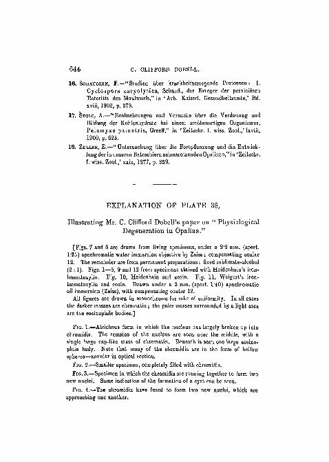
E X P L A N A T I O N OF P L A T E 38,
I l lustrat ing Mr. C. Clifford Dobell 's paper on " PhysiologicalDegeneration in Opaliua."
[Figs. 7 and 8 are drawn from living specimens, under a 2'5 mm. (apert.l-25) apochromatio water immersion objective by Zeiss ; compensating ocular12. The remainder are from permanent preparations: fixed sublimate-alcohol(2 :1). Pigs. 1—6, 9 and 12 from specimens stained with Heidenkain's iron-hscmatoxylin. Fig. 10, Heidenhain and eosin. Fig. 11, Weigert's iron-brematoxylin and eosin. Drawn under a 2 mm. (apert. l'4O) apochromaticoil immersion (Zeiss), with compensating ocular 12.
All figures are drawn in monochrome for sake of uniformity. In all casesthe darker masses are chromatin; the paler masses surrounded by a light areaare the eosinophile bodies.]
FIG. 1.—Afcrichous form in which the nucleus has largely broken up intochromidia. The remains of the nucleus are seen near the middle, with asingle large cap-like mass of chromatin. Beneath is seen one large eosino-phile body. Note that many of the chromidia are in the form of hollowspheres—annular in optical section.
FIG. 2.—Smaller specimen, completely filled with chromidia.FIG. 3.—Specimen in which the chromidia are running together to form two
new nuclei. Some indication of the formation of a cyst can be seen.
FIG. 4.—The chromidia have fused to form two new nuclei, which areapproaching one another.

PHYSIOLOGICAL DEGENERATION IN OPALINA. 645
FIG. 5.—A cyst-wall lias been formed, and the two solid chromatin nucleiare applied to one another.
FIG. G.—Specimen showing a still later stage in the fusion of the nuclei.A thick, soft cyst lias been formed.
FIG. 7.—Fresh preparation in which a cyst has been formed and the nucleuslias broken up into chromidia.
ElG. 8.—Atrichous form in fresh condition, showing nucleus with peri-pherally placed chromatin masses (annular in optical section).
FIG. 9.—Large multinucleate atrichous form breaking up to form smallerones. At least one nucleus is dividing.
FIG. 10.—An individual in the act of forming buds. Small blister-likenuclei are budded off, and (.he cytoplasm has become constricted round one ofthese, forming a complete bud.
FIG. 11.—Similar form, in which nucleus is seen in optical section. Onenucleus has been completely separated off, and another is almost so.
FIG. 12.—Association of eleven typical atrichous forms. None of thesehave as yet formed chromidia, though one appears to be enucleate.

9nn,ri. 4(»i/rn, MrA-A\-jj. Ifni.,')/ ,'VSW 3
OPALINA