physiology of tear film it’s drainage
TRANSCRIPT

PARTH VORA
T.Y. B. OPTOM

The tear film constitutes Three layers :-
An outermost lipid (oily) layer
An aqueous (watery) layer that makes up 90% of the tear film volume; and
A mucin layer that coats the corneal surface.

To form smooth optical surface on cornea.
To keep the surface of cornea & conjunctiva moist
It serve as lubricant
It transfer oxygen
Provide antibacterial action
Wash debris out
It provides a pathway for WBC in case of injury
Functions of lipid layer Retards evaporation of tear film
Prevents the overflow of tears

Function of Aqueous Layer Flushes, buffers and lubricates the corneal
surface
Delivers oxygen and other nutrients to the corneal surface
Wash out debris
Delivers antibacterial enzymes and antibodies such as lysozyme.

Functions of Mucin Layer Spreads tears over corneal surface.
Protects the cornea against foreign substances .
Makes corneal surface smooth by filling in surface irregularities.

Secretion of tears
Formation of tear film
Retention & redistribution of tear film
Displacement phenomenon
Evaporation form of tear film
Drying & breakup of tear film
Dynamic events during blinking
Elimination of tears

Two types:-
Basal and Reflex Secretion.
Tears are continuously secreted through out the day by Accessory lacrimal gland basal secretion.
The reflex tears is caused due to irritation of a foreign particle or an external stimulus. They are secreted by main lacrimal glands.
The reflex tears attempt to wash out irritants that may have come in contact with the eye.

Lids surfacing cornea with thin layer of mucus
on this new surface aqueous component of tear now spread spontaneously
Then the superficial lipid layer spreads over aqueous film, probably contributing to it’s stability & retarding evaporation between blink.

Retained at a uniform thickness over the corneal surface against gravitational force-positioned vertically.
Redistribution occurs in the form of bringing of new tear fluid by way of marginal strip where there is constant tear flow.

If with a finger the lower lid is carefully displaced upwards over eyeball , the particles in the film are seen to move up the cornea.(IN SLE)
Based on this , it has been concluded that the cornea is covered by a film of certain stability , compressibility and elasticity.

All lipid films including wax esters & cholesterol esters retard evaporation of water.
Important low humidity & turbulent air flow near cornea like windy & arid climate.
Evaporation estimated to be about 10% of production rate 0.12 μl/min.(1.2μl/min production)

In the normal human eye the precorneal tear film has a short lived stability.
When blinking is present after time internal of 15-40 second tear film rupture & dry spot appear on various part of cornea.
Drying of corneal surface – not only because of evaporation but also due to break up tear film.

Holly has described a mechanism of tear film rupture – Holly & Lemp’s mechanism.
Dry spots occur twice more in temporal side than nasal side - nasal areas are more protected against air currents & have comparatively higher temperature.

Tear film thins uniformly by evaporation
When thinned out to some critical thickness, significant number of lipid molecules begin to be attracted by mucin layer & migrate down
This migration enhanced if there is any spontaneous local thinning
After contamination of mucin layer by lipid migration from top surface of tear film mucin becomes hydrophobic & tear film rupture
Blinking repair rupture by removing lipid contaminant from mucin layer & restoring thick layer


As upper lid moves downwards, the superficial layer is compressed.
The compressed lipid layer has a thickness of 0.1 μm.
The lipid contaminated mucus is rolled up in a thread like shape and dragged into lower fornix.
When eye opens, lipids spread as a monolayer against the upper eyelid.


Fluid flows over preocular surface & reaches ciliary margin of each eyelid and collects in the inner canthus.
Fluid is drained by lacrimal passage into nasal cavity [“active lacrimal pump mechanism”]
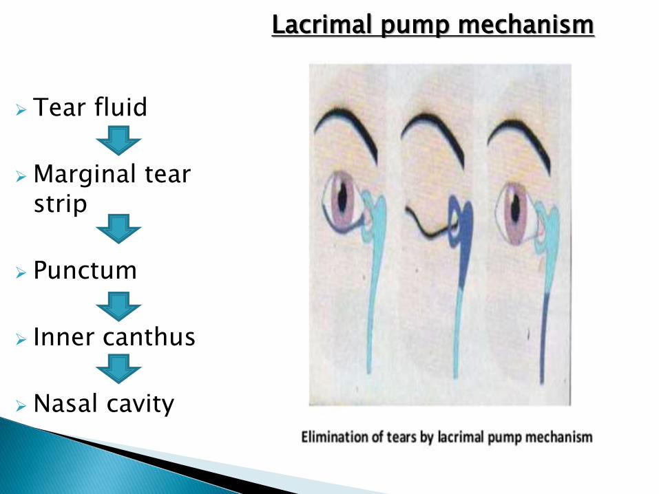
Tear fluid
Marginal tear strip
Punctum
Inner canthus
Nasal cavity
Lacrimal pump mechanism

Contraction of pretarsal fiber of orbicularis
Compress the punctum & shortens canaliculi
Fluid present in punctum & horizontal part of canaliculi toward lacrimal sac
Contraction of preseptal fiber of orbicular pulls lacrimal fascia & lacrimal wall laterally - opening of lacrimal sac
Produce the negative pressure & draws tear into lacrimal sac

Relaxation of pretarsal fibers of orbicularis allows canaliculi to expand & reopen.
Draw the fluid through puncti into canaliculi.
Relaxation of preseptal fibers-lacrimal sac collapse.
Expels the fluid downwards into open naso lacrimal duct. (NLD)

After entering into NLD,influence of eyelid movement on it’s further downward flow ends
Gravity help in downward flow
Air current movement within nose : Air current passing (inward & outward)Include negative pressure within NLD & draw fluid down into nose
Hasner’s valve present at lower end of the NLD remain open till the pressure within nose is less than NLD & allows fluid flow in to nose from NLD
From nose tears pass posteriorly with nasal mucus secretion

Anatomy and physiology – A.K. khurana – pg. 378 to 389
www.wikipedia.com
www.eyepedia.co.uk
www.slideshare.com