phytase from aspergillus niger ncim 563: isolation ...ncl.csircentral.net/769/1/th1752.pdfphytase...
TRANSCRIPT
Phytase from Aspergillus niger NCIM 563:
Isolation, Purification, Characterization
and its Applications.
A THESIS SUBMITTED TO THE UNIVERSITY
OF PUNE FOR THE DEGREE OF DOCTOR OF PHILOSOPHY
IN THE DEPARTMENT OF MICROBIOLOGY
By
SARVESH KUMAR SONI
Research Supervisor Dr. J.M. KHIRE
NCIM RESOURCE CENTRE,
NATIONAL CHEMICAL LABORATORY, PUNE-411008,
INDIA
JULY - 2009

Dedications
This thesis is dedicated to my family who I couldn’t have survived
through my stay in Pune without. First, to my father Jai prakash soni and
mother Geeta rani for never doubting in my ability to achieve a goal and being
very supportive of every endeavor I have ventured to take. Second, to my
brothers Brijesh and Bhartesh soni their wives Vinita, Parul and both of my
niece Vanshika and Rhidhima., who instilled in me the desire to learn and
confidence to achieve.
“My mind is still tender, my thoughts are still young
My dreams are still soar, there are battles to be won”

TABLE OF COTENTS
DECLARATION BY RESEARCH GUIDE i
DECLARATION BY THE RESEARCH SCHOLAR ii
ACKNOWLEDGEMENT iii- iv
ABSTRACT v- ix
ABBREVIATIONS x
Chapter 1. General Introduction 1-61
1.1 Phytic acid 3
1.2 Phytase 7
1.3 Enzymatic properties of phytases 18
1.4 Application of Phytases 33
1.5 Market trends and future prospects 38
1.6 Synthesis of calcium phosphate 39
1.7 Future studies 43
1.8 Aims and objectives 45
1.9 References 46
Chapter 2.
Production of Phytase I (Highly Acidic), II by Aspergillus niger (NCIM 563) under submerged fermentation condition.
62-84
2.1 Summary 62
2.2 Introduction 62
2.3 Materials and Methods 63
2.4 Results and Discussion 67
2.5 Conclusion 81
2.6 References 82
Chapter 3. Purification and characterization of phytase I (Highly Acidic) and II from Aspergillus niger (NCIM 563).
85-123
3.1 Summary 85
3.2 Introduction 86
3.3 Materials and Methods 87
3.4 Results and Discussion 92
3.5 Conclusion 119
3.6 References 120
Chapter 4. Application of phytase of Aspergillus niger (NCIM 563) in biomimetic synthesis of Hydroxyapatite and its polymorphs
124-146
4.1 Summary 124
4.2 Introduction 124
4.3 Materials and Methods 126
4.4 Results and Discussion 130
4.5 Conclusion 141
4.6 References 142
Chapter 5. Biocompatibility studies of enzymatically synthesized Hydroxyapatite and its polymorphs on Human Osteoblast like MG-63 cell line
147-164
5.1 Summary 147
5.2 Introduction 147
5.3 Materials and Methods 149
5.4 Results and Discussion 152
5.5 Conclusion 161
5.6 References 162
Chapter 6. Application of phytase in cell culture studies and myo-inositol production.
165-181
6.1 Summary 165
6.2 Introduction 165
6.3 Materials and Methods 169
6.4 Results and Discussion 172
6.5 Conclusion 177
6.6 References 178
Conclusions 182
Publications
i
DECLARATION
This is to certify that the work incorporated in the thesis entitled “Phytase from
Aspergillus niger NCIM 563 : Isolation, purification, characterization and its
applications” submitted by Sarvesh Kumar Soni was carried out under my
supervision at NCIM Resource centre, National Chemical Laboratory, Pune 411008,
Maharashtra, India. Materials obtained from other sources have been duly
acknowledged in the thesis.
Dr. J. M. Khire
(Research Guide)

ii
DECLARATION BY RESEARCH SCHOLAR
I hereby declare that the thesis entitled “Phytase from Aspergillus niger NCIM
563: Isolation, purification, characterization and its applications”, submitted for the
Degree of Doctor of Philosophy to the University of Pune, has been carried out by me
at NCIM Resource centre, National Chemical Laboratory, National Chemical
Laboratory, Pune - 411 008, Maharashtra, India, under the supervision of Dr. J.M.
Khire (Research supervisor). The work is original and has not been submitted in part or
full by me for any other degree or diploma to any other University.
Sarvesh Kumar Soni (Research Scholar)
iii
ACKNOWLEDGEMENTS
Obtaining a PhD is a group effort. While the work presented in this thesis is my own, it
would never have been possible without the help of numerous people who have given me
immense support in many ways during my long and arduous five years of study. I am deeply
indebted to my mentor, Dr. J.M. Khire who has provided me with many gifts, the greatest being
a completely different mindset from the one I had at the beginning of my doctoral studies. It was
his enthusiasm on phytase research which attracted me to join his laboratory. The ability to
think critically, to ask the right questions, and to resolve the problems through experiments are
the traits that I didn’t possess before meeting him. I thank him for allowing me freedom and
resources to pursue different lines of research. I will miss picking his brain for answers to my
questions and I just hope I can cultivate a fraction of knowledge that he possesses. Extending
this gift from my Ph.. D. work to the way I go through life has been an added bonus.
Another important influence, whose guidance has been wonderful, are Dr. S.B. Ogale and
Dr. V. Premnath. I cannot thank him enough for all the years of encouragement. He has had a
continuous faith in me, pushing me towards academic success, and has been the voice of reason
and support during my entire PhD tenure. I owe a huge debt of gratitude to Director NCL, Dr.
S. Sivaram and our HOD, Dr. D.V. Gokhale for excellent leadership and an inspirational
working environment they provided in the institute. As no one person contains complete
wisdom, I have been blessed to be a part of NCL scientific community for which a high level of
excellence and commitment to research has been established far and beyond most other
institutions.
Dr. Mahesh J. Kulkarni and his students Arvind and Hemangi were instrumental for
MALDI-TOF analysis, western blotting and analysis of the results and thoughtful discussions
for chapter 3. I extend my special thanks to Dr. Ashok Giri for their invaluable comments and
unconditional support. My thanks are due to Dr. Manoj Gote for his help during the initial
period of this work. The learning experience under him has only further increased my interest
in research. I also admire the help rendered by my ex-colleague during this work I am also
thankful for the guidance and encouragement I received from my PhD research committee
members Dr. Devpurkar, Dr. M. I. Khan and Dr. B.A. chopade.
I would like to convey my sincere thanks to Dr. K.B. Bastawde, Dr. Ulka Puntambekar,
Dr. S. M. Kotwal ,Mrs.Sandhya Sudge, Dr. Nutan Mahadik, Mrs. Shalaka, Mrs. Shivani, Mr.
S. Roychoudhary for their helping and caring nature.
Special thanks goes to my lab members Mukund, Sachin, Sudarshan, Kavita, Satish,
Pareen, Ajit, Mamata, Asawari, Deepti, Kapil and Anupama for unreserved support I have

iv
received countless time from them. They were always helped me during my difficult times and
were incredibly supportive. I would like to show sincere gratitude to my four amazing friends
Arshad Khan, Sampa Sarkar, Dilip Depan, Sunil Gothwal, and Ashwini, for their support,
encouragement, patience and advice during this challenging period in my life. Without them I
am not sure I would have made it this far. I owe you all dearly and wish success in your future
endeavors. I also owe a big thanks to my friends Vijay, Neeraj, Vipin, Amit, Narendra (Kailash)
and Parul who were not here in Pune but always with me silently.
I would like to thank Council of Scientific and Industrial Research New Delhi, for the
financial support. Finally none of this would have been possible were it not for the lifelong love
and encouragement of my parents and family. What I am today I owe to them.
Sarvesh K Soni

v
ABSTRACT
Phytic acid (myo-inositol 1,2,3,4,5,6 hexa kis di hydrogen phosphate) is the principle
storage form of phosphorus comprising 1-5% by weight in cereals, legumes, oil seeds
and nuts. It is the mixture of calcium –magnesium salts of inositol hexa phosphophoric
acid considered to be an anti nutritional part of human and animal diet because:
Negatively charged phytic acid chelates with positively charged divalent cations
as Fe+2, Ca+2, Mg+2, and Zn+2 and thus interferes with the assimilation of
important trace metals. This is partially attributed to the wide spreading human
nutritional deficiencies of calcium, iron and zinc in developing countries, where
the staple foods are of plant origin.
It binds to proteins and make them more resistant to proteolytic digestion.
Phytate phosphorus is poorly available to monogastrics (because of phytase
absence)
Thus inorganic phosphorus is supplemented in diets for poultry, fish and swine to
meet their nutritional requirement for phosphorus, after digestion the unutilized phytate
phosphorus from plant feeds is excreted, becoming an environmental pollutants in areas
of intensive animal agriculture; excessive phosphorus in soil runs off in lakes and the
seas causing eutrofication and stimulating growth of aquatic organisms that may
produce neurotoxins, injurious to humans.
Phytase (EC 3.1.3.8) (myo-inositol hexa kis phosphate phosphohydrolase)
catalyzes the hydrolysis of myo- inositol hexakis phosphate (phytic acid, myo – inositol
–p6) to inorganic monophosphate and lower phosphoric esters of myo-inositol, in some
cases to free myo-inositols.
Due to its selectivity for phytic acid, phytase can be used not only as feed
additive but also for selective phytate hydrolysis leading to lower myo-inositol
phosphates and myo-inositol, those are being used extensively in cell signaling
pathways, blood glucose response, lowering of cholesterol and triglycerides, in
treatment of Parkinson’s disease, Alzheimer’s disease and Multiple sclerosis.
Thermo tolerant fungus, Aspergillus niger NCIM 563, was found to produce novel,
highly acidic extracellular phytase at 30°C under submerged fermentation conditions.
Phytase plays an important role in biochemistry of inositol phosphates, so our interest
vi
has concentrated to large-scale production of this commercially interesting enzyme,
especially microbial phytases that are secreted extracellularly.
As the report of Abelson, P.H.,( Science. 283(1999), pp.2015) annual sale of phytase
as an animal food additive were estimated in 1999 to be 500 $ million and rising.
The thermo stability of Phytase enzyme suggests potential biotechnological
applications in pulp and paper industry as a novel biological agent to remove plant
phytic acids. Enzyme may also be used via coupling for diagnostic purposes in clinical
medicine such as diagnosis of Hyperinositolemia.
Hydroxyapatite is an element contained in bones and is compatible with the
human body, an artificial bone material and which in nano form can be used as good
tool for localized drug (antibiotics, anti cancer drugs) delivery. So far there is no report
for biological synthesis of hydroxyapatite. In the present study biomimetic synthesis of
Hydroxyapatite and its polymorphs (β Tri calcium phosphate(β TCP) and Di calcium
phosphate(DCP) ) using cheap agro-based waste materials i.e. wheat bran and extra
cellular enzyme phytase, produced by fungus Aspergillus niger NCIM 563 is
demonstrated.
So the major potential uses of Phytase in these three areas: -
1. Animal nutrition
2. Environmental protection
3. Human health
has induced the fast emerging of phytase science and biotechnology.
This thesis entitled “Phytase from Aspergillus niger NCIM: 563 Isolation,
purification, characterization and its applications ” was designed to study the
Phytases production from fungi(Aspergillus), purification and characterization of two
types of phytases under submerged fermentation condition and their applications in
synthesis of artificial bone material (calcium phosphate), production of myo-inositol
and in different cell culture studies.
Chapter 1: General Introduction This chapter covers the literature on phytase enzyme production, different sources,
applications and recent advancements in phytase research. It also covers biomimetic
synthesis of bone materials and different chemical methods of synthesis of

vii
Hydroxyapatite and its biocompatibility studies on cell lines. The significance and
objectives of the study is included in this chapter.
Chapter 2: Production of Phytase I (Highly Acidic), II by Aspergillus niger (NCIM
563) under submerged fermentation condition.
This chapter describes Novel extracellular phytase (I) was produced by
Aspergillus niger NCIM 563 under submerged fermentation conditions at 300C in
medium containing dextrin and glucose as carbon sources along with sodium nitrate as
nitrogen source. Maximum phytase activity (41.47 IU/ml at pH 2.5 and 10.71 IU/ml at
pH 4.0) was obtained when dextrin was used as carbon source along with glucose and
sodium nitrate as nitrogen source. Nearly 13 times increase in phytase activity was
observed when phosphate in the form of KH2PO4 (0.004 g/100 ml) was added in the
fermentation medium. Physic-chemical properties of partially purified enzyme indicate
the possibility of two distinct forms of phytases, Phy I and Phy II. Optimum pH and
temperature for Phy I was 2.5 and 600C while Phy II was 4.0 and 600C, respectively.
Phy I was stable in the pH range 1.5–3.5 while Phy II was stable in the wider pH range,
2.0–7.0. Molecular weight of Phy I and Phy II on Sephacryl S-200 was approximately
304 kDa and 183 kDa, respectively. Phy I activity was moderately stimulated in the
presence of 1 mM Mg2+, Mn2+, Ca2+ and Fe3+ ions and inhibited by Zn2+ and Cd2+ ions
while Phy II activity was moderately stimulated by Fe3+ ions and was inhibited by Hg2+,
Mn2+ and Zn2+ ions at 1 mM concentration in reaction mixture. The Km for Phy I and II
was 3.18 and 0.514 mM while Vmax was 331.16 and 59.47 µmols/min/ mg protein,
respectively.
Chapter 3: Purification and characterization of phytase I (Highly Acidic) and II from Aspergillus niger (NCIM 563). Biochemical characteristics suggest the enzyme isolated (Acidic) to be a novel
phytase (I). Optimum pH, pH stability studies and purified protein bands on SDS –
PAGE and Native page confirms two different phytases under submerged fermentation
conditions viz. active at pH 2.5 and 5.0. So we purified these 2 phytases Phy I and Phy
II by using hydrophobic chromatography (phenyl sepharose) and gel filtration
chromatography (S-200). Both the enzymes were characterized for optimum pH and
stability, optimum temp.& stability, molecular weight (determined by SDS-PAGE,
viii
MALDI- TOF, Gel filtration chromatography), isoelectric point, peptide mass finger
printing, N-terminal sequencing, substrate specificity and kinetics, total amino acid
analysis, effect of different metal ions & solvents, effect of gastric enzymes (protease,
pepsin, trypsin) and glycosylation.
Chapter 4: Application of phytase of Aspergillus niger (NCIM 563) in biomimetic synthesis of Hydroxyapatite and its polymorphs Wheat bran is a cheap agro-based waste material, which have a substantial amount
of bound phosphorous in the form of phytic acid. However, there have been no attempts
at harnessing the enormous amount of phosphorous present in most of the agro wastes
included wheat bran in to nanosized hydroxyapatite particle and its polymorphs. In this
chapter, the exciting possibility of a novel bio-inspired enzymatic synthesis of
nanosized Hydroxy apatite (HAP) Ca10(PO4)6(OH)2 and its polymorphs (β Tri calcium
phosphate(β TCP) and Di calcium phosphate(DCP) ) using cheap agro-based waste
materials i.e. wheat bran and a novel extra cellular enzyme phytase, produced by
fungus Aspergillus niger NCIM 563 is demonstrated. The as prepared apatite powder
with ~100 nm interconnected hollow spheres capped by stabilizing proteins devoid of
any contamination of carbonate was synthesized by the hydro catalytic action of
phytase on phytic acid i.e. extracted from wheat bran and calcium ions at 50ºC and pH
of 5.5, under inert environment of nitrogen. Calcination of white solid precipitate leads
to loss of occluded protein and converts it to highly crystalline material comprising
HAP, β TCP and DCP.
Biomimic synthesis of nanosized biocomposites using novel enzymes from micro
organisms starting from potential cheap agro-industrial waste materials is an exciting
possibility and could lead to an energy-conserving and economically viable green
approach towards the large-scale synthesis of crystalline artificial bone nanomaterials.
Chapter 5: Biocompatibility studies of enzymatically synthesized Hydroxyapatite
and its polymorphs on Human Osteoblast like MG-63 cell line
This chapter describes the studies of artificial bone material Hydroxyapatite (HAP)
synthesized by phytase enzyme and commercially available HAP (Sigma) on
Osteosarcoma MG-63 cell line for its biocompatibility. Human Osteosarcoma cells
(MG-63 cells) readily seed over 90% of the available biomaterials under static culture
conditions, and the attached cells proliferate to extensively cover the biomaterials as

ix
seen by DAPI staining and scanning electron microscopy. Cell growth on biomaterials
was also monitored by total protein assay. The attached cells are over 90% viable after
7 days in culture as assessed by MTT staining. MG-63 cells also show significant
increase in alkaline phosphatase activity 14 days post-addition of oestrogenic
supplement. This biomaterial (enzymatically synthesized HAP) show promise for use as
vehicles for cell delivery to place large numbers of cells directly into a wound site or
onto a tissue engineering scaffold and can potentially be used as artificial bone material.
Chapter 6: Application of phytase in cell culture studies and myo-inositol production This chapter describes the effect of phytase on cell line and its stability in different
cell culture media. Partially purified phytase was stable up to 89 to 92 % till 72 hour in
different cancer cell line culture media i.e. DMEM, MEM and RPMI 1640, showed
19.23 % proliferation of cancerous HL-60 cell line with 0.8 IU/3ml of phytase in MEM
media. Inorganic phosphorus (98.45μg/ml) also released in culture media from cells
after 24 hour of incubation at 370C.
Myo-inositol is well-known to play a major role in many cell signaling pathways
of Ca+2 uptake. HPLC analysis of complete phytate degradation by phytase produced by
A. niger NCIM: 563 showed myo-inositol as the main product. Produced myo-inositol
and intermediates are very cheap in comparison to those synthesized by chemical
methods since we are getting them from very cheap raw agricultural products like
wheat bran.

x
ABBREVIATIONS
EDTA Ethylene diamine tetraacetic acid NCIM National Collection of Industrial Microorganisms PVDF Polyvinylidene difluoride SDS Sodium dodecyl sulphate WRK Woodward's Reagent K ADP Adenosine diphosphate ATCC American Type Culture Collection ATP Adenosine triphosphate E. coli Escherichia coli MW Molecular Weight NADP Nicotinamide adenine dinucleotide phosphate SDS-PAGE Sodium Dodecyl Sulphate- Polyacrylamide Gel Electrophoresis SEM Scanning Electron Microscopy TEM Transmission Electron Microscopy MG-63 Human osteosarcoma Fibroblast cell line HAP Hydroxyapatite DAPI 4’, 6-diamidino-2-phenylindole, dilactate MTT 3-(4,5-Dimethylthiazol-2-yl)-2,5-diphenyl tetrazolium bromide ALP Alkaline phosphatase DMEM Dulbecco’s modified Eagle’s medium PBS phosphate buffered saline RT Room temperature BCA Bicinchoninic acid PNPP p-nitrophenyl phosphate, disodium salt CM Conditioned media PLAGA Poly lactideco- glycolide DMEM Dulbecco’s modified Eagle’s medium MEM Minimum Essential Medium RPMI 1640 Roswell Park Memorial Institute 1640 HL-60 Human promyelocytic leukemia cells
HPLC High-performance liquid chromatography
GRAS Generally Recognised as Safe PDA Potato Dextrose Agar A 431 Human epithelial carcinoma cell line MCF-7 Human breast adenocarcinoma cell line THP1 Human acute monocytic leukemia cell line IU International Unit
1
CHAPTER 1
Introduction
In biological systems, phosphomonoester hydrolysis is an important
reaction for energy metabolism, metabolic regulation, and signal transduction
pathways. Phosphorus is an essential element for the growth of all organisms and
in livestock production; feed must be supplemented with inorganic phosphorus (1).
Cereals, legumes and oilseed crops are grown over 90% of the world’s harvested
area. Together they serve as a major source of nutrients for the animal kingdom.
An important constituent of these crops is phytic acid (myo-inositol
hexakisphosphate). In forage, one-third of phosphorus is present as digestible
inorganics and two-thirds as organic phosphorus in the form of Phytin, which is a
mixture of calcium–magnesium salts of inositol hexaphosphoric acid, known as
phytic acid. The salt form, phytate, is an anhydrous storage form of phosphate
accounting for more than 80% of the total phosphorus in cereals and legumes.
Phytic acid is also a storage form of myo-inositol – an important growth factor. In
addition phytic acid, and myo-inositol derivatives derived from it, serves several
other important physiological functions in plants (1).
Due to its chemical structure phytic acid is a very stable molecule. It differs
from other organo-phosphate molecules in having a high phosphate content, which
results in a high negative charge over a wide pH range. Under normal
physiological conditions phytic acid chelates essential minerals such as calcium,
magnesium, iron and zinc. Phytic acid also binds to amino acids and proteins and
inhibits digestive enzymes (3). Thus, phytic acid is an antinutritive component in
plant-derived food and feed, and therefore enzymatic hydrolysis of phytic acid is
desirable.
Phosphatases are a diverse class of enzymes catalyzing the cleavage of
monophosphoester bonds in various organo-phosphate compounds. However,
these enzymes are virtually unable to hydrolyze the monophosphoester bonds in
phytic acid. Since the hydrolysis of phytic acid is of great importance a special
class of enzymes hydrolyzing phytic acid has evolved – the phytases. These
enzymes (myo-inositol hexakisphosphate phosphohydrolases) hydrolyze phytic

2
acid to less phosphorylated myo-inositol derivatives (in some cases to free myo-
inositol), releasing inorganic phosphate. Phytase is widespread in nature, occurring
in microorganisms, plants, as well as in some animal tissues. Several phytases have
been cloned and characterized, such as fungal phytase from Aspergillus ficuum (4),
bacterial phytase from Escherichia coli (5) and a mammalian phytase (6). These
enzymes share a highly conserved sequence motif that is found at the active sites
of acid phosphatases (7, 8). The reaction mechanism of E. coli phytase has been
revealed (9, 10) and is likely to be common for most phytases. Therefore, these
enzymes are said to form the phytase subfamily of histidine acid phosphatases
(11).
The ruminants digest phytic acid through the action of phytases produced by
the anaerobic gut fungi and bacteria present in their rumenal microflora. However,
monogastric animals such as pig, poultry and fish utilize phytate phosphorus
poorly because they are deficient in gastrointestinal tract phytases. Therefore,
supplemental inorganic phosphate is added to their feed to meet the phosphate
requirement and to ensure good growth. However, supplemental inorganic
phosphate does not diminish the antinutritive effect of phytic acid. The
antinutritive effect of phytic acid is especially problematic in the feeding of fish
(12), due to their short gastrointestinal tract. This hinders the use of plant-derived
protein in fish feed.
The problems mentioned above could be solved by hydrolysis of phytate
using supplemental phytase (13). Therefore, phytase has become an important
industrial enzyme and is the object of extensive research. By working efficiently
on the substrate in the prevailing conditions, supplemental phytase could diminish
the antinutritive effects of phytic acid and reduce the cost of diets by removing or
reducing the need for supplemental inorganic phosphate.
In addition, phytase would be an environmentally friendly product, reducing
the amount of phosphorus entering the environment. The Netherlands, Germany,
Korea and Taiwan have enacted or are enacting legislation to reduce the
phosphorus pollution created by monogastric livestock production (14).
Myo-inositol phosphates are also found in animal cells. However, the primary
function of these compounds in animal cells is not to serve as a storage form of
3
phosphorus or myoinositol. Instead, their major role is in transmembrane signalling
and mobilization of calcium from intracellular reserves. Therefore, these myo-
inositol phosphates can be used as enzyme substrates for metabolic investigation,
as enzyme inhibitors and therefore potentially as drugs (15). Chemical synthesis of
these compounds is difficult, requiring protection and deprotection steps (16). Thus
phytase, which converts phytic acid to lower myo-inositol phosphates, could be
used for industrial production of these special myo-inositol phosphate derivatives.
The enzyme was first discovered by Suzuki (17) during the course of rice bran
hydrolysis studies, which found that the phosphatidylinositols exhibiting varying
degree of phosphorylation were generated as intermediates or in some cases as end
products.
The first commercial preparation of phytase came to the market in Europe in
1994 via Gist-Brocades. This required not only a practical use and delivery of
enzyme but also the ability to produce the enzyme economically. Society’s
awareness and increasingly demanding recent regulations worldwide on
controlling the agricultural pollution, particularly phosphorus pollution with limits
on the phosphorus content in manure, have intensified the phytase research. The
focus has mainly been on its production and use as a means of reducing inorganic
phosphorus supplementation. At the close of 20th century, annual sales of phytase
as an animal feed additive were estimated to be $500 million, and these are rising
further.3 The growth of the market for phosphate to supplement animal feed
fostered a critical step in the commercial development of phytase. The data based
on the most recent livestock production showed that if phytase were used as a feed
ingredient in the diets of all of the monogastric animals reared in the United States,
it would release phosphorus with a value $1.68 × 10⁸.
1.1 Phytic acid
Phytic acid, which was discovered in 1903, has been found to be a nearly
ubiquitous component in cereals and grains (18). Phytic acid is the major storage
form of phosphorus in cereals, legumes and oilseeds. It serves several
physiological functions and also significantly influences the functional and
nutritional properties of cereals, legumes and oilseed (and food and feed derived
thereof) by forming complexes with proteins and minerals. The correct chemical

4
description of phytic acid is myo-inositol 1,2,3,4,5,6-hexakis dihydrogen phosphate
(IUPAC-IUB, 1977). The salts of phytic acid are described as phytates. More
accurately, phytate is a mixed potassium-, magnesium- and calcium salt of phytic
acid that is present as a chelate in cereals, legumes and oilseed.
1.1.1 Chemical Structure of Phytic acid
The conformational structures for phytic acid have been derived from 31P-
NMR (19) X-ray analysis (20). Johnson and Tate (19) suggested that the phosphate
at 2-position is in axial position, the other phosphates being in an equatorial
position. In contrast, Blank et al (20) concluded that the phosphate groups at the 1-,
3-, 4-, 5-, and 6- positions are axial, that at the 2-position being equatorial. Data of
Costello et al (21) supports the conformation suggested by Johnson and Tate (19).
This energetically most favourable conformation of phytic acid is shown in Figure
1. Costello and co-workers (21) also determined pKa values for dissociating
groups of phytic acid using 31P-NMR and pH titration methods. They concluded
that six groups were in the strong acid range (pKa 1.1 to 2.1), one in the weak acid
range (pKa 5.70), two with pKa 6.80 to 7.60, and three in the very weak acid range
(pKa 10.0 to 12.0). This suggests that phytic acid has a strong potential for
complexing multivalent cations and positively charged proteins, since it exists as a
strongly negatively charged molecule over a wide pH range.
(a) (b) Figure 1(a). Phytic acid, the predominate storage form of phosphorus in mature
seeds (figure courtesy of W. Schmidt – USDA/ARS)
(b) Energetically the most favourable conformation of phytic acid (myo- inositol hexakisphosphate). Circles represents the phosphate groups. The carbon atoms are numbered for D-configuration.
5
1.1.2 Physiological Functions of Phytic acid
Several physiological roles have been suggested for phytic acid in seeds and
grains. These include functioning (i) as a phosphorus store, (ii) as an energy store,
(iii) as a source of cations, (iv) as a source of myo-inositol (a cell wall precursor),
and (v) initiation of dormancy. In addition phytic acid probably serves several
other unknown functions in seeds (2). The role of phytic acid as a natural
antioxidant in seeds during dormancy was suggested by Graf et al (22). The
antioxidant property of phytic acid is based on the assumption that phytic acid
effectively blocks iron-driven hydroxyl radical formation. Phytic acid has been
shown to exert an antineoplastic effect in animal models of both colon and breast
carcinomas.
The presence of undigested phytic acid in the colon may protect against the
development of colonic carcinoma (23). Studies in the late 1980s and early 1990s
have established the role of inositol phosphate intermediates in the transport of
materials into the cell. Their role, especially that of inositol triphosphates, in signal
transduction and regulation of cell functions in plant and animal cells is a very
active area of research (14). An antagonist-stimulated increase in inositol (1,4,5)-
triphosphate (and inositol (1,3,4,5)-tetraphosphate) is often associated with an
increase in cytosolic free Ca2+, which subsequently triggers a variety of
physiological events. Many reviews on inositol phosphates are available in the
literature (16, 24).
1.1.3 Occurrence, Distribution and Content of Phytic acid
Phytic acid occurs primarily as salts of mono- and divalent cations (e.g.
potassium magnesium salt in rice and calcium-magnesium-potassium salt in
soybeans) in discrete regions of cereal grains and legumes. It accumulates in seeds
and grains during ripening, accompanied by other storage substances such as starch
and lipids. In cereals and legumes phytic acid accumulates in the aleurone particles
and globoid crystals, respectively (2). The endosperm of wheat and rice kernels is
almost devoid of phytate, as it concentrates in the germ and aleurone layers of the
cells of the kernel. Ferguson and Bollard (25) found that 99% of the phytate in dry
peas was in the cotyledons and 1% in the embryo taxis. The highest amount of

6
phytate among cereals is found in maize (0.83 - 2.22%) and among legumes in
dolique beans (5.92 - 9.15%) (2).
Table 1. Phytin-phosphorus (PP) content of feed ingredients as a percent of total
phosphorus (TP)
Ingredient Total P PP, % (SD) PP, % of TP
Soy beans G max G soja
0.59 0.77
0.41 (0.22) 0.56 (0.18)
69.5 72.7
SBM (50% protein) 0.52 0.37 (0.03) 71
Corn 0.25 0.17 (0.02) 66
Corn Gluten Meal 0.58 0.36 62
Wheat Middlings 0.47 0.35 74
Cereals/Millets
Maize 0.39 0.25 64
Rice 0.15 0.09 60
Wheat 0.44 0.27 61
Sorghum 0.30 0.22 73
Barley 0.33 0.20 61
Bajara 0.31 0.23 74
Oilseed meals
Groundnut meals 0.60 0.46 77
Soybean meal 0.88 0.56 64
Cotton seed meal 0.93 0.78 82
Sunflower meal 0.90 0.45 51
1.1.4 Antinutritive Effects of Phytic acid
Phytic acid has been shown to have a strong antinutritive effect (3). This effect
is based on the unusual molecular structure of phytic acid. At complete
7
dissociation, the six phoshate groups of phytic acid carry a total of twelve negative
charges. Therefore, phytic acid effectively binds different mono-, di-, and trivalent
cations and their mixtures, forming insoluble complexes (2). The formation of
insoluble phytate mineral complexes in the intestinal tract prevents mineral
absorption. This reduces the bioavailability of essential minerals (26). Zinc appears
to be the trace element of which the bioavailability is most influenced by phytic
acid. Rimbach and Pallauf (27) showed that graduated phytic acid
supplementations had a negative influence on apparent Zn2+ absorption and
lifeweight gain of growing rats.
Phytic acid interacts with proteins over a wide pH range, forming phytate-
protein complexes. At a low acidic pH, phytic acid has a strong negative charge
due to total dissociation of phosphate groups. Under these conditions a negative
influence of phytic acid on the solubility of proteins can be expected because of the
ionic binding between the basic phosphate groups of phytic acid and protonized
amino acid (lysyl, histidyl and arginyl) residues (28, 29). Under acidic conditions
phytic acid is likely to bind tightly to plant proteins, since the isoelectric point of
plant proteins is generally around pH 4.0 - 5.0. In the intermediate pH range (6.0 to
8.0) both phytic acid and plant proteins have a net negative charge. However,
under these conditions complex formation occurs between phytic acid and proteins.
Possible mechanisms include direct binding of phytic acid to protonated α-NH2
terminal groups and ε-NH2 groups of lysine residues, and a multivalent cation-
mediated interaction (30). By binding to plant proteins, phytic acid decreases their
solubility and digestability, therefore also reducing their nutritive value.
In addition to complexing with minerals and proteins, phytic acid interacts with
enzymes such as trypsin, pepsin, α-amylase and β-galactosidase, resulting in a
decrease in the activity of these important digestive enzymes (31, 34, 35).
1.2 Phytase
Phytase (myo-inositol hexakisphosphate phosphohydrolase) catalyzes the
hydrolysis of myo-inositol hexakisphosphate (phytic acid) to inorganic
monophosphate and lower myo-inositol phosphates, and in some cases to free myo-
inositol. The Enzyme NomenclatureCommittee of the International Union of
Biochemistry distinguishes two types of phytase: 3- phytase (EC 3.1.3.8) and 6-

8
phytase (EC 3.1.3.26). This classification is based on the first phosphate group
attacked by the enzyme (see numbering in Fig. 1). 3-Phytase is typical for
microorganisms and 6-phytase for plants. Phytase is widespread in nature. Phytase
activity has been reported in plant and animal tissues and in a variety of
microorganisms. Some of the reported phytases from various sources are
summarized in Table 2 and 3.
Figure 2. Hydrolysis of phytic acid to inositol and phosphoric acid by phytase
In order for an enzyme to be a phytase it must display phosphatase activity.
Depending on the pH versus activity profile and the optimum pH for catalysis,
these enzymes are further broadly classified as acid, neutral, or alkaline
phosphatases. Since most of the recent interest generated in phytase research had
centered on identifying an enzyme that would function effectively in the digestive
tract of monogastric animals, most of these studies have focused on acid
phosphatases with high specific activity for the preferred substrate, phytic acid.
Within this subdivision, three structurally distinct classes of enzymes have been
described to date as phytases. These three classes include representatives of
histidine acid phosphatases (HAP), β propeller phytase (BPP), and purple acid
phosphatases (PAP) (36).
9
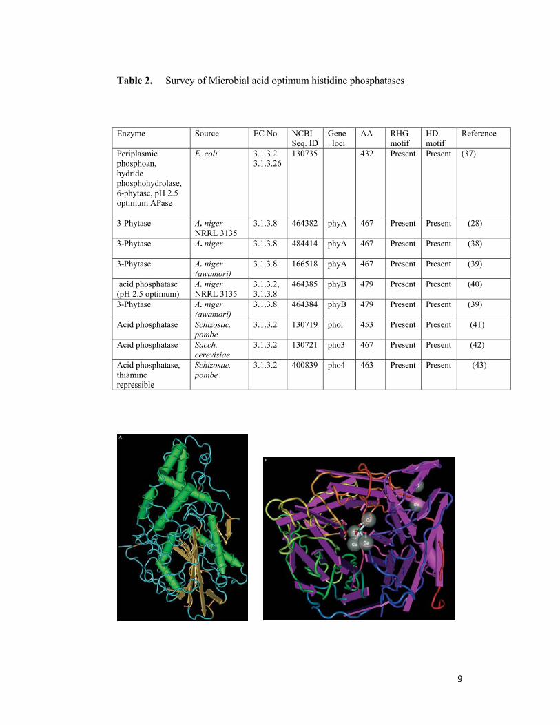
Table 2. Survey of Microbial acid optimum histidine phosphatases
Enzyme Source EC No NCBI Seq. ID
Gene . loci
AA RHG motif
HD motif
Reference
Periplasmic phosphoan, hydride phosphohydrolase, 6-phytase, pH 2.5 optimum APase
E. coli 3.1.3.2 3.1.3.26
130735 432 Present Present (37)
3-Phytase
A. niger NRRL 3135
3.1.3.8 464382 phyA 467 Present Present (28)
3-Phytase
A. niger
3.1.3.8 484414 phyA 467 Present Present (38)
3-Phytase
A. niger (awamori)
3.1.3.8 166518 phyA 467 Present Present (39)
acid phosphatase (pH 2.5 optimum)
A. niger NRRL 3135
3.1.3.2, 3.1.3.8
464385 phyB 479 Present Present (40)
3-Phytase
A. niger (awamori)
3.1.3.8 464384 phyB 479 Present Present (39)
Acid phosphatase
Schizosac. pombe
3.1.3.2 130719 phol 453 Present Present (41)
Acid phosphatase
Sacch. cerevisiae
3.1.3.2 130721 pho3 467 Present Present (42)
Acid phosphatase, thiamine repressible
Schizosac. pombe
3.1.3.2 400839 pho4 463 Present Present (43)
10
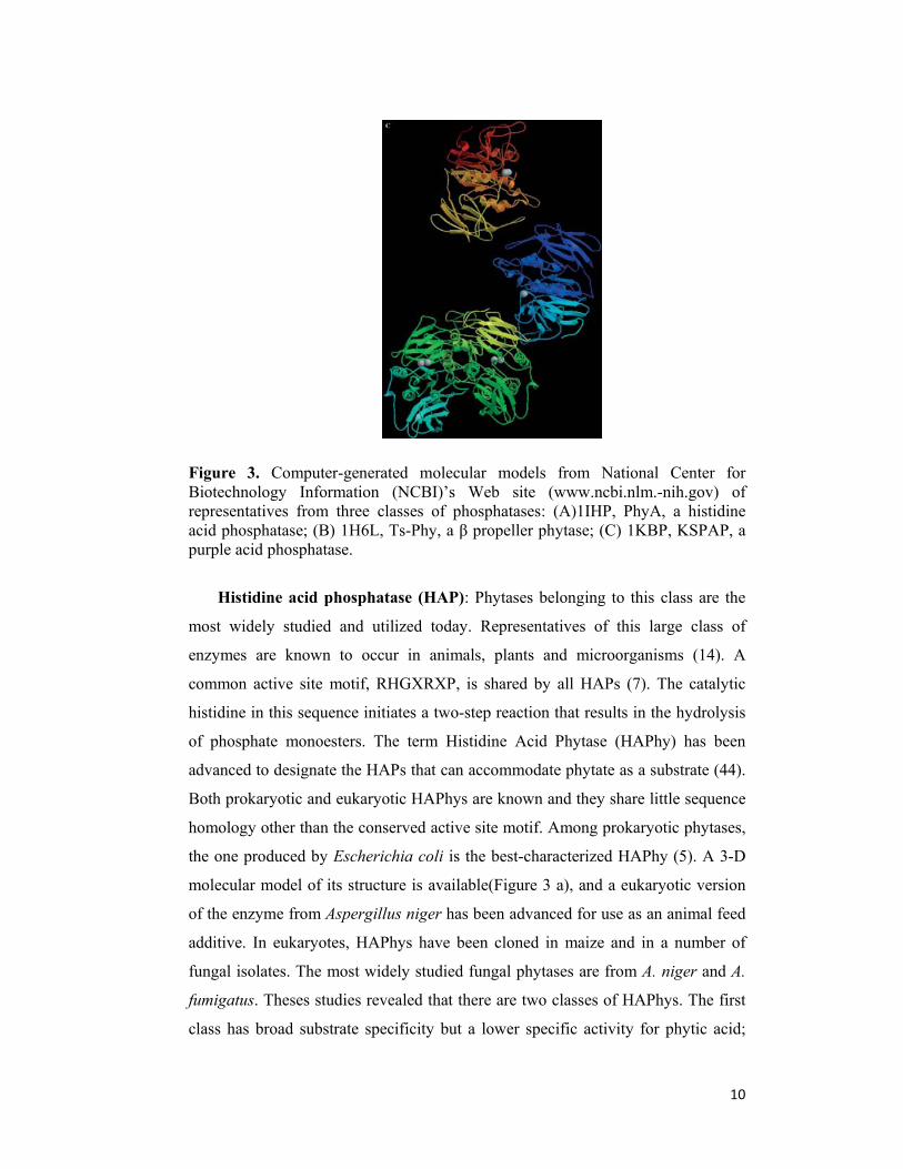
Figure 3. Computer-generated molecular models from National Center for Biotechnology Information (NCBI)’s Web site (www.ncbi.nlm.-nih.gov) of representatives from three classes of phosphatases: (A)1IHP, PhyA, a histidine acid phosphatase; (B) 1H6L, Ts-Phy, a β propeller phytase; (C) 1KBP, KSPAP, a purple acid phosphatase.
Histidine acid phosphatase (HAP): Phytases belonging to this class are the
most widely studied and utilized today. Representatives of this large class of
enzymes are known to occur in animals, plants and microorganisms (14). A
common active site motif, RHGXRXP, is shared by all HAPs (7). The catalytic
histidine in this sequence initiates a two-step reaction that results in the hydrolysis
of phosphate monoesters. The term Histidine Acid Phytase (HAPhy) has been
advanced to designate the HAPs that can accommodate phytate as a substrate (44).
Both prokaryotic and eukaryotic HAPhys are known and they share little sequence
homology other than the conserved active site motif. Among prokaryotic phytases,
the one produced by Escherichia coli is the best-characterized HAPhy (5). A 3-D
molecular model of its structure is available(Figure 3 a), and a eukaryotic version
of the enzyme from Aspergillus niger has been advanced for use as an animal feed
additive. In eukaryotes, HAPhys have been cloned in maize and in a number of
fungal isolates. The most widely studied fungal phytases are from A. niger and A.
fumigatus. Theses studies revealed that there are two classes of HAPhys. The first
class has broad substrate specificity but a lower specific activity for phytic acid;
11
the second class has narrow substrate specificity but a high specific activity for
phytase (45). Evidence from site-directed mutagenesis studies established the
importance of certain amino acid residues that make up the substrate specificity
site in fungal HAPhys. Mutating these key amino acids leads to changes in
substrate affinity and the pH profile (46). While not directly involved in the
catalytic mechanism of HAPhys, the conservation of an eight-cysteine motif
appears to be essential to maintain the proper molecular structure necessary for the
enzyme activity in fungal phytases (47). Today, the major application for HAPhys
is in the hydrolysis of phytate in cereal and grains in animal feed. Future
applications extend from the development of plant cultivars that require less P
fertilizer to modification for use as a peroxidase.
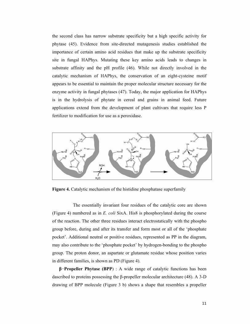
Figure 4. Catalytic mechanism of the histidine phosphatase superfamily
The essentially invariant four residues of the catalytic core are shown
(Figure 4) numbered as in E. coli SixA. His8 is phosphorylated during the course
of the reaction. The other three residues interact electrostatically with the phospho
group before, during and after its transfer and form most or all of the ‘phosphate
pocket’. Additional neutral or positive residues, represented as PP in the diagram,
may also contribute to the ‘phosphate pocket’ by hydrogen-bonding to the phospho
group. The proton donor, an aspartate or glutamate residue whose position varies
in different families, is shown as PD (Figure 4).
β−Propeller Phytase (BPP) : A wide range of catalytic functions has been
dascribed to proteins possessing the β-propeller molecular architecture (48). A 3-D
drawing of BPP molecule (Figure 3 b) shows a shape that resembles a propeller
12
with six blades (49). A novel calcium dependent Bacillus phytase that has this
configuration has been cloned and characterized. It lacks the RHGXRXP sequence
motif and therefore, it is not a member of HAP. It requires Ca2+
for both activity
and thermostability (50). This phytase employs two phosphate-binding sites, a
cleavage site for substrate hydrolysis and an affinity site to bind the substrate (51).
β−propeller phytases share an optimum pH range with some alkaline plant
phytases. The molecular structure of these plant phytases has yet to be determined,
but they display some common traits with β-propeller phytases. They both have a
narrow substrate range while requiring calcium for activity and only remove three
phosphates from phytic acid to yield inositol trisphospahte as a final product. No
commercial applications are available thus far for BPP, but it has been advanced as
an animal feed additive and as a means to promote plant growth under phosphate
limiting conditions.
Cysteine Phosphatase (CP): Recently, another class of phytase has been
reported from an anaerobic ruminal bacterium, Selenomonas ruminantium (52). Its
optimum temperature ranged between 50–55oC with optimal activity in the pH
range of 4.0–5.0 depending on the buffer used. Lead cations enhance activity,
while Fe2+
, Fe3+
, Hg2+
, and Zn2+
ions strongly inhibited the enzyme. Sequence
homology studies support similarities between this phytase and the catalytic
domain found in the cysteine phosphatase superfamily. Its 3-D structure’s
accession number is 1U24. The structure of this phytase consists of one large and
one small domain. Towards the C-terminal, near the edge of the large domain is a
shallow pocket containing a two loop structure similar to the active site found in
protein tyrosine phosphatase with the catalytically important HCXXGXXR(T/S).
This enzyme catalyzes dephosphorylation of phytic acid to myo-inositol
monophosphate.
Purple acid phosphatase (PAP): Characterization of a soybean (Glycine max L.
Merr.) phytase has revealed the purple acid phosphatases sequence motif,
DXG..GDXXY. .GNH(E,D)..VXXH..GHXH. The GmPhy phytase, found in
germinating soybean seedlings, apparently contains the catalytic mechanism
associated with this large class of metalloenzymes (53). This and a putative rice

13
(Oryza sativa) phytase are the only PAP phytases currently deposited in GenBank.
As compared to fungal phytase, this soybean seed phytase has a relatively low
specific activity for phytic acid. It has been proposed that the low catalytic activity
of GmPhy may be advantageous in plant seed where a slow and balanced
breakdown of phytate during germination could be efficacious. No 3-D model of
soybean phytase is available, and no commercial applicant is envisioned.
1.2.1 Microbial Sources
Microbial phytase activity is most frequently detected in fungi, particularly
in Aspergillus species. Shieh and Ware (54) screened over 2000 microorganisms
isolated from soil for phytase production. Most of the positive isolates produced
only intracellular phytase. Extracellular phytase activity was observed only in 30
isolates. All extracellular phytase producers were filamentous fungi. Twenty-eight
belonged to the genus Aspergillus, one to Penicillum and one to Mucor. Of the 28
phytase-producing Aspergillus isolates 21 belonged to the A. niger group. Other
studies (55-59) confirmed A. niger strains to be the best producers of extracellular
phytase. Phytase has also been detected in various bacteria, e.g. Aerobacter
aerogenes (60), Pseudomonas sp. (61), Bacillus subtilis (62), Klebsiella sp. (63),
B. subtilis (natto) (64), Escherichia coli (5), Enterobacter sp. 4 (65) and Bacillus
sp. DS 11 (later designated as B. amyloliquefaciens) (50). The only bacteria
producing extracellular phytase are those of the genera Bacillus and Enterobacter.
E. coli phytase is a periplasmic enzyme.
Some yeasts, such as Saccharomyces cerevisiae, Candida tropicalis,
Torulopsis candida, Debaryomyces castelii, Debaryomyces occidentalis,
Kluyveromyces fragilis and Schwanniomyces castelii, have also been shown to
produce phytase (66-70).
Recently Phytase production has been reported from Sachharomyces
cerevisiae CY (70), Pedobacter nyackensis MJ11 CGMCC 2503 (71), Yersinia
rohdei (2008)), Penicillium expansum (72), rabbit cecal bacteria (73), lactic acid
bacteria (74), Sporotrichum thermophile (75), Megasphaera elsdenii (76),
Bifidobacterium animalis (77), Aspergillus niger van Teighem (78), Selenomonas

14
lacticifex (79), Marine Yeast Kodamea ohmeri BG3 (80), Mucor indicus MTCC
6333 (81), Debaryomyces castellii CBS 2923 (82), marine yeast Kodamaea ohmeri
BG3 (83), antarctic yeast strain Cryptococcus laurentii AL27 (84), Mucor hiemalis
(85), Streptomyces hygroscopicus NRRL B-1476 (86).
1.2.2 Plant Sources
Phytase occurs widely in the plant kingdom. Phytase has been isolated and
characterized from cereals such as triticale, wheat, maize, barley and rice and from
beans such as navy beans, mung beans, dwarf beans and California small white
beans. Phytase activity has also been detected in white mustard, potato, radish,
lettuce, spinach, grass and lily pollen (23). Laboure et al (87) purified and
characterized phytase from germinating maize seedlings (Zea mays), and the
cDNA coding for this phytase was cloned (88). This cDNA was used to screen a
maize genomic library and two distinct genes were isolated and sequenced.
1.2.3 Animal Sources
Phytase has been found to exist in monogastric animals (89-92).Generally,
however, intestinal phytase does not play a significant role in food-derived phytate
digestion in monogastrics (93). Craxton et al (6) cloned and expressed a rat hepatic
multiple inositol polyphosphate phosphatase (MIPP) having phytase activity. The
MIPP mRNA was present in all rat tissues examined, but was most highly
expressed in kidney and liver. A phytase-like enzyme was also decribed in the
protozoan Paramecium (94).
1.2.4 Sequence Homology of phytases
The primary sequences of several fungal phytases have been determined in
recent years. A phytase cloned from A. niger var. awamori had over 97% identity
to A. niger NRRL 3135 phytase (phyA). Less homologous to the A. niger NRRL
15
3135 phytase are the phytases from A. fumigatus (65%), A. terrus (62%), E.
nidulans (62%), T. thermophilus (61%) and M. thermophila (46%). The PhyB
from A. niger NRRL 3135 shows 99% identity to the corresponding protein from
A. niger var. awamori. Surprisingly, two phytases (PhyA and PhyB) from A. niger
NRRL 3135 are only 25% homologous. Bacterial phytase from Escherichia coli
and a mammalian phytase (rat hepatic MIPP) do not exhibit apparent sequence
similarity to A. niger NRRL 3135 phytase. However, they share a highly conserved
sequence motif - RHG - that is found at the active sites of acid phosphatases (7,
95). Furthermore, they contain a remote C-terminal motif with histidine and
aspartic acid residues that probably take part in the catalysis. Therefore, these
phytases are said to form the phytase subfamily of histidine acid phosphatases
(11). The two plant phytases from Zea mays (PHYT I and PHYT II) are practically
identical, but do not show any homology to other phytases or to any phosphatases.
However, a region of 33 amino acids was detected that showed similarity to A.
niger NRRL 3135 phytase. This region is probably the acceptor site for phosphate
(88). The phytase from B. amyloliquefaciens (88, 96) shows 72% identity to an
open reading frame revealed in the genomic sequencing of the Bacillus subtilis
(97), but is not homologous to any phytases or to any phosphatases. Similarly, the
phytase from Enterobacter sp. is not homologous to any phytases or histidine acid
phosphatases. However, it is 30-38% homologous to low molecular weight acid
phosphatases from Chryseobacterium meningosepticum and Streptococcus
equisimilis. Especially certain lysine and tryptophan residues appears to be
conserved.
16
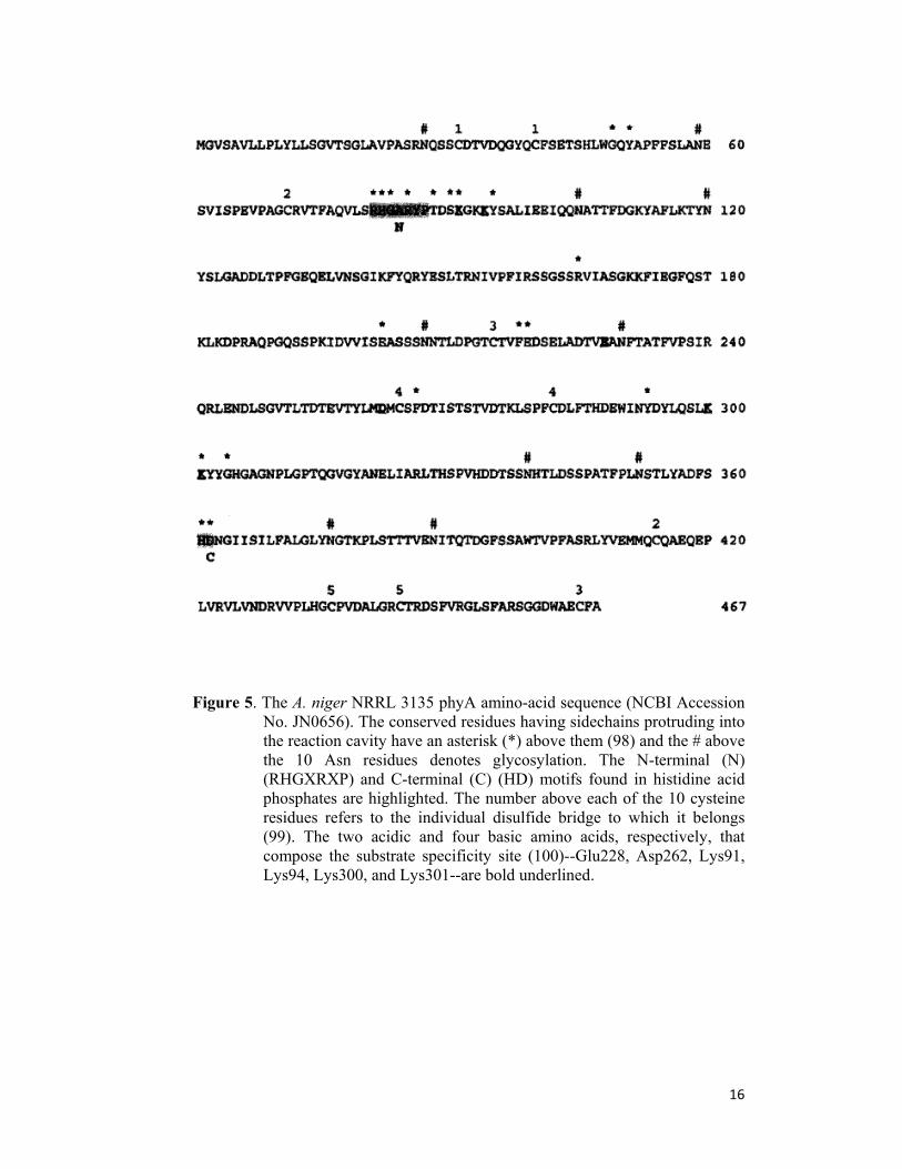
Figure 5. The A. niger NRRL 3135 phyA amino-acid sequence (NCBI Accession No. JN0656). The conserved residues having sidechains protruding into the reaction cavity have an asterisk (*) above them (98) and the # above the 10 Asn residues denotes glycosylation. The N-terminal (N) (RHGXRXP) and C-terminal (C) (HD) motifs found in histidine acid phosphates are highlighted. The number above each of the 10 cysteine residues refers to the individual disulfide bridge to which it belongs (99). The two acidic and four basic amino acids, respectively, that compose the substrate specificity site (100)--Glu228, Asp262, Lys91, Lys94, Lys300, and Lys301--are bold underlined.
17
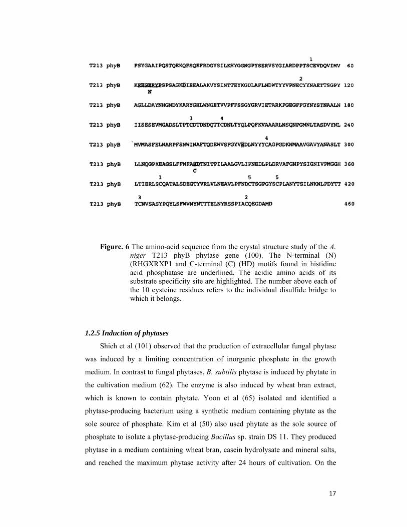
Figure. 6 The amino-acid sequence from the crystal structure study of the A. niger T213 phyB phytase gene (100). The N-terminal (N) (RHGXRXP1 and C-terminal (C) (HD) motifs found in histidine acid phosphatase are underlined. The acidic amino acids of its substrate specificity site are highlighted. The number above each of the 10 cysteine residues refers to the individual disulfide bridge to which it belongs.
1.2.5 Induction of phytases
Shieh et al (101) observed that the production of extracellular fungal phytase
was induced by a limiting concentration of inorganic phosphate in the growth
medium. In contrast to fungal phytases, B. subtilis phytase is induced by phytate in
the cultivation medium (62). The enzyme is also induced by wheat bran extract,
which is known to contain phytate. Yoon et al (65) isolated and identified a
phytase-producing bacterium using a synthetic medium containing phytate as the
sole source of phosphate. Kim et al (50) also used phytate as the sole source of
phosphate to isolate a phytase-producing Bacillus sp. strain DS 11. They produced
phytase in a medium containing wheat bran, casein hydrolysate and mineral salts,
and reached the maximum phytase activity after 24 hours of cultivation. On the
18
basis of these results it is difficult to say whether the production of these two
enzymes is induced by phytate itself or by phosphate starvation. Klebsiella phytase
production is induced by phytate (63, 102, 103). This situation is different from
the production of phytate-degrading enzymes in E. coli, the synthesis of which has
been shown to be stimulated by phosphate starvation or anaerobiosis (5, 103).
Various investigators have reported that in plants, during seed germination,
phytate is rapidly degraded and that the levels of phytase increase by several orders
of magnitude. It is not clear whether the increase in phytase activity is a result of
expression of phytase genes or simple activation of existing enzyme. Nayini and
Markakis (66) concluded that seeds contain both constitutive and germination-
inducible phytases. Northern blot analysis and in situ hybridization showed a high
accumulation of phytase mRNA during the early steps of germination in
coleorhiza, radical cortex and coleoptile parenchyma (104).This indicates
germination-induced synthesis of maize phytase.
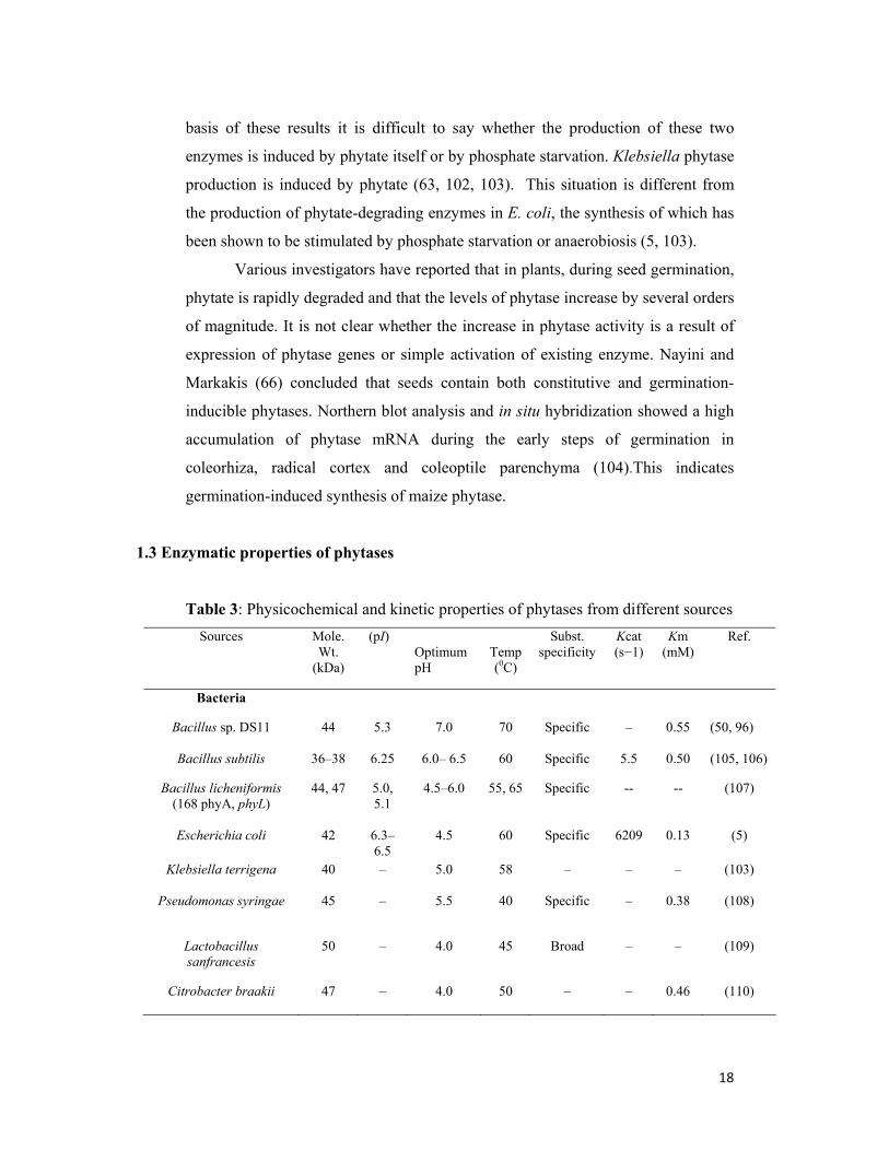
1.3 Enzymatic properties of phytases
Table 3: Physicochemical and kinetic properties of phytases from different sources
Sources Mole. Wt.
(kDa)
(pI)
Optimum pH
Temp (0C)
Subst. specificity
Kcat (s−1)
Km (mM)
Ref.
Bacteria
Bacillus sp. DS11
44 5.3 7.0 70 Specific – 0.55 (50, 96)
Bacillus subtilis 36–38 6.25 6.0– 6.5 60 Specific 5.5 0.50 (105, 106)
Bacillus licheniformis (168 phyA, phyL)
44, 47 5.0, 5.1
4.5–6.0 55, 65 Specific -- -- (107)
Escherichia coli 42 6.3–6.5
4.5 60 Specific 6209 0.13 (5)
Klebsiella terrigena
40 – 5.0 58 – – – (103)
Pseudomonas syringae 45
– 5.5 40 Specific – 0.38 (108)
Lactobacillus sanfrancesis
50 – 4.0 45 Broad –
– (109)
Citrobacter braakii
47 – 4.0 50 – – 0.46 (110)
19
Yeasts
Arxula adeninivorans adeninivorans
– – 4.5 75 Specific
– 0.23 (111)
Schwanniomyces castellii 490 -- 77 (112)
Sacch. cerevisiae 120 –
2.0–2.5, 5.0–5.5
55–60 – – – (113)
Pichia pastoris 95 –
2.5, 5.5 60 -- -- -- (114)
Fungi
Aspergillus ficuum (phyA)
85 4.5 2.5, 5.0 58 Specific 348 0.027 (115)
A. ficuum (phyB) 68 4.0 2.5 63 Broad 628 0.103 (116,
117)
A. oryzae 120 4.15 5.5 50 Broad – 0.33 (118)
Aspergillus niger SK-57 60 – 2.5,5.5 50 Specific -- 0.019 (119)
A. niger ATCC 9142
84 – 5.0 65 Broad – 0.10 (120)
Peniophora lycii (phyA)
72 3.61 4–4.5 50–55 Specific -- -- (121)
1.3.1 Biophysical characteristics
Published molecular size and the calculated theoretical molecular size of the
mature protein, and the isoelectric point (pI) of phytases from various sources are
shown in Table 3. Most phytases hitherto characterized are monomeric enzymes.
This is the case with fungal phytases (115, 122, 123) with E. coli and K. terrigena
phytases (5, 103) and with B. subtilis (natto) Phytase (64). However, some plant
and animal phytases appear to be built up of, multiple subunits. A phytase
accumulating in maize seedlings during germination is a dimeric enzyme built up
from two 38 kDa subunits (87). Purified rat intestinal phytase exhibited two
protein bands in SDS-PAGE with estimated molecular masses of 70 and 90 kDa
(124). However, since only the 90 kDa subunit is induced by phytic acid, it is
likely that these protein bands represent two different enzymes (alkaline
phosphatase and phytase, respectively). An inositol hexakisphosphate
dephosphorylating enzyme from the protozoan Paramecium has been proposed to
have a hexameric structure (94).
20
Two different forms of Klebsiella aerogenes phytase have been reported.
One, possibly the native enzyme, has an exceptionally large size (700 kDa). The
other is probably a fraction of the native enzyme, with a full complement of
activity and an exceedingly low molecular weight between 10 and 13 kDa (102).
Bacterial phytases are generally smaller than their fungal counterparts. The
predicted size of fungal phytases is around 50 kDa and the experimental size is
between 65 and 70 kDa, indicating heavy glycosylation. A. niger NRRL 3135
native phytase is 27% glycosylated. It contains a substantial proportion of N-linked
mannose chains and galactose (4, 45) reported that glycosylation of recombinant
phytases was highly variable. Whereas glycosylation was moderate in A. niger, it
was excessive and highly variable in Hansenula polymorpha and Saccharomyces
cerevisiae. Surprisingly, glycosylation differed not only between the different
expression systems used but also between different batches of a phytase produced
in the same expression system. Analysis of the glycosylation pattern of A. niger
phytase showed that the heterogeneity was due to incomplete glycosylation of two
out of ten potential N-glycosylation sites.
In general, glycosylation may have several effects on the properties of an
enzyme. Firstly, it may influence the catalytic properties or have an impact on the
stability of the enzyme. Secondly, it may influence the pI of the protein. Thirdly,
by consuming metabolic energy it may lower the level of expression of the protein.
Surprisingly, different extents of glycosylation had no effect on the catalytic
properties, thermostability or refolding properties of A. nigerphytase (122). The
importance of glycosylation for the structure and function of phytase is further
brought into question by the fact that only two potential N-glycolysation sites are
conserved in fungal phytases (125). Han and Lei (126) studied the role of
glycosylation in the functional expression of A. niger phytase (phyA) in Pichia
pastoris. Their results indicated an identical capacity of phytic acid hydrolysis and
slightly improved thermostability in glycosylated enzyme produced in P. pastoris
compared to the same enzyme overexpressed in A. niger. Deglycosylation of the
phytase resulted in 34% reduction in thermostability. Suppression of glycosylation
by tunicamycin during expression resulted in significant reduction of phytase
production, indicating that glycosylation is vital for the biosynthesis of
21
recombinant PhyA in P. pastoris. However, tunicamycin might also impair the
production by other means. Because there was no accumulation of intracellular
phytase protein, the impairment did not appear to occur at the level of translocation
of phytase. On the other hand, studies by Wyss et al (122) suggest that
glycosylation has no or only a minor effect on the pI of the fungal phytases tested.
The only exceptions were the phytases expressed in H. polymorpha, in which a
pronounced shift to acidic pI values was observed. All the fungal, bacterial, and
plant phytases hitherto investigated have acidic pI values, with the exception of A.
fumigatus phytase, which has a basic pI. Bacterial phytases seem to be less acidic
than fungal phytases: their pI is generally above 6, whereas fungal enzymes have
pI values below 5.5. A. fumigatus, Emericella nidulans, A. terrus, and
Myceliophthora thermophila phytases have a tendency to undergo proteolytic
degradation when expressed in A. niger and stored as concentrated culture
supernatants at 4oC (122). The activity of phytase from B. subtilis is unaffected by
proteases such as trypsin, papain and elastase (62), indicating a stronger protease
resistance than that of fungal phytases.
1.3.2 Temperature and pH stabilities and optima
The pH and temperature optima of phytases from various sources are
presented in Table 3. The pH optimums of phytases vary from 2.2 to 8. Most
microbial phytases, especially those of fungal orgin, have a pH optimum between
4.5 and 5.6. In contrast to most fungal phytases, A. fumigatus phytase has a broad
pH optimum; at least 80% of the maximal activity is observed at pH values
between 4.0 and 7.3. Some bacterial phytases, especially those from Bacillus, have
a pH optimum at 6.5 - 7.5. The pH optima of plant seed phytases range from 4.0 to
7.5, most having an optimum between 4.0 and 5.6. Two alkaline plant phytases
having a pH optimum at about 8.0 have been described in legume seed (127) and
lily pollen (128). A. niger NRRL 3135 and Citrobacter freundii phytases differ
from other phytases in having two pH optima. The temperature optima of phytases
vary from 45 to 77oC. Wyss and co-workers (129) studied the thermostability of
three acid phosphatases of fungal orgin (A. fumigatus and A.niger phytase, and A.
niger pH 2.5 optimum acid phophatase) by circular dichroism (CD) spectroscopy
and fluorescence, and by measuring the enzymatic activity. They concluded that A.

22
niger phytase was not thermostable, neither did it have the capacity to refold after
heat denaturation. At temperatures between 50 and 55oC it underwent an
irreversible conformational change that resulted in 70-80%loss of enzyme activity.
The A. fumigatus phytase was not thermostable, but had the remarkable property of
being able to refold completely into native like, fully active conformation after 20
min heat denaturation at 90oC. Compared to two phytases, A. niger pH 2.5 acid
phosphatase had higher intrinsic thermostability. At temperatures up to 80oC, only
minor changes in CD spectral characteristics and only slight, but irreversible
enzyme inactivation were observed. However, exposure to 90oC resulted in an
irreversible conformational change and complete loss of activity. Bacillus sp. strain
DS11 phytase (50) had a temperature optimum at 70oC, which is higher than the
temperature optimum of phytases in general. It was also very thermostable: 100%
residual activity after 10 min incubation at 70oC (in the presence of CaCl2). The
enzyme stability of Bacillus sp. strain DS11 phytase was drastically reduced above
50oC in the absence of CaCl2, whereas it was rather stable up to 90oC in the
presence of CaCl2. After incubation at 90oC for 10 min, the residual enzyme
activity was approximately 50% of the initial activity. This indicates that the Ca2+
ion has a strong protecting effect on the enzyme against thermal denaturation.
1.3.3 Modulators of Enzyme activity
Metal ions have been shown to modulate phytase activity. However, it is
difficult to determine whether the inhibitory effect of various metals is due to
direct binding to the enzyme, or whether the metal ions form poorly soluble
complexes with phytic acid and therefore decrease the active substrate
concentration. Phytase from Enterobacter sp. 4 was greatly inhibited by Zn2+,
Ba2+, Cu2+ and Al3+ (65).Similarly, the phytase from B. subtilis (natto) N- 77 was
greatly inhibited by metal ions such as Zn2+, Cd2+, Ba2+, Cu2+, Fe2+, and Al3+ (64).
Both of these enzymes, as well as two other Bacillus phytases (50, 62), were
greatly inhibited by EDTA, indicating that a metal ion (calcium) is needed for the
activity. Wyss et al (45) reported that Cu2+ considerably depressed the enzyme
activities of E. nidulans and A. terrus phytases, and that several metal ions
inhibited A. fumigatus phytase. The activity of A. fumigatus phytase was stimulated
up to 50% by EDTA, whereas EDTA had no major effects on the enzymatic
23
activities of other fungal phytases tested (E. nidulans, A. niger and A. terrus). The
effects of metal ions and the fact that EDTA either has no effect or even stimulates
phytase activity indicates that fungal phytases clearly differ from Ca2+dependent
Bacillus phytases that are readily inhibited by EDTA. This conclusion is supported
by the lack of metal ions in the crystal structure of A. niger Phytase (99). In
addition to calcium-dependent Bacillus phytases, a phytase from pollen of Typha
latifolia and phytases from some other plants require Ca2+ for optimal activity (87,
128, 130, 131).
Reducing reagents, such as 2-mercaptoethanol, dithiotreitol and reduced
glutathione have no major effects on microbial phytases. This suggests that these
enzymes either have no free and accessible sulfhydryl groups or that the free
sulfhydryl groups play a negligible role in the enzyme activity and structure. This
interpretation is supported by the fact that most mature phytases have an even
number of cysteine residues that might be implicated in disulfide bridges, as is the
case with A. niger phytase (99). The function of disulfide bonds in A. ficuum
phytase was elucidated by unfolding studies performed by Ullah and Mullaney
(132). These authors concluded that disulfide bonds are necessary for the structure
and activity of the enzyme and play an important role in the folding of the protein.
Mature Bacillus phytases appear to have no cysteine residues.
A structural analog of the substrate, myo-inositol hexasulfate, has been shown
to be a potent competitive inhibitor of both PhyA and PhyB enzymes from A.
ficuum (133). The Ki of inhibiton for the PhyA and PhyB enzymes were estimated
to be 4.6 and 0.2 μM, respectively. Fluoride is a known inhibitor of different
phytases and phosphatases (56, 134). The phytase from cotyledons of germinating
soybean seeds was strongly inhibited by fluoride, vanadate and inorganic
phosphate (130). Inorganic phosphate was a competitive inhibitor of soybean seed
phytase. The fact that soybean seed phytase is competitively inhibited by
orthophosphate with a Ki value of 28 μM implies that the activity of the enzyme is
tightly regulated. Competitive product -inhibiton of phytate hydrolysis caused by
inorganic phosphate is recognized for bacterial, fungal and oat spelt phytases (5,
55, 135). Fluoride also inhibited the alkaline phytase from lily pollen (131, 136)
and competitively inhibited the phytase from K. terrigena (103) and phosphate,
24
molybdate and vanadate. Molybdate and vanadate are known to inhibit
phosphatase enzymes. It has been suggested that these transition metal oxoanions
inhibit phosphomonoesterases by forming complexes that resemble the trigonal
bipyramidal geometry of the transition state (137).
Substrate concentrations above 300 μM have been reported to be inhibitory for
the phytase like enzyme from Paramecium (94). The Klebsiella sp. and Rhizopus
oligosporus phytases were also inhibited by the substrate (63, 138), but only in
higher substrate concentrations. Fungal phytase activity has been shown to be
inhibited by substrate concentrations exceeding 1 mM (139). Maize root and
soybean phytases were found to be inhibited at 300 μM and 20 mM substrate
concentration, respectively (140, 141). In high substrate concentrations, the charge
due to the phosphate groups may affect the local environment of the catalytic
domain of the enzyme. This might inhibit conversion of the enzyme-substrate
complex to enzyme and product (142), although inhibition due to the formation of
poorly soluble protein-phytate complex cannot be ruled out.
1.3.4 Substrate Specificity and Kinetic parameters
Phytases show relatively broad substrate specificity. ADP, ATP, p-
nitrophenyl phosphate, phenyl phosphate, fructose 1,6-bisphosphate, glucose 6-
phosphate, α-, and β-glycerophosphate and 3-phosphoglycerate, that are not
structurally similar to phytic acid, are frequently hydrolyzed by phytases. Only a
few phytases have been described as highly specific for phytic acid: the Bacillus
phytases (62, 64) and alkaline phytase isolated from lily pollen (136). Most
phytases hitherto studied follow Michaelis- Menten kinetics, with the exceptions of
M. thermophila and E. nidulans phytases which display non-Michaelis-Menten
behavior. It should be noted that under the standard assay conditions (i.e. 2 mM
phytic acid), only the rate of the reaction from myo-inositol hexakisphosphate to
pentakisphosphate is measured. Ullah and Phillippy (142) determined the kinetic
parameters of A. ficuum phytase and two acid phosphatases for myo-inositol hexa-,
penta-, tetra, and triphosphates. Phytate had the lowest Km value for all three
enzymes. They concluded that both phytase and pH 2.5 optimum acid phosphatase
effectively hydrolyzed the tested myo-inositol phosphates. Poor hydrolysis of
tested forms of myo-inositol phosphates by pH 6.5 optimum acid phosphatase was
25
demostrated by low Vmax and Kcat values. The kinetic efficiency of an enzyme is
validated by means of the Kcat/Km values for a substrate. The highest Kcat/Km
values for phytase and pH 2.5 optimum acid phosphatase were those for phytic
acid (1.29 × 10⁷ and 6.10 × 10⁶ M-1 s-1, respectively). E. coli phytase has a
Kcat/Km value of 4.78 × 10⁷ M-1 S-1 (5), which is the highest value reported for a
phytase. The specific activities for fungal phytases with phytic acid as substrate
vary almost 10- fold, from 23 to 198 U mg-1 (A. fumigatus and A. terrus,
respectively). The different extent and patterns of glycosylation have no significant
effect on the specific activities of fungal phytases (45). Specific activities reported
for bacterial phytases vary almost 100-fold, from 8.7 to 811 U mg-1, (B. subtilis
and E. coli, respectively). On the basis of substrate specificity, phytases can be
divided into two classes - phytases with broad substrate specificity (e.g. A.
fumigatus, E. nidulans and M. thermophila) and phytases with rather high
specificity for phytic acid (e.g. A. niger, A. terrus and E. coli). Phytases with broad
substrate specificity inherently have rather low specific activity for phytic acid (23
to 43 U mg-1), whereas phytases with narrow substrate specificity have specific
activities of 103 - 811 U mg-1. Bacillus phytases do not fit into this classification.
They appear to be very specific for phytic acid, but have apparently low specific
activity. The low specific activity is likely to hinder their use in industrial
applications. The Km values and specific activities of some published phytases for
phytic acid are presented in Table 4.
1.3.5 Kinetics and End Products of Phytic acid degradation
Phytic acid has six phosphate groups that may be released by phytases at
different rates and in different order. Wyss et al (45) investigated the kinetics of
phosphate release and the kinetics of accumulation of reaction intermediates, as
well as the end products of phytic acid degradation by various phytases. They
concluded that all fungal phytases studied released five of the six phosphate
groups, the end product being myo-inositol 2-monophosphate when excess enzyme
is used. This indicates that all of these phytases have a pronounced
stereospecificity and a strong preference for equatorial phosphate groups, whereas
they are virtually unable to cleave the axial phosphate group. Only in rare cases
were traces of free myo-inositol or myo-inositol 1-monophosphate detected. A.
26
fumigatus phytase readily degraded phytic acid to myo-inositol 2-monophosphate,
and only myo-inositol bisphophate (stereoisomer not known) accumulated to some
extent. In contrast, A. niger and A. terrus phytases had to be used at much higher
initial activities in order to obtain degradation down to myo-inositol 2-
monophoshate, and considerable amounts of myo-inositol tris- and bisphosphates
accumulated during the degradation. When E. coli phytase was used at an even
higher initial activity, there was a pronouced accumulation of myo-inositol
tetrakisphosphate during phytic acid degradation.
Myo-inositol bis- and triphosphates comprised more than 90% of the end
products after a 90-min incubation period (with excess enzyme) and almost no
myo-inositol monophosphate was detected. Therefore, lower myo-inositol
phosphates appears to be less suitable substrates for A. niger, A. terrus and
especially E. coli phytases than phytic acid. The stereoisomer assignment of the
reaction intermediates and degradation pathway was not determined for these
enzymes. The fact that the end products of phytic acid hydrolysis for most phytases
is identical does not necessarely mean that the degradation pathways for phytic
acid are identical. 3-Phytase starts hydrolyzing the phosphate esters at the D-3
position, giving rise to D-Ins(1,2,4,5,6)P5 as the first intermediate (103, 143). 6-
Phytase starts the hydrolysis at the L-6 (or D-4) position, yielding L-
Ins(1,2,3,4,5)P5 as the first intermediate. An alkaline phytase from lily pollen
(131) was shown to start the hydrolysis of phytic acid at position D-5, with two
subsequent dephosphorylation steps to yield Ins(1,2,3)P3 as the final product
(136). Inositol triphosphate is also the end product of phytic acid hydrolysis for the
phytase from Typha latifolia pollen (128). Rat hepatic multiple inositol
polyphosphate phosphatase (MIPP) catabolizes inositol Hexakisphosphate without
specificity towards a particular phosphate group. However, it hydrolyzed
Ins(1,3,4,5,6)P5 via Ins(1,4,5,6)P4 to Ins(1,4,5)P3 by consecutive 3- and 6-phytase
activities (6). A detailed characterization of the phytase from the protozoan
Paramecium by Freund et al (94) revealed that this enzyme degrades phytic acid
by stepwise dephosphorylation via D/L-Ins(1,2,3,4,5)P5, D/L-Ins(1,2,3,4)P4 and
Ins(1,2,3)P3 finally to D/ L-Ins(1,2)P2. Appearance of D/L-Ins(1,2,3,4)P4 clearly
preceeds that of Ins(1,2,3)P3. The slow conversion of inositol triphosphate to
27
inositol bisphosphate indicates that Ins(1,2,3)P3 is the main end product. Powar
and Jagannathan (62) showed that myo-inositol monophosphate (phosphate
position not determined) is the end product for B. subtilis phytase. Kinetics,
reaction intermediates and degradation pathways of phytic acid degradation have
not been reported for Bacillus phytases, neither is it known whether these enzymes
are 3- or 6-phytases. The strong stereospecificity for the equatorial phosphate
groups over the axial phosphate appears to be common to all phytases. This might
indicate that only the phosphate groups protruding equatorically from the inositol
ring can access the catalytic sites of these enzymes.
1.3.6 Active Site and Reaction mechanism
Acid phosphatases are a heterologous group of enzymes that hydrolyze
phosphate esters, optimally at low pH. A number of acid phosphatases, from both
prokaryotes and eukaryotes, share two regions of sequence similarity, each
centered around a conserved histidine residue (in bold) (95). The consensus pattern
for these two regions reported in the SWISS-PROT protein domain data base are
[LIVM]-X(2)-[LIVMA]-X(2)-[LIVM]-X-R-H- [GN]-X-R-X-[PAS] and [LIVMF]-
X-[LIVMFFAG]-X(2)-[STAGI]-H-D-[STANQ]-X- [LIVM]-X(2)-[LIVMFY]-
X(2)-[STA]. Sequence alignment of pho3 and pho5 gene products in yeast, human
prostatic and lysosomal acid phosphatase, and PhyA and PhyB from A. niger
NRRL 3153 reveals a conserved heptapeptide of RHGXRXP near the N-terminus
(compare to the former consensus pattern). The acid phosphatases containing this
active site motif are grouped as histidine acid phosphatases. This active site motif
is totally conserved in all fungal phytases and is also present in the E. coli phytase.
Sequence alignment of fungal and E. coli phytases reveals a conserved HD-motif
near the C-terminus (compare to the latter consensus pattern). Protein data base
searches for the sequence motifs RHG and HD reveal that they are present in a
number of acid phosphatases. In general, two classes of acid phosphatases can be
identified in terms of molecular mass. A low molecular weight form lacks both
motifs. A high molecular form is divided into two subclasses. One exhibits either
the RHG or the HD motif, the other both (144). Therefore, phytases are said to
form the phytase sub-family of high molecular weight histidine acid phosphatases
(11). Ullah and co-workers have used amino acid residue specific modifying
28
reagents to probe the active sites of fungal phytases (7, 8). Their results clearly
establish the crucial role of histidine and arginine residues for the activity of
phytase. Ullah and Dischinger (144) showed that some tryptophan residues might
also be involved in the phosphohydrolytic cleavage of phytic acid.
The results of Ullah and co-workers correlate with the site-directed
mutagenesis studies on E. coli Phytase (9, 10). On the basis of these results the
following two-step reaction mechanism for the high molecular weight histidine
acid phosphatases has been suggested: the positive charge of the guanido group of
the arginine residue in tripeptide RHG interacts directly with the phosphate group
in the substrate, making it more susceptible to nucleophilic attack, while the
histidine residue serves as a nucleophile in the formation of covalent
phosphohistidine intermediate; the aspartic acid residue (from the C-terminal HD-
motif) protonates the group leaving the substrate. The histidine residue in the HD-
motif also has a critical role in the enzyme activity. Due to the existence of these
motifs in fungal phytases and in the E. coli phytase, the proposed reaction
mechanism is likely to be characteristic to the members of the phytase sub-family
of histidine acid phosphatases.
The active site of phytases shows remarkable homology to the active site
residues of the members of a particular class of acid phosphatase termed "histidine
phosphatase" (7, 95). Chemical probing at the active site of human prostatic acid
phosphatase suggested that an arginine residue is involved in catalysis (145). A
similar observation was also made in A. niger NRRL 3135 (7). Sequence similarity
search among diverse phosphate metabolizing enzymes, i.e., fructose 2,6-
biphosphatase, phosphate glycerate mutase, and acid phosphatase, led to the
identification of a tripeptidic region with the sequence RHG (146). When A. niger
NRRL 3135 N-terminal amino acid sequence was compared with these enzymes, it
was noted that the conserved tripeptidic region was also present not only in fungal
phyA but also in the N-terminal region of phyB (147). Further chemical probing of
the fungal phytase also suggested a sensitive histidine at the active site (8). On
close examination of the active site residues of phyA and phyB in A. niger NRRL
3135, pH 2.5 optimum acid phosphatase in E. coli, pho3 and pho5 gene products in
yeast, human prostatic, and lysosomal acid phosphatase, it was observed that the
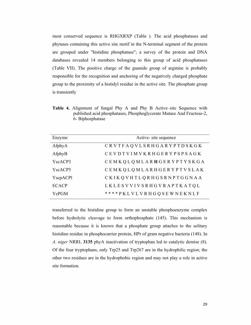
29
most conserved sequence is RHGXRXP (Table ). The acid phosphatases and
phytases containing this active site motif in the N-terminal segment of the protein
are grouped under "histidine phosphatase"; a survey of the protein and DNA
databases revealed 14 members belonging to this group of acid phosphatases
(Table VII). The positive charge of the guanido group of arginine is probably
responsible for the recognition and anchoring of the negatively charged phosphate
group to the proximity of a histidyl residue in the active site. The phosphate group
is transiently
Table 4. Alignment of fungal Phy A and Phy B Active–site Sequence with published acid phosphatases, Phosphoglycerate Mutase And Fructose-2, 6- Biphosphatase
Enzyme Active- site sequence
AfphyA C R V T F A Q V L S R H G A R Y P T D S K G K
AfphyB C E V D T V I M V K R H G E R Y P S P S A G K
YscACP3 C E M K Q L Q M L A R H G E R Y P T Y S K G A
YscACP5 C E M K Q L Q M L A R H G E R Y P T V S L A K
YscpACPl C K I K Q V H T L Q R H G S R N P T G G N A A
ECACP L K L E S V V I V S R H G V R A P T K A T Q L
YePGM * * * * P K L V L V R H G Q S E W N E K N L F
transferred to the histidine group to form an unstable phosphoenzyme complex
before hydrolytic cleavage to form orthophosphate (145). This mechanism is
reasonable because it is known that a phosphate group attaches to the solitary
histidine residue in phosphocarrier protein, HPr of gram negative bacteria (148). In
A. niger NRRL 3135 phyA inactivation of tryptophan led to catalytic demise (8).
Of the four tryptophans, only Trp25 and Trp267 are in the hydrophilic region; the
other two residues are in the hydrophobic region and may not play a role in active
site formation.
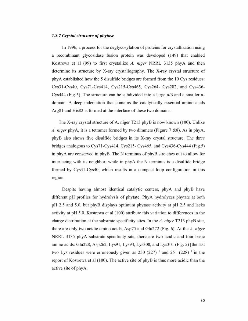
30
1.3.7 Crystal structure of phytase
In 1996, a process for the deglycosylation of proteins for crystallization using
a recombinant glycosidase fusion protein was developed (149) that enabled
Kostrewa et al (99) to first crystallize A. niger NRRL 3135 phyA and then
determine its structure by X-ray crystallography. The X-ray crystal structure of
phyA established how the 5 disulfide bridges are formed from the 10 Cys residues:
Cys31-Cys40, Cys71-Cys414, Cys215-Cys465, Cys264- Cys282, and Cys436-
Cys444 (Fig 5). The structure can be subdivided into a large α/β and a smaller α-
domain. A deep indentation that contains the catalytically essential amino acids
Arg81 and His82 is formed at the interface of these two domains.
The X-ray crystal structure of A. niger T213 phyB is now known (100). Unlike
A. niger phyA, it is a tetramer formed by two dimmers (Figure 7 &8). As in phyA,
phyB also shows five disulfide bridges in its X-ray crystal structure. The three
bridges analogous to Cys71-Cys414, Cys215- Cys465, and Cys436-Cys444 (Fig.5)
in phyA are conserved in phyB. The N terminus of phyB stretches out to allow for
interfacing with its neighbor, while in phyA the N terminus is a disulfide bridge
formed by Cys31-Cys40, which results in a compact loop configuration in this
region.
Despite having almost identical catalytic centers, phyA and phyB have
different pH profiles for hydrolysis of phytate. PhyA hydrolyzes phytate at both
pH 2.5 and 5.0, but phyB displays optimum phytase activity at pH 2.5 and lacks
activity at pH 5.0. Kostrewa et al (100) attribute this variation to differences in the
charge distribution at the substrate specificity sites. In the A. niger T213 phyB site,
there are only two acidic amino acids, Asp75 and Glu272 (Fig. 6). At the A. niger
NRRL 3135 phyA substrate specificity site, there are two acidic and four basic
amino acids: Glu228, Asp262, Lys91, Lys94, Lys300, and Lys301 (Fig. 5) [the last
two Lys residues were erroneously given as 250 (227) 1 and 251 (228) 1 in the
report of Kostrewa et al (100). The active site of phyB is thus more acidic than the
active site of phyA.

31
Recently, a recombinant form of E. coli phytase was purified and crystallized
to provide a three-dimensional structure of a 6-phytase (150). This X-ray-deduced
model can be used with the crystal structure of A. niger phyA, a 3-phytase (99) to
define the structural basis for their different catalytic pathways.
1The amino-acid residue location in the abbreviated sequence in Fig. 6 (100)
Enzyme reaction for phytase E.C.3.1.3.8
Myo-inositol
hexakisphosphate
+ H2O =
1D-myo-inositol 1,2,4,5,6-
pentakisphosphate
+
phosphate
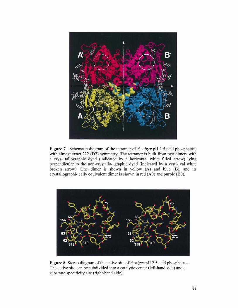
32
Figure 7. Schematic diagram of the tetramer of A. niger pH 2.5 acid phosphatase with almost exact 222 (D2) symmetry. The tetramer is built from two dimers with a crys- tallographic dyad (indicated by a horizontal white filled arrow) lying perpendicular to the non-crystallo- graphic dyad (indicated by a verti- cal white broken arrow). One dimer is shown in yellow (A) and blue (B), and its crystallographi- cally equivalent dimer is shown in red (A0) and purple (B0).
Figure 8. Stereo diagram of the active site of A. niger pH 2.5 acid phosphatase. The active site can be subdivided into a catalytic center (left-hand side) and a substrate specificity site (right-hand side).

33
Figure 9. Stereo picture of the three- dimensional fold of A. ficcum phytase. The α/β- domain is coloured red, the α-domain blue, the N- terminal lid green, and the disulphide bridges yellow. There is a gap on the left hand side of the α-domain for residues 249-252, which were not visible in the electron density. The active site is indicated with a red circle and the catalytically essential amino acid residues Arg 58 and His 59 are shown in blue ball-and-stick representation. This figure was prepared with the programs MOLSCRIPT (151) and RASTER3D (152).
1.4. Application of Phytases
1.4.1 Phytase as Feed Additive
Since phytic acid can not be metabolized by monogastric animals due to lack
of phytate-degrading enzyme in their gastrointestinal tract, feeds for pigs and
poultry are commonly supplemented with inorganic phosphate in order to meet the
phosphorus requirements. It is seen that the use of phytase in feed fodder improves
the phosphorus bioavailability and reduces the chelating ability of phytic acid, thus
resulting in reduction of phosphorus excretion in the areas of intensive livestock.
The superior activity of Aspergillus niger NRRL 3135 phytase and its practical
application to animal feed for the removal of phytic acid were demonstrated in
several studies (153, 154) collaborated to confirm the earlier feed studies by
34
feeding solvent precipitated phytase to chick diets, besides demonstrating the
efficacy of feeding phytase to swine. They concluded that the addition of enzyme
(1000 μmol P/h · ml phytase/kg diet) was sufficient to improve performance
further than that obtained by adding supplemental inorganic phosphate, thus
establishing the role of phytase as feed-additive in combating phosphorus pollution
in soil and water. The Natuphos produced by Gist-Brocades, when supplemented
in feed, resulted in enhanced utilization of phytin phosphorus by monogastric
animals.8 Alko Biotech tested Finase F for its use in improving the phosphorus
availability in swine and found results parallel to those obtained by phytases from
other sources (155-157). It was observed that the units of enzyme required for the
hydrolysis of set amount of phytin phosphorus were fairly consistent and one can
estimate, within a narrow range, the amount of phytase required to hydrolyze
phytin-P present in any commercial diet used in livestock units. Depending on the
specific diet, 380–1000 μmol P/h (6408– 16,600 nkat/ml) phytase is required to
replace 1 g phosphorus supplied by inorganic source. It was seen from the
livestock production statistics of the USDA that if phytase were used as a feed
ingredient in the diets of all the monogastric animals in United States, it would
release phosphorus with a value of $1.68 × 10⁸ per year, besides diminishing the
amount of phosphate in the manure and subsequently entering the environment.
The quantitative determination of released phosphate upon phytase
supplementation indicated that if the phytase were used in the diets of all
monogastric animals in the United States, it would preclude 8.23 × 10⁷ kg
phosphorus from entering the environment (14).
1.4.2 Food Application
A diet rich in cereal fibers, legumes and soy protein results in an increased
intake of phytate. Vegetarians, eldery people consuming unbalanced food with
high amounts of cereals, people in undeveloped countries who eat unleavened
bread, and babies eating soy-based infant formulas take in large amounts of phytate
(13). Undigested phytate in the small intestine negatively affects the absorption of
zinc, calcium, magnesium and iron. It also reduces the digestability of dietary
protein and inhibits digestive enzymes. Using phytase, Simell and co-workers (13)
reported the preparation of a phytate-free soy protein isolate with increased
35
solubility at low pH (pH 3) compared to the control soy protein isolate (158)
phytate from soybean milk using wheat phytase. Additions of A. niger phytase to
flour containing wheat bran increased iron absorption in humans (159). However,
more studies should be performed before accepting phytase as a food additive.
1.4.3 Pulp and Paper industry
Aging of papers is one of the main problems faced by the pulp and paper
industries. Lignin-free paper degraded faster than the lignin-containing paper, even
when it was sized with acid alum. Lignin acts as an antioxidant because it is
oxidized faster than cellulose and forms stable radicals; this shows that the process
aging is oxidative. Attempts to prevent oxidation by use of ethylenediamine
tetraacetic acid (EDTA) only accelerated the degradation process. Only a few
complexing agents block the Fenton reaction (formation of hydroxyl radicals); one
of them is phytic acid, found in seeds. A thermostable phytase can have potential
as a novel biological agent to degrade phytic acid during pulp and paper
processing. The enzymatic degradation of phytic acid would not result in the
production of toxic and mutagenic by-products, and at the same time aging of
paper can be prevented. Therefore, the exploitation of phytases in the pulp and
paper process could be environmentally friendly and would assist in the
development of cleaner technologies (160).
1.4.4 Phytase as Soil amendment
Phytic acid and its derivatives represent up to 50% of the total organic
phosphorus in the soil at certain locations, suggesting the possibility of phytase
addition that might stimulate plant growth in these soils. Findenegg and Nelemans
(161) studied the effect of phytase (phyA) on the availability of phosphorus from
phytic acid in the soil for maize plants. It was seen that phytin hydrolysis increased
when phytase was added to the soil, which further resulted in stimulation of plant
growth. However, the amount of phytase necessary for a significant effect
indicated that it is not a practical technique. This further indicated that the
expression of phytase in the roots of transgenic plants might increase the

36
availability of phosphorus to plants. Thus, expression of phytase in the roots of
plants might increase the phosphorus bioavailability (162).
1.4.5 Semisynthesis of Peroxidase
Based on structural similarity between the active sites of vanadium dependent
haloperoxidase, fungal phytase, and acid-phosphatase, a semisynthetic peroxidase
was designed.91 The Delft group incorporated vanadate ion into the active site of
A. niger NRRL 3135 phytase, thus transforming native phosphohydrolase into
semisynthetic peroxidase. The “new” enzyme was able to catalyse enantioselective
oxidation of prochiral sulphides and was found to be stable for 3 days with only
slight loss in turnover number. Being water miscible, the enzyme could be used in
the presence of organic solvents up to 30% concentration (v/v) with only a slight
loss in enzyme activity. It was seen that of all the phytases and acid phosphatases
tested, only histidine acid phosphatase (HAPs) with “RHGXRXP” active site
sequence could function as a peroxidase when vanadate ion was incorporated into
the active site.
1.4.6 Preparation of myo-Inositol phosphates
Besides the adverse effects of phytate and other highly phosphorylated
inositolphosphates on mineral bioavailability, some novel metabolic effects of
some of its degradation products have been recognized. The most extensively
studied positive aspect of myo-inositol phosphate (InsP1,4,5 and InsP1,3,4,5) is the
potential for reducing the risk of colon cancer. InsP5 plays an important role in
modulation of the oxygen-binding affinity of hemoglobin in the erythrocytes of
avian, amphibian, and reptilian species,5 while InsP4 and InsP3 was found to act
as secondary messenger in synergistically controlling intracellular calcium level.6 .
Surprisingly, the esters of inositol triphosphate have been shown to exert
significant inhibitory effects against retroviral infections including HIV (163). The
position of phosphate group on inositol ring is thereby of great significance for
their physiological function. To investigate the physiological effects of defined
myo-inositol phosphate isomers, these compounds have to be available in pure
form and sufficient quantity. . The chemical syntheses of myo-inositol phosphates
37
include difficult protection and deprotection steps, and are performed at extreme
temperatures and pressures (16).
Attempts to produce them nonenzymatically results in a mixture of
isomers, making the purification very uneconomical and arduous. Since phytases
hydrolyze myo-inositol hexaphosphate sequentially, the production of myo-inositol
phosphate derivates and free myo-inositol using phytase is a potential alternative to
chemical synthesis. Phytases are distributed widely in nature and different types of
phytases are known, 3-phytases, 4-phytases, and 6- phytases, indicating the
predominant attack at the susceptible phosphoester bond. Thus, using phytases of
different origin may lead to the production of different isomers. Siren (164)
prepared myo-inositol-1,2,6-triphosphate and myoinositol- 1,2,5-triphosphate by
enzymatic hydrolysis of phytic acid using S.cerevisiae phytase. The esters of
inositol triphosphate were found to alleviate conditions associated with abnormal
levels of neuropeptideY (NPY), such as arthritis and asthma, besides acting as a
pain killer (163). Greiner and Konietzny reported the synthesis of Ins-1,2,3,4,5-P,
Ins-2,3,4,5-P, Ins-2,4,5-P, and Ins-2,5- P using E. coli phytase covalently
immobilized on NHS-activated Sepharose. As only one major isomer of each InsP
was formed, further purification could be easily achieved by ion-exchange
chromatography. Using a combination of high-performance liquid chromatography
(HPLC) and kinetic studies, the stereospecificity of E. coli phytase P2 was
established, indicating sequential removal of phosphate groups via the 6/1/3/4/5
route (165).
Naturally, the advantages of enzymatic hydrolysis are stereospecificity and
mild reaction conditions. In addition to usage as drugs, myo-inositol phosphate
derivatives can be used as enzyme substrates for biochemical and metabolic
investigations and as chiral building blocks (15).
1.4.7 Environmental Benefits of Enzymes as Additives to Animal feed
The major enzyme application in the animal feed industry is the use of
phytase, which releases the phosphate bound in the grain and thus diminishes the
need for addition of inorganic phosphate to the feed. Phytase was developed for
use in animal feed based on the assumption that the substitution of inorganic
38
phosphate with phytase would offer a significant environmental advantage. To
check this assumption, an LCA study has been carried out on the use of phytase for
intensive pig farming. The study builds on Danish conditions, which means that
the average phosphate binding capacity for agricultural soils has been assumed to
be 95%. The results are shown in the Table 15. Most significant is the reduction of
the pollution of the aquatic environment with phosphate, which in Denmark due to
the intensive pig farming is a major problem. It can be calculated that the effect of
using phytase for all the pigs in Denmark (23 million) reduces the emission of P to
the aquatic environment with 260 t P. This corresponds to approximately 25% of
the diffuse emission of P from Danish agriculture. Another significant benefit of
phytase is the saving of the consumption of phosphate, which is an essential, but
limited resource. The phosphate saving from feeding all Danish pigs phytase
corresponds to the annual consumption of phosphate from 1 million people. Most
other enzyme applications within animal feed address the efficiency of digestion of
the grain nutrients. It is expected that such efficiency gains will drive significant
environmental benefits – just as it has been demonstrated for the food industry. (C)
1.5 Market trends and future prospects
The growth of the market for phosphate to supplement animal feed fostered a
critical step in the commercial development of phytase. Recent trends in market
have clearly shown the phytases to be an important enzyme as feed supplement.
Feed enzymes (protease, xylanase, phytase, amylase, cellulase, lipase) form an
important segment of animal nutrition market. Presently, only about 6% of
manufactured animal feeds contain enzymes. Due to concerns about environmental
pollution, 22 countries have adopted the use of phyA, produced from Aspergillus
niger NRRL 3135, as a feed additive. Gist-Brocades cloned multiple copies of A.
niger NRRL 3135 phyA gene into their PluGBug system resulting in the high
yields of phytase and marketing it under the trade name Natuphos. The largest
market share (~ 40%) in feed enzymes was of Finnfeeds International followed by
BASF. Finnfeeds recently developed phytase for commercial applications. Novo-
Nordisk too is marketing phytase in Europe under the trade name Phytase Novo
and is replacing it with another phytase cloned from Peniophora lycii, a
39
basidomycete. This phytase will be overexpressed in the A. niger expression
system and sold under the trade name Bio-Feed Phytase. Cenzone is producing a
concentrate of digestive enzymes, Cenzyme, which is a unique blend of various
digestive enzymes besides phytase from a fungal source. Alltech, Inc., produces
another phytase,Allzyme Phytase (166), from a wild-type isolate. Researchers at
the University of Wisconsin cloned and expressed fungal phytase gene in alfalfa
plants to commercially produce phytase.
1.6 Synthesis of Calcium phosphate
The development of new biomaterials for medical applications is one of the
challenging tasks for materials science today. In particular, there is an obvious
need for better implants as well as the manufacturing of artificial tissues. A
characteristic feature of this research area is the importance of biologically inspired
approaches. Bio-inspired materials open new models for manufacturing implants
for bone replacement. Bone is the major calcification present in a human body
(167a), which serves as structural (mechanical) support for the body. Bone is an
inorganic–bioorganic composite material consisting mainly of collagen proteins
and hydroxyapatite, and its properties depend intimately on its nano-scale
structures (168a). From the chemical point of view, bone is a composite material of
calcium phosphate and collagen (168a, 169a, 170). Different routes for new bone
implant materials are presented using the principle of functional gradation. The
scaffold has to be functionally graded in order to create an optimized mechanical
behavior as well as the intended improvement of the bone in growth (171).
Consider the example of orthopedic implants for shoulder and knee joint
replacements (171). In order to improve the acceptance of artificial implants by
living tissues, a functionally graded interlayer of biopolymers is a favorable
approach (171). The development and optimization of bone regeneration
techniques represent a major clinical need (172). A large number of bone fractures
have been treated by bone grafting. High amounts of autogenous bone grafts are
needed in revision surgery with severe loss of bone (173). Bone fractures are
usually treated with metallic wires, nails, screws and plates; joints are replaced by

40
artificial endoprostheses (hip or knee), and lost teeth are replaced by metallic
implants in the jaw, to name a few examples (167a). As soon as foreign materials
come into internal contact with the body, the question of biocompatibility becomes
paramount, as any adverse effect (namely toxicity, allergy, inflammation,
corrosion, and mechanical failure) must be strictly avoided. The search for
optimally designed biomaterials is still ongoing as a joint effort of physicians,
engineers, chemists, and physicists (174). Calcium phosphate ceramics have
received much attention as potential bone graft substitutes during the past decades
primarily because of their biocompatibility, bioactivity, and osteoconduction
characteristics (175a).
Biomedical applications of polymeric materials have faced many critical
obstacles such as undesirable protein adsorption and cell adhesion, due to the poor
biocompatibility of conventional polymer surfaces. To solve these problems, an
enormous number of basic and applied researches have been devoted to the surface
modifications of polymeric materials (176). Synthetic polymers have the advantage
of sustaining the release of the encapsulated therapeutic agent over a period of days
to several weeks compared to natural polymers, which have a relatively short
duration of drug release; they are in general limited by the use of relatively harsher
formulation conditions (177). The synthetic polymeric scaffold for medical
applications like implants and tissue engineering matrices must have sufficient
mechanical stability and elasticity as well as desired stability towards degradation,
and should be non-toxic (178). It has been shown that methods of implant surface
preparation can significantly affect the resultant properties of the surface and
subsequently the biologic responses (179).
The mineral-protein biocomposites (Hydroxyapatite – enzymes) produced by
enzymatic (chapter 4) protocols has been characterized by a host of techniques
such as Fourier Transform Infrared Spectroscopy (FTIR), UV-visible Spectroscopy
(UV-vis), Thermal Gravimetric Analysis (TGA), Scanning Electron Microscopy
(SEM), Energy Dispersive Analysis of X-Rays (EDAX), Transmission Electron
Microscopy (TEM), X-ray diffraction (XRD), and Gel Electrophoresis. This
section is devoted in explaining the basic principles used for characterization.
41
1.6.1 Fourier transform infrared spectroscopy
Fourier transform infrared (FTIR) spectroscopy is a characterization method by
which infrared radiation is passed through a sample. The main importance of IR
spectroscopic analysis is to determine the chemical functional groups and
molecular orientations in the sample. Different functional groups absorb
characteristic frequencies of IR radiation. IR spectroscopy is also an important and
popular tool for structural elucidation and compound identification. It is bound by
the red end of the visible region at high frequencies and the microwave region at
low frequencies. IR absorption positions are generally presented as wavenumbers.
Samples may be prepared in a solid, liquid, or gas form. The nature of the sample
determines which technique should be used. Common sample preparation methods
include: the salt pellet technique (sample powder is diluted in an IR-transparent
salt like KBr); Nujol Mull method (sample powder is diluted in an IR-transparent
oil); thin sample technique; attenuated total reflectance (the sample is sandwiched
between two IR-transparent crystals); and diffuse reflectance infrared FT (optics
focus beam on the top surface of the sample) (180). .
1.6.2 Scanning electron microscopy
Scanning electron microscope (SEM) is designed for direct studying the surfaces
of solid objects. By scanning with an electron beam that has been generated and
focused by the operation of the microscope, an image is formed in much the same
way as a television. SEM gives better resolution and depth of field than optical
microscope. For this reason the SEM can produce an image that is a good
representation of the three dimensional sample. In this thesis we have extensively
used SEM for observing the morphology of minerals synthesized via various
methods described in the following chapters. SEM measurements were performed
on a Leica Stereoscan-440 scanning electron microscope instrument equipped with
a Phoenix EDAX attachment.
1.6.3 Energy dispersive analysis of X-rays
Energy Dispersive analysis of X-rays (EDAX) is a chemical microanalysis
technique. The technique utilizes X-rays that are emitted from the sample during
bombardment by the electron beam to characterize the elemental composition of
the analyzed volume. Features or phases as small as about 1 μm can be analyzed.
42
When the sample is bombarded by the electron beam of the SEM, electrons are
ejected from the atoms comprising the sample’s surface. A resulting electron
vacancy is filled by an electron from a higher shell, and an X-ray is emitted to
balance the energy difference between the two electrons (Fig.2.6). The EDAX X-
ray detector measures the number of emitted X-rays versus their energy. The
energy of the X-ray is characteristic of the element from which the X-ray was
emitted. A spectrum of the energy versus relative counts of the detected X-rays is
obtained and evaluated for qualitative and quantitative determinations of the
elements present in the sampled volume. In this thesis we have extensively used
EDAX measurements in ordered to determine the chemical composition (both
qualitative and quantitative) of mineral-surfactant and mineral-protein composites.
1.6.4 Transmission electron microscopy
This technique requires that the thickness of the films under study will be less than
300 Å; however, direct observation of one monolayer is not possible because of
lack of contrast. The electron energy in TEM is very high (100 KeV) and the
resolution ranges from 1000 Å in to a few tens of Angstroms. We have used TEM
for observing the crystal morphologies with finer details. Samples for TEM
analysis were prepared by dropcoating films of the re dispersed powder in
deionized water on carbon-coated copper TEM grids, allowing the grid to stand for
2 minutes following which the extra solution was removed using a blotting paper.
TEM analysis was performed on a JEOL model 1200EX instrument operated at an
accelerating voltage at 120 kV.
1.6.5 X-ray diffraction
X-ray Diffraction (XRD) is one of the primary techniques used by mineralogists
and solid state chemists to examine the physico-chemical make-up of unknown
solids. It is the most effective way of observing the atomic structure of crystals. X-
ray diffraction is scattering of x-rays by periodic arrangement of atoms forming
lattice or crystals. X rays are only a small part of the electromagnetic spectrum
with wavelengths (λ) ranging from 0.02 Å to 100 Å. X-rays used to study crystals
have λ on the order of 1 to 2 Å (i.e. copper Kα = 1.5418 Å). Visible light has much
larger λ's (4000-7200 Å) and thus, x-rays are much more energetic (i.e. can
penetrate deeper into a material). This can easily be seen by inspection of the

43
Einstein equation (E = hν = hc/λ; E is Energy, ν frequency, c speed of light which
is constant for electromagnetic radiation, λ wavelength, h Plank's constant).
1.6.6 Thermal gravimetric analysis
The determination of changes in chemical or physical properties of material as a
function of temperature in a controlled atmosphere can be obtained by a thermal
analysis. Thermal analysis is a good analytical tool to measure thermal
decomposition of solids and liquids, solid-solid and solid-gas chemical reactions,
material specification, purity and identification, inorganic solid material adsorption
and phase transitions. Thermo gravimetric analysis (TGA) is based on the
measurement of the weight loss of the material as a function of temperature. TGA
operates on a null-balance principle, using an electromechanical transducer
coupled to a taut-band suspension system. The sensitivity of the balance is 0.1 mg.
TGA curve provides information concerning the thermal stability of the initial
sample, intermediate compounds that may be formed and of the residue if any. In
addition to thermal stability, the weight losses observed in TGA can be quantified
to predict the pathway of degradation or to obtain compositional information. The
ability to vary atmosphere during the TGA evaluation, particularly from an inert to
a reactive gas, provides additional information about a material composition and its
stability. The experimental data offer more sophisticated understanding of
reactions occurring at materials heating. This ability to obtain measurements at
higher temperatures is most useful for inorganic materials such as minerals used in
this thesis.
1.7 Future Studies
Future studies will probably be concentrated on (i) enzyme engineering to
improve the heat stability of the enzyme, reduce the molecular mass, and construct
a chimeric enzyme with the acid phosphatase; (ii) elucidation of the 3-dimensional
structure of the enzyme and precise glycosylation of the enzyme especially in
different plant and microbial systems; (iii) increasing yields in microbial and plant
44
systems by use of various promoters and leader sequences: (iv) application
research to find additional uses of the enzyme; (v) basic research on inositol
intermediates in plant and animal systems that may create demand for an
immobilized enzyme to produce those intermediates; (vi) additional titration of the
enzyme in animals for use of least-cost formulations; and (vii) research on delivery
systems for the enzyme for use in animal feeds (cloning of the enzyme into various
plants high in phytin-P that are used in commercial diets).
Despite the considerable economic interest, low yield and high cost of
enzyme production are the limiting factors in using this enzyme in animal diet.
However, with the development of heterologous microbial expression systems,
large-scale phytase production for animal feed is now possible at relatively low
cost. India is presently using dicalcium phosphate (DCP) in animal feeds and it
was seen that the phytase supplementation could replace 50–60% dicalcium
phosphate. It is estimated that 10 kg DCP can be replaced by 250 g of phytase
enzyme, thus, the potential demand for phytase in cattle and poultry feed will be
around 4000 tonnes per annum . In India all the livestock units do not depend upon
commercial feed, therefore, the actual demand level is approximately 200 tonnes
per annum. Thus, to obtain better and alternative source of phytases, there is an on
going interest in screening new organisms producing novel and efficient phytases
with the ultimate aim to produce this enzyme to cost effective level and establish
the suitability for its industrial application.

45
1.8 Aims and objectives:
Due to our interest in thermostable, acid tolerant phytase with reference
to the required properties for animal feed applications, we decided to study further
the enzyme accounting for Phytic acid hydrolyzing activity. Although number of
phytases has been studied from plants, animals and microorganisms, only few are
available commercially. A commercial enzyme should be thermo stable and fast
reacting during reaction, should have a high acid tolerant and should be produced
by organisms free of toxicity. Aspergillus niger have been widely used in food
fermentation in the orient for hundreds of years. They are generally recognized as
safe (GRAS) by many investigators. Therefore we decided to isolate and study the
phytase from Aspergillus niger NCIM 563.
Objectives of the study: “Phytase from Aspergillus niger NCIM: 563 Isolation,
purification, characterization and its applications” was taken up with the
following objectives.
1. Screening of microorganism producing Phytase.
2. Media optimization and effect of culture conditions on production of
phytase I (highly acidic) and II by Aspergillus niger NCIM 563 strain in
submerged fermentation.
3. Purification and characterization of both the enzymes i.e. Phytase I and II.
4. Application of phytase in biomimetic synthesis of Hydroxyapatite and
polymorphs.
5. Biocompatibility studies of enzymatically synthesized Hydroxyapatite &
Polymorphs and commercially available Hydroxyapatite on human
Osteosarcoma MG-63 cell line.
6. Study of phytase on different cancer cell lines and myo-inositol production

46
1.9 References
1. Vats P., Uttam C. Banerjee Production studies and catalytic properties of phytases (myo-inositolhexakisphosphate phosphohydrolases): an overview, , Enzyme and Microbial Technology 35 (2004) 3–14
2. Reddy N. R., Pierson M. D., Sathe S. K. and Salunkhe, D. K. (1989) Phytates
is cereals and legumes. CRC Press, Inc., Boca Raton, Fla. 3. Pallauf J., Rimbach G. (1996) Nutritional significance of phytic acid and
hytase. Arch. Anim. Nutr. 50: 301-319. 4. Ullah A. H. J. (1988a) Aspergillus ficuum phytase: partial primary structure,
ubstrate selectivity, and kinetic characterization. Prep. Biochem. 18: 459-471. 5. Greiner, R., Konietzny U., Jany K. D. (1993) Purification and characterization
of two phytases from Escherichia coli. Arch. Biochem. Biophys. 303: 107-113.
6. Craxton A., Caffrey J. J., Burkhart W., Safrany S. T., Shears S. B. (1997)
Molecular cloning and expression of a rat hepatic multiple inositol polyphosphate phosphatase. Biochem. J. 328: 75-81.
7. Ullah A. H. J., Cummins B. J. Dischinger H. C. Jr. (1991) Cyclohexanedione
modification of arginine at the active site of Aspergillus ficuum phytase. Biochem. Biophys. Res. Commun. 178: 45-53.
8. Ullah A. H. J., Dischinger, H. C. Jr. (1992) Identification of residues involved
in active site formation in Aspergillus ficuum phytase. Ann. N.Y. Acad. Sci. 672: 45-51.
9. Ostanin K., Harms E. H., Stevis P. E., Kuciel R., Zhou M. M., van Etten R. L.
(1992) Overexpression, site-directed mutagenesis, and mechanism of Escherichia coli acid phosphatase. J. Biol. Chem. 267: 22830-22836.
10. Ostanin, K., van Etten, R. L. (1993) Asp304 of Escherichia coli acid
phosphatase is involved in leaving group protonation. J. Biol. Chem. 268: 20778-20784.
11. Michell D. B., Vogel K., Weimann B. J., Pasamontes L., van Loon, A. P.
(1997) The phytase subfamily of histidine acid phosphatases: isolation of two genes for two novel phytases from the fungi Aspergillus terrus and Myceoliophthora thermophila. Microbiology 143: 245-252.
12. Richardson N. L., Higgs D. A., Beames R. M., McBride J. R. (1985) Influence
of dietary calcium, phosphorus, zinc and sodium phytate level on cataract incidence, growth and histopathology in juvenile chinook salmon (Oncorhynchus tshawytscha). J. Nutr. 115: 553-567.

47
13. Simell M., Turunen M., Piironen, J., Vaara T. (1989) Feed and food
applications of phytase. Lecture at 3rd Meet. Industrial Applications of Enzymes, Barcelona, Spain.
14. Wodzinski, R. J. and Ullah, A. H. J. (1996) Phytase. Adv. Appl. Microbiol. 42:
263-302. 15. Laumen K., Ghisalba O. (1994) Preparative scale chemo-enzymatic synthesis
of optically pure D-myo-inositol 1-phosphate. Biosci. Biotech. Biochem. 58, 2046-2049.
16. Billington, D. C. (1993) The Inositol Phosphates. Chemical Synthesis and
Biological Significance. Verlag Chemie, Weinheim. 17. Suzuki U., Yoshimura K., Takaishi M. (1907) Ueber ein Enzym “Phytase” das
Anhydro-oxy-methylen diphosphorsaure” Spaltet. Tokyo Imper. Univ. Coll. Agric. Bull. 7: 503–512.
18. Posternak S. (1903) Compt. Rend. 137, 202. Phytase: In Advances in Applied
Microbiology, 1996, 42, 263-302
19. Johnson L., Tate M. (1969) The structure of myo-inositol pentaphosphates.
Ann. A. N. Acad. Sci. 165: 526-535. 20. Blank G. E., Pletcher J., Sax M. (1971) The structure of myo-inositol
hexaphosphate, dodecasodium salt octama-contahydrate: a single crystal x-ray analysis. Biochem. Biophys. Res. Commun. 44: 319-325.
21. Costello A. J. R., Glonek T., Myers, T. C. (1976) Phosphorus-31 nuclear
magnetic resonance– pH titration of hexaphosphate (phytic acid). Carbohydr. Res. 46:156-171.
22. Graf E., Empson K., Eaton J. W. (1987) Phytic acid. A natural antioxidant. J.
Biol. Chem. 262:11647-11651. 23. Dvorakova J. (1998) Phytase: Sources, Preparation and Exploitation. Folia
Microbiol. 43:323-338. 24. Aschroft S. J.(1997) Intracellular second messengers. Adv. Exp. Med. Biol. 42:
673- 80. 25. Ferguson I. B., Bollard E. G. (1976) The movement of calcium in germinating
pea seeds. Ann. Bot. (London) 40:1047-1051.

48
26. Davies N. T. (1982) Effects of phytic acid on mineral availability. In: Dietary Fiber in Health and Disease. Vahoung G. V., Kritchevsky D. Eds., Plenum Press, New York.
27. Rimbach G., Pallauf J. (1992) The effect of a supplement of microbial phytase
on zinc availability. Z. Ernahrungswiss. 31:269-277. 28. De Rham O., Jost T. (1979) Phytate protein interactions in soybean extracts
and lowphytate soy protein products. J. Food Sci. 44:596-600. 29. Fretzdorff B., Brummer J. M., Rochen W., Greiner R., Konietzny U., Jany K.
D. (1995) Reduktion des Phytinsaure-Gehaites bei der Herstellung von Backwaren und Getreidenahrmitteln. AID-Verhraucherdienst 40: 12-20.
30. Cheryan M. (1980) Phytic acid interactions in food systems. CRC Crit. Rev.
Food Sci. Nutr. 13: 297-335. 31. Deshpande S. S., Cheryan M. (1984) Effect of phytic acid, divalent cations,
and their interactions on alpha-amylase activity. J. Food Sci. 49: 516-524. 32. Shen Y., Wang H., Pan G. (2008) Improving inorganic phosphorus content in
maize seeds by introduction of phytase gene. Biotechnol. 7: 323-327. 33. Wang Y., Gao X., Su Q., An L. (2007) Secretive expression of Aspergillus
fumigatus phytase in tobacco improves phosphorus nutrition in plant. High Technol. letters: 13 : 436-440.
34. Singh M., Krikorian A. D. (1982) Inhibiton of trypsin activity in vivo by
phytate. J.Agric. Food Chem. 30: 799-805. 35. Inagawa J., Kiyosawa I., Nagasawa, T. (1987) Effect of phytic acid on the
hydrolysis of lactose with beta-galactosidase. Agric. Biol. Chem. 51: 3027-3032.
36. Edward J., Mullaney E. J., Ullah A. H. J. (2003) The term phytase comprises
several different classes of enzymes. Biochem. Biophys. Res.Commu. 312:179–184.
37. Dassa, J., C. Marck, Boquet, P. L.( 1990) The complete nucleotide sequence of
the Escherichia coli gene appA reveals significant homology between pH 2.5 acid phosphatase and glucose 1-phosphatase. J. Bacteriol. 172: 5497-5500.
38. Van Hartingsveldt W., van zeijl C. M. J., Harteveld G. M., Gouka R. J.,
Suukerbuyk M. E. G., Luiten R. G. M., van Paridon P. A., Selten G. C. M., Veenstra A. E., van Gorcom R. F. M., van den Hondel A. M. J. J. ( 1993)
49
Cloning, characterization and over expression of the phytase-encoding gene (phyA) of Aspergillus niger. Gene 127:87-94
39. Piddington C. S., Houston C. S., Paloheimo M., Cantrell M., Micttinen-
Oinonen A., Nevalainen H., Rambosek J. (1993) The cloning and sequencing of the genes encoding phytase (phy) and pH 2.5-optimum acid phosphatase (aph) from Aspergillus niger var. awamori. Gene 133: 55-62.
40. Ehrlich K. C., Montalbano B. G., Mullaney E. J., Dischinger H. C., Ullah A. H.
J. (1993). Biochem. Biophys. Res. Commun. 195: 53-57. 41. Elliot S., Chang C., Schweingruber M. E., Schaller J., Rickli E. E., Carbon, J.
(1986). J. Biol. Chem. 261: 2936-2941. 42. Bajawa W., Meyhack B., Rudolph H., Schweingruber A. M., Hinnen A. (1984)
Nucleic Acids Res. 20: 7721-7729. 43. Yang J., Schweingruber M. E. (1990) The structural gene coding fro thiamin-
repressible acid phosphatase in Schizosaccharomyces pombe. Curr. Genet. 18: 269-272.
44. Oh B. C., Choi W. C., Park S., Kim Y. O., Oh T. K. (2004) Biochemical properties and substrate specificities of alkaline and histidine acid phytases. Appl. Microbiol. Biotechnol. 63: 362–372.
45. Wyss M., Brugger R., Kronenberger A., Remy R., Fimbel R., Oesterhelt G.,
Lehmann M., van Loon, A. P. G. M. (1999b) Biochemical characterization of fungal phytases (myo-inositol hexakisphosphate phosphohydrolases): catalytic properties. Appl. Environ. Microbiol. 65:367-373.
46. Mullaney E. J., Daly C. B., Kim T., Porres J. M., Lei X. G., Sethumadhavan
K., Ullah A. H. J. (2002) Site-directed mutagenesis of Aspergillus niger NRRL 3135 phytase at residue 300 to enhance catalysis at pH 4.0. Biochem. Biophys. Res. Commun. 297:1016-1020
47. Mullaney E., Ullah A. (2005) Conservation of cysteine residues in fungal histidine acid phytases. Biochem. Biophys. Res. Commun. 328: 404-408.
48. T. Pons, R. Gómez, G. Chinea, A. Valencia, (2003) Beta-propellers: Associated functions and their role in human diseases. Curr. Med. Chem. 10: 505-524.
50
49. Ha N.C. , Oh B.C., Shin S., Kim H.J., Oh T.K., Kim Y.O., Choi K.Y., Oh B.H. (2000) Crystal structures of a novel, thermostable phytase in partially and fully calcium-loaded states. Nat. Struct. Biol. 7: 147-153.
50. Kim Y. O., Kim H. K., Bae K. S., Yu J. H., Oh T. K. (1998a) Purification and properties of a thermostable phytase from Bacillus sp. DS11. Enzyme Microbiol. Technol. 22, 2-7.
51. Shin S., Ha N., Oh B., Oh T., Oh B. (2001) Enzyme Mechanism and Catalytic
Property of β Propeller Phytase. Structure, 9 : 851-858.
52. Chu H., Guo R., Lin T., Chou C., H. Shr, Lai H., Tang T., Cheng K., Selinger B., Wang A. (2004) Structures of Selenomonas ruminantium Phytase in Complex with Persulfated Phytate DSP Phytase Fold and Mechanism for Sequential Substrate Hydrolysis. Structure, 12: 2015-2024.
53. Hegeman C., Grabau E. (2001) A Novel Phytase with Sequence Similarity to Purple Acid Phosphatases Is Expressed in Cotyledons of Germinating Soybean Seedlings Plant Physiol. 126: 1598-1608.
54. Shieh T. R., Ware J. H. (1968) Survey of microorganisms for the production of extracellular phytase. Appl. Microbiol. 16:1348-1351. 55. Howson S. J., Davis R. J. (1983) Production of phytate-hydrolysing enzyme by
fungi. Enzyme Microbiol. Technol. 5, 377-382. 56. Volfova O., Dvorakova J., Hanzlikova A., Jandera, A. (1994) Phytase from
Aspergillus niger. Folia Microbiol. 39: 481-484. 57. Vohra A., Satyanarayana T. (2003) Phytases: Microbial sources, production,
purification and potential biotechnological applications. Critical Rev in Biotechnol 23: 29-60.
58. Vats P., Banerjee U. C. (2004) Production studies and catalytic properties of
phytases (myoinositolhexakisphosphate phosphohydrolases): an overview. Enzyme Microb Technol. 35: 3-14.
59. Vats P., Sahoo D. K., Banerjee U. C. (2004) Production of phytase (myo-
inositolhexakis phosphate phosphohydrolase) by Aspergillus niger van Teighem in laboratory scale fermenter. Biotechnol Prog 20: 737-743.
60. Greaves M. P., Anderson G., Webley D. M. (1967) The hydrolysis of inositol
phosphates by Aerobacter aerogenes. Biochim. Biophys. Acta 132: 412-418.
51
61. Irving G. C. J., Cosgrove., D. J. (1971) Inositol phosphate phosphatase of microbial origin. Observations on the nature of the active center of a bacterial (Pseudomonas sp.) phytase. Austral. J. Biol. Sci. 24: 1559-1564.
62. Powar V. K., Jagannathan V. (1982) Purification and properties of phytate-
specific phosphatase from Bacillus subtilis. J. Bacteriol. 151: 1102-1108. 63. Shah V., Parekh L. J. (1990) Phytase from Klebsiella sp. No. PG-2:
Purification and properties. Indian J. Biochem. Biophys. 27: 98-102. 64. Shimizu M. (1992) Purification and characterization of phytase from Bacillus
subtilis (natto) N-77. Biosci. Biotechnol. Biochem. 56: 1266-1269. 65.Yoon S. J., Choi Y. J., Min H. K., Cho K. K., Kim J. W., Lee S. C., Jung Y. H.
(1996) Isolation and identification of phytase-producing bacterium, Enterobacter sp. 4, and enzymatic properties of phytase enzyme. Enzyme Microbiol. Technol. 18: 449-454.
66. Nayini N. R., Markakis P. (1986) Phytases. In Phytic Acid: Chemistry and
Applications. Pilatus Press, Minneapolis. 67.Lambrechts C., Boze H., Moulin G., Galzy P. (1992) Utilization of phytate by
some yeast. Biotechnol. Lett. 14: 61-66. 68. Mochizuki D., Takahashi H. (1999) Method for producing phytase. Patent
application EP 0931837-A. 69. Kaur P., Kunze, G., Satyanarayana T. (2007) 'Yeast Phytases: Present Scenario
and Future Perspectives', Critical Reviews in Biotechnology, 27: 93 – 109. 70. In M. J., Seo S. W., Kim D. C., Oh N. S. (2009) Purification and biochemical
properties of an extracellular acid phytase produced by the Sachharomyces cerevisiae CY strain. Process Biochem. 44: 122-126.
71. Huang H., Shao N., Wang Y., Luo H., Yang P., Zhou Z., Zhan Z., Yao B.
(2009) A novel beta-propeller phytase from Pedobacter nyackensis MJ11 CGMCC 2503 with potential as an aquatic feed additive. Appl. Microbiol. Biotechnol. 83: 249-259
72.Canquil N., Villarroel M., Bravo S., Vergara C., Shene C. (2008) Kinetic
parameters of the extracellular phytase from Penicillium expansum. Afinidad, 65 : 451-456.
73. Marounek M., Břeňová N., Suchorská O., Mrázek J., (2009 )Phytase activity in
rabbit cecal bacteria. Folia Microbiologica 54: 111-114.

52
74. Raghavendra P., Halami P.M. (2009) Screening, selection and characterization of phytic acid degrading lactic acid bacteria from chicken intestine.International J. Food Microbiolo. 133: 129-134.
75. Singh B., Satyanarayana T. (2009) Plant Growth Promotion by an Extracellular
HAP-Phytase of a Thermophilic Mold Sporotrichum thermophile . Appl. Biochem. Biotechnol. DOI 10.1007/s12010-009-8593-0 , March 2009.
76. Puhl A.A., Greiner R., Selinger L.B. (2009) Stereospecificity of myo-inositol
hexakisphosphate hydrolysis by a protein tyrosine phosphatase-like inositol polyphosphatase from Megasphaera elsdenii .Appl. Microbiol. Biotechnol. 82(1):95-103.
77. Oh N.S., Lee B.H. (2007) Phytase properties from Bifidobacterium animalis.
Food Sci. Biotechnol. 16: 580-583. 78. Vats, P., Bhushan, B., Banerjee, U.C. (2009) Studies on the dephosphorylation
of phytic acid in livestock feed using phytase from Aspergillus niger van Teighem. Bioresource Technol. 100 : 287-291.
79. Puhl A.A., Greiner R., Selinger L.B. (2008) Kinetics, substrate specificity, and
stereospecificity of two new protein tyrosine phosphatase-like inositol polyphosphatases from Selenomonas lacticifex. Biochem. Cell Biol. 86: 322-330.
80. Li X., Chi Z., Liu Z., Yan K., Li H. (2008) Phytase production by a Marine
Yeast Kodamea ohmeri BG3. Appl. Biochem. Biotechnol. 149(2):183-93.
81.Gulati H.K., Chadha B.S., Saini H.S. (2007) Production of feed enzymes
(phytase and plant cell wall hydrolyzing enzymes) by Mucor indicus MTCC 6333: Purification and characterization of Phytase. Folia Microbiol. 52: 491-497.
82. Ragon M., Aumelas A., Chemardin P., Galvez S., Moulin G., Boze H. (2007)
Complete hydrolysis of myo-inositol hexakisphosphate by a novel phytase from Debaryomyces castellii CBS 2923. Appl. Microbiol. Biotechnol. 1-7.
83. Li X.Y., Liu Z.Q., Chi Z.M. (2008) Production of phytase by a marine yeast
Kodamaea ohmeri BG3 in an oats medium: Optimization by response surface methodology. Bioresource Technol. 99: 6386-6390.
84. Pavlova K., Gargova S., Hristozova T., Tankova Z. (2008) Phytase from
antarctic yeast strain Cryptococcus laurentii AL27 Folia Microbiolo. 53: 29-34.

53
85.Wehmer B. A., Walsh G. (2007) Purification and characterisation of an acid
phosphatase with phytase activity from Mucor hiemalis. Journal of Biotechnol. 132 : 82-87.
86. Ali T.H., Mohamed L.A., Ali N.H. (2007) Phytic acid (Myo-inositol
hexakisphosphate) phosphohydrolase from Streptomyces hygroscopicus NRRL B-1476. J. Biol. Sci. 7 (4): 607-613.
87. Laboure A. M., Gagnon J., Lescure A. M. (1993) Purification and characterization of a phytase (myo-inositol hexakisphosphate phosphohydrolase) accumulated in maize (Zea mays) seedlings during germination. Biochem. J. 295: 413-419.
88. Maugenest S., Martinez I., Lescure A. M. (1997) Cloning and characterization
of a cDNA encoding maize seedling phytase. Biochem. J. 322: 511-517. 89. Bitar K., Reinhold J. G. (1972) Phytase and alkaline phosphatase activities in
intestinal mucose of rat, chicken, calf, and man. Biochim. Biophys. Acta 268: 442-452.
90. Copper J. R., Gowing H. S. (1983) Mammalian small intestine phytase. Br. J.
Nutr. 50: 673-678. 91.Yang W. J., Matsuda Y., Sano S., Masutani H., Nakagawa H. (1991a)
Purification and characterization of phytase from rat intestinal mucosa. Biochim. Biophys. Acta 1075: 75-82.
92. Chi H., Tiller G. E., Dasouki M. J., Romano P. R., Jun Wang, O'keefe R. J.,
Puzas J. E., Rosier R. N., Reynolds P. R. (1999) Multiple inositol polyphosphate phosphatase: evolution as a distinct group within the histidine phosphatase family and chromosomal localization of the human and mouse genes to chromosomes 10q23 and 19. Genomics 56: 324-336.
93. Williams P. J., Taylor T. G. (1985) A comparative study of phytate hydrolysis
in the gastrointestinal track of the golden hamster (Mesocricetus auratus) and the laboratory rat. Br. J. Nutr. 54: 429-435.
94. Freund W. D., Mayr G. W., Tietz C., Schultz J. E. (1992) Metabolism of
inositol phosphates in the protozoan Paramecium. Eur. J. Biochem. 207:359-367.
95. van Etten R. L., Davidson R., Stevis P. E., MacArthur H., Moore, D. L. (1991) Covalent structure, disulfide bonding, and identification of reactive surface and active site residues of human prostatic acid phosphatase. J. Biol. Chem. 266:2313-2319. 96. Kim Y. O., Lee J. K., Kim H. K., Yu J. H., Oh T. K. (1998b) Cloning of the
54
thermostable phytase gene (phy) from Bacillus sp. DS11 and its overexpression in Escherichia coli. FEMS Microbiol. Lett. 162:185-191.
97. Kunst F., Ogasawara N., Mosner I., Albertini A. M., Alloni G., Azevedo V.,
Bertero M. G., Bessieres, P., Bolotin, A., Borcher, S. (1997) The complete genome of Gram-positive bacterium Bacillus subtilis. Nature 390: 249-256.
98. Pasamontes L., Haiker M., Henriquez-Huecas M., Mitchell D. B., van Loon A. P. G. M. (1997b). Cloning of the phytases from Emericella nidulans and the thermophilic fungus Talaromyces thermophilus. Biochim. Biophys. Acta 1353: 217-223.
99. Kostrewa D., Gruninger-Leitch F., D’Arcy A., Broger C., Mitchell D., van
Loon, A. P. G. M. (1997) Crystal structure of phytase from Aspergillus ficuum at 2.5 A resolution. Nat. Struct. Biol. 4:185-190.
100. Kostrewa D., Wyss M., D'Arcy A.,van Loon, A. P. G. M. (1999). Crystal
structure of Aspergillus niger pH 2.5 acid phosphatase at 2.4 • resolution. J. Mol. Biol. 288: 965-974.
101. Shieh T. R., Wodzinski R. J., Ware, J. H. (1969) Regulation of the formation
of acid phosphatase by inorganic phosphate in Aspergillus ficuum. J. Bacteriol. 100: 1161-1165.
102. Tambe S. M., Kaklij G. S., Kelkar S. M., Parekh, L. J. (1994) Two distinct
molecular forms of phytase from Klebsiella aerogenes: evidence for unusually small active enzyme peptide. J. Ferment. Bioeng. 77: 23-27.
103. Greiner R., Haller E., Konietzny U., Jany K. D. (1997) Purification and
characterization of a phytase from Klebsiella terrigena. Arch. Biochem. Biophys. 341:201-206.
104. Maugenest S., Martinez I., Godin B., Perez P., Lescure, A. M. (1999) Structure of two maize phytase genes and their spatio-temporal expression
during seedling development. Plant Mol. Biol. 39: 503-514. 105. Kerovuo J., Lauraeus M., Nurminen P., Kalkkinen N., Apajalahti J.(1998) Isolation, characterization, molecular gene cloning and sequencing of a novel phytase from Bacillus subtilis. Appl. Environ. Microbiol. 64:2079–2085. 106. Kerovuo J., Lappalainen I., Reinikainen T. (2000) The metal dependence of Bacillus subtilis phytase. Biochem. Biophys Res. Comm. 268:365–369. 107. Tye A. J., Siu F. K. Y., Leung T. Y. C., Lim B. L. (2002) Molecular cloning and biochemical characterization of two novel phytases from B.subtilis168 and B. licheniformis. Appl. Microbiol. Biotechnol.59: 190–197. 108. Cho J. S., Lee C. W., Kang S. H., Lee J. C., Bok J. D., Moon Y. S., Cho J.S.,

55
Lee C.W., Kang S.H., Lee J.C., Bok J.D., Moon Y.S., Lee H.G., Kim S.C., Choi Y.J. (2003) Purification and characterization of a phytase from Pseudomonas syringae MOK1. Curr Microbiol 47: 290-294.
109. De Angelis M., Gallo G., Corbo M.R., McSweeney P.L.H., Faccia M., Giovine M., Gobbetti M. (2003) Phytase activity in sourdough lactic acid bacteria: purification and characterization of a phytase from Lactobacillus sanfranciscensis CB1. Int J Food Microbiol 87:259–270.
110. Kim H. W., Kim Y. O., Lee J. H., Kim K. K., Kim Y. J. (2003) Isolation and characterization of a phytase with improved properties from Citrobacter braakii. Biotechnol. Lett. 25:1231–1234.
111. Sano L., Fukuhara H., Nakamura Y. (1999) Phytase of the yeast Arxula deninivorans. Biotech Lett 21:33–38. 112. Segueilha L., Lambrechts C., Boze H., Moulin G., Galzy P. (1992)
Purification and properties of phytase from Schwanniomyces castellii. J Ferment Bioeng. 74:7–11.
113. Han Y. W., Wilson D. B., Lei X. G. (1999) Expression of an Aspergillus
niger phytase gene (phyA) in Saccharomyces cerevisiae. App. Environ. Microbiol. 65: 1915–1918.
114.Han Y.W., Lei X. G. (1999) Role of glycosylation in functional expression of
an Aspergillus niger phytase (phyA) in Pichia pastoris. Arch. Biochem. Biophys. 364: 83–90.
115. Ullah, A. H. J., Gibson, D. M. (1987) Extracellular phytase (E.C. 3.1.3.8)
from Aspergillus ficuum NRRL 3135: Purification and characterization. Prep. Biochem. 17: 83-91.
116.Ehrlich K. C., Montalbano B. G., Mullaney E. J., Dischinger H. C., Ullah A.
H. J. (1993) Identification and cloning of a second phytase gene (phyB) from Aspergillus niger(ficuum). Biochem. Biophy.. Res Commun.195:53–57.
117.Ullah A. H. J., Cummins B. J. (1987) Purification, N-terminal amino acid
sequence and characterisation of pH 2.5 optimum acid phosphatase (E.C.3.1.3.2) from Aspergillus ficuum. Prep. Biochem. 17:397– 422.
118. Shimizu M. (1993) Purification and characterization of phytase and acid
phosphatase by Aspergillus oryzae K1. Biosci. Biotech. Biochem. 57: 1364-1365.
119. Nagashima T., Tange T., Anazawa H. (1999) Dephosphorylation of phytate

56
by using the Aspergillus niger phytase with a high affinity for phytate. Appl. Environ. Microbiol.65:4682–4684.
120. Casey A., Walsh G. (2003) Purification and characterization of extracellular phytase from Aspergillus niger ATCC 9142. Bioresource Technol. 86:183–
188. 121. Lassen S.F., Breinholt J., Østergaard P.R., Brugger R., Bischoff A., Wyss M.,
Fuglsang C.C. (2001) Expression, gene cloning and characterization of five novel phytases from four basidomycete fungi: Peniophora lycii, Agrocybe pediades, Ceriporia sp., and Trametes pubescens. Appl. Environ. Microbiol.67:4701–4707.
122. Wyss M., Pasamontes L., Friedlein A., Remy R., Tessier M., Kronenberger
A.,Middendorf A., Lehmann M., Schnoebelen L., Röthlisberger U., Kusznir E., Wahl G., Muller F., Lahm H. W., Vogel K., van Loon, A. P. G. M. (1999a) Biophysical characterization of fungal phytases (myo-inositol hexakisphosphate phosphohydrolases) : molecular size, glycosylation pattern, and engineering of proteolytic resistance. Appl. Environ. Microbiol. 65:359-366.
123. Dvorakova J., Volfova O., Kopecky J. (1997) Characterization of phytase
produced by Aspergillus niger. Folia Microbiol. 42: 349-352. 124. Yang W. J., Matsuda Y., Inomata M., Nakagawa H. (1991b) Developmental
and dietary induction of 90 kDa subunit of rat intestinal phytase. Biochim. Biophys. Acta 1075: 83-87.
125. Pasamontes L., Haiker M., Henriquez-Huecas M., Mitchell D. B.,van Loon, A. P. G. M. (1997) Cloning of the phytases from Emericella nidulans and the thermophilic fungus Talaromyces thermophilus. Biochim. Biophys. Acta 1353: 217-223.
126. Han Y., Lei X. G. (1999) Role of glycosylation in the functional expression of
an Aspergillus niger phytase (phyA) in Pichia pastoris. Arch. Biochim. Biophys. 364, 83-90.
127. Scott J. J. (1991) Alkaline phytase activity in nonionic detergent extracts of
legume seeds. Plant Physiol. 95: 1298-1301. 128. Hara A., Ebina S., Kondo A., Funaguma T. (1985) A new type of phytase
from pollen of Typha latifolia L. Agric. Biol. Chem. 49: 3539-3544. 129. Wyss M., Pasamontes L., Remy R., Kohler J., Kusznir E., Gadient M., Muller
F., van Loon, A. P. G. M. (1998) Comparison of the thermostability properties of three acid phosphatases from molds: Aspergillus fumigatus phytase, A. niger phytase, and A. niger pH 2.5 acid phosphatase. Appl. Environ. Microbiol. 64: 4446-4451.

57
130. Gibson D. M. and Ullah A. H. J. (1988) Purification and characterization of
phytase from cotyledons of germinating soybean seeds. Arch. Biochem. Biophys. 260: 503-513.
131. Scott J. J., Loewus F. A. (1986) A calcium activated phytase from pollen of
Lilium longiflorum. Plant Physiol. 82: 333-335. 132. Ullah A. H. J., Mullaney E. J. (1996) Disulfide bonds are necessary for structure and activity in Aspergillus ficuum phytase. Biochem. Biophys. Res. Commun. 227: 311- 317. 133. Ullah A. H. J., Sethumadhavan K. (1998) Myo-inositol hexasulfate is a potent inhibitor of Aspergillus ficcum Phytase. Biochem. Biophy. Res. Commun. 251: 260- 263. 134. Irving, G. C. J. (1980) In Inositol Phosphates: Their Chemistry, Biochemistry and Physiology. Ed., Cosgrove D. J.Elsevier, Amsterdam. 135. Konietzny U., Greiner R., Jany, K. D. (1995) Purification and characterization of a phytase from oat spelt. J. Food Biochem. 18: 165-183. 136. Barrientos L., Scott J. J., Murthy P. P. (1994) Specificity of hydrolysis of phytic acid by alkaline phytase from lily pollen. Plant Physiol. 106: 1489- 1495. 137. Zhang M., Zhou M., van Etten R. L., Stauffacher C. V. (1997) Crystal structure of bovine low molecular weight phosphotyrosyl phosphatase complexed with the transition state analog vanadate. Biochemistry 36: 15-23. 138. Sutardi M., Buckle K. A. (1988) Characterization of extra- and intracellular phytase from Rhizopus oligosporus used in tempeh production. Int. J. Food Microbiol. 6: 67-69. 139. Ullah A. H. J. (1988b) Production, rapid purification and catalytic characterization of extracellular phytase from Aspergillus ficuum. Prep. Biochem. 18: 443-458. 140. Hubel F., Beck E. (1996) Maize root phytase: purification, characterization, and localization of enzyme activity and its putative substrate. Plant Physiol. 112, 1429-1436. 141. Sutardi M., Buckle K. A. (1986) The characteristics of soybean phytase. J. Food Biochem.10: 197-216. 142. Ullah A. H. J., Phillippy B. Q. (1994) Substrate selectivity in Aspergillus ficuum phytase and acid phosphatase using myo-inositol phosphates. J. Agric. Food Chem. 42 : 423-425.
58
143. Cosgrove D. J. (1980) Inositol Phosphates: Their Chemistry, Biochemistry and Physiology. Elsevier, New York. 144. Ullah A. H. J., Dischinger H. C. Jr. (1995) Aspergillus ficuum phytase active site:involvement of Arg and Trp residues. Ann. N.Y. Acad. Sci. 750: 51-57. 145. Van Etten R.L. (1982) Human prostatic acid phosphatase: a histidine
phosphatase. Ann N Y Acad Sci 390: 27-51.
146. Bazan J. F., Fletterick R. J., Pilkis S. J. (1989). Evolution of a bifunctional
enzyme: 6-phosphofructo-2-kinase/fructose-2,6-bisphosphatase, Proc. Nutl. Acad. Sci. USA, 86: 9642-9646.
147. Ullah A.H.J., Dischinger, Jr H.C. (1993) Aspergillus ficuum phytase: complete primary structure elucidation by chemical sequencing. Biochem. Biophys. Res. Commun. 192: 747–753.
148. Anderson J. W., Pullen K., Georges F., Klevit R. E., Waygood E. B. (1993). The involvement of the arginine 17 residue in the active site of the histidine-containing protein, HPr, of the phosphoenolpyruvate:sugar phosphotransferase system of Escherichia coli. J. Biol. Chem. 268:12325-12333.
149. Grueninger-Leitch E., D'Arcy A., D'Arcy B., Chene C. (1996}. Deglycosylation of proteins for crystallization using recombinant fusion protein glycosidases. Protein Sci. 5: 2617-2622. 150. Jia Z., Golovan S., Ye Q., Forsberg C. W. (1998). Purification, crystallization and preliminary X-ray analysis of the Escherichia coli phytase. Acta Crystallogr. D54: 647-649. 151. Kraulis, P.J. (1991) MOLSCRIPT: a program to produce both detailed and
schematic plots of protein structures, J Appl. Crystallogr. 24, 946-950.
152. Merritt E. A., Murphy M.E.P. (1994) Raster3D Version 2.0. A program for photorealistic molecular graphics Acta Crystallogr. D50: 869-873
153. Simons P.C.M., Versteegh A.J., Jongbloed A.W., Kemme P., Slump K.D., Bos K.D., Wolters R.F., Buedeker R.F., Verschoor G.J. (1990) Improvement of phosphorus bioavailability by microbial phytase in broilers and pigs. Br. J. Nutr. 64:525–540.

59
154. Jongbloed A.W., Mroz Z., Kemme,P.A. (1992) The effect of supplementary Aspergillus niger phytase in diets for pigs on concentration and apparent digestibility of dry matter, total phosphorus and phytic acid in different sections of alimentary tract. J. Anim. Sci. 70: 1159–1168. 155. Lei X., Pao K., Elwyn R.M., Ullrey D.E., Yokoyama M.T. (1993) Supplemental microbial phytase improves bioavailability of dietary zinc to weanling pigs. J.Nutr. 123: 1117–1123. 156. Lei X.G., Ku P.K., Miller E.R., Yokoyama M.T., Ullrey D.E.(1994) Calcium level affects the efficacy of supplemental microbial phytase in corn–soybean meal diets of weanling pigs. J. Anim. Sci. 72: 139–143. 157. Young L.G., Leunissen M., Atkinson J.L. (1993) Addition of microbial phytase to the diets of young pigs. J. Anim. Sci. 71: 2147–2150. 158. Anno T., Nakanishi K,. Matsuno R., Kamikubo T. (1985) Enzymatic elimination of phytate in soybean milk. J. Japan Soc. Food Sci. Technol. 32: 174- 180. 159. Sandberg A. S., Hulthen L. R. Turk M. (1996) Dietary Aspergillus niger phytase increases iron absorption in humans. J. Nutr. 126: 476-480. 160. Liu B.L., Rafiq A., Tzeng Y.M., Rob A. (1998) The induction and characterization of phytase and beyond. Enzyme Microbiol. Technol. 22: 415–424. 161. Findenegg G.R., Nelemans J.A. (1993) The effect of phytase on the bioavailability of P from myo-inositolhexaphosphate (phytate) for maize roots. Plant Soil 154: 189–196. 162. Day P.R. (1996) Genetic modification of plants: significant issues and hurdles to success. Am. J. Clin. Nutr. 64: 651S–656S. 163. Siren M. (1998) Use of an ester of inositoltriphosphate for the preparing of medicaments. U.S. Patent 5846957. 164. Siren M., Linne L., Persson L. (1991) Pharmacological effects of D-myo- inositol- 1,2,6-triphosphate. In inositol-phosphates and derivatives, ed. A. B. Reitz,103–110. 165. Greiner R., Carlson N.-G., Alminger M.L.(2000) Stereospecificity of myoinositol hexaphosphate dephosphorylation by phytate degrading enzyme by Escherichia coli. J. Biotechnol. 84: 53–62. 166. Noe P., Chevalier J., Mora F., Comtate J. (1986) Action of xylanases on
chemical pulp fibers. J Wood Chem Technol 6:167- 184.

60
167. (a) Dorozhkin S. V., Epple M.(2002) Biological and medical significance
of calcium phosphates. Angew. Chem. Int. Ed. 41(17), 3130-46. (b) Koutsopoulos S., Dalas, E. (2001) Hydroxyapatite Crystallization in the Presence of Amino Acids with Uncharged Polar Side Groups: Glycine, Cysteine, Cystine and Glutamine. Langmuir 17: 1074- 1079.
168. Zhang Y., Zhou L., Xue N., Xu X., Li J. (2003) Oriented nano-structured hydroxyapatite from the template Chem. Phys. Lett. 376 (3): 493-497. 169. (a) Lowenstam H. A.(1981) Minerals formed by organisms. Science, 211: 1126-1131. (b) Lowenstam H. A., Weiner S. (1989) On Biomineralization: Oxford University Press, New York, 324 pp. 170. Tracy B. M., Doremus R. H. (1984) Direct electron microscopy studies of the bone - hydroxylapatite interface. J. Biomed. Mater. Res.18(7): 719-726. 171. Pompe W., Worch H., Epple M., Friess W., Gelinsky M., Greil P., Hempele U., Scharnweber D., Schulte K.(2003) Functionally graded materials for biomedical applications. Mat. Sci. Eng.A 362: 40-60. 172. Rodrigues C. V. M., Serricella P., Linhares A. B. R., Guerdes R. M., Borojevic R., Rossi M. A., Duarte M. E. L., Farina M. (2003) Characterization of a bovine collagen–hydroxyapatite composite scaffold for bone tissue engineering. Biomaterials 24: 4987- 4997 173. Schnettler R., Alt V., Dingeldein E., Pfefferle H.J., Kilian O., Meyer C., Heiss C., Wenisch S. (2003) Bone ingrowth in bFGF-coated hydroxyapatite ceramic implants.Biomaterials 24: 4603-4608. 174. Langer R. (2000) Biomaterials in Drug Delivery and Tissue Engineering: One Laboratory's Experience. Acc. Chem. Res. 33: 94- 101. 175. JunY. K., Kim W. H., Kweon O. K., Hong S. H. (2003) The fabrication and biochemical evaluation of alumina reinforced calcium phosphate porous implants. Biomaterials 24: 3731-3735. 176. Kato K., Uchida E., Kang E. T., Uyama Y., Ikada Y. (2003) Polymer surface with graft chains. Prog. Polym. Sci. 28: 209- 259. 177. Panyam J., Labhasetwar V. (2003) Nanoparticles for drug and gene delivery to cells and tissue. Advanced Drug Delivery Reviews, 55: 329- 347 178. Hersel U., Dahmen C., Kessler H.(2003) RGD modified polymers: biomaterials for stimulated cell adhesion and beyond. Biomaterials 24: 4385-

61
415. 179. Xavier S.P., Carvalho P. S. P., Beloti M. M., Rosa A. L.(2003) Response of rat bone marrow cells to commercially pure titanium submitted to different surface treatments J. Dentistry 31: 173- 180. 180. Condrate R. (2000) “Fourier transform infrared spectroscopy” (CIS 348
Spectroscopy) New York eds.

62
CHAPTER 2 Production of phytase (Phy I and Phy II) by Aspergillus niger NCIM 563 under submerged fermentation conditions 2.1 Summary
Aspergillus niger NCIM 563 produced two types of phytase under submerged
fermentation condition at 30°C in medium containing dextrin and glucose as carbon
sources along with sodium nitrate as nitrogen source. These two enzymes were
purified to near homogeneity using heat treatment, ion-exchange and gel-filtration
chromatography. Physico-chemical properties of purified enzymes indicate the
presence of two distinct forms of phytases, Phy I and Phy II. Optimum pH and
temperature for Phy I was 2.5 and 60°C while Phy II was 4.0 and 60°C, respectively.
Phy I was stable in the pH range 1.5 to 3.5 while Phy II was stable in the wider pH
range, 2.0 to 7.0. Molecular weight of Phy I and Phy II on Sephacryl S-200 was
approximately 304 kDa and 183 kDa, respectively. Phy I activity was moderately
stimulated in the presence of 1 mM Mg2+, Mn2+, Ca2+ and Fe3+ ions and inhibited by
Zn2+ and Cd2+ ions while Phy II activity was moderately stimulated by Fe3+ ions and
was inhibited by Hg2+, Mn2+ and Zn2+ ions at 1 mM concentration in reaction mixture.
The Km for Phy I and II was 3.18 and 0.514 mM while Vmax was 331.16 and 59.47
μmols/min/mg protein, respectively.
2.2 Introduction
Phytic acid, myo-inositolhexakisphosphate, is a major storage form of
phosphorus in cereals and legumes, representing 18-88 % of total phosphorus content
(1). Phytases (EC 3.1.3.8 and EC 3.1.3.26) belong to the family of histidine acid
phosphatases which catalyses the hydrolytic degradation of phytic acid and its salts
(phytates), generally yielding inositol, inositol monophosphate and inorganic
phosphate (2). Monogastric animals are incapable of digesting phytate phosphorus due
to lack or low level of the phytase activity in intestine (3, 4). Consequently, the
phytate in animal feeds is discharged in the feces of these animals into rivers and seas,
63
resulting in severe pollution of water resources (2, 5). Similarly, phytic acid is an anti-
nutrient, which complexes with protein and nutritionally important metals such as
calcium, zinc, magnesium and iron, decreasing their bioavailability (6, 7). To
overcome this difficulty the feed has to be supplemented with inorganic phosphate to
meet the nutritional requirements of the animals (8). Source of phosphates are non-
renewable so addition or treatment of feed with phytase is a biotechnological
challenge. Thus supplementation of feed with phytase will increase the bioavailability
of phosphate, decreasing phosphorus pollution in areas of intensive animal agriculture
(9, 10).
Although large number of micro-organisms viz. bacteria, yeast and fungi are
reported to produce phytase (11, 12) fungal phytases are widely used in animal feed
due to their acid tolerance and higher yield (13) in comparison to the bacterial phytase.
Moreover in poultry the gut pH varies from 2.5 to 6.0, thus phytase active and stable
in acidic environment is highly preferred (14). Among fungi many Aspergilli (11, 12,
15-18) are known to be active phytase producers. As Aspergillus niger is Generally
Recognized as Safe (GRAS) it is frequently used in food and feed applications. Earlier
we have reported phytase production by solid state fermentation (SSF) of agriculture
residue using Aspergillus niger NCIM 563 which was highly active at pH 5.0 (19).
The present communication reports culture conditions for production of two novel
phytases (Phy I and Phy II) under submerged fermentation condition and their partial
characterization, which according to us, is the first report of two distinct forms of
phytase produced under submerged fermentation.
2.3 Materials and Methods
Chemicals
Phytic acid sodium salt was purchased from Sigma Chemical Company, St Louise,
MO, U.S.A. All other chemicals used were of analytical grade and obtained from
leading manufacturers including BDH, Sigma and Glaxo.

64
Identification of Fungal strain
For confirming the identification of the fungal strain, 5S ribotyping was carried
out using universal fungal primers. The sequence analysis was done at Ribosomal
Database Project (RDP) I & II and the Basic Local Alignment SearchTool (BLAST-n)
analysis at National Centre for Biotechnology Information (NCBI) server. The
alignment of the sequence was done using CLUSTALW program at European
Bioinformatics site. The strain used throughout the present work was Aspergillus niger
NCIM 563. It was maintained on Potato Dextrose Agar (PDA) slants. PDA contains
(per litre distilled water: extract from 200 g potatoes; glucose, 20 g; Difco yeast
extract, 1 g; and agar 20 g).
Scanning Electron Microscopy study
Vegetative mycelium of the organism was collected, washed thrice with
physiological saline and spread on an aluminium foil and air dried. The samples were
then fixed with gluteraldehyde (2.0 % v/v), for 4 hours at 25ºC. The fixed samples
were then washed serially with 10-90 % of acetone; each fraction of acetone was kept
for the period of 10 minutes. The film was air dried and coated with thin layer of gold
in a gold coating unit (model E-5000, Polaron equipment Ltd.) and viewed with SEM
Leica Stereoscan 440 model (Leica Cambridge Ltd. UK) at an accelerating voltage of
10 kV and beam current of 25 Pa. The photographs were recorded by a 35 mm camera
attached to the resolution recording unit.
Medium and Culture conditions
Fermentation medium for phytase production was according to Shieh and Ware
(20) with few modifications. Soluble starch was replaced with dextrin and potassium
dihydrogen phosphate was used as phosphorus source. Thus modified fermentation
medium contained (per 100 ml): Dextrin 5 g; Glucose 2.5 g; NaNO3 0.86 g; KH2PO4

64 a
Figure a. Scanning electron Micrograph of Vegetative mycelium of Aspergillus niger NCIM 563.
65
0.004 g; KCl 0.05g; MgSO4.7H2O 0.05g; FeSO4.7H2O 0.01g. pH 5.5 before
sterilization.
Fermentation medium (100 ml in 250 ml Erlenmeyer flask) was inoculated with 1
% (v/v) of spore suspension (5 x 10⁷ spores per ml) prepared by suspending the spores
from 7 day old sporulated slant of Aspergillus niger NCIM 563 grown on PDA in 10
ml of sterile distilled water containing 0.01 % (v/v) Tween 80 and incubated at 30C
at 200 rpm. Samples were removed after every 24 h and checked for pH, growth, total
residual reducing sugar, extra cellular protein and phytase activity. Various carbon
(different starches) and nitrogen (organic and inorganic) sources and metabolizable
sugars were supplemented in the fermentation medium to study their effect on
production of phytase. The effect of inorganic phosphorus (end product of phytic acid
degradation) on the phytase was checked at conc. range between 0.001 to 0.006 g/ 100
ml medium. The effect of various surfactants on phytase production was checked at
0.5 ml/100 ml medium. Effect of various volumes of fermentation medium in either
250 or 500 ml Erlenmeyer flasks was also studied. Effect of water or buffer for
preparation of fermentation medium was studied.
Phytase assay
Phytase activity was measured at 50C as described earlier (19). The
reaction for Phy I and Phy II was carried out at pH 2.5 (100 mM Glycine-HCl buffer)
and pH 4.0 (100 mM acetate buffer) at 50C for 30 min, respectively. The liberated
inorganic phosphate was measured by a modification of the ammonium molybdate
method (21). A freshly prepared four ml solution of acetone: 5 N H2SO4: 10 mM
ammonium molybdate (2:1:1 v/v/v) and 400 μl of 1M citric acid were added to the
assay mixture. Absorbance was measured at 370 nm. One unit of phytase activity (IU)
was expressed as the amount of enzyme that liberates 1μmol phosphorus per minute
under standard assay conditions.
Each experiment was carried out in triplicate and the values reported are the
mean of three such experiments in which a maximum of 3-5% variability was
observed.
66
Protein estimation
Protein concentration in the culture filtrate was determined by the method of
Lowry et al (22), using Bovine serum albumin as a standard. The biomass was
measured after drying at 105C for 24 hour.
Partial purification of Phy I and Phy II
After fermentation mycelium was separated by filtration followed by
centrifugation at 10,000 x g for 30 min and the clear supernatant was collected. Solid
ammonium sulphate was added to the supernatant to 95 % saturation with constant
stirring. The precipitate was collected by centrifugation at 15, 000 x g for 20 min and
dissolved in smallest possible volume of Glycine HCl buffer (100 mM, pH 2.5) and
salt was removed by passing through Sephadex G-25 column and active fractions were
concentrated through YM-30 membrane (Millipore) and used for phytase activity
measurement.
Electrophoresis
Native PAGE (8 %) was performed in a vertical gel apparatus at pH 7.5, according to
the method described by Davis (23). Samples containing approximately 5-10 μg of
protein were applied to the gel and electrophoresis was carried out at 200 V for 3-4
hours. Protein bands were visualized by Silver staining (0.4 % w/v) (24).
Molecular weight determination by Gel filtration
The molecular weight of native enzyme was estimated by gel filtration on
Sephacryl S-200 (1 x 150 cm) column equilibrated with 50 mM Glycine-HCl buffer,
pH 2.5 using cytochrome C (12.4 kDa), carbonic anhydrase (29 kDa), bovine serum
albumin (66 kDa), alcohol dehydrogenase (150 kDa) and β-amylase (200 kDa) as
standard proteins by the method of Andrews (25).

67
Sugar content
Total residual reducing sugar concentration was estimated by DNSA method (26)
and HPLC system (Dionex India Limited, Mumbai, India) equipped with UV- or RI-
detectors. An ion exclusion column (Aminex HPX-87H; Bio-Rad, Hercules, CA,
USA) was used at a temperature of 380C with 8 mmol H2SO4 as a mobile phase at a
flow rate of 6 ml per min.
Characterization of phytase
Ammonium sulphate precipitated and desalted by Sephadex G-25 enzyme was
used for characterization of phytase. The effect of pH on phytase activity was
determined at different pH values, by using 100 mM glycine-HCl buffer (pH 1.5-3.5),
acetate buffer (pH 4.0-6.0) and tris-HCl buffer (6.5-7.0). The pH stability of phytase
was determined by incubating suitable concentration of enzyme in above buffers, at
4C for 18 h. After incubation, small aliquots were removed and checked for residual
activity. The optimum temperature was determined over the temperature range 40-
65C and thermal stability was investigated up to 70C for 60 min.
The effect of various metal ions on phytase activity was carried out at 50C for
30 min as described earlier by adding metal ion (1 mM final concentration) to the
phytase reaction mixture viz. 100 mM Glycine-HCl buffer, pH 2.5 and acetate buffer,
pH 4.0 containing 3 mM sodium phytate along with control, i.e. phytase reaction
without metal ion at 50C.
The rate of hydrolysis of substrate (sodium phytate) was determined by varying
the concentrations in the range of 0.5-10 mM. Assays were carried out at 50 °C under
standard assay conditions. The kinetic rate constants, Km and Vmax were determined
graphically from Lineweaver-Burk plotting (27).
2.4 Results and Discussions
Identification of the isolate The 5S ribotyping and BLAST-n analysis of the 595 base pairs was done at the
National Centre for Biotechnology Information (NCBI) server (Accession No.
68
DQ444287) which confirmed the identified organism as Aspergillus niger. Below is
the sequence of the 5S r RNA gene fragment (595 base pair).
tccgtaggtgaacctccggaaggatcattaccgaatgtgggtctttgggcccaacctcccatccgtgtctattgtaccctgttg
cttcggcgggcccgccgcttgtcggccgccgggggggcgcctctgccccccgggcccgtgcccgccggagaccccaa
cacgaacactgtctgaaagcgtgcagtctgagttgattgaatgcaatcagttaaaactttcaacaatggatctcttggttccgg
catcgatgaagaacgcagcgagatgcgataactaatgtgaattgcagaattcagtgaatcatcgagtctttgaacgcacattg
cgccccctggtattccggggggcatgcctgtccgagcgtcattgctgccctcaagcccggcttgtgtgttgggtcgccgtcc
ccctctccggggggacgggcccgaaaggcagcggcggcaccgcgtccgatcctcgagcgtatggggctttgtcacatgc
tctgtaggattggccggcgcctgccgacgttttccaaccattctttaccaggttgacctcggatcaggtagggatacccgctg
aacttaagcatatcaataagcgg.
Optimization of phytase production under shake flask conditions
Preliminary results indicate that Aspergillus niger NCIM 563 grew well and
produced phytase in modified basal medium (100 ml in 250 ml flask) containing 5 g
dextrin and 2.5 g glucose as carbon source and 0.86 g NaNO3, 0.004 g KH2PO4 with
mineral salts. There was no increase in phytase activity with use of soluble starch,
corn starch or potato starch when used as starch source (less significant data is not
shown). Reports of phytase production from various Aspergillus sp. indicate use of
soluble starch or corn starch as a source of carbon for fermentation (17, 28). Dextrin
(partially hydrolyzed starch) was utilized by present strain of Aspergillus niger NCIM
563 for phytase production with specific activity of 224 IU/mg.
Time course of phytase production
The time course of phytase production under submerged fermentation
conditions in medium (100 ml in 250 ml flask) containing 5 g dextrin and 2.5 g
glucose and inorganic salts are shown in Fig. 1. The fungus grew rapidly as indicated
by an increase in biomass (2.24 g/100 ml) and extracellular protein (199.5 μg/ml) with
corresponding increase in phytase activity up to fifteen day of submerged
fermentation. Phytase showed high activity at pH 2.5 (41.47 IU/ml) than at pH 4.0
(10.71 IU/ml). About 80 % of glucose was utilized rapidly with decrease in pH of the
fermentation medium to 2.80. After fifteen day, fermentation medium started to turn
black due to formation of spores by the fungus and resulted in decline in phytase

69
activity. Phytase activity reported during present work on fifteen day of submerged
fermentation is very high i.e. 41.47 IU/ml i.e. 688 nkat/ml at pH 2.5 as compared to
184 nkat/ml by Aspergillus niger van Teigham after seventeen days of submerged
fermentation (18) and 110 nkat/ml by Aspergillus niger syn. Aspergillus ficuum NRRL
3135 (29).
0 2 4 6 8 10 12 14 16 18 20 22 24-1
0
1
2
3
4
5
6
7
8
9
10
11
0 2 4 6 8 10 12 14 16 18 20 22 24
0
20
40
60
80
100
120
140
160
180
200
220
pH,R
educ
ing
suga
r,B
iom
ass,
Phyt
ase
Act
ivity
at p
H 4
.0
Ext
race
llula
r pr
otei
n ,P
hyta
se A
ctiv
ity a
t pH
2.5
Time (Days)
Figure 1. Time Course of phytase production during shake flask : —— Phytase activity (IU/ml) at pH 2.5, —▼— Phytase activity (IU/ml) at pH 4.0,
—□— Biomass (g/100ml), —▲— Reducing sugar (%), — — pH,
— ■—Extra cellular protein (g/ml)
Effect of easily metabolizable sugars on phytase production
To study the effect of various easily metabolizable sugars on phytase production,
fermentation medium was supplied either with glucose, sucrose, lactose, maltose and
fructose at 2.5 g/100 ml medium. All these sugars had positive effect on phytase
activity with more pronounced effect on activity was observed with glucose or
fructose in fermentation medium. With 2.5 g glucose in 100 ml fermentation medium
phytase activity at pH 2.5 was 2.8 times higher (41.47 IU/ml) while phytase activity at
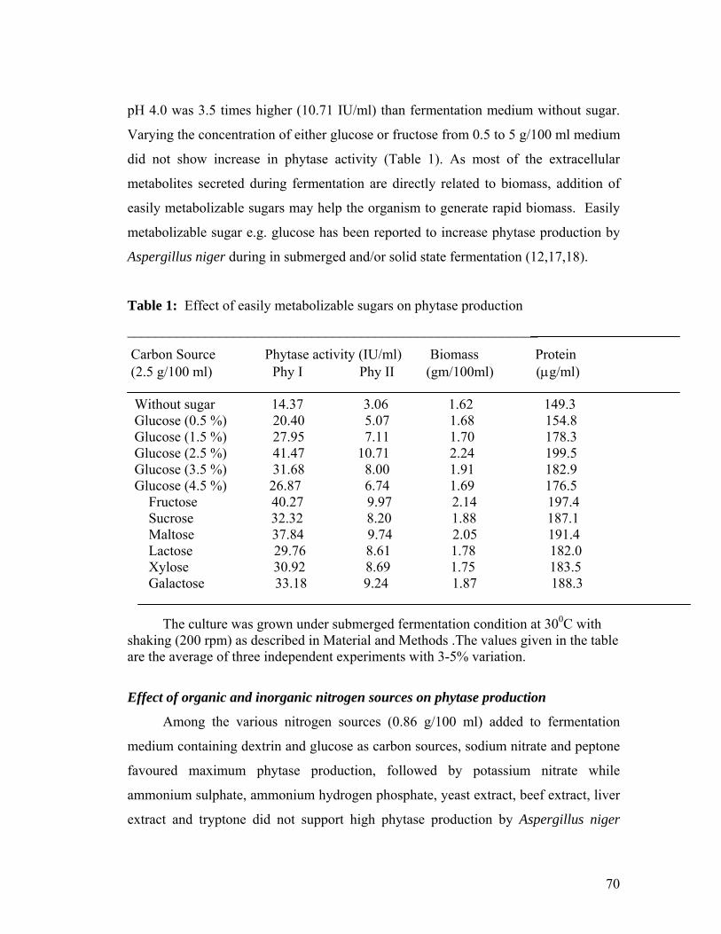
70
pH 4.0 was 3.5 times higher (10.71 IU/ml) than fermentation medium without sugar.
Varying the concentration of either glucose or fructose from 0.5 to 5 g/100 ml medium
did not show increase in phytase activity (Table 1). As most of the extracellular
metabolites secreted during fermentation are directly related to biomass, addition of
easily metabolizable sugars may help the organism to generate rapid biomass. Easily
metabolizable sugar e.g. glucose has been reported to increase phytase production by
Aspergillus niger during in submerged and/or solid state fermentation (12,17,18).
Table 1: Effect of easily metabolizable sugars on phytase production
__________________________________________________________
Carbon Source Phytase activity (IU/ml) Biomass Protein (2.5 g/100 ml) Phy I Phy II (gm/100ml) (g/ml) Without sugar 14.37 3.06 1.62 149.3 Glucose (0.5 %) 20.40 5.07 1.68 154.8 Glucose (1.5 %) 27.95 7.11 1.70 178.3 Glucose (2.5 %) 41.47 10.71 2.24 199.5 Glucose (3.5 %) 31.68 8.00 1.91 182.9 Glucose (4.5 %) 26.87 6.74 1.69 176.5 Fructose 40.27 9.97 2.14 197.4 Sucrose 32.32 8.20 1.88 187.1 Maltose 37.84 9.74 2.05 191.4 Lactose 29.76 8.61 1.78 182.0 Xylose 30.92 8.69 1.75 183.5 Galactose 33.18 9.24 1.87 188.3
The culture was grown under submerged fermentation condition at 300C with shaking (200 rpm) as described in Material and Methods .The values given in the table are the average of three independent experiments with 3-5% variation.
Effect of organic and inorganic nitrogen sources on phytase production
Among the various nitrogen sources (0.86 g/100 ml) added to fermentation
medium containing dextrin and glucose as carbon sources, sodium nitrate and peptone
favoured maximum phytase production, followed by potassium nitrate while
ammonium sulphate, ammonium hydrogen phosphate, yeast extract, beef extract, liver
extract and tryptone did not support high phytase production by Aspergillus niger

71
NCIM 563 (Table 2). Yeast extract, peptone and tryptone have been reported to
stimulate maximum phytase production by Aspergillus niger van Teigham (18).
Table 2: Effect of various organic and inorganic nitrogen sources on phytase
production
___________________________________________________________________
Nitrogen source Phytase activity (IU/ml) (0.86 g/100 ml) pH 2.5 pH 4.0 ___________________________________________________________________
NaNO3 41.47 10.71 KNO3 34.25 9.11 (NH4)2SO4 1.32 0.46 (NH4)2HPO4 1.58 0.57 Urea 1.02 0.23 NaNO2 0.26 0.06 Yeast extract 1.25 0.94 Beef extract 1.37 0.75 Liver extract 1.18 0.44 Peptone 39.43 10.37 Tryptone 1.77 0.46 Corn Steep Liquor 19.14 4.17 __________________________________________________________________
The culture was grown under submerged fermentation condition at 300C with shaking (200 rpm) as described in Material and Methods .The values given in the table are the average of three independent experiments with 3-5% variation.
Effect of phosphorus on phytase production
Addition of various inorganic phosphorus sources in fermentation medium at
0.004 g/100 ml concentrations increases the phytase activity. Nearly 13 times
increase in phytase activity at pH 2.5 (41.47 IU/ml) and pH 4.0 (10.71 IU/ml) was
found by addition of potassium dihydrogen phosphate as compared to fermentation
medium without inorganic phosphate. However increasing the concentration of
potassium dihydrogen phosphate to 0.006 g/100 ml medium had adverse effect on
phytase activity (Table 3). Various reports established that the phytase production by
submerged or solid state fermentation is limited by the amount of inorganic

72
phosphorus in fermentation medium (1, 2, 10, 11, 20). High levels of inorganic
phosphorus repress the biosynthesis of phytase. However, according to Vats et al (18)
phytase production by Aspergillus niger van Teighem in seven litre laboratory scale
fermenter was influenced by acidic pH (1.5-1.8) of the medium than phosphorus
concentration.
Table 3: Effect of phosphates on phytase production
______________________________________________________________
Phosphate source Phytase activity (IU/ml) (0.004 g/100 ml medium) pH 2.5 pH 4.0 ______________________________________________________________ No phosphate 3.03 0.78 KH2PO4 (1 mg%) 8.19 2.06 KH2PO4 (2 mg%) 18.89 4.56 KH2PO4 (4 mg%) 41.47 10.71 KH2PO4 (6 mg%) 26.07 6.48 K2HPO4 39.73 10.64 Na2HPO4 26.14 6.93 NaH2PO4 22.71 5.91 NH4HPO4 23.95 6.23 CaHPO4 24.78 6.45 Ca3(PO4)2 26.17 7.11 ______________________________________________________________
The culture was grown submerged fermentation condition at 300C with shaking (200 rpm) as described in Material and Methods .The values given in the table are the average of three independent experiments with 3-5% variation. Effect of surfactant on phytase production
To check the effect of surfactants on phytase production the fermentation
medium was supplied with either Tween 80 or Triton X-100 (0.5 ml/100 ml) on either
third or fifth day of fermentation.
Addition of surfactants increased the release of phytase activity. Tween 80 was
found much superior to Triton X100. Addition of Tween-80 on the third day of
fermentation resulted in 35 % increase in phytase activity at pH 2.5 (56.24 IU/ml) and
43 % increase (15.31 IU/ml) in phytase activity at pH 4.0 as compared to fermentation
medium without surfactant (Fig 2). Similar effect was observed during lipase
production by Aspergillus niger NCIM 1207 when Triton X-100 (0.5 ml/100 ml

73
medium) was added to fermentation medium. (30). Surfactants increased the cell
wall/cell membrane permeability, which may be the reason for increase rate of
metabolite secretion by the cells in fermentation medium.
1 2 3 4 50
5
10
15
20
25
30
35
40
45
50
55
60
65
Phyt
ase
Act
ivity
(IU
/ml)
Surfactant
Assay at pH 2.5 Assay at pH 4.0
Figure 2. Effect of surfactants on phytase production 1: No Surfactant , 2: Triton X 100 Added on 3rd day, 3: Triton x 100 Added on 5th day, 4: Tween 80 Added on 3rd day, 5: Tween 80 Added on 5th day Effect of volume of fermentation medium during shake flask studies
The effect of volume of fermentation medium in either 250 or 500 ml Erlenmeyer
flasks indicate that maximum phytase activity at pH 2.5 (41.47 IU/ml) and pH 4.0
(10.71 IU/ml) produced when 100 ml fermentation medium was taken in 250 ml flask
as compared to 50 ml medium in the same flask. Similar trend was found when
fermentation was carried out in 500 ml Erlenmeyer flasks with 100 and 200 ml
fermentation medium (Fig 3). This may be due to perhaps requirement of
microaerophilic atmosphere for fungus, Aspergillus niger NCIM 563 to secrete higher
amount of phytase.
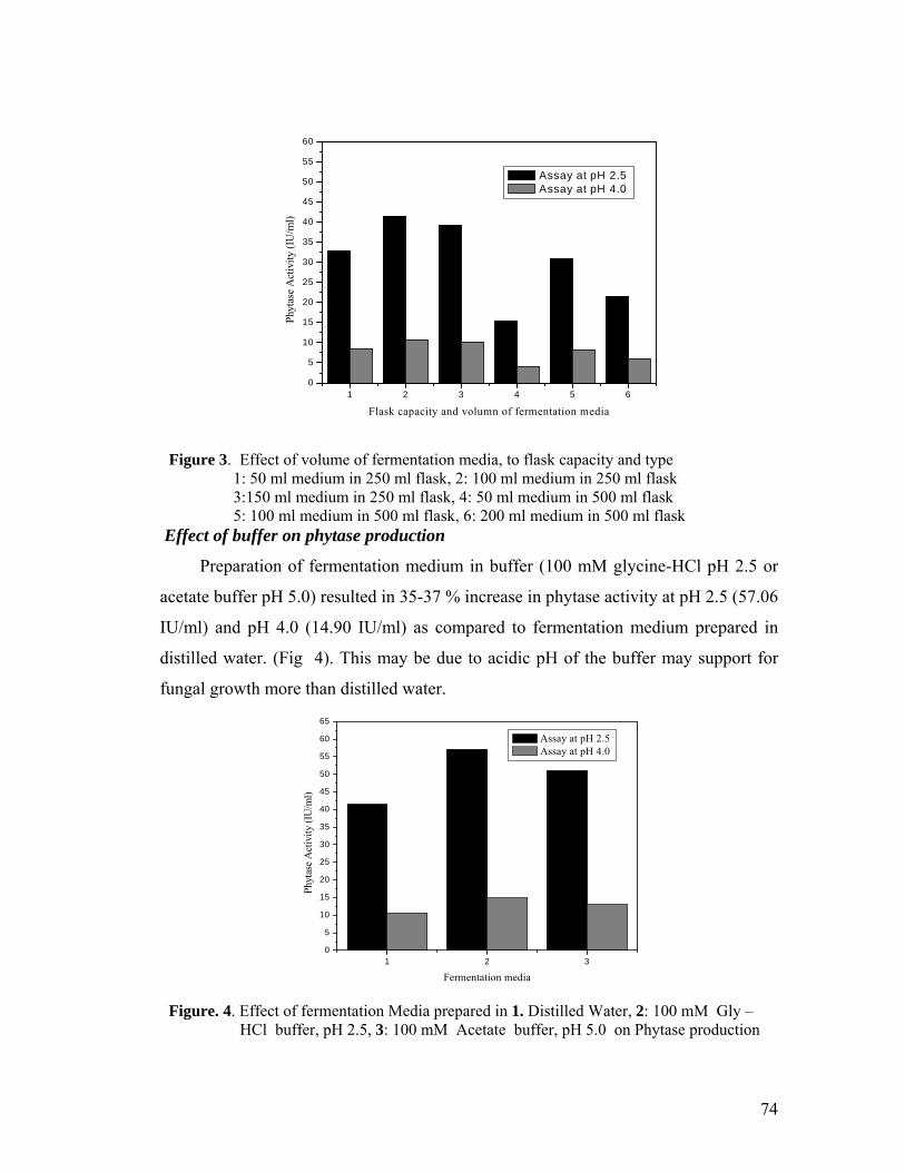
74
1 2 3 4 5 60
5
10
15
20
25
30
35
40
45
50
55
60
Phyt
ase
Act
ivity
(IU
/ml)
Flask capacity and volumn of fermentation media
Assay at pH 2.5 Assay at pH 4.0
Figure 3. Effect of volume of fermentation media, to flask capacity and type 1: 50 ml medium in 250 ml flask, 2: 100 ml medium in 250 ml flask 3:150 ml medium in 250 ml flask, 4: 50 ml medium in 500 ml flask 5: 100 ml medium in 500 ml flask, 6: 200 ml medium in 500 ml flask Effect of buffer on phytase production
Preparation of fermentation medium in buffer (100 mM glycine-HCl pH 2.5 or
acetate buffer pH 5.0) resulted in 35-37 % increase in phytase activity at pH 2.5 (57.06
IU/ml) and pH 4.0 (14.90 IU/ml) as compared to fermentation medium prepared in
distilled water. (Fig 4). This may be due to acidic pH of the buffer may support for
fungal growth more than distilled water.
1 2 30
5
10
15
20
25
30
35
40
45
50
55
60
65
Phyt
ase
Act
ivity
(IU
/ml)
Fermentation media
Assay at pH 2.5 Assay at pH 4.0
Figure. 4. Effect of fermentation Media prepared in 1. Distilled Water, 2: 100 mM Gly –HCl buffer, pH 2.5, 3: 100 mM Acetate buffer, pH 5.0 on Phytase production

75
Characterization of partially purified phytase
The ammonium sulphate precipitation procedure resulted in 95 % enzyme
recovery with purification of 3.0-fold. The enzyme showed two pH optima i.e. 2.5 and
4.0 (Fig 5). According to Oh et al (31) phytases can be categorized into two major
classes, the histidine acid phytases (HAP) and alkaline phytases and based on their pH
optima HAP is further divided into two groups, PhyA showing two pH optima viz. 2.5
and 5.5 with higher activity at pH 5.5 while PhyB show only single pH optima of 2.5.
The phytase from Aspergillus niger NCIM 563 in the present communication belongs
to PhyA with two pH optima with unusual high activity at pH 2.5 than at pH 4.0. The
maximum enzyme activity at pH 2.5 was (41.47 IU/ml) and 10.71 IU/ml at pH 4.0.
Thus activity at pH 4.0 was 25 % as compared to at pH 2.5. On the other hand
extracellular phytase from Aspergillus ficuum NRRL 3135 even though exhibit two
pH optima of 2.5 and 5.0, shows 48 % less activity at pH 2.5 than at pH 5.0 (15).
1.0 1.5 2.0 2.5 3.0 3.5 4.0 4.5 5.0 5.5 6.0 6.5 7.0 7.5-10
0
10
20
30
40
50
60
70
80
90
100
110
Rel
ativ
e Ph
ytas
e A
ctiv
ity(%
)
pH
Optimum pH pH Stability
Assay at pH 2.5 Assay at pH 4.0
Figure 5 Effect of pH on phytase activity and stability: —▲— Optimum pH —□— Assay at pH 2.5, — ■—Assay at pH 4.0

76
Storage of phytase at various pHs for 18 h at 4C reveled that phytase activity at
pH 2.5 was stable at pH range 1.5-3.5 whereas phytase activity at pH 4.0 was more
stable in the pH range 2.0-7.0 (Fig. 5). The optimum temperature for phytase activity
was 60C for both the activities at pH 2.5 and 4.0 (Fig. 6). Phytase from various
Aspergilli show optimum temperature in the temperature range 40-65C (11, 12).
Thermostability of phytase active at pH 2.5 and 4.0 was different (Fig 4a and 4b). At
65C phytase active at pH 2.5 (Fig. 7a) retained 80 % of its original activity after 15
min but at 70C activity decreased sharply with only 40 % of its original activity
remained after 15 min while phytase active at pH 4.0 retained only 40 % of its original
activity after 15 min exposure at 65C and 36 % of its original activity could be
detected after 5 min exposure at 70C (Fig.7b).
40 45 50 55 60 6530
40
50
60
70
80
90
100
Rel
ativ
e Ph
ytas
e A
ctiv
ity (
%)
Tem p.(0C )
A ssay at pH 2.5 A ssay at pH 4.0
Figure 6. Effect of temperature on phytase activity: —■— Assay at pH 2.5, ——
Assay at pH 4.0
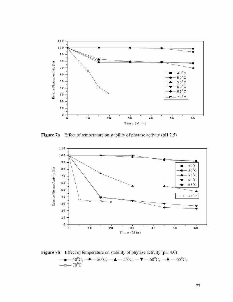
77
0 1 0 2 0 3 0 4 0 5 0 6 00
1 0
2 0
3 0
4 0
5 0
6 0
7 0
8 0
9 0
1 0 0
1 1 0
0 1 0 2 0 3 0 4 0 5 0 6 00
1 0
2 0
3 0
4 0
5 0
6 0
7 0
8 0
9 0
1 0 0
1 1 0
7 0 0 CRel
ativ
e Ph
ytas
e A
ctiv
ity (
%)
T im e (M in . )
4 0 0 C
5 0 0 C
5 5 0 C
6 0 0 C
6 5 0 C
Figure 7a Effect of temperature on stability of phytase activity (pH 2.5)
0 1 0 2 0 3 0 4 0 5 0 6 00
1 0
2 0
3 0
4 0
5 0
6 0
7 0
8 0
9 0
1 0 0
1 1 0
0 1 0 2 0 3 0 4 0 5 0 6 00
1 0
2 0
3 0
4 0
5 0
6 0
7 0
8 0
9 0
1 0 0
1 1 0
4 0 0 C
5 0 0 C
5 5 0 C
6 0 0 C
6 5 0 C
Rel
ativ
e Ph
ytas
e A
ctiv
ity (
%)
T im e (M in )
7 0 0 C
Figure 7b Effect of temperature on stability of phytase activity (pH 4.0)
—■— 400C, —— 500C, —▲— 550C, —▼— 600C, —— 650C, —□— 700C
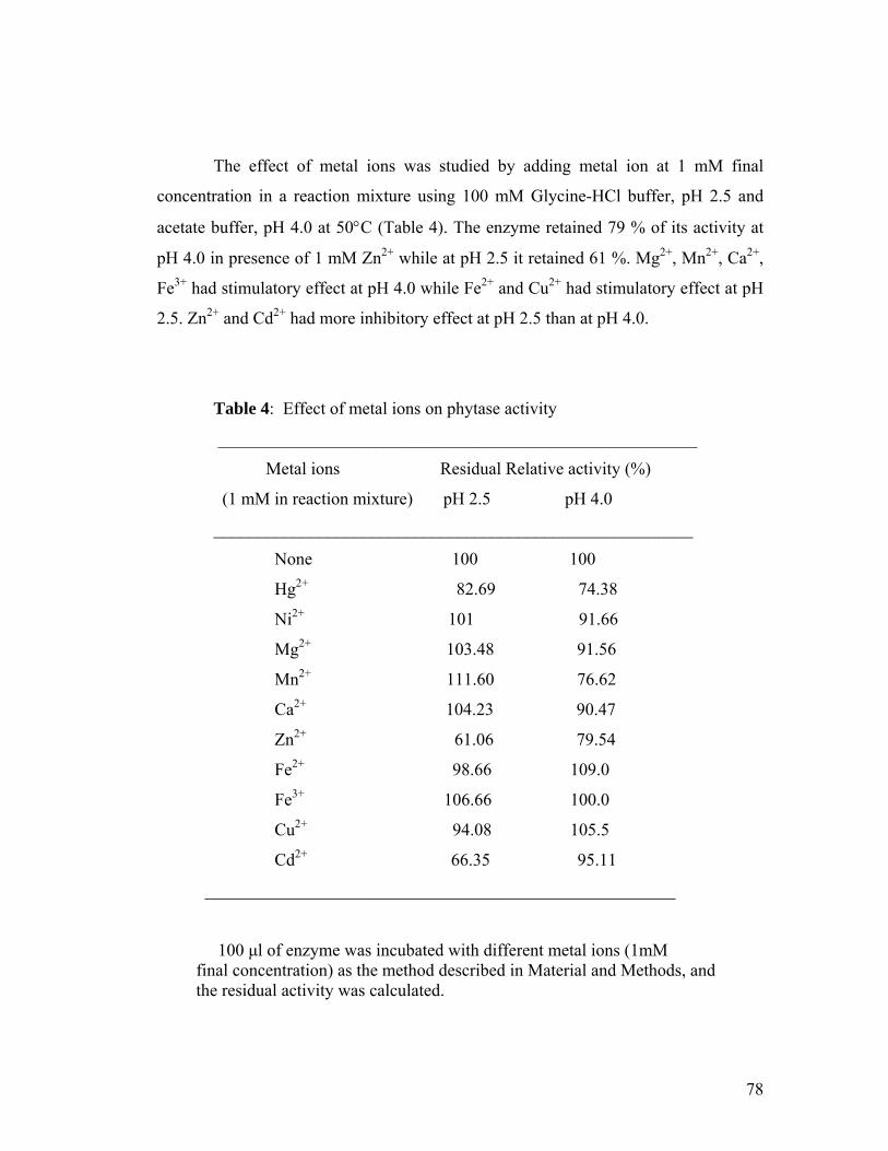
78
The effect of metal ions was studied by adding metal ion at 1 mM final
concentration in a reaction mixture using 100 mM Glycine-HCl buffer, pH 2.5 and
acetate buffer, pH 4.0 at 50C (Table 4). The enzyme retained 79 % of its activity at
pH 4.0 in presence of 1 mM Zn2+ while at pH 2.5 it retained 61 %. Mg2+, Mn2+, Ca2+,
Fe3+ had stimulatory effect at pH 4.0 while Fe2+ and Cu2+ had stimulatory effect at pH
2.5. Zn2+ and Cd2+ had more inhibitory effect at pH 2.5 than at pH 4.0.
Table 4: Effect of metal ions on phytase activity
_______________________________________________________
Metal ions Residual Relative activity (%)
(1 mM in reaction mixture) pH 2.5 pH 4.0
_______________________________________________________
None 100 100
Hg2+ 82.69 74.38
Ni2+ 101 91.66
Mg2+ 103.48 91.56
Mn2+ 111.60 76.62
Ca2+ 104.23 90.47
Zn2+ 61.06 79.54
Fe2+ 98.66 109.0
Fe3+ 106.66 100.0
Cu2+ 94.08 105.5
Cd2+ 66.35 95.11
______________________________________________________
100 μl of enzyme was incubated with different metal ions (1mM final concentration) as the method described in Material and Methods, and the residual activity was calculated.
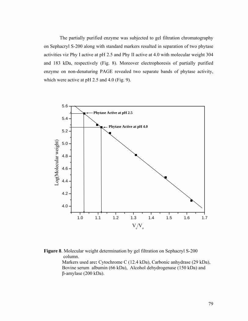
79
The partially purified enzyme was subjected to gel filtration chromatography
on Sephacryl S-200 along with standard markers resulted in separation of two phytase
activities viz Phy I active at pH 2.5 and Phy II active at 4.0 with molecular weight 304
and 183 kDa, respectively (Fig. 8). Moreover electrophoresis of partially purified
enzyme on non-denaturing PAGE revealed two separate bands of phytase activity,
which were active at pH 2.5 and 4.0 (Fig. 9).
1.0 1.1 1.2 1.3 1.4 1.5 1.6 1.7
4.0
4.2
4.4
4.6
4.8
5.0
5.2
5.4
5.6
Phytase Active at pH 4.0
Phytase Active at pH 2.5
Log
(Mol
ecul
ar w
eigh
t)
Ve/V
o
Figure 8. Molecular weight determination by gel filtration on Sephacryl S-200 column. Markers used are: Cytochrome C (12.4 kDa), Carbonic anhydrase (29 kDa), Bovine serum albumin (66 kDa), Alcohol dehydrogenase (150 kDa) and β-amylase (200 kDa).

80
Figure 9. Native gel electrophoresis (8 %) of partially purified enzyme showing
multiple forms of phytase
81
The Km for Phy I and II was 3.18 and 0.514 mM, respectively while Vmax was
331.16 and 59.47 μmols/min/mg when sodium phytate was used as substrate which is
slightly higher than the Km (0.04-0.44 mM) reported from purified phytase of
Aspergillus niger 92 (10) and Aspergillus niger NRRL 3135 (32). Fujita et al (33) also
reported two types of phytase from Aspergillus oryzae under solidstate fermentation,
which show same pH optima (5.0), thermal stability (100 % stable up to 500C) but
differ in their temperature optima (65 0C and 55 0C, respectively) and molecular
weight (120 and 116 kDa, respectively).
2.5 Conclusions
Earlier we have reported production of phytase by solid state fermentation
using Aspergillus niger NCIM 563 with pH optimum 5.0 (19). Present studies on
phytase production under submerged fermentation conditions by Aspergillus niger
NCIM 563 indicate the possibility of two different forms of phytases, Phy I and Phy II
which differ in their molecular weight, pH optima, pH stability and their inhibition by
metal ions. High activity at pH 2.5 than at pH 4.0 indicates its application in poultry
feed supplement.
82
2.6 References
1. Reddy N. R., Sathe S. K., Salunkhe D. K. (1982) Phytases in legumes and cereals.Adv. Food Res. 82 : 1-92.
2. Mullaney E. J., Daly C., Ullah A. B. (2000) Advances in phytase research. Adv. Appl. Microbiol. 47 :157-199. 3. Bitar K., Reinhold J. G. (1972) Phytase and alkaline phosphate activities in intestinal mucosa of rat, chicken, calf and man. Biochim. Biophys. Acta 268: 442-452. 4. Common F. H. (1989) Biological availability of phosphorus for pigs. Nature 143: 370- 380. 5. Walsh G. A., Power R. F., Headon D. R.( 1994) Enzymes in the animal feed industry. Trends in Food Sci. Technol 5: 81-87. 6. Erdman J. W., Poneros-Schneier A.( 1989) Phytic acid interactions with divalent cations in food and in the gastrointestinal tract. Adv. Exp. Med. Biol. 249:167- 171. 7. Fox M. R. S., Tao S. H. ( 1989) Antinutritive effects of phytate and other phosphorylated Derivatives. Nutr. Toxicol. 3:59-96. 8. Mitchell D. B., Vogel K., Weimann B. J., Pasamontes L., van Loon A. (1977) The phytase subfamily of histidine acid phosphatases: isolation of genes for two novel phytases from fungi Aspergillus terreus and Myceliophthora thermophila. Microbiol. 143: 245-252. 9. Zyta K. (1992) Mold phytases and their application in the food industry. World J. Microbiol. Bitechnol. 8: 467-472. 10. Dvorakova, J. (1998) Phytase: Source, preparation and exploitation. Folia Microbiol. 43:323-338. 11.Vohra A, Satyanarayana T. (2003) Phytases: Microbial sources, production, purification and potential biotechnological applications. Critical Rev. in Biotechnol. 23:29-60. 12.Vats P, Banerjee U. C.(2004) Production studies and catalytic properties of phytases (myoinositolhexakisphosphate phosphohydrolases): an overview. Enzyme Microbiol. Technol. 35 :3-14. 13. Kim Y. O., Kim H. K., Bae K. S., Yu J. H., Oh. T. K. (1998) Purification and
83
properties of a thermostable Phytase from Bacillus spp DS11. Enzyme Microbiol. Technol. 22: 2-7. 14. Radcliffe J. S., Zhang Z., Kornegay E. T. (1998) The effect of microbial phytase, citric acid and their interaction in a corn soybean meal based diet for weaning pigs. J. Animal Sci. 76: 1880-1886. 15. Ullah A.H.J (1988). Production, rapid purification and catalytical characterization of extracellular Phytase from Aspergillus ficuum. Prep. Biochem. 18: 443-458. 16. Volfova O., Dvorakova J., Hanzlikova A., Jandera A. (1994) Phytase from Aspergillus niger. Folia Microbiol. 39:479-482. 17. Vats P., Banerjee U. C. (2002) Studies on the production of Phytase by a newly isolated strain of Aspergillus niger van Teigham obtained from rotten wood-logs. Process Biochem.38:211-217. 18.Vats P., Sahoo D. K., Banerjee, U. C. (2004) Production of Phytase (myo- Inositolhexakisphosphate phosphohydrolase) by Aspergillus niger van Teighem in laboratory-scale fermenter. Bioechnol. Progress 20:737-743. 19. Mandviwala T. N., Khire J. M. (2000) Production of high activity thermostable Phytase from thermotolerant Aspergillus niger in solid state fermentation. J. Industrial Microbiol. Biotechnol. 24: 237-243. 20. Shieh T. R., Ware J. H. (1968) Survey of microorganisms for the production of extracellular phytase. Appl. Microbiol. 16 :1348-1351.
21. Heinohen J. K., Lathi R. J. (1981) A new and convenient colorimetric determination of inorganic orthophosphate and its application to the assay to inorganic pyrophosphatase. Anal. Biochem. 113:313-317.
22.Lowry O. H., Rosebrough N. J., Farr A. L., Randall R. L. (1951) Protein measurement with the folin phenol reagent. J. Biol. Chem. 193: 265-275. 23. Davis B. J. (1964) Disc electrophoresis II. Method and application to human Serum proteins. Ann. N. Y. Acad. Sci. 121: 404-427. 24. Deutscher M. P. (1990) Guide to protein purification., Methods. Enzymol., 182, pp430. Academic press, Toronto.
25. Andrews P. (1964) Estimation of molecular weight of proteins by Sephadex gel filtration. Biochem. J. 92: 222-223.
84
26.Miller G. L. (1959) Use of dinitrosalicylic acid reagent for determination of reducing sugar. Anal. Chem. 31: 427- 431. 27.Lineweaver H., and Burk D.( 1934) Determination of enzyme dissociation constants. J. Amer. Chem.Soc. 56: 658. 28. Gargova S., Sariyska M. (2003) Effect of culture conditions on the biosynthesis of Aspergillus niger phytase and acid phosphatase. Enzyme Microbial Technol. 32: 231-235. 29.Wodzinski R., Ullah A. H. L. (1996) Phytase. Advances in Appl. Microbiol. 42: 263- 283. 30. Mahadik N. D., Bastawde K. B., Puntambekar U. S., Khire J. M., Gokhale D. V. (2004) Production of acidic lipase by a mutant of Aspergillus niger NCIM 1207 in submerged fermentation. Process Biochem. 39: 2031-2034. 31.Oh B. C., Choi S., Park S., Kim Y. O., Oh T. K. (2004) Biochemical properties and substrate specificities of alkaline and histidine acid phytases. Appl. Microbiol. Technol. 63: 362-372. 32.Ullah A.H.J., Gibson DM (1987) Extracellular Phytase (EC 3.1.3.8) from Aspergillus ficuum NRRL 3135: Purification and characterization. Prep Biochem 17: 63-91.
33.Fujita J., Yamane Y., Fukuda H., Kizaki Y., Wakabayashi S., Shigeta S., Suzuki O., Ono K. (2003) Production and properties of Phytase and acid phosphatase From a sake koji mold, Aspergillus oryzae. J. of Bioscience and Bioengineering 95: 348- 353.
85
CHAPTER 3 Purification and characterization of phytase I (Highly Acidic) and II from Aspergillus niger (NCIM 563)
3.1 Summary
An extracellular acid tolerant phytase (Phy I) from Aspergillus niger
(NCIM-563) has been purified along with totally different phytase (Phy II) to
homogeneity by two chromatographic steps, using Phenyl Sepharose CL4B and
Sephacryl 200, with total yield of 30% and 26% respectively. The molecular mass
of the enzymes as determined by SDS-PAGE and gel filtration column were 66,
264 kDa and 150, 148 respectively, suggested that Phy I consists of four identical
subunits and Phy II is a monomer. The purified phytase I is a glycosylated (2.01%)
protein with a pI value of 3.55, Phy II is a nonglycosylated protein with a pI value
of 3.91. The pH and temperature optima for the purified enzymes are 2.5, 5.0 and
55 ºC, respectively. The Phy I was stable over a broad pH range (1.5-9) and its
half-life of inactivation (t1/2) at 70ºC was 2-3 minutes. Phy II was stable (3.5-9.0)
and t1/2 at 70 ºC was 2-2.5 hour. The Phy I exhibited very broad substrate
specificity and Phy II was more specific for sodium phytate. The relative substrate
specificity of Phytase I towards the various synthetic substrates is in the order of
Na-pyro-phosphate> ATP> p-Nitrophenyl phosphate> Sodium phenyl phosphate >
-NADP > 1-Naphthyl phosphate > 2-Naphthyl phosphate > ADP> D-Glucose 6-
phosphate > -D-Glucose 1-phosphate > Phenyl phosphate > Sodium phytate >
Calcium phytate > Glycerol-1-phosphate > AMP.
The kinetics of hydrolysis of number of substrates by Phy I showed that
Na-pyro-phosphate and Sodium phytate has lower Km (higher affinity) and
Calcium phytate and ADP has higher Kcat (higher catalytic efficiency). For Phy II
Sodium phytate has lowest Km and ADP and p-Nitrophenyl phosphate has highest
Kcat among all the substrates. For sodium phytate Km is 2.01, .145 mM and Vmax
is 5018, 1671 μmoles min-1 mg-1 for Phy I and II respectively. Phy II was
completely inhibited by Ag+, Hg2+ metal ions, and Phy I was partially inhibited.
Peptide analysis of both the purified proteins by Mass Spectrometry (MS)
86
(MALDI-TOF) after in gel trypsin digestion showed totally different mass (m/z)
pattern, proves that both the proteins are totally different. The N-terminal amino
acid sequences of Phy I and Phy II were FSYGAAIPQQ and GVDERFPYTG,
respectively. Activity and stability of both phytases in acidic range suggests their
potential application in poultry feed.
3.2 Introduction
Phytase- a phosphomonoesterase- is an enzyme capable of hydrolyzing
phytate [myo-inositol [1,2,3,4,5,6] hexakisphosphate], the major storage form of
phosphorus in cereals and legumes, representing 18-88 % of total phosphorus
content (1). Phytases (EC 3.1.3.8 and EC 3.1.3.26) belong to the family of histidine
acid phosphatases which hydrolyses phytate to liberate inositol and inorganic
phosphorus (2). Phytate phosphorus is biologically unavailable to nonruminants as
they lack or secrete very low level of phytase activity in intestine. Consequently,
the phytate in animal feeds is discharged in the faeces of these animals into rivers
and seas, resulting in severe pollution of water resources (2). Similarly, phytic acid
is an anti-nutrient; due to its strong chelating potential can bind essential minerals
such as calcium, zinc and copper, rendering them unavailable or poorly available
for absorption (3). To overcome this difficulty the feed has to be supplemented
with inorganic phosphate to meet the nutritional requirements of the animals (4).
Supplementation of feed with phytases increases the bioavailability of phytic acid
bound phosphate, facilitating a reduction in the addition of inorganic phosphate to
the feed and decrease phosphorus excretion in areas of intensive animal
agriculture. Although large number of bacteria, yeast and fungi are reported to
produce phytase (5-7) fungal phytases are preferred in animal feed due to their
high yield and more acid tolerance as compared to phytases from bacteria (8).
Similarly pH in poultry gut varies from 2.5 to 6.0, thus phytase active and stable in
acidic environment is highly preferred (9). Several phytases have been cloned and
characterized, such as fungal phytase from Aspergillus ficuum (10), bacterial
phytase from Escherichia coli (11) and a mammalian phytase (12). These enzymes
share a highly conserved sequence motif that is found at the active sites of acid
phosphatases (13, 14). The reaction mechanism of E. coli phytase has been
87
revealed (15, 16) and is likely to be common for most phytases. Therefore, these
enzymes are said to form the phytase subfamily of histidine acid phosphatases
(17).
Among fungi many Aspergilli (5, 6,18, 19) are known to be active phytase
producers. As Aspergillus niger is Generally Recognised as Safe (GRAS) it is
frequently used in food and feed applications. Earlier we have reported phytase
production by Aspergillus niger NCIM 563 under submerged fermentation (20-22)
which includes production and partial characterization of two types of phytase
from Aspergillus niger NCIM 563.
In order to study structure function relationship of enzyme, it is necessary
to obtain it in a pure form and in large quantity through simple and efficient
method of purification. In the present study we have described simple and single
method of chromatographic step purification of phytases (I and II) from
Aspergillus niger (NCIM 563). The purified enzyme has been extensively
characterized for its biochemical and molecular properties and also for its substrate
specificity towards the different substrates comprising phosphorus.
3.3 Material & Methods
Chemicals
Sodium phytate, Calcium phytate, p-Nitrophenyl phosphate, Sodium phenyl
phosphate, -D-Glucose 1-phosphate , D-Glucose 6-phosphate, 1-Naphthyl
phosphate, 2-Naphthyl phosphate, ATP, ADP, AMP, -NADP, Na-pyro-
phosphate, Glycerol-1-phosphate, Phenyl phosphate, SDS and gel filtration
markers, Coomassie Brilliant Blue R 250, Bromophenol Blue, Phenyl Sepharose
CL-4B, Sephacryl-300, Sephacryl-200 were purchased from Sigma Chemical
company , U.S.A.. External mass standards Calmix 1 and 2 obtained from Applied
Biosystems. All other chemicals used were of analytical grade and obtained from
leading manufacturers including BDH, Hi media and Glaxo.

88
Organism and culture condition
The strain used throughout the present work was Aspergillus niger NCIM
563. It was maintained as described earlier (20). For purification of Phytases (I and
II), isolate was grown in a medium containing (per100 mL): Dextrin 5 g; Glucose
2.5 g; NaNO3 0.86 g; KH2PO4 0.004 g; KCl 0.05 g; MgSO4.7H2O 0.05 g;
FeSO4.7H2O 0.01 g. pH 5.5 before sterilization. Fermentation medium (100 mL in
250 mL Erlenmeyer flask) was inoculated with 1% (v/v) of spore suspension
(5X10⁷ spores per mL) prepared by suspending the spores from 7 day old
sporulated slant of Aspergillus niger NCIM 563 grown on PDA in 10 mL of sterile
distilled water containing 0.01% (v/v) Tween 80 and incubated at 300C at 200 rpm
for 10-12 days. After fermentation, the fungal biomass was separated by filtration
and then centrifugation (10,000 x g, 10 minutes) and clear supernatant was used as
source of extracellular enzyme.
Phytase assay and protein, glycosylation determination
Phytase activity was measured at 550C as described earlier (20). The
reaction for Phy I and Phy II was carried out at pH 2.5 (100 mM Glycine-HCl
buffer) and pH 5.0 (100 mM acetate buffer) at 550 C for 30 min, respectively. The
liberated inorganic phosphate was measured by a modification of the ammonium
molybdate method (23). A freshly prepared 4 mL solution of acetone: 5 N H2SO4:
10 mM ammonium molybdate (2:1:1 v/v/v) and 400 µl of 1M citric acid were
added to the assay mixture. Absorbance was measured at 370 nm. One unit of
phytase activity (IU) was expressed as the amount of enzyme that liberates 1µmol
phosphorus per minute under standard assay conditions. Each experiment was
carried out in triplicate and the values reported are the mean of three such
experiments in which a maximum of 3–5% variability was observed. Protein
concentration in the culture filtrate was determined by the method of Lowry et al.
(24) as well as measurement of absorbance at 280 nm using Bovine serum albumin
as a standard. The neutral sugar content (glycosylation) of the purified enzyme
preparation was determined by modified phenol sulphuric acid method (25).

89
Purification of Phytase I and II
Mycelia free culture broth was concentrated 10 times by rota vapour at 400C
(both the enzymes are 100% stable at this temperature) under vacuum. The same
enzyme sample was then applied to hydrophobic column chromatography using
Phenyl Sepharose CL-4B (30 ml bed volume), previously equilibrated with 30%
Ammonium sulphate in 20 mM Acetate buffer (pH 2.5). The column was washed
thoroughly with 20 bed volumes of above buffer. Elution was done with 120 ml
linear decreasing gradient of ammonium sulphate (30-0 %) with a flow rate of 20
ml per hour and approximately 3.0 ml fractions were collected. Fractions showing
activity at pH 2.5 (Phy I) and pH 5.0 (Phy II) were pooled separately, concentrated
by rota vapour and loaded on gel filtration Sephacryl (S-200) column with a flow
rate of 12 ml per hour and 2ml fractions were collected. Unless otherwise
mentioned all the purification procedures were carried out at 4 °C.
Electrophoretic techniques and N-terminal sequence
SDS-PAGE (10%) was performed in a vertical slab gel apparatus at pH 8.3,
according to the method described by Laemmli (26). Native PAGE (8%) was done
at pH 7.0 under non-denaturing conditions Davis (27). Protein bands were detected
either by coomassie Brilliant Blue R-250 (0.2%, w/v) or by Silver staining (0.2%
w/v), The Isoelectric Focusing Polyacrylamide Gel Electrophoresis (IEF-PAGE)
was performed in a precasted acrylamide gel 7.5% (Biorad) using wide range
Ampholytes (pH, 3-10) by the method of Chinnathambi et al (28) using a mini
scale density gradient isoelectric focusing unit. Protein bands were detected either
by Coomassie Brilliant Blue R-250 (0.2 % w/v) or by Silver staining (0.2 % w/v)
and phytase band was revealed by sodium phytate as a substrate.
For N-terminal sequence analysis, the purified enzyme was electrophoresed
on 10% SDS-PAGE at pH 8.8. After electrophoresis, the gel and
polyvinylidenedifluoride (PVDF) membrane were sandwiched between Whatman
filter paper strips. The strips and membrane were previously equilibrated with
CAPS buffer (10 mM, pH 11.0) containing 10 % methanol. The protein was
electrotransfered using a semi dry blotting system (Pharmacia Biotech, Nova Blot)

90
under a constant current of 52 mA for 30 minutes. After transfer, the membrane
was removed and washed thoroughly with milli-Q water and protein band was
visualized by Coomassie Blue R-250 stain. The N-terminal amino acid sequence of
the first 10 residues of the both enzymes was determined by Edman degradation on
an Applied Bio systems Procise sequencer at commercial services (Commonwealth
Biotechnologies, Inc., Richmond, Virginia, USA). Reported sequences of
Phytases from various sources were searched from GenBank and SwissProt
Databases and a sequence alignment were performed using a Clustal-W sequence
alignment program (29).
Determination of molecular mass (Mr)
The native molecular mass of α-galactosidase was estimated by gel filtration,
according to the method described by Andrews (30). The gel filtration column
(Sephacryl, S-300, 1x150 cm), was equilibrated in phosphate buffer (50 mM, pH
7.0) and calibrated using gel filtration standard molecular weight markers: Bovine
serum albumin (Mr 66 kDa); Alcohol dehydrogenase (Mr 150 kDa); β-amylase (Mr
200 kDa), Apoferetin (Mr 443 kDa) and Thyroglobulin (Mr 669 kDa). The column
void volume was determined with Blue dextran (Mr 2000 kDa). The subunit
molecular mass of the purified Phytases was determined by SDS-PAGE according
to Weber and Osborn (31) using sigma high molecular mass markers.
MALDI-TOF analysis
Mass spectral analysis was performed on a Voyager-De-STR (Applied Bio
systems) MALDI-TOF. A nitrogen laser (337 nm) was used for desorption and
ionization. Spectra were acquired in the range of 10 to 100 kDa, in linear mode
with delayed ion extraction and with an accelerating voltage of 25 kV. The low
mass ion gate was set at 4500 Da. All the analyses were performed in four
replications. The instrument was calibrated with myoglobulin and bovine serum
albumin. In-gel tryptic digestion and analysis of peptides was done according to
Bar et al (32).

91
pH and temperature effect
pH optimum of phytase was determined by assaying phytase activity over a
pH range 1.5-9.0, using 50 mM Gly-HCL buffer (pH, 1.5-4.5),Acetate buffer (pH,
4.0-6.5) and Tris- HCL buffer(pH, 6.5-9.0) by standard assay method. The pH
stability was also determined in a same pH range by pre-incubating enzyme
samples in above buffers at room temperature for the period of 12 hours. Small
aliquots (10 μl) were withdrawn from all the samples and residual enzyme activity
was determined by standard assay method.
The optimum temperature of phytase was determined by performing assays
at temperatures, 30 to 70°C. The Temperature stability was also determined by
incubating enzyme samples over a same temperature range for the period of one
hour. Aliquots (100 μl) were withdrawn after suitable time interval and residual
enzyme activity was determined by standard assay method.
Effect of various metal ions and inhibitor reagents
A suitably diluted enzyme (100 μl) in 50 mM phosphate buffer, pH 7.0, was
pre incubated with various metal salts, and inhibitor reagents at room temperature
for 10 minutes; subsequently residual activity of the samples was determined by
standard assay method.
A suitably diluted enzyme was pre-incubated with various Metal ion
solutions at 40C for 24 hour and subsequently residual activity of phytase was
determined to check the stability of enzymes under different metal ion solutions.
Effect of different additives on thermal stability of phytases
Purified enzyme samples (5.0 U/ml) were incubated at 70ºC with different
additives (10 % W/V). Aliquots (~10 μl) were removed after 0, 10 and 30 min and
assayed for residual enzyme activity. Enzyme activity of 0 min sample was taken
as 100 %.

92
Substrate specificity and kinetic studies
The relative substrate specificity of both the phytases towards various
substrates was determined at final concentration of 3.0 mM. The initial rate of
hydrolysis of various substrates was determined by varying the substrate
concentrations in different ranges (.1mM to 10mM). Assays were carried out at
55°C under standard assay conditions. The kinetic rate constants, Km, Vmax, Kcat
and Kcat / Km were determined graphically from Lineweaver-Burk plotting (33).
3.4 Result and discussion
Purification of Phytase I and II
The purification of Phytase I and Phytase II was carried out as summarized
in Table 1a and 1b. The crude culture supernatant was first concentrated (50%) on
rota vapour followed by adsorption and elution ion exchange chromatography on
Phenyl Sepharose CL-4B followed by molecular sieving on Sephacryl S-300.
Major activities were eluted in two peaks with a reverse gradient of ammonium
sulphate (30-0 %) in 20 mM acetate buffer, pH 2.5. First peak was obtained at 15
% ammonium sulphate with higher enzyme activity at pH 5.0 (Phy II) while
second peak was obtained at 5-0 % ammonium sulphate concentration in 20 mM
acetate buffer with higher enzyme activity at pH 2.5 (Phy I). These fractions were
dialyzed, concentrated and applied on gel filtration column. The purified Phy I and
II had a specific activity of 3436.37 and 1215.33 Umg-1 with 16.67 and 14.29 fold
purification, respectively. Nagashima et al (34) purified phytase from A. niger SK-
57 to homogeneity in four steps by using ion-exchange chromatography, gel
filtration chromatography and chromatofocussing. Recently Xiaoyu Li et al (35)
reported purification of extracellular phytase from marine yeast, Kodamaea ohmeri
BG3 by ammonium sulphate fractionation, gel filtration on Sephadex G-75
followed by fast flow anion exchange chromatography on DEAE Sepharose.
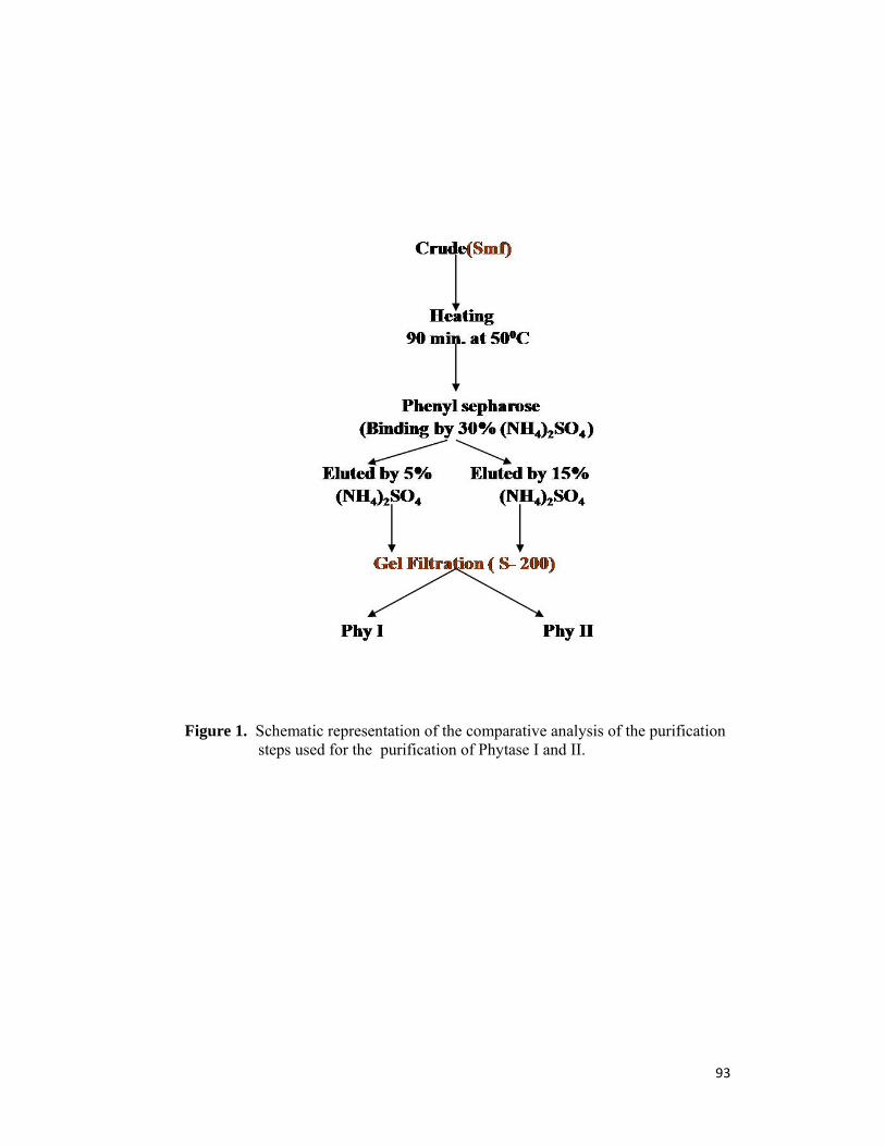
93
Figure 1. Schematic representation of the comparative analysis of the purification
steps used for the purification of Phytase I and II.
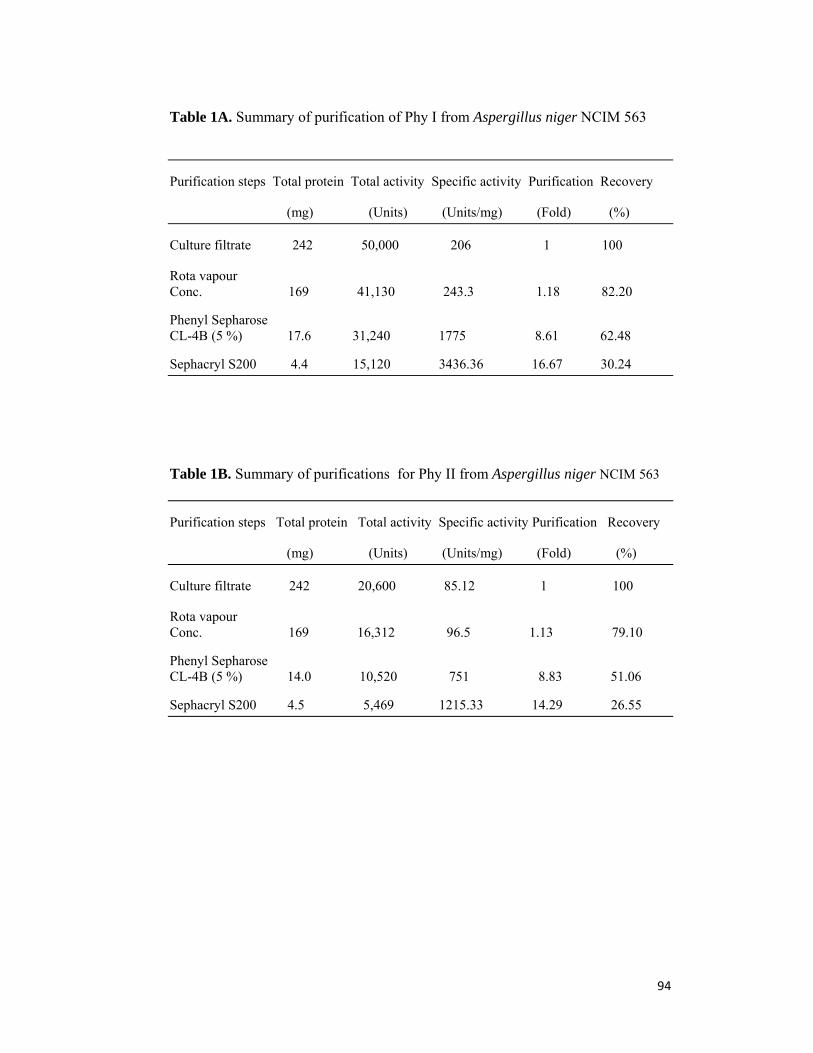
94
Table 1A. Summary of purification of Phy I from Aspergillus niger NCIM 563
Purification steps Total protein Total activity Specific activity Purification Recovery
(mg) (Units) (Units/mg) (Fold) (%)
Culture filtrate 242 50,000 206 1 100
Rota vapour Conc. 169 41,130 243.3 1.18 82.20
Phenyl Sepharose CL-4B (5 %) 17.6 31,240 1775 8.61 62.48
Sephacryl S200 4.4 15,120 3436.36 16.67 30.24
Table 1B. Summary of purifications for Phy II from Aspergillus niger NCIM 563
Purification steps Total protein Total activity Specific activity Purification Recovery
(mg) (Units) (Units/mg) (Fold) (%)
Culture filtrate 242 20,600 85.12 1 100
Rota vapour Conc. 169 16,312 96.5 1.13 79.10
Phenyl Sepharose CL-4B (5 %) 14.0 10,520 751 8.83 51.06
Sephacryl S200 4.5 5,469 1215.33 14.29 26.55

95
Figure 2. (A) SDS-PAGE of purified Phy I and Phy II from Aspergillus niger NCIM 563
Lane M:Molecular weight markers, Lane 1: Purified Phy II, Lane 2: Purified Phy I.
Molecular properties of phytase
The subunit molecular mass of purified Phy I and II was determined to be
66 kDa and 150-160 kDa, respectively, by SDS-PAGE analysis (Fig 2 A), while
that of native enzyme was found to be 264 kDa and 150-160 kDa, respectively, by
gel filtration on Sephacryl S300 (Figure 3). This suggests that Phy I is tetramer and
Phy II is monomer. Dimeric Phy B has been reported from Aspergillus niger
NRRL 3135 which has molecular mass of 68 kDa (36). However MALDI data
revealed the molecular weight of Phy I and II to be 66 kDa and 74 kDa [Fig 4 A &
B (inset)]. Thus molecular weight of Phy II by MALDI-TOF was half the
molecular weight shown by SDS gel electrophoresis. This could be because of the

96
doubly charge species of Phy II during ionization process of MALDI-TOF
spectrometry (37).
Isoelectric point (pI)
Single protein bands were observed on IEF-PAGE (Figure 2B) indicated
that the both the enzymes are homogenous and contain single molecular form of
Phytases. The pI of the Phytase I and II estimated by Isoelectric focusing was ~
3.65 and 3.95 respectively (Fig 2b) revealing that the both the enzymes are acidic
in nature. Golovan et al.(38) purified phytase (45 kDa) from E. coli of 45 kDa
molecular weight and further separated it into two isoforms of identical size with
pI of 6.5 and 6.3 by chromate focussing. The isoforms showed similar optimum
temperature and pH 4.5 of 60 ◦C, respectively.
Figure 2 (B) Iso-electric focusing of purified Phy I and Phy II.Lane M: Standard pI markers (Pharmacia), Lane 1: Purified Phy I Lane 2: Purified Phy IIIsoelectric point of purified enzymes M- Marker, I- Phytase I, II- Phytase II Protein (source) pI Protein (source) pI Cytochrome C (horse, heart) 10 .7 Ribonuclease A (bovine, pancreas) 9.5 Lectin (Lens culinaris) 8.3, 8.0, 7.8 Myoglobin (horse, muscle) 7.4, 6.9 Carbonic anhydrase (bovine, ß-Lactoglobulin (bovine, milk) 5.3, 5.2 erythrocytes) 6.0 Trypsin inhibitor (soybean) 4.5 Glucose oxidase (A. niger) 4.2 Amyloglucosidase (A. niger) 3.5
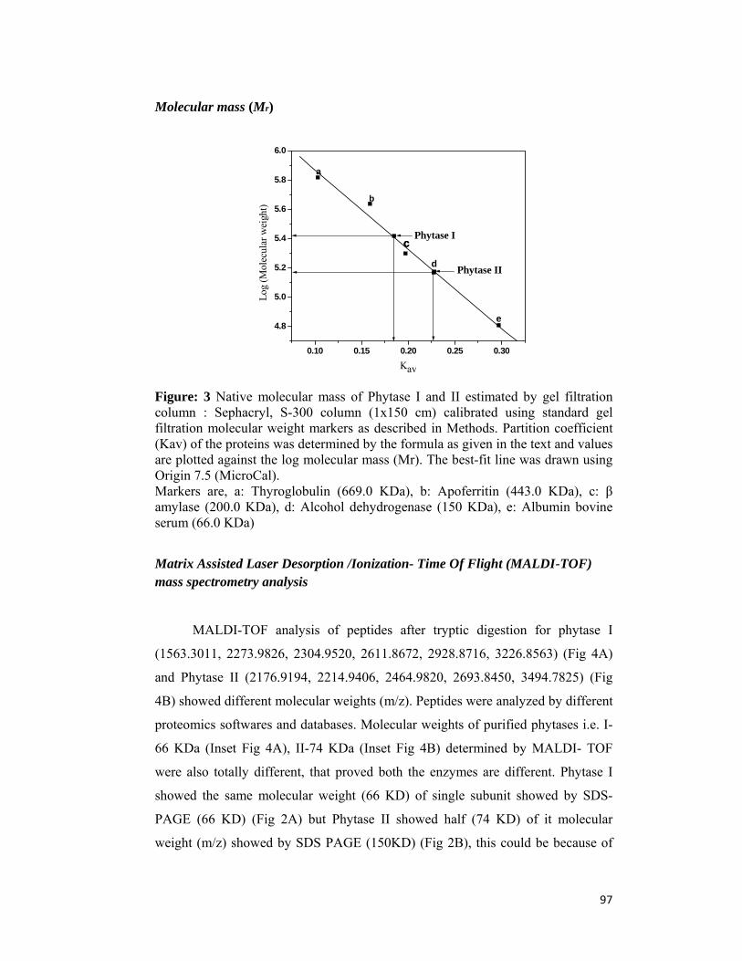
97
Molecular mass (Mr)
0.10 0.15 0.20 0.25 0.30
4.8
5.0
5.2
5.4
5.6
5.8
6.0
Phytase II
c
Log
(M
olec
ular
wei
ght)
Kav
a
b
c
d
e
Phytase I
Figure: 3 Native molecular mass of Phytase I and II estimated by gel filtration column : Sephacryl, S-300 column (1x150 cm) calibrated using standard gel filtration molecular weight markers as described in Methods. Partition coefficient (Kav) of the proteins was determined by the formula as given in the text and values are plotted against the log molecular mass (Mr). The best-fit line was drawn using Origin 7.5 (MicroCal). Markers are, a: Thyroglobulin (669.0 KDa), b: Apoferritin (443.0 KDa), c: β amylase (200.0 KDa), d: Alcohol dehydrogenase (150 KDa), e: Albumin bovine serum (66.0 KDa)
Matrix Assisted Laser Desorption /Ionization- Time Of Flight (MALDI-TOF) mass spectrometry analysis
MALDI-TOF analysis of peptides after tryptic digestion for phytase I
(1563.3011, 2273.9826, 2304.9520, 2611.8672, 2928.8716, 3226.8563) (Fig 4A)
and Phytase II (2176.9194, 2214.9406, 2464.9820, 2693.8450, 3494.7825) (Fig
4B) showed different molecular weights (m/z). Peptides were analyzed by different
proteomics softwares and databases. Molecular weights of purified phytases i.e. I-
66 KDa (Inset Fig 4A), II-74 KDa (Inset Fig 4B) determined by MALDI- TOF
were also totally different, that proved both the enzymes are different. Phytase I
showed the same molecular weight (66 KD) of single subunit showed by SDS-
PAGE (66 KD) (Fig 2A) but Phytase II showed half (74 KD) of it molecular
weight (m/z) showed by SDS PAGE (150KD) (Fig 2B), this could be because of
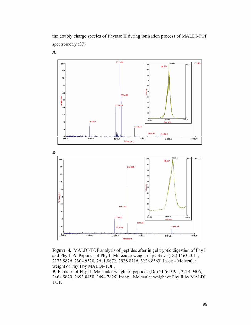
98
the doubly charge species of Phytase II during ionisation process of MALDI-TOF
spectrometry (37).
A
B
Figure 4. MALDI-TOF analysis of peptides after in gel tryptic digestion of Phy I and Phy II A. Peptides of Phy I [Molecular weight of peptides (Da) 1563.3011, 2273.9826, 2304.9520, 2611.8672, 2928.8716, 3226.8563] Inset: - Molecular weight of Phy I by MALDI-TOF. B. Peptides of Phy II [Molecular weight of peptides (Da) 2176.9194, 2214.9406, 2464.9820, 2693.8450, 3494.7825] Inset: - Molecular weight of Phy II by MALDI-TOF.
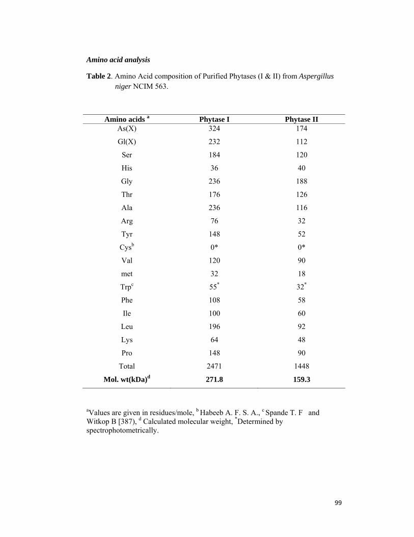
99
Amino acid analysis
Table 2. Amino Acid composition of Purified Phytases (I & II) from Aspergillus niger NCIM 563.
Amino acids a Phytase I Phytase II As(X) 324 174
Gl(X) 232 112
Ser 184 120
His 36 40
Gly 236 188
Thr 176 126
Ala 236 116
Arg 76 32
Tyr 148 52
Cysb 0* 0*
Val 120 90
met 32 18
Trpc 55* 32*
Phe 108 58
Ile 100 60
Leu 196 92
Lys 64 48
Pro 148 90
Total 2471 1448
Mol. wt(kDa)d 271.8 159.3
aValues are given in residues/mole, b Habeeb A. F. S. A., c Spande T. F and Witkop B [387), d Calculated molecular weight, *Determined by spectrophotometrically.
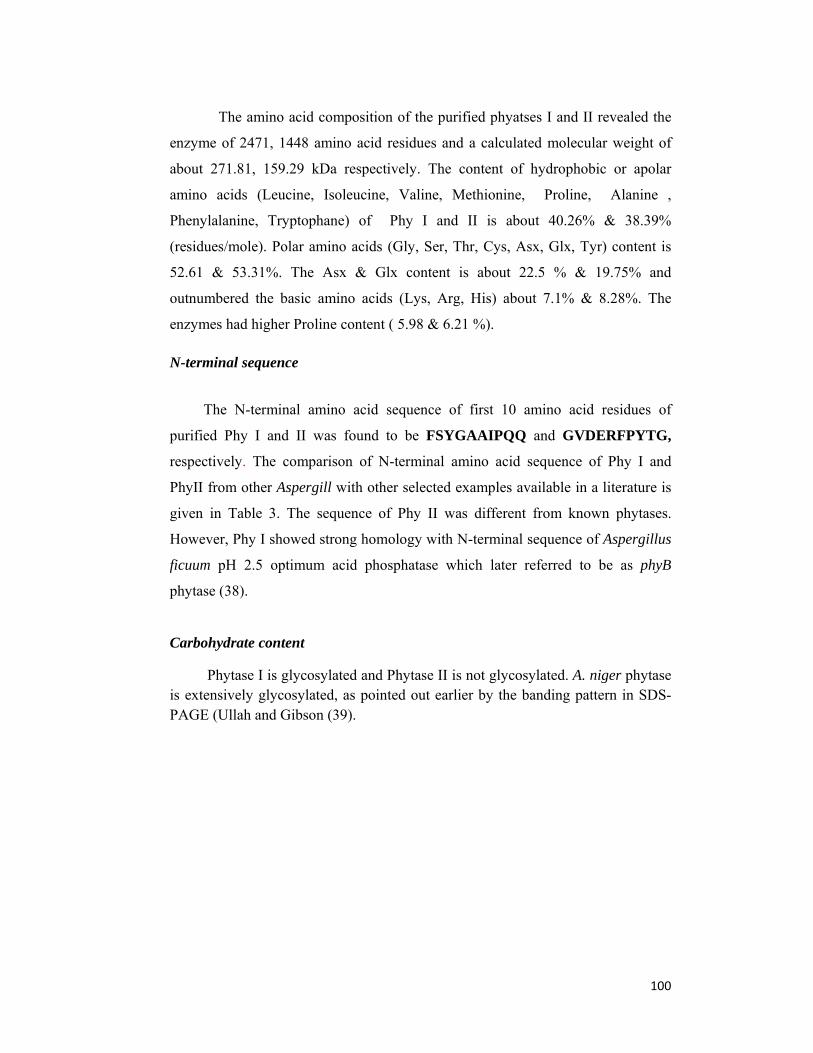
100
The amino acid composition of the purified phyatses I and II revealed the
enzyme of 2471, 1448 amino acid residues and a calculated molecular weight of
about 271.81, 159.29 kDa respectively. The content of hydrophobic or apolar
amino acids (Leucine, Isoleucine, Valine, Methionine, Proline, Alanine ,
Phenylalanine, Tryptophane) of Phy I and II is about 40.26% & 38.39%
(residues/mole). Polar amino acids (Gly, Ser, Thr, Cys, Asx, Glx, Tyr) content is
52.61 & 53.31%. The Asx & Glx content is about 22.5 % & 19.75% and
outnumbered the basic amino acids (Lys, Arg, His) about 7.1% & 8.28%. The
enzymes had higher Proline content ( 5.98 & 6.21 %).
N-terminal sequence
The N-terminal amino acid sequence of first 10 amino acid residues of
purified Phy I and II was found to be FSYGAAIPQQ and GVDERFPYTG,
respectively. The comparison of N-terminal amino acid sequence of Phy I and
PhyII from other Aspergill with other selected examples available in a literature is
given in Table 3. The sequence of Phy II was different from known phytases.
However, Phy I showed strong homology with N-terminal sequence of Aspergillus
ficuum pH 2.5 optimum acid phosphatase which later referred to be as phyB
phytase (38).
Carbohydrate content
Phytase I is glycosylated and Phytase II is not glycosylated. A. niger phytase is extensively glycosylated, as pointed out earlier by the banding pattern in SDS-PAGE (Ullah and Gibson (39).
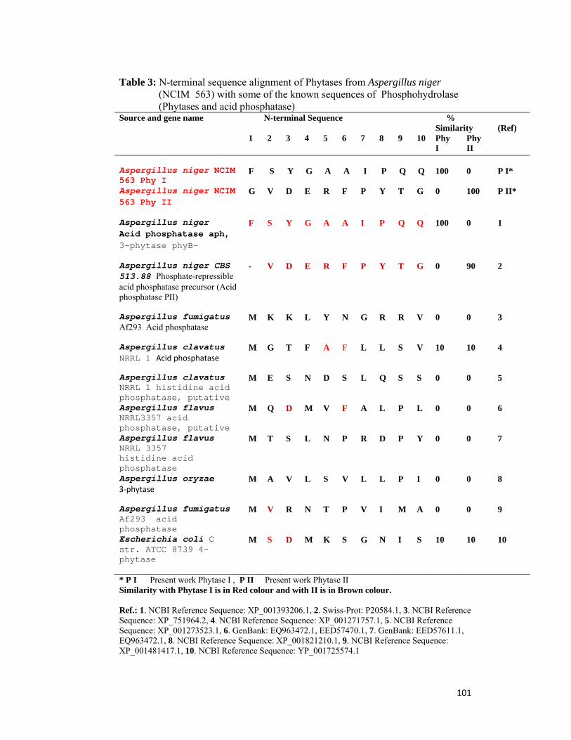
101
Table 3: N-terminal sequence alignment of Phytases from Aspergillus niger (NCIM 563) with some of the known sequences of Phosphohydrolase (Phytases and acid phosphatase)
N-terminal Sequence % Similarity
Source and gene name
1 2 3 4 5 6 7 8 9 10 Phy I
Phy II
(Ref)
Aspergillus niger NCIM 563 Phy I
F
S
Y
G
A
A
I
P
Q
Q
100
0
P I*
Aspergillus niger NCIM 563 Phy II
G V D E R F P Y T G 0 100 P II*
Aspergillus niger Acid phosphatase aph, 3-phytase phyB-
F S Y G A A I P Q Q 100 0 1
Aspergillus niger CBS 513.88 Phosphate-repressible acid phosphatase precursor (Acid phosphatase PII)
- V D E R F P Y T G 0 90 2
Aspergillus fumigatus Af293 Acid phosphatase
M K K L Y N G R R V 0 0 3
Aspergillus clavatus NRRL 1 Acid phosphatase
M G T F A F L L S V 10 10 4
Aspergillus clavatus NRRL 1 histidine acid phosphatase, putative
M E S N D S L Q S S 0 0 5
Aspergillus flavus NRRL3357 acid phosphatase, putative
M Q D M V F A L P L 0 0 6
Aspergillus flavus NRRL 3357 histidine acid phosphatase
M T S L N P R D P Y 0 0 7
Aspergillus oryzae 3‐phytase
M A V L S V L L P I 0 0 8
Aspergillus fumigatus Af293 acid phosphatase
M
V R N T P V I M A 0 0 9
Escherichia coli C str. ATCC 8739 4-phytase
M S D M K S G N I S 10 10 10
* P I Present work Phytase I , P II Present work Phytase II Similarity with Phytase I is in Red colour and with II is in Brown colour. Ref.: 1. NCBI Reference Sequence: XP_001393206.1, 2. Swiss-Prot: P20584.1, 3. NCBI Reference Sequence: XP_751964.2, 4. NCBI Reference Sequence: XP_001271757.1, 5. NCBI Reference Sequence: XP_001273523.1, 6. GenBank: EQ963472.1, EED57470.1, 7. GenBank: EED57611.1, EQ963472.1, 8. NCBI Reference Sequence: XP_001821210.1, 9. NCBI Reference Sequence: XP_001481417.1, 10. NCBI Reference Sequence: YP_001725574.1

102
Biochemical properties of phytases Figure 7 and 9 indicates that optimum temperature of Phy I and II was 55ºC
but optimum pH was 2.5 and 5.0, respectively (Fig 5 & 6). Purified phytase from
Aspergillus niger van Teighem also exhibited maximum activity at pH 2.5 and 52-
55ºC (6). However phytase produced from Aspergillus niger CFR335 either under
solid state or submerged fermentation showed phytase activity at pH 4.5 only (40).
Similarly Phy I had half life of 6-8 h at 55ºC while Phy II had 10-12 h. Several
other microbial phytases (1) and various commercial phytases display optimum
temperature range of 45-55ºC (41). Phy I showed broad pH stability (pH 2-9)
while Phy II was stable between pH 4.0 to 8.5 (Fig. 5&6). At 65º C for 1.5 h Phy I
retained 40 % activity while Phy II retained 55 % original activity (Fig.8 &10).
However crude phytase was found to retain 100 % original enzyme activity in
presence of 12 % skim milk after treatment at 70ºC for 3 min (22).
pH optimum and stability
1 2 3 4 5 6 7 8 9 100
10
20
30
40
50
60
70
80
90
100
110
Rel
ativ
e P
hyta
se A
ctiv
ity
(%)
pH
% ( Optimum pH) % ( pH Stability)
Figure: 5 Effect of pH on activity and stability of purified Acidic Phytase(Phy I) from Aspergillus niger (NCIM 563)
pH Optimum ( ) : A Suitably diluted enzyme (~ 0.05 U) was assayed in a series of pH (1.5-9) at 55 °C as described in Methods and the relative enzyme activity was determined. Maximum enzyme activity obtained was taken as 100 %. pH Stability () : A suitable concentration of purified enzyme (~ 5.0 U/ml) was incubated in a series of pH (1.5-9) at R.T. for the period of 12 hours. Aliquots (10 μl) were removed and residual enzyme activity was determined under standard assay conditions. Maximum activity obtained was taken as 100 %.

103
1 2 3 4 5 6 7 8 9 10-10
0
10
20
30
40
50
60
70
80
90
100
110
Rel
ativ
e Ph
ytas
e A
ctiv
ity (
%)
pH
Optim um pH pH Stability
Figure 6. Effect of pH on activity and stability of purified Phytase(Phy II) from Aspergillus niger (NCIM 563)
pH Optimum ( ) : A Suitably diluted enzyme (~ 0.1 U) was assayed in a series of pH (1.5-9) at 55 °C as described in Methods and the relative enzyme activity was determined. Maximum enzyme activity obtained was taken as 100 %.
pH Stability () : A suitable concentration of purified enzyme (~10.0 U /ml)was incubated in a series of pH (1.5-9) at R.T for the period of 12 hours. Aliquots (10 μl) were removed and residual enzyme activity was determined under standard assay conditions. Maximum activity obtained was taken as 100 %.
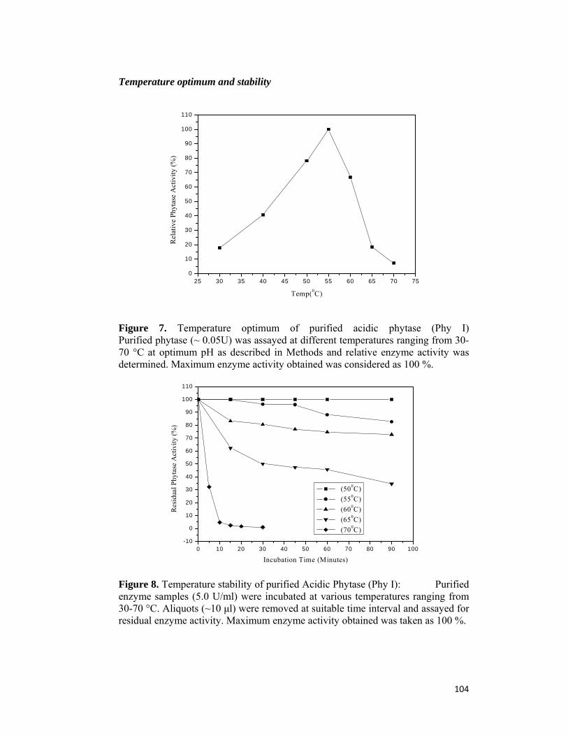
104
Temperature optimum and stability
25 30 35 40 45 50 55 60 65 70 750
10
20
30
40
50
60
70
80
90
100
110
Rel
ativ
e P
hyta
se A
ctiv
ity
(%)
Temp(0C)
Figure 7. Temperature optimum of purified acidic phytase (Phy I) Purified phytase (~ 0.05U) was assayed at different temperatures ranging from 30-70 °C at optimum pH as described in Methods and relative enzyme activity was determined. Maximum enzyme activity obtained was considered as 100 %.
0 10 20 30 40 50 60 70 80 90 100-10
0
10
20
30
40
50
60
70
80
90
100
110
Res
idua
l Phy
tase
Act
ivit
y (%
)
Incubation Time (Minutes)
(500C)
(550C)
(600C)
(650C)
(700C)
Figure 8. Temperature stability of purified Acidic Phytase (Phy I): Purified enzyme samples (5.0 U/ml) were incubated at various temperatures ranging from 30-70 °C. Aliquots (~10 μl) were removed at suitable time interval and assayed for residual enzyme activity. Maximum enzyme activity obtained was taken as 100 %.
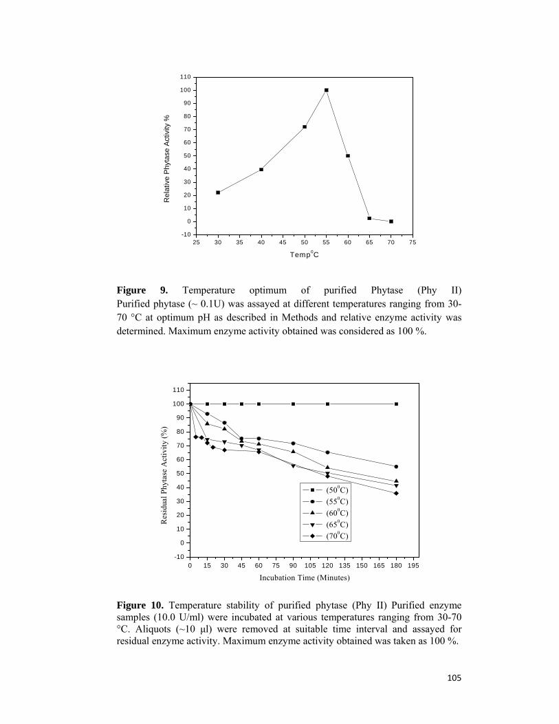
105
25 30 35 40 45 50 55 60 65 70 75-10
0
10
20
30
40
50
60
70
80
90
100
110
Re
lativ
e P
hyt
ase
Act
ivity
%
Temp0C
Figure 9. Temperature optimum of purified Phytase (Phy II) Purified phytase (~ 0.1U) was assayed at different temperatures ranging from 30-70 °C at optimum pH as described in Methods and relative enzyme activity was determined. Maximum enzyme activity obtained was considered as 100 %.
0 15 30 45 60 75 90 105 120 135 150 165 180 195-10
0
10
20
30
40
50
60
70
80
90
100
110
Res
idua
l Phy
tase
Act
ivit
y (%
)
Incubation Time (Minutes)
(500C)
(550C)
(600C)
(650C)
(700C)
Figure 10. Temperature stability of purified phytase (Phy II) Purified enzyme samples (10.0 U/ml) were incubated at various temperatures ranging from 30-70 °C. Aliquots (~10 μl) were removed at suitable time interval and assayed for residual enzyme activity. Maximum enzyme activity obtained was taken as 100 %.

106
Table 4. t1/2 of Phytases at various temperatures.
Temp. 0C Phytase I Phytase II
50 24-30 hour 48-56 hour
55 6-8 hour 10-12 hour
60 3.5 hour 4 hour
65 30 min 3 hour
70 2-3 min 2-2.5 hour
Substrate specificity
The hydrolysis of several phosphate esters (3 mM) by purified Phy I and II
was examined by measuring the release of free phosphate. The relative rate of
hydrolysis of various substrates is summarized in Table 4. Phy I showed
considerably broad substrate specificity and was noticeably specific for other
phosphate esters including p-nitrophenyl phosphate, a general substrate for acidic
phosphatase. Similar results were also recorded by phytase from Saccharomyces
cerevisiae CY strain (42) and Mucor hiemalis (43). Phytases derived from
Aspergillus niger, namely AllzymeTM and Natuphos® also displayed broad
substrate specificity (44). Many other fungal phytases also exhibit broad substrate
specificity which is desirable in terms of animal feed application (45).
Kinetics
Results of the kinetic studies (Table 5) indicated that Km values for sodium
phytate were lower than calcium phytate indicating that Phy I and II showed more
affinity towards sodium phytate. However Phy II showed higher Vmax (26674
μmoles min-1 mg-1) than Phy I (5018 μmoles min-1 mg-1). Lower Km (0.04 –
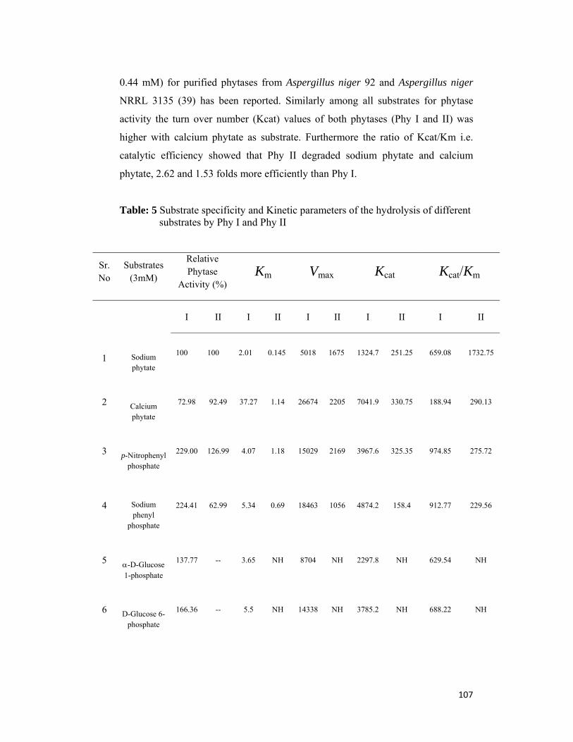
107
0.44 mM) for purified phytases from Aspergillus niger 92 and Aspergillus niger
NRRL 3135 (39) has been reported. Similarly among all substrates for phytase
activity the turn over number (Kcat) values of both phytases (Phy I and II) was
higher with calcium phytate as substrate. Furthermore the ratio of Kcat/Km i.e.
catalytic efficiency showed that Phy II degraded sodium phytate and calcium
phytate, 2.62 and 1.53 folds more efficiently than Phy I.
Table: 5 Substrate specificity and Kinetic parameters of the hydrolysis of different substrates by Phy I and Phy II
Sr.No
Substrates (3mM)
Relative Phytase
Activity (%)
Km Vmax Kcat Kcat/Km
I II I II I II I II I II
1
Sodium phytate
100 100 2.01 0.145 5018 1675 1324.7 251.25 659.08 1732.75
2
Calcium phytate
72.98 92.49 37.27 1.14 26674 2205 7041.9 330.75 188.94 290.13
3
p-Nitrophenyl phosphate
229.00 126.99 4.07 1.18 15029 2169 3967.6 325.35 974.85 275.72
4
Sodium phenyl
phosphate
224.41 62.99 5.34 0.69 18463 1056 4874.2 158.4 912.77 229.56
5
-D-Glucose 1-phosphate
137.77 -- 3.65 NH 8704 NH 2297.8 NH 629.54 NH
6
D-Glucose 6-phosphate
166.36 -- 5.5 NH 14338 NH 3785.2 NH 688.22 NH
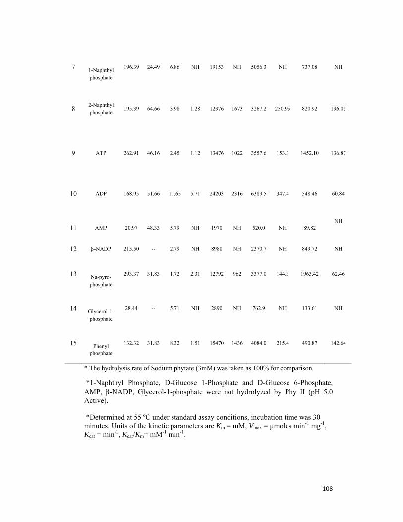
108
7
1-Naphthyl phosphate
196.39 24.49 6.86 NH 19153 NH 5056.3 NH 737.08 NH
8
2-Naphthyl phosphate
195.39 64.66 3.98 1.28 12376 1673 3267.2 250.95 820.92 196.05
9 ATP 262.91 46.16 2.45 1.12 13476 1022 3557.6 153.3 1452.10
136.87
10 ADP 168.95 51.66 11.65 5.71 24203 2316 6389.5 347.4 548.46
60.84
11 AMP 20.97 48.33 5.79 NH 1970 NH 520.0 NH 89.82 NH
12 -NADP 215.50 -- 2.79 NH 8980 NH 2370.7 NH 849.72 NH
13
Na-pyro-phosphate
293.37 31.83 1.72 2.31 12792 962 3377.0 144.3 1963.42 62.46
14
Glycerol-1-phosphate
28.44 -- 5.71 NH 2890 NH 762.9 NH 133.61 NH
15
Phenyl phosphate
132.32 31.83 8.32 1.51 15470 1436 4084.0 215.4 490.87 142.64
* The hydrolysis rate of Sodium phytate (3mM) was taken as 100% for comparison.
*1-Naphthyl Phosphate, D-Glucose 1-Phosphate and D-Glucose 6-Phosphate, AMP, -NADP, Glycerol-1-phosphate were not hydrolyzed by Phy II (pH 5.0 Active). *Determined at 55 ºC under standard assay conditions, incubation time was 30 minutes. Units of the kinetic parameters are Km = mM, Vmax = μmoles min-1 mg-1, Kcat = min-1, Kcat/Km= mM-1 min-1.

109

110
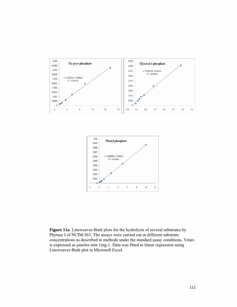
111
Figure 11a. Lineweaver-Burk plots for the hydrolysis of several substrates by Phytase I of NCIM-563. The assays were carried out at different substrate concentrations as described in methods under the standard assay conditions. Vmax is expressed as μmoles min-1mg-1. Data was fitted to linear regression using Lineweaver-Burk plot in Microsoft Excel.

112
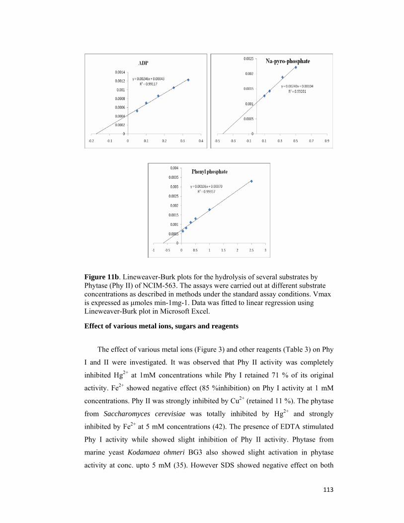
113
Figure 11b. Lineweaver-Burk plots for the hydrolysis of several substrates by Phytase (Phy II) of NCIM-563. The assays were carried out at different substrate concentrations as described in methods under the standard assay conditions. Vmax is expressed as μmoles min-1mg-1. Data was fitted to linear regression using Lineweaver-Burk plot in Microsoft Excel.
Effect of various metal ions, sugars and reagents
The effect of various metal ions (Figure 3) and other reagents (Table 3) on Phy
I and II were investigated. It was observed that Phy II activity was completely
inhibited Hg2+ at 1mM concentrations while Phy I retained 71 % of its original
activity. Fe2+ showed negative effect (85 %inhibition) on Phy I activity at 1 mM
concentrations. Phy II was strongly inhibited by Cu2+ (retained 11 %). The phytase
from Saccharomyces cerevisiae was totally inhibited by Hg2+ and strongly
inhibited by Fe2+ at 5 mM concentrations (42). The presence of EDTA stimulated
Phy I activity while showed slight inhibition of Phy II activity. Phytase from
marine yeast Kodamaea ohmeri BG3 also showed slight activation in phytase
activity at conc. upto 5 mM (35). However SDS showed negative effect on both

114
Phy I and II. Oxalic acid and EDTA also showed stimulating effect on Phy I
activity. Phytase from Aspergillus niger van Teighem, however, was not affected
by most metal ions, inhibitors and organic solvents (6).
Table 6. Effect of different metal ion solutions on Phytase activity: -
Phy I PhyII
Effective Conc. in Enzyme Assay
1mM 5mM 10mM 1mM 5mM 10mM
Sr.No. Metal ions Residual Phytase Activity (%)
1 None 100 100 100 100 100 100
2 Co2+ 103 72 35 100 101 106
3 Cu2+ 94 94 94 65 45 32
4 Ba2+ 98 ppt ppt 93 83 73
5 Fe3+ 108 100 97 60 70 80
6 Fe2+ 98 57 ppt 58 73 94
7 Ca2+ 100 94 93 73 87 104
8 Mg2+ 101 89 75 82 93 116
9 Ni2+ 100 98 93 77 82 94
10 K+ 100 100 101 73 80 91
11 Na+ 100 100 102 70 82 98
12 Mn2+ 100 83 72 80 85 91
13 Pb+ ppt ppt ppt 65 46 36
14 Hg2+ 100 100 87 00 00 00
15 Ag+ 92 51 20 00 00 00
16 Cd2+ 89 85 80 81 80 80
17 Zn2+ 98 91 85 43 39 32
18 NH4VO3 ppt ppt ppt 11 8 4
19 K2Cr2O7 86 56 10 93 51 25
A suitably diluted enzyme is assayed with varying final concentrations of Metal ion solutions i.e. 1mM, 5mM and 10mM in reaction mixture. Activities are expressed as percentage of activity compare to the control sample.

115
Table 7. Stability of purified enzymes under different metal ion solutions.
Sr.No. Metal ions (1mM) PhyI PhyII
1 None 100 100
2 Co2+ 100 94
3 Cu2+ 95 11
4 Ba2+ 100 91
5 Fe3+ 94 74
6 Fe2+ 15 65
7 Ca2+ 94 89
8 Mg2+ 90 104
9 Ni2+ 100 93
10 K+ 97 98
11 Na+ 98 84
12 Mn2+ 80 89
13 Pb+ 73 64
14 Hg2+ 71 00
15 Ag+ 80 53
16 Cd2+ 89 84
17 Zn2+ 86 54
18 NH4VO3 15 46
19 K2Cr2O7 90 104
A suitably diluted enzyme was pre-incubated with various Metal ion solutions at 40C for 24 hour and subsequently residual activity of Phytase was determined as described in methods. Activities are expressed as percentage of activity compare to the control sample.
116
Table 8. Effect of different solvents and reagents on phytase activity.
Sr.No. Solvents and reagents PhyI PhyII
1 None (Control) 100 100
2 Acetic acid (0.1%) 95 92
3 Oxalic acid (1mM) 194 91
(10mM) 208 82
4 DTT (1mM) 101 97
5 EDTA (1mM) 181 88
(10mM) 187 93
6 Triton-X-100 (0.1%) 101 93
7 Tween 80 (0.1%) 113 89
8 Tween 20 (0.1%) 114 94
9 SDS (1mM) 4 4
10 Glycerol (0.1%) 97 95
11 Toluene (0.1%) 105 91
12 Sodium azide (1mM) 101 97
13 -mercepto ethanol (0.1%) 105 93
14 Ethylene Glycol (0.1%) 104 92
A suitably diluted enzyme is assayed with different solvents (0.1%) and reagents (1mM) in reaction mixture. Activities are expressed as percentage of activity compare to the control sample.

117
Table 9. Effect of different additives on thermal stability of Acidic Phytase (PhyI):
Purified enzyme samples (5.0 U/ml) were incubated at 700C with different additives (10 % W/V). Aliquots (~10 μl) were removed after 0, 10 and 30 min and assayed for residual enzyme activity. Enzyme activity of 0 min sample was taken as 100 %.
Sr.No. Additive (10%W/V) Remaining Phytase Activity (%)
700C 10 min 700C 30 Min
1 700C 0 min. 100 100
2 Control (with out additive)
5.08 2.92
3 Maltose 6.28 4.48
4 Xylose 2.67 2.11
5 Glucose 9.94 4.50
6 2-Deoxy Glucose 3.64 2.50
7 Lactose 10.98 7.39
8 Galactose 3.11 1.14
9 Sucrose 6.23 3.14
10 Cellobiose 5.62 2.55
11 Stachyose 2.98 1.14
12 CaCl2.2H2O 2.23 1.04
13 PEG 3.56 2.11
14 Glycerol 2.56 1.45
15 Mannose 2.34 1.23
118
Table 10. Effect of different additives on thermal stability of Phytase II
Purified enzyme samples (10.0 U/ml) were incubated at 700C with different additives (10 % W/V). Aliquots (~10 μl) were removed after 0, 2 hour and 4 hour and assayed for residual enzyme activity. Enzyme activity of 0 min sample was taken as 100 %.
Sr.No. Additive (10%W/V) Remaining Phytase Activity (%)
700C 2 hour 700C 4 hour
1 700C 0 min. 100 100
2 Control (without additive)
63.68 35.15
3 Maltose 49.77 15.79
4 Xylose 42.27 7.89
5 Glucose 52.70 22.56
6 2-Deoxy Glucose 64.41 33.08
7 Lactose 62.03 35.15
8 Galactose 5.49 2.45
9 Sucrose 57.09 28.38
10 Cellobiose 48.67 26.50
11 Stachyose 23.86 12.56
12 CaCl2.2H2O 12.07 4.67
13 PEG 28.18 12.88
14 Glycerol 56.91 29.51
15 Mannose 38.24 .376
119
3.5. Conclusion
Fujita et al (46) reported three types of acid phosphatases in which one
was phytase from Aspergillus oryzae under submerged fermentation conditions.
Extracellular phytase with two pH optima (2.5 and 5.0) has been reported from
Aspergillus niger (18) and commercial Aspergillus niger phytase (Natuphos) (45).
Earlier we have reported production and partial characterization of two types of
phytase from Aspergillus niger NCIM 563 under submerged fermentation (20). In
the present studies we were able to purify both phytases (Phy I active at pH 2.5 and
Phy II active at pH 5.0) from Aspergillus niger NCIM 563 produced under
submerged fermentation to homogeneity and found that they differ with each other
in molecular and biochemical properties including pH optima, temperature and pH
stability, effect of various metal ions and other reagents, MALDI-TOF analysis,
affinity towards various substrates and N-terminal amino acid analysis. This,
according to our knowledge, is the first report of two distinct phytases from
Aspergillus niger produced under submerged fermentation conditions. High
phytase activity at pH 2.5 and stability under acidic conditions (pH 1.5 to 9.0)
suggests its application in poultry feed. However, further work is needed to
improve its thermostability by immobilization and/or micro encapsulation along
with site directed mutagenesis for its application in pelleted poultry feed which is
in progress.
120
3.6 References: 1. Konietzny U., Greiner R. (2002) Molecular and catalytic properies of phytate- degrading enzymes (phytases). Int J Food Sci Technol 37:791-812.. 2. Mullaney E. J., Daly C., Ullah A. B. (2000) Advances in phytase research. Adv Appl Microbiol 47: 157-199. 3. Singh P. K. (2008) Significance of phytic acid and supplemental phytase in chicken nutrition: a review. World’s Poultry Sci J. 64: 553-580. 4. Selle P. H., Ravindran V. (2007) Microbial phytase in poultry nutrition. Animal Feed Sci Technol 135: 1-41. 5. Vohra A., Satyanarayana T.(2003) Phytases: Microbial sources, production,
purification and potential biotechnological applications. Critical Rev in Biotechnol 23: 29-60.
6. Vats P, Banerjee U. C.(2004) Production studies and catalytic properties of phytases (myoinositolhexakisphosphate phosphohydrolases): an overview. Enzyme Microb Technol 35: 3-14. 7. Kaur P, Kunze G, Satyanarayana T. (2007) Yeast phytases: Present scenario
and future perspectives. Critical Rev in Biotechnol 27: 93-109. 8. Kim Y. O., Kim H. K., Baeks Y. J., Oh T. K. (1988) Purification and properties
of a thermostable phytase from Bacillus spp DS11. Enzyme Microb Technol 22: 2-7.
9. Radcliffe J. S., Zhang Z., Kornegay E. T. (1988) The effect of microbial
Phytase, citric acid and their interaction in a corn soybean meal based diet for Weaning pigs. J Animal Sci 76:1880-1886.
10. Ullah, A. H. J. (1988a) Aspergillus ficuum phytase: partial primary structure, substrate selectivity, and kinetic characterization. Prep. Biochem. 18: 459-471. 11. Greiner, R., Konietzny, U. and Jany, K. D. (1993) Purification and characterization of Two phytases from Escherichia coli. Arch. Biochem. Biophys. 303: 107-113. 12. Craxton A, Caffrey J. J., Burkhart W., Safrany S. T., Shears, S. B. (1997) Molecular cloning and expression of a rat hepatic multiple inositol polyphosphate phosphatase. Biochem. J. 328: 75-81. 13. Ullah A. H. J., Cummins B. J. Dischinger H. C. Jr. (1991) Cyclohexanedione modification of arginine at the active site of Aspergillus ficuum phytase. Biochem. Biophys. Res. Commun.178: 45-53.
121
14. Ullah A. H. J., Dischinger H. C. Jr. (1992) Identification of residues involved in active site formation in Aspergillus ficuum phytase. Ann. N.Y. Acad. Sci. 672:45-51. 15. Ostanin K., Harms E. H., Stevis P. E., Kuciel R., Zhou M. M., van Etten R. L. (1992) Overexpression, site-directed mutagenesis, and mechanism of Escherichia coli acid phosphatase. J. Biol. Chem. 267: 22830-22836. 16. Ostanin K., van Etten, R. L. (1993) Asp304 of Escherichia coli acid phosphatase is involved in leaving group protonation. J. Biol. Chem. 268: 20778-20784. 17. Michell D. B., Vogel K., Weimann B. J., Pasamontes L., van Loon A. P. (1997) The phytase subfamily of histidine acid phosphatases: isolation of two genes for two novel phytases from the fungi Aspergillus terrus and Myceoliophthora thermophila. Microbiology 143: 245-252. 18. Ullah A. H. J. (1998) Production, rapid purification and catalytical
characterization of extracellular Phytase from Aspergillus ficuum. Prep Biochem 18: 443-458.
19. Vats P, Sahoo D. K., Banerjee U. C. (2004) Production of phytase (myo-
nositolhexakis phosphate phosphohydrolase) by Aspergillus niger van Teighem in laboratory scale fermenter. Biotechnol Prog 20: 737-43.
20. Soni S. K., Khire J. M. (2007) Production and partial characterization of two
types of phytase from Aspergillus niger NCIM 563 under submerged fermentation conditions. World J Microbiol Biotechnol 23: 1585-1593.
21. Bhavsar K., Shah P., Soni S. K., Khire J.M. (2008) Influence of pretreatment of
agriculture residues on phytase production by Aspergillus niger NCIM 563 under submerged fermentation conditions. African J Biotechnol 7, 1101-1106. 22. Shah P., Bhavsar K., Soni S. K., Khire J. M. (2009) Strain improvement and up Scaling of phytase production by Aspergillus niger NCIM 563 under submerged fermentation conditions. J Ind Microbiol Biotechnol 36: 373- 80. 23. Heinohen J. K., Lathi R. J. (1981) A new and convenient colorimetric determination of inorganic orthophosphate and its application to the assay of inorganic pyrophosphatase. Anal Biochem 113: 313-317. 24. Lowry O. H., Rosebrough N. J., Farr A. L., Randall R. L. (1951) Protein Measurement with the Folin Phenol reagent. J Biol Chem 193: 265-275. 25. Dubois M., Gilles K.A., Hamilton J.K., Rebers P.A., Smith F. (1956) Colorimetric method for determination of sugars and related substances, Anal. Chem. 28: 350-356.
122
26. Laemmli U. K. (1970) Cleavage of structural proteins during the assembly of the head of bacteriophage T4, Nature (London). 227, 680-685. 27. Davis B. J (1964) Disc electrophoresis II. Method and application to human serum proteins. Ann N Y Acad. Sci 121: 404–427. 28. Chinnatambi S., Bodhe A. M., Lachke A. H. (1994) Fabrication of improved mini scale eletrofocusing unit. Biotechnol Tech 8: 681-682. 29. Clustal W, Multiple sequence alignment program., URL: http://www.ebi.ac.uk/clustalw/. 30. Andrews P, Estimation of the molecular weights of proteins by Sephadex gel filtration. (1964) Biochem J. 91: 222-233. 31. Weber K., Osborn M. (1969) The Reliability of Molecular Weight determination by Dodecyl Sulfate-Polyacrylamide Gel Electrophoresis. J. Bio. Chem. 244: 4406-4412. 32. Bar C., Patil R., Doshi J., Kulkarni M. J. (2007) Characterization of the proteins of bacterial strains isolated from contaminated site involved in heavy metal resistance-A proteomic approach. J Biotechnol 128: 44-451. 33. Lineweaver H., Burk D. (1934) Determination of enzyme dissociation constants. J. Amer Chem Soc. 56: 656-58.. 34. Nagashima T., Tange T., Anazawa H. (1999) Dephosphorylation of phytase by using the Aspergillus niger phytase with a high affinity for phytate. Appl Enviorn Microbiol 65: 4682-4684. 35. Xiaoyu L., Zhenming C., Zhiqiang L., Jing L., Xianghong W., Hirimuthugoda N.Y. (2008) Purification and characterization of extracellular phytase from a marine yeast Kodamaea ohmeri BG3. Marine Biotechnol 10: 190-197. 36. Ullah A. H. J., Sethumadhavan K. (1988) Differences in the active site environment of Aspergillus ficuum phytases. Biochem Biophys Res Commun 243: 458- 462. 37. Korwar A. M., Santhakumari B., Kulkarni M. J. (2008) Identification of ripening specific proteins in tomato by intact tissue MALDI-TOF-MS. Electronic J Food Plants Chem 3:10-13. 38. Ehrlich K. C., Montalbano B. G., Mullaney E. J., Dischinger H. C., Ullah A. H. J. (1993) Identification and cloning of a second phytase gene (phyB) from Aspergillus niger (ficuum). Biochem Biophys Res Commun 192: 747-53.
123
39. Ullah A. H. J., Gibson D. M. (1987) Extracellular phytase (EC 3.1.3.8) from Aspergillus ficuum NRRL 3135: purification and characterization. Prep Biochem 17: 63-91. 40. Gunashree B. S., Venkateswaran G. (2008) Effect of different cultural Conditions for production of phytase by Aspergillus niger CFR 335 under submerged and solid state fermentations. J Ind Microbiol Biotechnol 35: 1587- 1596. 41. Boyce A., Walsh G. (2006) Comparision of selected physicochemical characteristics of commercial phytases relevant to their application in phosphate pollution abatement. J Enviorn Sci Health Part A: Toxic/Hazard Subst Enviorn Eng 41: 789-798. 42. In M. J., Seo S. W., Kim D. C., Oh N. S. (2009) Purification and biochemical properties of an extracellular acid phytase produced by the Sachharomyces cerevisiae CY strain. Process Biochem 44: 122-126. 43. Boyce A., Walsh G. (2007) Purification and characterization of an acid Phosphatase with phytase activity from Mucor hiemalis. J Biotechnol 132: 82- 87. 44. Martin J. A., Murphy R. A., Power R. F. G. (2006) Purification and physico- chemical characterization of genetically modified phytases expressed in Aspergillus awamori. Bioresour Technol 97: 1703-1708. 45. Wyss M, Brugger R, Kronenberger A, Remy R, Fimbel R., Oesterhelt G., Lehmann M., van Loon A.P.G.M. (1999) Biochemical characterization of fungal phytases (myo-inositol hexakisphosphate phosphohydrolase): catalytic properties. Appl Enviorn Microbiol 65: 367-73. 46. Fujita J., Yamane Y., Fukuda H., Kizaki Y., Wakabayashi S., Shigeta S, Suzuki O., Ono K. (2003) Production and properties of phytase and acid phosphatase from a sake koji mold, Aspergillus oryzae. J Biosci Bioeng 95: 348-53.

124
CHAPTER 4
Application of phytase of Aspergillus niger (NCIM 563) in biomimetic synthesis of Hydroxyapatite and its polymorphs
4.1 Summary
Wheat bran is a cheap agro-based waste material, which has a substantial
amount of bound phosphorous in the form of phytic acid. However, surprisingly
there have been no attempts at harnessing such phosphorous in wheat bran or other
agro wastes into nanosized hydroxyapatite particles and its polymorphs. In the
present study, we demonstrate novel bio-inspired enzymatic synthesis of nanosized
Hydroxy apatite (HAP) Ca10(PO4)6(OH)2 and its polymorphs (β Tri calcium
phosphate(β TCP) and Di calcium phosphate(DCP) ) using wheat bran and a
novel extra cellular enzyme phytase, produced by solid state fermentation of
fungus Aspergillus niger NCIM 563. The as prepared apatite powder with ~100 nm
interconnected hollow spheres capped by stabilising proteins and devoid of any
carbonate contamination was synthesized by the hydro catalytic action of phytase
on phytic acid i.e. extracted from wheat bran and calcium ions at 50ºC and pH of
5.5 under inert environment of nitrogen. Calcination of white solid precipitate
leads to loss of occluded protein and converts it to highly crystalline material
comprising HAP, β TCP and DCP.
Biomimic synthesis of nanosized biocomposites using novel enzymes from
micro organisms starting from cheap agro-industrial waste materials is an exciting
possibility that could lead to an energy-conserving and economically viable green
approach towards the large-scale synthesis of crystalline artificial bone
nanomaterials.
4.2 Introduction
There is a high demand of fabrication of bone like biomaterials for tissue
revitalization for orthopaedic and dental applications. Biomimetic mineralization
of advanced man-made materials with properties similar to bone is a challenge

125
because of their complex nature. Synthetic calcium hydroxyapatite [HAP,
Ca10(PO4)6(OH)2], is a bioactive material that is chemically similar to biological
apatite, the mineral component of bone (1).
HAP has immense applications as artificial bone material, supportive media
for drug delivery, purification of protein (2), nucleic acid, isolation of virus etc.
The calcium phosphate matrix is capable of various chemical interactions with
DNA and proteins, including charge repulsion by crystal phosphates and metal
affinity interactions with crystalline calcium (3-7). Indeed, human bone is a natural
composite comprising of nano-apatite rods (which are ~100 nm) arranged in
lamellae and bound to collagen (8). Thus, synthetic HAP is of interest as
biocompatible phase reinforcement in biomedical composites, for bulk filling, bone
defects and for coatings on metal implants (9). HA and other calcium phosphates
(calcium deficient hydroxyapatite, CDHA) are also of interest as components in
injectable bone cements; controlling particle properties (e.g. size and shape) and
are often used to modulate cement setting behaviour (10).
HAP powders and coatings can be synthesized using a number of methods
including sol-gel processing (11), co-precipitation (12, 13), emulsion techniques
(14-17), mechano-chemical methods (18) and chemical vapour deposition (19).
The disadvantages of these methods include requirement of very precise control
over reaction conditions, expensive starting materials, large amounts of toxic
organic solvents and time consuming processes. For example, in wet chemical
synthesis of HAP, a maturation step (>18 h), followed by a heat treatment of
650C, is required (20). Failure to allow sufficient maturation, gives a phase-
separated product upon heat treatment, which can adversely affect biological
properties in vivo. Furthermore, the aforementioned synthesis approaches give little
or no control over HAP particle characteristics i.e. particle size, agglomeration
surface area, etc (21, 22).
To gain a better understanding of the factors that produce particles with the
desired properties, current synthesis methods are essentially too slow and
unpredictable. Thus, there is interest in developing faster synthesis techniques for
synthetic apatite that allow a greater degree of control over particle properties.
126
Wheat (Triticum sativum) is a member of family Gramineace, typically
constitutes 20-25% of its total product in the form of wheat bran and a great deal of
wheat bran is either used as fodder or disposed off by farmers as waste. Wheat
bran has a substantial amount of bound phosphorous in the form of phytic acid; a
very well known fact. However, to our knowledge there have been no attempts at
harnessing this enormous amount of phosphorous present in most of the agro
wastes including wheat bran in-to nanosized hydroxyapatite particles.
Phytases (EC 3.1.3.8 and EC 3.1.3.26) are a class of phosphohydrolases that
catalyzes the hydrolysis of phytic acid (myo- inositol hexakis phosphate) to
inorganic monophosphate and lower phosphoric esters of myo-inositol, in some
cases to free myo-inositols (23, 24, 25). Our approach involves the use of GRAS
(Generally recognised as safe) fungus Aspergillus niger for the extracellular
production of phytase enzyme by solid state fermentation of wheat bran.
The present Chapter describes the rapid, single step synthesis of crystalline
nano-particle of HAP, β TCP and DCP by a wet biomimetic method. Briefly,
aqueous 50mM solution of calcium chloride was mixed with microbial phytase and
phytic acid, under continuous stirring at 50C in acetate buffer of 50mM at pH 5.5.
The precipitation of calcium phosphate particles started in the biocatalysed
reaction, which was aged for 24 h, filtered, washed with deionized water, and dried
at 40C. This is the first report, to our knowledge, of biomimetic synthesis of
nanosized hydroxyapatite and its precursors starting from wheat bran.
4.3 Materials and Methods
Chemicals
The wheat bran used in this study was obtained from an animal feed stuff
outlet at Pune, Maharashtra, India. Phytic acid sodium salt from corn was
purchased from Sigma Chemical Company, St Louis, MO, USA. All other
chemicals used were of analytical grade and obtained from leading manufacturers
including BDH, Sigma, and Glaxo.
127
Fungal strain and maintenance
The strain used throughout the present work was Aspergillus niger NCIM
563. It was maintained on Potato Dextrose Agar (PDA) slants. PDA contains (per
litre distilled water: extract from 200 g potatoes; glucose, 20 g; Difco yeast extract,
1 g; and agar 20 g).
Production of phytase, culture conditions and Extraction of phytate from
agriculture residue
Submerged fermentation for production of phytase was carried out accordingly
to a reported work (25). Fermentation medium was inoculated with 1 % (v/v) of
spore suspension (5 x 10⁷ spores per ml) prepared by suspending the spores from 7
day old sporulated slant of Aspergillus niger NCIM 563 grown on PDA in 10 ml of
sterile distilled water containing 0.01 % Tween 80 and incubated at 30C at
stationary condition. Phytase produced by the same strain under submerged
fermentation condition (25) was having optimum pH 2.5 so it wasn’t used in the
present study. Extraction of phytate from wheat bran was carried out according to
reported by Harland (27) and Latta (28).
Partial purification of phytase II produced by submerged fermentation
After fermentation mycelium was separated by filtration followed by
centrifugation at 10,000g for 30 min and the clear supernatant was collected. Solid
ammonium sulphate was added to the supernatant to 95% saturation with constant
stirring. The precipitate was collected by centrifugation at 15,000g for 20 min and
dissolved in smallest possible volume of Glycine HCl buffer (100 mM, pH 2.5)
and salt was removed by passing through Sephadex G-25column and active
fractions were concentrated through YM-30 membrane (Millipore) and used for
phytase activity measurement.
Enzyme assay
Phytase activity was measured at 50C and using acetate buffer of pH 5.5
100mM as described earlier (26). The liberated inorganic phosphate was measured
128
by a modification of the ammonium molybdate method (29). A freshly prepared
solution of acetone: 5 N H2SO4: 10 mM ammonium molybdate (2:1:1 v/v/v) and
400 μl of 1M citric acid were added to the assay mixture. Absorbance was
measured at 370 nm. One unit of phytase activity (IU) was expressed as the
amount of enzyme that liberates 1μmol phosphorus per minute under standard
assay conditions.
Synthesis of HAP and reaction conditions
Reaction was carried out strictly under inert environment of nitrogen and
solutions were prepared in distilled deionized water (resistivity 18.2 M. cm).
Deionised water was obtained with a Millipore ultra pure water system, which was
previously distilled and decarbonised by boiling and bubbling N2. Reaction was
carried out in acetate buffer (pH 5.5, 50 mM) while keeping the constant
temperature of 50C (optimum temperature for enzyme activity) (26) with
continuous stirring for 4 hrs. Extracted phytate from wheat bran (3mM final
concentration) and partially purified phytase (17.19 IU/ml) was added in a three
neck round bottom (RB) flask. Calcium chloride (50mM) was dissolved in distilled
water, under inert atmosphere and was added into this round bottom flask with an
addition funnel. The whole assembly (Fig.1) was kept under N2 atmosphere, to
avoid the carbonate contamination. A white precipitate was obtained after reaction
and aged in the mother liquid for 24h. The white solid products were isolated by
repeated centrifuging and washing with decarbonated water and finally dried at
40C. During the centrifuging, sealed containers were used to avoid contact with
air. The deionised-distilled water used for synthesis and washing, was
decarbonated prior to use by boiling and subsequent cooling in the absence of CO2
by means of a gas-washing bottle filled with KOH solution. During the washing
process, some of the HAP particles that remained suspended in the supernatant
after centrifuging were discarded. Dried samples were calcined at 12000C for 2
hour. Reaction was also carried out with out enzyme as control experiment keeping
all the conditions same.
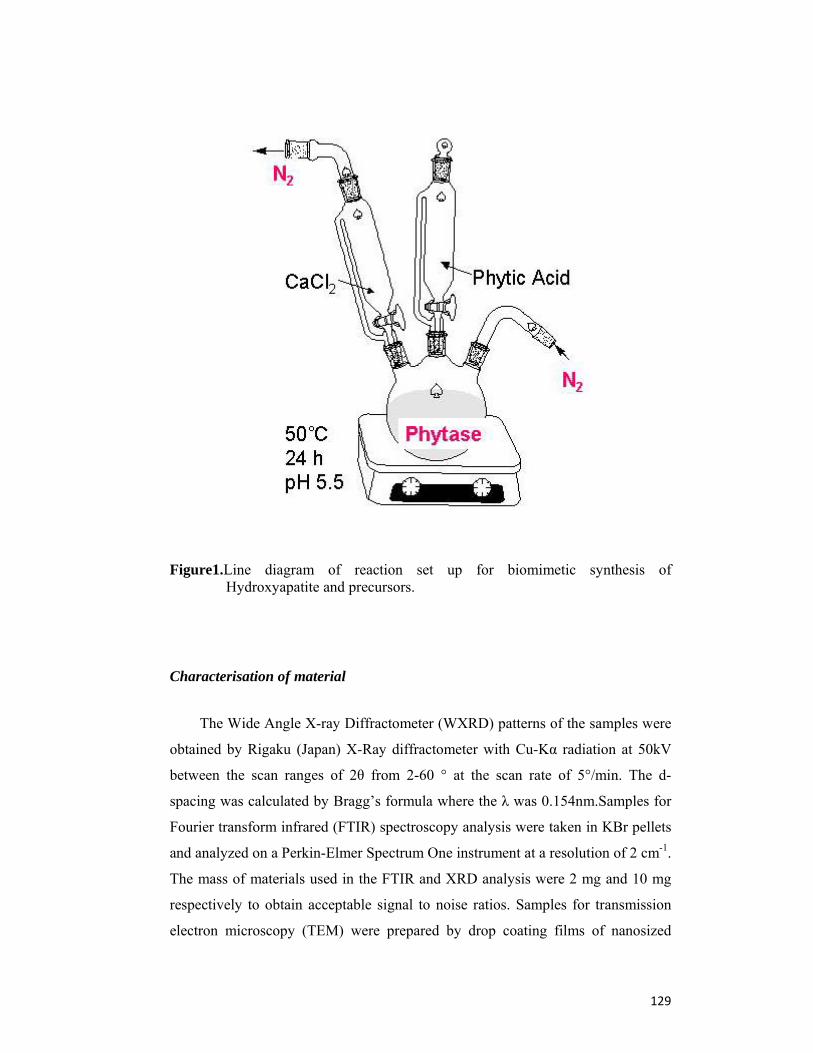
129
Figure1.Line diagram of reaction set up for biomimetic synthesis of Hydroxyapatite and precursors.
Characterisation of material
The Wide Angle X-ray Diffractometer (WXRD) patterns of the samples were
obtained by Rigaku (Japan) X-Ray diffractometer with Cu-Kα radiation at 50kV
between the scan ranges of 2θ from 2-60 ° at the scan rate of 5°/min. The d-
spacing was calculated by Bragg’s formula where the λ was 0.154nm.Samples for
Fourier transform infrared (FTIR) spectroscopy analysis were taken in KBr pellets
and analyzed on a Perkin-Elmer Spectrum One instrument at a resolution of 2 cm-1.
The mass of materials used in the FTIR and XRD analysis were 2 mg and 10 mg
respectively to obtain acceptable signal to noise ratios. Samples for transmission
electron microscopy (TEM) were prepared by drop coating films of nanosized
130
HAP powder dispersed in water onto carbon-coated copper grids. TEM and SAED
patterns of synthesized products were obtained on a JEOL 1200 EX instrument
operated at an accelerating voltage of 120 kV. For scanning electron microscopy
(SEM) analysis, solution cast film of biogenic calcium phosphate was made on Si
(111) substrates. SEM measurements were performed on a Leica Stereoscan-440
instrument equipped with a Phoenix energy dispersive analysis of X-rays (EDAX)
attachment. Thermo gravimetric analysis (TGA) profile of carefully weighed
quantity of the as prepared powder of calcium phosphate was recorded on a Seiko
Instruments model TG/DTA 32 instrument from 50 C to 900C with a heating
rate of 10C min-1 under nitrogen with flow rate 20ml min-1. Solid state 31P nuclear
magnetic resonance (NMR) spectra were obtained at 162 MHz on a Bruker
spectrometer Avance 400 (rotor 4mm, spinning rate 2-12 kHz) using Magic Angle
Spinning (MAS). Phosphoric acid (H3PO4) was used a reference sample.
Gel Electrophoresis
The biomolecules occluded in the nanostructures were analyzed using 10%
sodium dodecyl sulfate polyacrylamide gel electrophoresis (SDS-PAGE) according
to the procedure published (34). For protein analysis, the as synthesized particles
of HAP, along with occluded proteins were treated with 14.4mM β-mercapto
ethanol (2-mercapto ethanol) dissolved in double distilled water; for 5-10 min in
boiling water bath. Free proteins were removed by repeated washing and
centrifugation at 10000 rpm for 30 min in supernatant. Following dissolution, the
protein sample was dialyzed against deionized water for 48 h, lyophilized and
analyzed using SDS-PAGE along with protein molecular weight markers.
4.4 Results and Discussions
Production and partial purification of Phyatse II
The partial purified phytase enzyme preparation from the submerged
fermentation by fungus Aspergillus niger NCIM 563 used for the reaction was
having activity 20.6 IU/ml and specific activity 150.04 IU/mg. The purification

131
process showed 70.11 % recovery with 1.76 fold purification of crude enzyme
preparation.
Table 1. Purification table for phytase II produced by Aspergillus niger NCIM 563
Possible mechanism of calcium phosphate production
On mixing of partial purified enzyme preparation to phytic acid (Extracted
from wheat bran), inorganic monophosphate is released due to the hydrocatalysis
by phytase on the ester bond present between phosphorus and carbon of myo-
inositol ring. The released phosphate bind with the calcium ion and the white
precipitate of calcium phosphate was formed at 50C and pH 5.5. Remaining
proteins along with phytase and reaction end products like myo-inositol, lower
phosphoric esters of myo-inositol were separated from calcium phosphate
precipitate by subsequent washing with deionised double distilled water. (Fig.2)
Purification steps Phytase II
Total protein (mg)
Total activity (IU)/L
Specific activity U/mg
Fold purification
Yield %
Culture filtrate 242 20,600 85.12 1 100
Rota vapour conc. 169 16,312 96.5 1.13 79.10
95% Ammonium sulphate ppt. and desalting
96.26 14,443 150.04 1.76 70.11

132
Figure.2. Possible mechanism of production of calcium phosphate (Hydroxy apatite, β TCP, DCP) by phytase from Aspergillus niger NCIM 563 produced under submerged fermentation
X- ray diffraction studies
The crystallization of biomimetic product formed in the enzyme-phytic acid
reaction was studied by X-ray diffraction (XRD) analysis at 2 h of reaction (Figure
1A). XRD traces showed amorphous powders of low crystallinity, represented by
broad diffraction peak at around 28.16 and at 5.32, and free of secondary phases
owing to its low temperature processing. However, the obtained results are
comparable to the diffraction pattern of the biological apatite and are in good
agreement with the previous results (35).
To further verify the crystallinity of particles, XRD analysis of calcined
particles was performed. The results are shown in Fig 1B. The room temperature
XRD profile (2θ value) of the calcined material matches very well with HAP, β
TCP (Tri calcium phosphate), DCP (Di calcium phosphate) that reported in the
literature (36). The peaks of the resulting diffractogram were observed near 29.60
and 31.10 and are diagnostic for the presence of calcium hydroxyapatite (JCPDS
No. 09-0 432) (37). In addition, the presence of organic molecules greatly
influences the crystalline behaviour of the HAP.
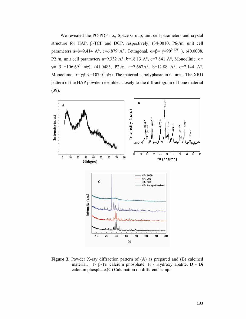
133
We revealed the PC-PDF no., Space Group, unit cell parameters and crystal
structure for HAP, β-TCP and DCP, respectively: (34-0010, P63/m, unit cell
parameters a=b=9.414 A°, c=6.879 A°, Tetragonal, α=β= γ=900 [38] ), (40.0008,
P21/n, unit cell parameters a=9.332 A°, b=18.13 A°, c=7.841 A°, Monoclinic, α=
γ≠ β =106.690. ≠γ), (41.0483, P21/n, a=7.667A°, b=12.88 A°, c=7.144 A°,
Monoclinic, α= γ≠ β =107.00. ≠γ). The material is polyphasic in nature .. The XRD
pattern of the HAP powder resembles closely to the diffractogram of bone material
(39).
Figure 3. Powder X-ray diffraction pattern of (A) as prepared and (B) calcined material. T- β-Tri calcium phosphate, H - Hydroxy apatite, D - Di calcium phosphate.(C) Calcination on different Temp.
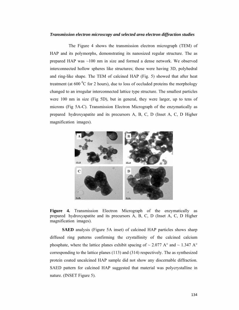
134
Transmission electron microscopy and selected area electron diffraction studies
The Figure 4 shows the transmission electron micrograph (TEM) of
HAP and its polymorphs, demonstrating its nanosized regular structure. The as
prepared HAP was ~100 nm in size and formed a dense network. We observed
interconnected hollow spheres like structures; those were having 3D, polyhedral
and ring-like shape. The TEM of calcined HAP (Fig. 5) showed that after heat
treatment (at 600 0C for 2 hours), due to loss of occluded proteins the morphology
changed to an irregular interconnected lattice type structure. The smallest particles
were 100 nm in size (Fig 5D), but in general, they were larger, up to tens of
microns (Fig 5A-C). Transmission Electron Micrograph of the enzymatically as
prepared hydroxyapatite and its precursors A, B, C, D (Inset A, C, D Higher
magnification images).
Figure 4. Transmission Electron Micrograph of the enzymatically as prepared hydroxyapatite and its precursors A, B, C, D (Inset A, C, D Higher magnification images).
SAED analysis (Figure 5A inset) of calcined HAP particles shows sharp
diffused ring patterns confirming the crystallinity of the calcined calcium
phosphate, where the lattice planes exhibit spacing of ~ 2.077 A° and ~ 1.347 A°
corresponding to the lattice planes (113) and (314) respectively. The as synthesized
protein coated uncalcined HAP sample did not show any discernable diffraction.
SAED pattern for calcined HAP suggested that material was polycrystalline in
nature. (INSET Figure 5).

135
Figure 5. Transmission Electron Micrograph of calcined material (Hydroxyapatite and its precursors). B,D Higher magnification images. Inset(A) shows selected area electron diffraction curve.
FTIR Analysis
The FT-IR spectrums of as prepared particles in Fig. 6(1), calcined in Fig.
6(2) and with commercial available HAP in Fig. 6(3) are given in Figure 6. An
intense band observed at 3451 cm-1 corresponds to the combination of OH group
(HAP phase). The apatitic PO43- 4 modes, in the region of 600- 500 cm-1 and 1
and 3 modes at 1150-1000cm-1, which corresponds to the factor group splitting of

136
PO43- tetrahedral, were also detected [40]. The bands at ~953-961 cm-1 corresponds
to 1 symmetric and 4 P-O stretching vibrations of the PO43- ion, respectively
(41). Samples have not been contaminated by atmospheric carbon dioxide, as no
peak corresponding to a secondary carbonate is recorded. The presence of intense
amide I and II bands at 1644 and 1547 cm-1 respectively are also observed in the
FTIR spectrum (1). Observation in Fig. 6(1) indicates that the particles in the
hollow spherical/circular morphology are present with proteins that are possibly
occluded into the particles or are bound to the surface of the particles (42). When
the as prepared HAP particles were calcined at 6000C for 2 h, the amide signatures
as well as the signatures from organic molecules around ca. 1400 cm-1 in FTIR
spectra fade away, indicating the removal of most of the biomolecules during
calcinations (43) as shown in Figure 6(2).
Figure 6. FTIR spectra of (1) as prepared material, (2) Calcined and (3) commercially available hydroxyapatite.
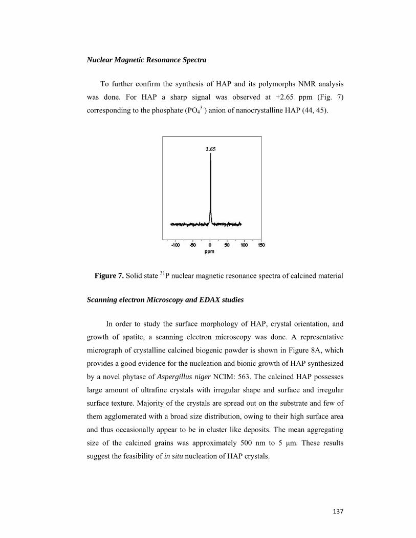
137
Nuclear Magnetic Resonance Spectra
To further confirm the synthesis of HAP and its polymorphs NMR analysis
was done. For HAP a sharp signal was observed at +2.65 ppm (Fig. 7)
corresponding to the phosphate (PO43-) anion of nanocrystalline HAP (44, 45).
Figure 7. Solid state 31P nuclear magnetic resonance spectra of calcined material
Scanning electron Microscopy and EDAX studies
In order to study the surface morphology of HAP, crystal orientation, and
growth of apatite, a scanning electron microscopy was done. A representative
micrograph of crystalline calcined biogenic powder is shown in Figure 8A, which
provides a good evidence for the nucleation and bionic growth of HAP synthesized
by a novel phytase of Aspergillus niger NCIM: 563. The calcined HAP possesses
large amount of ultrafine crystals with irregular shape and surface and irregular
surface texture. Majority of the crystals are spread out on the substrate and few of
them agglomerated with a broad size distribution, owing to their high surface area
and thus occasionally appear to be in cluster like deposits. The mean aggregating
size of the calcined grains was approximately 500 nm to 5 μm. These results
suggest the feasibility of in situ nucleation of HAP crystals.
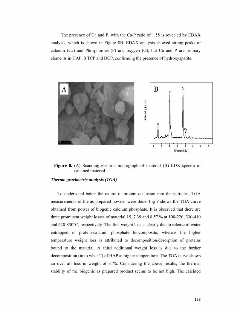
138
The presence of Ca and P, with the Ca/P ratio of 1.55 is revealed by EDAX
analysis, which is shown in Figure 8B. EDAX analysis showed strong peaks of
calcium (Ca) and Phosphorous (P) and oxygen (O), but Ca and P are primary
elements in HAP, β TCP and DCP, confirming the presence of hydroxyapatite.
Figure 8. (A) Scanning electron micrograph of material (B) EDX spectra of calcined material.
Thermo gravimetric analysis (TGA)
To understand better the nature of protein occlusion into the particles, TGA
measurements of the as prepared powder were done. Fig 9 shows the TGA curve
obtained from power of biogenic calcium phosphate. It is observed that there are
three prominent weight losses of material 15, 7.39 and 8.57 % at 100-220, 330-410
and 620-830°C, respectively. The first weight loss is clearly due to release of water
entrapped in protein-calcium phosphate biocomposite, whereas the higher
temperature weight loss is attributed to decomposition/desorption of proteins
bound to the material. A third additional weight loss is due to the further
decomposition (in to what??) of HAP at higher temperature. The TGA curve shows
an over all loss in weight of 31%. Considering the above results, the thermal
stability of the biogenic as prepared product seems to be not high. The calcined
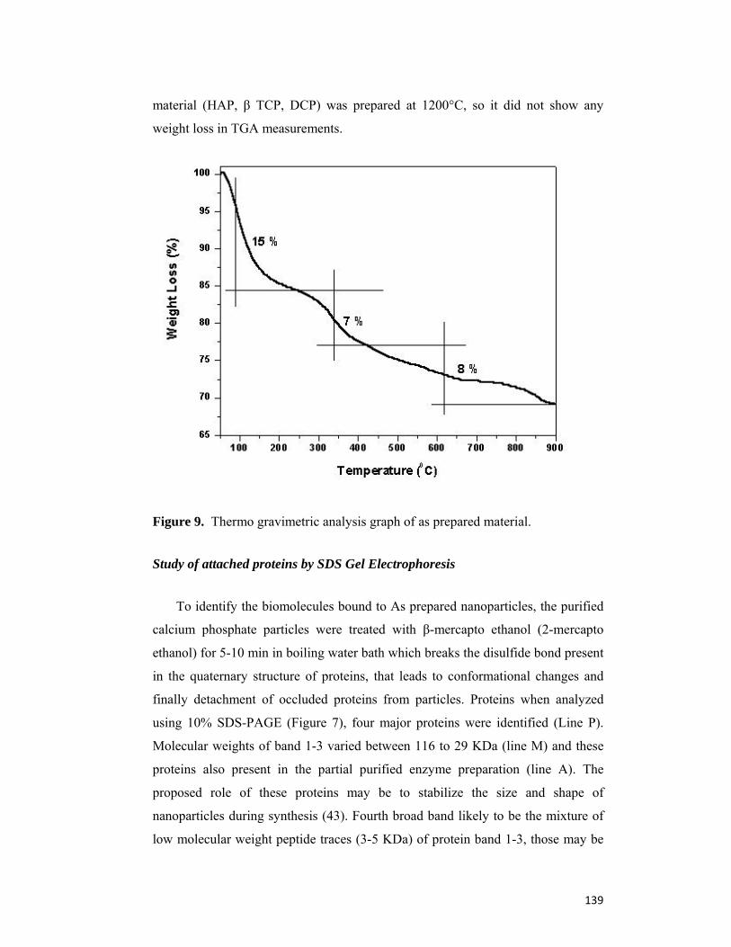
139
material (HAP, β TCP, DCP) was prepared at 1200°C, so it did not show any
weight loss in TGA measurements.
Figure 9. Thermo gravimetric analysis graph of as prepared material.
Study of attached proteins by SDS Gel Electrophoresis
To identify the biomolecules bound to As prepared nanoparticles, the purified
calcium phosphate particles were treated with β-mercapto ethanol (2-mercapto
ethanol) for 5-10 min in boiling water bath which breaks the disulfide bond present
in the quaternary structure of proteins, that leads to conformational changes and
finally detachment of occluded proteins from particles. Proteins when analyzed
using 10% SDS-PAGE (Figure 7), four major proteins were identified (Line P).
Molecular weights of band 1-3 varied between 116 to 29 KDa (line M) and these
proteins also present in the partial purified enzyme preparation (line A). The
proposed role of these proteins may be to stabilize the size and shape of
nanoparticles during synthesis (43). Fourth broad band likely to be the mixture of
low molecular weight peptide traces (3-5 KDa) of protein band 1-3, those may be

140
produced during the detachment process. These results suggest the possible
application of synthesized bionic particles for purification of certain proteins.
Figure 10. SDS-PAGE (10 %) data showing the silver stained fungal extracellular proteins bound onto the surface of synthesized uncalcined particles.
Lane A: Partially purified phytase along with other extracellular proteins after sub merged fermentation of fungus Aspergillus niger NCIM 563.
Lane M: Standard protein molecular weight markers indicated in kDa.
Lane P: Corresponds to the proteins bound to synthesized particles.

141
4.5 Conclusions
Hydroxyapatite and polymorphs have been successfully synthesized by
various chemical methods. According to current study, it is possible that nanoscale
and microscale mineral Hydroxyapatite can be synthesizes by biomimetic
enzymatic process. Pure synthesis of HAP nano particles by the direct use of
fungus biomass and understanding of the role of fungal mechanism would
definitely be an issue of great interest in future investigations. Strictly anaerobic
archaeobacterial microorganism (56) methanogans i.e. Methanobacterium,
Methanoccus etc. and sulfur metabolizers i.e. Desulfurococcus, Methanopyrus,
Sulfolobus etc. may have great potential for biosynthesis of pure hydroxyapatite
nanoparticles using their phosphohydrolase enzymes. The overall results therefore
suggest that the biomimetic synthesized nanospheres can be a good choice for bone
grafting and protein purification applications. This bionic approach could be
envisioned as an energy-conserving and economically viable green approach
towards the large-scale synthesis of crystalline artificial bone, using potential
cheap agro-industrial waste materials like wheat bran.

142
4.6 References
1. Alexandra E. P., Buckland T., Hing K., Best S. M., Bonfield W. (2004) The Structure of theBond Between Bone and Porous Silicon-Substituted Hydroxyapatite Bioceramicimplants. Journal of Biomedical Materials Research Part A, 8A:
25-33. 2. Tiselius A., S. Hjerten S., Levin O. (1956) Hydroxyapatite, mode of action and
use to optimise protein purification. Arch Biochem Biophys, 65:132-155.
3. Kawasaki, T. (1991) Hydroxyapatite as a Liquid Chromatographic Packing. J. Chromatogr. 544:147–184.
4. Martinson, H. (1973). Basis of fractionation of single-stranded nucleic acids on hydroxyapatite.Biochemistry 12: 2731–2736.
5. Martinson H., E. Wagenaar (1974). Hydroxylapatite-catalyzed degradation of ribonucleic acid. Biochemistry 13 (8), pp 1641–1645.
6. Bernardi G, Giro MG, Gaillard C. (1972). Chromatography of polypeptides and proteins on hydroxyapatite columns: some new developments. Biochim Biophys. Acta 278: 409 –420.
7. Bernardi, G. (1971). Chromatography of nucleic acids on hydroxyapatite columns. Methods Enzymol 21: 95–139.
8. Fratzl P., Gupta H. S., Paschalis E. P., Roschger P. (2004). Structure and mechanical quality of the collagen–mineral nano Composite in bone. J. Mater. Chem. 14: 2115-2123.
9. J.A. Cerri, I.M.G. Santos, E. Longo, E.R. Leite, R.M. Lebullenger, A.C. Hernandes, J.A., Varela (1998). Characteristics of PbO–BiO1.5–GaO1.5 Glasses Melted in SnO2 Crucibles. J. Am. Ceram. Soc. 81: 705- 708.
10. Ginebra M. P., Driessens F. C. M., Planell J. A. (2004). Effect of the particle size on the micro and nanostructural features of a calcium phosphate cement: A kinetic analysis.Biomaterials 25: 3453- 3462.
11. Bigi A., Boanini E., Rubini K. (2004) Hydroxyapatite gels and nanocrystals prepared through a sol–gel process. J. Solid State Chem. 177: 3092-3098.
12. Pang Y. X., Bao X. (2003) Influence of temperature, ripening time and calcination on the morphology and crystallinity of hydroxyapatite nanoparticles J EUR CERAM SOC 23: 1697-1704.
13. Philips M. J. (2005). Ph. D. Thesis, Queen Mary University of London.
14. Philips M. J., Darr J. A., Luklinska Z. B., Rehman I. (2003). Synthesis and characterization of nano-biomaterials with potential osteological applications. J. Mater. Sci.: Mater. Med. 14: 875-882.
143
15. Lim G. K., Wang J., Ng S. C., Chew C. H., Gan L. M.(1997). Processing of hydroxyapatite via microemulsion and emulsion routes. Biomaterials. 18:1433-1439.
16. Wang Y., Zhang S., Wei K, Zhao N., Chen J., Wang X. (2006) Hydrothermal synthesis of hydroxyapatite nanopowders using cationic surfactant as a template.Mater. Lett. 60: 1484-1487.
17. Riman R. E., Suchanek L., Byrappa K., Chen C., Shuk P., Oakes C. S. (2002) Solution synthesis of hydroxyapatite designer particulates. Solid State Ionics 151: 393-402.
18. Rhee, S. (2002). Synthesis of hydroxyapatite via mechanochemical treatment. Biomaterials 23: 1147-1152.
19. Darr J. A., Guo Z. X., Raman V., Bououdina M., Rehman I. U. (2004). Metal organic chemical vapour deposition (MOCVD) of bone mineral like carbonated hydroxyapatite coatings. Chem. Comm. 6: 696-697.
20. Kim S., Ryu H., Shin H., Jung H. S., Hong K. S. (2005). In situ observation of hydroxyapatite nanocrystal formation from amorphous calcium phosphate in calcium-rich solutions. Mater. Chem. Phys. 91: 500-506.
21. Arce H., Montero M. L., Saenz A., Castano V. M. (2004). Effect of pH and temperature on the formation of hydroxyapatite at low temperatures by decomposition of a Ca–EDTA complex. Polyhedron, 23:1897-1901.
22. Cao L. Y., Zhang C. B., Haung J. F. (2005) Influence of temperature, [Ca2+], Ca/P ratio and ultrasonic power on the crystallinity and morphology of hydroxyapatite nanoparticles prepared with a novel ultrasonic precipitation method. Mater. Lett. 59:1902-1906.
23. Wodzinski R., Ullah A.H.J. (1996) Phytase. Adv Appl Microbiol. 42:263-302.
24. Mullaney, E. J., Daly, C., Ullah, A. B. (2000) Advances in phytase research. Adv. Appl. Microbiol. 47:157-199.
25. Soni, S. K., Khire, J. M. (2007) Production and partial characterization of two type of phytase from Aspergillus niger NCIM 563 under submerged fermentation conditions.World J. of Microbiol. Biotechnol. 23:1585-1593.
26. Mandviwala, T. N., Khire, J. M. (2000) Production of high activity thermostable Phytase from thermotolerant Aspergillus niger in solid state fermentation. Journal of Industrial Microbiol. and Biotechnol. 24: 237-243.

144
27. Harland, B. F., Oberleas, D. (1977) A modified method for phytate analysis using an ion- exchange procedure: Application to textured vegetable proteins. Cereal Chem. 54: 827- 832.
28. Latta, M., Eskin, M. J. (1980) A simple and rapid colorimetric method for phytate determination. Agric. Food Chem. 28:1313-1315.
29. Heinohen, J.K., Lathi, R.J. (1981) A new and convenient colorimetric determination of inorganic orthophosphate and its application to the assay to inorganic pyrophosphatase. Anal Biochem. 113: 313–317.
30. Murugan R., Sampath Kumar T. S., Yang F., Ramakrishna S. (2005) Hydroxyl Carbonateapatite Hybrid Bone Composites Using Carbohydrate Polymer. Journal of Composite Materials 39: 1159-1167.
31. ISO 10993-5:1992(E). Biological evaluation of medical devices—Part 5: Tests for cytotoxicity: in vitro methods.
32. Nablo, B.J., Schoenfisch, M. H. (2005) In vitro cytotoxicity of nitric oxide-releasing sol –gel derived materials. Biomaterials 26: 4405-4415.
33. Wilson, A. P., Cytotoxicity and Viability Assays in Animal Cell Culture: A Practical Approach, 3rd ed. (ed. Masters, J. R. W.) 1, 175-219 (Oxford University Press: Oxford 2000)
34. Laemmli, U. K. (1970) Cleavage of structural proteins during the assembly of the head of bacteriophage T4. Nature 227: 680-685.
35. Groot D. K. (1980) Bioceremics consisting of calcium phosphate salts. Biomat. 1: 47-50.
36. Jarcho, M., C. H. Bolen, M. B. Thomas, J. Bobick, J. F. Kay and R. H. Doremus (1976) Hydroxylapatite synthesis and characterization in dense polycrystalline form. J. Mat. Sci. 11: 2027-2035.
37. Wang, F. (2006) Synthesis and microstructure of hydroxyapatite nanofibers synthesized at 370C. Materials chemistry and Physics 95:145-149.
38. Kay M. I., Young R. A., Posner A. S. (1964) Crystal structure of hydroxy apatite, Nature 204:1050-1052.
39. Sasaki, N., Sudoh, Y.(1997) X- ray pole figure analysis of apatite crystals and collagen molecules in bone. Calcil Tissue Int. 60: 361-367.
40. Chu T. M. G., Halloran J. W., Hollister S. J., Feinberg S. E. (2001) Hydroxyapatite implants with designed internal architecture. J. Mater. Sci.: Mater. Med. 12: 471-478.
41. Miyaji F., Kono Y., Suyama Y. (2005) Formation and structure of zinc-substituted calcium hydroxyapatite Mater. Res. Bull. 40: 209-220.

145
42. Rautaray D., Sanyal A., Adyanthaya S. D., Ahmad A., Sastry M (2004) Biological Synthesis of Strontium Carbonate Crystals Using the Fungus Fusarium oxysporum. Langmuir 20, 6827–6833.
43. Bansal V., Ahmad A., Sastry M. (2006) Fungus-Mediated Biotransformation of Amorphous Silica in Rice Husk to Nanocrystalline Silica. J. Am. Chem. Soc. 128:14059 –14066.
44. Aue W. P., Roufosse A. H., Glimcher M. J., Griffin R. G. (1984) Solid-state Phosphorus-3 1 Nuclear Magnetic Resonance Studies of Synthetic Solid Phases of Calcium Phosphate: Potential Models of Bone Mineral. Biochemistry 23: 6110-6114.
45. Wilson, R. M. (2005) Rietveld refinements and spectroscopic studies of the structure of Ca- deficient apatite. Biomaterials 26: 1317-1327.
46. Komath, M., Varma, H.K. (2003) Development of fully injectable calcium phosphate cement for orthopedic and dental applications. Bull. Mater. Sci. 26: 415–422.
47. Kumari T. V., Vasudev U., Kumar A., Menon B. (2002) Cell surface interactions in the study of biocompatibility. Trends biomater. Artif. Organs. 15: 37-41.
48. Quan R., Yang D., Wu X, Wang H., Miao X., Li W (2008) In vitro and in vivo biocompatibility of graded hydroxyapatite– zirconia composite bioceramic. J Mater Sci: Mater. 19: 183-187.
49 Laing P. G., Ferguson A. B., Hodge E. S. (1967) Tissue reaction in rabbit muscle exposed to metallic implants. J Biomed Mater Res. 1: 135–149.
50. Tanaka K., Tamura J., Kawanabe K., Nawa M., Oka M., Uchida M. (2002) Ce-TZP/Al2O3 nanocomposite as a bearing material in total joint replacement. J Biomed Mater Res. (Appl Biomater.) 63: 262–270.
51. Vande Vord P. J., Matthew H. W. T., De Silva S. P., Mayton L., Wu B., Wooley P. H. (2002) Evaluation of the biocompatibility of a chitosan scaffold in mice. J Biomed Mater Res. 59: 585–590.
52. Murugan R., K.P. K. P., Sampath Kumar T. S. (2003) Heat-deproteinated xenogeneic bone from slaughterhouse waste: physico-chemical properties. Bull. Mater. Sci. 26: 523- 528.
53. Dong, Y., Feng, S.S. (2005) Poly(D,L- Lactide- co-glycolide) /montmorillonite Nanoparticles for oral delivery of anty cancer drugs. Biomaterials 26: 6068-6076.
54. Shieh, T.R., Ware, J. H. (1968) Survey of Microorganisms for the Production of extracellular phytase. Applied Microbiology 16: 1348-1351.

146
55. Rautaray D., Ahmad A., Sastry M. (2004) Biological synthesis of metal carbonate minerals using fungi and actinomycetes. J.Mater. Chem. 14: 2333-2340.
56. L.M. Prescott L. M., Harley J. P., Clein D. A., Microbiology, 4th ed., Ch. 20, WCB/Mcgraw- Hill, USA, 1999, 427-428.

147
CHAPTER 5
Biocompatibility studies of enzymatically synthesized Hydroxyapatite and its polymorphs on Human Osteoblast like MG-63 cell line
5.1 Summary
This chapter describes the studies of artificial bone material Hydroxyapatite
(HAP) synthesized by phytase enzyme and commercially available HAP (Sigma) on
osteosarcoma MG-63 cell line for its biocompatibility. Human osteosarcoma cells
(MG-63 cells) readily seed over 90% of the available biomaterials under static culture
conditions, and the attached cells proliferate to extensively cover the biomaterials as
seen by DAPI staining and scanning electron microscopy. Cell growth on biomaterials
was also monitored by total protein assay. The attached cells are over 90% viable after
7 days in culture as assessed by MTT staining. MG-63 cells also show significant
increase in alkaline phosphatase activity 7 days post-addition of oestrogenic
supplement. This biomaterial (enzymatically synthesized HAP) show promise for use
as vehicles for cell delivery to place large numbers of cells directly into a wound site
or onto a tissue engineering scaffold and can potentially be used as artificial bone
material.
5.2 Introduction
New materials have been developed for orthopaedic implants. Among these
materials, HAP which is known for its high biological activity and its unique property
to integrates with, has attracted attention .The most common tissue for transplant
procedures is bone tissue. Out of the 500,000 bone graft procedures per year, half
relate to spinal surgeries (1). Synthetic materials are man-made materials such as
metals, plastic or ceramics that mimic natural bone in its strength, flexibility and/or
structure (2). Most are calcium composites designed to be resorbed within the body
over time. Autografts, allografts and ceramics and the rationale, benefits and
drawbacks of choosing them are discussed next.Bone tissue in itself is composed of
four different cell types: osteoblasts, osteoclasts, bone lining cells on the surfaces of
148
bone, and osteocytes within the mineralized component (3). Osteoblasts lay down new
bone while osteoclasts digest away old bone. Osteocytes are mature osteoblasts
contained in a calcified matrix are responsible for intercellular communication. The
bone lining cells are considered to be fairly inactive carrying out neither absorption
nor formation. Little is known about their function but it is speculated that they could
also be precursors for osteoblasts (4). Osteoblasts are cells that lie at the surface of the
bone matrix that is already formed and they deposit new matrix or osteoid. They are
fully differentiated cells. They lay down bone matrix, by secreting collagen Type I and
other non-collagenous proteins (4, 5). The osteoblasts themselves often embed
themselves in the new matrix that they form and are then termed osteocytes. The
matrix hardens as calcium phosphate (hydroxyapatite) crystals are deposited in it. The
osteocyte is considered to be the most terminally differentiated cell of the osteoblast
lineage. Osteocytes are embedded in the bone matrix and communicate with other
cells through gap junctions present in the canaliculi (6). Before becoming trapped in
the matrix, the osteocytes test positive for the presence of alkaline phosphatase
activity, as functional mature osteoblasts (6). These osteoblasts can also be identified
by the other products they produce such as Type I collagen, osteopontin, osteonectin,
osteocalcin and bone sialoprotein (6). Osteoblasts that are active show alkaline
phosphatase (ALP) activity that can be detected with a histochemical stain (7- 9) and
by a biochemical assay that can quantify enzyme activity (10, 11).
This chapter describes the high bioactivity and biocompatibility of artificial bone
material Hydroxyapatite (HAP) synthesized by phytase enzyme and commercially
available HAP (Sigma) on MG-63 cells, an osteosarcoma cell line that is commonly
used in osteoblast and osteoblast/biomaterial research (12-17). A number of osteoblast
cell lines and osteoblast precursor cell lines are used in the study of bone development,
physio logy and bone-tissue engineering. The MG-63 cell line is derived from a
human osteosarcoma (isolated from a 14 year old boy) (18). When induced to
differentiate with 1, 25-dihydroxyvitamin D3 (1,25D3), they express increased
alkaline phosphatase activity and secrete osteocalcin (19). Another point to note is that
the MG-63 cells differ from osteoblasts in that they do not mineralize their
149
extracellular matrix upon addition of osteogenicinducing media but express all other
markers of differentiation. Also, in a study that evaluated these cells and another
osteosarcoma cell line, both cell lines showed adhesion or differentiation
characteristics similar to those of human osteoblast- like cells (20). The MG-63 cells
exhibit features of an undifferentiated Osteoblast and express increased alkaline
phosphatase activity when they are induced to differentiate (21). The MG-63 cell line
cannot be used for in vivo applications in humans, but are a useful in vitro model to
assess attachment, proliferation and differentiation similar to that of osteoblasts.
HAP which is known for its high biological activity and its unique property
to integrate with bones has attracted attention. Human Osteosarcoma cells (MG-63
cells) readily seed over 90% of the available biomaterials under static culture
conditions, and the attached cells proliferate to extensively cover the biomaterials as
seen by DAPI staining and scanning electron microscopy. Cell growth on biomaterials
was also monitored by total protein assay. The attached cells are over 90% viable after
7 days in culture as assessed by MTT staining. MG-63 cells also show significant
increase in alkaline phosphatase activity 7 days post-addition of oestrogenic
supplement. This biomaterial (enzymatically synthesized HAP) show promise for use
as vehicles for cell delivery to place large numbers of cells directly into a wound site
or onto a tissue engineering scaffold and can potentially be used as artificial bone
material.
5.3 Materials and Methods
MG-63 osteosarcoma cell line
The osteosarcoma MG-63 cell line was obtained from National Centre for Cell
Science (NCCS), Pune, India. Cells obtained were at passage numbers 90-93. Cells
were cultured at 37°C with 5% CO2 and 90% humidity in T-75 tissue culture flasks
(Corning 430641). Cells were maintained in the culture medium used include 1:1
mixture of Ham’s F12 medium and Dulbecco’s modified Eagle’s medium (DMEM)
with out phenol red with 2.5 mM L-glutamine (Invitrogen, US), 0.3 mg/ml G418

150
(Sigma, US) and supplemented with 10% fetal bovine serum (FBS; Gibco 10082-
139)
In vitro culture of osteoblasts
As synthesized material was converted in pallets by pressure and pallets were
autoclaved and imbedded in 70 % ethanol twice for a period of 20 minutes at each
time for sterilization purposes. The pallets were then washed with phosphate buffered
saline (PBS) thrice to remove any residual ethanol. Human osteosarcoma cells (MG-
63) cultured until passage 5, were seeded on the HA surface at a cell concentration of
104 cells/sample. The constituents of the culture medium used include 1:1 mixture of
Ham’s F12 medium and Dulbecco’s modified Eagle’s medium (DMEM) with out
phenol red with 2.5 mM L-glutamine (Invitrogen, US), 0.3 mg/ml G418 (Sigma, US)
and 10% fetal bovine serum (Invirogen, US). The medium was changed every two
days.
Cell attachment study
One mL of the solution containing 10,000 cells osteosarcoma was added to
each of the wells containing the samples. The cells were incubated with the test
sample in co2 incubator supplied with 5% co2, 95% humidity at 37ºC for 7days. At
the end of the incubation period, the solution containing the unattached cells was
discarded and each well containing the test samples was washed thrice with 1 ml of
PBS. For the purpose of cell counting, the nuclei of the attached cells were stained
with DAPI (4’, 6-diamidino-2-phenylindole, dilactate) (InvitrogenTM) using the
following protocol. Pre-chilled methanol was added to the wells containing the
attached cells and allowed for fixation at room temperature for 10 minutes. Next, the
nuclei of the attached cells were stained with 0.25 mL/well of 1µg/mL DAPI for 30
minutes at room temperature. Each test sample was photographed using a fluorescence
microscope Leica DM IRB) at 100 times magnification. For each test sample, the
number of cells in 5 predetermined rectangular areas (top, centre, bottom, left and
right portion of the culture well) was counted based on the nuclei count. This cell

151
count method was also used to calculate the cell number at the day 0, day 3, day 5 and
day 7 for cell proliferation quantification.
Cell proliferation quantification
Cell count method was calculate the cell number at the day 0, day 3, day 5 and
day 7 for cell proliferation quantification by the MTT cytotoxicity assay. One mL of
the solution containing 10,000 cells osteosarcoma was added to each of the wells
containing the samples. The cells were incubated with the test sample in CO2
incubator supplied with 5% CO2, 95% humidity at 37ºC for 7days. At the end of the
incubation period, the solution containing the unattached cells was discarded and each
well containing the test samples was washed thrice with 1 ml of PBS. For the purpose
of cell counting, via the MTT cytotoxicity assay is the conversion of the tetrazolium
salt (MTT) to the colored product formazan. The concentration can be measured photo
metrically at 490 nm. The formation of formazan takes place via intact mitochondria.
MTT assay was performed by using the following protocol. Pre-chilled methanol was
added to the wells containing the attached cells and allowed for fixation at room
temperature for 10 minutes. Next, add MTT reagent (5mg/ml), incubate at 37ºC for
1hr.add 200µl of 100% isopropyl alcohol and keep it at RT for minimum 4hrs.After
incubation ensure that formazan precipitate is dissolved by pipetting up and down
until any precipitate is visible. Take the observance at 490nm.
Total protein assay
Osteoblasts (10,000 cells/sample) were seeded on blank controls, commercial
HA and as prepared HA samples, respectively, in three 24 well plates and cultured in
osteosarcoma cell culture medium under standard cell culture conditions for 1, and 7
days respectively. At the end of the prescribed times, the culture medium was removed
from culture well, and washed by PBS for 3 times, Samples were stained by
bicinchoninic acid (BCA) protein assay working reagent (BCA Kit No. 23225, Pierce)
at 37ºC for 30 minutes. Once room tempetature was reached, the protein content was
152
determined at 590 nm using a FLUO star optima microplate reader (BMG Labtech
Gmbh). The total intracellular protein (expressed in mg) synthesized by the osteoblast
cells cultured in the medium was determined from standard curve of absorbance
versus known concentrations of albumin by the BCA kit.
Alkaline phosphatase activity assay
The prescribed cells were cultured on the materials, and alkaline phosphatase
(ALP) activity was measured at days 0, 3, 5 and 7. Four hundred mL of p-nitrophenyl
phosphate, disodium salt (PNPP) solution (Phosphatase Substrate Kit, No. 37620,
Pierce Biotechnology) was added to the 24-well culture plate and incubated at room
temperature for 1 h. The reaction of p-nitrophenol conversion to p-nitrophenylate was
stopped by the addition of 200 mL of 2M of sodium hydroxide. Light absorbance of
these samples was measured at 405nm on a FLUO Star Optima microplate reader.
SEM of cell morphology
Scaffolds with cells that were cultured for 0, 3, 5 and 7 days were fixed in 4%
paraformaldehyde solution for 30 min at room temperature. Scaffolds were dehydrated
in 50%, 70%, 95%, and 100% (twice) ethanol (5 cycles with increasing concentrations
of ethanol) for 10 min. Samples were subsequently air-dried overnight and observed
using SEM.
5.4 Results and discussion
Approach in evaluating the attachment, proliferation, and differentiation of
MG-63 cells on HAP in this work is outlined by the following diagram (Fig1).

153
Figure 1. Flow Chart of Experimental Design
The attachment, viability, proliferation and the ability to differentiate into osteoblasts
were evaluated.
HAP is not cytotoxic MG-63 cells
MG-63 cells, showed high viabilities when cultured in un-conditioned
media, conditioned media (CM) and subsequent dilutions of CM (Table1). Five mL
media was conditioned by soaking in one gram quantities of artificially synthesized
HAP and commercially available HAP (Sigma) for five days. Cells were plated on
glass cover slips and cultured overnight in the appropriate media and LIVE/DEAD
staining (data not shown) was carried out to assess viability. Ten fields were measured
on each cover slip and live and dead cells quantified. Approximate number of morsels
in each field was 5. Percentage viabilities for MG-63 cells were greater than 90% in
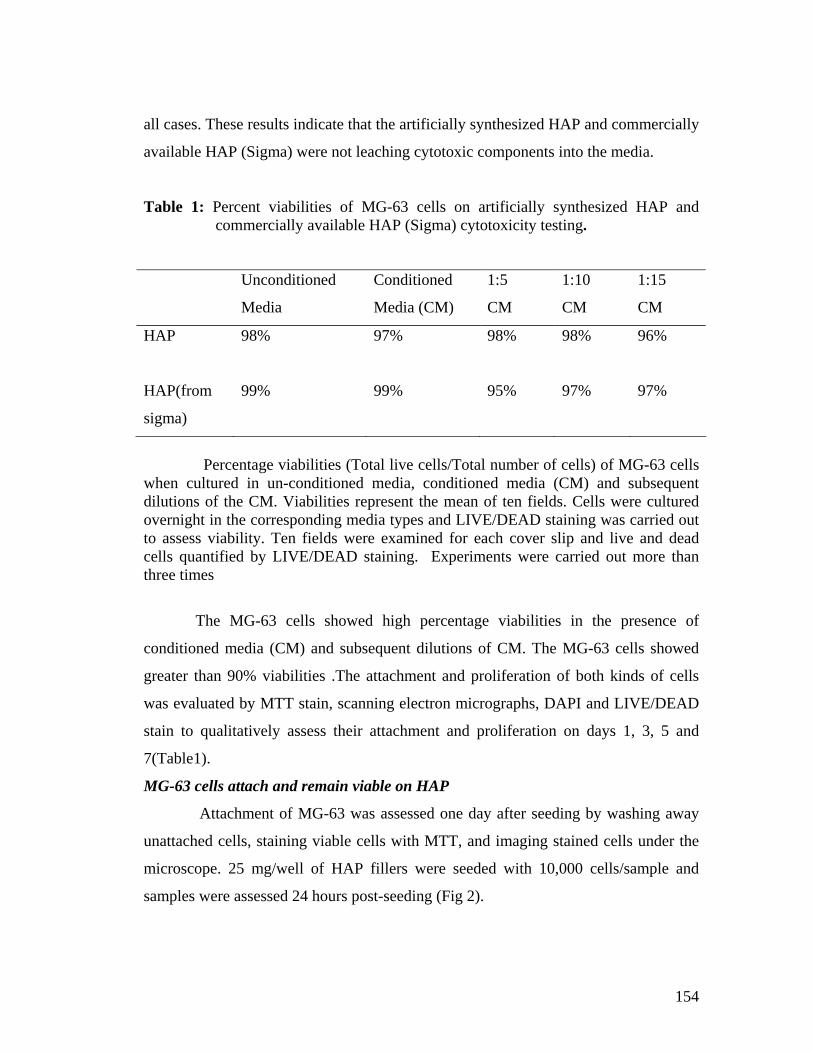
154
all cases. These results indicate that the artificially synthesized HAP and commercially
available HAP (Sigma) were not leaching cytotoxic components into the media.
Table 1: Percent viabilities of MG-63 cells on artificially synthesized HAP and commercially available HAP (Sigma) cytotoxicity testing.
Percentage viabilities (Total live cells/Total number of cells) of MG-63 cells when cultured in un-conditioned media, conditioned media (CM) and subsequent dilutions of the CM. Viabilities represent the mean of ten fields. Cells were cultured overnight in the corresponding media types and LIVE/DEAD staining was carried out to assess viability. Ten fields were examined for each cover slip and live and dead cells quantified by LIVE/DEAD staining. Experiments were carried out more than three times
The MG-63 cells showed high percentage viabilities in the presence of
conditioned media (CM) and subsequent dilutions of CM. The MG-63 cells showed
greater than 90% viabilities .The attachment and proliferation of both kinds of cells
was evaluated by MTT stain, scanning electron micrographs, DAPI and LIVE/DEAD
stain to qualitatively assess their attachment and proliferation on days 1, 3, 5 and
7(Table1).
MG-63 cells attach and remain viable on HAP
Attachment of MG-63 was assessed one day after seeding by washing away
unattached cells, staining viable cells with MTT, and imaging stained cells under the
microscope. 25 mg/well of HAP fillers were seeded with 10,000 cells/sample and
samples were assessed 24 hours post-seeding (Fig 2).
Unconditioned
Media
Conditioned
Media (CM)
1:5
CM
1:10
CM
1:15
CM
HAP
98% 97% 98% 98% 96%
HAP(from
sigma)
99% 99% 95% 97% 97%
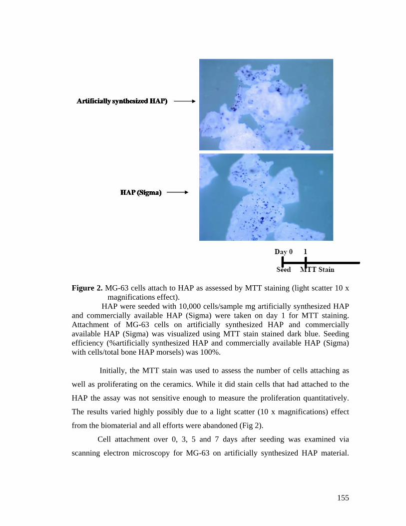
155
Figure 2. MG-63 cells attach to HAP as assessed by MTT staining (light scatter 10 x magnifications effect).
HAP were seeded with 10,000 cells/sample mg artificially synthesized HAP and commercially available HAP (Sigma) were taken on day 1 for MTT staining. Attachment of MG-63 cells on artificially synthesized HAP and commercially available HAP (Sigma) was visualized using MTT stain stained dark blue. Seeding efficiency (%artificially synthesized HAP and commercially available HAP (Sigma) with cells/total bone HAP morsels) was 100%. Initially, the MTT stain was used to assess the number of cells attaching as
well as proliferating on the ceramics. While it did stain cells that had attached to the
HAP the assay was not sensitive enough to measure the proliferation quantitatively.
The results varied highly possibly due to a light scatter (10 x magnifications) effect
from the biomaterial and all efforts were abandoned (Fig 2).
Cell attachment over 0, 3, 5 and 7 days after seeding was examined via
scanning electron microscopy for MG-63 on artificially synthesized HAP material.

156
Scanning electron microscopy (Fig3) revealed that MG-63 cells attach to the material
surfaces of artificially synthesized HAP In all protocols unattached cells were washed
away after the second day. The MG-63 cells populated all topographies on the
surfaces including concavities and pores (Fig 3).
Figure 3. MG-63 cells attach to HAP. Scanning electron microscopy shows MG-63 cells on the surfaces of HAP on day 0, 3, 5 and 7. 10,000 cells/sample HAP was seeded. Scanning electron micrographs (SEM) showed that MG-63 cells can attach
onto the surfaces as well as pores of HAP. SEM studies examining of attachment and
proliferation of osteoblasts and fibroblasts on a copolymer poly (lactideco- glycolide
(PLAGA) scaffold showed that cells grew in concentric patterns and had an ability to
fill (100-250 m) pores (22) (Fig 3).

157
Attachment and viability were also assessed by DAPI (Fig 4) and
LIVE/DEAD (Fig 5) staining showing that MG-63 cells attach and maintain high
viabilities up to seven days after seeding. The cells densely populated the surfaces as
early as Day 1 making individual cell counts impossible. High viability (few dead
cells) indicated high survival rates of the cells on HAP. The seeding efficiency of HAP
populated with MG-63 cells was always 100%.
Figure 4. MG-63 cells attach and remain viable on HAP seen by DAPI stain. HAP was seeded in each well and showed attachment and high viabilities
through 7 days post-seeding. Unattached cells were washed away on day 1 after
seeding .DAPI stained cellular nuclei blue, excitation358 nm and emission 461 nm.
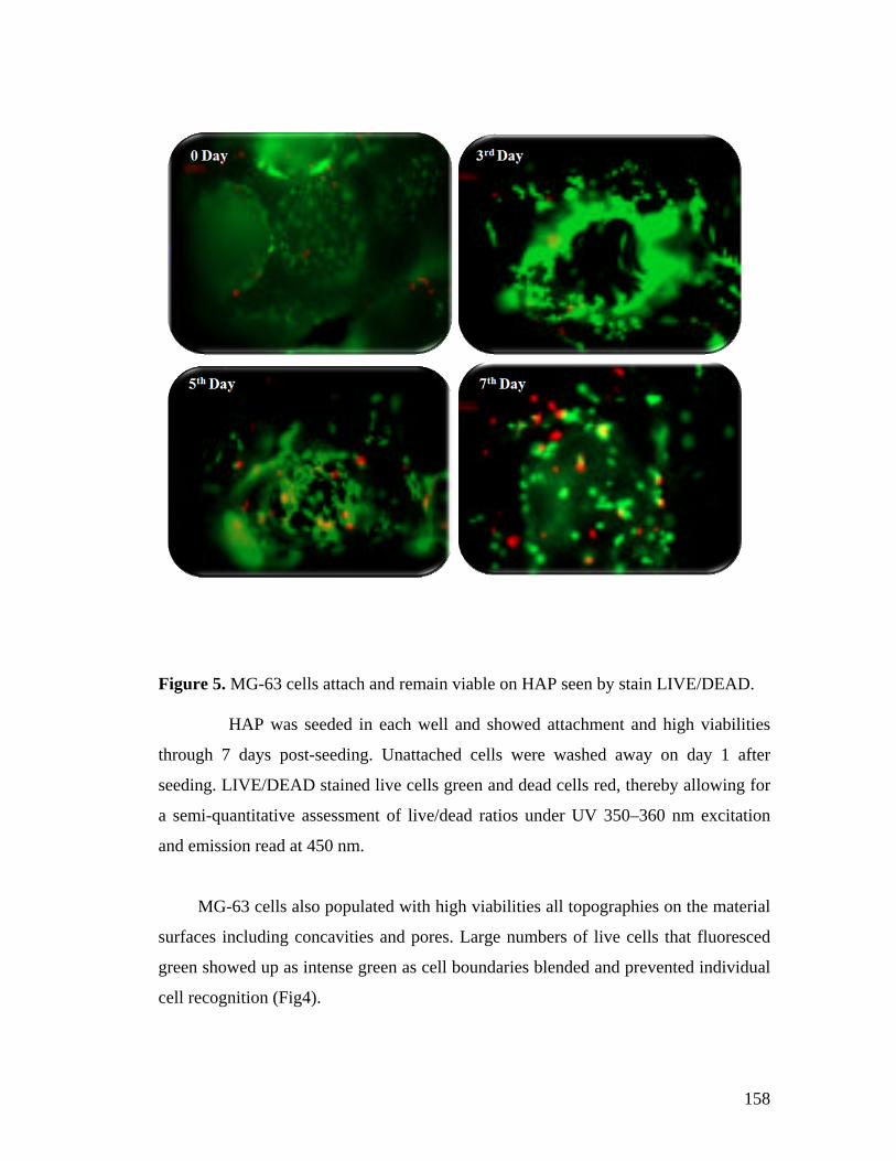
158
Figure 5. MG-63 cells attach and remain viable on HAP seen by stain LIVE/DEAD. HAP was seeded in each well and showed attachment and high viabilities
through 7 days post-seeding. Unattached cells were washed away on day 1 after
seeding. LIVE/DEAD stained live cells green and dead cells red, thereby allowing for
a semi-quantitative assessment of live/dead ratios under UV 350–360 nm excitation
and emission read at 450 nm.
MG-63 cells also populated with high viabilities all topographies on the material
surfaces including concavities and pores. Large numbers of live cells that fluoresced
green showed up as intense green as cell boundaries blended and prevented individual
cell recognition (Fig4).
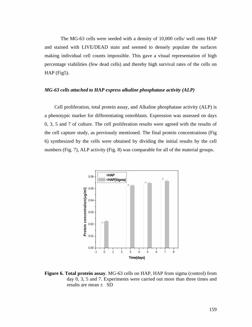
159
The MG-63 cells were seeded with a density of 10,000 cells/ well onto HAP
and stained with LIVE/DEAD stain and seemed to densely populate the surfaces
making individual cell counts impossible. This gave a visual representation of high
percentage viabilities (few dead cells) and thereby high survival rates of the cells on
HAP (Fig5).
MG-63 cells attached to HAP express alkaline phosphatase activity (ALP)
Cell proliferation, total protein assay, and Alkaline phosphatase activity (ALP) is
a phenotypic marker for differentiating osteoblasts. Expression was assessed on days
0, 3, 5 and 7 of culture. The cell proliferation results were agreed with the results of
the cell capture study, as previously mentioned. The final protein concentrations (Fig
6) synthesized by the cells were obtained by dividing the initial results by the cell
numbers (Fig. 7), ALP activity (Fig. 8) was comparable for all of the material groups.
-1 0 1 2 3 4 5 6 7 80.00
0.01
0.02
0.03
0.04
0.05
0.06
Pro
tein
co
nce
ntr
atio
n(u
g/m
l)
Time(days)
=HAP =HAP(Sigma)
Figure 6. Total protein assay. MG-63 cells on HAP, HAP from sigma (control) from day 0, 3, 5 and 7. Experiments were carried out more than three times and results are mean ± SD
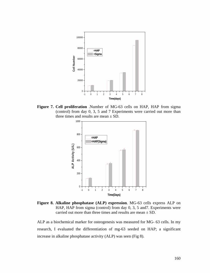
160
-1 0 1 2 3 4 5 6 7 80
20000
40000
60000
80000
100000
Cel
l Nu
mb
er
Time(days)
=HAP =Sigma
Figure 7. Cell proliferation .Number of MG-63 cells on HAP, HAP from sigma (control) from day 0, 3, 5 and 7 Experiments were carried out more than three times and results are mean ± SD.
-1 0 1 2 3 4 5 6 7 80
200
400
600
800
1000
AL
P A
ctiv
ity
(U/L
)
Time(Days)
=HAP=HAP(Sigma)
Figure 8. Alkaline phosphatase (ALP) expression. MG-63 cells express ALP on HAP, HAP from sigma (control) from day 0, 3, 5 and7. Experiments were carried out more than three times and results are mean ± SD.
ALP as a biochemical marker for osteogenesis was measured for MG- 63 cells. In my
research, I evaluated the differentiation of mg-63 seeded on HAP; a significant
increase in alkaline phosphatase activity (ALP) was seen (Fig 8).
161
5.5 Conclusions
The MG-63 cell line was used as an initial assessment to evaluate whether
cells would survive, attach and proliferate on the HAP. Owing to the fact that the cell
line is an osteosarcoma cell line, it cannot be implanted in vivo and was thus evaluated
as a preliminary, qualitative assessment of cells cultured in the HAP in vitro
environment. Most of the applications for using MG-63 cells in studies assess the cells
as a preliminary investigation to see whether they attach to certain biomaterials,
remain viable on the biomaterials or express morphological or biochemical
characteristics similar to those of osteoblasts (21, 24, 17, and 23). We observed that
1) Artificially synthesized HAP does not exert a cytotoxic effect on MG-63 cells.
2) MG-63 cells attach and proliferate on artificially synthesized HAP.
3) MG-63 cells express alkaline phosphatase activity on artificially synthesized HAP.
More studies are necessary in order to assess HAP as a vehicle for delivery
into bone defects. Our results evaluating the attachment, proliferation and
differentiation of MG-63 on HAP. Granulat show great promise for their use. The
analysis of further osteoblastic phenotypical markers such as calcium deposition is
impossible due to the beta tri-calcium phosphate nature of the HAP, but measurement
of osteocalcin, osteonectin or osteopontin mRNA would be useful.
Another interesting study would be to conduct studies of convex surfaces of
pores when compared to concave surfaces and to investigate which surfaces are
preferred by the MG-63. Observations made during LIVE/DEAD staining showed
more cells colonizing the insides of concave pores. More studies need to be done in
order to show whether the cells have a preference and such characteristics can be used
to design better biomaterials and better hybrid therapies for bone healing. Thus, in-
depth studies of surface undulations and its effect on cell attachment, proliferation and
differentiation are needed. Further SEM studies could be carried out to evaluate how
far into the centre of the bone void filler, the cells attach.
162
5.6 References
1.Linovitz R. J., Peppers T. A. (2002) Use of an advanced formulation of betatricalcium phosphate as a bone extender in interbody lumbar fusion. Orthopedics 25: s585-589.
2. Lewandrowski K. U., Gresser J. D., Wise D. L., Trantol D. J. (2000) Bioresorbable bone graft substitutes of different osteoconductivities: a histologic evaluation of
osteointegration of poly(propylene glycol-co-fumaric acid)-based cement implants in rats. Biomaterials 21: 757-764.
3. Rodan, G. A. (1992) Introduction to bone biology. Bone 13 Suppl 1: S 3-6.
4. Bilezikian J. P., Raisz L. G., Rodan G. A. (1996) Principles of bone biology, San Diego, Academic Press. 5. Mackie E. J. (2003) Osteoblasts: novel roles in orchestration of skeletal architecture. IntJ Biochem Cell Biol. 35: 1301-1305.
6. Knothe Tate, M. L., Adamson, J. R., Tami, A. E. & Bauer, T. W. (2004) The osteocyte. Int J Biochem Cell Biol. 36, 1-8.
7. Abdallah B. M., Jensen C. H., Gutierrez G., Leslie R. G., Jensen T. G., Kassem M.(2004) Regulation of Human Skeletal Stem Cells Differentiation by Dlk1/Pref-1. J Bone Miner Res. 19: 841-852.
8. Justesen J., Stenderup K., Eriksen E. F., Kassem M. (2002) Maintenance of osteoblastic and adipocytic differentiation potential with age and osteoporosis in human marrow stromal cell cultures. Calcif Tissue Int. 71: 36-44.
9. Miao D., Scutt A. (2002) Histochemical localization of alkaline phosphatase activity in decalcified bone and cartilage. J Histochem Cytochem. 50:333-340.
10. Wang Y. X., Li S. L., Wu D. C., Zhang K. H., Yu S. F.,Cui S. X. (2003) [Bone
formation in vitro and in vivo by human bone marrow-derived mesenchymal stem cells]. Zhonghua Kou Qiang Yi Xue Za Zhi. 38: 467-469. 11 Jaiswal N., Haynesworth S. E., Caplan A. I., Bruder S. P. (1997) Osteogenic
differentiation of purified, culture-expanded human mesenchymal stem cells in vitro. J Cell Biochem. 64: 295-312.

163
12. Martin J. Y., Schwartz Z., Hummert T. W., Schraub D. M., Simpson J., Lankford J.,Jr., Dean D. D., Cochran D. L., Boyan, B. D. (1995) Effect of titanium surface roughness on proliferation, differentiation, and protein synthesis of human osteoblast- like cells (MG63). J Biomed Mater Res. 29:389-401.
13. Martin J. Y., Dean D. D., Cochran D. L., Simpson J., Boyan B. D., Schwartz,
Z.(1996) Proliferation, differentiation, and protein synthesis of human osteoblastlike cells (MG63) cultured on previously used titanium surfaces. Clin Oral Implants Res. 7, 27-37.
14. Lincks J., Boyan B. D., Blanchard C. R., Lohmann C. H., Liu Y., Cochran D. L.,
Dean D. D., Schwartz, Z. (1998) Response of MG63 osteoblast- like cells to titanium and titanium alloy is dependent on surface roughness and composition. Biomaterials 19: 2219-2232.
15. Boyan B. D., Lohmann C. H., Sisk M., Liu Y., Sylvia V. L., Cochran D. L., Dean D.D., Schwartz Z. (2001) Both cyclooxygenase-1 and cyclooxygenase-2 mediate osteoblast response to titanium surface roughness. J Biomed Mater Res. 55, 350-359.
16. Scheven B. A., Marshall D., Aspden R. M. (2002) In vitro behaviour of human
osteoblasts on dentin and bone. Cell Biol Int. 26: 337-346. 17. Amaral M., Costa M. A., Lopes M. A., Silva R. F., Santos J. D., Fernandes, M.
H.(2002) Si(3)N(4)-bioglass composites stimulate the proliferation of MG63 osteoblast- like cells and support the osteogenic differentiation of human bone marrow cells. Biomaterials 23: 4897-4906.
18. Heremans H., Billiau A., Cassiman J. J., Mulier J. C., de Somer P. (1978) In vitro
cultivation of human tumor tissues. II. Morphological and virological characterization of three cell lines. Oncology 35, 246-252.
19. Franceschi R. T., Young J. (1990) Regulation of alkaline phosphatase by 1,25-
dihydroxyvitamin D3 and ascorbic acid in bone-derived cells. J Bone Miner Res. 5, 1157-1167.
20. Clover J., Gowen M. (1994) Are MG-63 and HOS TE85 human osteosarcoma cell
lines representative models of the osteoblastic phenotype? Bone 15: 585-591. 21. Massaro C., Baker M. A., Cosentino F., Ramires P. A., Klose S., Milella E. (2001)
Surface and biological evaluation of hydroxyapatite-based coatings on titanium deposited by different techniques. J Biomed Mater Res. 58: 651-657.
164
22. Borden M., Attawia M., Laurencin C. T. (2002) The sintered microsphere matrix for bone tissue engineering: in vitro osteoconductivity studies. J Biomed Mater Res. 61: 421-429.
23. Kim H. W., Noh Y. J., Koh Y. H., Kim H. E., Kim H. M. (2002) Effect of CaF2
on densification and properties of hydroxyapatite- zirconia composites for biomedical applications. Biomaterials 23: 4113-4121.
24. Lee S. J., Choi J. S., Park K. S., Khang G., Lee Y. M., Lee H. B. (2004) Response
of MG63 osteoblast- like cells onto polycarbonate membrane surfaces with different micropore sizes. Biomaterials 25: 4699-4707.

165
CHAPTER 6
Application of phytase in cell culture studies and myo-inositol production.
6.1 Summary
This chapter describes the application of phytase in cell culture studies and
myo-inositol production. Effect of phytase on cell line and its stability in different
cell culture media was checked. Partially purified phytase was stable up to 89 to 92
% till 72 hour in different cancer cell line culture media i.e. DMEM, MEM and
RPMI 1640, showed 19.23 % proliferation of cancerous HL-60 cell line with 0.8
IU/3ml of phytase in MEM media. Inorganic phosphorus (98.45μg/ml) also
released in culture media from cells after 24 hour of incubation at 370C.
Myo-inositol is well-known to play a major role in many cell signalling
pathways of Ca+2 uptake. HPLC analysis of complete phytate degradation by
phytase produced by A. niger NCIM 563 showed myo-inositol as the main end
product. Produced myo-inositol and intermediates are very cheap in comparison to
those synthesized by chemical methods since we are getting them from very cheap
raw agricultural products like wheat bran.
6.2 Introduction
Phytic acid, myo-inositol hexa kis phosphate, is a major storage form of
phosphorus in cereals and legumes, representing 18-88 % of total phosphorus
content (1). Phytases (EC 3.1.3.8 and EC 3.1.3.26) catalyses the hydrolytic
degradation of phytic acid and its salts (phytates), yielding inositol, inositol
monophosphate and inorganic phosphate (2). Phytic acid is also an anti-nutrient,
which complexes with protein and nutritionally important metals such as calcium,
zinc, magnesium and iron, decreasing their bioavailability (3, 4). To overcome this
problem the feed has to be supplemented with inorganic phosphate to fulfill the
nutritional requirements (5).

166
Many bacteria, yeast and fungi are reported to produce phytase (6, 7). Among
them fungal phytases are widely used in animal feed due to their acid tolerance and
higher yield (8). In poultry the gut pH is acidic (pH2.5 to 6.0) so phytase active
and stable in acidic environment is highly preferred (9). Among fungi many
Aspergilli ( 6, 7, 10-12) are known to be active phytase producers. As Aspergillus
niger is Generally Recognised as Safe (GRAS) it is frequently used in food and
feed applications.
The properties of myo-inositol phosphates strongly depend on the number
and distribution of the phosphate residues on the myo-inositol ring. myo-Inositol
phosphates containing the 1,2,3-trisphosphate cluster, for example, are iron binding
anti-oxidants (13), and D-myo-inositol (1,2,3,6) tetrakisphosphate [D-
Ins(1,2,3,6)P4] is moderately effective in opening Ca channels (14). In addition, D-
Ins(1,2,6)P3 has anti-inflammatory and anti-secretory properties (15). Until now,
the diversity and unavailability of individual myo-inositol phosphate intermediates
precluded investigation of their bioactivity. Phytate-degrading enzymes, especially
the histidine acid phytate-degrading enzymes (16), are good candidates for the
production of lower myo-inositol phosphates from phytate, because they release
only one major myo-inositol pentakis-, -tetrakis-, tris-, -bis-, and -monophosphate
(17). Only the alkaline phytate-degrading enzymes from Bacillus species have
been demonstrated to degrade phytate using a dual pathway (18). To exploit the
full potential of naturally occurring phytate-degrading enzymes, identification of
the myo-inositol phosphates generated during enzymatic phytate degradation is of
great importance.
Myo-inositol is also known as inositol, hexahydroxycyclohexane,
cyclohexanehexol, mouse antialopecia factor and, chemically, as cis-1,2,3,5-trans-
4,6-cyclohexanehexol. Myo-inositol is abbreviated as Ins and sometimes as just I.
It is represented by the following chemical structure (Fig. 1).

167
Figure 1. myo-Inositol
Myo-inositol phosphates are being extensively studied for their
pharmacological and nutraceutical properties including their ability to reduce risk
of colon cancer, affecting intracellular release of calcium in neuro degenerative
diseases such as Alzheimer’s disease, Parkinson’s disease, multiple sclerosis and
dementia and also for their positive effects on the heart.
Synthesis of myo-inositol phosphates by chemical method is time
consuming and expensive, requiring vigorous reaction conditions and leading to
168
optically impure product. Attempts to produce defined isomers of the different
lower myo-inositol phosphates nonenzymatically have resulted in mixtures of
myo-inositol pentakis-, tetrakis-, tris-, and bisphosphate isomers. Purification of
these isomers from the mixtures is arduous and uneconomical.
Different metal ion chelators have major application in prevention of
neurodegenerative diseases. Phytate consists of a myo-inositol ring and six
symmetrically distributed phosphate moieties. It is remarkably stable and forms
tight complexes with a variety of polyvalent metals such as iron, zinc, calcium and
magnesium. This pronounced ability of phytate to form metal chelates lowers the
nutritional bioavailability of minerals (19). However, in the last few years
epidemiological evidences have strongly supported the hypothesis that phytate and
degradation products of phytate, found in bioprocessed foods of plant origin, also
have various physiological effects of importance for health (20). Certain myo-
inositol phosphates have been proposed to have positive effects on heart disease by
controlling hypercholesterolemia and atherosclerosis and also to prevent renal
stone formation, diabetes complications and inflammation. The most extensively
studied positive aspect of myo-inositol phosphates is their potential for reducing
the risk of colon cancer. Much attention has been focused on lower myo-inositol
phosphates, in particular the intracellular second messengers D-myo-inositol(
l,4,5)trisphosphate and D-myo-inositol (1,3,4,5) tetrakisphosphate, stimulating
intracellular release of calcium affecting cellular metabolism and secretion. The
physiological role of different myo-inositol phosphates is presently undergoing
extensive research.
Nicoletti (21) have demonstrated an increase of calcium ion influx in
cerebellar neurons. They provide evidence for a direct activating effect of phytic
acid and its salts on the functional activity of the central nervous system. There are
not so many reports of effect of phytase enzyme on different cell lines. Different
kind of phospholipids which are present in cell membrane (23), may affect phytase
(phosphohydrolase) enzyme of these membrane phospholipids. The present chapter
describes the application of purified phytase II in different cell line culture medium
and its effect on growth of cells, which according to us, is the first of this kind of
work.

169
6.3 Materials and Methods
Chemicals
Phytic acid sodium salt was purchased from Sigma Chemical Company, St
Louise, MO, USA. Different cell culture media e.g. Dulbecco's Modified Eagle's
Medium (DMEM), Minimum Essential Medium (MEM), Roswell Park Memorial
Institute (RPMI) 1640 were purchased from Bioresource (GIBCO). All other
chemicals used were of analytical grade and obtained from leading manufacturers
including BDH, Sigma and Glaxo.
Fungal strain
The strain used throughout the present work was Aspergillus niger NCIM 563.
It was maintained on Potato Dextrose Agar (PDA) slants. PDA contains (per litre
distilled water: extract from 200 g potatoes; glucose, 20 g; Difco yeast extract, 1 g;
and agar 20 g).
Cell line and maintenance
HL60 human leukemia cell line procured from NCCS, Pune, India. The
cells were cultured in MEM media supplemented with L-glutamine, sodium
bicarbonate, glucose, sodium pyruvate and 10% fetal bovine serum. The cells were
grown at 370C under 5%CO2 and 95 % humidity. 1X10⁶cells/ml maintained in 25
cm2 tissue culture flask and the media was replaced three times a week.
Table1. Commonly used cell lines and time for their maximum growth
Cell Lines Specificity Time for maximum growth
A 431 Skin Cancer 5 days
MCF-7 Breast cancer 8 days
HL-60 Blood cancer 24 hours
THP-1 Monocyte 72 hours
As HL-60 grows in 24 hours so this cell line is used in MEM media for further
experiments.
170
Medium and Culture conditions
Production of enzyme was done by submerged fermentation. Medium and
culture conditions were used according to the methods described in Chapter 2.
Enzyme assay
Phytase activity was measured at 50C and using acetate buffer (100
mM, pH 5.5) as described earlier (24). The liberated inorganic phosphate was
measured by a modification of the ammonium molybdate method (25). A freshly
prepared solution of acetone: 5 N H2SO4: 10 mM ammonium molybdate (2:1:1
v/v/v) and 400 μl of 1M citric acid were added to the assay mixture. Absorbance
was measured at 370 nm. One unit of phytase activity (IU) was expressed as the
amount of enzyme that liberates 1μmol phosphorus per minute under standard
assay conditions.
Each experiment was carried out in triplicate and the values reported are the mean
of three such experiments in which a maximum of 3-5% variability was observed.
Purification of Phytase II produced by submerged fermentation
Phytase II was purified according the process described in Chapter 3.
Stability of phytase in different cell culture medium
Suitably diluted purified phytase II obtained after different chromatography
steps of concentrated broth was incubated in different animal cell culture medium
(DMEM, MEM, RPMI 1640) for its stability at 370C for 72 hour. After every 24
hour, small aliquots (100μl) were removed, diluted accordingly and checked for
residual phytase activity.
Effect of phytase on HL-60 cell line
Different concentrations of suitably diluted and filter sterilized partially
purified phytase incubated with HL60 cell line in MEM media. After 24 hour the
released free inorganic phosphorus was measured by Heinohen and Lathi (25) and
number of cells were counted by haemocytometer Fisher scientific No 026715
(26).

171
Production of myo-inositol by enzymatic digestion of phytase II
Production of myo-inositol and different myo-inositol phospho esters can be
understood by the Figure 2.
Figure 2. Schematic diagram for production of myo-inositol and myo-inositol phosphoesters
Purification of myo-inositol and other reaction products
Reactions of phytases from Aspergillus niger NCIM 563 and phytic acid
(commercial and extracted from wheat bran) were performed in different buffer
solutions and reaction product was then analysed and purified by HPLC system

172
(Dionex India Limited, Mumbai, India) equipped with UV- or RI detectors. An ion
exclusion column (Aminex HPX-87H; Bio-Rad, Hercules, CA, USA) used at a
temperature of 38 0C with 8 mmol H2SO4 as a mobile phase at a flow rate of 6 mL
per min. The production of myo-inositol is confirmed by chromatogram of reaction
end product devoid of proteins (Figure 3).
6.4 Results and discussions
Characterization of phytase II
Purification and characterization of phytase II has been described in detail in
Chapter 3. Storage of suitably diluted phytase II in different cell culture medium
revealed that it retained 97.55% and 92.45% of initial activity after 24 and 72
hours of incubation in MEM media (Table 2). As phytase II showed maximum
stability in MEM media, which is having least free PO4-3 among all other medium
used (100 μg/ml) so MEM can be used efficiently in further cell line experiment
(Table 3).
Table 2. Stability of Phytase I and Phytase II with different cell culture media.
Cell culture medium
% Stability of Enzyme
Phytase I Assay at (pH 2.5)
Phytase II Assay at (PH 5.0)
24 hr 48hr 72hr 24hr 48hr 72hr
DMEM 93.45% 87.77% 88.34% 94.56% 91.44% 89.78%
MEM 96.09% 92.98% 90.56% 97.55% 94.67% 92.45%
RPMI 94.67% 91.04% 87.66% 95.77% 92.88% 89.09%
The suitably diluted purified enzyme was incubated with different media and residual Phytase activity was determined as described in Materials and Methods. MEM Media is having the least free PO4
-3 (100μg/ml) and both the enzymes showed the highest stability in MEM media after the incubation of 72 hour (90-92%). So MEM media is used for further cell line experiments.

173
Effect of phytase on HL-60 cell line
After incubating filter sterilized purified phytase II (0.8 IU) with HL60
cell line for 24 hour in MEM media (3 ml), revealed 19.23% proliferation in HL-
60 cell line with release of inorganic phosphorus (98.45 μg/ml) which indicates as
a marker of phytase activity on cell surface in media. Mg2+ -independent
phosphatidate phosphohydrolase dephosphorylated different phospholipids
phosphatidate, ceramide 1-phosphate, lysophosphatidate and sphingosine 1-
phosphate. The enzyme could thus play an important role in regulating cell
activation and signal transduction (27). We used cancerous cell line instead of
normal cell line due to its easier maintenance in laboratory and more regeneration
frequency. Cell membrane permeability (28) increased due to action of phytase,
which enhanced the nutrient uptake by the cell and resulted in cell proliferation.
These results suggest the controlled use of phytase enzyme to enhance the cell
membrane permeability that can be used in cell transformation (29, 30) and
controlled drug release (31, 32). Increasing the phytase conc. from 0.8 IU to 6.0
IU/3ml of medium resulted in 73.07% decline in cell growth and released
inorganic phosphate-increased to 142.25 μg/ml (Table 3), which strongly suggests
that phytase reacted on the surface phosphate chains associated with membrane
lipid or proteins that leads to disintegration of plasma membrane and resulted in
declined cell growth. Curtis and Hill (33) used trypsin treatment of intact cells or
isolated plasmalemmae from embryonic chick neural retinae leads to an
accumulation of lysophospholipids in the plasmalemmae. Juckett and Rosenberg
(34) incubated nucleases on mouse tumor cells to probe for the existence of surface
nucleic acids. Nanjundan M. and Possmayer F. (35) examined the localization of
lipid phosphate phosphohydrolase in lipid-rich signalling platforms from rat lung,
isolated rat type II cells and mouse lung epithelial cell lines (MLE12 and
MLE15cells). Sphingolipids are major components of eukaryotic plasma
membrane which play major role in regulating signal transduction pathways,
directing protein sorting and mediating cell-to-cell interactions. Recent studies
have shown that sphingolipid-metabolizing enzymes function not only in

174
intracellular organelles but also in the extracellular spaces including the outer
leaflets of the plasma membrane (36)
There may be some role of enzyme in cell growth with the relation of
released phosphorus in culture media. When no. of cells per ml are maximum than
released phosphorus is also maximum when 0.8 IU/ml of purified Phytase II filter
sterilized enzyme was used.
Table 3. Effect of Phytase II on HL-60 (Human leukemia ) Cell line after 24 hrs of incubation.
______________________________________________________________
Enzyme concentration in Cell growth Released PO4--3 in media
media (IU/3ml) (Cells/ml) (μg/ml)
______________________________________________________________
0.0 5.2X10⁴ 12.34
0.2 5.2X10⁴ 54.25
0.4 5.8X10⁴ 86.9
0.8 6.2X10⁴ 98.45
1.60 4.84 X10⁴ 112.55
2.0 2.2 X10⁴ 128.15
4.0 1.8 X10⁴ 139.85
6.0 1.4 X10⁴ 142.25
______________________________________________________________
The suitably diluted and filter sterilized partially purified enzyme was incubated with HL 60 cell line. The released inorganic phosphorus and cell number was determined as described in Materials and Methods
175
Production of myo-inositol by enzymatic digestion of phytic acid The phytate-degrading enzyme of Aspergillus niger NCIM 563 is a
extracellular enzyme belonging to the group of histidine acid phosphatases (37).
The final product of enzymatic phytate degradation was identified by High
Performance Liquid Chromatography (HPLC) coupled with UV/IR detector as
Inositol. The full sequence of phytate hydrolysis by the phytate-degrading enzyme
from A.niger NCIM 563 was not elucidated but only the final concentration of
myo-inositol was calculated. From 100 mg of Phytic acid after the reaction of
phytase 8.6 mg of myo-inositol was recovered.
Phytic acid has six phosphate groups that may be released by phytases at
different rates and in different order. Wyss et al. (38) investigated the kinetics of
phosphate release and the kinetics of accumulation of reaction intermediates, as
well as the end products of phytic acid degradation by various phytases. They
concluded that all fungal phytases studied released five of the six phosphate
groups, the end product being myo-inositol 2-monophosphate when excess enzyme
is used. This indicates that all of these phytases have a pronounced
stereospecificity and a strong preference for equatorial phosphate groups, whereas
they are virtually unable to cleave the axial phosphate group. Only in rare cases
were traces of free myo-inositol or myo-inositol 1-monophosphate detected. A.
fumigatus phytase readily degraded phytic acid to myo-inositol 2-monophosphate,
and only myo-inositol bisphophate (stereoisomer not known) accumulated to some
extent. In contrast, A. niger and A. terrus phytases had to be used at much higher
initial activities in order to obtain degradation down to myo-inositol 2-
monophoshate, and considerable amounts of myo-inositol tris- and bisphosphates
accumulated during the degradation. When E. coli phytase was used at an even
higher initial activity, there was a pronouced accumulation of myo-inositol
tetrakisphosphate during phytic acid degradation.
Myo-inositol bis- and triphosphates comprised more than 90% of the end
products after a 90-min incubation period (with excess enzyme) and almost no
myo-inositol monophosphate was detected. Therefore, lower myo-inositol
phosphates appears to be less suitable substrates for A. niger, A. terrus and

176
especially E. coli phytases than phytic acid. The stereoisomer assignment of the
reaction intermediates and degradation pathway was not determined for these
enzymes. The fact that the end products of phytic acid hydrolysis for most phytases
is identical does not necessarely mean that the degradation pathways for phytic
acid are identical. The strong stereospecificity for the equatorial phosphate groups
over the axial phosphate appears to be common to all phytases. This might indicate
that only the phosphate groups protruding equatorically from the inositol ring can
access the catalytic sites of these enzymes.
Figure 3. HPLC profiles of (A) myo-inositol standard, (B) Reaction mixture
The chemical syntheses of myo-inositol phosphates include difficult protection and
deprotection steps, and are performed at extreme temperatures and pressures (39).
Since phytases hydrolyze myo-inositol hexaphosphate sequentially, the production
of myo-inositol phosphate derivates and free myo-inositol using phytase is a
potential alternative to chemical synthesis. The preparation of Dmyo- inositol
1,2,6-trisphosphate, D-myo-inositol 1,2,5-trisphosphate, L-myo-inositol 1,3,4-

177
trisphosphate and myo-inositol 1,2,3-trisphosphate by enzymatic hydrolysis of
phytic acid by S. cerevisiae phytase has been described (40). Immobilized phytases
have been used to produce various myo-inositol phosphates (17, 41). Naturally, the
advantages of enzymatic hydrolysis are stereospecificity and mild reaction
conditions. In addition to usage as drugs, myo-inositol phosphate derivatives can
be used as enzyme substrates for biochemical and metabolic investigations and as
chiral building blocks (42).
6.5 Conclusions
Phytase from Aspergillus niger NCIM 563 indicates its application in different
cell culture experiments to focus some light on phospho inositol lipids and role of
phytase in cell membrane permeability.
The role of phytase enzyme in different cell culture experiments and
controlled use of phytase enzyme to enhance the cell membrane permeability that
can be used in cell transformation and controlled drug release. In future it can
throw some light on phospholipid research associated with animal cell membrane.
Enzymatic production of important myo-inositol phosphate intermediates
molecules from cheap agricultural residues, those are having several important
roles in cell signaling pathways.
Offers stereo specificity and cost-effective method of synthesis in mild
reaction conditions.
This bionic approach could be envisioned as an energy-conserving and
economically viable green approach towards the large-scale synthesis of
myo-inositol and myo-inositol phosphates; using potential cheap agro-
industrial waste materials.

178
6.6 References
1. Reddy N. R., Sathe S. K., Salunkhe D. K. (1982) Phytases in legumes and cereals. Adv. Food Res. 82 : 1-92.
2. Mullaney E. J., Daly C., Ullah A. B. (2000) Advances in phytase research. Adv. Appl. Microbiol. 47 :157-199.
3. Erdman J. W., Poneros-Schneier A.( 1989) Phytic acid interactions with divalent cations in food and in the gastrointestinal tract. Adv. Exp. Med. Biol. 249 :167-171.
4. Fox M. R. S., Tao S. H.( 1989) Antinutritive effects of phytate and other phosphorylated Derivatives. Nutr. Toxicol. 3 :59-96.
5. Mitchell DB, Vogel K, Weimann BJ, Pasamontes L and van Loon A (1997) Microbiology 143, 245-252.
6. Vohra A, Satyanarayana T. (2003) Phytases: Microbial sources, production, purification and potential biotechnological applications. Critical Rev in Biotechnol. 23 :29-60.
7. Vats P, Banerjee U. C.(2004) Production studies and catalytic properties of phytases (myoinositolhexakisphosphate phosphohydrolases): an overview. Enzyme Microbiol Technol. 35 :3-14.
8. Kim Y. O., Kim H. K., BAE k. s., Yu J. H., Oh. T. K. (1998) Purification and properties of a thermostable Phytase from Bacillus spp DS11. Enzyme Microbiol. Technol. 22 :2-7.
9. Radcliffe J. S., Zhang Z., Kornegay E. T. (1998) The effect of microbial Phytase, citric acid and their interaction in a corn soybean meal based diet for weaning pigs. J. Animal Sci. 76: 1880-1886.
10. Vats P., Sahoo D. K., Banerjee, U. C. (2004) Production of Phytase (myo- Inositolhexakisphosphate phosphohydrolase) by Aspergillus niger van Teighem in laboratory-scale fermenter. Bioechnol. Progress. 20:737-743.
11. Ullah A.H.J (1988). Production, rapid purification and catalytical characterization of extracellular phytase from Aspergillus ficuum. Prep. Biochem. 18 :443-458.
12. Volfova O., Dvorakova J., Hanzlikova A., Jandera A. (1994) Phytase from Aspergillus niger. Folia Microbiol. 39:479-482.
13. Phillippy B. Q., Graf E. (1997) Antioxidant functions of inositol 1,2,3-trisphosphate and inositol 1,2,3,6-tetrakisphosphate. Free Radic. Biol. Med. 22: 939-946.

179
14. Burford N.T., Nahorski S. R., Chung S. K., Chang Y. T., Wilcox R. A. (1997) Binding and activity of the nine possible regioisomers of myo-inositol tertrakisphosphate at the inositol 1,4,5-triphosphate receptor. Cell Calcium 21: 301-310.
15. Claxon A., Morris C., Blake D., Siren M., Halliwell B., Gustafsson T., Löfkvist B., Bergelin I. (1990) The anti-inflammatory effects of D-myo- inositol-1.2,6- trisphosphate (PP56) on animal models of inflammation. Agents Actions. 29: 68-70.
16. Konietzny U., Greiner R. (2002) Molecular and catalytic properties of phytate- degrading enzymes (phytases). Int. J. Food Sci. Technol. 37: 791- 812. 17. Greiner R., Konietzny U. (1996) Construction of a bioreactor to produce special breakdown products of phytate. J. Biotechnol. 48:153-159. 18. Kerovuo J., Rouvinen J., Hatzack F. (2000) Analysis of myo-inositol hexakisphosphate by Bacillus Phytase: Indication of a novel reaction mechanism. Biochem. J. 352: 623-628.
19. Cheryan, M. (1980) Phytic acid interactions in food systems. CRC Crit. Rev. Food Sci. Nutr. 13, 297-335.
20. Konietzny, U. and R. Greiner(2004). Bacterial phytase: Potential application, in vivo function and regulation of its synthesis. Braz. J. Microbiology., 35; 11-18.
21. Nicoletti F., Bruno V., Fiore L., Cavallaro S., Canonico P.L.(1989) Inositol hexakisphosphate (phytic acid) enhances Ca2+ influx and D-[3H] aspartate. J Neurochem. 53: 1026-30.
22. Lodish H., Berk A., Zipursky L.S., (2004). Molecular Cell Biology (4th ed.). ISBN 0716731363.
23. Jesse Gray, Shana Groeschler, Tony Le, Zara Gonzalez (2002). "Membrane Structure" (SWF). Davidson College. http://www.bio.davidson.edu/people/macampbell/111/memb-swf/membranes.swf.
24. Mandviwala T. N., Khire J. M. (2000) Production of high activity thermostable Phytase from thermotolerant Aspergillus niger in solid state fermentation. Journal of Industrial Microbiology and Biotechnology 24 :237-243.

180
25. Heinohen J. K., Lathi R. J. (1981) A new and convenient colorimetric determination of inorganic orthophosphate and its application to the assay to inorganic pyrophosphatase. Anal. Biochem. 113 :313-317.
26. Absher, M. (1993) In Tissue Culture Methods and Applications, Eds. Kruse, P.F. and Patterson, M.K., Jr. Academic Press, N.Y., 1973, p.395.
27. Waggoner D.W. et al. (1996) Phosphatidate Phosphohydrolase Catalyzes the Hydrolysis of Ceramide 1-Phosphate, Lysophosphatidate, and Sphingosine1-Phosphate. J. Biological Chem. 271: 16506–16509.
28. Kost J, Horbett TA, Ratner BD. (1985) Singh M. Glucose-sensitive membranes containing glucose oxidase: activity, swelling, and permeability studies. J. Biomed Mater. Res. 19:1117–1133.
29. Gietz R.D., Woods R.A. (2002) Transformation of yeast by lithium acetate/single- stranded carrier DNA/polyethylene glycol method. Methods Enzymol. 350: 87-96.
30. Chen I, Dubnau D (2004). "DNA uptake during bacterial transformation". Nat. Rev. Microbiol. 2 (3): 241-9.
31. Langer R, Brown L, Edelman E. (1985) Controlled release and magnetically modulated release systems for macromolecules. Methods Enzymol. 112:399–422.
32. Fischel-Ghodsian F, Brown L, Mathiowitz E , Brandenburg D, and Langer R(1988) Enzymatically controlled drug delivery. Proc. Natl. Acad. Sci U S A. 85: 2403–2406.
33. Curtis A. S. G. and Hill O. (1979) Cell surface lipids and adhesion IV. The effects of trypsin on lipid turnover by the plasmalemma. J. Cell Sci. 38, 283-292
34. Juckett D.A. and Rosenberg B.(1982) Actions of c/s-Diamminedichloroplatinum on Cell Surface Nucleic Acids in Cancer Cells as Determined by Cell Electrophoresis Techniques. Cancer Research 42, 3565-3573.
35. Nanjundan M. and Possmayer F. (2001) Pulmonary lipid phosphate phosphohydrolase in plasma membrane signalling platforms. Biochem. J. 358, 637-646.
36. Tani M., Ito M., Igarashi Y.(2007) Ceramide/sphingosine/sphingosine 1-phosphate metabolism on the cell surface and in the extracellular space. Cellular Signalling 19, 229–237.
37. Rigden D. J. (2008) J The histidine phosphatase superfamily: structure and function Biochem. J. 409: 333–348.

181
38. Wyss M., Brugger R., Kronenberger A., Remy R., Fimbel R., Oesterhelt G., Lehmann, M. and van Loon, A. P. G. M. (1999b) Biochemical characterization of fungal phytases (myo-inositol hexakisphosphate phosphohydrolases): catalytic properties. Appl. Environ. Microbiol. 65:367-373.
39. Billington D.C., The Inositol Phosphates—Chemical Synthesis and
Biological Significance. , VCH, New York (1993).
40. Siren, M. 1986b. New myo-inositol triphosphoric acid isomer. Patent No. SW 052950.
41. Ullah A H; Phillippy B Q.( 1988) Immobilization of Aspergillus ficuum phytase: product characterization of the bioreactor. Prep.Biochem. 18:483-489.
42. Laumen K., Ghisalba O. (1994) Preparative scale chemo-enzymatic synthesis of optically pure D-myo-inositol 1-phosphate. Biosci. Biotech. Biochem. 58: 2046-2049.

182
Conclusions
1. Aspergillus niger (NCIM 563), produces high amount of two types of
extracellular thermostable phytases i.e. Phytase I and Phytase II, under
submerged fermentation conditions. Phytase I is highly acidic and show
very high activity at pH 2.5. Phytase II exhibited broad pH stability and
optimum activity at pH 5.0.
2. The crude phytase preparation has been purified to homogeneity by a
simple chromatographic step; using Phenyl Sepharose CL-4B and
sephacryl S-200, with final yield of 30.24 % and 26.55 % for Phytase I and
Phytase II respectively. Both the enzymes differ with each other in
molecular and biochemical properties including pH optima, temperature
and pH stability, effect of various metal ions and other reagents, MALDI-
TOF analysis, affinity towards various substrates and N-terminal amino
acid analysis.
3. The purified Phytase I is a Glycosylated and II is a non-glycosylated
protein. The native molecular mass of Phytase I is four times higher than its
mass under denaturing conditions, confirms tetrameric nature of native
protein. Phytase II is a monomer.
4. The partial N-terminal sequence of Phytase I from NCIM -563 exhibited
homology with the earlier reported Phytase B (3-Phytase) from Aspergillus
ficuum NRRL 3135. Phytase II shows similarity with the earlier reported
Phosphate-repressible acid phosphatase precursor (Acid phosphatase PII) of
Aspergillus niger CBS 513.88. Because of the homology with reported
enzymes, we can say that both the enzymes are Histidine acid phosphatase.
5. Phytase I is having broad substrate specificity and it releases phosphorus
from diverse group of substrate, in comparison to it, Phytase II exhibits
183
specific hydrolysis of substrates and shows maximum specificity toward
the phytate.
6. Apart from its conventional use in animal feed, phytase preparation from
Aspergillus niger NCIM 563 can be used in synthesis of Hydroxyapatite
(an artificial bone material). As phytase II has its optimum pH 5.0 and
synthesis of Hydroxyapatite is favourable under less acidic or basic
environment, so Phytase II is a good candidate for synthesis of HAP, in
comparison to Phytase I that is having optimum pH 2.5.
7. Enzymatically synthesized HAP does not exert a cytotoxic effect on
Osteosarcoma MG-63 cells. MG-63 cells attach and proliferate on
artificially synthesized HAP and express alkaline phosphatase activity on
artificially synthesized HAP. Commercially available HAP and HAP
synthesized in present study shows almost same biocompatibility.
8. The overall result of enzymatic synthesis therefore suggests that the
biomimetic synthesized HAP nanospheres can be a good choice for bone
grafting and protein purification applications. This bionic approach could
be envisioned as an energy-conserving and economically viable green
approach towards the large-scale synthesis of crystalline artificial bone,
using potential cheap agro-industrial waste materials like wheat bran.
9. Phytase from Aspergillus niger NCIM 563 indicates its application in
different cell culture experiments to focus some light on phospho inositol
lipids and role of phytase in cell membrane permeability.
10. The role of phytase enzyme in different cell culture experiments and
controlled use of phytase enzyme to enhance the cell membrane
permeability that can be used in cell transformation and controlled drug
release. In future it can throw some light on phospholipid research
associated with animal cell membrane.
11. Enzymatic production of important myo-inositol phosphate intermediates
molecules and finally myo-inositol from cheap agricultural residues, those

184
are having several important roles in cell signaling pathways, offers stereo
specificity and cost-effective method of synthesis in mild reaction
conditions.
12. Finally, since Aspergillus niger is generally recognized as a safe (GRAS)
for food applications. Moreover the present enzyme preparation is
thermostable and highly active at acidic pH, as per the requirement for
poultry feed. It has potential applications as additive for animal feed, phytic
acid hydrolysis, in other related industries and production of myo-inositol
phospho intermediates of therapeutic importance.
Publications
Publications
1. Sarvesh Kumar Soni, J. M. Khire* Production and partial characterization of two types of phytase from Aspergillus niger NCIM 563 under submerged fermentation conditions. World J Microbiol Biotechnol (2007) 23:1585–1593.
2. Kavita Bhavasar , Parin Shah , Sarvesh Kumar Soni, J. M. Khire.*Influence of pretreatment of agricultural residues on phytase production by Aspergillu niger NCIM 563. African Journal of Biotechnology. (2008) 7: 1101-1108.
3. Strain improvement and upscaling of phytase production by Aspergillus
nigerNCIM 563 under submerged fermentation conditions. Shah P., Bhavsar K., Soni S.K. & Khire J.M*. Journal of Industrial Microbiology and Biotechnology 2009 36: 373-380
4. S. K. Soni, A. Magdum and J. M. Khire* Purification and characterization of two distinct phytases from Aspergillus niger NCIM 563. Communicated
Patents
1. Sarvesh kumar soni, J.M. Khire. A process for preparation of novel acidic
phytase (pH 2.5) by Aspergillus niger NCIM 563 under submerged fermentation condition. (Applied for US patent) Indian patent CSIR no. NF-180/2005.
2. Sarvesh kumar Soni, Dilip Depen, J.M.Khire, R.P.Singh. Novel biocatalytic synthesis of nanosized hydroxyapatite, myo-inositol and several myo- inositol phosphates simultaneously from agricultural residues. CSIR no. NF-0049/2008. Applied for international patent.
Abstract
1. 31st FEBS congress Abstract Title ‘Highly acidic phytase from Aspergillus Niger NCIM563 under submerged fermentation.’ http://www.blackwell-synergy.com/toc/ejb/273/s1, Poster presentations (pdf), PP832 (Page no. 225 of 291).