pic1, an ancient permease in arabidopsis chloroplasts ... · pdf filepic1, an ancient permease...
TRANSCRIPT
PIC1, an Ancient Permease in Arabidopsis Chloroplasts,Mediates Iron Transport W
Daniela Duy,a Gerhard Wanner,a Anderson R. Meda,b Nicolaus von Wiren,b Jurgen Soll,a and Katrin Philippara,1
a Department fur Biologie 1, Botanik, Ludwig-Maximilians-Universitat Munchen, D-80638 Munich, Germanyb Molecular Plant Nutrition, Institute for Plant Nutrition, University of Hohenheim, D-70599 Stuttgart, Germany
In chloroplasts, the transition metals iron and copper play an essential role in photosynthetic electron transport and act as
cofactors for superoxide dismutases. Iron is essential for chlorophyll biosynthesis, and ferritin clusters in plastids store iron
during germination, development, and iron stress. Thus, plastidic homeostasis of transition metals, in particular of iron, is
crucial for chloroplast as well as plant development. However, very little is known about iron uptake by chloroplasts.
Arabidopsis thaliana PERMEASE IN CHLOROPLASTS1 (PIC1), identified in a screen for metal transporters in plastids, contains
four predicted a-helices, is targeted to the inner envelope, and displays homology with cyanobacterial permease-like proteins.
Knockout mutants of PIC1 grew only heterotrophically and were characterized by a chlorotic and dwarfish phenotype
reminiscent of iron-deficient plants. Ultrastructural analysis of plastids revealed severely impaired chloroplast development
and a striking increase in ferritin clusters. Besides upregulation of ferritin, pic1 mutants showed differential regulation of genes
and proteins related to iron stress or transport, photosynthesis, and Fe-S cluster biogenesis. Furthermore, PIC1 and its
cyanobacterial homolog mediated iron accumulation in an iron uptake–defective yeast mutant. These observations suggest
that PIC1 functions in iron transport across the inner envelope of chloroplasts and hence in cellular metal homeostasis.
INTRODUCTION
Some transition metals, and in particular iron, are essential
micronutrients in plants. Thus, to control metal homeostasis,
plants have developed specified strategies for metal ion acquisi-
tion, distribution to organs and tissues, and subcellular compart-
mentalization (for overview, see Hall and Williams, 2003; Curie and
Briat, 2003; Colangelo and Guerinot, 2006). Dicotyledonous plants
such as Arabidopsis thaliana take up ferrous iron [Fe(II)] after re-
duction of Fe(III) chelates from the soil. This first step is accom-
plished by the action of the plasmalemma root ferric chelate
reductase FERRIC REDUCTASE/OXIDASE2 (Robinson et al.,
1999) and the major root metal transporter IRON-REGULATED
TRANSPORTER1 (IRT1) (Eide et al., 1996; Henriques et al., 2002;
Varotto et al., 2002; Vert et al., 2002), which mediates Fe2þ
uptake into root epidermis cells. Distribution of iron in the plant is
achieved by long-distance transport of Fe chelates in the vas-
culature. A strong chelator of iron is the aminocarboxylate nico-
tianamine, and members of the YELLOW STRIPE1-LIKE (YSL)
transporter family in Arabidopsis are likely candidates that con-
tribute to iron distribution by loading and unloading Fe-nicotian-
amine from the vascular tissue (Le Jean et al., 2005; Waters et al.,
2006). Within the plant cell, iron has to be compartmentalized into
different organelles, such as chloroplasts, mitochondria, and
vacuoles. However, to date, only two members of the NRAMP
(for natural resistance-associated macrophage protein) family of
metal transporters, NRAMP3 and NRAMP4, have been shown to
play a role in Fe mobilization from the vacuole during seedling
development (Thomine et al., 2003; Lanquar et al., 2005). The
iron transport pathway across the envelopes of chloroplasts and
mitochondria remains unknown, although chloroplasts in partic-
ular represent a major sink for metal ions (see below).
Chloroplasts are organelles enclosed by an outer and an inner
envelope membrane and have evolved from the endosymbiosis
of free-living cyanobacteria with an ancient eukaryotic cell (for
review, see Vothknecht and Soll, 2005). Because chloroplasts
are the site of photosynthesis, they provide the basis for life on
earth in its present form. However, chloroplasts represent only
one type of the plastid organelle family in higher plants (for
overview, see Moller, 2005). Proplastids in meristematic tissue
and etioplasts in dark-grown plantlets develop into the mature,
autotrophic chloroplast of the green leaf. By contrast, storage
plastids are heterotrophic organelles that convert photosyn-
thates derived from source tissues into storage compounds.
Thus, in addition to photosynthesis, plastids harbor many more
vital biosynthetic functions, such as nitrogen and sulfur assim-
ilation or the biosynthesis of fatty acids and aromatic amino
acids. In consequence, these functions require an active solute
exchange across the outer and inner envelope membranes
surrounding the chloroplast stroma. Metal transport proteins in
both membrane systems thus provide a bottleneck to the control
of metal homeostasis in the chloroplast as well as in the plant cell.
Because of their potential for valency changes, the transition
metals Fe, Cu, and Mn play a vital role in photosynthetic elec-
tron transport in chloroplasts (Raven et al., 1999). Whereas the
photosynthetic apparatus represents one of the most iron-
enriched systems in the plant cell (photosystem II, photosystem
I, cytochrome b6-f complex, and ferredoxin), copper ions catalyze
1 To whom correspondence should be addressed. E-mail [email protected]; fax 49-89-17861-185.The author responsible for distribution of materials integral to thefindings presented in this article in accordance with the policy describedin the Instructions for Authors (www.plantcell.org) is: Katrin Philippar([email protected]).W Online version contains Web-only data.www.plantcell.org/cgi/doi/10.1105/tpc.106.047407
The Plant Cell, Vol. 19: 986–1006, March 2007, www.plantcell.org ª 2007 American Society of Plant Biologists
electron transfer via plastocyanin and a cluster of Mn atoms
is required as the catalytic center in the oxygen-evolving com-
plex. Furthermore, stroma-localized Fe and Cu/Zn superoxide
dismutases scavenge reactive oxygen species in the water–water
cycle (Kliebenstein et al., 1998; Asada, 1999). In addition, Zn is
known to function as a cofactor (RNA polymerase, zinc finger
domains) in plastid transcription. During germination, develop-
ment, and iron stress, ferritin clusters in plastids serve as iron
stores (Briat et al., 1999; Connolly and Guerinot, 2002). Further-
more, Fe-S cluster biogenesis in chloroplasts requires the import of
iron. Fe-S cluster proteins are essential components of the
photosynthetic electron transport chain and are involved in
protein import, chlorophyll biosynthesis, and breakdown as
well as in nitrogen and sulfur assimilation (for overview of Fe-S
biogenesis, see Balk and Lobreaux, 2005; Ye et al., 2006).
Despite these essential functions for metal ions in chloroplasts,
very little is known about metal transport proteins in plastid
envelopes. To date, the only chloroplast proteins demonstrated
to be involved in metal ion transport are the copper-transporting
P-type, heavy-metal ATPases PAA1, PAA2, and HMA1 as well as
the magnesium transport protein MRS2-11 (Shikanai et al., 2003;
Abdel-Ghany et al., 2005; Drummond et al., 2006; Seigneurin-
Berny et al., 2006). Whereas PAA1, HMA1, and MRS2-11 have
been reported to be localized in the inner chloroplast envelope,
PAA2 transports Cu across the thylakoid membrane. Direct
measurements of iron transport on isolated pea (Pisum sativum)
chloroplasts have shown that iron is transported in the ferrous
form across the inner envelope (Shingles et al., 2001, 2002). Fe2þ
uptake into chloroplasts is most likely energized by a proton
gradient and can be inhibited by Zn2þ, Cu2þ, or Mn2þ. The puta-
tive metal uptake protein might thus mediate Fe2þ/Hþ-uniport
and be able to transport Zn2þ, Cu2þ, and Mn2þ as well.
In this study, we characterized PERMEASE IN CHLOROPLASTS1
(PIC1), a transmembrane protein in the inner envelope of Arabidopsis
chloroplasts. PIC1 is of cyanobacterial origin and most likely func-
tions in iron permease in the chloroplast envelope. The detailed
characterization of pic1 knockout mutants reveals a phenotype
reminiscent of iron-deficient chloroplasts. This includes chlorosis,
an altered organization of leaf mesophyll cells, and severe defects
in chloroplast and thylakoid development. These findings are
supported by differential gene expression in pic1 mutants (e.g.,
downregulation of genes involved in photosynthesis and Fe-S
cluster biogenesis). By contrast, the accumulation of ferritin
clusters in pic1 mutant plastids as well as the upregulation of
stress-related genes indicates an iron-overload reaction in the
cytosol and impaired metal homeostasis at the cellular level.
Because PIC1 and its cyanobacterial homolog sll1656 were able
to complement the growth of an iron uptake–defective yeast
mutant and to confer iron uptake, we conclude that the protein is
involved in iron transport and homeostasis in chloroplasts.
RESULTS
A Putative Permease of Cyanobacterial Origin
in Chloroplast Envelopes
In our search for potential metal ion transporters in plastid
envelopes, we screened the Arabidopsis chloroplast proteome
for polypeptides that are hydrophobic, have a basic isoelectric
point, contain potential transmembrane domains, and show
significant homology with cyanobacterial proteins. The protein
At2g15290 was identified in a mixed envelope preparation of
Arabidopsis chloroplasts (Froehlich et al., 2003) and according to
in silico analyses was classified into the virtual hydrophobic pro-
teome from plastid envelope membranes (Rolland et al., 2003).
Furthermore, a screening of publicly available databases revealed
outstanding similarities to cyanobacterial proteins (Figure 1A). To
date, the function of all proteins homologous with At2g15290 is
annotated as unknown; however, some cyanobacterial proteins
were grouped into prokaryotic clusters of orthologous groups of
proteins (COGs) (Tatusov et al., 2003). Among these COGs were
high-affinity Fe2þ/Pb2þ permeases, permease components of
major facilitator proteins and ABC-type (for ATP binding cas-
sette) transport systems or amino acid transporters, indicating
that At2g15290 might function as a permease in solute transport
across the plastid envelope membranes. Thus, in this report, we
refer to At2g15290 as PIC1 (for PERMEASE IN CHLOROPLASTS1).
According to the ARAMEMNON database (Schwacke et al.,
2003), PIC1 represents a chloroplast-localized membrane pro-
tein with four a-helical transmembrane domains. The predicted
topology is depicted in Figures 1B and 1C. In Arabidopsis, the
nucleus-encoded PIC1 represents a single-copy gene with no
further homologs identified. A BLAST search against plant se-
quences in GenBank and PHYSCObase identified one homolog
in Lotus corniculatus var japonicus, two genes in each of the
monocots Oryza sativa and Zea mays, and one homologous pro-
tein in the moss Physcomitrella patens (Figure 1A). A chloroplast-
targeting signal peptide as well as four a-helical transmembrane
domains are predicted for all plant proteins, and the mature
polypeptide chains of this unique family are highly similar (60 to
74% amino acid identity) (Figure 1C; see Supplemental Figure
1 online).
The amino acid sequence of the mature PIC1, to our surprise,
showed significant homology only with proteins of cyanobacte-
rial origin. The protein with the highest similarity (24% amino acid
identity) is encoded by the gene sll1656 in Synechocystis sp PCC
6803 (Figure 1C). The sole eukaryotic relatives except in vascular
plants were found in the genomes of the green alga Chlamydo-
monas reinhardtii and the red alga Cyanidioschyzon merolae
(Figure 1A). Thus, PIC1 seems to originate directly from the endo-
symbiosis of a cyanobacterium with a eukaryotic cell, which led
to the formation of chloroplasts (Vothknecht and Soll, 2005).
Except in the ancient cyanobacterium Gloeobacter violaceus, at
least one homolog was present in all sequenced cyanobacterial
genomes. Interestingly, Gloeobacter is the only genus lacking a
thylakoid membrane system (Nakamura et al., 2003).
PIC1 Is Localized in the Inner Envelope
of Arabidopsis Chloroplasts
To verify the predicted chloroplast localization of PIC1, we
performed in vitro import experiments into isolated Arabidopsis
chloroplasts. The precursor protein PIC1 was imported into iso-
lated intact chloroplasts of 8-d-old seedlings and processed to a
mature protein of ;22 kD (Figure 2A). The protein was resistant
to protease treatment, confirming import into chloroplasts. In
Iron Transport in Arabidopsis Chloroplasts 987
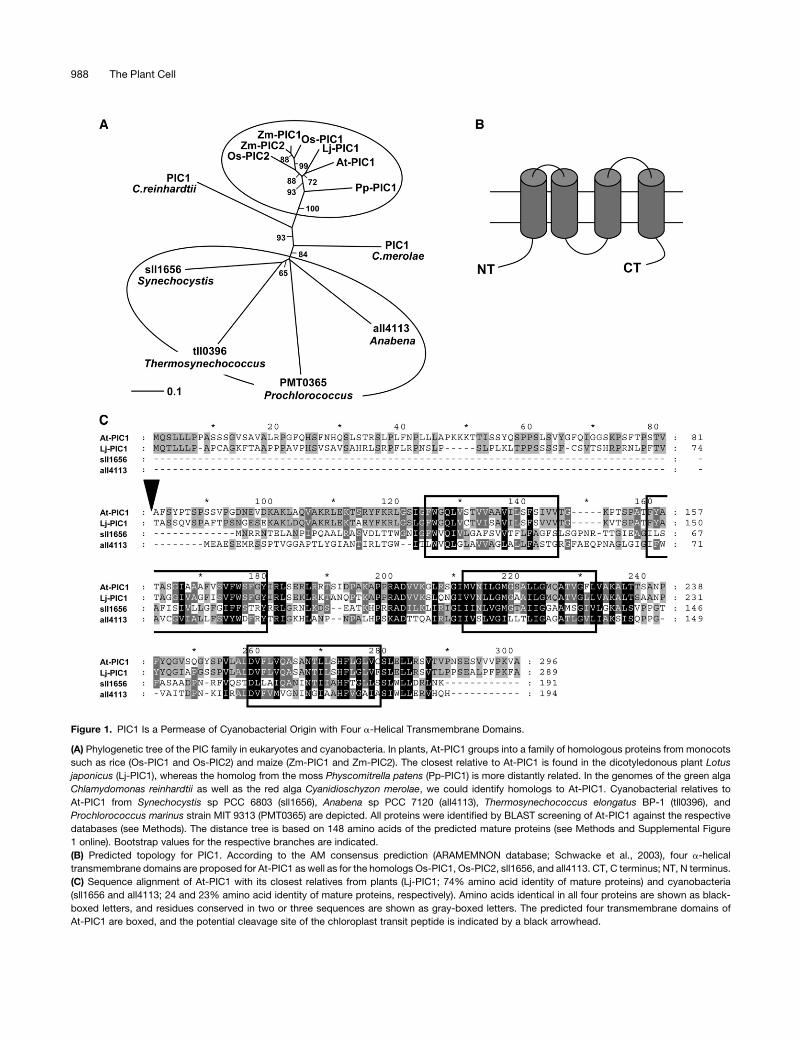
Figure 1. PIC1 Is a Permease of Cyanobacterial Origin with Four a-Helical Transmembrane Domains.
(A) Phylogenetic tree of the PIC family in eukaryotes and cyanobacteria. In plants, At-PIC1 groups into a family of homologous proteins from monocots
such as rice (Os-PIC1 and Os-PIC2) and maize (Zm-PIC1 and Zm-PIC2). The closest relative to At-PIC1 is found in the dicotyledonous plant Lotus
japonicus (Lj-PIC1), whereas the homolog from the moss Physcomitrella patens (Pp-PIC1) is more distantly related. In the genomes of the green alga
Chlamydomonas reinhardtii as well as the red alga Cyanidioschyzon merolae, we could identify homologs to At-PIC1. Cyanobacterial relatives to
At-PIC1 from Synechocystis sp PCC 6803 (sll1656), Anabena sp PCC 7120 (all4113), Thermosynechococcus elongatus BP-1 (tll0396), and
Prochlorococcus marinus strain MIT 9313 (PMT0365) are depicted. All proteins were identified by BLAST screening of At-PIC1 against the respective
databases (see Methods). The distance tree is based on 148 amino acids of the predicted mature proteins (see Methods and Supplemental Figure
1 online). Bootstrap values for the respective branches are indicated.
(B) Predicted topology for PIC1. According to the AM consensus prediction (ARAMEMNON database; Schwacke et al., 2003), four a-helical
transmembrane domains are proposed for At-PIC1 as well as for the homologs Os-PIC1, Os-PIC2, sll1656, and all4113. CT, C terminus; NT, N terminus.
(C) Sequence alignment of At-PIC1 with its closest relatives from plants (Lj-PIC1; 74% amino acid identity of mature proteins) and cyanobacteria
(sll1656 and all4113; 24 and 23% amino acid identity of mature proteins, respectively). Amino acids identical in all four proteins are shown as black-
boxed letters, and residues conserved in two or three sequences are shown as gray-boxed letters. The predicted four transmembrane domains of
At-PIC1 are boxed, and the potential cleavage site of the chloroplast transit peptide is indicated by a black arrowhead.
988 The Plant Cell
isolated pea chloroplasts, At-PIC1 displayed the same import
pattern (data not shown). All import experiments showed an
intermediate product of ;23.5 kD (Figure 2A) that can be pulse-
chased into the inner envelope membrane (E. Firlej-Kwoka,
personal communication). Thus, we conclude that PIC1 uses
the general import pathway into chloroplasts but that insertion
into the inner envelope membrane occurs via an intermediate.
Based on ChloroP (Emanuelsson et al., 1999), the precursor
protein PIC1 of 31.4 kD (296 amino acids) is predicted to have a
chloroplast transit peptide with a processing site after amino acid
residue 15. However, in vitro import experiments and immuno-
blot analysis showed a mature protein of ;22 kD (Figures 2A and
2B), requiring the cleavage of a far larger transit peptide. Fur-
thermore, the alignment with all cyanobacterial homologs and
the predicted mature proteins of the plant subfamily support this
observation (Figure 1A; see Supplemental Figure 1 online). Thus,
we conclude that the precursor PIC1 is processed after amino
acid residue 81 (Figure 1C), a cleavage site that is supported by
ChloroP analysis after removal of the first 15 residues. The
resulting mature-sized protein is 216 amino acids long and has a
molecular mass of 22.8 kD.
The envelope localization of PIC1 was confirmed by immuno-
blot analysis. Using antibodies directed against the C-terminal
part of PIC1, we detected the mature protein in the envelope
fraction of chloroplasts from 6-week-old Arabidopsis rosette
leaves (Figure 2B). Because the antiserum against PIC1 failed to
detect the protein in the entire organelle, chloroplasts were sub-
fractionated into envelopes, stroma, and thylakoid membranes.
The mature PIC1 protein runs at an apparent molecular mass of
22 kD. As marker proteins for the chloroplast subfractions, we
used antibodies against the chloroplast inner envelope pro-
tein Tic110, the large subunit of ribulose-1,5-bis-phosphate
carboxylase/oxygenase (LSU), and the thylakoid-bound light-
harvesting complex (LHCP) from pea.
To verify the chloroplast localization in intact cells, full-length
PIC1 was fused with the green fluorescent protein (GFP) and
Arabidopsis mesophyll protoplasts were transiently transformed
with this construct (Figure 2C). Merging the fluorescence images
of GFP and chlorophyll indicated an envelope insertion of PIC1.
GFP signals were located at the periphery of chloroplasts only
and were associated with neither thylakoid membranes nor
cytosolic or plasma membrane components of the cell. However,
the GFP distribution appeared more spotted than the even signal
of the outer envelope protein At-OEP7 (Lee et al., 2001), which
was used as a control. Again, this indicates an association of
PIC1 with the inner envelope membrane, because similar GFP sig-
nal patterns can be found for the inner membrane–linked VIPP1
(for vesicle-inducing protein in plastids) protein (Aseeva et al.,
2004) and the P-type ATPase PAA1 (Abdel-Ghany et al., 2005).
PIC1 Is Ubiquitously Expressed in Arabidopsis and Peaks
in Green Tissues
When we probed the Affymetrix full genome microarray (ATH1
chip) for the expression of PIC1 in rosette leaves and roots of
4-week-old Arabidopsis (Clausen et al., 2004), it demonstrated
that PIC1 mRNA was present in both tissues but that transcript
levels in leaves were approximately twofold higher than those in
roots (Figure 3A). This expression pattern could be confirmed by
quantitative real-time RT-PCR on cDNA of 7-d-old seedlings
(Figure 3B). Here, the relatively high transcript density, 1500 PIC1
transcripts per 10,000 actin transcripts, originated mostly from
cotyledons. Screening the array data of the AtGenExpress
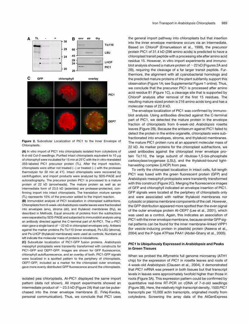
Figure 2. Subcellular Localization of PIC1 to the Inner Envelope of
Chloroplasts.
(A) In vitro import of PIC1 into chloroplasts isolated from cotyledons of
8-d-old Col-0 seedlings. Purified intact chloroplasts equivalent to 10 mg
of chlorophyll were incubated for 15 min at 258C with the in vitro–translated
35S-labeled PIC1 precursor protein (TL). After the import reaction,
chloroplasts were either not treated (�) or treated (þ) with the protease
thermolysin for 30 min at 48C. Intact chloroplasts were recovered by
centrifugation, and import products were analyzed by SDS-PAGE and
autoradiography. The precursor protein PIC1 is processed to a mature
protein of 22 kD (arrowheads). The mature protein as well as an
intermediate form of 23.5 kD (asterisks) are protease-protected, con-
firming import into intact chloroplasts. The translation mixture sample
(TL) represents 10% of the precursor added to the import reaction.
(B) Immunoblot analysis of PIC1 localization in chloroplast subfractions.
Chloroplasts from 6-week-old Arabidopsis rosette leaves were fractionated
into envelopes (env), stroma (str), and thylakoid membranes (thy), as
described in Methods. Equal amounts of proteins from the subfractions
were separated by SDS-PAGE and subjected to immunoblot analysis using
an antibody directed against the C-terminal part of At-PIC1. The a-PIC1
stain gave a single band of ;22 kD in chloroplast envelopes only. Antisera
against the marker proteins Ps-Tic110 (inner envelope), Ps-LSU (stroma),
and Ps-LHCP (thylakoid membranes) were used as controls. Numbers at
left indicate the molecular mass of proteins in kilodaltons.
(C) Subcellular localization of PIC1-GFP fusion proteins. Arabidopsis
mesophyll protoplasts were transiently transformed with constructs for
PIC1-GFP and OEP7-GFP. Images are shown for GFP fluorescence,
chlorophyll autofluorescence, and an overlay of both. PIC1-GFP signals
were localized in a spotted pattern to the periphery of chloroplasts.
OEP7-GFP, included as a marker for the chloroplast outer envelope,
gave more evenly distributed GFP fluorescence around the chloroplasts.
Iron Transport in Arabidopsis Chloroplasts 989
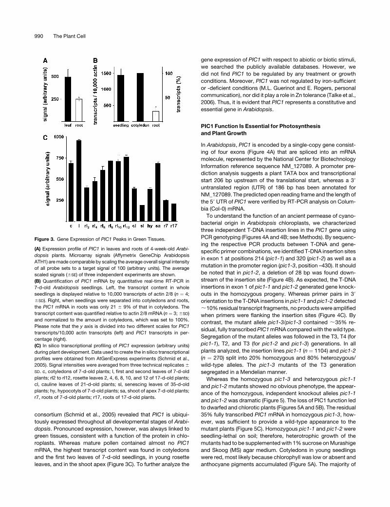
consortium (Schmid et al., 2005) revealed that PIC1 is ubiqui-
tously expressed throughout all developmental stages of Arabi-
dopsis. Pronounced expression, however, was always linked to
green tissues, consistent with a function of the protein in chlo-
roplasts. Whereas mature pollen contained almost no PIC1
mRNA, the highest transcript content was found in cotyledons
and the first two leaves of 7-d-old seedlings, in young rosette
leaves, and in the shoot apex (Figure 3C). To further analyze the
gene expression of PIC1 with respect to abiotic or biotic stimuli,
we searched the publicly available databases. However, we
did not find PIC1 to be regulated by any treatment or growth
conditions. Moreover, PIC1 was not regulated by iron-sufficient
or -deficient conditions (M.L. Guerinot and E. Rogers, personal
communication), nor did it play a role in Zn tolerance (Talke et al.,
2006). Thus, it is evident that PIC1 represents a constitutive and
essential gene in Arabidopsis.
PIC1 Function Is Essential for Photosynthesis
and Plant Growth
In Arabidopsis, PIC1 is encoded by a single-copy gene consist-
ing of four exons (Figure 4A) that are spliced into an mRNA
molecule, represented by the National Center for Biotechnology
Information reference sequence NM_127089. A promoter pre-
diction analysis suggests a plant TATA box and transcriptional
start 206 bp upstream of the translational start, whereas a 39
untranslated region (UTR) of 186 bp has been annotated for
NM_127089. The predicted open reading frame and the length of
the 59 UTR of PIC1 were verified by RT-PCR analysis on Colum-
bia (Col-0) mRNA.
To understand the function of an ancient permease of cyano-
bacterial origin in Arabidopsis chloroplasts, we characterized
three independent T-DNA insertion lines in the PIC1 gene using
PCR genotyping (Figures 4A and 4B; see Methods). By sequenc-
ing the respective PCR products between T-DNA and gene-
specific primer combinations, we identified T-DNA insertion sites
in exon 1 at positions 214 (pic1-1) and 320 (pic1-2) as well as a
mutation in the promoter region (pic1-3, position –430). It should
be noted that in pic1-2, a deletion of 28 bp was found down-
stream of the insertion site (Figure 4B). As expected, the T-DNA
insertions in exon 1 of pic1-1 and pic1-2 generated gene knock-
outs in the homozygous progeny. Whereas primer pairs in 39
orientation to the T-DNA insertions in pic1-1 and pic1-2 detected
;10% residual transcript fragments, no products were amplified
when primers were flanking the insertion sites (Figure 4C). By
contrast, the mutant allele pic1-3/pic1-3 contained ;35% re-
sidual, fully transcribed PIC1 mRNA compared with the wild type.
Segregation of the mutant alleles was followed in the T3, T4 (for
pic1-1), T2, and T3 (for pic1-2 and pic1-3) generations. In all
plants analyzed, the insertion lines pic1-1 (n ¼ 1104) and pic1-2
(n ¼ 270) split into 20% homozygous and 80% heterozygous/
wild-type alleles. The pic1-3 mutants of the T3 generation
segregated in a Mendelian manner.
Whereas the homozygous pic1-3 and heterozygous pic1-1
and pic1-2 mutants showed no obvious phenotype, the appear-
ance of the homozygous, independent knockout alleles pic1-1
and pic1-2 was dramatic (Figure 5). The loss of PIC1 function led
to dwarfed and chlorotic plants (Figures 5A and 5B). The residual
35% fully transcribed PIC1 mRNA in homozygous pic1-3, how-
ever, was sufficient to provide a wild-type appearance to the
mutant plants (Figure 5C). Homozygous pic1-1 and pic1-2 were
seedling-lethal on soil; therefore, heterotrophic growth of the
mutants had to be supplemented with 1% sucrose on Murashige
and Skoog (MS) agar medium. Cotyledons in young seedlings
were red, most likely because chlorophyll was low or absent and
anthocyane pigments accumulated (Figure 5A). The majority of
Figure 3. Gene Expression of PIC1 Peaks in Green Tissues.
(A) Expression profile of PIC1 in leaves and roots of 4-week-old Arabi-
dopsis plants. Microarray signals (Affymetrix GeneChip Arabidopsis
ATH1) are made comparable by scaling the average overall signal intensity
of all probe sets to a target signal of 100 (arbitrary units). The average
scaled signals (6SE) of three independent experiments are shown.
(B) Quantification of PIC1 mRNA by quantitative real-time RT-PCR in
7-d-old Arabidopsis seedlings. Left, the transcript content in whole
seedlings is displayed relative to 10,000 transcripts of actin 2/8 (n ¼ 4;
6SD). Right, when seedlings were separated into cotyledons and roots,
the PIC1 mRNA in roots was only 21 6 9% of that in cotyledons. The
transcript content was quantified relative to actin 2/8 mRNA (n ¼ 3; 6SD)
and normalized to the amount in cotyledons, which was set to 100%.
Please note that the y axis is divided into two different scales for PIC1
transcripts/10,000 actin transcripts (left) and PIC1 transcripts in per-
centage (right).
(C) In silico transcriptional profiling of PIC1 expression (arbitrary units)
during plant development. Data used to create the in silico transcriptional
profiles were obtained from AtGenExpress experiments (Schmid et al.,
2005). Signal intensities were averaged from three technical replicates 6
SD. c, cotyledons of 7-d-old plants; l, first and second leaves of 7-d-old
plants; rl2 to rl12, rosette leaves 2, 4, 6, 8, 10, and 12 of 17-d-old plants;
cl, cauline leaves of 21-d-old plants; sl, senescing leaves of 35-d-old
plants; hy, hypocotyls of 7-d-old plants; sa, shoot of apex 7-d-old plants;
r7, roots of 7-d-old plants; r17, roots of 17-d-old plants.
990 The Plant Cell
the slowly growing mutants produced almost transparent rosette
leaves; however, in approximately one-third of the plants, the
leaves appeared white. This subpopulation of mutants was even
smaller and did not produce inflorescences (data not shown).
Because pic1-1 and pic1-2 represent independently derived
mutant alleles, the described phenotype in both lines (Figures 5A
and 5B) can be linked to the loss of PIC1 function. Roots, like the
entire pic1 mutant plant, were smaller than in wild-type plants.
However, we did not observe any significant phenotype in roots
of pic1 mutants maintained on sucrose-supplemented medium.
To complement the mutant phenotype, a 3327-bp genomic
fragment containing the PIC1 gene and the promoter region
(gPIC1) was introduced into heterozygous PIC1/pic1-1 plants.
Six independent T1 lines that inherited the gPIC1 construct were
generated. According to segregation analysis of the PIC1 locus
by PCR genotyping in the T1 and T2 generations, one of these
lines proved to be homozygous for the pic1-1 allele (pic1-1/
pic1-1 gPIC1-1), four lines were still heterozygous (PIC1/pic1-1
gPIC1-2, -3, -4, and -5), and one line was wild type (PIC1/PIC1
gPIC1-6) at the endogenous PIC1 locus. The line pic1-1/pic1-1
gPIC1-1 had deep green leaves and showed normal develop-
ment in the T1 and T2 generations (Figure 5A), proving the
complementation of the homozygous pic1-1 mutant by gPIC1-1.
Interestingly, the shoot apex and the very young emerging
leaves of homozygous pic1-1 and pic1-2 mutants were pale
green and their leaves turned chlorotic while growing (Figure 5D).
After 6 weeks on sucrose-containing agar, surviving mutant plants
were able to produce an inflorescence; again, the young sepals
of the flower were pale green and became white subsequently
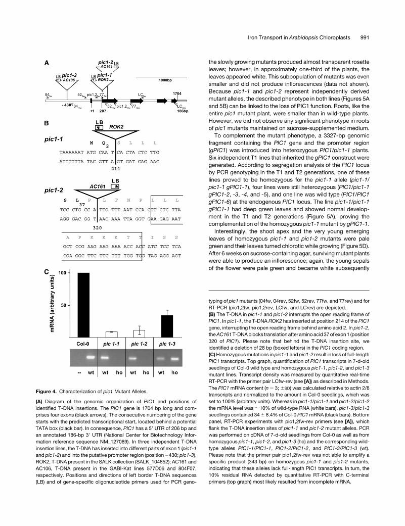
Figure 4. Characterization of pic1 Mutant Alleles.
(A) Diagram of the genomic organization of PIC1 and positions of
identified T-DNA insertions. The PIC1 gene is 1704 bp long and com-
prises four exons (black arrows). The consecutive numbering of the gene
starts with the predicted transcriptional start, located behind a potential
TATA box (black bar). In consequence, PIC1 has a 59 UTR of 206 bp and
an annotated 186-bp 39 UTR (National Center for Biotechnology Infor-
mation reference sequence NM_127089). In three independent T-DNA
insertion lines, the T-DNA has inserted into different parts of exon 1 (pic1-1
and pic1-2) and into the putative promoter region (position�430; pic1-3).
ROK2, T-DNA present in the SALK collection (SALK_104852); AC161 and
AC106, T-DNA present in the GABI-Kat lines 577D06 and 804F07,
respectively. Positions and directions of left border T-DNA sequences
(LB) and of gene-specific oligonucleotide primers used for PCR geno-
typing of pic1 mutants (04fw, 04rev, 52fw, 52rev, 77fw, and 77rev) and for
RT-PCR (pic1,2fw, pic1,2rev, LCfw, and LCrev) are depicted.
(B) The T-DNA in pic1-1 and pic1-2 interrupts the open reading frame of
PIC1. In pic1-1, the T-DNA ROK2 has inserted at position 214 of the PIC1
gene, interrupting the open reading frame behind amino acid 2. In pic1-2,
the AC161 T-DNA blocks translation after amino acid 37 of exon 1 (position
320 of PIC1). Please note that behind the T-DNA insertion site, we
identified a deletion of 28 bp (boxed letters) in the PIC1 coding region.
(C) Homozygous mutations in pic1-1 and pic1-2 result in loss of full-length
PIC1 transcripts. Top graph, quantification of PIC1 transcripts in 7-d-old
seedlings of Col-0 wild type and homozygous pic1-1, pic1-2, and pic1-3
mutant lines. Transcript density was measured by quantitative real-time
RT-PCR with the primer pair LCfw-rev (see [A]) as described in Methods.
The PIC1 mRNA content (n ¼ 3; 6SD) was calculated relative to actin 2/8
transcripts and normalized to the amount in Col-0 seedlings, which was
set to 100% (arbitrary units). Whereas in pic1-1/pic1-1 and pic1-2/pic1-2
the mRNA level was ;10% of wild-type RNA (white bars), pic1-3/pic1-3
seedlings contained 34 6 8.4% of Col-0 PIC1 mRNA (black bars). Bottom
panel, RT-PCR experiments with pic1,2fw-rev primers (see [A]), which
flank the T-DNA insertion sites of pic1-1 and pic1-2 mutant alleles. PCR
was performed on cDNA of 7-d-old seedlings from Col-0 as well as from
homozygous pic1-1, pic1-2, and pic1-3 (ho) and the corresponding wild-
type alleles PIC1-1/PIC1-1, PIC1-2/PIC1-2, and PIC1-3/PIC1-3 (wt).
Please note that the primer pair pic1,2fw-rev was not able to amplify a
specific product (343 bp) on homozygous pic1-1 and pic1-2 mutants,
indicating that these alleles lack full-length PIC1 transcripts. In turn, the
10% residual RNA detected by quantitative RT-PCR with C-terminal
primers (top graph) most likely resulted from incomplete mRNA.
Iron Transport in Arabidopsis Chloroplasts 991
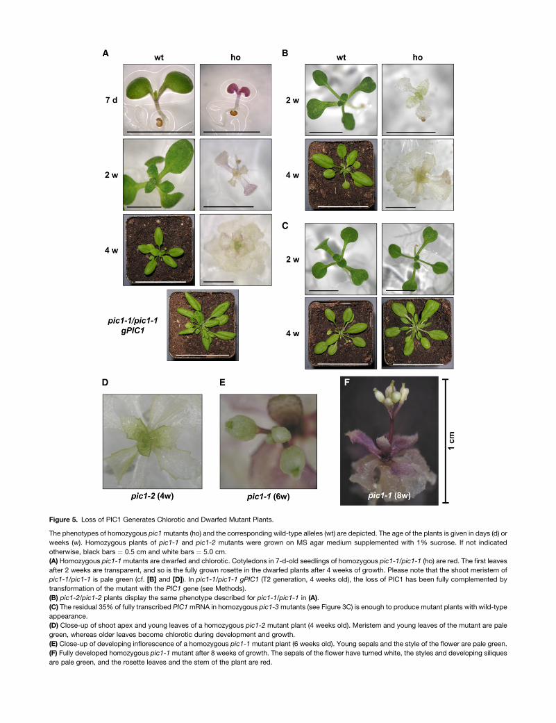
Figure 5. Loss of PIC1 Generates Chlorotic and Dwarfed Mutant Plants.
The phenotypes of homozygous pic1 mutants (ho) and the corresponding wild-type alleles (wt) are depicted. The age of the plants is given in days (d) or
weeks (w). Homozygous plants of pic1-1 and pic1-2 mutants were grown on MS agar medium supplemented with 1% sucrose. If not indicated
otherwise, black bars ¼ 0.5 cm and white bars ¼ 5.0 cm.
(A) Homozygous pic1-1 mutants are dwarfed and chlorotic. Cotyledons in 7-d-old seedlings of homozygous pic1-1/pic1-1 (ho) are red. The first leaves
after 2 weeks are transparent, and so is the fully grown rosette in the dwarfed plants after 4 weeks of growth. Please note that the shoot meristem of
pic1-1/pic1-1 is pale green (cf. [B] and [D]). In pic1-1/pic1-1 gPIC1 (T2 generation, 4 weeks old), the loss of PIC1 has been fully complemented by
transformation of the mutant with the PIC1 gene (see Methods).
(B) pic1-2/pic1-2 plants display the same phenotype described for pic1-1/pic1-1 in (A).
(C) The residual 35% of fully transcribed PIC1 mRNA in homozygous pic1-3 mutants (see Figure 3C) is enough to produce mutant plants with wild-type
appearance.
(D) Close-up of shoot apex and young leaves of a homozygous pic1-2 mutant plant (4 weeks old). Meristem and young leaves of the mutant are pale
green, whereas older leaves become chlorotic during development and growth.
(E) Close-up of developing inflorescence of a homozygous pic1-1 mutant plant (6 weeks old). Young sepals and the style of the flower are pale green.
(F) Fully developed homozygous pic1-1 mutant after 8 weeks of growth. The sepals of the flower have turned white, the styles and developing siliques
are pale green, and the rosette leaves and the stem of the plant are red.
(Figure 5E). Eight-week-old plants were fully grown with a mature
size of 1 cm. The style and developing silique of the flower were
pale green, whereas leaves and stem accumulated anthocyane
pigments (Figure 5F). Because all flowers of homozygous mu-
tants were sterile, pic1-1 and pic1-2 had to be propagated in the
heterozygous state.
In summary, the described phenotypes of pic1-1 and pic1-2
mutants suggest an essential function of PIC1 in photosynthesis
and plant growth. Obviously, loss of PIC1 function produces
chlorotic plants, which are not able to assimilate carbohydrates
by photosynthesis. Hence, their growth has to be supplemented
with sucrose.
Loss of PIC1 Function Leads to Impaired
Chloroplast Development
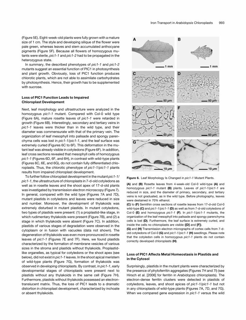
Next, leaf morphology and ultrastructure were analyzed in the
homozygous pic1-1 mutant. Compared with Col-0 wild type
(Figure 6A), mature rosette leaves of pic1-1 were retarded in
growth (Figure 6B). Interestingly, secondary and tertiary veins in
pic1-1 leaves were thicker than in the wild type, and their
diameter was commensurate with that of the primary vein. The
organization of leaf mesophyll into palisade and spongy paren-
chyma cells was lost in pic1-1/pic1-1, and the leaf surface was
extremely curled (Figures 6C to 6F). This deformation in the mu-
tant leaf was already visible in cotyledons (Figure 6F). In addition,
leaf cross sections revealed that mesophyll cells of homozygous
pic1-1 (Figures 6D, 6F, and 6H), in contrast with wild-type plants
(Figures 6C, 6E, and 6G), do not contain fully differentiated chlo-
roplasts. Thus, the chlorotic phenotype of pic1-1/pic1-1 plants
results from impaired chloroplast development.
To further follow chloroplast development in the mutant pic1-1/
pic1-1, the ultrastructure of chloroplasts in 7-d-old cotyledons as
well as in rosette leaves and the shoot apex of 17-d-old plants
was investigated by transmission electron microscopy (Figure 7).
In general, compared with the wild type (Figures 7A and 7D),
mutant plastids in cotyledons and leaves were reduced in size
and number. Moreover, the development of thylakoids was
extremely disturbed in mutant plastids. In mutant cotyledons,
two types of plastids were present: (1) a proplastid-like stage, in
which rudimentary thylakoids were present (Figure 7B), and (2) a
stage in which thylakoids were absent (Figure 7C). In addition,
plastids of various stages of degradation were observed in the
cytoplasm or in fusion with vacuoles (data not shown). The
degeneration of thylakoids was even more pronounced in rosette
leaves of pic1-1 (Figures 7E and 7F). Here, we found plastids
characterized by the formation of membrane vesicles of various
sizes in the stroma and plastids without thylakoids. Proplastid-
like organelles, as typical for cotyledons or the shoot apex (see
below), did not exist in pic1-1 leaves. In the shoot apical meristem
of wild-type plants (Figure 7G), formation of thylakoids was
observed in developing chloroplasts. By contrast, in pic1-1, early
developmental stages of chloroplasts were present next to
plastids without any thylakoids in the same cell (Figure 7H).
Furthermore, plastids without thylakoids possessed an electron-
translucent matrix. Thus, the loss of PIC1 leads to a dramatic
distortion in chloroplast development, characterized by inchoate
or absent thylakoids.
Loss of PIC1 Affects Metal Homeostasis in Plastids and
in the Cytosol
Surprisingly, plastids in the mutant plants were characterized by
the presence of phytoferritin aggregates (Figures 7H and 7I) (see
Hirsch et al. [2006] for ferritin in Arabidopsis chloroplasts). The
electron-dense ferritin clusters were detected in plastids of
cotyledons, leaves, and shoot apices of pic1-1/pic1-1 but not
in any chloroplasts of wild-type plants (Figures 7A, 7D, and 7G).
When we compared gene expression in pic1-1 versus the wild
Figure 6. Leaf Morphology Is Changed in pic1-1 Mutant Plants.
(A) and (B) Rosette leaves from 4-week-old Col-0 wild-type (A) and
homozygous pic1-1 mutant (B) plants. Leaves of pic1-1/pic1-1 are
reduced in size, and the diameter of primary, secondary, and tertiary
veins is not graduated, as in the wild type. Before photography, leaves
were destained in 70% ethanol.
(C) to (F) Semithin cross sections of rosette leaves from 17-d-old Col-0
wild-type (C) and pic1-1/pic1-1 (D) as well as from 7-d-old cotyledons of
Col-0 (E) and homozygous pic1-1 (F). In pic1-1/pic1-1 mutants, the
organization of the leaf mesophyll into palisade and spongy parenchyma
cells is lost (D). Furthermore, the leaf surface is extremely curled, and
inside the cells no chloroplasts are visible ([D] and [F]).
(G) and (H) Transmission electron micrographs of cortex cells from 7-d-
old cotyledons of Col-0 (G) and pic1-1/pic1-1 (H) seedlings. Please note
that the cotyledon cells in homozygous pic1-1 plants do not contain
correctly developed chloroplasts (H).
Iron Transport in Arabidopsis Chloroplasts 993
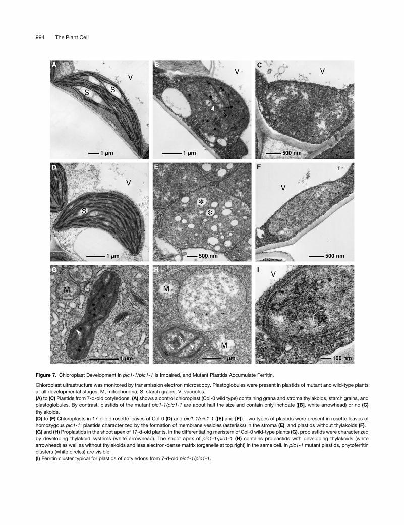
Figure 7. Chloroplast Development in pic1-1/pic1-1 Is Impaired, and Mutant Plastids Accumulate Ferritin.
Chloroplast ultrastructure was monitored by transmission electron microscopy. Plastoglobules were present in plastids of mutant and wild-type plants
at all developmental stages. M, mitochondria; S, starch grains; V, vacuoles.
(A) to (C) Plastids from 7-d-old cotyledons. (A) shows a control chloroplast (Col-0 wild type) containing grana and stroma thylakoids, starch grains, and
plastoglobules. By contrast, plastids of the mutant pic1-1/pic1-1 are about half the size and contain only inchoate ([B], white arrowhead) or no (C)
thylakoids.
(D) to (F) Chloroplasts in 17-d-old rosette leaves of Col-0 (D) and pic1-1/pic1-1 ([E] and [F]). Two types of plastids were present in rosette leaves of
homozygous pic1-1: plastids characterized by the formation of membrane vesicles (asterisks) in the stroma (E), and plastids without thylakoids (F).
(G) and (H) Proplastids in the shoot apex of 17-d-old plants. In the differentiating meristem of Col-0 wild-type plants (G), proplastids were characterized
by developing thylakoid systems (white arrowhead). The shoot apex of pic1-1/pic1-1 (H) contains proplastids with developing thylakoids (white
arrowhead) as well as without thylakoids and less electron-dense matrix (organelle at top right) in the same cell. In pic1-1 mutant plastids, phytoferritin
clusters (white circles) are visible.
(I) Ferritin cluster typical for plastids of cotyledons from 7-d-old pic1-1/pic1-1.
994 The Plant Cell
type by Affymetrix microarray analysis (SAM software; Tusher
et al., 2001), an increase of transcripts for FER1 and FER4 in
pic1-1/pic1-1 mutants was found (Table 1). Furthermore, ferritin
protein accumulated in homozygous pic1-1 and pic1-2 mutants
but was not detectable in wild-type leaves (Figure 8A). Because
all ferritin detected in the mutants was processed to the mature
size of 23.5 kD, it is evident that it is plastid-intrinsic (Figures 7H
and 7I). No ferritin precursor protein (28 kD) was detected. In
conclusion, pic1 mutant plants increased the levels of ferritin
transcripts and protein, leading to the formation of ferritin clus-
ters within plastids. Because ferritin in plants has been described
as a storage protein for iron in plastids (Briat and Lobreaux,
1997), the loss of PIC1 function might interfere with iron homeo-
stasis in mutant plastids.
In addition to ferritin, we found the transcripts of the copper
superoxide dismutases CSD1 and CSD2 to be upregulated in
pic1-1/pic1-1 (Table 1). Again, the transcriptional regulation
could be verified by immunoblot analysis, in which specific
antibodies for CSD1 and CSD2 detected the proteins in homo-
zygous pic1-1 and pic1-2 mutants only (Figure 8A). Whereas
CSD1 is localized in the cytosol, CSD2 is plastid-intrinsic
(Kliebenstein et al., 1998). The increase of both CSD proteins in
the cytosol and in plastids was accompanied by a twofold
increase of the Cu content in pic1-1 shoot tissue (see Supple-
mental Table 1 online). Thus, metal ion homeostasis was affected
not only in plastids but also within the cytosol of pic1 mutants. By
contrast, transcripts and protein levels of the plastid-localized,
Fe-dependent superoxide dismutase FSD1 (Kliebenstein et al.,
1998) were slightly downregulated (Table 1, Figure 8A).
Photosynthetic Capacity Is Lost but Protein Import Is Still
Functional in pic1 Plastids
Whereas metal homeostasis–associated proteins in pic1 mu-
tants are differentially regulated, the photosynthetic capacity of
mutant plastids was completely lost. This becomes apparent by
the heterotrophic growth and chlorosis of homozygous pic1-1
and pic1-2 (Figures 5 and 6), the impaired biogenesis and
degradation of thylakoids (Figure 7), and the downregulation of
genes associated with photosynthesis and carbon assimilation
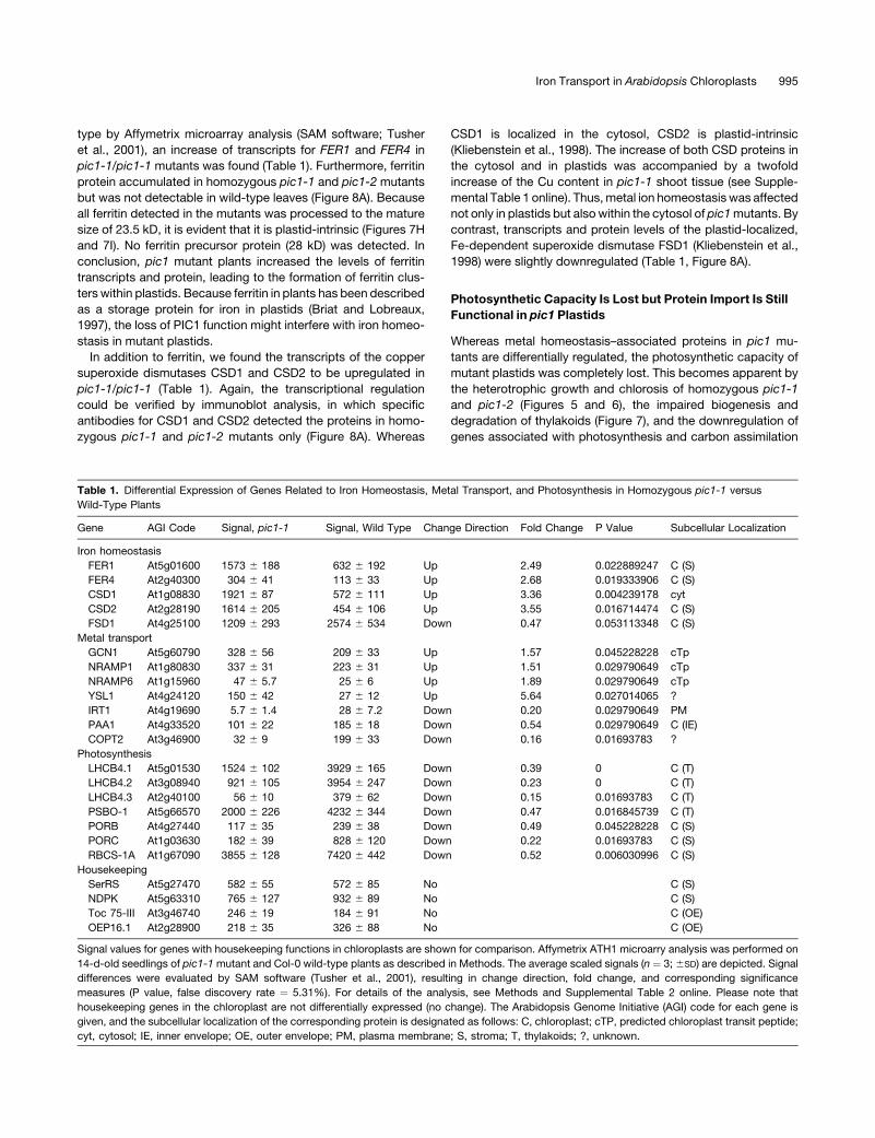
Table 1. Differential Expression of Genes Related to Iron Homeostasis, Metal Transport, and Photosynthesis in Homozygous pic1-1 versus
Wild-Type Plants
Gene AGI Code Signal, pic1-1 Signal, Wild Type Change Direction Fold Change P Value Subcellular Localization
Iron homeostasis
FER1 At5g01600 1573 6 188 632 6 192 Up 2.49 0.022889247 C (S)
FER4 At2g40300 304 6 41 113 6 33 Up 2.68 0.019333906 C (S)
CSD1 At1g08830 1921 6 87 572 6 111 Up 3.36 0.004239178 cyt
CSD2 At2g28190 1614 6 205 454 6 106 Up 3.55 0.016714474 C (S)
FSD1 At4g25100 1209 6 293 2574 6 534 Down 0.47 0.053113348 C (S)
Metal transport
GCN1 At5g60790 328 6 56 209 6 33 Up 1.57 0.045228228 cTp
NRAMP1 At1g80830 337 6 31 223 6 31 Up 1.51 0.029790649 cTp
NRAMP6 At1g15960 47 6 5.7 25 6 6 Up 1.89 0.029790649 cTp
YSL1 At4g24120 150 6 42 27 6 12 Up 5.64 0.027014065 ?
IRT1 At4g19690 5.7 6 1.4 28 6 7.2 Down 0.20 0.029790649 PM
PAA1 At4g33520 101 6 22 185 6 18 Down 0.54 0.029790649 C (IE)
COPT2 At3g46900 32 6 9 199 6 33 Down 0.16 0.01693783 ?
Photosynthesis
LHCB4.1 At5g01530 1524 6 102 3929 6 165 Down 0.39 0 C (T)
LHCB4.2 At3g08940 921 6 105 3954 6 247 Down 0.23 0 C (T)
LHCB4.3 At2g40100 56 6 10 379 6 62 Down 0.15 0.01693783 C (T)
PSBO-1 At5g66570 2000 6 226 4232 6 344 Down 0.47 0.016845739 C (T)
PORB At4g27440 117 6 35 239 6 38 Down 0.49 0.045228228 C (S)
PORC At1g03630 182 6 39 828 6 120 Down 0.22 0.01693783 C (S)
RBCS-1A At1g67090 3855 6 128 7420 6 442 Down 0.52 0.006030996 C (S)
Housekeeping
SerRS At5g27470 582 6 55 572 6 85 No C (S)
NDPK At5g63310 765 6 127 932 6 89 No C (S)
Toc 75-III At3g46740 246 6 19 184 6 91 No C (OE)
OEP16.1 At2g28900 218 6 35 326 6 88 No C (OE)
Signal values for genes with housekeeping functions in chloroplasts are shown for comparison. Affymetrix ATH1 microarry analysis was performed on
14-d-old seedlings of pic1-1 mutant and Col-0 wild-type plants as described in Methods. The average scaled signals (n ¼ 3; 6SD) are depicted. Signal
differences were evaluated by SAM software (Tusher et al., 2001), resulting in change direction, fold change, and corresponding significance
measures (P value, false discovery rate ¼ 5.31%). For details of the analysis, see Methods and Supplemental Table 2 online. Please note that
housekeeping genes in the chloroplast are not differentially expressed (no change). The Arabidopsis Genome Initiative (AGI) code for each gene is
given, and the subcellular localization of the corresponding protein is designated as follows: C, chloroplast; cTP, predicted chloroplast transit peptide;
cyt, cytosol; IE, inner envelope; OE, outer envelope; PM, plasma membrane; S, stroma; T, thylakoids; ?, unknown.
Iron Transport in Arabidopsis Chloroplasts 995
(Table 1). For example, transcript abundance of chlorophyll
binding proteins (e.g., LHCB4), subunits of the oxygen evolving
complex (e.g., PSBO-1), proteins involved in chlorophyll biosyn-
thesis (e.g., protochlorophyllide oxidoreductase; POR), and
ribulose-1,5-bis-phosphate carboxylase/oxygenase (e.g., RBCS-
1A) was decreased dramatically in pic1 mutants. Again, we
verified these data by immunoblot analysis, in which LHCB4 and
RBCS-1A proteins were not detectable and POR as well as
PSBO-1 were decreased in pic1 mutants compared with the wild
type (Figures 8A and 8B). It should be noted that all detected
plastid-localized proteins were processed to their mature form in
pic1 mutants (i.e., imported into the mutant plastids).
To further analyze the protein import capacity of pic1 mutant
plastids, we chose the following proteins: VIPP1, which is local-
ized at the inner chloroplast envelope (Aseeva et al., 2004); POR,
which is localized in the chloroplast stroma; and PSBO-1, which
is localized in the thylakoid lumen (Figure 8B). All three proteins
were imported into plastids and processed to their mature size,
as shown by immunoblot analysis. We never detected any
residual precursor proteins in pic1 mutants, indicating that
protein import into plastids is functional in pic1.
In contrast with proteins linked to metal homeostasis or pho-
tosynthesis, genes associated with housekeeping functions in
the chloroplast stroma, such as aminoacyl tRNA synthetases
(e.g., SerRS) or a nucleoside-diphosphate kinase (NDPK), as well
as the general protein import pore Toc75 (Toc 75-III) and the
amino acid–selective channel OEP16 (OEP16.1) in the outer chlo-
roplast envelope, were not regulated in pic1 mutants (Table 1,
Figure 8A).
PIC1 and Its Homolog from Synechocystis Mediate Iron
Uptake in Yeast
Several cyanobacterial relatives of At-PIC1 are annotated as
potential permeases for metal ions. In addition, the upregulation
of ferritin gene expression and the accumulation of ferritin
proteins in homozygous pic1-1 and pic1-2 mutants suggest a
possible function of PIC1 in iron transport into chloroplasts.
Furthermore, the phenotype of pic1 mutants, in particular leaf
chlorosis, inhibition of chloroplast development, and lack of
palisade parenchyma differentiation, is reminiscent of iron defi-
ciency (Henriques et al., 2002; Varotto et al., 2002).
To investigate a potential role of PIC1 in metal transport across
the plastidic inner envelope, we expressed the cDNA of PIC1 and
its cyanobacterial homolog sll1656 from Synechocystis in the
yeast double mutant fet3 fet4, which is defective in low- and
high-affinity Fe uptake (Dix et al., 1994). Because of its reliance
on additional and less efficient uptake mechanisms, the fet3 fet4
mutant requires high Fe concentrations in its growth medium.
PIC1 and sll1656 cDNAs in the yeast expression vector pFL61
were transformed into fet3 fet4 yeast mutants. The cDNA of the
iron transporter IRT1 from Arabidopsis roots (Eide et al., 1996)
and the empty vector pFL61 were used as positive and negative
controls, respectively. Because the fet3 fet4 mutant cannot grow
under iron-limited conditions, the ability of the transformants to
grow on minimal medium supplemented with Fe concentrations
ranging from 0 to 20 mM FeCl3 was investigated (Figure 9A).
Whereas yeast cells expressing the empty vector did not grow on
5 mM FeCl3, expression of the Fe2þ transporter gene IRT1 from
Arabidopsis efficiently complemented yeast growth. Expression
of PIC1 and sll1656 restored the ability of fet3 fet4 to grow when
supplemented with 5 mM or greater concentrations of FeCl3.
Similarly, in liquid minimal medium containing 20 mM FeCl3, fet3
fet4 mutant cells expressing either PIC1 or sll1656 entered the
exponential growth phase 2 to 3 h earlier than the vector control
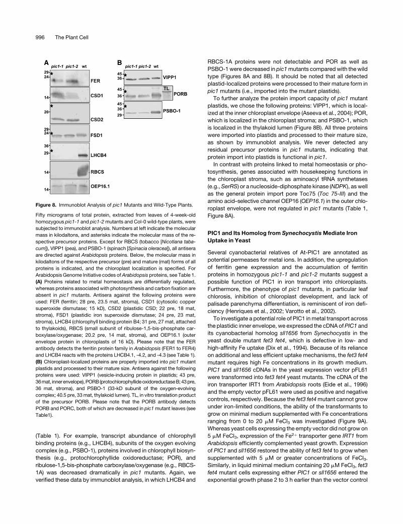
Figure 8. Immunoblot Analysis of pic1 Mutants and Wild-Type Plants.
Fifty micrograms of total protein, extracted from leaves of 4-week-old
homozygous pic1-1 and pic1-2 mutants and Col-0 wild-type plants, were
subjected to immunoblot analysis. Numbers at left indicate the molecular
mass in kilodaltons, and asterisks indicate the molecular mass of the re-
spective precursor proteins. Except for RBCS (tobacco [Nicotiana taba-
cum]), VIPP1 (pea), and PSBO-1 (spinach [Spinacia oleracea]), all antisera
are directed against Arabidopsis proteins. Below, the molecular mass in
kilodaltons of the respective precursor (pre) and mature (mat) forms of all
proteins is indicated, and the chloroplast localization is specified. For
Arabidopsis Genome Initiative codes of Arabidopsis proteins, see Table 1.
(A) Proteins related to metal homeostasis are differentially regulated,
whereas proteins associated with photosynthesis and carbon fixation are
absent in pic1 mutants. Antisera against the following proteins were
used: FER (ferritin; 28 pre, 23.5 mat, stroma), CSD1 (cytosolic copper
superoxide dismutase; 15 kD), CSD2 (plastidic CSD; 22 pre, 18 mat,
stroma), FSD1 (plastidic iron superoxide dismutase; 24 pre, 23 mat,
stroma), LHCB4 (chlorophyll binding protein B4; 31 pre, 27 mat, attached
to thylakoids), RBCS (small subunit of ribulose-1,5-bis-phosphate car-
boxylase/oxygenase; 20.2 pre, 14 mat, stroma), and OEP16.1 (outer
envelope protein in chloroplasts of 16 kD). Please note that the FER
antibody detects the ferritin protein family in Arabidopsis (FER1 to FER4)
and LHCB4 reacts with the proteins LHCB4.1, -4.2, and -4.3 (see Table 1).
(B) Chloroplast-localized proteins are properly imported into pic1 mutant
plastids and processed to their mature size. Antisera against the following
proteins were used: VIPP1 (vesicle-inducing protein in plastids; 43 pre,
36mat, innerenvelope),PORB(protochlorophyllideoxidoreductaseB;43pre,
36 mat, stroma), and PSBO-1 (33-kD subunit of the oxygen-evolving
complex; 40.5 pre, 33 mat, thylakoid lumen). TL, in vitro translation product
of the precursor PORB. Please note that the PORB antibody detects
PORB and PORC, both of which are decreased in pic1 mutant leaves (see
Table1).
996 The Plant Cell
(data not shown). However, the complementation was not as
efficient as with IRT1.
In addition, we determined whether PIC1 and sll1656 could
complement the ctr1 mutant, which is defective in Cu transport
(Dancis et al., 1994a). The ctr1 mutant cannot grow on copper-
limiting medium or on medium containing EDTA (Dancis et al.,
1994b; Korshunova et al., 1999). Expression of PIC1, sll1656,
and IRT1 in the ctr1 mutant strain did rescue its growth defect on
medium with concentrations of EDTA of >0.05 mM, whereas cells
transformed with the pFL61 vector control grew poorly (Figure
9B). However, because of the Cu dependence of Fe uptake in
yeast (Dancis et al., 1994b), we cannot distinguish whether the
growth complementation of ctr1 in this experiment is the result of
restored copper or iron uptake.
To show whether complementation of the fet3 fet4 mutant by
PIC1 and sll1656 is attributable to iron uptake into the cells, we
measured short-term 59Fe accumulation in fet3 fet4 cells trans-
formed with the empty vector pDR195 or the cDNAs of IRT1
(positive control), PIC1, and sll1656 in pDR195 (Table 2). The
expression of either protein, PIC1 or sll1656, significantly in-
creased the Fe uptake rates compared with the vector control. In
agreement, with the growth complementation assays, however,
Fe uptake rates mediated by PIC1 and sll1656 were severalfold
lower than those mediated by IRT1. These results support a
direct role of these two proteins in the membrane transport of
iron.
DISCUSSION
PIC1, an Ancient Permease in the Inner Envelope of Plastids
The protein PIC1 contains four predicted transmembrane a-helices
and is integral to the inner envelope of Arabidopsis chloroplasts.
The chloroplast membrane localization of PIC1 was already
indicated by proteome analysis (Froehlich et al., 2003). We
showed here the insertion of PIC1 into the Arabidopsis chloro-
plast envelope by immunoblot analysis, by in vitro protein import
experiments, and by in vivo GFP labeling of the protein (Figure 2).
Immunoblot and GFP analyses clearly excluded a thylakoid mem-
brane localization, and processing of the imported precursor
protein PIC1 suggested an integration into the inner rather than
the outer chloroplast envelope. In summary, the results demon-
strate an inner envelope localization of PIC1. The PIC1 homolog
sll1656 in Synechocystis most likely is integral to the plasma
membrane of the cyanobacterium.
Sequence comparison revealed that besides the PIC1 family in
vascular plants and green and red algae, the only relatives of
PIC1 are of cyanobacterial origin. Thus, it is very likely that PIC1
proteins evolved directly during endosymbiosis between a cya-
nobacterium and an ancient eukaryotic cell. This evolutionary
step led to the formation of chloroplasts and hence to their
function in photosynthesis in land plants and algae (Vothknecht
and Soll, 2005). Because of the severe phenotype of pic1
knockout plants, it became evident that PIC1 function is essential
for thylakoid biogenesis and, in turn, for photosynthesis. The fact
that the cyanobacterium G. violaceus contains neither thylakoid
membranes (Nakamura et al., 2003) nor PIC1 homologs empha-
sizes this observation.
PIC1 proteins of cyanobacterial origin belong to COGs
(Tatusov et al., 2003), which generally play a role in ion or solute
transport across membranes. Several cyanobacterial relatives
of PIC1 have been annotated as potential permease compo-
nents of ABC transporters. Unlike eukaryotic ABC transporters,
Table 2. Iron Uptake in Yeast Is Mediated by PIC1 and Its
Synechocystis Homolog sll1656
Sample Fe Accumulation
Fold
Increase P Value SNK Test
pDR195 104.61 6 8.93 c
IRT1 869.02 6 29.94 8.3 6 0.29 P < 0.001 a
PIC1 153.11 6 12.85 1.5 6 0.12 P ¼ 0.020 b
sll1656 141.29 6 1.92 1.4 6 0.02 P ¼ 0.030 b
The plasmid pDR195 (empty vector control) as well as the cDNA of IRT1,
PIC1, and sll1656 in pDR195 were introduced into fet3 fet4 yeast
mutants. After growth to the exponential phase, yeast cultures were
assayed for 59Fe uptake as described in Methods. Fe accumulation
(pmol Fe/106 yeast cells 6 SD) was measured after 10 min in 40 mM59Fe-labeled FeCl3 in three independent experiments. Compared with
the empty vector control, expression of all three proteins significantly
increased the iron content in yeast cells (fold increase; P value).
Significant differences among the transformants are indicated by differ-
ent letters as determined by analysis of variance followed by the
Student-Newman-Keuls (SNK) test (P < 0.05; see Methods for details).
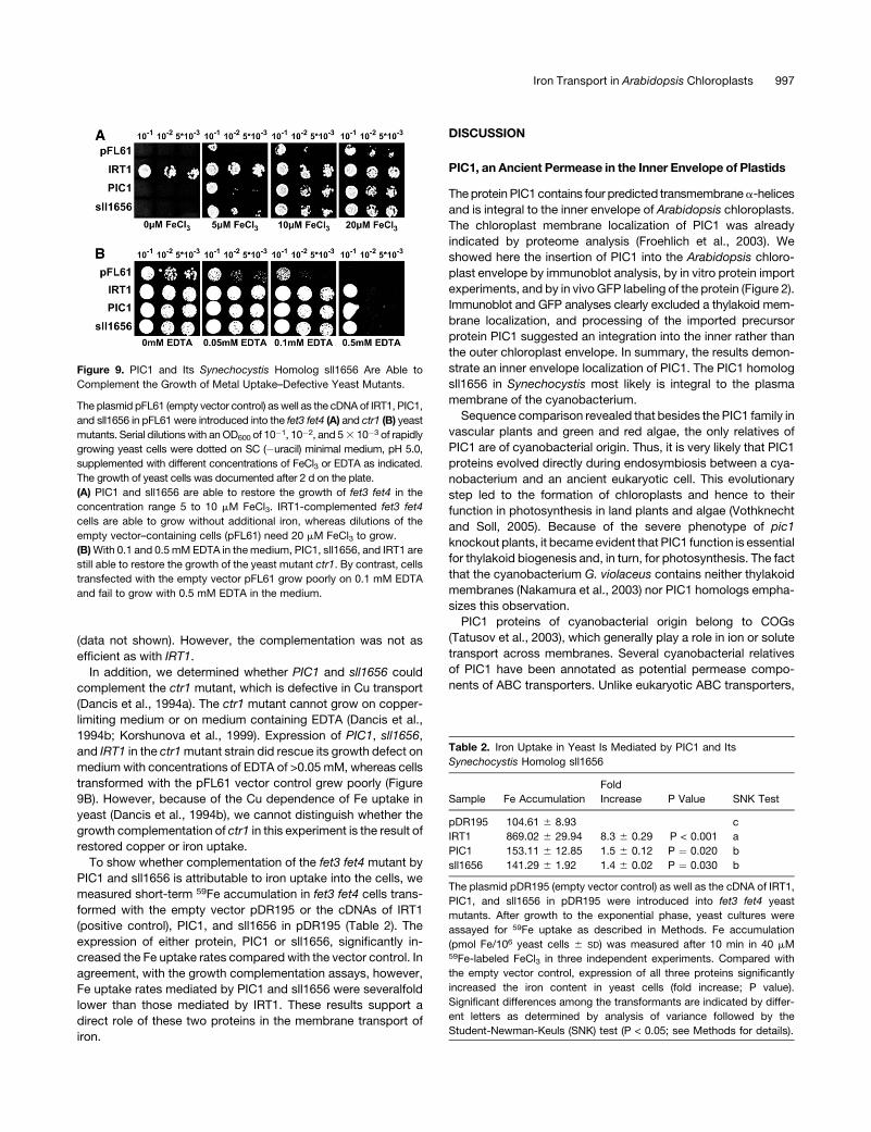
Figure 9. PIC1 and Its Synechocystis Homolog sll1656 Are Able to
Complement the Growth of Metal Uptake–Defective Yeast Mutants.
The plasmid pFL61 (empty vector control) as well as the cDNA of IRT1, PIC1,
and sll1656 in pFL61 were introduced into the fet3 fet4 (A) and ctr1 (B) yeast
mutants. Serial dilutions with an OD600 of 10�1, 10�2, and 5 3 10�3 of rapidly
growing yeast cells were dotted on SC (�uracil) minimal medium, pH 5.0,
supplemented with different concentrations of FeCl3 or EDTA as indicated.
The growth of yeast cells was documented after 2 d on the plate.
(A) PIC1 and sll1656 are able to restore the growth of fet3 fet4 in the
concentration range 5 to 10 mM FeCl3. IRT1-complemented fet3 fet4
cells are able to grow without additional iron, whereas dilutions of the
empty vector–containing cells (pFL61) need 20 mM FeCl3 to grow.
(B) With 0.1 and 0.5 mM EDTA in the medium, PIC1, sll1656, and IRT1 are
still able to restore the growth of the yeast mutant ctr1. By contrast, cells
transfected with the empty vector pFL61 grow poorly on 0.1 mM EDTA
and fail to grow with 0.5 mM EDTA in the medium.
Iron Transport in Arabidopsis Chloroplasts 997
prokaryotic transporters of this family are composed of inde-
pendent permease and ABC protein subunits (Higgins, 2001).
Here, the permease subunits represent the integral membrane
proteins and form the pore, whereas the soluble ABC subunits
bind to the permeases and energize the transport by ATP
cleavage. Several soluble, potential ABC subunits, which are
predicted to be located in plastids, exist in plants and might
represent interacting partners of PIC1 (Garcia et al., 2004).
Among them, the gene GCN1, which encodes two soluble ABC
subunits, is upregulated in pic1-1 knockout mutants (Table 1).
Thus, a function of PIC1 in membrane transport processes most
likely has been evolutionarily conserved.
pic1 Mutant Plants Display a Severe Chlorotic Phenotype
PIC1 is expressed ubiquitously throughout all organs and devel-
opmental stages of Arabidopsis (Figure 3). In agreement with an
essential function in chloroplasts, PIC1 transcript abundance
peaked in green tissues. However, PIC1 is also expressed in
roots and therefore might fulfill a similar function at the inner
envelope of root plastids as well.
Two independently derived T-DNA insertion lines of PIC1
(pic1-1 and pic1-2) could be characterized as knockout muta-
tions. Both mutant alleles generated the same phenotype in the
homozygous progeny (Figure 5). Furthermore, we were able to
complement the phenotype of pic1-1 by the gene PIC1 under the
control of its own promoter. Thus, it is evident that the loss of
PIC1 was responsible for the described phenotype. Interestingly,
;10% residual transcript fragments of PIC1 were detected in
pic1-1/pic1-1 and pic1-2/pic1-2 (Figure 4C). However, these
transcripts lacked the N-terminal part of the PIC1 coding se-
quence; therefore, they could not function as templates for the
synthesis of functional PIC1 proteins. Most likely, these tran-
script fragments originated from an ectopic promoter activity of
the T-DNA insert.
pic1 knockout mutants developed severe chlorosis and only
grew heterotrophically. Hence, PIC1 function is necessary for
chlorophyll synthesis and/or accumulation and is a prerequisite
for proper photosynthesis as well as carbohydrate assimilation in
chloroplasts. Differential regulation of transcript and protein
levels of genes related to photosynthesis in pic1 mutants (see
below) emphasizes this observation. As demonstrated by trans-
mission electron microscopy, functional thylakoid membrane
systems did not develop in the homozygous pic1-1 mutant.
Because rudimentary thylakoid membranes were present in
plastids of cotyledons and the meristem of pic1-1/pic1-1 but
not in mature rosette leaves, we conclude that thylakoid biogen-
esis in pic1-1/pic1-1 is arrested at a proplastid-like stage. This is
consistent with the observation that young leaf organs of pic1
mutants were pale green and contained some chlorophyll but
turned chlorotic while growing. As a consequence, vesicle for-
mation inside plastids of rosette leaves suggests that inchoate
thylakoids are degraded during maturation of the leaf (Figures 7E
and 7F). Furthermore, the fact that mutant leaves contain smaller
and fewer plastids than wild-type leaves suggested a parallel
organelle decomposition as well (Figure 6H). However, residual
but fewer plastids without thylakoids remained in leaf cells,
probably necessary for biosynthetic functions apart from pho-
tosynthesis (for functions of plastids, see Moller, 2005). This
phenotype of pic1 mutants was emphasized by the presence of
transcripts and proteins associated with housekeeping functions
of the chloroplast (see below).
A Role of PIC1 in Iron Transport across the Chloroplast
Inner Envelope
Growth complementation on low iron supply of a yeast mutant
defective in iron uptake, and uptake assays using radiolabeled
Fe, showed that PIC1 and its homolog sll1656 from Synecho-
cystis are capable of mediating membrane transport of iron.
Thus, to our knowledge, PIC1 appears to be the first protein
shown to be involved in iron transport across plastid mem-
branes. Other chloroplast proteins with a role in metal ion trans-
port across the chloroplast envelope are the copper-transporting
ATPases PAA1 and HMA1 and the magnesium transport pro-
tein MRS2-11 (Shikanai et al., 2003; Abdel-Ghany et al., 2005;
Drummond et al., 2006; Seigneurin-Berny et al., 2006). As most
of the plant transporters involved in metal ion transport across
the plasma membrane or the tonoplast, such as ZIP or NRAMP
proteins, usually transport a broad range of heavy metal ions
(Colangelo and Guerinot, 2006), it cannot be excluded that PIC1
and sll1656 might transport other metal ions besides iron as well.
Because PIC1 and sll1656 were able to complement the Cu
uptake–deficient yeast mutant ctr1, a function in copper trans-
port might be possible. However, because of the Cu dependence
of Fe uptake in yeast (Dancis et al., 1994b), it is not yet clear
whether the observed complementation of ctr1 is a result of
restored copper or iron transport. Moreover, we think that the
phenotypes, differential gene expression, and metal content in
the pic1 mutants favor a function in iron rather than copper
transport (see discussion below).
In short-term uptake studies with radiolabeled Fe, PIC1 and
sll1656 showed iron uptake rates that were much lower than
those mediated by IRT1 (Table 2). These lower Fe uptake rates
were in agreement with a retarded and weaker growth comple-
mentation of yeast cells on iron-limiting medium by PIC1 and
sll1656 (Figure 9). A similar difference from IRT1-mediated Fe
uptake in yeast has been observed in yeast cells expressing metal
transporters of the NRAMP family (Curie et al., 2000; Thomine
et al., 2000; Kaiser et al., 2003). Furthermore, because our assay
relied on the expression of a plastid protein in yeast, it is most likely
that iron uptake was mediated by a certain amount of plasma
membrane–localized, mistargeted protein, explaining the low Fe
uptake rates compared with the plasma membrane–intrinsic IRT1.
Chlorosis, caused by the inhibition of chlorophyll synthesis and
chloroplast development (Briat and Lobreaux, 1997), is a clas-
sical symptom of iron deficiency in leaves and particularly in
plastids. Furthermore, in iron-deficient leaves, the palisade pa-
renchyma cells do not differentiate properly, and in chloroplasts,
grana stacks of thylakoids are reduced drastically (Henriques
et al., 2002; Varotto et al., 2002). In the case of pic1 knockout
plants, we propose that the transport pathway of iron is blocked
at the inner chloroplast envelope, causing the described iron
deficiency symptoms: chlorosis, lack of cell differentiation in
leaf mesophyll parenchyma, and impaired thylakoid develop-
ment.
998 The Plant Cell
Because pic1 knockout mutants were able to germinate and
develop rudimentary thylakoids in plastids of cotyledons and
pale green young rosette leaves, an alternative iron uptake
pathway seems to exist in Arabidopsis plastids. Possible by-
passing transporters could be plastid-localized, iron-transport-
ing proteins, such as NRAMP1 (Curie et al., 2000) and NRAMP6,
which contain potential chloroplast transit peptides (for review,
see Curie and Briat, 2003; Colangelo and Guerinot, 2006).
Interestingly, both of these genes were slightly upregulated in
pic1-1 mutants (Table 1). NRAMP1 has a function in Arabidopsis
roots (Curie et al., 2000), but according to the data of the
AtGenExpress consortium (Schmid et al., 2005), transcripts of
NRAMP1 are further present in the vegetative shoot apex and
flowers, whereas NRAMP6 is very specifically expressed at later
stages in seed development. Thus, it might be possible that
these metal transport proteins confer a weak iron supplementa-
tion to pic1 mutant plastids in seeds and meristematic tissues.
The described phenotype of pic1 mutants as well as the in vitro
iron uptake mediated by PIC1 both suggest an in vivo function in
iron import into the plastid. However, impaired metal homeosta-
sis and ferritin accumulation in pic1 mutants might also be
caused by disturbed copper transport, a defect in iron export
from the plastid, or inefficient iron mobilization for Fe-S cluster
assembly in chloroplasts (see below). With regard to the strong
sink activity for Fe exerted by the plastidic expression of ferritin
(Van Wuytswinkel et al., 1998), a role of PIC1 in iron export
becomes unlikely. In conclusion, PIC1 most likely functions in Fe
uptake into chloroplasts, but other roles in plastid metal ion
homeostasis and transport (e.g., Fe export, Cu import/export)
might be possible.
Loss of PIC1 Disturbs Metal Homeostasis in Leaves
In addition to structural changes in pic1 knockout lines that
resemble iron deficiency, leaf cells apparently experience iron
stress and perturbed metal homeostasis. Unfortunately, we were
not able to isolate intact plastids from pic1 knockout plants to
determine their iron content. However, when we measured the
iron content in a total leaf extract from homozygous pic1-1 plants,
it was not altered relative to that of wild-type leaves. Further-
more, we were not able to rescue the pic1 mutant phenotype by
the addition of exogenously supplied Fe chelates fFe(III)-EDTA,
Sequestren [Fe(III)-EDDHA]g. When we tested the knockout
mutants pic1-1 and pic1-2 grown on agar plates (1% sucrose,
0.53 MS salts) under iron-deficient (addition of 300 mM ferro-
zine), iron-sufficient, and iron-abundant [addition of 50 mM
Fe(III)-EDTA] conditions, we did not detect significant differences
in the mutant phenotype. Furthermore, homozygous pic1-3
mutants in this assay did not display a phenotype compared
with wild-type plants. In all plants analyzed for plus/minus iron,
we monitored PIC1 transcripts by quantitative RT-PCR, which
demonstrated no regulation of PIC1 expression by iron (data not
shown). In publicly available microarray data, PIC1 was consti-
tutively expressed under all biotic and abiotic impacts described
(e.g., light, hormones, stress, Pb and Zn tolerance, and phos-
phate and potassium deficiency). Moreover, expression of the
permease was under the control of neither iron nor zinc (Talke
et al., 2006; M.L. Guerinot, E. Rogers, and U. Kramer, personal
communication). Thus, these results show that PIC1 represents a
constitutive and essential gene in Arabidopsis that seems not to
be regulated by exogenous metal availability, like the iron uptake
transporter IRT1 in roots (Eide et al., 1996; Connolly et al., 2002;
Vert et al., 2003).
But what happens to metal homeostasis of the plant cell when
PIC1 function is lost? In pic1 mutant plants, ferritin proteins
accumulated in plastids to much higher levels than in wild-type
plants. A major role of ferritin is to precipitate Fe in the inner core
of the ferritin protein cluster and thus to withdraw iron from an
intracellular generation of reactive oxygen species (for overview,
see Briat et al., 1999; Connolly and Guerinot, 2002). The unusual
accumulation of ferritin in pic1 knockout mutants was docu-
mented by electron microscopy (Figures 7H and 7I) at the
transcript level (Table 1) as well as at the protein level (Figure
8). Thus, the increase in ferritin protein and the formation of
ferritin clusters in mutant plastids was accompanied by the
upregulation of FER1 and FER4. Iron overload in plants has been
described to cause severe oxidative stress, because free and
excess Fe2þ ions lead to the formation of hydroxyl radicals via the
Fenton reaction (Briat and Lobreaux, 1997). As a consequence,
plants upregulate Cu-dependent as well as Fe-dependent su-
peroxide dismutases (CSD and FSD) (Kliebenstein et al., 1998;
Asada, 1999, Abdel-Ghany et al., 2005). Interestingly, the Cu
content of pic1-1 shoots was twice as great as in wild-type
shoots, whereas the Fe levels were the same (see Supplemental
Table 1 online). Compared with the wild type, CSD1 (cytosolic)
and CSD2 (plastid-localized) increased parallel to the Cu content
in leaves of pic1 mutants, whereas transcripts and protein levels
of the major plastid-localized iron superoxide dismutase (FSD1)
were slightly downregulated, suggesting a shortage of usable
iron in plastids. Furthermore, most transcripts that were in-
creased in homozygous pic1-1 are supposed to function in
stress-related processes (data not shown), suggesting that pic1
knockout mutants suffer from cytosolic iron stress, plastidic iron
shortage, and a general copper overload. In conclusion, we
propose that PIC1 function is closely linked to metal homeostasis
in plastids and the cytosol.
To understand the integration of PIC1 into the metal homeo-
stasis of the cell, we screened for differentially expressed metal
transport proteins in pic1-1 mutants (Table 1). Besides NRAMP1
and NRAMP6 (see above), we found the putative Fe-nicotiana-
mine transporter YSL1 to be upregulated in pic1-1. YSL1 has
been shown to be involved in the loading of iron into seeds, and
the expression of YSL1 is increased in response to iron excess
(Le Jean et al., 2005; Waters et al., 2006). Although YSL1 as its
relative YSL2 in Arabidopsis most likely is localized in the plasma
membrane, it is discussed that the protein might be involved in Fe
detoxification in specific cellular compartments (Le Jean et al.,
2005). By contrast, transcripts of the iron uptake transporter
IRT1, which is induced by iron deficiency in roots (Eide et al.,
1996; Connolly et al., 2002; Vert et al., 2003), are decreased in
pic1-1. Because IRT1 signals were at the detection limit of the
Affymetrix microarray, we verified this observation by quantita-
tive RT-PCR (see Supplemental Figure 2 online). Interestingly,
compared with wild-type signals, IRT1 transcripts in pic1-1 and
pic1-2 were decreased under iron-sufficient and iron-abundant
conditions but not under iron deficiency, indicating that IRT1
Iron Transport in Arabidopsis Chloroplasts 999
expression was repressed more strongly in pic1 mutants than in
the wild type. Thus, the loss of PIC1 might interfere with iron-
triggered signal transduction within the cell.
Furthermore, expression of the copper-transporting ATPase
PAA1 (inner envelope of chloroplasts) (Abdel-Ghany et al., 2005)
and the potential high-affinity Cu uptake protein COPT2 (for
overview, see Colangelo and Guerinot, 2006) was downregu-
lated. Thus, expression of genes related to iron stress and metal
transport in pic1 mutants reflects iron deficiency in plastids
(FSD1, NRAMP1, and NRAMP6), iron overload in the cytosol
(FER1, FER4, CSD1, YSL1, and IRT1), and an increased copper
content in leaves (CSD2, PAA1, and COPT2). The described
phenotype of pic1 knockout plants (ferritin increase, IRT1 de-
crease, and oxidative stress) and complementation of the copper
uptake–deficient yeast mutant ctr1 by PIC1 and sll1656 (see
above) suggest a role of PIC1 in Cu transport across the inner
chloroplast envelope. In this case, however, quantitatively more
Fe but less or unchanged Cu content should be expected in
shoots of pic1 mutants relative to wild-type shoots, as has been
shown for mutants of the Cu-transporting ATPases PAA1 and
HMA1 in chloroplast envelopes (Abdel-Ghany et al., 2005;
Seigneurin-Berny et al., 2006). In contrast with this expectation,
we made the observation that Cu concentrations in pic1 mutant
shoots were increased twofold and Fe levels did not change.
Moreover, genes that have been reported to be upregulated
under Cu but not under Fe deficiency (e.g., COPT2 [Wintz et al.,
2003]) apparently were not upregulated in pic1. In summary, we
conclude that the block of iron transport in plastids of pic1
knockout mutants causes impaired metal homeostasis, resulting
in the described phenotype and regulation of gene expression.
In addition to induction by an excess of iron, ferritin accumu-
lation in leaf chloroplasts can be induced by reactive oxygen
species (specific for the isoform FER1) or during senescence as a
result of the decomposition of the photosynthetic apparatus and
the release of iron from degraded Fe-S cluster proteins (Briat
et al., 1999). In contrast with the long-term storage of iron in
seeds, the leaf ferritin functions as a transient iron buffer for
important iron-dependent processes such as photosynthesis
and nitrogen fixation. In pic1 mutants, however, iron mobilization
from ferritin seems to be defective for Fe-S cluster assembly in
chloroplasts (see below), leading to the described Fe-deficiency
symptoms of pic1 mutant plastids and a simultaneous accumu-
lation of ferritin clusters.
Plastids of pic1 Mutants Lose Their Photosynthetic Capacity
and Downregulate the Fe-S Cluster Biogenesis Machinery
but Maintain Housekeeping Functions
In contrast with stress-related genes, all genes associated
with photosynthesis and carbon assimilation were massively
downregulated in pic1 mutants (Table 1, Figure 8). This decrease
in photosynthesis-associated proteins is reflected by the phe-
notype of pic1 mutants, characterized by heterotrophic growth,
chlorosis, and the degradation of thylakoids in plastids. Fe-S
cluster proteins are central for the function of the photosynthetic
electron transport chain in chloroplasts (Raven et al., 1999). The
assembly of these Fe-S proteins takes place in plastids and
involves the action of desulfurases and scaffold proteins, which
synthesize transient Fe-S clusters (for overview, see Balk and
Lobreaux, 2005; Ye et al., 2006). Whereas elemental sulfur is
provided by the desulfurases, iron is mobilized via a yet unknown
pathway and transferred to the scaffold proteins. To understand
the possible implication of PIC1 in Fe-S cluster biogenesis, we
monitored the transcript abundance of all genes described either
to function in the Fe-S biogenesis machinery or to code for the
major, plastid-localized Fe-S cluster–containing proteins (Ye
et al., 2006) in homozygous pic1-1 mutants by microarray
analysis (Table 3). Differential gene expression in pic1-1 versus
the wild type was further analyzed by SAM software (Tusher
et al., 2001). Interestingly, gene expression of the desulfurase Cp
NifS and the desulfurase activator Cp SufE was not differentially
regulated, whereas the scaffold proteins Nfu1, Nfu2, Nfu3, and
Cp IscA, which are proposed to integrate iron into the Arabidopsis
chloroplast Fe-S clusters (Ye et al., 2006), were downregulated in
pic1-1 mutants. By contrast, expression of the corresponding
mitochondrial proteins ISU1 (desulfurase), Nfu4, and Nfu5 (scaf-
fold) was not changed or increased, respectively. Thus, PIC1
function might be involved in the mechanism of iron mobilization
for Fe-S cluster assembly in chloroplasts.
Furthermore, expression of several chloroplast-localized Fe-S
cluster proteins in pic1-1 was downregulated (Table 3). These in-
clude the major leaf ferredoxins (FED A and FD1) and ferredoxin–
thioredoxin reductases (FTR b and FTR-A), the chlorophyllide a
oxygenase CAO (for chlorophyll b biosynthesis), and enzymes for
sulfur assimilation (the adenosine-5-phosphate reductases APR1
to APR3 and the sulfite reductase SiR). Again, transcripts of
mitochondrial ferredoxins (the adrenodoxin-like ferredoxins adr.-l
Fd1 and adr.-l Fd2) were not changed or increased. Expression
of Fe-S proteins for chlorophyll breakdown (PaO, for pheophor-
bide a oxygenase) and siroheme biosynthesis (SirB, for sirohy-
drochlorin ferrochelatase), however, was not changed. Interestingly,
siroheme in plants is found as a prosthetic group only for the
plastid-localized sulfite and nitrite reductases, and the iron re-
quired for siroheme biosynthesis by SirB is probably acquired via
a pathway distinct from that for Fe-S cluster assembly (Raux-
Deery et al., 2005).
Transcripts of genes coding for Fe-S proteins with house-
keeping functions in plastids such as protein import (Tic55, a
member of the translocon at the inner envelope of chloroplasts)
as well as the chloroplast-localized nitrogen fixation machinery
(NIR, for nitrite reductase; GLU1 and GLU2, two ferredoxin-
dependent GOGATs [glutamine:2-oxo-glutarate amido transfer-
ases]; GLT1, an NADH-dependent GOGAT; and Atase 2, for
amidophosphoribosyltransferase) were unchanged in pic1-1
(Table 3) (Ye et al., 2006), indicating that pic1 mutant chloroplasts
are able to import precursor proteins (see below) and to assim-
ilate nitrogen. Other genes, encoding non–Fe-S proteins with
housekeeping functions in the chloroplast stroma or envelope
(e.g., the major protein import pore Toc75 and the amino acid–
selective channel OEP16), also are not regulated in pic1 mutants
(Table 1, Figure 8). Thus, although pic1 mutant plastids are
deranged in metal homeostasis, lose their photosynthetic capac-
ity, and most likely are impaired in Fe-S cluster biogenesis, they
seem to provide housekeeping functions. This is underlined by
the fact that pic1 mutant plants are able to survive when
1000 The Plant Cell
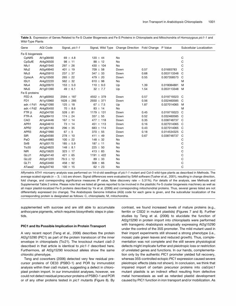
supplemented with sucrose and are still able to accumulate
anthocyane pigments, which requires biosynthetic steps in plas-
tids.
PIC1 and Its Possible Implication in Protein Transport
A very recent report (Teng et al., 2006) describes the protein
At2g15290 (PIC1) as part of the protein translocon of the inner
envelope in chloroplasts (Tic21). The knockout mutant cia5-3
described in that article is identical to pic1-1 described here.
Furthermore, all At2g15290 knockout alleles show the same
chlorotic phenotype.
Teng and coworkers (2006) detected very few residual pre-
cursor proteins of OE33 (PSBO-1) and POR by immunoblot
analysis within their cia5 mutant, suggesting a defect in chloro-
plast protein import. In our immunoblot analyses, however, we
could not detect residual precursor proteins of PSBO-1 and POR
or of any other proteins tested in pic1 mutants (Figure 8). By
contrast, we found increased levels of mature proteins (e.g.,
ferritin or CSD2 in mutant plastids) (Figures 7 and 8). Further
studies by Teng et al. (2006) to elucidate the function of
At2g15290 in protein import into chloroplasts were performed
with transgenic Arabidopsis ectopically expressing At2g15290
under the control of the 35S promoter. The mild mutant used in
their import experiments still showed a strong phenotype (i.e.,
mosaic pale green leaves and reduced growth). Thus, comple-
mentation was not complete and the still severe physiological
defects might implicate further and pleiotropic loss or restriction
of unrelated genes and functions. In our hands, complementa-
tion only by the authentic PIC1 promoter yielded full recovery,
whereas 35S-controlled ectopic PIC1 expression caused severe
pleiotropic effects (data not shown). In conclusion, we think that
impaired import of certain precursor proteins into cia5/pic1
mutant plastids is an indirect effect resulting from defective
metal homeostasis as well as retarded plastid development
caused by PIC1 function in iron transport and/or mobilization. As
Table 3. Expression of Genes Related to Fe-S Cluster Biogenesis and Fe-S Proteins in Chloroplasts and Mitochondria of Homozygous pic1-1 and
Wild-Type Plants
Gene AGI Code Signal, pic1-1 Signal, Wild Type Change Direction Fold Change P Value Subcellular Localization
Fe-S biogenesis
CpNifS At1g08490 69 6 4.8 120 6 44 No C
CpSufE At4g26500 98 6 11 88 6 12 No C
Nfu1 At4g01940 297 6 26 430 6 104 No C
Nfu2 At5g49940 401 6 19 709 6 58 Down 0.57 0.01693783 C
Nfu3 At4g25910 237 6 37 347 6 33 Down 0.68 0.053113348 C
CpIscA At1g10500 265 6 22 479 6 20 Down 0.55 0.007358573 C
ISU1 At4g22220 562 6 32 610 6 98 No M
Nfu4 At3g20970 153 6 5.0 110 6 9.0 Up 1.39 0.018084881 M
Nfu5 At1g51390 49 6 6.1 32 6 7.7 Up 1.54 0.053113348 M
Fe-S proteins
FED A At1g60950 2594 6 187 4552 6 379 Down 0.57 0.019719323 C
FD1 At1g10960 1628 6 285 2930 6 371 Down 0.56 0.032460585 C
adr.-l Fd1 At4g21090 125 6 18 67 6 7.5 Up 1.87 0.027014065 M
adr.-l Fd2 At4g05450 70 6 8.6 83 6 14 No M
FTR b At2g04700 528 6 81 1179 6 131 Down 0.45 0.019719323 C
FTR-A At5g08410 174 6 24 337 6 55 Down 0.52 0.032460585 C
CAO At1g44446 167 6 14 477 6 118 Down 0.35 0.038748737 C
APR1 At4g04610 73 6 14 451 6 113 Down 0.16 0.027014065 C
APR2 At1g62180 296 6 35 683 6 114 Down 0.43 0.027014065 C
APR3 At4g21990 67 6 5 370 6 55 Down 0.18 0.014534325 C
SIR At5g04590 278 6 10 411 6 49 Down 0.67 0.038748737 C
PaO At3g44880 100 6 22 148 6 32 No C
SirB At1g50170 185 6 5.9 187 6 11 No C
Tic55 At2g24820 148 6 8.1 225 6 50 No C
NIR At2g15620 323 6 17 413 6 83 No C
GLU1 At5g04140 421 6 65 1191 6 422 No C
GLU2 At2g41220 79.5 6 12 89 6 33 No C
GLT1 At5g53460 458 6 92 308 6 66 No C
ATase2 At4g34740 100 6 15 62 6 23 No C
Affymetrix ATH1 microarry analysis was performed on 14-d-old seedlings of pic1-1 mutant and Col-0 wild-type plants as described in Methods. The
average scaled signals (n ¼ 3; 6SD) are shown. Signal differences were evaluated by SAM software (Tusher et al., 2001), resulting in change direction,
fold change, and corresponding significance measures (P value, false discovery rate ¼ 5.31%). For details of the analysis, see Methods and
Supplemental Table 2 online. Please note that we listed all genes reported to be involved in the plastidic Fe-S cluster biogenesis machinery as well as
all major plastid-localized Fe-S proteins described by Ye et al. (2006) and corresponding mitochondrial proteins. Thus, several genes listed are not
differentially expressed (no change). The Arabidopsis Genome Initiative (AGI) code for each gene is given, and the subcellular localization of the
corresponding protein is designated as follows: C, chloroplasts; M, mitochondria.
Iron Transport in Arabidopsis Chloroplasts 1001
shown in Figure 8, several proteins are very well imported and
processed within pic1 mutant plastids.
In our opinion, the described chlorotic and dwarfed phenotype
of pic1 knockout mutants closely links PIC1 to metal homeosta-
sis of chloroplasts. Although PIC1 shows sequence homology
with prokaryotic solute permeases and not with known protein
import pores with four a-helical transmembrane domains (e.g.,
Tim proteins in the inner membrane of mitochondria [Murcha
et al., 2007]), a function of the protein in the translocation of
precursor proteins cannot be fully excluded. Finally, the func-
tional complementation of iron uptake–defective yeast mutants
and the measured iron uptake, mediated by PIC1 and sll1656,
clearly provide evidence for their function in iron transport and
metal homeostasis.
METHODS
Plant Material and Growth Conditions
All experiments were performed on Arabidopsis thaliana plants, ecotype
Col-0 (Lehle Seeds). The T-DNA insertion lines SALK_104852 (pic1-1),
GABI 577D06 (pic1-2), and 804F07 (pic1-3) were purchased from NASC
(University of Nottingham) and GABI-Kat (Max Planck Institute for Plant
Breeding Research). Before sowing, seeds were surface-sterilized with
ethanol and 5% hypochloride. To synchronize germination, all seeds
were kept at 48C for 3 d. Plants were grown on soil or on 0.3% Gelrite
medium (Serva) containing 1% D-sucrose and 0.53 MS salts at pH 5.7.
Plant growth occurred in growth chambers with a 16-h light (218C; photon
flux density of 100 mmol�m�2�s�1) and 8-h dark (168C) cycle.
Protein Import Experiments
Intact chloroplasts for import experiments were isolated from 8-d-old
Arabidopsis seedlings according to Aronsson and Jarvis (2002). The
cDNA of At-PIC1 was purchased as SSP pUNI51 clone U18531 (Yamada
et al., 2003). For in vitro transcription, PIC1 was subcloned into pGEM 4-Z
(EcoRI/SacI) (Promega). In vitro transcription was performed using the
mMessage mMachine kit (Ambion). Translation and protein import of the
precursor protein PIC1 were performed as described by Nada and Soll
(2004). The import reaction contained Arabidopsis chloroplasts equiva-
lent to 10 mg of chlorophyll and 4.5 mL of in vitro translation product in
100 mL of import buffer.
Antiserum Production and Immunoblot Analysis
To raise an antiserum against At-PIC1, the C-terminal end of the PIC1
cDNA was amplified by PCR on PIC1/pUNI51 using the oligonucleotide
primers PIC1(�154)-Ncofw (59-TGCCATGGTTTTCTGGTCGTTTGGGTA-
TATTC-39) and PIC1fl-Xhorev (59-GGCAGACTCGAGAGCAACCTTAG-
GAACTAC-39). The resulting truncated version PIC1(�154), which lacks
the first 154 amino acids of the protein, was subcloned into the pPROEx
Hta (NcoI/XhoI) plasmid vector (Invitrogen). The generated plasmid
PIC1(�154)/pPROEx Hta was used for overexpression after transforming
Escherichia coli BL21(DE3) cells (Novagen/Merck Biosciences). Rapidly
growing cells with an OD600 of 0.6 were induced with 0.6 mM isopropyl
1-thio-b-D-galactopyranoside for 3 h at 378C. Afterward, pelleted cells
were resuspended in cell lysis solution (50 mM Tris-HCl, pH 8.0, 25%
[w/v] sucrose, 1 mM EDTA, and 100 mg/mL DNase) and sonicated three
times for 30 s. Inclusion bodies were collected by centrifugation at 48C
and 20,000g for 30 min. The resulting pellet was resuspended in buffer A
(50 mM NaPP, pH 8.0, 100 mM NaCl, 2 mM b-mercaptoethanol, and 8 M
urea) and sonicated again. Remaining insoluble material was pelleted by
centrifugation (48C, 20,000g, 30 min). The major fraction of overex-
pressed recombinant PIC1(�154) protein was present in the urea-soluble
supernatant. Proteins in this fraction were separated by SDS-PAGE, and
the overexpressed PIC1(�154) of 15 kD was recovered by electroelution.
The protein was concentrated using an Amicon ultrafiltration cell with
YM10 membranes (Millipore) in 25 mM Tris-HCl, pH 8.0, 0.19 M Gylcin,
5 mM b-mercaptoethanol, and 1% (w/v) SDS and used as antigene to raise
antibodies in rabbit (BioGenes). Polyclonal antisera against Nt-RBCS, At-
OEP16.1, Ps-VIPP1, and At-PORB were raised in rabbit (Pineda Antibody
Service); anti-Lhcb4 antiserum was purchased from Agrisera.
Subfractionation of chloroplasts from 6-week-old Arabidopsis rosette
leaves was performed as described by Gerdes et al. (2006). For a total
protein extract from 4-week-old leaves, plant material was homogenized
in liquid nitrogen and proteins were extracted in 50 mM Tris-HCl, pH 8, 50
mM EDTA, 2% lithium dodecyl sulfate, 10 mM DTT, and 100 mM
phenylmethylsulfonyl fluoride on ice for 30 min. Cell debris was pelleted
by 15 min of centrifugation (48C, 14,000g). Proteins from envelopes,
stroma, and thylakoid membranes or from leaves of pic1 mutants and
wild-type plants were separated by SDS-PAGE and subsequently trans-
ferred to nitrocellulose filters. The antisera against PIC(�154) and PORB
were used in a 1:500 dilution, antiserum against At-FER was used at
1:2500, and all other antisera were diluted 1:1000; secondary anti-rabbit
IgG alkaline phosphatase antibodies (Sigma-Aldrich) were diluted
1:30,000. Nonspecific signals were blocked by 1% skim milk powder
and 0.3% BSA. Blots were stained using the alkaline phosphatase
reaction in the presence of nitroblue tetrazolium and bromochloroindolyl
phosphate as substrate; for FER detection, blots were stained with the
Immobilon Western kit (Millipore) using the chemiluminescent alkaline
phosphatase substrate.
Subcellular Localization of GFP Fusion Proteins
To generate a C-terminal fusion of GFP to the preprotein At-PIC1,
the entire coding sequence was amplified by PCR using the primers
flGATE fw (59-CACCATGCAATCACTACTCTTGCCGCC-39) and flGATE
rev (59-AGCAACCTTAGGAACTACGACAG-39) on PIC1/pUNI51. Accord-
ingly, the cDNA of At-OEP7 was amplified by OEP7forGFP (59-AAAAAG-
CAGGCTATATGGGAAAAACTTCGGGAG-39) and OEP7revGFP (59-AGA-
AAGCTGGGTACAAACCCTCTTTGGATGTGG-39) on Arabidopsis Col-0
cDNA. The resulting PCR products were subcloned into the binary
pK7FWG2 vector (Karimi et al., 2002) using the Gateway cloning system
(Invitrogen). pK7FWG2 provides a fusion of GFP to the C-terminal end of
the respective proteins, which are expressed under the control of the
constitutive 35S cauliflower mosaic virus promoter.
Arabidopsis mesophyll protoplasts were isolated from leaves of
4-week-old plants and transiently transfected with PIC1/pK7FWG2 and
OEP7/pK7FWG2 according to the protocol of the Sheen Lab (http://
genetics.mgh.harvard.edu/sheenweb/protocols_reg.html). After 16 h of
expression, fluorescence images were obtained with an epifluorescence
microscope (polychrome IV system; Till Photonics) using GFP and fluo-
rescein isothiocyanate filter sets. Photographs were taken with a cooled
infrared CCD camera and visualized with the TILLvision 4.0 software.
Transcript Level Profiling
To monitor transcript abundance, total RNA was extracted from the
respective Arabidopsis tissues using the Plant RNeasy extraction kit
(Qiagen). In general, three samples were harvested for each tissue type
from 10 (for mature plants) to 500 (for seedlings) individual plants to
decrease biological variation.
For microarray analysis, 5 mg of RNA from 14-d-old seedlings of the
pic1-1 mutant and the wild type was processed and hybridized to
Affymetrix GeneChip Arabidopsis ATH1 Genome Arrays using the Affy-
metrix One-Cycle Labeling and Control (Target) kit according to the
1002 The Plant Cell
manufacturer’s instructions. To provide biological replicates, RNA for
each sample was isolated from at least 40 individual seedlings. Further-
more, three mutant and three wild-type samples (n ¼ 3) were grown and
harvested with a time interval of 1 week. Microarray signals were made
comparable by scaling the average overall signal intensity of all probe
sets to a target signal of 100. Signals from pic1-1 (n¼ 3) and the wild type
(n ¼ 3) were analyzed by Affymetrix Data Mining Tool and GeneChip
Operating software (compare with Clausen et al., 2004). The statistical
significance of signal differences was evaluated by SAM software (Tusher
et al., 2001). Here, we performed a two-class unpaired t test on the signal
values (number of permutations ¼ 100). Setting the delta cutoff value at
1.55 resulted in a data set of 4235 significantly regulated genes with a
false discovery rate of 5.31%.
The RNA used in quantitative real-time RT-PCR was digested with
DNase and reverse-transcribed into cDNA (Clausen et al., 2004). Detec-
tion and quantification of transcripts were performed as described pre-
viously (Philippar et al., 2004) using a LightCycler (Roche). For PIC1, we
constructed the gene-specific primers LCfw (59-CTCCGTCGAACTT-
CCA-39) and LCrev (59-AACTACGACAGATTCCG-39), amplifying a pro-
duct of 333 bp. To prevent the amplification of contaminating genomic
DNA, the primers were selected to flank introns 2 and 3 (Figure 4A). To
analyze the residual N-terminal RNA in the mutants pic1-1 and pic1-2, the
primer pair pic1,2 fw (59-AGAACACCAATTCGCTAC-39) and pic1,2 rev
(59-CCTCATTATCACCGGG-39) was designed to amplify a product of 343
bp, spanning both sites of the T-DNA insertion. All quantifications were
normalized to the signal of actin cDNA fragments generated by the
primers Act fw (59-GGTGATGGTGTGTCT-39) and Act rev (59-ACTGAGC-
ACAATGTTAC-39), which amplified cDNA from actin 2 and 8 (At3g18780
and At1g49240).
Data used to create in silico transcriptional profiles were obtained
from the AtGenExpress developmental series leaves, shoots and stems,
and roots at the NASCArrays website (http://affymetrix.Arabidopsis.info/
narrays/experimentbrowse.pl.).
Characterization of the T-DNA Mutants pic1-1, pic1-2, and pic1-3
Genomic DNA of the heterozygous T-DNA insertion lines inside At-PIC1,
pic1-1 (SALK_104852) (Alonso et al., 2003), pic1-2 (GABI-Kat 577D06),
and pic1-3 (GABI-Kat 804F07) (Rosso et al., 2003) was screened by PCR
genotyping. PIC1 gene-specific primers in combination with T-DNA–
specific left border primers generated fragments of 524 bp (pic1-1), 716
bp (pic1-2), and 738 bp (pic1-3) on heterozygous and homozygous
plants. To identify plants with the T-DNA insertion in both alleles of PIC1,
we used gene-specific primers flanking the predicted T-DNA insertion
sites. DNA from homozygous, mutated pic1 gave no amplification pro-
duct, whereas the amplified regions on wild-type and heterozygous DNA
were 434 bp (pic1-1), 582 bp (pic1-2), and 519 bp (pic1-3). The following
primers were used: for pic1-1, 52fw (59-GTTTCGTTGTCCGCTGGC-
TTTAGC-39) and 52rev (59-GGTTAAATGAGTGCTGGAAACCTGGC-39);
for pic1-2, 77fw (59-TGCAATCACTACTCTTGCCGCCGG-39) and 77rev
(59-CAATCTCTTTGCAACCTGTGAATA-39); for pic1-3, 04fw (59-GTTCC-
TGTCTCAGTTCTTTATGCC-39) and 04rev (59-CAACAACTACCTCCTAT-
CACGATG-39); as well as SALK LBb1 (59-GCGTGGACCGCTTGCTG-
CAACT-39) and GABI LB1 (59-CCCATTTGGACGTGAATGTAGACAC-39).
For positions and orientations of the T-DNA inserts and oligonucleotide
primers, see Figure 4A. To verify PCR products and T-DNA insertion sites,
all amplified DNA fragments were sequenced.
To complement the mutant pic1-1, a 3327-bp genomic fragment of
PIC1 (gPIC1) was amplified by PCR on genomic DNA of Col-0 wild-type
plants and subcloned into the binary plasmid vector pKWG (Karimi et al.,
2002) using the Gateway cloning system (Invitrogen). The primers used to
clone gPIC1 were gPIC1fw (59-GTCAAACCTGAGTCTGATTTCTCTTC-39)
and gPIC1rev (59-AGCAACCTTAGGAACTACGACAG-39), flanking a 1830-bp
promoter region and the entire genomic sequence of PIC1 (1497 bp). The
construct gPIC1/pKWG was transformed into chemically competent Agro-
bacterium tumefaciens GV3101 cells. Heterozygous PIC1/pic1-1 plants (T4
generation) were transformed by vacuum infiltration as described by
Bechtold et al. (1993). Transformed T1 plants were selected on kanamycin
(100 mg/mL) containing agar medium (0.3% Gelrite, 1% D-sucrose, and
0.53 MS salts) and afterward transferred to soil. Plants in the T1 and T2
generations in addition were characterized by PCR genotyping. Oligonu-
cleotide primers for the gPIC1/pKWG T-DNA were PIC1 E3fw (59-GTTG-
TGAAAGGTTTGAGGAGCG-39) and attB2 adapter (59-ACCACTTTGTAC-
AAGAAAGCTGGGT-39), the latter being specific for the pKWG vector. The
T-DNA insertion in pic1-1 was detected as described above, and homo-
zygous pic1-1/pic1-1 alleles were identified by the absence of PCR
products from primers flanking the PIC1 gene: PIC1 59UTRfw (59-GCC-
CATTGCAACTGTGAAGAATAGACG-39) and PIC1 39UTRrev (59-CATTG-
AATCCGCAATCGTTGGTTCTC-39).
Electron Microscopy
Pieces of leaf tissue were fixed immediately with 2.5% glutaraldehyde in
75 mM sodium cacodylate and 2 mM MgCl2, pH 7.0, for 1 h at room
temperature, rinsed several times in fixative buffer, and post-fixed for 2 h
with 1% osmium tetroxide in fixative buffer at room temperature. After
two washing steps in distilled water, the cells were stained en bloc with
1% uranyl acetate in 20% acetone for 30 min. Dehydration was per-
formed with a graded acetone series. Samples were then infiltrated and
embedded in Spurr’s low-viscosity resin (Spurr, 1969). After polymeriza-
tion, ultrathin sections with thickness between 50 and 70 nm were cut with
a diamond knife and mounted on uncoated copper grids. The sections
were poststained with aqueous lead citrate (100 mM, pH 13.0). All micro-
graphs were taken with an EM 912 electron microscope (Zeiss) equipped
with an integrated Omega energy filter operated in the zero loss mode.
Semithin sections were cut with a diamond knife with a thickness of 500
nm and mounted on glass slides; phase contrast micrographs were taken
with a Zeiss Axiophot microscope and a Nikon D1000 digital camera.
Complementation and Iron Accumulation in Yeast Mutants
For functional expression in yeast, the entire coding sequence of At-PIC1
was amplified by PCR on the cDNA PIC1/pUNI51 using the primers
flGATE fw and flGATE rev (see above). The coding sequence of sll1656
from Synechocystis sp strain PCC 6803 was amplified by PCR on
genomic DNA with the following primers: sllfw (59-CACCATGAACC-
GACGCAACACCG-39) and sllrev (59-CTTGTTCAGACGGTCTAGCAAC-
CACAG-39). After subcloning into pCR2.1 TA vector (Invitrogen), both
cDNAs were subcloned into the yeast expression vectors pFL61 (PstI/
SpeI) and pDR195 (XhoI/BamHI). The cDNA of IRT1 from Arabidopsis was
purchased as GSLT cDNA (Castelli et al., 2004). The coding sequence of
IRT1 was amplified by PCR using the primers IRT1.NotIfw (59-ATT-
CAGGCGGCCGCATGAAAACAATCTTCCTCGTAC-39) and IRT1.NotIrev
(59-ATCTGGGCGGCCGCTTAAGCCCATTTGGCGATAATC-39) and sub-
cloned into pFL61 and pDR195 digested with NotI.
The yeast strains used in this study were fet3 fet4: DEY1453 (MATa/
MATa ade2/þ can1/can1 his3/his3 leu2/leu2 trp1/trp1 ura3/ura3 fet3-2T
HIS3/fet3-2THIS3 fet4-1TLEU2/fet4-1TLEU2) (Eide et al., 1996) and
ctr1: 64p (MATa gcn4 his3 leu2 trp1 ura3 ctr1TLEU2) (Dancis et al.,
1994a). Untransformed mutant yeast cells were grown in YPD (yeast
extract, peptone, and D-glucose) medium and, after transformation in
selective synthetic complete (SC) (�uracil) minimal medium, supplemented
with 20 g/L glucose and necessary auxotrophic supplements (Eide et al.,
1996). For optimal growth of the fet3 fet4 strain, the pH of YPD and SC
(�uracil) was decreased to 4.0 and 3.5, respectively. Yeast cells were
transformed with the respective plasmids using standard procedures
(Gietz and Schiestl, 1991). For complementation experiments, 10-mL
drops of yeast cultures were spotted in different dilutions onto SC
Iron Transport in Arabidopsis Chloroplasts 1003
(�uracil) plates, pH 5.0, supplemented with different concentrations of
FeCl3 or EDTA. For iron accumulation, the fet3 fet4 mutant transformants
were grown to exponential phase in synthetic defined medium containing
30 g/L glucose, 1.7 g/L yeast nitrogen base without amino acids, 5 g/L
(NH4)2SO4, and necessary auxotrophic amino acids without uracil. This
medium was also supplemented with 50 mM Fe(III)-EDTA. Cells were
centrifuged for 10 min at 3500 rpm at 48C and washed once in ice-cold
ultrapure water and once in ice-cold MGN medium (10 mM MES, 3%
glucose, and 1 mM nitrilotriacetic acid, pH 6.1). Cells were resuspended
in MGN medium to reach an OD600 of 4.0. Iron accumulation was assayed
in MGN medium supplemented with 1 mM ascorbate and 40 mM 59Fe-
labeled FeCl3. Cells were incubated for 15 min at 308C before the addition
of MGN medium with 59Fe-labeled FeCl3 (specific activity of 740 MBq/
mol; Perkin-Elmer) for 10 min. Finally, cells were vacuum-filtered through
Whatman GF/C filters and washed with 20 mL of ice-cold SSW solution
(Eide et al., 1992). The filters were used for the determination of total
activity by scintillation counting (Wallac). Mean values for iron accumu-
lation resulted from the measurement of three replicates for each trans-
formant. Data were subjected to one-way analysis of variance followed by
a Student-Newman-Keuls test (P < 0.05) using the statistical software
Sigmastat for Windows (version 2.0; Systat Software).
Computational Analysis of Protein and DNA Sequences
To identify proteins homologous with At-PIC1, BLAST searches against
GenBank (http://www.ncbi.nlm.nih.gov/BLAST/; Altschul et al., 1997),
PHYSCObase (http://moss.nibb.ac.jp/; Nishiyama et al., 2003), and the
genome databases of Chlamydomonas reinhardtii (Chlamydomonas
reinhardtii version 3.0 database; http://genome.jgi-psf.org/Chlre3/Chlre3.
home.html) and Cyanidioschyzon merolae (Cyanidioschyzon merolae
Genome Project; http://merolae.biol.s.u-tokyo.ac.jp/; Matsuzaki et al.,
2004) were performed. COGs (Tatusov et al., 2003) are accessible at http://
www.ncbi.nlm.nih.gov/COG/. The plant membrane protein database
ARAMEMNON (version 3.2; http://aramemnon.botanik.uni-koeln.de/;
Schwacke et al., 2003) was used to determine consensus transmem-
brane topologies. The phylogenetic tree was constructed with the pro-
gram PAUP version 4.0b10 (Swofford, 2002). A minimal evolution tree
was calculated using the default settings. In the analysis, 148 amino acids
were included that were situated within or adjacent to the predicted
transmembrane helices (see Supplemental Figure 1 online). The data
were bootstrap-resampled 100 times. Protein sequence alignments were
generated by ClustalX (Thompson et al., 1997) using the Gonnet series
protein weight matrix with default settings. Promoter prediction was per-
formed with Neural Network Promoter Prediction (http://www.fruitfly.org/
seq_tools/promoter.html; Reese, 2001), and the plant TATA box was
defined by MatInspector at GenomatixSuite 3.4.1 (http://www.genomatix.
de; Cartharius et al., 2005).
Accession Numbers
Sequence data from this article can be found in the GenBank, GenPept,
and PHYSCObase data libraries, the genome databases of C. reinhardtii
and C. merolae, or under the respective Arabidopsis gene index (Arabi-
dopsis Genome Initiative locus identifier): Arabidopsis thaliana At-PIC1
(AY057510, At2g15290); rice (Oryza sativa) Os-PIC1 (XM_464386,
Os02g0187600) and Os-PIC2 (AP003573, Os06g0638100); maize (Zea
mays) Zm-PIC1 (BT017876) and Zm-PIC2 (BT018285); Lotus japonicus
Lj-PIC1 (AP006404); Physcomitrella patens Pp-PIC1 (contig 5795);
C. reinhardtii PIC1 (protein identifier 178114); C. merolae PIC1 (protein
identifier CMN128C); Synechocystis (sll1656, BAA16830); Anabena
sp PCC 7120 (all4113, BAB75812); Thermosynechococcus elongatus
BP-1 (tll0396, BAC07948); Prochlorococcus marinus strain MIT 9313
(PMT0365, NP_894198); and Arabidopsis IRT1 (BX827853, At4g19690)
and OEP7 (NM_115102, At3g52420).
Supplemental Data
The following materials are available in the online version of this article.
Supplemental Figure 1. Multiple Sequence Alignment of the PIC1
Family in Eukaryotes and Cyanobacteria.
Supplemental Figure 2. IRT1 mRNA in pic1 Mutant and Col-0 Wild-
Type Plants under Iron-Sufficient, Iron-Deficient, and Iron-Abundant
Conditions.
Supplemental Table 1. Fe and Cu Content in Shoots of pic1-1 Mutant
and Col-0 Wild-Type Plants.
Supplemental Table 2. Raw Array Data (Affymetrix ATH1 GeneChip)
of pic1-1 Mutant versus Wild-Type Analysis.
ACKNOWLEDGMENTS
The skillful technical assistance of Daniela Eder, Karl Mayer, and Silvia
Dobler is gratefully acknowledged. We thank Ulrike Oster for microarray
data analysis, Manuela Baumgartner for help with phylogenetic analysis
and chloroplast subfractionation, Ewa Firley-Kwotka and Bettina Bolter
for discussion of protein import, and Sabine Molik and Roland Lill for
advice and critical discussion of yeast iron metabolism. We also thank
Petra Dietrich for the yeast mutant strains fet3 fet4 and ctr1. For the
donation of antisera, we thank Frederic Gaymard for ferritin, Marinus
Pilon for CSD1, CSD2, and FSD1, and R. Berzborn for PSBO-1 anti-
serum. This work was supported by grants to K.P. from the Deutsche
Forschungsgemeinschaft.
Received September 15, 2006; revised December 29, 2006; accepted
February 2, 2007; published March 2, 2007.
REFERENCES
Abdel-Ghany, S.E., Muller-Moule, P., Niyogi, K.K., Pilon, M., and
Shikanai, T. (2005). Two P-type ATPases are required for copper
delivery in Arabidopsis thaliana chloroplasts. Plant Cell 17: 1233–
1251.
Alonso, J.M., Stepanova, A.N., Leisse, T.J., Kim, C.J., Chen, H.,
Shinn, P., Stevenson, D.K., Zimmerman, J., Barajas, P., Cheuk, R.,
and Heller, C. (2003). Genome-wide insertional mutagenesis of
Arabidopsis thaliana. Science 301: 653–657.
Altschul, S.F., Madden, T.L., Schaffer, A.A., Zhang, J., Zhang, Z.,
Miller, W., and Lipman, D.J. (1997). Gapped BLAST and PSI-BLAST:
A new generation of protein database search programs. Nucleic Acids
Res. 25: 3389–3402.
Aronsson, H., and Jarvis, P. (2002). A simple method for isolating
import-competent Arabidopsis chloroplasts. FEBS Lett. 529: 215–220.
Asada, K. (1999). The water-water cycle in chloroplasts: Scavenging of
active oxygen and dissipation of excess photons. Annu. Rev. Plant
Physiol. Plant Mol. Biol. 50: 601–639.
Aseeva, E., Ossenbuhl, F., Eichacker, L.A., Wanner, G., Soll, J., and
Vothknecht, U.C. (2004). Complex formation of Vipp1 depends on its
alpha-helical PspA-like domain. J. Biol. Chem. 279: 35535–35541.
Balk, J., and Lobreaux, S. (2005). Biogenesis of iron-sulfur proteins in
plants. Trends Plant Sci. 10: 324–331.
Bechtold, N., Ellis, J., and Pelletier, G. (1993). In planta Agrobacterium-
mediated gene transfer by infiltration of adult Arabidopsis thaliana
plants. C. R. Acad. Sci. Paris/Life Sci. 316: 1194–1199.
Briat, J.F., and Lobreaux, S. (1997). Iron transport and storage in
plants. Trends Plant Sci. 2: 187–193.
1004 The Plant Cell
Briat, J.F., Lobreaux, S., Grignon, N., and Vansuyt, G. (1999). Reg-
ulation of plant ferritin synthesis: How and why. Cell. Mol. Life Sci. 56:
155–166.
Cartharius, K., Frech, K., Grote, K., Klocke, B., Haltmeier, M.,
Klingenhoff, A., Frisch, M., Bayerlein, M., and Werner, T. (2005).
MatInspector and beyond: Promoter analysis based on transcription
factor binding sites. Bioinformatics 13: 2933–2942 .
Castelli, V., et al. (2004). Whole genome sequence comparisons and
‘‘full-length’’ cDNA sequences: A combined approach to evaluate
and improve Arabidopsis genome annotation. Genome Res. 14:
406–413.
Clausen, C., Ilkavets, I., Thomson, R., Philippar, K., Vojta, A.,
Mohlmann, T., Neuhaus, E., Fulgosi, H., and Soll, J. (2004). Intra-
cellular localization of VDAC proteins in plants. Planta 220: 30–37.
Colangelo, E.P., and Guerinot, M.L. (2006). Put the metal to the petal:
Metal uptake and transport throughout plants. Curr. Opin. Plant Biol.
9: 322–330.
Connolly, E.L., Fett, J.P., and Guerinot, M.L. (2002). Expression of the
IRT1 metal transporter is controlled by metals at the levels of tran-
script and protein accumulation. Plant Cell 14: 1347–1357.
Connolly, E.L., and Guerinot, M. (2002). Iron stress in plants. Genome
Biol. 3: REVIEWS1024.1–REVIEWS1024.4.
Curie, C., Alonso, J.M., Le Jean, M., Ecker, J.R., and Briat, J.F.
(2000). Involvement of NRAMP1 from Arabidopsis thaliana in iron
transport. Biochem. J. 347: 749–755.
Curie, C., and Briat, J.F. (2003). Iron transport and signaling in plants.
Annu. Rev. Plant Biol. 54: 183–206.
Dancis, A., Haile, D., Yuan, D.S., and Klausner, R.D. (1994a). The
Saccharomyces cerevisiae copper transport protein (Ctr1p). Biochem-
ical characterization, regulation by copper, and physiologic role in
copper uptake. J. Biol. Chem. 269: 25660–25667.
Dancis, A., Yuan, D.S., Haile, D., Askwith, C., Eide, D., Moehle, C.,
Kaplan, J., and Klausner, R.D. (1994b). Molecular characterization of
a copper transport protein in S. cerevisiae: An unexpected role for
copper in iron transport. Cell 76: 393–402.
Dix, D.R., Bridgham, J.T., Broderius, M.A., Byersdorfer, C.A., and
Eide, D.J. (1994). The FET4 gene encodes the low affinity Fe(II)
transport protein of Saccharomyces cerevisiae. J. Biol. Chem. 269:
26092–26099.
Drummond, R.S.M., Tutone, A., Li, Y.C., and Gardner, R.C. (2006). A
putative magnesium transporter AtMRS2-11 is localized to the plant
chloroplast envelope membrane system. Plant Sci. 170: 78–89.
Eide, D., Broderius, M., Fett, J., and Guerinot, M.L. (1996). A novel
iron-regulated metal transporter from plants identified by functional
expression in yeast. Proc. Natl. Acad. Sci. USA 93: 5624–5628.
Eide, D., Davis-Kaplan, S., Jordan, I., Sipe, D., and Kaplan, J. (1992).
Regulation of iron uptake in Saccharomyces cerevisiae. The ferrir-
eductase and Fe(II) transporter are regulated independently. J. Biol.
Chem. 267: 20774–20781.
Emanuelsson, O., Nielsen, H., and von Heijne, G. (1999). ChloroP, a
neural network-based method for predicting chloroplast transit pep-
tides and their cleavage sites. Protein Sci. 8: 978–984.
Froehlich, J.E., Wilkerson, C.G., Ray, W.K., McAndrew, R.S.,
Osteryoung, K.W., Gage, D.A., and Phinney, B.S. (2003). Proteomic
study of the Arabidopsis thaliana chloroplastic envelope membrane
utilizing alternatives to traditional two-dimensional electrophoresis.
J. Proteome Res. 2: 413–425.
Garcia, O., Bouige, P., Forestier, C., and Dassa, E. (2004). Inventory
and comparative analysis of rice and Arabidopsis ATP-binding cas-
sette (ABC) systems. J. Mol. Biol. 343: 249–265.
Gerdes, L., Bals, T., Klostermann, E., Karl, M., Philippar, K., Hunken,
M., Soll, J., and Schunemann, D. (2006). A second thylakoid mem-
brane-localized Alb3/OxaI/YidC homologue is involved in proper
chloroplast biogenesis in Arabidopsis thaliana. J. Biol. Chem. 281:
16632–16642.
Gietz, R.D., and Schiestl, R.H. (1991). Applications of high efficiency
lithium acetate transformation of intact yeast cells using single-
stranded nucleic acids as carrier. Yeast 7: 253–263.
Hall, J.L., and Williams, L.E. (2003). Transition metal transporters in
plants. J. Exp. Bot. 54: 2601–2613.
Henriques, R., Jasik, J., Klein, M., Martinoia, E., Feller, U., Schell, J.,
Pais, M.S., and Koncz, C. (2002). Knock-out of Arabidopsis metal
transporter gene IRT1 results in iron deficiency accompanied by cell
differentiation defects. Plant Mol. Biol. 50: 587–597.
Higgins, C.F. (2001). ABC transporters. Physiology, structure and
mechanism—An overview. Res. Microbiol. 152: 205–210.
Hirsch, J., Marin, E., Floriani, M., Chiarenza, S., Richaud, P.,
Nussaume, L., and Thibaud, M.C. (2006). Phosphate deficiency
promotes modification of iron distribution in Arabidopsis plants.
Biochimie 88: 1767–1771.
Kaiser, B.N., Moreau, S., Castelli, J., Thomson, R., Lambert, A.,
Bogliolo, S., Puppo, A., and Day, D.A. (2003). The soybean NRAMP
homologue, GmDMT1, is a symbiotic divalent metal transporter
capable of ferrous iron transport. Plant J. 35: 295–304.
Karimi, M., Inze, D., and Depicker, A. (2002). GATEWAY vectors for
Agrobacterium-mediated plant transformation. Trends Plant Sci. 7:
193–195.
Kliebenstein, D.J., Monde, R.A., and Last, R.L. (1998). Superoxide
dismutase in Arabidopsis: An eclectic enzyme family with disparate
regulation and protein localization. Plant Physiol. 118: 637–650.
Korshunova, Y.O., Eide, D., Clark, W.G., Guerinot, M.L., and Pakrasi,
H.B. (1999). The IRT1 protein from Arabidopsis thaliana is a metal
transporter with a broad substrate range. Plant Mol. Biol. 40: 37–44.
Lanquar, V., Lelievre, F., Bolte, S., Hames, C., Alcon, C., Neumann,
D., Vansuyt, G., Curie, C., Schroder, A., Kramer, U., Barbier-
Brygoo, H., and Thomine, S. (2005). Mobilization of vacuolar iron by
AtNRAMP3 and AtNRAMP4 is essential for seed germination on low
iron. EMBO J. 24: 4041–4051.
Lee, Y.J., Kim, D.H., Kim, Y.W., and Hwang, I. (2001). Identification of
a signal that distinguishes between the chloroplast outer envelope mem-
brane and the endomembrane system in vivo. Plant Cell 13: 2175–2190.
Le Jean, M., Schikora, A., Mari, S., Briat, J.F., and Curie, C. (2005). A
loss-of-function mutation in AtYSL1 reveals its role in iron and
nicotianamine seed loading. Plant J. 44: 769–782.
Matsuzaki, M., et al. (2004). Genome sequence of the ultrasmall
unicellular red alga Cyanidioschyzon merolae 10D. Nature 428: 653–657.
Moller, S.G., ed (2005). Plastids. (Oxford, UK: Blackwell Publishing).
Murcha, M.W., Elhafez, D., Lister, R., Tonti-Filippini, J., Baumgartner,
M., Philippar, K., Carrie, C., Mokranjac, D., Soll, J., and Whelan, J.
(2007). Characterisation of the preprotein and amino acid transporter
gene family in Arabidopsis. Plant Physiol. 143: 199–212.
Nada, A., and Soll, J. (2004). Inner envelope protein 32 is imported into
chloroplasts by a novel pathway. J. Cell Sci. 117: 3975–3982.
Nakamura, Y., et al. (2003). Complete genome structure of Gloeobacter
violaceus PCC 7421, a cyanobacterium that lacks thylakoids. DNA
Res. 10: 137–145.
Nishiyama, T., et al. (2003). Comparative genomics of Physcomitrella
patens gametophytic transcriptome and Arabidopsis thaliana: Impli-
cation for land plant evolution. Proc. Natl. Acad. Sci. USA 100: 8007–
8012.
Philippar, K., Ivashikina, N., Ache, P., Christian, M., Luthen, H.,
Palme, K., and Hedrich, R. (2004). Auxin activates KAT1 and KAT2,
two Kþ-channel genes expressed in seedlings of Arabidopsis thaliana.
Plant J. 37: 815–827.
Raux-Deery, E., Leech, H.K., Nakrieko, K.A., McLean, K.J., Munro,
A.W., Heathcote, P., Rigby, S.E.J., Smith, A.G., and Warren, M.J.
Iron Transport in Arabidopsis Chloroplasts 1005
(2005). Identification and characterization of the terminal enzyme of
siroheme biosynthesis from Arabidopsis thaliana. J. Biol. Chem. 280:
4713–4721.
Raven, J.A., Evans, M.C.E., and Korb, R.E. (1999). The role of trace
metals in photosynthetic electron transport in O2-evolving organisms.
Photosynth. Res. 60: 111–149.
Reese, M.G. (2001). Application of a time-delay neural network to
promoter annotation in the Drosophila melanogaster genome. Com-
put. Chem. 26: 51–56.
Robinson, N.J., Procter, C.M., Connolly, E.L., and Guerinot, M.L.
(1999). A ferric-chelate reductase for iron uptake from soils. Nature
397: 694–697.
Rolland, N., Ferro, M., Seigneurin-Berny, D., Garin, J., Douce, R.,
and Joyard, J. (2003). Proteomics of chloroplast envelope mem-
branes. Photosynth. Res. 78: 205–230.
Rosso, M.G., Li, Y., Strizhov, N., Reiss, B., Dekker, K., and
Weisshaar, B. (2003). An Arabidopsis thaliana T-DNA mutagenized
population (GABI-Kat) for flanking sequence tag-based reverse ge-
netics. Plant Mol. Biol. 53: 247–259.
Schmid, M., Davison, T.S., Henz, S.R., Pape, U.J., Demar, M.,
Vingron, M., Scholkopf, B., Weigel, D., and Lohmann, J.U.
(2005). A gene expression map of Arabidopsis thaliana development.
Nat. Genet. 37: 501–506.
Schwacke, R., Schneider, A., van der Graaff, E., Fischer, K., Catoni,
E., Desimone, M., Frommer, W.B., Flugge, U.I., and Kunze, R.
(2003). ARAMEMNON, a novel database for Arabidopsis integral
membrane proteins. Plant Physiol. 131: 16–26.
Seigneurin-Berny, D., et al. (2006). HMA1, a new Cu-ATPase of the
chloroplast envelope, is essential for growth under adverse light
conditions. J. Biol. Chem. 182: 2882–2892.
Shikanai, T., Muller-Moule, P., Munekage, Y., Niyogi, K.K., and
Pilon, M. (2003). PAA1, a P-type ATPase of Arabidopsis, functions in
copper transport in chloroplasts. Plant Cell 15: 1333–1346.
Shingles, R., North, M., and McCarty, R.E. (2001). Direct measure-
ment of ferrous ion transport across membranes using a sensitive
fluorometric assay. Anal. Biochem. 296: 106–113.
Shingles, R., North, M., and McCarty, R.E. (2002). Ferrous ion trans-
port across chloroplast inner envelope membranes. Plant Physiol.
128: 1022–1030.
Spurr, A.R. (1969). A low-viscosity epoxy resin embedding medium for
electron microscopy. J. Ultrastruct. Res. 26: 31–43.
Swofford, D.L. (2002). PAUP*. Phylogenetic Analysis Using Parsimony
*and Other Methods, Version 4. (Sunderland, MA: Sinauer Associates).
Talke, I.N., Hanikenne, M., and Kramer, U. (2006). Zinc-dependent
global transcriptional control, transcriptional deregulation, and higher
gene copy number for genes in metal homeostasis of the hyper-
accumulator Arabidopsis halleri. Plant Physiol. 142: 148–167.
Tatusov, R., et al. (2003). The COG database: An updated version
includes eukaryotes. BMC Bioinformatics 4: 41.
Teng, Y.S., Su, Y.S., Chen, L.J., Lee, Y.J., Hwang, I., and Li, H.M.
(2006). Tic21 is an essential translocon component for protein trans-
location across the chloroplast inner envelope membrane. Plant Cell
18: 2247–2257.
Thomine, S., Lelievre, F., Debarbieux, E., Schroeder, J.I., and
Barbier-Brygoo, H. (2003). AtNRAMP3, a multispecific vacuolar
metal transporter involved in plant responses to iron deficiency. Plant
J. 34: 685–695.
Thomine, S., Wang, R., Ward, J.M., Crawford, N.M., and Schroeder,
J.I. (2000). Cadmium and iron transport by members of a plant metal
transporter family in Arabidopsis with homology to Nramp genes.
Proc. Natl. Acad. Sci. USA 97: 4991–4996.
Thompson, J.D., Gibson, T.J., Plewniak, F., Jeanmougin, F., and
Higgins, D.G. (1997). The CLUSTAL_X windows interface: Flexible
strategies for multiple sequence alignment aided by quality analysis
tools. Nucleic Acids Res. 25: 4876–4882.
Tusher, V.G., Tibshirani, R., and Chu, G. (2001). Significance analysis
of microarrays applied to the ionizing radiation response. Proc. Natl.
Acad. Sci. USA 98: 5116–5121.
Van Wuytswinkel, O., Vansuyt, G., Grignon, N., Fourcroy, P., and
Briat, J.F. (1998). Iron homeostasis alteration in transgenic tobacco
overexpressing ferritin. Plant J. 17: 93–97.
Varotto, C., Maiwald, D., Pesaresi, P., Jahns, P., Salamini, F., and
Leister, D. (2002). The metal ion transporter IRT1 is necessary for iron
homeostasis and efficient photosynthesis in Arabidopsis thaliana.
Plant J. 31: 589–599.
Vert, G., Grotz, N., Dedaldechamp, F., Gaymard, F., Guerinot, M.L.,
Briat, J.F., and Curie, C. (2002). IRT1, an Arabidopsis transporter
essential for iron uptake from the soil and for plant growth. Plant Cell
14: 1223–1233.
Vert, G.A., Briat, J.F., and Curie, C. (2003). Dual regulation of the
Arabidopsis high-affinity root iron uptake system by local and long-
distance signals. Plant Physiol. 132: 796–804.
Vothknecht, U.C., and Soll, J. (2005). Chloroplast membrane trans-
port: Interplay of prokaryotic and eukaryotic traits. Gene 354:
99–109.
Waters, B.M., Chu, H.H., DiDonato, R.J., Roberts, L.A., Eisley, R.B.,
Lahner, B., Salt, D.E., and Walker, E.L. (2006). Mutations in
Arabidopsis Yellow Stripe-Like1 and Yellow Stripe-Like3 reveal their
roles in metal ion homeostasis and loading of metal ions in seeds.
Plant Physiol. 141: 1446–1458.
Wintz, H., Fox, T., Wu, Y.Y., Feng, V., Chen, W., Chang, H.S., Zhu, T.,
and Vulpe, C. (2003). Expression profiles of Arabidopsis thaliana in
mineral deficiencies reveal novel transporters involved in metal ho-
meostasis. J. Biol. Chem. 278: 47644–47653.
Yamada, K., et al. (2003). Empirical analysis of transcriptional activity in
the Arabidopsis genome. Science 302: 842–846.
Ye, H., Pilon, M., and Pilon-Smits, E.A.H. (2006). CpNifS-dependent
iron-sulfur cluster biogenesis in chloroplasts. New Phytol. 171: 285–292.
1006 The Plant Cell
DOI 10.1105/tpc.106.047407; originally published online March 2, 2007; 2007;19;986-1006Plant Cell
PhilipparDaniela Duy, Gerhard Wanner, Anderson R. Meda, Nicolaus von Wirén, Jürgen Soll and Katrin
Chloroplasts, Mediates Iron TransportArabidopsisPIC1, an Ancient Permease in
This information is current as of May 22, 2018
Supplemental Data /content/suppl/2007/03/05/tpc.106.047407.DC1.html
References /content/19/3/986.full.html#ref-list-1
This article cites 75 articles, 30 of which can be accessed free at:
Permissions https://www.copyright.com/ccc/openurl.do?sid=pd_hw1532298X&issn=1532298X&WT.mc_id=pd_hw1532298X
eTOCs http://www.plantcell.org/cgi/alerts/ctmain
Sign up for eTOCs at:
CiteTrack Alerts http://www.plantcell.org/cgi/alerts/ctmain
Sign up for CiteTrack Alerts at:
Subscription Information http://www.aspb.org/publications/subscriptions.cfm
is available at:Plant Physiology and The Plant CellSubscription Information for
ADVANCING THE SCIENCE OF PLANT BIOLOGY © American Society of Plant Biologists