piezo1 ion channel pore properties are dictated by c ... · pdf filepiezo1 ion channel pore...
TRANSCRIPT
ARTICLE
Received 29 Mar 2015 | Accepted 20 Apr 2015 | Published 26 May 2015
Piezo1 ion channel pore properties aredictated by C-terminal regionBertrand Coste1,2,*, Swetha E. Murthy2,*, Jayanti Mathur3, Manuela Schmidt2,w, Yasmine Mechioukhi1,
Patrick Delmas1 & Ardem Patapoutian2,3
Piezo1 and Piezo2 encode mechanically activated cation channels that function as mechano-
transducers involved in vascular system development and touch sensing, respectively.
Structural features of Piezos remain unknown. Mouse Piezo1 is bioinformatically predicted
to have 30–40 transmembrane (TM) domains. Here, we find that nine of the putative inter-
transmembrane regions are accessible from the extracellular side. We use chimeras between
mPiezo1 and dPiezo to show that ion-permeation properties are conferred by C-terminal
region. We further identify a glutamate residue within a conserved region adjacent to the last
two putative TM domains of the protein, that when mutated, affects unitary conductance and
ion selectivity, and modulates pore block. We propose that this amino acid is either in the
pore or closely associates with the pore. Our results describe important structural motifs of
this channel family and lay the groundwork for a mechanistic understanding of how Piezos are
mechanically gated and conduct ions.
DOI: 10.1038/ncomms8223 OPEN
1 Aix Marseille Universite, CNRS, CRN2M-UMR7286, 13344 Marseille, France. 2 Howard Hughes Medical Institute, Molecular and Cellular Neuroscience, TheScripps Research Institute, La Jolla, California 92037, USA. 3 Genomics Institute of the Novartis Research Foundation, San Diego, California 92121, USA.* These authors contributed equally to this work. w Present Address: Somatosensory Signaling Group; Max Planck Institute of Experimental Medicine, 37075Goettingen, Germany. Correspondence and requests for materials should be addressed to B.C. (email: [email protected]).
NATURE COMMUNICATIONS | 6:7223 | DOI: 10.1038/ncomms8223 | www.nature.com/naturecommunications 1
& 2015 Macmillan Publishers Limited. All rights reserved.
Mechanotransduction is the process by which mechanicalstimuli are converted into biological activity. Piezos aremechanically activated (MA) cation channels conserved
through evolution and act as mechanotransducers in variousbiological processes. The single Piezo gene in flies is involved innociception1; zebrafish and mouse Piezo2 in touch sensation2–6;zebrafish Piezo1 in red blood cell volume regulation7; and mousePiezo1 in vascular development8,9. In humans, mutations thatalter channel gating of Piezo1 and 2 are linked to variousdisorders with dominant inheritance10–12. Piezo proteins containover 2,000 amino acid residues with an estimated 30–40transmembrane (TM) segments and are likely to form homo-tetramers in a complex weighing over 1.2 million daltons13,14.Piezos lack homology with other proteins, and their structuralfeatures remain unknown. The large size and numeroushydrophobic domains of Piezos constitute technical challengesfor structural analysis of the intact channel15. Basic questionsregarding Piezo topology and the location of the ion-permeationpathway remain unanswered, and yet these questions are crucialfor a mechanistic understanding of how the channel is gatedby mechanical forces, and how human disease-related pointmutations affect channel function16.
We aimed to determine the topology of these large proteinsand delineate the amino-acid residues involved in the ion-permeation pathway. Here, we provide experimental evidence toconfirm the position of the amino (N)- and carboxy (C)-terminals and 13 of the putative inter-hydrophobic loop regionsof the protein. We show that the C-terminal region of the Piezoprotein encompasses the pore. Within this region, we identify aglutamate residue involved in ion conduction properties. Ourresults lay the groundwork for understanding how Piezos aremechanically gated and conduct ions, and how Piezo mutationsaffect human biology.
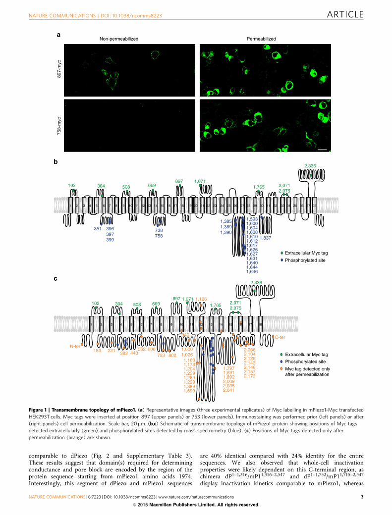
ResultsTransmembrane topology of Piezo channels. To characterizethe transmembrane topology of mPiezo1 we combined bioin-formatics analysis, immunostaining to detect extracellular tagsinserted in predicted loop regions, and detection of intracellularphosphorylation sites by mass spectrometry (Fig. 1). First, weused hydrophobicity plots, transmembrane segment predictionsand sequence alignment of functionally tested Piezo proteins(human, mouse and fly Piezos) to generate a predicted trans-membrane topology of mPiezo1. This virtual topology was thentested experimentally by inserting Myc tags at mPiezo1 terminiand in each predicted loop. In some cases, more than one tag wastested per predicted loop. Each of these constructs was subjectedto an immunostaining protocol to test whether an anti-Mycantibody could recognize the Myc epitope in mPiezo1 in non-permeabilized cells, suggesting an extracellular topology (Fig. 1aand Supplementary Fig. 1). A negative signal would suggest anintracellular epitope or one that is masked by the cell membraneor another such mechanism17. Therefore, only tags that gave apositive signal were used to predict the topology. A total of 48Myc constructs were designed. Forty-five of these constructsshowed staining with the Myc antibody after permeabilization,among which only 10 constructs were positive for stainingwithout permeabilization, and therefore predicted to be presentextracellularly (Supplementary Fig. 2 and SupplementaryTable 1). Two among the 10 Myc constructs were four aminoacids apart and can account only for a single extracellular loop,therefore nine extracellular loops are labelled. The orientationof these extracellular loops agreed with the hydropathy/hydrophilicity plot predictions. Insertion of Myc tags at theN- or C-terminal, and at seven out of the nine extracellular loop
positions did not affect channel function, and resulted in MAcurrents of amplitude comparable to WT currents. Whole-cellcurrents could not be detected from constructs with Myc tags atamino acid position 2,071, 2,075 or 2,336, suggesting that taginsertions there compromised channel function. Next, using massspectrometry, we analysed phosphorylated sites within mPiezo1purified protein to identify intracellular loops in the protein. Atotal of 24 phosphorylated peptides were detected correspondingto phosphorylation of 23 positions (Supplementary Table 2).Combining Myc-immunostaining and phosphorylation sitestogether with the bioinformatics analysis allowed us toconstruct a model topology, which predicts 38 transmembranedomains with intracellular N- and C-termini (Fig. 1b). However,this might be an over-estimation of transmembrane domains,since the prediction heavily relies on bioinformatics, andbecause we were unable to get experimental evidence for manyof the predicted extracellular loops. If we instead consider thatMyc tags detected only after permeabilization may be indicativeof intracellular or transmembrane positions, then a second modelwith less TM domains can be proposed (Fig. 1c). In this model,among the 35 Myc tags detected only after permeabilization,only one is predicted to be extracellular. Indeed, Myc 1126 isexpected to be present in the extracellular loop labelled byMyc 1071 in both models because no predicted TM domainor hydrophobic domain is present between Myc 1071 andMyc 1126. Therefore, absence of Myc 1126 staining withoutpermeabilization is likely due to accessibility issue. In this secondmodel, mPiezo1 is predicted to have a minimum of 18 TMdomains. In addition, if we further assume that the inter-hydrophobic loops with phosphorylated residues are the only fourintracellular loops, then only 10 of the hydrophobic regionswould be counted as transmembrane domains. This extremescenario would predict a topology with a minimum of 10 TMdomains.
Pore-related properties of mPiezo1/dPiezo chimeras. We nextset out to identify residues involved in the ion-permeationpathway of the channel. mPiezo1 and dPiezo differ in their bio-physical pore properties, such as sensitivity to the polycationicpore blocker ruthenium red (RR) and single-channel con-ductance14. We therefore generated chimeras between mPiezo1and dPiezo based on our predicted mPiezo1 topology, andassayed them for these pore-related properties (Fig. 2). Allchimeras were expressed in HEK293T cells and tested in whole-cell configuration to determine RR sensitivity of MA currents,and in cell-attached configuration to determine unitaryconductance of stretch-activated channels.
Replacing the C-terminal half of mPiezo1 amino-acid sequencewith dPiezo (chimera mP11–1,315/dP1,317–2,548) generated a non-functional channel but the reciprocal substitution (chimeradP1–1,316/mP11,316–2,547) gave rise to a chimeric channel withMA currents sensitive to 30 mM RR, similar to mPiezo1 but not todPiezo (Fig. 2a–c and Supplementary Table 3). Single-channelconductance of this chimeric channel was also similar to mPiezo1(Fig. 2d–f and Supplementary Table 3), demonstrating thatbiophysical pore properties of Piezo channels are determined bythe C-terminal half of the protein sequence. We subsequentlygenerated more chimeras by swapping shorter C-terminal regionsbetween the two proteins. Chimera mP11–1,714/dP1,753–2,548 wasnot functional, and chimera dP1–1,929/mP11,974–2,547 could notbe generated, but the complementary chimeras, dP1–1,752/mP11,715–2,547 and mP11–1,973/dP1,930–2,548, were functionaland analysed. Chimera dP1–1,752/mP11,715–2,547 displayed RRsensitivity and unitary conductance similar to mPiezo1,whereas chimera mP11–1,973/dP1,930–2,548 had channel properties
ARTICLE NATURE COMMUNICATIONS | DOI: 10.1038/ncomms8223
2 NATURE COMMUNICATIONS | 6:7223 | DOI: 10.1038/ncomms8223 | www.nature.com/naturecommunications
& 2015 Macmillan Publishers Limited. All rights reserved.
comparable to dPiezo (Fig. 2 and Supplementary Table 3).These results suggest that domain(s) required for determiningconductance and pore block are encoded by the region of theprotein sequence starting from mPiezo1 amino acids 1974.Interestingly, this segment of dPiezo and mPiezo1 sequences
are 40% identical compared with 24% identity for the entiresequences. We also observed that whole-cell inactivationproperties were likely dependent on this C-terminal region, aschimera dP1–1,316/mP11,316–2,547 and dP1–1,752/mP11,715–2,547
display inactivation kinetics comparable to mPiezo1, whereas
102 304 508 669897 1,071
1,765 2,0712,075
2,336
351 396397399
738758
1,3851,3891,390
1,5931,6001,6041,6081,6101,6121,6171,6261,6271,6311,6401,6441,646
1,837
Extracellular Myc tag
Phosphorylated site
897-
myc
753-
myc
Non-permeabilized
1 2 3 4 5 6 7 8 9 10 11 12 13 14 15 16 17 18 19 20 21 22 23 24 25 26 27 28 29 30 31 32 33 34 35 36 37 38
Permeabilized
Extracellular Myc tag
Phosphorylated site
Myc tag detected onlyafter permeabilization
102 304 508 669897
1,765 2,071
2,336
1 2 3 4 5 6 7 8 9 10 11 13 14 15 17 1812 16
N-ter153 221
382 443562 606
753 802
940
1,0001,026
1,126
1,1631,1781,2041,2391,2691,2991,3891,699
1,7971,8911,8922,0092,0352,041
1,071
2,0862,1042,1262,1432,1462,1572,173
C-ter
2,075
Figure 1 | Transmembrane topology of mPiezo1. (a) Representative images (three experimental replicates) of Myc labelling in mPiezo1-Myc transfected
HEK293T cells. Myc tags were inserted at position 897 (upper panels) or 753 (lower panels). Immunostaining was performed prior (left panels) or after
(right panels) cell permeabilization. Scale bar, 20mm. (b,c) Schematic of transmembrane topology of mPiezo1 protein showing positions of Myc tags
detected extracellularly (green) and phosphorylated sites detected by mass spectrometry (blue). (c) Positions of Myc tags detected only after
permeabilization (orange) are shown.
NATURE COMMUNICATIONS | DOI: 10.1038/ncomms8223 ARTICLE
NATURE COMMUNICATIONS | 6:7223 | DOI: 10.1038/ncomms8223 | www.nature.com/naturecommunications 3
& 2015 Macmillan Publishers Limited. All rights reserved.
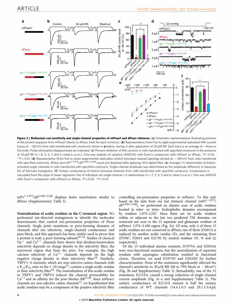
mP11–1,973/dP1,930–2,548 displays faster inactivation similar todPiezo (Supplementary Table 3).
Neutralization of acidic residues in the C-terminal region. Weperformed site-directed mutagenesis to identify the moleculardeterminants that control ion-permeation properties of Piezochannels. Single point mutations in pore-forming domains ofchannels alter ion selectivity, single-channel conductance andpore block, and this approach has been widely used to prove thata protein is truly a pore-forming subunit18–24. Studies of classicalNaþ and Ca2þ channels have shown that divalent/monovalentselectivity depends on charge density in the selectivity filter, thenarrowest region that lines the pore. For example, the highcalcium selectivity of Ca2þ channels depends on the highnegative charge density in their selectivity filter18. Similarly,TRPV1–4 channels, which are non-selective cation channels witha PCa/PNa ratio in the 1–10 range25, contain a single acidic residuein their selectivity filter26. The neutralization of this acidic residuein TRPV1 and TRPV4 reduces the channel permeability forCa2þ and its affinity for the pore blocker RR27,28. Since mPiezo1channels are non-selective cation channels13, we hypothesized thatacidic residues may be a component of the putative selectivity filter
controlling ion-permeation properties in mPiezo1. To this end,based on the data from our last chimeric channel (mP11–1,973/dP1,930–2,548), we performed an alanine scan of acidic residueslocated in inter- or intra- hydrophobic domains encompassedby residues 1,974–2,547. Since there are no acidic residueswithin or adjacent to the last two predicted TM domains, werestricted our scan to the 15 aspartate and glutamate residues ofmPiezo1 1,974–2,188 region (Fig. 3a). Of note, only 4 of these 15acidic residues are not conserved in dPiezo; one of them (D2013) isreplaced by another acidic residue (E), and the remaining three(D1987, D2014 and E2170) by neutral residues (N, N and Q,respectively).
Of the 15 individual alanine mutants, D1975A and D2034Awere non-functional mutants, but the neutralization of aspartateresidues with asparagine substitution resulted in functionalclones. Therefore, we used D1975N and D2034N for furthercharacterization. None of the mutations significantly affected thechannel’s sensitivity to 30 mM RR (60 to 79% block at � 80 mV)(Fig. 3b and Supplementary Table 3). Remarkably, one of the 15mutations, E2133A, caused a strong reduction of single-channelcurrent amplitude (Fig. 3c–e and Supplementary Table 3). Theunitary conductance of E2133A mutant is half the unitaryconductance of WT channels (14.4±0.5 and 29.1±0.4 pS,
50 ms500
pA
10 µm 10 µm
5 µm 5 µm
50 p
A
50 ms
200
pA
50 ms
5 µm 5 µm
50 ms50 p
A
10 µm 10 µm
Control 30 µM RR Washout
500
pA
50 ms
7 µm 7 µm
mPiezo1
dPiezo
dP1–1,316/mP11,316–2,547
dP1–1,316/mP11,316–2,547
dP1–1,752/mP11,715–2,547
dP1–1,752/mP11,715–2,547
mP11–1,973/dP1,930–2,548
mP11–1,973/dP1,930–2,548
dP1–
1,31
6 /mP1
1,31
6–2,
547
dP1–
1,75
2 /mP1
1,71
5–2,
547
mP1
1–1,
973 /d
P1,
930–
2,54
8
mPiez
o1dP
iezo
**
*
**
*
mPiezo1
dPiezo
dP1–
1,31
6 /mP1
1,31
6–2,
547
dP1–
1,75
2 /mP1
1,71
5–2,
547
mP1
1–1,
973 /d
P1,
930–
2,54
8
mPiez
o1
dPiez
o
dP1–1,316mP11,316–2,547
dP1–1,752/mP11,715–2,547
mP11–1,973/dP1,930–2,548
mPiezo1dPiezo
100 ms
5 pA
1 pA
1 pA
5 pA
–180 mV
5 pA
–20 mmHg
0
20
40
60
80
100
0
10
20
30
40
–200 –160 –120 –80 –40 0
–6
–5
–4
–3
–2
–1
0
(pA)
(mV)
30 µ
M R
R c
urre
nt b
lock
at –
80 m
V (
%)
Uni
tary
con
duct
ance
(pS
)
Figure 2 | Ruthenium red sensitivity and single-channel properties of mPiezo1 and dPiezo chimeras. (a) Schematic representations illustrating portions
of the protein sequence from mPiezo1 (black) or dPiezo (red) for each construct. (b) Representative (from five to eight experimental replicates) MA current
traces at � 80 mV from cells transfected with constructs shown in a before, during or after application of 30 mM RR. Each trace is an average of —three to
five trials. Probe stimulation displacements are indicated. (c) Percent inhibition of MA currents in cells transfected with specified constructs in the presence
of 30mM RR (n¼ 8, 5, 6, 5 and 5; mean±s.e.m.). One-way analysis of variance (ANOVA) with Dunn’s comparison with mPiezo1 or dPiezo, *Po0.05,
**Po0.01. (d) Representative (from five to seven experimental replicates) stretch-activated channel openings elicited at � 180 mV from cells transfected
with specified constructs. dPiezo and mP11–1,973/dP1,930–2,548 traces are displayed after applying 1 kHz digital filter. (e) Average I–V relationships of stretch-
activated single channels in cells transfected with specified constructs. Single-channel amplitude was determined as the amplitude difference in Gaussian
fits of full-trace histograms. (f) Unitary conductance of stretch-activated channels from cells transfected with specified constructs. Conductance is
calculated from the slope of linear regression line of individual cell single-channel I–V relationships (n¼ 7, 5, 5, 5 and 6; mean±s.e.m.). One-way ANOVA
with Dunn’s comparison with mPiezo1 or dPiezo, *Po0.05, **Po0.01.
ARTICLE NATURE COMMUNICATIONS | DOI: 10.1038/ncomms8223
4 NATURE COMMUNICATIONS | 6:7223 | DOI: 10.1038/ncomms8223 | www.nature.com/naturecommunications
& 2015 Macmillan Publishers Limited. All rights reserved.
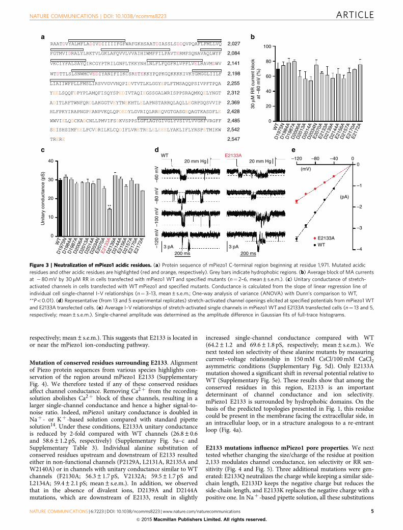
respectively; mean±s.e.m.). This suggests that E2133 is located inor near the mPiezo1 ion-conducting pathway.
Mutation of conserved residues surrounding E2133. Alignmentof Piezo protein sequences from various species highlights con-servation of the region around mPiezo1 E2133 (SupplementaryFig. 4). We therefore tested if any of these conserved residuesaffect channel conductance. Removing Ca2þ from the recordingsolution abolishes Ca2þ block of these channels, resulting in alarger single-channel conductance and hence a higher signal-to-noise ratio. Indeed, mPiezo1 unitary conductance is doubled inNaþ - or Kþ -based solution compared with standard pipettesolution14. Under these conditions, E2133A unitary conductanceis reduced by 2-fold compared with WT channels (26.8±0.6and 58.6±1.2 pS, respectively) (Supplementary Fig. 5a–c andSupplementary Table 3). Individual alanine substitution ofconserved residues upstream and downstream of E2133 resultedeither in non-functional channels (P2129A, L2131A, R2135A andW2140A) or in channels with unitary conductance similar to WTchannels (F2130A; 56.3±1.7 pS, V2132A; 59.5±1.7 pS andL2134A; 59.4±2.1 pS; mean±s.e.m.). In addition, we observedthat in the absence of divalent ions, D2139A and D2144Amutations, which are downstream of E2133, result in slightly
increased single-channel conductance compared with WT(64.2±1.2 and 69.6±1.8 pS, respectively; mean±s.e.m.). Wenext tested ion selectivity of these alanine mutants by measuringcurrent–voltage relationship in 150 mM CsCl/100 mM CaCl2asymmetric conditions (Supplementary Fig. 5d). Only E2133Amutation showed a significant shift in reversal potential relative toWT (Supplementary Fig. 5e). These results show that among theconserved residues in this region, E2133 is an importantdeterminant of channel conductance and ion selectivity.mPiezo1 E2133 is surrounded by hydrophobic domains. On thebasis of the predicted topologies presented in Fig. 1, this residuecould be present in the membrane facing the extracellular side, inan intracellular loop, or in a structure analogous to a re-entrantloop (Fig. 4a).
E2133 mutations influence mPiezo1 pore properties. We nexttested whether changing the size/charge of the residue at position2,133 modulates channel conductance, ion selectivity or RR sen-sitivity (Fig. 4 and Fig. 5). Three additional mutations were gen-erated: E2133Q neutralizes the charge while keeping a similar side-chain length, E2133D keeps the negative charge but reduces theside-chain length, and E2133K replaces the negative charge with apositive one. In Naþ -based pipette solution, all these substitutions
Uni
tary
con
duct
ance
(pS
)
10
20
0
30
40
WT
D19
84A
D19
87A
D20
06A
D20
13A
D20
14A
E20
70A
E21
33A
D21
39A
D21
44A
E21
56A
D21
57A
E21
70A
E21
72A
0–40–80–120
–2
–1
0
–3
–4
(pA)
(mV)
WTE2133A
20 mm Hg 20 mm Hg
3 pA 3 pA200 ms 200 ms
–60
mV
–80
mV
–100
mV
–120
mV
WT E2133A
20
40
0
60
100
30 µ
M R
R c
urre
nt b
lock
at –
80 m
V (
%)
80
**
D20
34N
D19
75N
WT
D19
84A
D19
87A
D20
06A
D20
13A
D20
14A
E20
70A
E21
33A
D21
39A
D21
44A
E21
56A
D21
57A
E21
70A
E21
72A
D20
34N
D19
75N
RAATDVYALMFLADIVDIIIIIFGFWAFGKHSAATDIASSLSDDQVPQAFLFMLLVQ 2,027
FGTMVIDRALYLRKTVLGKLAFQVVLVVAIHIWMFFILPAVTERMFSQNAVAQLWYF 2,084
VKCIYFALSAYQIRCGYPTRILGNFLTKKYNHLNLFLFQGFRLVPFLVELRAVMDWV 2,141
WTDTTLSLSNWMCVEDIYANIFIIKCSRETEKKYPQPKGQKKKKIVKYGMGGLIILF 2,198
LIAIIWFPLLFMSLIRSVVGVVNQPIDVTVTLKLGGYEPLFTMSAQQPSIVPFTPQA 2,255
YEELSQQFDPYPLAMQFISQYSPEDIVTAQIEGSSGALWRISPPSRAQMKQELYNGT 2,312
ADITLRFTWNFQRDLAKGGTVEYTNEKHTLELAPNSTARRQLAQLLEGRPDQSVVIP 2,369
HLFPKYIRAPNGPEANPVKQLQPDEDYLGVRIQLRREQVGTGASGEQAGTKASDFLE 2,428
WWVIELQDCKADCNLLPMVIFSDKVSPPSLGFLAGYGIVGLYVSIVLVVGKFVRGFF 2,485
SEISHSIMFEELPCVDRILKLCQDIFLVRETRELELEEELYAKLIFLYRSPETMIKW 2,542
TRERE 2,547
Figure 3 | Neutralization of mPiezo1 acidic residues. (a) Protein sequence of mPiezo1 C-terminal region beginning at residue 1,971. Mutated acidic
residues and other acidic residues are highlighted (red and orange, respectively). Grey bars indicate hydrophobic regions. (b) Average block of MA currents
at �80 mV by 30 mM RR in cells transfected with mPiezo1 WT and specified mutants (n¼ 2–6, mean±s.e.m.). (c) Unitary conductance of stretch-
activated channels in cells transfected with WT mPiezo1 and specified mutants. Conductance is calculated from the slope of linear regression line of
individual cell single-channel I–V relationships (n¼ 3–13, mean±s.e.m.; One-way analysis of variance (ANOVA) with Dunn’s comparison to WT,
**Po0.01). (d) Representative (from 13 and 5 experimental replicates) stretch-activated channel openings elicited at specified potentials from mPiezo1 WT
and E2133A transfected cells. (e) Average I–V relationships of stretch-activated single channels in mPiezo1 WT and E2133A transfected cells (n¼ 13 and 5,
respectively; mean±s.e.m.). Single-channel amplitude was determined as the amplitude difference in Gaussian fits of full-trace histograms.
NATURE COMMUNICATIONS | DOI: 10.1038/ncomms8223 ARTICLE
NATURE COMMUNICATIONS | 6:7223 | DOI: 10.1038/ncomms8223 | www.nature.com/naturecommunications 5
& 2015 Macmillan Publishers Limited. All rights reserved.
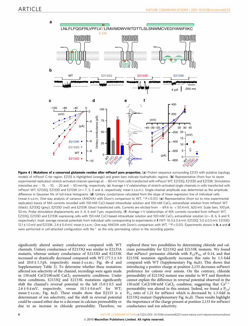
significantly altered unitary conductance compared with WTchannels. Unitary conductance of E2133Q was similar to E2133Amutants, whereas unitary conductance of E2133D and E2133Kincreased or drastically decreased compared with WT (77.5±3.0and 20.0±1.3 pS, respectively; mean±s.e.m.; Fig. 4b–d andSupplementary Table 3). To determine whether these mutationsaffected ion selectivity of the channel, recordings were again madein 150 mM CsCl/100 mM CaCl2 asymmetric conditions. Underthese conditions, E2133Q and E2133K mutations significantlyshift the channel’s reversal potential to the left (5.0±0.5 and2.6±0.4 mV, respectively, versus 10.3±0.6 mV for WT;mean±s.e.m.; Fig. 4e,f). These results show that E2133 is adeterminant of ion selectivity, and the shift in reversal potentialcould be caused either due to a decrease in calcium permeability ordue to an increase in chloride permeability. Therefore, we
explored these two possibilities by determining chloride and cal-cium permeability for E2133Q and E2133K mutants. We foundthat mPiezo1 permeates chloride with PCl/PNa of 0.14, and thatE2133K mutation significantly increases this ratio by 1.5-foldcompared with WT (Supplementary Fig. 6a,b). This shows thatintroducing a positive charge at position 2,133 decreases mPiezo1preference for cations over anions. On the contrary, chloridepermeability of E2133Q mutant was similar to WT and thereforecannot explain the difference in reversal potential observed in the150 mM CsCl/100 mM CaCl2 condition, suggesting that Ca2þ
permeability was altered in this mutant. Indeed, we found a PCa/PCs ratio of 1.21 for mPiezo1 which is decreased by 1.3-fold inE2133Q mutant (Supplementary Fig. 6c,d). These results highlightthe importance of the charge present at position 2,133 for mPiezo1conductance and ion selectivity.
–120 –80 –40 00
–2
–4
–6
–8
–10
(mV)
(pA)
WT
E2133K
E2133Q
E2133D WT
E2133
K
E2133
Q
E2133
D
100
75
50
25
0
**
**
**
Uni
tary
con
duct
ance
(pS
)
4 pA 200 ms
LNLFLFQGFRLVPFLVELRAVMDWVWTDTTLSLSNWMCVEDIYANIFIIKC2,133
20
400
200
–600
–800
–400
–40 –30 –20 4030
(pA)
(mV)
–200
****
0
5
10
15
20
WT
E2133
Q
E2133
D
E2133
K
(mV)
WT E2133KE2133Q E2133D
Figure 4 | Mutations of a conserved glutamate residue alter mPiezo1 pore properties. (a) Protein sequence surrounding E2133 with putative topology
models of mPiezo1 C-ter region. E2133 is highlighted (orange) and green bars indicate hydrophobic regions. (b) Representative (from four to seven
experimental replicates) stretch-activated channel openings at � 80 mV from cells transfected with mPiezo1 WT, E2133Q, E2133D and E2133K. Stimulation
intensities are � 15, � 10, � 20 and � 50 mm Hg, respectively. (c) Average I–V relationships of stretch-activated single channels in cells transfected with
mPiezo1 WT, E2133Q, E2133D and E2133K (n¼ 7, 5, 5 and 4, respectively; mean±s.e.m.). Single-channel amplitude was determined as the amplitude
difference in Gaussian fits of full-trace histograms. (d) Unitary conductance calculated from the slope of linear regression line of individual cells
(mean±s.e.m.; One-way analysis of variance (ANOVA) with Dunn’s comparison to WT, **Po0.01). (e) Representative (from six to nine experimental
replicates) traces of MA currents recorded with 150 mM CsCl-based intracellular solution and 100 mM CaCl2 extracellular solution from mPiezo1 WT
(black), E2133Q (grey), E2133D (red) and E2133K (blue) transfected cells. Currents are elicited from � 69.6 to þ 50.4 mV, D20 mV. Scale bars, 100 pA,
50 ms. Probe stimulation displacements are 3, 4, 6 and 7 mm, respectively. (f) Average I–V relationships of MA currents recorded from mPiezo1 WT,
E2133Q, E2133D and E2133K expressing cells with 150 mM CsCl-based intracellular solution and 100 mM CaCl2 extracellular solution (n¼ 8, 6, 6 and 9,
respectively). Inset: average reversal potentials from individual cells corresponding to experiments in f (WT: 10.3±0.6 mV; E2133Q: 5.0±0.5 mV; E2133D:
12.1±1.0 mV and E2133K: 2.6±0.4 mV; mean±s.e.m.; One-way ANOVA with Dunn’s comparison with WT, **Po0.01). Experiments shown in b, c and d
were performed in cell-attached configuration with Naþ as the only permeating cation in the recording pipette.
ARTICLE NATURE COMMUNICATIONS | DOI: 10.1038/ncomms8223
6 NATURE COMMUNICATIONS | 6:7223 | DOI: 10.1038/ncomms8223 | www.nature.com/naturecommunications
& 2015 Macmillan Publishers Limited. All rights reserved.
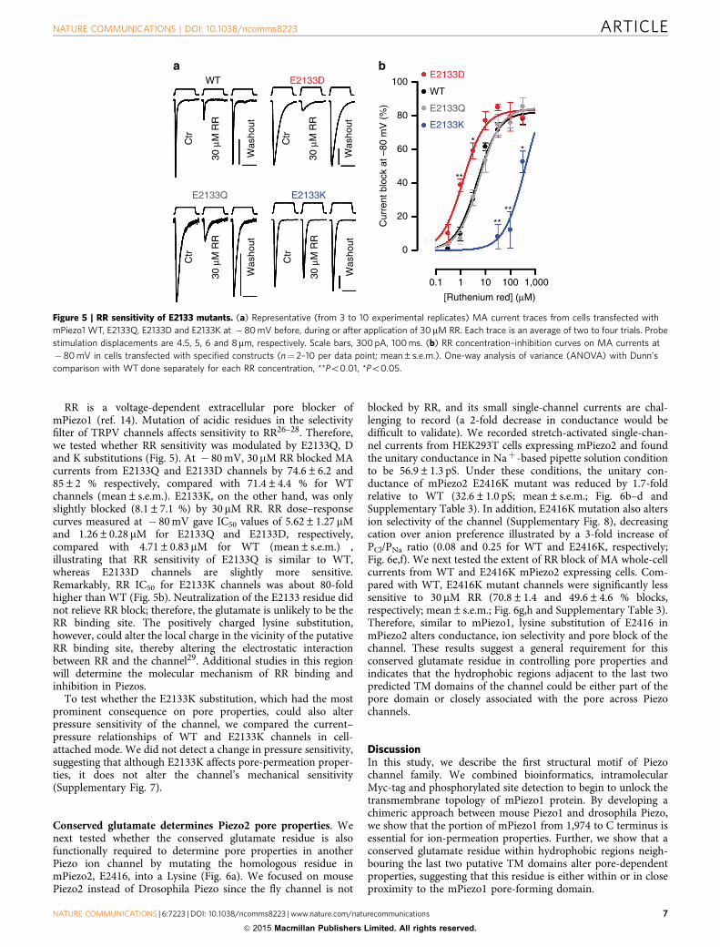
RR is a voltage-dependent extracellular pore blocker ofmPiezo1 (ref. 14). Mutation of acidic residues in the selectivityfilter of TRPV channels affects sensitivity to RR26–28. Therefore,we tested whether RR sensitivity was modulated by E2133Q, Dand K substitutions (Fig. 5). At � 80 mV, 30 mM RR blocked MAcurrents from E2133Q and E2133D channels by 74.6±6.2 and85±2 % respectively, compared with 71.4±4.4 % for WTchannels (mean±s.e.m.). E2133K, on the other hand, was onlyslightly blocked (8.1±7.1 %) by 30 mM RR. RR dose–responsecurves measured at � 80 mV gave IC50 values of 5.62±1.27 mMand 1.26±0.28 mM for E2133Q and E2133D, respectively,compared with 4.71±0.83 mM for WT (mean±s.e.m.) ,illustrating that RR sensitivity of E2133Q is similar to WT,whereas E2133D channels are slightly more sensitive.Remarkably, RR IC50 for E2133K channels was about 80-foldhigher than WT (Fig. 5b). Neutralization of the E2133 residue didnot relieve RR block; therefore, the glutamate is unlikely to be theRR binding site. The positively charged lysine substitution,however, could alter the local charge in the vicinity of the putativeRR binding site, thereby altering the electrostatic interactionbetween RR and the channel29. Additional studies in this regionwill determine the molecular mechanism of RR binding andinhibition in Piezos.
To test whether the E2133K substitution, which had the mostprominent consequence on pore properties, could also alterpressure sensitivity of the channel, we compared the current–pressure relationships of WT and E2133K channels in cell-attached mode. We did not detect a change in pressure sensitivity,suggesting that although E2133K affects pore-permeation proper-ties, it does not alter the channel’s mechanical sensitivity(Supplementary Fig. 7).
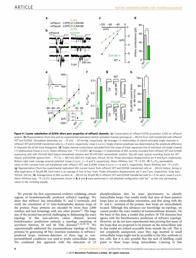
Conserved glutamate determines Piezo2 pore properties. Wenext tested whether the conserved glutamate residue is alsofunctionally required to determine pore properties in anotherPiezo ion channel by mutating the homologous residue inmPiezo2, E2416, into a Lysine (Fig. 6a). We focused on mousePiezo2 instead of Drosophila Piezo since the fly channel is not
blocked by RR, and its small single-channel currents are chal-lenging to record (a 2-fold decrease in conductance would bedifficult to validate). We recorded stretch-activated single-chan-nel currents from HEK293T cells expressing mPiezo2 and foundthe unitary conductance in Naþ -based pipette solution conditionto be 56.9±1.3 pS. Under these conditions, the unitary con-ductance of mPiezo2 E2416K mutant was reduced by 1.7-foldrelative to WT (32.6±1.0 pS; mean±s.e.m.; Fig. 6b–d andSupplementary Table 3). In addition, E2416K mutation also altersion selectivity of the channel (Supplementary Fig. 8), decreasingcation over anion preference illustrated by a 3-fold increase ofPCl/PNa ratio (0.08 and 0.25 for WT and E2416K, respectively;Fig. 6e,f). We next tested the extent of RR block of MA whole-cellcurrents from WT and E2416K mPiezo2 expressing cells. Com-pared with WT, E2416K mutant channels were significantly lesssensitive to 30 mM RR (70.8±1.4 and 49.6±4.6 % blocks,respectively; mean±s.e.m.; Fig. 6g,h and Supplementary Table 3).Therefore, similar to mPiezo1, lysine substitution of E2416 inmPiezo2 alters conductance, ion selectivity and pore block of thechannel. These results suggest a general requirement for thisconserved glutamate residue in controlling pore properties andindicates that the hydrophobic regions adjacent to the last twopredicted TM domains of the channel could be either part of thepore domain or closely associated with the pore across Piezochannels.
DiscussionIn this study, we describe the first structural motif of Piezochannel family. We combined bioinformatics, intramolecularMyc-tag and phosphorylated site detection to begin to unlock thetransmembrane topology of mPiezo1 protein. By developing achimeric approach between mouse Piezo1 and drosophila Piezo,we show that the portion of mPiezo1 from 1,974 to C terminus isessential for ion-permeation properties. Further, we show that aconserved glutamate residue within hydrophobic regions neigh-bouring the last two putative TM domains alter pore-dependentproperties, suggesting that this residue is either within or in closeproximity to the mPiezo1 pore-forming domain.
E2133D
Ctr
30 µ
M R
R
Was
hout
Ctr
30 µ
M R
R
Was
hout
E2133KC
tr
30 µ
M R
R
Was
hout
Ctr
30 µ
M R
R
Was
hout
E2133Q
WT
E2133K
E2133Q
E2133D
20
40
0
60
100
Cur
rent
blo
ck a
t –80
mV
(%
)
80
0.1 1 10 100 1,000
[Ruthenium red] (µM)
WT
****
*
**
*
Figure 5 | RR sensitivity of E2133 mutants. (a) Representative (from 3 to 10 experimental replicates) MA current traces from cells transfected with
mPiezo1 WT, E2133Q, E2133D and E2133K at � 80 mV before, during or after application of 30mM RR. Each trace is an average of two to four trials. Probe
stimulation displacements are 4.5, 5, 6 and 8mm, respectively. Scale bars, 300 pA, 100 ms. (b) RR concentration–inhibition curves on MA currents at
� 80 mV in cells transfected with specified constructs (n¼ 2–10 per data point; mean±s.e.m.). One-way analysis of variance (ANOVA) with Dunn’s
comparison with WT done separately for each RR concentration, **Po0.01, *Po0.05.
NATURE COMMUNICATIONS | DOI: 10.1038/ncomms8223 ARTICLE
NATURE COMMUNICATIONS | 6:7223 | DOI: 10.1038/ncomms8223 | www.nature.com/naturecommunications 7
& 2015 Macmillan Publishers Limited. All rights reserved.
We provide the first experimental evidence validating certainregions of bioinformatically predicted mPiezo1 topology. Weshow that mPiezo1 has intracellular N- and C-terminals, andverify the orientation of 13 inter-hydrophobic domain loops ofthe protein. Piezo proteins are encoded by more than 2,000residues and lack homology with any other protein13. The largesize of the protein has proved challenging in delineating the exacttopology of this non-selective cation channel. Severalbioinformatics predictions estimate a topology consisting ofanywhere between 30 and 40 TMs domains11,16,30–36. Weexperimentally addressed the transmembrane topology of Piezoproteins by generating 48 Myc insertion mutations in mPiezo1-predicted loops. Immuno-detection of these tags in non-permeabilized conditions was used to probe extracellular loops.We combined this approach with the detection of 23
phosphorylation sites by mass spectrometry to identifyintracellular loops. Our results verify that nine of these putativeloops have an extracellular orientation, and that along with theN- and C- terminal of the protein, four loops are intracellularlylocated. Although this advances our knowledge on topology, wecannot predict the exact number of transmembrane domains. Onthe basis of this data, a model that predicts 38 TM domains bestagrees with the bioinformatics prediction of mPiezo1 topology.However, we do not have experimental data proving that many ofthe loops that are proposed to be present on the extracellular sidein this model are indeed accessible from outside the cell. This isnot completely unexpected, since Myc tags inserted in smallextracellular loops might not be accessible to the Myc antibody inlive cells. But it is also possible that the lack of labelling couldpoint to these loops being intracellular. Catering to this
20
40
0
60
80
Uni
tary
con
duct
ance
(pS
)0
–2
–4
–6
–8
–120 –80 –40 0
(mV)
(pA)
WT
E2416K
WT
E2416
K
***
mPiezo1 LVPFLVELRAVM mPiezo2 LVPFLTELRAVM
2,133
2,416
5 pA200 ms
E2416KmPiezo2
0.0
0.1
0.2
0.3
WT
E2416
K
**
–80 –60 –40 40 60
–200
400
600
(pA)
(mV)
–40
–30
–20
Ere
v (m
V)
WT
E2416
K
**20
40
0
60
100
30 µ
M R
R c
urre
nt b
lock
at –
80 m
V (
%)
80
WT
E2416
K
**
Ctr
30 µ
M R
R
Was
hout
E2416K
Ctr
30 µ
M R
R
Was
hout
WT
PC
I– /P
Na+
Figure 6 | Lysine substitution of E2416 alters pore properties of mPiezo2 channels. (a) Conservation of mPiezo1 E2133 at position 2,416 on mPiezo2
protein. (b) Representative (from nine and six experimental replicates) stretch-activated channel openings at � 80 mV from cells transfected with mPiezo2
WT and E2416K. Stimulation intensities are � 15 and � 20 mm Hg, respectively. (c) Average I–V relationships of stretch-activated single channels in
mPiezo2 WT and E2416K transfected cells (n¼9 and 6, respectively; mean±s.e.m.). Single-channel amplitude was determined as the amplitude difference
in Gaussian fits of full-trace histograms. (d) Single-channel conductance calculated from the slope of linear regression line of individual cell single-channel
I–V relationships (mean±s.e.m.; Mann–Whitney test, ***Po0.001). (e) Average I–V relationships of MA currents recorded from mPiezo2 WT and E2416K
expressing cells with 150 mM NaCl-based intracellular solution and 30 mM NaCl extracellular solution. Top left inset: typical recording traces for WT
(black) and E2416K (green) from � 71.7 to þ48.3 mV, D20 mV. Scale bars, 100 pA, 50 ms. Probe stimulation displacements are 9 and 8mm, respectively.
Bottom right inset: average reversal potential (mean±s.e.m.; n¼6 and 5, respectively; Mann–Whitney test **Po0.01). (f) PCl/PNa permeability
ratios of MA currents from cell transfected with mPiezo2 WT, and E2416K (mean±s.e.m.; n¼6 and 5, respectively; Mann–Whitney test **Po0.01).
(g) Representative (from five experimental replicates) MA current traces from mPiezo2 WT and E2416K transfected cells at �80 mV before, during or
after application of 30 mM RR. Each trace is an average of two to four trials. Probe stimulation displacements are 5 and 7 mm, respectively. Scale bars,
100 pA, 100 ms. (h) Average block of MA currents at �80 mV by 30 mM RR in mPiezo2 WT and E2416K transfected cells (n¼ 5 for each; mean±s.e.m.;
Mann–Whitney test, **Po0.01). Experiments shown in b, c and d were performed in cell-attached configuration with Naþ as the only permeating
cation in the recording pipette.
ARTICLE NATURE COMMUNICATIONS | DOI: 10.1038/ncomms8223
8 NATURE COMMUNICATIONS | 6:7223 | DOI: 10.1038/ncomms8223 | www.nature.com/naturecommunications
& 2015 Macmillan Publishers Limited. All rights reserved.
possibility, if we assume that all Myc tags not observed in liveunpermeabilized cells are intracellular or within transmembraneregions, then mPiezo1 could have only 18 TM domains.Furthermore, if we assume that only regions that arephosphorylated form intracellular loops, then a topology with aminimum of 10 putative TM domains could be proposed. Theactual number of TM domains could be anywhere in between thetwo extreme cases. Regardless, our results here represent the firstexperimentally based information on Piezo topology, and will beinvaluable for future studies addressing structural features of thischannel.
We generated chimeric proteins between mPiezo1 anddPiezo and assayed their pore-related properties14. We showthat the mPiezo1 C-terminal domains encoded by the residues1,974–2,547 confer unitary conductance, ion selectivity and RRsensitivity. Therefore, we conclude that C-terminal portion ofmPiezo1, which is 1/5th of the whole protein, includes most if notall of the pore domain. Interestingly, according to our predictedmPiezo1 topology, B70% of the reported hPiezo1 and hPiezo2mutations map to this region, including hPiezo1 (T2127M) andhPiezo2 (T2356M), which are in close proximity to our identifiedglutamate residue (see below)16, emphasizing the importance ofthis region in channel function.
Following up on our chimeric channel screen, we used site-directed mutagenesis of acidic residues within hydrophobicregions encoded by residues 1,974–2,188 to show that neutraliza-tion of a single glutamate residue results in a 2-fold decrease inunitary conductance. Additional substitutions of aspartate, lysineor glutamine instead of the glutamate residue altered unitaryconductance, ion selectivity and sensitivity to RR, therebysuggesting that E2133 contributes to mPiezo1 pore properties.The role of this glutamate residue in dictating ion-permeationproperties of Piezos was further confirmed when the lysinesubstitution of the homologous residue in mPiezo2 (E2416K) alsomodulated unitary conductance, cation over anion selectivity andRR sensitivity.
Alignment of Piezo proteins from distant species such asparasites shows that mPiezo1 E2133 glutamate residue is the onlycharged residue in a conserved stretch of amino acids that form aPF(X2)E(X6)W motif 34. We found that P and W residues areindeed essential for channel function, as mutations at thesepositions result in non-functional channels. The impact of theglutamate residue on pore properties and its location in a highlyconserved motif is suggestive of its presence in a structure similarto a selectivity filter present in most members of known ionchannel families. However, only lysine substitution of the E2133altered mPiezo1 RR sensitivity, suggesting that this residue is notthe RR binding site. Furthermore, the magnitude of the impactthat lysine substitution has on ion selectivity and single-channelconductance is rather mild for a model where the selectivity filterof mPiezo1 or 2 channels would constitute a ring of E2133 orE2416 residues from associated subunits. This suggests that theglutamate residue may not lie in the selectivity filter but could belocated close enough to the pore to allosterically modulate itsproperties.
The E2133 residue lies in a region flanked by hydrophobicresidues. However, the different topology models differ in theprediction of how these hydrophobic domains are oriented. It ispossible that the hydrophobic domains form full transmembranesegments, and that E2133 could be accessible from or close to theextracellular side (Fig. 1b). Alternatively, it might be located closerto the intracellular side, potentially as part of a re-entrant loop(Fig. 1c). The true orientation of this E2133 region relative to therest of the channel will be elucidated by future structural analysis.
Although mutating the glutamate residue impacts poreproperties in mouse Piezos, this residue is conserved between
mouse (E2133/2416 in mPiezo1/2) and fly Piezos (E2091 indPiezo) and therefore cannot explain their pore-related differ-ences. Therefore, other unidentified differences between mouseand fly Piezos in the C-terminal region must explain the largevariations in their biophysical pore properties.
Overall, we provide strong evidence that the protein regioncorresponding to mPiezo1 residues 1,974 to C terminusencompass the domains that form the pore of the channel, andthat mPiezo1 E2133 residue (or mPiezo2 E2416) is at least in thevicinity of the pore-permeation pathway and can influence Piezopore properties.
MethodsmPiezo1 topology prediction. Using the sequence of mammalian Piezos, topologypredictions were made using Hopp–Woods hydrophilicity/antigenicity plots as wellas Kyte Doolittle hydrophobicity plots. A combination of these results was used topredict the final topology. Our approach was validated to predict the topology ofvarious TRP channels and CLC channels37. For Hopp–Woods plots, we used awindow size of 7 to predict highly hydrophilic/antigenic regions. For Kyte Doolittlehydrophobicity plots, we used a window size of 17 to predict TM regions and awindow size of 9 to predict surface regions over the full-length amino-acid sequence.For ambiguous regions, fragments of the amino-acid sequence were analysedseparately using the same parameters. The results were then compared with putativetransmembrane domains predicted by TMHMM2 and Phobius algorithms. A totalof 38 transmembrane regions were predicted. Myc tags were inserted in predictedinside and outside loops near hydrophilic residues, preferably in the middle of theloop. All Myc constructs were generated using the QuickChange II XL site-directedmutagenesis kit (Agilent Technologies). Myc tags were also introduced at the N- andC-terminal regions. All constructs were verified by sequencing.
HEK293T cells were plated on poly-D-lysine-coated MatTek dishes, which wereadditionally coated with laminin (10 or 20 mg ml� 1). Cells were transfected witheach construct separately (2 mg cDNA each) using Fugene HD (Promega). Twenty-four to 48 h after transfection live labelling was carried out by incubatingtransfected HEK293T cells with Myc 9E11 (1:50; Santa Cruz Biotechnology) at37 �C for 20 min or 1 h. After five washes with warm medium, cells were incubatedwith secondary antibodies conjugated to Alexa Fluor 546 (1:200; Life Technologies)for 10–20 min at room temperature. Cells were washed five times with PBS, fixedwith 2% PFA/PBS for 20–30 min and imaged at an Olympus (Tokyo, Japan)Fluoview 500 confocal microscope by illumination with the HeNe green 543 nmlaser.
Immunostaining after fixation and permeabilization of cells was carried outessentially as described13. In brief, cells were fixed with 4% PFA/PBS for 10 min,washed five times with PBS, permeabilized in PBS containing 0.4% Triton X-100and blocked with 10% normal goat serum in PBS followed by incubation withantibodies. The Myc 9E11 (1:100; Santa Cruz Biotechnology) antibody was used toconfirm predicted outside and inside loops and Piezo1 antibodies (1:100) toconfirm Piezo1 expression. After three washes in PBS, secondary antibodiesconjugated to Alexa Fluor 546 and 633 (1:200; Life Technologies) were applied.Immunostainings were imaged at an Olympus (Tokyo, Japan) Fluoview 500confocal microscope by sequential illumination using the HeNe green 543 nm laserand the HeNe red 633 nm laser. The live labelling and permeabilized staining wasrepeated two or more times for each construct to confirm results.
Detection of phosphorylation sites. mPiezo1-GST proteins were purifiedfrom HEK293T cells transfected with mPiezo1-GST cDNAs14. After incubationwith cell lysates overnight at 4 �C, the glutathione beads were washed four timesin a buffer containing 25 mM NaPIPES, 140 mM NaCl, 0.6% CHAPS, 0.14%phosphatidylcholine (PC), 2.5 mM dithiothreitol (DTT) and a cocktail of proteaseinhibitors and eluted with 100 mM glutathione in a buffer containing 25 mMNaPIPES, 50 mMTris, 0.6% CHAPS, 0.14% PC, 2.5 mM DTT and a cocktail ofprotease inhibitors. The eluant was dialysed against a buffer containing 25 mMNaPIPES, 0.6% CHAPS, 0.14% PC, 2.5 mM DTT and a cocktail of proteaseinhibitors. The protein samples were denatured, reduced and alkylated with2-iodoacetamide before precipitation with acetonitrile. The pellet was re-dissolvedin 8 M urea and ProteaseMax detergent, then diluted to 1 M urea and digested withtrypsin or chymotrypsin. A portion of each digest was analysed by nanoscale liquidchromatography-mass spectrometry (nanoLC/MS) on the Orbitrap Velos (ThermoScientific) without further processing. The remainder of the digests was enrichedfor phosphopeptides using TiO2 beads and also analysed by nanoLC/MS on theOrbitrap Velos. The data were searched using a small database (B450 proteins) toidentify peptides from mPiezo1 (B73% sequence coverage) and sites ofphosphorylation and the search results combined in Scaffold 3. The search resultswere filtered for high confidence (95% at the peptide level) and MS/MS indicatingsites of phosphorylation were inspected.
Generation of chimeras and mutants. mPiezo1 and dPiezo chimeras were gen-erated by swapping specific regions in the C-terminal end of the protein. The
NATURE COMMUNICATIONS | DOI: 10.1038/ncomms8223 ARTICLE
NATURE COMMUNICATIONS | 6:7223 | DOI: 10.1038/ncomms8223 | www.nature.com/naturecommunications 9
& 2015 Macmillan Publishers Limited. All rights reserved.
beginning of a swapped region was chosen at a stretch of amino acids that wereconserved between mPiezo1 and dPiezo. These junction sites are marked inSupplementary Fig. 2. Domain swapping was carried out using the QuickChange IIXL site-directed mutagenesis kit from Agilent Technologies. Mega-primers weredesigned flanking the region of interest with overlapping base pairs between thedonor and the recipient cDNA. The mega-primer was generated by PCR ampli-fication and gel purification. The PCR product was then used as a primer in themutagenesis reaction with the recipient cDNA as the template. PCR cycling con-ditions were according to the manufacturer’s instructions. Reactions were trans-formed into XL-gold competent cells and colonies screened. All positive chimeraclones were verified by full-length DNA sequencing.
Point mutants were also generated using the QuickChange II XL site-directedmutagenesis kit according to the manufacturer’s instructions and confirmed byfull-length DNA sequencing. dPiezo chimeras were cloned into pIres2-EGFP vectorand all mPiezo1 chimeras and mutants were cloned into pcDNA3.1(� ) vector,which was modified to include an IRES-EGFP element. mPiezo2 (E2416K) wascloned in pCMV-Sport6 vector.
Cell culture and transient transfection. Human embryonic kidney 293T(HEK293T) cells were grown in Dulbecco’s Modified Eagle Medium containing4.5 mg ml� 1 glucose, 10% fetal bovine serum, 50 units ml� 1 penicillin and50mg ml� 1 streptomycin. Cells were plated onto 12-mm round glass poly-D-lysinecoated coverslips placed in 24-well plates and transfected using lipofectamine 2000(Invitrogen) according to the manufacturer’s instruction. All plasmids weretransfected at a concentration of 600 to 1,000 ng ml� 1. mPiezo2(E2416K) was co-transfected with GFP at a concentration of 300 ng ml� 1. Cells were recorded from12 to 48 h post transfection.
Electrophysiology. Patch-clamp experiments were performed in standard whole-cell or cell-attached mode using Axopatch 200B amplifier (Axon Instruments).Currents were sampled at 20 kHz and filtered at 2 kHz. Leak currents beforemechanical stimulations were subtracted off-line from the current traces. Voltageswere not corrected for a liquid junction potential except for ion selectivityexperiments (Figs 4e–g and 5e,f, Supplementary Fig. S6D,E and S7). Liquid junc-tion potential was calculated using Clampex 10.3 software. All the experimentswere done at room temperature.
Solutions. For whole-cell patch-clamp recordings, recording electrodes had aresistance of 2–3 MO when filled with internal solution composed of (in mM)133 CsCl, 1 CaCl2, 1 MgCl2, 5 EGTA, 10 HEPES (pH 7.3 with CsOH), 4 MgATPand 0.4 Na2GTP. The extracellular solution was composed of (in mM) 133 NaCl, 3KCl, 2.5 CaCl2, 1 MgCl2, 10 HEPES (pH 7.3 with NaOH) and 10 glucose. Forion-selectivity experiments, internal solution used was (in mM) 150 CsCl, 10HEPES (pH 7.3 with CsOH) and extracellular solution consisted of (in mM) 100CaCl2 and 10 HEPES (pH 7.3 with CsOH). PCl/PNa was measured in extracellularsolution composed of (in mM) 30 NaCl, 10 HEPES and 225 Sucrose (pH 7.3 withNaOH) and intracellular solution consisted of (in mM) 150 NaCl and 10 HEPES(pH 7.3 with NaOH). PCa/PCs was measured in extracellular solution composed of(in mM) 50 Ca-gluconate, 0.5 CaCl2, 10 HEPES and 170 sucrose (pH 7.3 withCsOH), and intracellular solution composed of (in mM) 149 Cs-methanesulfonate,1 CsCl and 10 HEPES (pH 7.3 with CsOH). For ruthenium red (RR) experiments,10 or 100 mM RR stock solution was prepared in water and diluted to workingconcentrations in extracellular solution.
For cell-attached patch-clamp recordings, external solution used to zero themembrane potential consisted of (in mM) 140 KCl, 1 MgCl2, 10 glucose and 10HEPES (pH 7.3 with KOH). Recording pipettes were of 2–3 MO resistance whenfilled with standard solution composed of (in mM) 130 mM NaCl, 5 KCl, 1 CaCl2,1 MgCl2, 10 TEA-Cl and 10 HEPES (pH 7.3 with NaOH) or divalent-free pipettesolution consisted of (in mM) 150 mM NaCl, 10 HEPES (pH 7.3 with NaOH).
Whole-cell mechanical stimulation. For whole-cell recordings, mechanical sti-mulation was achieved using a fire-polished glass pipette (tip diameter 3–4 mm)positioned at an angle of 80� relative to the cell being recorded. Downward dis-placement of the probe towards the cell was driven by Clampex-controlled pie-zoelectric crystal microstage (E625 LVPZT Controller/Amplifier; PhysikInstrumente). The probe had a velocity of 1 mm ms� 1 during the ramp phase of thecommand for forward movement and the stimulus was applied for 150 ms. Toassess the mechanical sensitivity of a cell, a series of mechanical steps in 0.5 or 1 mmincrements was applied every 10–20 s. For RR experiments, once an efficient sti-mulus eliciting MA current for a cell was achieved, repeated stimuli at that distancewere then given every 20–30 s. Cells were continuously perfused and at least threeconsecutive stable recording traces were achieved before switching between solu-tions. Current parameters were measured from three or more averaged traces. ForI–V relationship recordings, voltage steps were applied 700 ms before mechanicalstimulation (150 ms) from a holding potential of � 60 mV. Voltage steps weregiven from � 80 mV to þ 80 in 20 mV increments. The starting position of thestimulation probe relative to the cell is not tightly controlled and varies betweencells. Therefore, responses were recorded at different probe displacements from cellto cell allowing recording of MA currents with similar amplitude.
Permeability ratio measurements. Reversal potential for each cell in the men-tioned solution was determined by interpolation of the respective current–voltagedata. Permeability ratios were calculated by using the following Goldman–Hodgkin–Katz (GHK) equations:
PCl/PNa ratios:
Erev ¼RTF
lnPNa½Na�o þ PCl½Cl�iPNa½Na�i þ PCl½Cl�o
PCa/PCs ratios:
Erev ¼RTF
ln
ffiffiffiffiffiffiffiffiffiffiffiffiffiffiffiffiffiffiffiffiffiffiffiffiffiffiffiffi4PCa Ca½ �oPCs Cs½ �i
þ 14
s� 1
2
!
where, RT/F has the value 25.69 at 25 �C.
Unitary conductance measurement. Stretch-activated currents were recorded inthe cell-attached patch-clamp configuration. Membrane patches were stimulatedwith 500 ms negative pressure pulses through the recording electrode usingClampex-controlled pressure clamp HSPC-1 device (ALA-scientific). Since thesingle-channel amplitude is independent of the pressure intensity, the most optimalpressure stimulation was used to elicit responses that allowed single-channelamplitude measurements. These stimulation values were largely dependent on thenumber of channels in a given patch of the recording cell. Single-channel ampli-tude at a given potential was measured from trace histograms of 5 to 20 repeatedrecordings. Histograms were fitted with Gaussian equations using Clampfit 10.3software or multi-peak fitting analysis of IGOR Pro software. Single-channel slopeconductance for each individual cell was calculated from linear regression curve fitto single-channel I–V plots. We sometimes observed sub-conductance states insingle-channel recording of chimeric and mutant channels, but we focused theanalysis on the state that was prevalent.
Concerning mPiezo2 WT and mutant, stretch-activated currents weredetected in only B50% of HEK293T transfected cells but never in cells expressingires2-EGFP only. This frequency is lower than the one of mPiezo1 and relatedmutants (B90% positive patches). It may be due to either lower expression ofmPiezo2-related constructs or to unknown functional difference between mPiezo1and 2. In standard pipette solution, mPiezo2 unitary conductance is 23.7±0.6 pS(Supplementary Table 3).
References1. Kim, S. E., Coste, B., Chadha, A., Cook, B. & Patapoutian, A. The role of
Drosophila Piezo in mechanical nociception. Nature 483, 209–212 (2012).2. Faucherre, A., Nargeot, J., Mangoni, M. E. & Jopling, C. piezo2b regulates
vertebrate light touch response. J. Neurosci. 33, 17089–17094 (2013).3. Ikeda, R. et al. Merkel cells transduce and encode tactile stimuli to drive
abeta-afferent impulses. Cell 157, 664–675 (2014).4. Maksimovic, S. et al. Epidermal Merkel cells are mechanosensory cells that tune
mammalian touch receptors. Nature 509, 617–621 (2014).5. Woo, S. H. et al. Piezo2 is required for Merkel-cell mechanotransduction.
Nature 509, 622–626 (2014).6. Ranade, S. S. et al. Piezo2 is the major transducer of mechanical forces for
touch sensation in mice. Nature 516, 121–125 (2014).7. Faucherre, A., Kissa, K., Nargeot, J., Mangoni, M. E. & Jopling, C. Piezo1
plays a role in erythrocyte volume homeostasis. Haematologica 99, 70–75(2014).
8. Li, J. et al. Piezo1 integration of vascular architecture with physiological force.Nature 515, 279–282 (2014).
9. Ranade, S. S. et al. Piezo1, a mechanically activated ion channel, is required forvascular development in mice. Proc. Natl Acad. Sci. USA 111, 10347–10352(2014).
10. Albuisson, J. et al. Dehydrated hereditary stomatocytosis linked to gain-of-function mutations in mechanically activated PIEZO1 ion channels. Nat.Commun. 4, 1884 (2013).
11. Bae, C., Gnanasambandam, R., Nicolai, C., Sachs, F. & Gottlieb, P. A.Xerocytosis is caused by mutations that alter the kinetics of the mechano-sensitive channel PIEZO1. Proc. Natl Acad. Sci. USA 110, E1162–E1168 (2013).
12. Coste, B. et al. Gain-of-function mutations in the mechanically activated ionchannel PIEZO2 cause a subtype of distal Arthrogryposis. Proc. Natl Acad. Sci.USA 110, 4667–4672 (2013).
13. Coste, B. et al. Piezo1 and Piezo2 are essential components of distinctmechanically activated cation channels. Science 330, 55–60 (2010).
14. Coste, B. et al. Piezo proteins are pore-forming subunits of mechanicallyactivated channels. Nature 483, 176–181 (2012).
15. Kamajaya, A., Kaiser, J. T., Lee, J., Reid, M. & Rees, D. C. The structure of aconserved piezo channel domain reveals a topologically distinct beta sandwichfold. Structure 22, 1520–1527 (2014).
16. Volkers, L., Mechioukhi, Y. & Coste, B. Piezo channels: from structure tofunction. Pflugers Arch. 467, 95–99 (2014).
ARTICLE NATURE COMMUNICATIONS | DOI: 10.1038/ncomms8223
10 NATURE COMMUNICATIONS | 6:7223 | DOI: 10.1038/ncomms8223 | www.nature.com/naturecommunications
& 2015 Macmillan Publishers Limited. All rights reserved.
17. Schmidt, M., Dubin, A. E., Petrus, M. J., Earley, T. J. & Patapoutian, A.Nociceptive signals induce trafficking of TRPA1 to the plasma membrane.Neuron 64, 498–509 (2009).
18. Hille, B. Ion Channels of Excitable Membranes 3rd edn (Sinauer, 2001).19. Voets, T. & Nilius, B. The pore of TRP channels: trivial or neglected? Cell
Calcium 33, 299–302 (2003).20. Brelidze, T. I., Niu, X. & Magleby, K. L. A ring of eight conserved negatively
charged amino acids doubles the conductance of BK channels and preventsinward rectification. Proc. Natl Acad. Sci. USA 100, 9017–9022 (2003).
21. Hansen, S. B., Wang, H. L., Taylor, P. & Sine, S. M. An ion selectivity filter inthe extracellular domain of Cys-loop receptors reveals determinants for ionconductance. J. Biol. Chem. 283, 36066–36070 (2008).
22. Kellenberger, S., Auberson, M., Gautschi, I., Schneeberger, E. & Schild, L.Permeability properties of ENaC selectivity filter mutants. J. Gen. Physiol. 118,679–692 (2001).
23. Kang, L., Gao, J., Schafer, W. R., Xie, Z. & Xu, X. Z. C. elegans TRP familyprotein TRP-4 is a pore-forming subunit of a native mechanotransductionchannel. Neuron 67, 381–391 (2010).
24. Yan, Z. et al. Drosophila NOMPC is a mechanotransduction channel subunitfor gentle-touch sensation. Nature 493, 221–225 (2013).
25. Owsianik, G., Talavera, K., Voets, T. & Nilius, B. Permeation and selectivity ofTRP channels. Annu. Rev. Physiol. 68, 685–717 (2006).
26. Liao, M., Cao, E., Julius, D. & Cheng, Y. Structure of the TRPV1 ion channeldetermined by electron cryo-microscopy. Nature 504, 107–112 (2013).
27. Garcia-Martinez, C., Morenilla-Palao, C., Planells-Cases, R., Merino, J. M. &Ferrer-Montiel, A. Identification of an aspartic residue in the P-loop ofthe vanilloid receptor that modulates pore properties. J. Biol. Chem. 275,32552–32558 (2000).
28. Voets, T. et al. Molecular determinants of permeation through the cationchannel TRPV4. J. Biol. Chem. 277, 33704–33710 (2002).
29. MacKinnon, R. & Miller, C. Mutant potassium channels with altered binding ofcharybdotoxin, a pore-blocking peptide inhibitor. Science 245, 1382–1385(1989).
30. Andolfo, I. et al. Multiple clinical forms of dehydrated hereditarystomatocytosis arise from mutations in PIEZO1. Blood 121, 3925–3935 (2013).
31. Bagriantsev, S. N., Gracheva, E. O. & Gallagher, P. G. Piezo proteins: regulatorsof mechanosensation and other cellular processes. J. Biol. Chem. 289,31673–31681 (2014).
32. Delmas, P. & Coste, B. Mechano-gated ion channels in sensory systems. Cell155, 278–284 (2013).
33. Nilius, B. & Honore, E. Sensing pressure with ion channels. Trends Neurosci.35, 477–486 (2012).
34. Prole, D. L. & Taylor, C. W. Identification and analysis of putative homologuesof mechanosensitive channels in pathogenic protozoa. PLoS ONE 8, e66068(2013).
35. Xiao, R. & Xu, X. Z. Mechanosensitive channels: in touch with Piezo. Curr. Biol.20, R936–R938 (2010).
36. Zarychanski, R. et al. Mutations in the mechanotransduction proteinPIEZO1 are associated with hereditary xerocytosis. Blood 120, 1908–1915(2012).
37. Dutzler, R., Campbell, E. B., Cadene, M., Chait, B. T. & MacKinnon, R. X-raystructure of a ClC chloride channel at 3.0 A reveals the molecular basis of anionselectivity. Nature 415, 287–294 (2002).
AcknowledgementsWe thank Tim Jegla for discussions on pore prediction, Allain Francisco for help withmolecular biology, Dan Mason from Genomics Institute of the Novartis ResearchFoundation for assistance with mass spectrometry experiments, and Jorg Grandl andNancy Hong for critical reading of the manuscript. We thank Bailong Xiao for technicalhelp and for sharing unpublished data. This work was supported by grants from AgenceNationale de la Recherche (ANR-12-PDOC-0005-01) to B.C. and by National Institutesof Health (NS083174) to A.P., who is also an investigator of the Howard Hughes MedicalInstitute.
Author contributionsB.C. and S.E.M. performed the electrophysiological experiments. M.S. designed andinitiated the immunostaining experiments. J.M. generated chimera, mutant and taggedconstructs and performed the immunostaining experiments. Y.M. provided technicalhelp. B.C., S.E.M. and A.P. designed the experiments and wrote the manuscript. All theauthors discussed the paper.
Additional informationSupplementary Information accompanies this paper at http://www.nature.com/naturecommunications
Competing financial interests: The authors declare no competing financial interests.
Reprints and permission information is available online at http://npg.nature.com/reprintsandpermissions/
How to cite this article: Coste, B. et al. Piezo1 ion channel pore properties are dictatedby C-terminal region. Nat. Commun. 6:7223 doi: 10.1038/ncomms8223 (2015).
This work is licensed under a Creative Commons Attribution 4.0International License. The images or other third party material in this
article are included in the article’s Creative Commons license, unless indicated otherwisein the credit line; if the material is not included under the Creative Commons license,users will need to obtain permission from the license holder to reproduce the material.To view a copy of this license, visit http://creativecommons.org/licenses/by/4.0/
NATURE COMMUNICATIONS | DOI: 10.1038/ncomms8223 ARTICLE
NATURE COMMUNICATIONS | 6:7223 | DOI: 10.1038/ncomms8223 | www.nature.com/naturecommunications 11
& 2015 Macmillan Publishers Limited. All rights reserved.