piezoelectricity in the dielectric component of nanoscale ...xray.engr.wisc.edu/publications/jo et...
TRANSCRIPT

Piezoelectricity in the Dielectric Component of Nanoscale Dielectric-Ferroelectric Superlattices
Ji Young Jo,1 Rebecca J. Sichel,1 Ho Nyung Lee,2 Serge M. Nakhmanson,3 Eric M. Dufresne,4 and Paul G. Evans1,*1Department of Materials Science and Engineering and Materials Science Program, University of Wisconsin,
Madison, Wisconsin 53706, USA2Materials Science and Technology Division, Oak Ridge National Laboratory, Oak Ridge, Tennessee 37831, USA
3Materials Science Division, Argonne National Laboratory, Argonne, Illinois 60439, USA4Advanced Photon Source, Argonne National Laboratory, Argonne, Illinois 60439, USA
(Received 29 December 2009; published 19 May 2010)
The origin of the functional properties of complex oxide superlattices can be resolved using time-
resolved synchrotron x-ray diffraction into contributions from the component layers making up the
repeating unit. The CaTiO3 layers of a CaTiO3=BaTiO3 superlattice have a piezoelectric response to an
applied electric field, consistent with a large continuous polarization throughout the superlattice. The
overall piezoelectric coefficient at large strains, 54 pm=V, agrees with first-principles predictions in which
a tetragonal symmetry is imposed on the superlattice by the SrTiO3 substrate.
DOI: 10.1103/PhysRevLett.104.207601 PACS numbers: 77.65.�j, 61.05.cf, 68.65.Cd, 77.55.Px
Complex oxide superlattices present an opportunity todesign structures with nonequilibrium properties that arevastly different from the bulk forms of their components.Superlattices consisting of alternating dielectric and ferro-electric oxides possess an average spontaneous polariza-tion, even with unit-cell-scale thicknesses of theferroelectric layer, because the electrostatic energy of thestructure as a whole is reduced by polarizing the normallyunpolarized dielectric [1–4]. The average polarization canexceed the bulk polarization of the ferroelectric component[1], providing a new route for the enhancement of func-tional properties including piezoelectricity. This averagepolarization and structural evidence for a static polariza-tion in the nonferroelectric layers have been observedexperimentally [5,6]. The desirable functional propertiesof unit-cell-scale superlattices, however, are defined bytheir responses to applied fields including mechanicalstress and electric fields, and have yet to be fully exploited.Fundamentally, the functionality of ferroelectrics arisesbecause electrostatic polarization causes electrical andmechanical phenomena to be strongly coupled [7]. In thisLetter, we show that the relationship between polarizationand functional properties applies at the nanometer scale insuperlattices where there is a large induced polarization inthe dielectric component. Our approach allows us to com-pare the predicted nonequilibrium properties of superlatti-ces with experimental measurements. We find excellentagreement between experiment and first-principles predic-tions of both structure and piezoelectric response.
In the mean-field free energy description of ferroelec-tricity the components of the piezoelectric tensor are pro-portional to the remnant polarization P, and to factorsquantifying dielectric and electrostrictive properties [7].The piezoelectric strain, the mechanical response to theapplied field E, provides insight into both electrical andmechanical phenomena. In this sense, the dielectric layersof the superlattice should have a large piezoelectric re-
sponse arising from their large polarization [8]. Here, wetest this prediction by deriving the contributions of thepiezoelectric responses of individual components to theoverall piezoelectric response of the superlattice usingtime-resolved synchrotron x-ray microdiffraction as anin situ probe of a superlattice capacitor.The components of the superlattice for our study have
well-defined bulk properties: BaTiO3 is a common ferro-electric in its room-temperature tetragonal phase, andCaTiO3 is a centrosymmetric dielectric. The superlatticewas prepared by pulsed laser deposition on a 4 nm-thickSrRuO3 bottom electrode on a SrTiO3 substrate [2]. Thegrowth of 480 individual atomic layers, i.e., 80 periods ofthe 2ðBaTiO3Þ=4ðCaTiO3Þ repeating unit in Fig. 1(a), wasmonitored using oscillations of the intensity of the specularreflection in reflection high-energy electron diffraction.The in-plane lattice parameter of the superlattice is coher-ently strained to the SrTiO3 substrate.The x-ray diffraction pattern of a superlattice, as shown
schematically in Fig. 1(b), exhibits a series of reflectionswith a reciprocal-space separation set by the thickness ofthe repeating unit. With E ¼ 0 the average lattice parame-ter is tavg, and superlattice reflections along the specular
rod are indexed with l andm such that reflections appear atqz ¼ 2�
tavgðmþ l
nÞ, where m ¼ 0; 1; 2; . . . and l ¼ . . .� 2;
�1; 0; 1; 2; . . . . Here n is the total number of atomic layersin the repeating unit and qz is the reciprocal-space coor-dinate along the surface-normal direction. For the super-lattice shown in Fig. 1(a), the reflection at l ¼ 0m ¼ 2, for
which qz depends only on tavg, gives tavg ¼ 3:98 �A in zero
field.Time-resolved superlattice diffraction patterns were ac-
quired at station 7ID-C of the Advanced Photon Source ofArgonne National Laboratory. X rays with a photon energyof 10 keV were focused using a Fresnel zone plate onto a300 nm spot positioned within a capacitor defined by a
PRL 104, 207601 (2010) P HY S I CA L R EV I EW LE T T E R Sweek ending21 MAY 2010
0031-9007=10=104(20)=207601(4) 207601-1 � 2010 The American Physical Society
100 �m-diameter Pt top electrode [9]. The electrome-chanical properties of the superlattice were obtained byapplying a triangle-wave electric field E. A multichannelscaler synchronized to the applied electric field sortedx rays detected using an avalanche photodiode detectorinto 500 counting channels during each electric-field pulse[10]. The process was repeated for several values of qz toobtain a map of intensity as a function of qz and time. Inorder to achieve reasonable counting statistics, the inten-sity at each qz was obtained by summing over 20 cycles ofthe applied electric field.
When P and E are parallel piezoelectric expansion dis-places each superlattice reflection to lower qz, as is evidentin the diffraction patterns acquired with a peak magnitudeof 1:25 MV=cm in Fig. 1(c). Piezoelectricity increases thethickness of the repeating unit by ntavg", where " is the
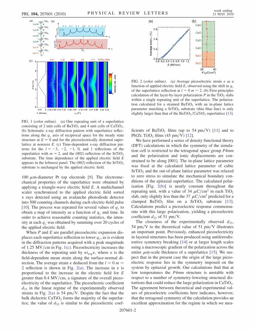
field-dependent mean strain along the surface-normal di-rection. The average strain " deduced from the l ¼ 0 m ¼2 reflection is shown in Fig. 2(a). The increase in " isproportional to the increase in the electric field for Egreater than 0:4 MV=cm, a signature of the overall piezo-electricity of the superlattice. The piezoelectric coefficientd33 in the linear regime of the experimentally observedstrains in Fig. 2(a) is 54 pm=V. Despite the fact that thebulk dielectric CaTiO3 forms the majority of the superlat-tice, the value of d33 is similar to the piezoelectric coef-
ficients of BaTiO3 films (up to 54 pm=V) [11] and toPbðZr;TiÞO3 films (45 pm=V) [12].We have performed a series of density functional theory
(DFT) calculations in which the symmetry of the simula-tion cell is restricted to the tetragonal space group P4mmand the polarization and ionic displacements are con-strained to be along [001]. The in-plane lattice parameterwas fixed at the calculated lattice parameter of cubicSrTiO3 and the out-of-plane lattice parameter was relaxedto zero stress to simulate the mechanical boundary con-ditions of the epitaxial superlattice. The calculated polar-ization [Fig. 2(b)] is nearly constant throughout therepeating unit, with a value of 34 �C=cm2 in each TiO2
slab, only slightly less than the 37 �C=cm2 predicted for aclamped BaTiO3 film on a SrTiO3 substrate [13].Calculations predict a piezoelectric response commensu-rate with this large polarization, yielding a piezoelectriccoefficient d33 of 51 pm=V.The closeness of the experimentally observed d33,
54 pm=V to the theoretical value of 51 pm=V illustratesan important point. Previously, enhanced piezoelectricityin layered structures has been produced using antiferrodis-tortive symmetry breaking [14] or at larger length scalesusing a macroscopic gradient of the polarization across theentire �m-scale thickness of a superlattice [15]. We sus-pect that in the present case the origin of the large piezo-electric response lies in the symmetry imposed on thesystem by epitaxial growth. Our calculations find that atlow temperatures the P4mm structure is unstable withrespect to a number of symmetry-lowering structural dis-tortions that could reduce the large polarization in CaTiO3.The agreement between theoretical and experimental val-ues of piezoelectric coefficients here indicates, however,that the tetragonal symmetry of the calculation provides anexcellent approximation for the regime in which we mea-
FIG. 2 (color online). (a) Average piezoelectric strain " as afunction of applied electric field E, observed using the shift in qzof the superlattice reflection at l ¼ 0 m ¼ 2. (b) First-principlescalculation of the layer-by-layer polarization P in the TiO2 slabswithin a single repeating unit of the superlattice. The polariza-tion calculated for a strained BaTiO3 with an in-plane latticeparameter matching a SrTiO3 substrate (thin blue line) is onlyslightly larger than that of the BaTiO3=CaTiO3 superlattice [13].
FIG. 1 (color online). (a) One repeating unit of a superlatticeconsisting of 2 unit cells of BaTiO3 and 4 unit cells of CaTiO3.(b) Schematic x-ray diffraction pattern with superlattice reflec-tions along the qz axis of reciprocal space for the steady statestructure at E ¼ 0 and for the piezoelectrically distorted super-lattice at nonzero E. (c) Time-dependent x-ray diffraction pat-terns for the l ¼ �3, �2, �1, 0, and 1 reflections of thesuperlattice with m ¼ 2, and the (002) reflection of the SrTiO3
substrate. The time dependence of the applied electric field Eappears in the leftmost panel. The (002) reflection of the SrTiO3
substrate is unchanged by the applied electric field.
PRL 104, 207601 (2010) P HY S I CA L R EV I EW LE T T E R Sweek ending21 MAY 2010
207601-2

sure the piezoelectric response of the superlattice, i.e.,applying E at room temperature.
The initial nonlinearity of the strain in Fig. 2(a) showsthat the highly responsive state is reached only when thesystem is distorted by a high electric field, above E ¼0:4 MV=cm. This leads to the tantalizing prospect thatsuperlattices can be produced in which electric-field in-duced phase transitions yield enhanced piezoelectric prop-erties. A second potential origin of the nonlinearity at lowfields lies in the decomposition of the polarization of thefilm into domains at zero field, an effect which has pre-viously been surmised based on the static properties ofsuperlattices [16,17]. Further investigation will give moredetailed insights into understanding the possible role ofsuperlattice phase transitions under applied electric field.These considerations, however, go beyond the scope of thisLetter and thus will be discussed elsewhere in the future.
Further indication that the CaTiO3 layers play a crucialrole in the piezoelectricity of the superlattice lies in thelayer-by-layer origin of the piezoelectric response. Theintensities of superlattice reflections result from samplingthe structure function of the repeating unit at a smallnumber of points, as in Fig. 3(a). Under nonequilibriumconditions, the structure function, and thus the intensitiesof superlattice reflections, is changed by the relative dis-placements of atoms within the repeating unit. This effectprovides a route to measure experimentally (i) how theaverage piezoelectric strain " is divided between the twocomponents of the superlattice and (ii) via electromechani-cal coupling, whether the layer-by-layer polarization isindeed continuous.
In an analytical representation using a sinusoidal modu-lation of lattice parameters and scattering factors [18], theintensities of the l ¼ �1 and l ¼ þ1 superlattice reflec-tions are proportional to�ðmnþ lÞ ðtBaTiO3
� tCaTiO3Þ
ðtBaTiO3þ tCaTiO3
Þ � lðfBaTiO3
� fCaTiO3Þ
ðfBaTiO3þ fCaTiO3
Þ�2:
(1)
Here ti and fi are the lattice parameter and the structurefactor for component i (i ¼ BaTiO3 or CaTiO3). We definer to be the fraction of the average piezoelectric strainarising from distortion in the BaTiO3 component. Whenr ¼ nBaTiO3
tBaTiO3ð" ¼ 0Þ=ntavg � nBaTiO3
=n ¼ 1=3 with
tBaTiO3ð" ¼ 0Þ=tavg close to 1, both components have equal
strain. In terms of r, the lattice parameters in the BaTiO3
and CaTiO3 layers are tBaTiO3ð"Þ ¼ tBaTiO3
ð" ¼0Þ þ r"tavgn=nBaTiO3
and tCaTiO3ð"Þ ¼ tCaTiO3
ð" ¼ 0Þ þð1� rÞ"tavgn=nCaTiO3
. For " � 1, the change in intensity
with increasing " includes only terms proportional to
"
�r� nBaTiO3
n
�; (2)
and to the square of this quantity. Expression (2) predictsthat for r ¼ 1=3, when BaTiO3 and CaTiO3 are equallystrained, the intensities of the l ¼ �1 and the l ¼ þ1satellite reflections will not be changed by piezoelectricexpansion.Profiles of the l ¼ �1 and l ¼ þ1 reflections at m ¼ 2
are shown in Figs. 3(b) and 3(c), for zero field and E ¼1 MV=cm, respectively. Both reflections decrease in peakintensity and broaden at high E. We attribute the broad-ening to inhomogeneity of the piezoelectric response orelectric field either within the lateral spot size of thefocused x-ray beam or across the thickness of the super-lattice. At E ¼ 1 MV=cm, corresponding to an averagestrain of 0.45%, the change in the integrated intensitywith respect to zero field is þ2% for the l ¼ �1 satelliteand �4% for the l ¼ þ1 satellite.A numerical kinematic diffraction calculation provides
the intensities of the l ¼ �1 and l ¼ þ1 reflections as acontinuous function of r. This model differs from thesinusoidal approximation in that it uses atomic positionsderived from the zero-field DFT calculations and extrapo-lates to nonzero E by stretching the CaTiO3 and BaTiO3
components according to the parameter r. Simulated andexperimentally observed changes in the intensities of thel ¼ �1 and l ¼ þ1 reflections are shown as a function of" and r in Figs. 4(a) and 4(b). The best agreement betweenthe kinematic diffraction calculations and the observedsmall changes in intensity occurs when r is close to 1=3,at which the dielectric and ferroelectric layers have equalpiezoelectric response.A hypothetical superlattice composed of materials with
their bulk properties, in which r ¼ 1, and only the BaTiO3
has spontaneous polarization and resulting piezoelectricityis an extremely poor fit for the experimental results. In the
FIG. 3 (color online). (a) The intensity of superlattice reflec-tions (red solid line) is determined by sampling the structurefunction of the repeating unit (black dashed line) at integervalues of l. Electric-field dependence of the (b) l ¼ �1 m ¼ 2and (c) l ¼ þ1 m ¼ 2 reflections. The shaded regions representthe area integrated to obtain the integrated intensity.
PRL 104, 207601 (2010) P HY S I CA L R EV I EW LE T T E R Sweek ending21 MAY 2010
207601-3

hypothetical case, kinematic diffraction predicts that thel ¼ �1 and the l ¼ þ1 reflections at m ¼ 2 would in-crease in intensity by 20% and 120%, respectively, at " ¼0:45%, an effect clearly not present in the data. The oppo-site limiting case of r ¼ 0, corresponding to localizing thepiezoelectric strain in CaTiO3, would lead to a decrease inintensity by 45% at l ¼ þ1, which is similarly not ob-served. A large fraction of the piezoelectric distortion thusunambiguously occurs in the CaTiO3 component of thesuperlattice.
Equal magnitudes of the piezoelectric strains in BaTiO3
and CaTiO3 are consistent with mean-field expectation thatlarge, nearly equal, polarizations in these layers producepiezoelectric coefficients of similar magnitudes in a simpleassumption on the continuity of permittivity and electro-strictive coefficient. This prediction of the functional prop-erties using the continuity of the polarization can becompared with the atomic-scale structure derived fromDFT calculations. Figure 4(c) compares DFT calculationsof the fractional changes of Ca-Ca and Ba-Ba distances foraverage strains of 0.5% and 1% with the changes expectedfrom a uniform distortion with r ¼ 1=3. The uniformdivision of the distortion between BaTiO3 and CaTiO3 isin excellent agreement with the DFT results for strains upto " ¼ 0:5%, which is consistent with the experimentalresults.
An approach similar to the one we have demonstratedhere, combining DFT calculations and nonequilibrium
structural probes, can be used to probe the stabilizationof structural phases in low dimensional systems [19], thecoupling between ferroelectricity and magnetism via struc-tural distortions [20], and to develop new methods tocontrol thermal properties in dielectrics using composi-tional grading [21]. Our results show that even in thesesymmetric superlattices there can be an important role ofthe imposed crystallographic symmetry in determining thepiezoelectric response. The role of compositional symme-try breaking in the properties of superlattices can likewisebe resolved by a similar approach [22]. The functionalityof complex oxides is now being engineered with layerthicknesses at which these new approaches are necessaryto extend the conventional volume-average characteriza-tion of the properties of these materials to far smallerspatial scales.This work was supported by the U.S. Department of
Energy, Office of Basic Energy Sciences, through ContractNo. DE-FG02-04ER46147. H. N. L. acknowledges supportfrom the Division of Materials Sciences and Engineering,U.S. Department of Energy, through Contract No. DE-AC05-00OR22725. S.M.N. and the use of the AdvancedPhoton Source were supported by the U. S. Department ofEnergy, Office of Science, Office of Basic EnergySciences, under Contract No. DE-AC02-06CH11357.
*[email protected][1] J. B. Neaton and K.M. Rabe, Appl. Phys. Lett. 82, 1586
(2003).[2] H. N. Lee et al., Nature (London) 433, 395 (2005).[3] D. A. Tenne et al., Science 313, 1614 (2006).[4] S. S. A. Seo and H.N. Lee, Appl. Phys. Lett. 94, 232904
(2009).[5] M. Dawber et al., Phys. Rev. Lett. 95, 177601 (2005).[6] W. Tian et al., Appl. Phys. Lett. 89, 092905 (2006).[7] M. E. Lines and A.M. Glass, Principles and Applications
of Ferroelectrics and Related Materials (OxfordUniversity Press, New York, 1977), p. 156.
[8] X. Wu et al., Phys. Rev. Lett. 101, 087601 (2008).[9] D.-H. Do et al., Nature Mater. 3, 365 (2004).[10] D. H. Do et al., Integr. Ferroelectr. 101, 174 (2008).[11] I.-D. Kim et al., Appl. Phys. Lett. 86, 192907 (2005).[12] A. Grigoriev et al., Phys. Rev. Lett. 100, 027604 (2008).[13] S.M. Nakhmanson, K.M. Rabe, and D. Vanderbilt, Appl.
Phys. Lett. 87, 102906 (2005).[14] E. Bousquet et al., Nature (London) 452, 732 (2008).[15] R. Nath et al., Appl. Phys. Lett. 92, 012916 (2008).[16] V. A. Stephanovich, I. A. Lukyanchuk, and M.G. Karkut,
Phys. Rev. Lett. 94, 047601 (2005).[17] S. Lisenkov, I. Ponomareva, and L. Bellaiche, Phys. Rev.
B 79, 024101 (2009).[18] R.M. Fleming et al., J. Appl. Phys. 51, 357 (1980).[19] D. D. Fong et al., Science 304, 1650 (2004).[20] H. Zheng et al., Science 303, 661 (2004).[21] S. Zhong et al., Appl. Phys. Lett. 89, 142913 (2006).[22] N. Sai, B. Meyer, and D. Vanderbilt, Phys. Rev. Lett. 84,
5636 (2000).
FIG. 4 (color online). Kinematic simulation of the change inthe intensities of the (a) l ¼ �1 m ¼ 2 and (b) l ¼ þ1 m ¼ 2reflections, as a function of the average strain " and the fraction rof the strain occurring in BaTiO3. Symbols represent the inter-section of the experimental strain and integrated intensity withthe simulated intensity. The solid lines indicate the limits set bythe statistical uncertainty in the integrated intensity. The experi-mental results are consistent with r ¼ 1=3, corresponding toequal strains in the BaTiO3 and CaTiO3 components. (c) DFTpredictions of the fractional change in the Ca-Ca and Ba-Badistances (symbols) are consistent with the sharing of strainaccording to r ¼ 1=3.
PRL 104, 207601 (2010) P HY S I CA L R EV I EW LE T T E R Sweek ending21 MAY 2010
207601-4