plant natural product glycosyl- and...
TRANSCRIPT

Plant Natural Product Glycosyl- and Methyltransferases
HABILITATIONSSCHRIFT
zur Erlangung des akademischen Grades
doctor rerum naturalium habilitatus (Dr. rer. nat. habil.)
vorgelegt der
Mathematisch-Naturwissenschaftlich-Technischen Fakultät
(mathematisch-naturwissenschaftlicher Bereich)
der Martin-Luther-Universität Halle-Wittenberg
von
Dr. rer. nat. Thomas Vogt
geb. am 06.08.1960 in St. Goar (Rh.-Pfalz)
Gutachter: 1. Prof. Dr. Toni M. Kutchan 2. Prof. Dr. Eran Pichersky 3. Prof. Dr. Jonathan Gershenzon Halle, den 8.März 2006
urn:nbn:de:gbv:3-000010563[http://nbn-resolving.de/urn/resolver.pl?urn=nbn%3Ade%3Agbv%3A3-000010563]

Wissen zu erwerben, ohne über das Erlernte
nachzudenken, ist sinnlos; nur nachzudenken, ohne zu
lernen, führt zu gefährlichen Überlegungen.
(Konfuzius)
2
TABLE OF CONTENTS
Acknowledgements
pp.
1. Summary and Rationale………………………………………….. 5
2. Introduction……………………………………………………….. 6-7
3. Identification and Purification of Transferases…………………. 7-11
4. Substrate Specificity………………………………………………. 11-17
5. Structural Characterisation and Catalytic Mechanisms……….. 17-23
6. Cellular Localisation……………………………………………… 23-26
7. Biological Significance …………………………………………… 26-29
8. Molecular Evolution……………………………………………… 29-34
9. Economical Relevance……………………………………………. 34-37
10. Summary and Outlook…………………………………………… 37-38
11. References ………………………………………………………… 38-50
Declaration
Curriculum vitae
List of Publications (included as reprints)
List of Reviews (not included as reprints)
Supplemental publications
3
Acknowledgements First of all, I would like to thank my former PhD students Mwafaq Ibdah and Judith Isayenkova for their enthusiastic and dedicated work. I also like to thank former diploma students Elke Zimmermann and Stefan Ebert for the amount of work and interest they put into this project. I also would like to thank many people in the Department of Secondary Metabolism at the Leibniz-Institute of Plant Biochemistry. Dagmar Knöfel for a long time provided excellent technical assistance and personal advices. Dr. Willibald Schliemann and Dr. Alfred Baumert provided scientific, technical, and personal support, helping out at any time without hesitation. I thank the head of the Department of Secondary Metabolism, Prof. Dieter Strack, for helpful discussions and for giving me a chance to develop my own research at his Department. Thanks also to those other people who have been or are still members of the Department of Secondary Metabolism. To mention especially Dr. Joachim Hans, Dr. Bettina Hause, Barbara Kolbe, Kerstin Manke, Ingrid Otschik, Dr. Michael Stephan, Ute Vinzens, Silvia Wegener, Dr. Markus Weiss and Dr. Jochen Winter. Thanks to my other colleagues from our Institute, especially to Dr. Wolfgang Brandt, Dr. Jürgen Schmidt and Dr. Jörg Ziegler for their dedicated help and important experimental and theoretical support. Thanks to the greenhouse and EDV-staff and the administration, especially Kerstin Balkenhohl and Christine Kaufmann. Special thanks also to many scientists I got to know during the last years and who helped me with advice, enthusiasm, and action: Prof. Mark Bernards (London, Canada), Prof. Dr. Hans Bohnert (Illinois, USA), Prof. Vince De Luca (St. Catharines, Canada), Prof. Brian Ellis (Vancouver, Canada), Dr. Rudi Grimm (Munich, Germany), Dr. Paul-Gerhard Gülz (Cologne, Germany), Dr. Werner Heller (Munich, Germany), Dr. Andreas Krins (Dresden, Germany), Dr. Daniel Rauh (San Francisco, USA), Dr. Harald Seidlitz (Munich, Germany), Prof. Milton Stubbs (Halle, Germany), Prof. Loverine Taylor, (Pullman, USA), Prof. Eckhard Wollenweber (Darmstadt, Germany), Dr. Victor Wray (Braunschweig, Germany) and many other colleagues, whom I had a chance to talk to or discuss with during my career and work. I am grateful to Dr. J. Isayenkova, Dr. W. Schliemann, Prof. D. Strack, Dr. C. Tretner and Dr. J. Ziegler for critical reading of this manuscript. I thank the referees of this manuscript, especially Prof. Toni Kutchan, for their interest and support. I gratefully acknowledge the financial support by the Deutsche Forschungsgemeinschaft. I thank Jan and Lance for their guidance in mental and physical strength.
This work is dedicated to my parents Maria and Alois
and I feel somewhat ashamed that I write it in English which both do not read or understand.
4

1. Summary and Rationale
Plant natural product biosynthesis is probably the most versatile and flexible
biochemical system on earth. Glycosylation and methylation significantly contribute to the
large diversity of the abundance of natural products. This project was initiated to understand
and characterize the accumulation of a unique class of glycosylated plant compounds, the
betacyanins, which have replaced the anthocyanins as fruit and flower pigments in the
majority of the Caryophyllales. Two model plants, Dorotheanthus bellidiformis (livingstone
daisy) and the halophyte Mesembryanthemum crystallinum (ice plant) were chosen due to the
unique features of betacyanin accumulation in cell cultures and epidermal bladder cells,
respectively. During these investigations not only the accumulation of these and accom-
panying flavonoid pigments was investigated, but purification techniques for several regio-
and substrate specific enzymes involved in the glucosylation or methylation of betacyanin and
flavonoids were established, the corresponding genes were cloned and the respective
recombinant enzymes characterized. Substrate specificity and sequence alignments of these
enzymes provided evidence that betacyanin biosynthesis was likely developed after the
structures of flavonoids, including anthocyanins had already been established. The observed
parallel accumulation of betacyanin and flavonoid conjugates in the bladder cells of the ice
plant led to the identification of a novel subset of cation-dependent O-methyltransferases with
a broad substrate specificity including glucose esters. This observation contradicts the current
perception that these enzymes in plants are only involved in the methylation of the lignin
monomers caffeoyl- and 5-hydroxy feruloyl coenzyme A, respectively.
To understand the substrate specificity and the properties of both types of enzymes it
was of utmost importance to obtain information on the enzyme structures and reaction
mechanisms. Initial attempts to crystallise a betanidin 5-O-glucosyltransferase, were
hampered by its low abundance in heterologous systems and the resulting multistep
purification procedure. Therefore, an alternative approach was used with a combination of
sequence similarity search, site-directed mutagenesis, and computer-assisted molecular
modelling to resolve the first 3-D structure of a plant natural product glucosyltransferase. A
plausible reaction mechanism for this enzyme could be established to explain the inversion of
configuration of the attached sugar from α-linked in the donor UDP-glucose to a β-linkage in
the glucosylated plant natural product. In case of PFOMT, the novel cation-dependent O-
methyltransferase discovered during this work from the ice plant, the high yields of
recombinant protein combined with the rapid success in crystallisation facilitated our efforts
to establish a crystal structure for this subcluster of methyltransferases.
5

2. Introduction
Sessile plants were forced during evolution to develop effective mechanisms to detoxify
all kinds of reactive and toxic compounds within their cells and to repulse possible predators
by endogenous chemicals, rather than rely on kinetic and mechanical strength. They have
achieved both goals by synthesis, modification and storage of a wide array of natural
compounds including alkaloids, cyanogenic glucosides, glucosinolates, phenolics and
terpenoids, to name only the most prominent groups. Research in plant natural product
biosynthesis has been focussed on the identification and synthesis of these compounds for
more than a century. The isolation of morphine by Wilhelm Sertürner as early as 1806, and
the chemical synthesis of indigo by Adolf von Baeyer in 1878 are hallmarks of plant derived
biochemistry with a tremendous scientific and socio-economic impact to the 21st century.
The parallel development of biochemical and molecular tools during the last 20 years
has allowed an in-depth characterisation of plant natural product biosynthetic enzymes, the
corresponding genes and an array of regulatory elements organizing the biosynthesis and
storage of metabolites (Dixon, 1999). Although much of the research on plant natural product
biosynthesis is still at the level of gene discovery, future steps are already taken combining
experimental and computer-based analysis of model plants, like Arabidopsis thaliana
(http://www.arabidopsis.org/tools/aracyc). The abundance of plant natural products can be
reduced to only a few biosynthetic pathways and to a few simple starter-molecules originating
from primary metabolism. The polyketide- and the shikimate-pathway initiate a variety of
target structures from activated acetate units and from the two aromatic amino acids
phenylalanine and tyrosine, respectively. In addition, acetate/mevalonate and glycerin-
aldehyde 3-phosphate/pyruvate are considered as precursors of terpenoids (Gershenzon and
Kreis, 1999; Lichtenthaler, 1999).
The first step to achieve this diversity is generated by ligations, cyclisations and
oxidations and already results in arrays of core structures. These are further diversified by a
second armada of enzymes, mostly members of the transferase families, including glycosyl-
(and glucuronosyl-), methyl-, acyl-, sulfo-, or prenyltransferases. Single or successive
modifications by enzymes of these superfamilies result in the observed myriads of natural
products, which are not only characteristic for the plant, the fungal, or the microbial kingdom,
but even enable the identification of individual species and subspecies by a quantitative or
qualitative metabolite pattern (Harborne and Turner, 1984; Watermann and Gray, 1988).
The array of plant natural products is the result of evolutionary diversification of genes
and the corresponding enzymes. Changing environmental conditions with a tremendous array
6

of potential substrates provide the most challenging tests for any enzymatic system. The
impact of evolutionary and environmental factors are reflected in the development of large
gene families or gene clusters from a single or a few hypothetical ancestors (Moore and
Purugganan, 2005). In the genome of A. thaliana more than 100 glycosyltransferase (GT) like
sequences have been identified (Li et al., 2001) and similar numbers of these enzymes are
expected for other plant species. These and other modifying enzymes may hold essential keys
for the survival of plants under changing environmental conditions (Lu and Rausher, 2003).
Their natural variation and functional redundancy may be compared to the large diversity of
antibodies in the vertebrate immune system (Cannon et al., 2004), although immunological
reaction cascades appear more complex and target whole cells or proteins rather than small
molecules.
Our research is focused on two superfamilies of these enzymes, the plant glycosyl- and
methyltransferases (referred to as GTs and MTs). With an emphasis on O-glucosylation and
O-methylation the progress on structural and functional aspects our and other laboratories
have made throughout the last decade will be discussed, despite initial difficulties associated
with the isolation and characterization of both types of enzymes. Similar enzymes exist in
animals, fungi and microbial systems and are referenced sporadically throughout this report
(Axelrod and Vesell, 1970; Lampe et al., 1999).
3. Identification and Purification of Transferases
The properties of natural compounds are in part the result of hydrophilic and
hydrophobic modifications governed by glycosylation or methylation, respectively. In
addition, sulfatation, acylation, and prenylation further enhance the structural complexity and
influence the properties and location of the corresponding conjugates.
Glycosylation is considered as one of the final modifications in the biosynthesis of many
compounds. Glycosides of all major classes of secondary metabolites, i.e. phenylpropanoids,
terpenoids, alkaloids, thiocyanides, cyanohydrins, and steroids have been identified (Figure
1). The large number of naturally occurring glycosides does not correlate with a detailed
knowledge of the corresponding enzymes leading to their formation, the glycosyltransferases.
Except for their well-documented role in detoxification of xenobiotics and their ability to
increase the hydrophilicity of hydrophobic or amphiphilic aglycones, the function of the
variety of glycosyltransferases in plant secondary metabolism remained poorly characterized
(Jones and Vogt, 2001).
7
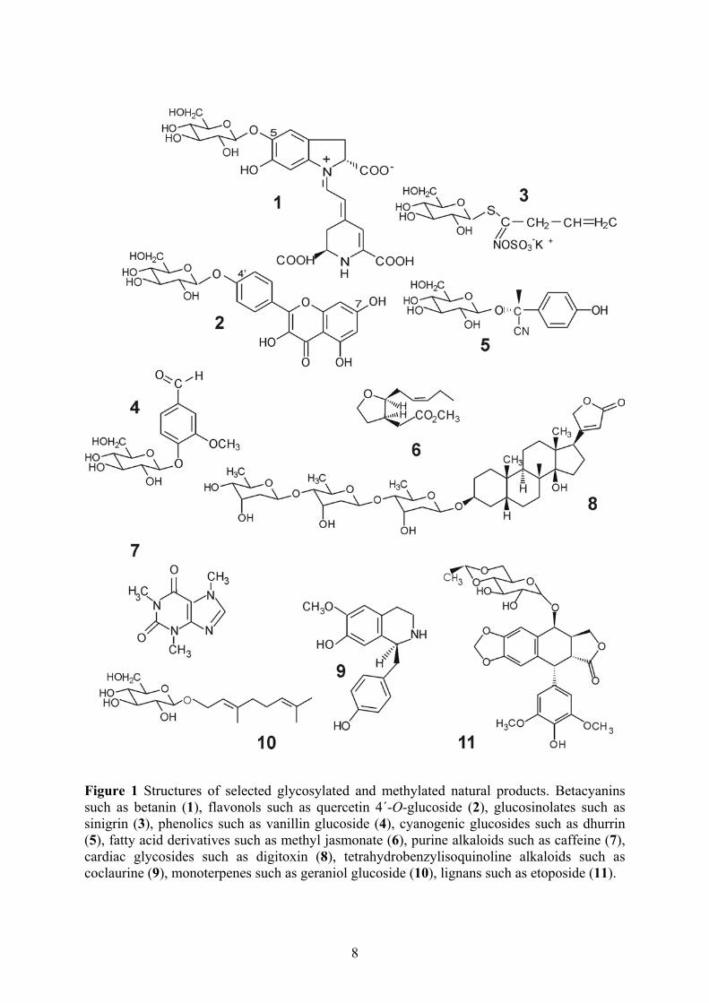
Figure 1 Structures of selected glycosylated and methylated natural products. Betacyanins such as betanin (1), flavonols such as quercetin 4´-O-glucoside (2), glucosinolates such as sinigrin (3), phenolics such as vanillin glucoside (4), cyanogenic glucosides such as dhurrin (5), fatty acid derivatives such as methyl jasmonate (6), purine alkaloids such as caffeine (7), cardiac glycosides such as digitoxin (8), tetrahydrobenzylisoquinoline alkaloids such as coclaurine (9), monoterpenes such as geraniol glucoside (10), lignans such as etoposide (11).
8

Only in the last decade many GTs have been characterized and associated with
individual pathways based on functional expression, substrate specificity, and plant model
systems (summarized in several reviews, e.g. Vogt, 2000; Lim and Bowles, 2004).
Methylation of natural compounds in plants is performed by S-adenosyl-L-methionine
(AdoMet) dependent methyltransferases (MTs) and is a characteristic feature of most
secondary metabolites like phenylpropanoids, alkaloids or terpenoids (Figure 1). Plant MTs
are able to methylate four different polarized nucleophiles (O, N, and S) or activated C-atoms
(carbanions). O-Methyltransferases (OMTs) act on a wide variety of target structures and can
be classified as either cation-dependent (class I) or cation-independent (class II) proteins
(Joshi and Chiang, 1998). Irrespectively of their substrates and specificity, class II enzymes
comprise a family of dimeric proteins with an average molecular mass of 40-60 kDa per
monomer (Ibrahim and Muzac, 2000). A third class of O-methylating enzymes without any
cation-dependence has recently been indroduced and termed SABATH-enzymes (based on the
first two initials of the plants they were found in). These are involved in the formation of
volatile aromatic carboxymethylated compounds, like methyl salicylate or methyl benzoate
(Ross et al., 1999; Pott et al., 2004). These OMTs share more sequence identities to some N-
methyltransferases, like theobromine synthase than to any other class II OMTs and therefore,
were positioned into a separate subcluster.
In contrast to the universal array of substrates of cation-indepedent enzymes, class I
OMTs apparently serve only one function in plants and animals: the methylation of vicinal
dihydroxy systems. In animals, catechol O-methyltransferase (catechol OMT) inactivates
aromatic neurotransmitters in the brain and mutagenic phenolics in the liver and kidney
(Mannisto and Kaakkola, 1999; Zhu et al., 1994). In plants, the corresponding enzymes, due
to their preferred substrate, are referred to as caffeoyl coenzyme A OMTs (CCoAOMT) and
are part of a complex grid of enzymatic reactions to build the structure of plant lignin, besides
cellulose the most prominent polymer on earth (Humphreys and Chapple, 2002). Their
involvement in the methylation of complex pigment conjugates has recently been established
by our results and suggests additional roles of these class I OMTs in plants (Ibdah et al.,
2003). Whereas the animal enzymes are considered monomeric, the plant enzymes have a
dimeric structure with a monomeric molecular weight between 25 and 30 kDa (Schmitt et al.,
1991; Ferrer et al., 2005).
Methylation and glycosylation in plants are usually associated with inactivation or
detoxification of natural compounds rather than promoting their biological activities. This is
illustrated by a most intriguing example, the cyanogenic glucosides (Conn, 1980), where only
9

the association of the bound sugar precludes the liberation of the toxic cyanide. A recent
report emphasizes the stabilization of cytokinins by glucosylation to prevent these hormones
from degradation by oxidases/dehydrogenases (Mok et al., 2005). On the other hand,
modification of physical properties by methylation can result in the release of biologically
active scents and flavours (Dudareva et al., 2004) or enable the free diffusion of plant
hormones, like jasmonic acid (Reinbothe et al., 1994). In coffee beans and in many other
plants, several N-methylations of xanthosine essentially lead to the formation of the purine
alkaloid caffeine, the most prominent legal, bioactive drug (Uefuji et al., 2003; Misako and
Kouichi, 2004). In some cases, glycosylation or methylation may have no effect on the
properties of biologically active compounds but can be used in vitro to specifically label
active molecules and decipher their biological function (Vogt et al., 1995; Xu et al., 1997).
The first reports describing the transfer of a sugar moiety from UDP-glucose to
natural products were already published in the late 50´s (Hutchinson et al., 1958; Cardini and
Yamaha, 1958; Yamaha and Cardini, 1960). A few years later, S-adenosyl-L-methionine
dependent O-methylation of caffeic acid was discovered in plant tissues (Finkle and Nelson,
1963; Legrand et al., 1976). In subsequent reports, the characterisation of purified transferase
activities was rendered quite difficult due to the low abundance of these enzymes in plant
tissues and the restraint availability of reference compounds to characterize the substrate
specificity. Partly purified proteins were characterized based on enzyme properties or, if
purified further, to obtain antibodies for cellular localisation (Latchinian-Sadek and Ibrahim,
1991; Hrazdina, 1992).
Figure 2 Efficient use of affinity matrices: A Purification of native betanidin 6-O-gluco-syltransferase by dye ligand chromatography on Reactive Yellow 3 (from Vogt et al., 1997). B Heterologously expressed CCoAOMT from Ammi majus purified by metal affinity chromatography on Talon (from Lukačin et al., 2004). Purification was achieved although the protein does not contain any HisTag. 1, molecular weight markers; 2, crude extracts; 3A, after ion-exchange chromatography; 3B, 10 mM imidazole wash; 4A, 1 M UDP-glucose eluate; 4B, 30 mM imidazole eluate. Arrows indicate the positions of purified proteins.
10

The purification and unequivocal identification of many transferases was facilitated by
the use of affinity or pseudo-affinity matrices, i.e. glucosyltransferases can be sometimes
purified in a single step by dye-ligand chromatography (Vogt et al., 1997; Jones et al., 1999;
Figure 2), or metal affinity chromatography (Marcinek et al., 2000). Methyltransferase
purification was simplified by S-adenosyl-L-homocysteine agarose or by adenosine agarose
(Sharma and Brown, 1979; Cacace et al., 2003). Recently, our lab developed a purification
for cation-dependent OMTs based on immobilized-metal affinity chromatography which was
originally established to capture and purify recombinant His-tagged proteins (Lukačin et al.,
2004; Figure 2).
DNA-sequencing, combined with rapid developments in mutational analysis and
molecular cloning lead to the identification and functional characterisation of several
transferases, e.g. the annotation of the bronze-1 locus in Zea mays as a glucosyltransferase
encoding gene (Fedoroff et al., 1984) and to the first description and cloning of a plant class I
CCoAOMT from tobacco (Nicotiana tabacum) (Schmitt et al., 1991). In parallel, improved
recovery of proteins combined with enhanced sensitivity to obtain amino acid sequence
information enabled the identification of elusive plant GTs and other rare proteins (Ziegler et
al., 1997; Matsudaira, 1991; Grimm and Eckerskorn, 1996). Only recent developments in
plant biochemistry begin to stress the important role of these modifying enzymes compared to
the so-called “key enzymes” of plant natural product biosynthesis, e.g. phenylalanine
ammonia lyase (PAL), chalcone synthase (CHS), or hydroxymethylglutaryl (HMG)-CoA
reductase.
4. Substrate Specificity
The most obvious questions to be asked about any transferase are two very simple ones:
What are their substrates? How specific are the individual enzymes? Earlier reports were
often forced to work with only partly purified proteins or crude cell extracts, which were not
suited to unequivocally correlate one individual enzyme with the observed specificity. Only
by recombinant techniques developed throughout the last decade a more thorough
investigation of the specificities was accessible.
To answer the second question first, plant GTs and MTs are quite specific for the
individual sugar or methyl group donor, respectively. Especially OMTs use exclusively
AdoMet as the substrate donor, whereas in case of the plant GTs an apparent specificity for
UDP-glucose is observed. In a few reports, UDP-glucose was only the preferred rather than
11

the solely accepted sugar donor (Taguchi et al., 2001; Sasaki et al., 2004). In several cases
rhamnosyl-, galactosyl-, xylosyl- or even glucuronosyltransferases have also been described
(Brugliera et al., 1994; Frydman et al. 2004; Miller et al., 1999; Martin et al., 1999; Schulz
and Weissenböck, 1988). The imbalance in favour of the GTs is only in part due to the
preferred modification of natural compounds by glucose, but also resides in the limited
experimental access of available activated sugars other than glucose, galactose, rhamnose, or
xylose. This is evident in case of the pharmaceutically important foxglove (Digitalis) species
(Figure 3), where only the final glucosylation step in the formation of bioactive cardiac
glycosides has been characterized. In contrast, the attachment of the pharmacologically
important dideoxy sugars, like digitoxose, has only been described yet for micro-organisms
(Albrecht, 1999; Trefzer et al., 1999; Thorson and Vogt, 2003).
Figure 3 The pharmacologically active cardiac glycoside digitoxin, characteristic for several Digitalis species, like Digitalis purpurea (left).
In contrast to plants, microbial GTs are often observed to be less specific for the
individual sugar (Blanco et al., 2001). This is in part compensated by their strong preference
for the sugar acceptor, which is usually not observed for plant GTs (Jones and Vogt, 2001). If
this specificity has somehow evolved together with the potent antibiotic properties of many
microbial products, is an intriguing question (Trefzner et al., 1999; Thorson and Vogt, 2003).
In plants, glycosylation like methylation is seen as a “tranquilliser” with a lower bioactivity of
the glycosylated product compared to the glycoside. From the few known glycosylated plant-
derived and biologically active compounds like the saponins, the attachment of several
hydrophilic sugars, rather than of one specific sugar is responsible for the observed
detrimental effect on membranes (Osbourn, 1996; Kasai et al., 1999). Apparently, procaryotes
and eucaryotes have evolved two different strategies to detoxify or inactivate harmful
compounds.
In the last decade it became increasingly evident that functional characterisation of
either GTs or MTs should strictly differentiate between in vitro and in vivo properties of
12

characterized proteins. In vitro specificities are only limited by the supply of possible
substrates. True in vivo functions of these proteins, however, are much more difficult to
access. We and other have repeatedly emphasized this important point.
Cloning and functional expression of the p-hydroxymandelonitrile GT leading to the
formation of dhurrin in vivo proved the remarkable specificity of this enzyme towards the p-
hydroxylated mandelonitrile as compared to the similar p-hydroxymandelic acid and similar
structures in vitro (Jones et al., 1999). In contrast, the same enzyme in vitro also converts
structurally very different hydroxylated terpenoids, like geraniol, to the corresponding
glycoside, although with a lower catalytic efficiency (Jones et al., 1999; Hansen et al., 2003).
Although the observed kinetic properties suggest that the in vitro and in vivo substrates are
identical, which was proven using A. thaliana as a host plant for the entire dhurrin
biosynthetic pathway (Tattersall et al., 2001), the flexibility of these enzymes in vitro is
striking.
The red-violet betacyanins have replaced the anthocyanins as pigments in the majority
of the Caryophyllales (Strack et al., 2003), but only recently the biosynthetic pathway based
on the presence of a novel 4,5-extradiol cleaving enzyme, the DOPA 4,5-dioxygenase has
been resolved (Christinet et al., 2004). The question wether glycosylation takes part before or
after the spontaneous condensation of betalamic acid with cyclo-DOPA in the biosynthetic
pathway stimulated our own interest in this pathway and still is an ongoing debate (Vogt,
2002; Sasaki et al., 2004). Two alternative routes have been proposed and in vitro
experimental data for two distinct, heterologously expressed and functionally characterized
subgroups of enzymes were presented (Heuer et al., 1996; Vogt et al., 1999a; Sasaki et al.,
2004, 2005). In livingstone daisy (Dorotheanthus bellidiformis), two enzymes specific for the
aglycone betanidin have been described, which also glucosylate a variety of flavonoids with
similar catalytic efficiency as the substrate betanidin (Vogt et al., 1997; Vogt, 2002). A
homologous set of enzymes was observed in red beet (Beta vulgaris), with identical properties
towards the tested flavonoids, but with a strong preference for flavonols as compared to
betanidin (Isayenkova J, Wray V, Strack D, Vogt T, in preparation). In contrast, two distantly
related enzymes described from two related species Mirabilis jalapa and Celosia argentea
(Sasaki et al., 2004, 2005), promote the glucosylation of cyclo-DOPA. The problem therefore
persists, whether glucosylation in vivo is performed at the betanidin or at the cyclo-DOPA
level or two parallel routes exist. Unless knockout mutations or in vivo functional
complementation do not oppose the observed specificities, it seems plausible, that in this
13

monophyletic pathway, present in only 2% of all vascular plants, specificity may have
evolved two times independently from an existing pool of transferases.
The specificities of class II OMTs of Thalictrum tuberosum associated with isoquinoline
alkaloid biosynthesis in vivo, are also not restricted to the corresponding substrates.
Methylation of a variety of phenolic substrates in vitro was observed (Frick and Kutchan,
1999). A total of six different alleles may have arisen by gene duplication and the specificity
of two of those enzymes investigated resides in the change of a single amino acid, tyrosine
instead of cysteine. In their search for 16-hydroxy tabersonine MT of Catharanthus roseus
Schröder and co-workers purified four class II OMTs with activities towards this alkaloid.
Unexpectedly, with the recombinant enzymes no activity towards the alkaloid was observed,
but two sequential methylations of the common flavonol myricetin (Cacace et al., 2003).
Either the specificity of the heterologously expressed enzymes has changed or, if both
activities belong to different enzymes which co-eluted in all chromatographic steps, the
biochemical similarity between alkaloid and flavonol methylating OMTs in this plant is
remarkable. That this similarity may sometimes be misleading can be exemplified for a pair of
regiospecific isoflavone daidzein 7- and 4´-OMTs. The 7-OMT was originally proposed to
catalyse the methyl transfer also to the 4´- group of daidzein in vivo (Liu and Dixon, 2001).
Only by cloning of the “correct” enzyme it was proven that two separate enzymes exist and
daidzein biosynthesis requires a specific 4´-OMT in vitro and in vivo (Akashi et al., 2003).
The 5-hydroxyferulate/caffeic acid OMT from alfalfa (Medicago sativa) involved
primarily in the formation of syringyl lignin has a much broader specificity than initially
anticipated. This multifunctional enzyme methylates the hydroxyl group in meta position of
small benzaldehyde like aromatics more efficiently than caffeic acid, although the position
specificity remains the same (Kota et al., 2004). In contrast, other class II enzymes may retain
their high specificity in vitro and in vivo. Eugenol and chavicol OMT show a conserved high
substrate specificity and are both expressed in the same tissue, in basil peltate glands (Gang et
al., 2002).
These few examples emphasize that care should be taken to predict the substrate
specificity of any GT and MT in question. Knockout mutations combined with
complementation studies, RNAi-mediated gene silencing and metabolite profiling may be
required to elucidate the true in vivo role of a given enzyme. A final example, selected from
our own work on class I OMTs, illustrates several additional problems associated with in vitro
versus in vivo specificity. These include redundancy of similar proteins with different
14

specificities, posttranslational in vivo modifications and artificially high in vitro ratios of
substrates compared to enzyme quantities.
In addition to betacyanins, the ice plant (Mesembryanthemum crystallinum) accumulates
large amounts of glycosylated and methylated flavonoid conjugates in epidermal tissues (Vogt
et al., 1999b; Ibdah et al., 2002). Methylation is performed by a unique class I cation-
dependent CCoAOMT (termed PFOMT), which displayed a broad substrate specificity
towards various flavonoids as well as caffeic acid and its conjugates (Ibdah et al., 2003). This
broad specificity was in contrast to previously reported caffeoyl CoA-specific properties of
these highly conserved enzymes involved in lignin modification (Pakusch et al., 1989; Maury
et al., 1999). At least two subsets of functionally active enzymes of the same subgroup exist in
many species (Ibdah et al., 2003). In the databases all of these enzymes are classified as
CCoAOMTs involved in lignin biosynthesis. Therefore, it is not only difficult to predict the
specificity based on sequence data, but to compare the observed substrate promiscuity in vitro
with the in vivo situation. Are there any developmental or spatial separations of these
activities in vivo? PFOMT has the unique property to methylate caffeoylglucose in vitro and
could be involved in the methylation of ester in vivo. The product feruloylglucose could then
be the potential acyl donor for the abundant feruloylated betacyanin conjugates in epidermal
bladder cells (Bokern et al., 1991; Vogt et al., 1999). A second, substrate-specific CCoAOMT
already cloned from the ice plant may then only be involved in caffeoyl- and 5-hydroxy
feruloyl CoA methylation in vascular tissues, resulting in a strict tissue specificity. However,
the situation is further complicated, since in A. thaliana tissues (and most likely in the ice
plant) one member of the class II OMTs is described with the same overlapping substrate
preferences as both class I enzymes (Muzac et al., 2000, T. Vogt, unpublished). The corres-
ponding transcripts of all these class I and class II OMT-genes could already be detected in A.
thaliana plants (Vogt, unpublished). Whether the corresponding enzymes are active in the
same tissues or are spatially separated remains to be proven by further transcript analysis and
immunolocalization studies.
A further level of complexity can be reached by post-translational modifications. Upon
sequencing of the native PFOMT, an N-terminal truncation of this protein has been observed
(Ibdah et al., 2003) which may lead to an altered substrate specificity (Vogt, 2004). Consistent
with the subsequent methylation properties of the native enzyme, only 6- and 3´-O-methyl
derivatives of the endogenous quercetagetin were found in epidermal tissues (Vogt et al.,
1999b). In contrast, the “full-length” recombinant enzyme also methylates the 5-hydroxy
group of quercetagetin (Ibdah et al., 2003) resulting in two strikingly yellow fluorescent
15

compounds. No trace of any yellow fluorescent compound was ever observed in the ice plant.
Therefore, a full-length PFOMT with the in vitro properties is unlikely to exist in vivo. The
observed truncation of the PFOMT in planta could be experimentally mimicked and a
comparable in vivo specificity could be reproduced with in vitro designed proteins (Vogt et
al., 2004; Figure 4) confirming the data obtained with the native plant enzyme.
Figure 4 Variation in reaction products of recombinant PFOMT isoforms. HPLC run of full length (a,d) (N0), N-terminally five amino acids shorter (N-5) (b,e), and N-terminally eleven amino acids shorter (N-11) (c,f ), respectively. Endogenous quercetagetin (queg) was used as the substrate. UV-detection at 364 nm (a-c); Fluorescence detection (d-e), excitation at 370 nm and emission at 520 nm. Peak identification: 1, queg-5-OMe; 2, queg (substrate); 3 queg-5,3´-diOMe; 4, queg-3´-OMe; 5, queg-6-OMe, 6, queg-6,3´-diOMe. Substrate 2, and products 5, 6, and traces of 4 are also observed with the native enzyme. Yellow fluorescing peaks 1 and 3 are products of N0 and N-5 recombinant enzymes only (from Vogt, 2004).
Cellular in vivo ratios of enzyme and substrate might also influence the specificity even
of in vitro “absolute specific” enzymes. When highly concentrated solutions of the full length
PFOMT were incubated with the flavonol quercetin at a 1:1 enzyme to substrate ratio, very
rapid methylation of the substrate was accompanied by the formation of a new yellow
fluorescent product. This product was never observed in the usual in vitro assay, with
saturating substrate to enzyme ratios (T. Vogt, unpublished). HPLC-analysis indicated that
about 1% of the predicted product, quercetin 3´-OMe, was also methylated at the 5-hydroxy
position which is not part of a required vicinal dihydroxy system. The new product gives the
16

observed bluish/yellow fluorescence of que-5,3´-diOMe under UV-light (Figure 5)
comparable to that of the quercetagetin 5-OMe derivatives (Ibdah et al., 2003). This
phenomenon could result in products, not observed under standard experimental conditions. If
only a few molecules of these compounds provide an advantage for the species, they may
contribute to natural selection, a key element in molecular evolution.
Figure 5 A. Structure of fluorescent quercetin-5,3´-diOMe and B. enzyme assays with concentrated PFOMT (N0) at 10 mg/ml. 1, enzyme only; 2, enzyme + 1 mM Mg2+; 3, enzyme + 200 µM quercetin; 4, enzyme + 200 µM quercetin + 1 mM Mg2+; 5, enzyme + 200 µM quercetin + 1 mM AdoMet; 6, enzyme + 1 mM Mg2+ + 200 µM quercetin + 1 mM AdoMet. Fluorescence under UV light (λmax 365 nm). Traces of bivalent cations in the double destilled H2O cause the observed slight yellow/blue fluorescence in vial 5.
Enantioselective properties may further enhance the specificity of some GTs and
probably also of some MTs. An A. thaliana GT was reported to glucosylate selectively the
plant hormone (+)-abscisic acid, whereas some enantioselectivity was also reported earlier for
a similar abscisic acid-specific GT from adzuki bean (Vigna angularis) (Lim et al., 2005; Xu
et al., 2002). The enantioselective properties may become more prominent, once further
enzymes have been analysed which modify optically active aliphatic and cyclic structures.
In summary, these examples raise doubt, how accurate substrate and regiospecificity
observed in vitro can be correlated with the corresponding activity in vivo, unless an enzyme
is characterized on the biochemical level and mutants with the expected, altered chemotype
are observed.
5. Structural Characterisation and Catalytic Mechanisms
In the new millenium, considerable progress has been made on the structural elucidation
of plant methyltransferases, more than on the glucosyl- or glucuronosyltransferases. In 2001
and 2002, Zubieta et al. published the first crystal structures of several natural product class II
17

OMTs from plants, a chalcone, an isoflavone, and a caffeic acid/5-OH-ferulic acid O-
methyltransferase from alfalfa, termed ChOMT, IOMT, and COMT respectively (pdb
signatures: 1FP1, 1FP2, 1KYZ). This breakthrough enabled for the first time an in-depth look
at the organisation of the dimeric structure and the possibility to critically examine the
plausibility of motif predictions based on sequence similarities (Ibrahim, 1997; Joshi and
Chiang, 1998). The X-ray data also paved the way to understand the catalytic mechanism,
correlating substrate specificity with the active site topology, and served as a template to
model similar enzymes with different specificities (Gang et al., 2002; Yang et al., 2004).
All crystallized AdoMet-dependent MTs, including the class II plant enzymes, share a
common core structure, the “AdoMet-dependent MT fold”. Central to this shared core
structure is a mixed seven-stranded β-sheet, with strand number seven inserted antiparallel
between strands five and six (Cheng and Roberts, 2001). The C-terminal catalytic domains in
the COMT, ChOMT, IOMT maintain a highly conserved binding pocket for S-adenosyl-L-
homocysteine (SAH) fixed through a network of hydrogen bonds as well as van-der-Waals
interactions (Figure 6). Methylation proceeds via a typical SN-2-mechanism, a base-assisted
deprotonation of the hydroxyl group by a histidine residue, followed by a nucleophilic attack
at the reactive methyl group in AdoMet (Zubieta et al., 2001, 2002).
Figure 6 Schematic close-ups of the active site cavity of COMT. A. Connolly surface of the protein illustrating the perfect match and complementary shape of bound ferulic acid (FA) and SAH. B. Active site arrangement for the COMT–SAH–5-hydroxyconiferaldehyde complex. Bonds of the COMT active site residues are colour coded. Carbon atoms of SAH and 5-hydroxyconiferaldehyde are shown in black to distinguish them from the active site residues. His 269 acts as the catalytic base. (from Zubieta et al., 2002).
18

Mutation of the catalytic base histidine completely inhibits the methyl transfer. Once
AdoMet is bound, specificity is governed by single amino acids, which more or less facilitate
the binding of the different methyl acceptors by unique arrangements of spatial, hydrophobic,
or hydrophylic constraints.
In class I metal dependent O-methylation reactions, as performed by the mammalian
catechol OMT, deprotonation is facilitated by a bivalent cation, usually Mg2+. This is bound
after AdoMet-binding and converts the vicinal dihydroxy groups of the acceptor to be more
easily ionisable (Vidgren et al., 1994). Again by an SN-2-type reaction, a lysine residue acts as
the catalytic base and accepts the proton specifically from the hydroxyl group of the catechol
like structure in the 3´-position, accompanied by a nucleophilic attack of the methyl group of
AdoMet. Interestingly, and in contrast to the corresponding plant proteins, the catechol OMT
exists in a soluble (liver, kidney) and a membrane-bound (brain) form with a 20 amino acid
extended N-terminal anchor. In both cases these enzymes are monomeric. There is no
dramatic effect on catalytic efficiency, however, the association with the hydrophobic
environment slightly promotes substrate binding and a lower apparent Km for the catechol
substrates is observed (Rivett and Roth, 1982; Mannisto and Kaakkola, 1999). This indicates
that also the physico-chemical environment of enzyme withotherwise identical amino acid
sequences can be important to correctly assess the substrate specificity and kinetic parameters.
Most recently, the structure of the class I OMTs from alfalfa (a substrate specific
CCoAOMT) and from the ice plant (the promiscuous PFOMT) were resolved (Ferrer et al.,
2005; D. Rauh, T. Vogt, J. Kopycki, M.T. Stubbs, in preparation; Figure 7).
Figure 7 Dimeric 3D-structure of PFOMT from the iced plant (left). Superimposed 3D-structures of the CCoAOMT from alfalfa in grey (from Ferrer et al., 2005) and PFOMT from the ice plant in green illustrate the structural similarity of both enzymes (right). 5-OH feruloylCoA (alfalfa) and SAH (PFOMT) are illustrated in red and blue, respectively. Ca2+ (CCoAOMT) and Mg2+ (PFOMT) are marked as purple and blue dots, respectively. The design and overlay of both structures is a courtesy of Jacub Kopycki, MLU Halle-Wittenberg, Germany using PyMol (DeLano Scientific, San Carlos, CA, USA).
19

Despite the low sequence identities between the rat catechol, the alfalfa, and ice plant
OMTs, the metal dependence and the requirements for vicinal dihydroxy groups already
suggests similar structural motifs and catalytic centers of both sets of enzymes (Vidgren et al.,
1994; Ferrer et al., 2005). Each monomer of the CCoAOMT and the PFOMT form a catalytic
domain with a core α/β/α Rossman fold that provides the binding site for SAH (Rossman et
al., 1974). The catalytic SN-2-like mechanism was described for the alfalfa CCoAOMT,
mediated by a stabilized and chelated cation (Mg2+, Ca2+, Mn2+ or Zn2+), abstraction of a
proton by a catalytic lysine residue, and subsequent nucleophilic attack of the methyl group
(Ferrer et al., 2005). The dimeric structure of both plant enzymes are not critical for substrate
recognition like in the case of class II OMTs, and each substrate and cofactor only interacts
with one monomer. However, the first 20 amino acids of the CCoAOMT which up to now
could not be resolved by electronic density maps, appear to be critical for substrate
specificity, at least in case of the PFOMT (Vogt, 2004). The structure of this enzyme is very
similar to the alfalfa enzyme, except for a C-terminal loop and this flexible N-terminal
domain likely related to the profound differences in the substrate specificity. The observed
drastic changes in catalytic efficiencies of different truncated isoforms obtained from
heterologously expressed PFOMT (Vogt, 2004) should reward a thorough examination of this
part of the protein and may lead to the identification of catalytically important amino acids in
this extremely variable domain of class I OMTs.
In contrast to the microbial, animal, or plant MTs, much less is known about the
glycosyltransferase/glucuronosyltransferase structures and catalytic mechanisms. This is in
part due to low overexpression yields of these enzymes in pro- and eucaryotic systems,
combined with problems to crystallize the proteins in sufficient quality to obtain high
resolution electron density maps (Gustafsson et al., 2004; Wang, X, Samual Roberts Nobel
Foundation Inc., Ardmore, OK, USA, pers. communication). In addition, the mammalian
enzymes may be glycosylated and are usually associated with the endoplasmatic reticulum
membranes, enhancing the problems of purification and expression (Radominska-Pandya et
al., 1999). From the array of more than 70 GT families described in the carbohydrate active
enzyme database (http://afmb.cnrs-mrs.fr/CAZY/; Coutinho and Henrissat, 1999), only a few
structures are solved and none of these are related to plant Family 1 enzymes. This family
displays the observed activity towards small natural products, and its members are
characterized by a nucleotide recognition domain and a mechanism leading to the inversion of
the sugar configuration (NRD1β-GTs).
20

The solved crystal structures can be divided into two distinct structural types, the GT-A
and the GT-B fold (Hu and Walker, 2002), with the structure of the vancomycin
glucosyltransferase from Amycolatopsis orientalis, recently crystallized as the first member of
the GT-B family, to which the plant natural product GTs belong (Mulichak et al., 2001). They
both show the inversion of configuration and belong to the NRD1β-GTs. The overall
sequence similarities of the plant Family 1 proteins with this microbial sequence is less than
20%. But the secondary structure predictions for one member of the Family I enzymes, the
betanidin 5-GT from livingstone daisy, UGT73A5 (nomenclature by MacKenzie et al., 1997),
was sufficiently similar to consider building of a model structure. Similar molecular
modelling studies were applied previously to P450 monooxygenases with sequence identities
as low as 13%, again based on common motifs and very similar secondary structure
predictions (Rupasinghe et al., 2003). Our efforts finally succeeded to build the first 3-D
model of a plant natural product GT. The model structure of UGT73A5 was simultaneously
supported by site-directed mutagenesis of the enzyme (Hans et al., 2004). The proposed
structure for UGT73A5 not only showed the expected two Rossman-fold-like domains with a
deep cleft already known from the A. orientalis structure. The model also explained the loss
of enzymatic activity after mutations of the conserved glutamate to alanine in the sugar
binding domain, the so-called PSPG-box (Hughes and Hughes, 1994; Vogt and Jones, 2000),
and of the N-terminally conserved histidine 22 to leucine, respectively.
Based on this model a catalytic mechanism was proposed, which explains the observed
inversion of the bound sugar, by an SN-1-type mechanism, contrary to the scenario with the
SN-2-type mechanism proposed for the OMTs, but with similar amino acid residues involved
(Figure 8). Histidine 22 acts as the catalytic base, abstracting a proton from the substrate
betanidin with subsequent transfer of the proton to the conserved glutamate. In a second step,
a proton transfer from the charged lysine 31 to the oxygen atom of the ester bond in the
activated sugar is proposed. After cleavage of this bond, the resulting sugar carbocation forms
a glycosidic bond with the nucleophile, in this case the deprotonated betanidin. The inversion
of the configuration is the result of a fixed orientation of the substrates and the preferred
direction of the attack of the nucleophile at the Cα-sugar carbon. The alternative SN-2-reaction
mechanism, previously proposed to explain the inversion of conformation for NRD1β-types
of enzymes (Kapitonov and Yu, 1999), is kinetically less favoured. The activation enthalpy
for the direct attack of the nucleophile betanidin at the Cα-atom of the bound sugar (30.4
kcal/mol) as compared to 11 kcal/mol observed for cleavage of the Cα-O(P) bond is
significantly higher (Figure 8c). The proposed glucosyltransferase model has recently been
21

confirmed by modelling of the dhurrin GT from millet (Sorghum bicolor) (BL Møller,
Kopenhagen, DK, pers. communication).
N
COOO
HO
N
H
COOHOOC
HO
H
HO
H
HO
H
OOHH
H
OH
NH
O
ON
O
OHOHHHHH
OPO
OH
O
P
O
OH
N
HN
H3C C
O
CH3H22
O
E394
Betanidin
UDP-glucose
++
∆rH = -8.9 kcal/mol
∆Hf = -1169.1 kcal/mol
NH3
K282NH3
K31
N
COOO
HO
N
H
COOHOOC
H O
H
HO
H
HO
H
OOHH
H
OH
NH
O
ON
O
OHOHHHHH
OPO
OH
O
P
O
OH
NH
N
H3C C
O
CH3H22
OH
E394
Betanidin
UDP-glucose
++
NH3
K282N
K31
H
HH
N
OOCO
OH
N
H
HOOC COO
H
O
HOH
H
HO
H
HO
OH
H
H
HO
NH
O
ON
O
OHOHHHHH
OPO
OH
O
P
O
OH
NH
N
H3C C
O
CH3H22
OH
E394
BetanidinUDP-glucose
++
NH2K31
N
OOC
O
HO
NH
HOOC
COO
H
O
HOH
H
HO
H
HOOH
H
H
HO
NH
O
ON
O
OHOHHHHH
OPO
OH
O
P
O
OH
NH
N
H3C C
O
CH3H22
OH
E394
Betanin
++
NH3
K282
∆Hf = -1178.0 kcal/mol
∆Hf = -1173.5 kcal/mol
∆Hf = -1204.5 kcal/mol
∆rH = 4.5 kcal/mol
∆rH = -40.3 kcal/mol
∆H# = 13.2 kcal/mol
∆H# = 3.3 kcal/mol
+
+
+
+
+
+
+ +
++
NH3
K282+
a
b
c
e
r1ar1b
r2
r4
r3
H
N
OOCO
OH
N
H
HOOC COO
H
O
HOH
H
HO
H HOOH2
H
H
HO
NH
O
ON
O
OHOHHHHH
OPO
OH
O
P
O
OH
NHN
H3C C
O
CH3H22
OH
E394
Betanidin
++
NH2K31
∆Hf = -1164.2 kcal/mol
+
+ NH3
K282+
d
NH2K31
+
∆H# = 11.2 kcal/mol ∆rH = 9.3 kcal/mol
UDP
UDP
∆H# = 14.0 kcal/mol
Figure 8 Proposed reaction scheme to explain the SN-1-type mechanism and the inversion of sugar configuration for the NRD1β-GT, UGT73A5 from D. bellidiformis. (from Hans et al., 2004). Successive catalytic steps are indicated (a-e).
22

While a model structure combined with site-directed mutagenesis may explain the
observed reaction mechanism and subsequent docking studies confirm that betanidin is the
best substrate, these algorithms are less capable to describe the constraints of substrate
binding and specificity as detailed as a high resolution crystal structure. In fact, as seen from
an early proposed model of the CCoAOMT, these may be incorrect in predicting the
involvement of certain amino acids in the catalysis (Hoffmann et al., 2001; Ferrer et al.,
2005), and they are not well suited to describe the flexible loops of the enzyme with high
accuracy. In case of the plant Family I GTs, the involvement of the conserved PSPG-box as a
scaffold for UDP-glucose binding was predicted, but the involvement of a histidine from the
N-terminal domains of the protein was unexpected. Analogous to the plant OMTs or the
mammalian glucuronosyltransferases, some amino acids of the N-terminal domain are not
only involved in substrate binding (Moehs et al., 1997), but also play an important role in
catalytic activity (Hans et al., 2004; Radominska-Pandya et al., 1999).
Still, the overall heterogeneity of the N-terminal domains of the GTs is striking.
Currently, it is just a more or less educated guess that this heterogeneity could be essential for
other tasks. It may be essential for individual GTs to recognize or interact with other
associated proteins or protein complexes, already suggested for the N-terminal domain of
glycoprotein glucosyltransferases (Guerin and Parodi, 2003). Gel filtration and analytical
ultrafiltration performed with concentrated, recombinant UGT73A4 from red beet did not
raise any doubts on the monomeric nature of this protein. On the other hand, low angle X-ray
scattering on the same solution obtained from recombinant protein suggested a tendency to
form dimeric units in vitro (H. Lilie and S. König, Martin-Luther-University Halle-
Wittenberg, Germany, pers. communications). Whether plant GTs in vivo are soluble and
monomeric or, similar to class II OMTs (Zubieta et al., 2001) require association to another
GT or a cellular proteins for full enzymatic activity remains to be solved and may be directly
correlated with the question of their intracellular or compartmental localisation.
6. Cellular Localisation
The most challenging questions are concerned with cellular or compartmental
localisation of individual GTs and MTs and their interaction with other proteins. At a first
glance this problem is simple: The corresponding plant proteins are soluble and their presence
may be expected therefore in the cytosol. However, nobody seriously expects a free floating
movement of the NRD1β-GTs in search for their substrates. Based on immunolocalisation
23
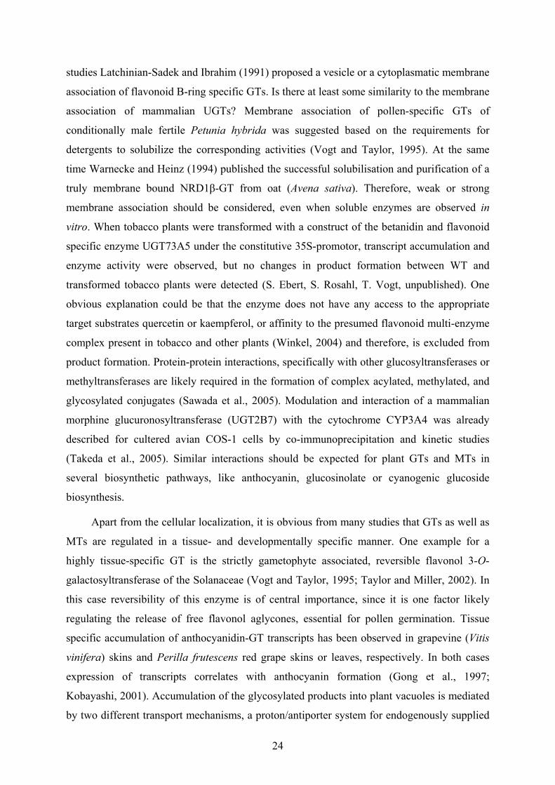
studies Latchinian-Sadek and Ibrahim (1991) proposed a vesicle or a cytoplasmatic membrane
association of flavonoid B-ring specific GTs. Is there at least some similarity to the membrane
association of mammalian UGTs? Membrane association of pollen-specific GTs of
conditionally male fertile Petunia hybrida was suggested based on the requirements for
detergents to solubilize the corresponding activities (Vogt and Taylor, 1995). At the same
time Warnecke and Heinz (1994) published the successful solubilisation and purification of a
truly membrane bound NRD1β-GT from oat (Avena sativa). Therefore, weak or strong
membrane association should be considered, even when soluble enzymes are observed in
vitro. When tobacco plants were transformed with a construct of the betanidin and flavonoid
specific enzyme UGT73A5 under the constitutive 35S-promotor, transcript accumulation and
enzyme activity were observed, but no changes in product formation between WT and
transformed tobacco plants were detected (S. Ebert, S. Rosahl, T. Vogt, unpublished). One
obvious explanation could be that the enzyme does not have any access to the appropriate
target substrates quercetin or kaempferol, or affinity to the presumed flavonoid multi-enzyme
complex present in tobacco and other plants (Winkel, 2004) and therefore, is excluded from
product formation. Protein-protein interactions, specifically with other glucosyltransferases or
methyltransferases are likely required in the formation of complex acylated, methylated, and
glycosylated conjugates (Sawada et al., 2005). Modulation and interaction of a mammalian
morphine glucuronosyltransferase (UGT2B7) with the cytochrome CYP3A4 was already
described for cultered avian COS-1 cells by co-immunoprecipitation and kinetic studies
(Takeda et al., 2005). Similar interactions should be expected for plant GTs and MTs in
several biosynthetic pathways, like anthocyanin, glucosinolate or cyanogenic glucoside
biosynthesis.
Apart from the cellular localization, it is obvious from many studies that GTs as well as
MTs are regulated in a tissue- and developmentally specific manner. One example for a
highly tissue-specific GT is the strictly gametophyte associated, reversible flavonol 3-O-
galactosyltransferase of the Solanaceae (Vogt and Taylor, 1995; Taylor and Miller, 2002). In
this case reversibility of this enzyme is of central importance, since it is one factor likely
regulating the release of free flavonol aglycones, essential for pollen germination. Tissue
specific accumulation of anthocyanidin-GT transcripts has been observed in grapevine (Vitis
vinifera) skins and Perilla frutescens red grape skins or leaves, respectively. In both cases
expression of transcripts correlates with anthocyanin formation (Gong et al., 1997;
Kobayashi, 2001). Accumulation of the glycosylated products into plant vacuoles is mediated
by two different transport mechanisms, a proton/antiporter system for endogenously supplied
24

substrates or a typical ATP-binding cassette type of transporters for exogenously applied
xenobiotica (Frangne et al., 2002). Interactions of GTs with glutathione S-transferases or
vacuolar membranes may occur, but remain to be established.
The localization of MTs in plants has been the focus of several recent investigations. In
general, the transcripts are usually tissue and developmentally specific distributed. Two
OMTs, the class II COMT and the class I CCoAOMT, both involved in lignin formation, are
strictly separated in Zinnia elegans (Ye and Varner, 1995). COMT is mainly found in the
phloem tissue whereas higher signal intensities of CCoAOMT at both, the mRNA and the
protein level were localized in developing xylem elements. Consistent with this observation,
both enzymes are associated with developing vascular tissue in alfalfa (Inoue et al., 1998).
The association of modifying enzymes in the same tissues than the corresponding
products is expected and plausible. Expression of (iso)eugenol OMT (IEMT), associated with
fragrance production in Clarkia breweri, is associated with petal tissues, the site of volatile
production, but also with stamens (Dudareva and Pichersky, 2000; Figure 9).
Figure 9: Tissue specific accumulation of OMT transcripts. In situ localization of IEMT in cross-sections of a petal (A) and stamen (B) (From Dudareva and Pichersky, 2000) (left). CCoAOMT (CCOMT) and caffeic acid OMT (COMT) signals in RNA from peltate glands and leaves of basil (from Gang et al., 2001) (right).
A similar situation was observed for peltate glands of Ocimum basilicum (basil), where
the high abundance of transcripts in a gland-specific EST-database for eight marginally
different CCoAOMT isoforms perfectly matched the peltate specific high expression of this
transcript in Northern blot analyses, as compared to the caffeic acid OMT (Gang et al., 2001;
Figure 9). Immunolocalization of benzoic acid OMT in epidermal tissues of snapdragon
(Anthirrhinum majus) flowers, is also consistent with these observations (Kolosova et al.,
2001).
Immunolocalization of two class II OMTs involved in alkaloid biosynthesis in opium
poppy (Papaper somniferum), the (R,S)-3'-hydroxy-N-methylcoclaurine 4'- and reticuline 7-
OMTs is associated with parenchyma cells of vascular bundles, adjacent to, but not within
25
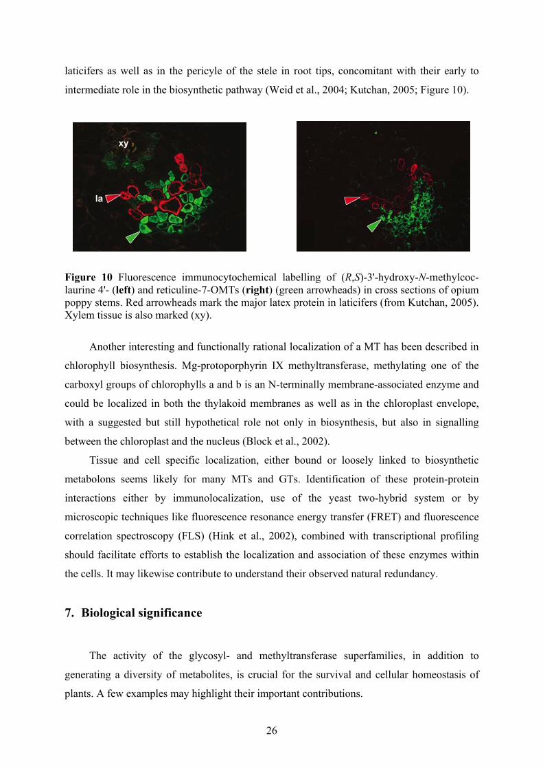
laticifers as well as in the pericyle of the stele in root tips, concomitant with their early to
intermediate role in the biosynthetic pathway (Weid et al., 2004; Kutchan, 2005; Figure 10).
Figure 10 Fluorescence immunocytochemical labelling of (R,S)-3'-hydroxy-N-methylcoc-laurine 4'- (left) and reticuline-7-OMTs (right) (green arrowheads) in cross sections of opium poppy stems. Red arrowheads mark the major latex protein in laticifers (from Kutchan, 2005). Xylem tissue is also marked (xy).
Another interesting and functionally rational localization of a MT has been described in
chlorophyll biosynthesis. Mg-protoporphyrin IX methyltransferase, methylating one of the
carboxyl groups of chlorophylls a and b is an N-terminally membrane-associated enzyme and
could be localized in both the thylakoid membranes as well as in the chloroplast envelope,
with a suggested but still hypothetical role not only in biosynthesis, but also in signalling
between the chloroplast and the nucleus (Block et al., 2002).
Tissue and cell specific localization, either bound or loosely linked to biosynthetic
metabolons seems likely for many MTs and GTs. Identification of these protein-protein
interactions either by immunolocalization, use of the yeast two-hybrid system or by
microscopic techniques like fluorescence resonance energy transfer (FRET) and fluorescence
correlation spectroscopy (FLS) (Hink et al., 2002), combined with transcriptional profiling
should facilitate efforts to establish the localization and association of these enzymes within
the cells. It may likewise contribute to understand their observed natural redundancy.
7. Biological significance
The activity of the glycosyl- and methyltransferase superfamilies, in addition to
generating a diversity of metabolites, is crucial for the survival and cellular homeostasis of
plants. A few examples may highlight their important contributions.
26

Attraction of pollinators is of utmost importance to all flowering plants. Glycosylation
and methylation play pivotal, yet contrary roles, especially B-ring-specific glucosylation and
subsequent acylation promote intra- and intermolecular stacking, essential for enhanced
colour formation by flavonoids and anthocyanins (Goto and Kondo, 1991). Although
glycosylation is not the reason for the enhanced colour by itself, B-ring attached sugars form
bridges between the coloured flavylium cation of anthocyanins and the esterified colourless
phenylpropanoid residues, like caffeic acid, which then stabilize the chromophore by
intramolecular co-pigmentation. This leads to a very intensive colour appearance as shown for
several Gentiana species (Yoshida et al., 2000; Fukuchi-Mizutani et al., 2003). In addition to
hydroxylation and glycosylation, different methylation patterns of anthocyanins from simple
pelargonidin to highly methylated capensinidin, have a potent effect on flower colour (Strack
and Wray, 1989). In contrast to floral colours, floral scent is the dominant means of long
distance attraction, independent of daylight, and therefore indispensable to attract night-active
insects (Dudareva and Pichersky, 2000; Guterman et al., 2002). Methylation, resulting in
aromatic ester and methyl ether formation strongly enhances floral scent volatility. In several
cases emission of methylated fragrances and volatiles is accompanied by resin formation.
Leaf resin is a complex mixture of terpenoids, waxes, and usually methylated flavonoids (Dell
and McComb, 1978). The resultant exudate protects young developing flower and leaf buds
from mechanical stress, damaging UV-radiation, and serves as an antimicrobial protectant
(Rhoades, 1977; Vogt et al., 1991).
Key players in response to stress are jasmonic acid and salicylic acid carboxyl OMTs,
(Seo et al., 2001; Ross et al., 1999). Airborne methyl jasmonate and methyl salicylate, the
products of these enzymes, play a pivotal role in many plant-insect interactions (Miller et al.,
2005; Shulaev et al., 1997; Chen et al., 2003). Methyl jasmonate can then further activate
specific pathways and enzymes involved in herbivore or pathogen defense (Martin, 2003).
Methyl jasmonate formation may be regarded as the general defence response and can also
induce other MTs, like those involved in methoxylated indol glucosinolate or those of methyl
salicylate biosynthesis in Arabidopsis (Hudgins and Franceschi, 2004; Mikkelsen et al., 2003;
Chen et al., 2003).
Cellular homeostasis is maintained and regulated by various hormones, like auxins,
cytokinins, and abscisic acid. In this case, several suitable candidate GTs have been identified
and functionally characterized, to modify the structure, polarity and action of these hormones
e.g. UGT84B1 from Arabidopsis (Figure 11), glucosylating the auxin 3-indol acetic acid
27

(Jackson et al., 2001), a cytokinin GT from maize (Mok et al., 2005), and the abscisic acid-
specific GT from adzuki bean (Xu et al., 2002).
Figure 11 Phenotype of wildtype A. thaliana (left) and the constitutive transgenic over-expressor UGT84B1 (right) encoding a GT of the auxin indole-3-acetic acid. Note especially the impaired gravitropism of the root system. (from Lim and Bowles, 2004).
A variety of GTs and MTs are transcriptionally up-regulated upon biotic or abiotic
stresses (Horvath and Chua; 1996; Schmitt et al., 1991; Ibdah et al., 2003; Sachan and
Falcone, 2002). Oxidative stress and reduced virus resistance could be correlated with
antisense-mediated down-regulation of the scopoletin- and phenylpropanoid GT (TOGT)
from tobacco (Chong et al., 2002). Whether up-regulation of transcripts, combined with an
observed accumulation of these potentially active phytoalexins indeed has any effect on viral
replication, is still controversial (Gachon et al. 2004; Matros and Mock, 2004), and may likely
be dependent on the virus strain.
Induction of naringenin 7-OMT was reported from rice plants upon radiation with UV-
light (Rakwal et al., 1996). The specific accumulation of the product sakuranetin might point
at an important function of this enzyme in protecting rice plants from oxidative stress. This
protection may be dependent on external factors, endogenous substrate concentrations, or
even on the virus strains. Detoxification of exogenous toxic compounds, like the mycotoxin
deoxynivalenol, a sesquiterpenoid produced by several Fusarium species, by UGT73C5 from
A. thaliana stresses the importance of a promiscuous specificity of many GTs (Poppenberger
28

et al., 2003). On the other hand fungi also fight back with similar “weapons” as shown for the
glucosyltransferase mediated detoxification of the cruciferous phytoalexin brassinin by the
stem rot fungus Scelotinia sclerotiorum (Pedras et al., 2004). Due to the ongoing application
of herbicides, pesticides, and fungicides which are targets for the reservoir of O- and N-
glycosyltransferases, promoting endogenous, inducible detoxification and defence responses
will be of immediate relevance and economically rewarding (Loutre et al., 2003; Messner et
al., 2003).
In mammals, the methylation of reactive mutagenic flavonoids by class I COMT has
been mainly attributed to their lack of mutagenic activity in vivo as compared to the in vitro
situation (Zhu et al., 1994). Deglycosylation, followed by glucuronylation and methylation of
natural products from food and beverages by the intestine or the liver, is of ongoing debate.
Specific modifications are reported to promote the uptake of “healthy” metabolites into the
bloodstream and thereby increase or decrease their health beneficial properties, like protection
from atherosclerosis and congestive heart failure (Reed, 2002; Nielsen et al., 2003).
8. Molecular Evolution
All superfamilies of plant enzymes, either the P450s, the glycosyl-, the
methyltransferases, and many others essentially derive from one or a few remnant ancestors.
As we did not witness the diversification, we are faced with the tremendous challenge to
describe the observed diversity and analyze the organizing principles while simultaneously
explaining the genomic and evolutionary constraints leading to this diversification.
The first comprehensive summary of plant Family 1 GTs established 107 putative
glycosyltransferase genes from A. thaliana (Li et al., 2001) and was later complemented by a
second analysis (Paquette et al., 2003). Both studies essentially grouped this allelic diversity
into 14 phylogenetic subclusters, strongly supported by bootstrap analysis. Most of the A.
thaliana glycosyltransferases have a monoexonic coding sequence, and neither has a signal
sequence nor any membrane-spanning domains (Lim and Bowles, 2004). This is in contrast to
many animal glucuronosyltransferases, where complex architectures with up to six exons
spanning about 25 kb have been described (Turgeon et al., 2000). The observed clustering of
many GT-genes in plants is consistent with at least three whole genome duplications in A.
thaliana (Maere et al., 2005). On the other hand, specific GTs e.g. those involved in the
biosynthesis of the triterpenoid avenacin in oat (Avena sativa), are clustered with several other
genes of the avenacin biosynthetic pathway, which is non-consistent with a gene duplication
29

event for these GTs (Qi et al., 2004). Still, the presence of large gene families is the result of
an expanding genome with a gain of function surpassing loss of genes and gene function.
Let us take a look at the organising principles in GT-cluster formation. Is it specificity
for the donor or the acceptor? Is it regio- or position specific? From a detailed investigation of
50 functionally expressed GTs from Arabidopsis with the model substrate esculetin, it was
evident that several members of different subclusters were able to glucosylate this coumarin
in vitro, although with different regiospecificities (Lim et al., 2003). Other subclusters were
highly specific for the substrates and the positions.
Among all plants investigated up to now the members of anthocyanidin 3-GTs and
anthocyanin 5-GTs are highly homologous, up to 60% identity even when enzymes from
different species are compared. This is significantly higher than the less than 20% homology
over the whole natural product GT-superfamily (Vogt and Jones, 2000). In addition they
display a uniform and strict specificity for the individual hydroxyl groups (See also Figure
12). This specificity compared with sequence data can then be used to annotate other
unknown sequences from the databases. An A. thaliana GT-sequence with the database
accession number CAC01717 (Figure 12) can therefore easily be correlated with a flavonoid
3-GT (Figure 12). Conservation of sequence identities and functional specificity represent the
importance of the conserved flavonoid and anthocyanin biosynthetic pathway.
Other subclusters are far less specific for a single substrate, not even for the glucose
donor. Figure 12 illustrates various other protein sequences which in vitro are considered to
glycosylate flavonoids and anthocyani(di)ns. Although the two large clusters to the right
contain a variety of, at a first glance, different enzymes from taxonomically unrelated species,
a close-up view reveals striking similarities within one subcluster. All members specified as
“Flavonoid A- and B-Ring specific GTs” marked in blue (Figure 12), transfer glucose
preferentially to the 3´- or 4´-hydroxyl group of the flavonoid B-ring or the 7-OH group of the
A-ring, but not to the 3-hydroxy position of the heterocycle. In case of the enzyme from
scullap (Scutellaria baicalensis) (BAA83484, Figure 12) glucose is transferred exclusively to
the 7-OH of the A-ring, since no hydroxylated B-ring is present in substrates baicalein or
scutellarein (Hirotani et al., 2000).
This specificity for oligohydroxylated structural motifs is crucial to the whole cluster to
which flavonoid 7-GTs from A. thaliana (Figure 12, AAR01231) and rice (Oryza sativa),
UGT73A5 from livingstone daisy (Figure 12, Y18871), UGT73A4 from red beet (Figure 12,
AY526080), a 2´-chalconaringenin GT from Dianthus caryophyllus (Fig. 12, BAD52007)
(Ogata et al., 2004), sequences from tomato (Lycopersicon esculentum) (Figure 12, X85138),
30

tobacco (Fig. 12, U32644), gentian (Figure 12, BAC54092) and scullap (Figure 12,
BAA83484) belong to.
Figure 12 Various flavonoid and anthocyanidin GTs (indicated by their NCBI-database accession numbers) cluster according to position specificity towards individual hydroxyl groups. A. thaliana sinapic acid GT (AB019232) was used as an outlier. Solid numbers and colours indicate that the corresponding proteins were functionally characterized, whereas soft numbers and colours indicate proteins with poorly or uncharacterized functions (Cladogram created with Clustal W, PAM 250 matrix, Thompson et al., 1994)
This subclass UGT73 (MacKenzie et al., 1997) together with the second very
heterogenous subcluster of “phenolic and flavonoid GTs referenced by a naphtol GT from
tobacco (Fig. 12, BAB60721), by arbutin synthase (Figure 12, CAC35167.1), UGT71F2
characterized as a betanidin 6-GT ((Figure 12, AF374004), a red beet flavonoid and betanidin
GT, UGT71F1 (Figure 12, AY536081) and other GTs with a broad acceptor specificity,
including flavonoids and anthocyanidins is clearly separated from the conserved clusters of
flavonoid and anthocyanin 3- and 5-GTs, respectively (Figure 12). Due to this heterogeneity it
will be difficult to predict the substrate profile of new members of both subclusters. This
31

profile is likely species specific and a detailed analyses of the aglycone pattern of the plant
may be required to suggest potential candidates.
Diversification into the individual specificities, as we observe them today has occurred
by gene duplication and subsequent loss and gain of functions (Moore and Purugganan,
2005). Genomic clustering as observed for some A. thaliana GTs may be regarded as a proof
for gene duplications, which have occurred during evolution of vascular plants. Clusters like
UGT73 and UGT71 may have developed at some time from one or several ancestors and due
to their promiscuity they “aquired” or new functions. One excellent example to illustrate this
hypothesis may be mentioned briefly. Ogata et al. (2005), most recently described an
anthocyanidin GT from Rosa hybrida cultivar (rose) with the unusual property to
simultaneously glucosylate anthocyanidins at the 5-OH and the 3-OH position (Figure 12,
BAD99560) combining the specificities of an anthocyanidin 3-GT and the anthocyanin 5-GT.
Apparently, plants have maintained these sets of promiscuous and “flexible” enzymes,
different from the conserved clusters of specific enzymes, with the obvious evolutionary
advantage to adjust to changing or potentially lethal conditions. It is noteworthy that several
putative “Flavonoid A- and B-Ring specific GTs” (Figure 12) were reported to be
transcriptionally up-regulated during stress-responses, like the wound-inducible Is5A and
Is10A genes from tomato or the twi1 gene from tomato (Horvath and Chua, 1996; O´Donnell
et al., 1999). Again, in response to environmental or endogenous stimuli a pool of enzymes
with high promiscuity combines the required, overlapping specificity to modify a variety of
compounds with the precise position selectivity to ensure high affinity.
An overlapping substrate specificity but precise position specificity of clusters UGT71
and UGT73 should again be illustrated for betanidin 5- and 6-GTs from D. bellidiformis,
which share less than 20% amino acid sequence identity, yet glucosylate the same substrate
betanidin and flavonoids with a different regiospecificity (Vogt et al., 1999a; Vogt, 2002).
This polyphyletic origin of two enzymes accepting identical substrates, yet show no
significant sequence identity is also nicely illustrated by the set of scutellarein glucosyl- and
glucuronosyltransferases, which share the same acceptor and even identical position
specificities, but use different sugars (Hirotani et al., 2000; Nagashima et al., 2000). Both sets
of enzymes therefore, have evolved from at least two different ancestors, presumably both
involved in the modification of unknown, extinct or remnant substrates and at some point
have “gained” the properties to modify novel endogenous metabolites, like betanidin or
scutellarein, respectively.
32

MTs are classified based on their dependence on a metal cofactor and the class I and the
class II nomenclature (Joshi and Chiang, 1998). Although all AdoMet-dependent enzymes
share identical structural features (Cheng and Roberts, 2001), a single ancient separation of
both classes of enzymes has occurred to explain the astonishing 35% amino acid sequence
identities between class I OMTs from plants, cyano-, and myxobacteria (Ibdah et al., 2003).
Dimerization apparently is restricted to the plant class I OMTs, since the mammal and also
the functionally not characterized cyanobacterial enzymes are monomers. Plant class I OMTs
can be further divided into at least two subclasses based on substrate and regiospecificity
(Ibdah et al., 2003; Vogt, 2004). Exceptionally high sequence conservation (up to 90%) points
to an ancient, important, and conserved function of one subset of the class I OMTs, the
CCoAOMTs involved in lignin monomer formation. A less rigid conservation of amino acid
sequences is observed for those few promiscuous enzymes detected recently (Ibdah et al.,
2003). Diversification and specialization of class I OMTs resulted in at least six different
genes in Arabidopsis, of which only two have been functionally characterized yet (Vogt,
unpublished). A similar situation is observed in other plant species, e.g. in sweet basil, where
several slightly different CCoAOMT isoforms were observed in peltate glands (Gang et al.,
2001). Probably only one is specifically involved in the methylation of guaiacyl lignin
monomers, whereas the substrates of the other members, all targeted to vicinal dihydroxy
functions, remain elusive. The precursors of the basil peltate phenylpropenes chavicol or
eugenol (Gang et al., 2002), which are modified by two distinct class II enzymes chavicol
OMT and eugenol OMT could be targeted initially by one or two different class I isoenzymes.
The diversification of plant species and the rapid evolution of substrate diversity
apparently is concomitant with the development of class II OMTs, where sequence
similarities are much less conserved (Ibrahim and Muzac, 2000). Predicting their in vivo
substrates based on sequence identities is a tremendous challenge (Schröder et al., 2002).
Flexible and overlapping substrate specificities are described for class II OMTs, e.g. the
caffeic acid OMT methylates also caffeoyl CoA, the sole substrate of the lignin specific class
I cluster enzymes. The lignin specific class I enzyme, however, does not or very poorly
methylate caffeic acid (at least in vitro). OMTs from T. tuberosum which do not distinguish
between a benzylisoquinoline alkaloid or a simple phenylpropanoid have already been
mentioned (Frick and Kutchan, 1999). Seoighe and Gehring (2004) explain that gene
duplication is a selective process and expansion of a gene family is dependent on a series of
evolutionary constraints. If this is the case, the multiplicity of class II OMTs may be the result
of more flexible structural motifs of these enzymes as compared to a rigid, cation-dependent
33

structure of the class I OMTs. The structurally very similar subcluster of promiscuous class I
OMTs, as compared to the highly conserved and stringent CCoAOMTs (Ibdah et al., 2003),
was possibly established later in evolution to fill a niche, not immediately accessible to the
class II enzymes for yet unknown reasons. Similar principles may also hold true for the
evolution of N-, S-, and C-methyltransferases.
9. Economical Relevance
Glycosylation and methylation of endogenous unstable precursors of cyanogenic
glycosides, glucosinolates, or anthocyanidins are of major ecochemical importance for the
plants (Zagrobelny et al., 2004; Wittstock et al., 2004). Designing natural products by genetic
engineering of the corresponding enzymes may not only protect plants from pathogens and
herbivores (von Rad et al., 2001; Tattersall et al., 2001), but could also lead to desired
modifications of colour, taste, or fragrance of flowers, nutraceuticals, and beverages with a
promise for potential economical benefits (Dudareva et al., 2004; Marillia, 2001; Kazuma et
al., 2003).
Beside the expected in vivo use of these enzymes, their in vitro application as potential
biocatalysts is already established (Arend et al., 2001). By mixing recombinant E. coli strains,
which over-expressed a mixture of one A. thaliana GT and a set of peppermint MTs, Willits
et al. (2004) produced glucosylated and methylated flavonol conjugates in vitro. In principle,
both types of enzymes, can therefore be used in biofermentation or by combinatorial
enzymology to produce a desired mixture of compounds in vitro without the necessity to use
organic solvents or toxic catalysts.
Stevia rebaudiana contains a complex mixture of intensely sweet terpenoid glycosides,
the steviosides, which have been proposed to, but finally rejected by the European
Commission as natural, non toxic sugar replacements (CS/ADD/EDUL167final, 17/06/1999).
The rejection was in part based on the poorly defined formulation of the drug. Nevertheless,
steviol glycoside preparations are legally sold and used as food additives in Asia and the US.
Successful cloning and functional expression of several glucosyltransferases involved in
rebaudioside A biosynthesis may now promote further in vitro studies to critically examine
the potentially carcinogenic nature of these low calorie sweeteners and circumvent the risk of
toxic plant contaminants (Richman et al., 2005). The market for low calorie sweenteners from
plants is up to US$ 1.5 billion just in the US is governing the search for plants and enzymes
with desired properties to result in healthy natural sweeteners (Kinghorn and Soejarto, 2002).
34

The monoglucuronide of the similar terpenoid sweetener glycyrrhizin from liquorice
roots (Glycyrrhiza glabra) is already commercially produced using a β-glucosidase which
cleaves selectively the 1,2-β linkage of the two glucuronic acid residues (Tanaka, 1997).
TABLE 1 Plant glycoside sweeteners and bitter glycosides
Name of the Compound
Plant source Structure (+) fold sweeter than sucrose (-) bitter, threshold
Stevioside Stevia rebaudiana Diterpene glycoside + 210
Rebaudioside A Stevia rebaudiana Diterpene glycoside + 240
Rubososide Rubus suavissimus Diterpene glycoside + 110
Glycyrrhizin Glycyrrhiza glabra Triterpene glycoside + 170
Neohesperidin dihydrochalcone
Citrus spec. Hydrogenated flavanone glycoside
+ 1000
Naringin Citrus pummelo Flavanone diglycoside (-)
Limonoid glycoside
Citrus unshiu Diterpenoid glycosdide
(-)
Gentiopikrin Gentiana lutea Lactone glycoside (-)
The importance of a position-specific transfer, combined with the high position
specificity of some GTs should be emphasized again as essential for sensoric properties. The
successful cloning and expression of a position-specific naringenin 7-O-glucoside-1,2
rhamnosyltransferase from Citrus fruits resulted in the production of the bitter
neohesperidoside (Bar-Peled et al., 1991; Frydman et al., 2004). In contrast, the 1,6-linked
disaccharid is a tasteless compound. Like rebaudioside A and the Solanaceae pollen
flavonoids, neohesperidoside contains a 1,2-α-glycosidic bound between two sugars, which
may be a characteristic feature for the interaction and perception with unknown receptors
(Taylor and Miller, 2002). Irrespectively of the unknown function in plants, cloning and
characterization of the newly discovered Citrus GT will now likely result in fruits and
vegetables with altered aromatic properties.
Glycosylation is a process difficult to be controlled organo-chemically in vitro.
Therefore, isolation of GTs with desired specificities are also of major interest for organic
synthesis. Arendt et al. (2001), by heterologous expression of arbutin synthase from Rauvolfia
serpentina, produced arbutin, a potent inhibitor of human melanin biosynthesis, which was
directly released into the culture medium. The extremely promiscuous specificity of this
35

enzyme promises rewarding discoveries within the GT or MT superfamily in the search for
specific targets (Hefner et al., 2002).
Production of pharmacologically important compounds is often hampered by their low
water solubility, like in case of the yellow coloured curcumin (diferuloylmethane), used as
food additive and in traditional folk medicine for its anti-inflammatory properties, its anti-
cancer properties and recently, to reduce alcohol induced liver disease. Two
glucosyltransferases from Catharanthus roseus (whatever their physiological function might
be) with broad substrate specificities have been functionally expressed and displayed activity
towards the diferuloyl conjugate curcumin and its monoglucoside, to give curcumin
diglycoside (Kaminaga et al., 2004). Strongly increased water solubility of the resulting
glycosides may promote their further use as potent antioxidants. Increased solubility and
stabilization was also achieved for the bitter apocarotinoid aglycone crocetin by a GT-activity
isolated from the stigmas of Crocus sativus (Moraga et al., 2004). Crocetin, and the
corresponding glycosides crocin and picrocrocin are responsible for the yellow colour of
saffron, the dried stigmas of this plant, the world´s most expensive spice. These examples not
only illustrate the power of plant biosynthetic systems, but may also encourage researchers to
screen exotic plant species for activities and properties, which are otherwise difficult to
achieve by synthetic chemistry.
Health promoting in vitro antioxidative effects of a variety of anthocyanins and vitamins
in fruits and vegetables are often scavenged by the rapid oxidation, degradation, and poor
uptake of these compounds in vivo (Rice-Evans et al., 1997; Arts et al., 2004). Rational design
and production of compounds with enhanced bioavailability either by transgenic approaches
in vivo or by combinatorial enzymology in vitro could facilitate the production of non-toxic
antioxidants and vitamins for human consumption (Křen and Martinková, 2001).
Today the market for ornamental plants and cut flowers totals over US$ 90 billion.
Therefore, variation of flower colour is not only a matter of plant reproduction, but promises
huge economical benefits for the cut-flower industry. The search for a blue or a black rose, is
an ongoing, and money consuming vision for years (Mol et al., 1998). Recently, after the
commercial release of the first bluish carnation “Moondust” in 2004 the Japanese company
Suntory Ltd. successfully created the first blue rose by genetically engineering the presence of
the highly substituted anthocyanidin delphinidin, which does not naturally occur in roses. To
match the demands and needs of the market, successful introduction of the anthocyanidin core
structure, will likely be followed by efforts to enhance colour formation e.g. by genetically
engineering regiospecific GTs and MTs which might promote intra- and intermolecular
36

stacking (Markham et al., 2004; Mori et al., 2005). This will not only result in a set of
transgenic cut-flowers, but pave the way to understand the molecular principles of flower
colouration.
What holds true for flower colour, definitely is the case for volatiles, where the
complexity of genes and enzymes involved in the emission of floral scents is just emerging
(Schieberle and Hofmann, 2003; Dudareva et al., 2004). Genetically engineered design of
methylated or esterified aroma compounds in planta or in vitro, should result in a stable
bouquet of flavours with a potential use in health promoting issues like aroma therapy or the
control of plant-insect interactions (Pare and Tumlinson, 1999). In addition, controlling the
specificity and selectivity of the sugar transfer could facilitate the trapping and release of
aromatic compounds, with the aim to produce nutrients with desired aromatic properties
(Schieberle and Hofmann, 2003).
Class I OMTs involved in lignin modification are key players in various efforts to
modify lignin contents of plants (e.g. Zeneca, British patent 9119279.9). A decrease in the
lignin content of the raw material or a shift of the ratio of guaicyl/syringyl lignin could
increase pulp yield and aid delignification. For forage crops this would increase the
digestibility and energy value of animal food. In contrast, production of woody materials with
a greater percentage of lignin than normal should result in stronger and more resistant wood
products, as well as improve the fuel value.
10. Summary and Outlook
The development of both molecular and biochemical tools have greatly accelerated the
research on plant natural product glycosyl- and methyltransferases. We and others have
shown that regio- and position specificity are more consistent with amino acid sequence based
cluster formation than substrate specificities. Although it may not yet be possible to predict
precisely the specificity of a newly discovered enzyme, progress in functional characterisation
of both superfamilies of enzymes have greatly facilitated the annotation of newly described
sequences. Difficulties to unequivocally correlate the in vivo specificity with the observed in
vitro properties are largely due to the promiscuous nature of both classes of enzymes. But this
promiscuity may be an important strategy in evolution to guarantee that local and global
environmental changes, accompanied by ecochemical challenges, are not necessarily
perceived as a risk, but as the driving force to promote new properties and developments.
37

Two key issues may influence research on GTs and MTs in the future. One will be
concerned with the elucidation of specific protein structures to describe and understand how
enzyme specificity is governed by individual protein domains. A solid basis has already been
generated by our lab and the work of others using molecular modelling and crystallography to
understand the principles of sugar and methyl group transfer to plant products. Progress to
determine protein structures in solution by NMR spectroscopy could help to solve currently
difficult or impossible to crystallize enzymes, like the soluble or membrane-bound glycosyl-
and glucuronosyltransferases. Visions to create new enzymes with desired properties in silico
or by rational experimental design may not be too far fetched. The second, even more
ambitious task will be concerned with the function, regulation and integration of these
enzymes into complex metabolic systems and requires support by genomics, proteomics,
metabolomics, and bioinformatics. Based on the wealth of information and knowledge
currently generated it will be only be a question of time that this research will result not only
in scientific but also in economical benefits.
11. References
Akashi T, Sawada Y, Shimada N, Sakurai N, Aoki T, Ayabe S (2003) cDNA cloning and biochemical characterization of S-adenosyl-L-methionine: 2,7,4'-trihydroxyisoflavanone 4'-O-methyltransferase, a critical enzyme of the legume isoflavonoid phytoalexin pathway. Plant Cell. Physiol. 44, 103-112. Albrecht HP (1999) Cardiac glycosides, in Naturally Occurring Glycosides, pp. 83-124, Ikan R (ed.), Wiley & Sons, Chichester Arend J, Warzecha H, Hefner T, Stöckigt J (2001) Utilizing genetically engineered bacteria to produce plant-specific glucosides. Biotechnol. Bioeng. 76, 126-131. Arts IC, Sesink AL, Faassen-Peters M, Hollman PC (2004) The type of sugar moiety is a major determinant of the small intestinal uptake and subsequent biliary excretion of dietary quercetin glycosides. Brit. J. Nutr. 91, 841-847. Axelrod J, Vesell ES (1970) Heterogeneity of N- and O-methyltransferases. Mol. Pharmacol. 6, 78-84. Bar-Peled M, Lewinsohn E, Fluhr R, Gressel J (1991) UDP-rhamnose:flavanone-7-O-glucoside-2''-O-rhamnosyltransferase. Purification and characterisation of an enzyme catalyzing the production of bitter compounds in citrus. J. Biol. Chem. 266, 20953-20959. Blanco G, Patallo EP, Brana AF, Trefzer A, Bechthold A, Rohr J, Mendez C, Salas JA (2001) Identification of a sugar flexible glycosyltransferase from Streptomyces olivaceus, the producer of the antitumor polyketide elloramycin. Chem. Biol. 8, 253-263. Block MA, Tewari AK, Albrieux C, Marechal E, Joyard J (2002) The plant S-adenosyl-L-methionine:Mg-protoporphyrin IX methyltransferase is located in both envelope and thylakoid chloroplast membranes. Eur. J. Biochem. 269, 240-248.
38

Bokern M, Wray V, Strack D (1991) Phenolic acid conjugates and betacyanin accumulation and activity changes of enzymes involved in feruloylglucose metabolism in cell suspension cultures of Chenopodium rubrum. Planta 184, 261-270. Brugliera F, Holton TA, Stevenson TW, Farcy E, Lu CY, Cornish EC (1994) Isolation and characterisation of a cDNA clone corresponding to the Rt locus of Petunia hybrida. Plant J. 5, 81-92. Cacace S, Schröder G, Wehinger E, Strack D, Schmidt J, Schröder J (2003) A flavonol O-methyltransferase from Catharanthus roseus performing two sequential methylations. Phytochemistry 62, 127-137. Cannon JP, Haire RN, Rast JP, Litman GW (2004) The phylogenetic origins of the antigen-binding receptors and somatic diversification mechanisms. Immunol. Review 200, 12-22. Cardini CE, Yamaha T (1958) Biosynthesis of plant glycosides from uridine diphosphate glucose. Nature 22, 1446-1447. Chen F, D´Auria JC, Tholl D, Ross JR, Gershenzon J, Noel JP, Pichersky E (2003) An Arabidopsis thaliana gene for methylsalicylate biosynthesis, identified by a biochemical genomics approach, has a role in defense. Plant J. 36, 577-588. Cheng X, Roberts RJ (2001) AdoMet-dependent methylation, DNA methyltransferases and base flipping. Nucl. Acids Res. 29, 3784-3795. Chong J, Baltz R, Schmitt C, Beffa R, Fritig B, Saindrenan P (2002) Down regulation of a pathogen-responsive tobacco UDP-glc:phenylpropanoid glucosyltransferase reduces scopoletin glucoside accumulation, enhances oxidative stress, and weakens virus resistance. Plant Cell 14, 1093-1107. Christinet L, Burdet FX, Zaiko M, Hinz U, Zryd JP (2004) Characterisation and functional identification of a novel plant 4,5-extradiol dioxygenase involved in betalain pigment biosynthesis in Portulaca grandiflora. Plant Physiol. 137, 265-274. Conn EE (1980) Cyanogenic compounds, in Annu. Rev. Plant Phyisol. 31, 433-451. Coutinho PM Henrissat, B (1999) Carbohydrate-Active Enzymes server at URL: http://afmb.cnrs-mrs.fr/CAZY/ Dell B, McComb AJ (1978) Plant resins – their formation, secretion and possible functions in, Advances in Botanical Research, Vol. 6, Woolhouse RV (ed.), pp. 277-316, Academic Press, New York. Dixon RA (1999) Plant natural products: the molecular genetic basis of biosynthetic diversity. Curr. Opin. Plant Biotechnol. 10, 192-197. Dudareva N, Pichersky E (2000) Biochemical and molecular genetic aspects of floral scents. Plant Physiol. 122, 623-634. Dudareva N, Pichersky E, Gershenzon J (2004) Biochemistry of plant volatiles. Plant Physiol. 135, 1893-1902. Fedoroff NV, Furtek DB, Nelson OE (1984) Cloning of the Bronze locus in maize by a simple and generalizable procedure using the transposable controlling element Ac. P. Natl. Acad. Sci. USA 81, 3825-3829.
39

Ferrer JL, Zubieta C, Dixon RA, Noel JP (2005) Crystal structures of alfalfa caffeoyl coenzyme A 3-O-methyltransferase. Plant Physiol. 137, 1009-1017. Finkle BJ, Nelson RF (1963) Enzyme reactions with phenolic compounds: a meta O-methyltransferase in plants. Biochim. Biophys. Acta 78, 747-749. Frangne N, Eggmann T, Koblischke C, Weissenböck G, Martinoia E, Klein M (2002) Flavone glucoside uptake into barley mesophyll and Arabidopsis cell culture vacuoles. Energization occurs by H+-antiport and ATP-binding cassette-type mechanisms. Plant Physiol. 128, 726-733. Frick S, Kutchan TM (1999) Molecular cloning and functional expression of O-methyltransferases common to isoquinoline alkaloid and phenylpropanoid biosynthesis. Plant J. 17, 329-339. Frydman A, Weisshaus O, Bar-Peled M, Huhman DV, Sumner LW, Marin FR, Lewinsohn E, Fluhr R, Gressel J, Eyal Y (2004) Citrus fruit bitter flavors: isolation and functional characterisation of the gene Cm1,2RhaT encoding a 1,2 rhamnosyltransferase, a key enzyme in the biosynthesis of the bitter flavonoids of Citrus. Plant J. 40, 88-100. Fukuchi-Mizutani M, Okuhara H, Fukui Y, Nakao M, Katsumoto Y, Yonekura-Sakakibara K, Kusumi T, Hase T, Tanaka Y (2003) Biochemical and molecular characterisation of a novel UDP-glucose: anthocyanin 3'-O-glucosyltransferase, a key enzyme for blue anthocyanin biosynthesis, from gentian. Plant Physiol. 132, 1652-1663. Gachon C, Baltz R, Saindrenan P (2004) Overexpression of a scopoletin glucosyltransferase in Nicotiana tabacum leads to precocious lesion formation during the hypersensitive response to tobacco mosaic virus but does not effect virus resistance. Plant Mol. Biol. 54, 137-146. Gang DR, Wang J, Dudareva N, Nam KH, Simon JE, Lewinsohn E, Pichersky E (2001) An investigation of the storage and biosynthesis of phenylpropenes in sweet basil. Plant Physiol. 125, 539-555. Gang DR, Lavid N, Zubieta C, Chen F, Beuerle T, Lewinsohn E, Noel JP, Pichersky E (2002) Characterisation of phenylpropene O-methyltransferases from sweet basil: facile change of substrate specificity and convergent evolution within a plant O-methyltransferase family. Plant Cell 14, 505-519. Gershenzon J, Kreis W (1999) Biochemistry of terpenoids: monoterpenes, sesquiterpenes, diterpenes, sterols, cardiac glycosides and steroid saponins in Biochemistry of Plant Secondary Metabolism, Vol. 2, Annual Plant Reviews, (Wink M, ed.), pp. 222-299, CRC Press, Boca Raton, Fl., USA. Gong Z, Yamazaki M, Sugiyama M, Tanaka Y, Saito K (1997) Cloning and molecular analysis of structural genes involved in anthocyanin biosynthesis and expressed in a forma-specific manner in Perilla frutescens. Plant Mol. Biol. 35, 915-927. Goto T, Kondo T (1991) Structure and molecular stacking of anthocyanins - flower color variation. Angew. Chem. Int. Ed. Engl. 30, 17-33. Grimm R, Eckerskorn C (1996) Enhanced in situ gel digestion of electrophoretically separated proteins with automated peptide elution onto mini reversed-phase columns. Electrophoresis 17, 899-906. Guerin M, Parodi AJ (2003) The UDP-glucose:glycoprotein glucosyltransferase is organized in at least two tightly bound domains from yeast to mammals. J. Biol. Chem. 278, 20540-20546. Gustafsson C, Govindarajan S, Minshull J (2004) Codon bias and heterologous protein expression. Trends Biotechnol. 22, 346-353.
40

Guterman I, Shalit M, Menda N, Piestun D, Dafny-Yelin M, Shalev G, Bar E, Davydov O, Ovadis M, Emanuel M, Wang J, Adam Z, Pichersky E, Lewinsohn E, Zamir D, Vainstein A, Weiss D. (2002) Rose scent: genomics approach to discovering novel floral fragrance-related genes. Plant Cell 14, 2325-2338. Hans J, Brandt W, Vogt T (2004) Site-directed mutagenesis and protein 3D-homology modelling suggest a catalytic mechanism for UDP-glucose dependent betanidin 5-O-glucosyltransferase from Dorotheanthus bellidiformis. Plant J. 39, 319-333. Hansen KS, Kristensen C, Tattersall DB, Jones PR, Olsen CE, Bak S, Møller BL (2003) The in vitro substrate regiospecificity of recombinant UGT85B1, the cyanohydrin glucosyltransferase from Sorghum bicolor. Phytochemistry 64, 143-51. Harborne JB, Turner BL (1984) Plant Chemosystematics. Academic Press, London Hefner T, Arend J, Warzecha H, Siems K, Stöckigt J (2002) Arbutin synthase, a novel member of the NRD1beta glycosyltransferase family, is a unique multifunctional enzyme converting various natural products and xenobiotics. Bioorg. Med. Chem. 10, 1731-1741. Heuer S, Vogt T, Böhm H, Strack D (1996) Partial purification and characterisation of UDP-glucose: betanidin-5-O- and 6-O-glucosyltransferases from cell suspension cultures of Dorotheanthus bellidiformis (Burm.f.) N.E.Br. Planta 199, 244-250. Hink MA, Bisselin T, Visser AJ (2002) Imaging protein-protein interactions in living cells. Plant Mol. Biol. 50, 871-883. Hirotaqni M, Kuroda R, Suzuki H, Yoshikawa T (2000) Cloning and expression of UDP-glucose: flavonoid 7-O-glucosyltransferase from hairy root cultures of Scutellaria baicalensis. Planta 210, 1006-1013. Hoffmann L, Maury S, Bergdoll M, Thion L, Erard M, Legrand M (2001) Identification of the enzymatic active site of tobacco caffeoyl-coenzyme A O-methyltransferase by site-directed mutagenesis. J. Biol. Chem. 276, 36831-36838. Horvath DM, Chua NH (1996) Identification of an immediate-early salicylic acid-inducible tobacco gene and characterisation of induction by other compounds. Plant Mol. Biol. 31, 1061-1072. Hrazdina G (1992) Compartmentation in aromatic metabolism. in Recent Advances in Phytochemistry, Vol. 24, Phenolic Metabolism in Plants, (Stafford HA, Ibrahim RK, eds.), pp. 1-23, Plenum Press, New York. Hu Y, Walker S (2002) Remarkable structural similarities between diverse glycosyltransferases. Chem. Biol. 9, 1287-1296. Hudgins JW, Franceschi VR (2004) Methyl jasmonate-induced ethylene production is responsible for conifer phloem defense responses and reprogramming of stem cambial zone for traumatic resin duct formation. Plant Physiol. 135, 2134-2149. Hughes J, Hughes MA (1994) Multiple secondary plant product UDP-glucose glucosyltransferase genes expressed in cassava. DNA Sequence 5, 41-49. Humphreys JM, Chapple C (2002) Rewriting the lignin roadmap. Curr. Opin. Plant Biol. 5, 224-229. Hutchinson A, Roy, C, Towers GHN (1958) Synthesis of phlorin and other phenolic glucosides by plant tissues. Nature 181, 841-842.
41

Ibdah M, Krins A, Seidlitz HK, Heller W, Strack D, Vogt T (2002) Spectral dependence and dose response of flavonol and betacyanin accumulation in Mesembryanthemum crystallinum under enhanced UV radiation. Plant Cell Environ. 25, 1145-1154. Ibdah M, Zhang X-H, Schmidt J, Vogt T (2003) A novel Mg++-dependent O-methyltransferase in the phenylpropanoid metabolism of Mesembryanthemum crystallinum. J. Biol. Chem. 278, 43961-43972. Ibrahim RK (1997) Plant O-methyltransferase signatures. Trends Plant Sci. 2, 249-250. Ibrahim R, Muzac I (2000) The methyltransferase gene superfamily: A tree with multiple branches. in Recent Advances in Phytochemistry, Vol. 34, Evolution of Metabolic Pathways (Romeo JT, Ibrahim R, Varin L and DeLuca V, eds), pp. 349-385, Pergamon Press, Amsterdam. Inoue K, Sewalt VJH, Balance GM, Ni W, Stürzer C, Dixon RA (1998) Developmental expression and substrate specificities of alfalfa caffeic acid 3-O-methyltransferase and caffeoyl coenzyme A 3-O-methyltransferase in relation to lignification. Plant Physiol. 117, 761-770. Jackson RG, Lim EK, Li Y, Kowalczyk M, Sandberg G, Hoggett J, Ashford DA, Bowles DJ (2001) Identification and biochemical characterisation of an Arabidopsis indole-3-acetic acid glucosyl-transferase. J. Biol. Chem. 276, 4350-4356. Jones PR, Møller BL, Hoj PB (1999) The UDP-glucose:p-hydroxymandelonitrile-O-glucosyl-transferase that catalyzes the last step in synthesis of the cyanogenic glucoside dhurrin in Sorghum biArabidopsis. Isolation, cloning, heterologous expression, and substrate specificity. J. Biol. Chem. 274, 35483-35491. Jones P, Vogt T (2001) Glucosyltransferases in secondary plant metabolism: tranquilizers and stimulant controllers. Planta 213, 164-174. Joshi CP, Chiang VL (1998) Conserved sequence motifs in plant S-adenosyl-L-methionine-dependent methyltransferases. Plant Mol. Biol. 37, 663-674. Kapitonov D, Yu RK (1999) Conserved domains of glycosyltransferases. Glycobiology 10, 961-978. Kaminaga Y, Sahin FP, Mizukami H (2004) Molecular cloning and characterisation of a glucosyl-transferase catalyzing glucosylation of curcumin in cultured Catharanthus roseus cells. FEBS Lett. 567, 197-202. Kasai R, Yamasaki K, Tanaka O (1999) Saponins, in Naturally Occurring Glycosides, pp.295-310, (Ikan R, ed.) Wiley & Sons, Chichester Kazuma K, Noda N, Suzuki M (2003a) Malonylated flavonol glycosides from the petals of Clitoria ternatea. Phytochemistry 62, 229-237. Kazuma K, Noda N, Suzuki M (2003b) Flavonoid composition related to petal color in different lines of Clitoria ternatea. Phytochemistry 64, 1133-1139. Kinghorn AD, Soejarto DD (2002) Discovery of terpenoid and phenolic sweeteners from plants. Pure Appl. Chem. 74, 1169-1179. Kobayashi S, Ishimaru M, Ding CK, Yakushiji H, Goto N (2001) Comparison of UDP-glucose:flavonoid 3-O-glucosyltransferase (UFGT) gene sequences between white grapes (Vitis vinifera) and their sports with red skin. Plant Sci. 160,:543-550.
42

Kolosova N, Sherman D, Karlson, Dudareva N (2001) Cellular and subcellular localisation of S-adenosyl-L-methionine: benzoic acid carboxyl methyltransferase, the enzyme responsible for biosynthesis of the volatile ester methylbenzoate in snapdragon flowers. Plant Physiol. 126, 956-964. Křen V, Martinková L (2001) Glycosides in medicine: The role of glycosidic residue in biological activity. Curr. Med. Chem. 8, 1313-1338. Kutchan T (2005) A role for intra- and intercellular translocation in natural product biosynthesis. Curr. Opin. Plant Biol. 8, 292-300. Lampe JW, Bigler J, Horner NK, Potter JD (1999) UDP-glucuronosyltransferase (UGT1A1*28 and UGT1A6*2) polymorphisms in Caucasians and Asians: relationships to serum bilirubin concen-trations. Pharmacogenetics 3, 341-349. Latchinian-Sadek L, Ibrahim RK (1991) Flavonol ring B-specific O-glucosyltransferases: purification, production of polyclonal antibodies, and immunolocalisation. Arch. Biochem. Biophys. 289, 230-236. Legrand M, Fritig B, Hirth L (1976) Catechol O-methyltransferases of tobacco: evidence for several enzymes with para- and meta-O-methylating activities. FEBS Lett. 1, 131-136. Li Y, Baldauf S, Lim EK, Bowles D (2001) Phylogenetic analysis of the UDP-glucosyltransferase multigene family of Arabidopsis thaliana. J. Biol. Chem. 276, 4338-4343. Lichtenthaler, HK (1999) The 1-deoxy-D-xylulose-5-phosphate pathway of isoprenoid biosynthesis in plants. Annu. Rev. Plant Physiol. Plant Mol. Biol. 50, 47-65. Lim EK, Baldauf S, Li Y, Elias L, Worrall D, Spencer SP, Jackson RG, Taguchi G, Ross J, Bowles DJ (2003) Evolution of substrate recognition across a multigene family of glycosyltransferases in Arabidopsis. Glycobiology 13, 139-145. Lim EK, Bowles DJ (2004) A class of plant glycosyltransferases involved in cellular homeostasis. EMBO J. 23, 2915-2922. Lim EK, Doucet CJ, Hou B, Jackson RG, Abrahms SH, Bowles DJ (2005) Resolution of (+)-abscisic acid using an Arabidosis glycosyltransferase. Tetrahedron-Asymmetr. 16, 143-147. Liu CJ, Dixon RA (2001) Elicitor-induced association of isoflavone O-methyltransferase with endomembranes prevents the formation and 7-O-methylation of daidzein during isoflavonoid phytoalexin biosynthesis. Plant Cell 13, 2643-2658. Loutre C, Dixon DP, Brazier M, Slater M, Cole DJ, Edwards R (2003) Isolation of a glucosyltransferase from Arabidopsis thaliana active in the metabolism of the persistent pollutant 3,4-dichloroaniline. Plant J. 34, 485-493. Lu Y, Rausher MD (2003) Evolutionary rate variation in anthocyanin pathway genes. Mol. Biol. Evol. 20, 1844-1853 Lukačin R, Matern U, Specker S, Vogt T (2004) Cations modulate the substrate specificity of bifunctional class I O-methyltransferase from Ammi majus. FEBS Lett. 577, 367-370. Mackenzie PI, Owens IS, Burchell B, Bock KW, Bairoch A, Belanger A, Fournel-Gigleux S, Green M, Hum DW, Iyanagi T, Lancet D, Louisot P, Magdalou J, Chowdhury JR, Ritter JK, Schachter H, Tephly TR, Tipton KF, Nebert DW (1997) The UDP glycosyltransferase gene superfamily: recommended nomenclature update based on evolutionary divergence. Pharmacogenetics 7, 255-269.
43

Maere S, De Bodt S, Raes J, Casneuf T, Van Montagu M, Kuiper M, Van de Peer Y (2005) Modeling gene and genome duplications in eukaryotes. P. Natl. Acad. Sci. USA 102, 5454-5459. Mannisto PT, Kaakkola S (1999) Catechol-O-methyltransferase (COMT): biochemistry, molecular biology, pharmacology, and clinical efficacy of the new selective COMT inhibitors. Pharmacol. Rev. 51, 593-628. Marcinek H, Weyler W, Deus-Neumann B, Zenk MH (2000) Indoxyl-UDPG-glucosyltransferase from Baphicacanthus cusia. Phytochemistry 53, 201-207. Marillia EF, MacPherson JM, Tsang EW, Van Audenhove K, Keller WA, GrootWassink JW. (2001) Molecular cloning of a Brassica napus thiohydroximate S-glucosyltransferase gene and its expression in Escherichia coli. Physiol. Plant. 113, 176-184. Markham KR, Bloor SJ, Nicholson R, Rivera R, Shemlock M, Kevan PG, Michener C (2003) Black flower coloration in wild Lisianthius nigrescens: its chemistry and ecological consequences. Z. Naturforsch. 59c, 625-630. Martin RC, Mok MC, Mok DW (1999) A gene encoding the cytokinin enzyme zeatin O-xylosyl-transferase of Phaseolus vulgaris. Plant Physiol. 120, 553-558. Martin DM, Gershenzon J, Bohlmann J (2003) Induction of volatile terpene biosynthesis and diurnal emission by methyl jasmonate in foliage of Norway spruce. Plant Physiol. 132, 1586-1599. Matros A, Mock HP (2004) Ectopic expression of a UDP-glucose:phenylpropanoid glucosyltrans-ferase leads to increased resistance of transgenic tobacco plants against infection with Potato Virus Y. Plant Cell Physiol. 45, 1185-1193. Matsudaira P (1991) Sequence from picomole quantities of proteins electroblotted onto polyvinylidene difluoride membranes. J. Biol. Chem. 262, 10035-10038. Maury S, Geoffroy P, Legrand M (1999) Tobacco O-methyltransferases involved in phenylpropanoid metabolism. The different caffeoyl-coenzyme A/5-hydroxyferuloyl-coenzyme A 3/5-O-methyl-transferase and caffeic acid/5-hydroxyferulic acid 3/5-O-methyltransferase classes have distinct substrate specificities and expression patterns. Plant Physiol. 121, 215-224. Meßner B, Thulke O, Schäffner AR (2002) Arabidopsis glucosyltransferases with activities toward both endogenous and xenobiotic substrates. Planta 217, 138-146. Mikkelsen MD, Petersen BL, Glawischnig E, Jensen AB, Andreasson E, Halkier BA (2003) Modulation of CYP79 genes and glucosinolate profiles in Arabidopsis by defense signaling pathways. Plant Physiol. 131, 298-308. Miller KD, Guyon V, Evans JN, Shuttleworth WA, Taylor LP (1999) Purification, cloning, and heterologous expression of a catalytically efficient flavonol 3-O-galactosyltransferase expressed in the male gametophyte of Petunia hybrida. J. Biol. Chem. 274, 34011-34019. Miller KD, Strommer J, Taylor LP (2002) Conservation in divergent solanaceous species of the unique gene structure and enzyme activity of a gametophytically-expressed flavonol 3-O-galactosyl-transferase. Plant Mol Biol 48, 233-42. Miller B, Madilao LL, Ralph S, Bohlmann J. (2005) Insect-induced conifer defense. White pine weevil and methyl jasmonate induce traumatic resinosis, de novo formed volatile emissions, and accumulation of terpenoid synthase and putative octadecanoid pathway transcripts in sitka spruce. Plant Physiol. 137, 369-382.
44
Misako K, Kouichi M (2004) Caffeine synthase and related methyltransferases in plants. Front. Biosci. 9, 1833-1842. Moehs CP, Allen PV, Friedman M, Belknap WR (1997) Cloning and expression of solanidine UDP-glucose glucosyltransferase from potato. Plant J. 11, 227-236. Mol J, Grotewold E, Koes R (1998) How genes paint flowers and seeds. Trends Plant Sci. 3, 212-217. Mok CM, Martin RC, Dobrev PI, Vankova R, Ho PS, Yonekura-Sakakibara K, Sakakibara H, Mok DSW (2005) Topolins and hydroxylated thidiazuron derivatives are substrates of cytokinin O-glucosyltransferases with position specificity to receptor recognition. Plant Physiol. 137, 1057-1066. Moore RC, Purugganan MD (2005) The evolutionary dynamics of plant duplicate genes. Curr. Opin. Plant Biol. 8, 122-128. Moraga AR, Nohales PF, Perez JA, Gomez-Gomez L (2004) Glucosylation of the saffron apocarotenoid crocetin by a glucosyltransferase isolated from Crocus sativus stigmas. Planta 219, 955-966. Mori M, Yoshida K, Ishigaki Y, Matsunaga T, Nikaido O, Kameda K, Kondo T (2005) UV-B protective effect of a polyacylated anthocyanin, HBA, in flower petals of the blue morning glory, Ipomoea tricolor cv. Heavenly Blue. Bioorg. Med. Chem. 13, 2015-2020. Mulichak AM, Losey HC, Walsh CT, Garavito RM (2001) Structure of the UDP-glucosyltransferase GtfB that modifies the heptapeptide aglycone in the biosynthesis of vancomycin group antibiotics. Structure 9, 547-557. Muzac I, Wang J, Anzellotti D, Zhang H, Ibrahim RK (2000) Functional expression of an Arabidopsis cDNA clone encoding a flavonol 3´-O-methyltransferase and characterisation of the gene product. Arch. Biochem. Biophys. 375, 385-388. Nagashima S, Hirotani M, Yoshikawa T (2000) Purification and characterisation of UDP-glucuronate: baicalein 7-O-glucuronosyltransferase from Scutellaria baicalensis Georgi. cell suspension cultures. Phytochemistry 53, 533-538. Nielsen IL, Dragsted LO, Ravn-Haren G, Freese R, Rasmussen SE (2003) Absorption and excretion of black currant anthocyanins in humans and watanabe heritable hyperlipidemic rabbits. J. Agric. Food Chem. 51, 2813-2820. O'Donnell PJ, Truesdale MR, Calvert CM, Dorans A, Roberts MR, Bowles DJ (1999) A novel tomato gene that rapidly responds to wound- and pathogen-related signals. Plant J. 14, 137-142. Ogata J, Itoh Y, Ishida M, Yoshida H, Ozeki Y (2004) Cloning and functional expression of cDNAs encoding flavonoid glucosyltransferases from Dianthus caryophyllus. Plant Biotechnol. 21, 367-375. Ogata J, Kanno Y, Itoh Y, Tsugawa H, Suszuki M (2005) Plant biochemistry: anthocyanin biosynthesis in roses. Nature 435, 757-758. Osbourn A (1996) Preformed antimicrobial compounds and plant defence against fungal attack. Plant Cell 8, 1821-1831. Pakusch AE, Kneusel RE, Matern U (1989) S-Adenosyl-L-methionine:trans-caffeoyl-coenzyme A 3-O-methyltransferase from elicitor-treated parsley cell suspension cultures. Arch. Biochem. Biophys. 271, 488-494.
45

Paquette S, Møller BL, Bak S (2003) On the origin of family 1 plant glycosyltransferases. Phyto-chemistry 62, 399-413. Pare PW, Tumlinson JH (1999) Plant volatiles as a defense against insect herbivores. Plant Physiol. 121, 325-332. Poppenberger B, Berthiller F, Lucyshyn D, Sieberer T, Schuhmacher R, Krska R, Kuchler K, Glossl J, Luschnig C, Adam G (2003) Detoxification of the Fusarium mycotoxin deoxynivalenol by a UDP-glucosyltransferase from Arabidopsis thaliana. J. Biol. Chem. 278, 47905-47914. Pott MB, Hippauf F, Saschenbrecker S, Chen F, Ross J, Kiefer I, Slusarenko A, Noel JP, Pichersky E, Effmert U, Piechulla B (2004) Biochemical and structural characterization of benzenoid carboxyl methyltransferases involved in floral scent production in Stephanotis floribunda and Nicotiana suaveolens. Plant Physiol. 135, 1946-1955. Qi X, Bakht S, Leggett M, Maxwell C, Melton R, Osbourn A (2004) A gene cluster for secondary metabolism in oat: implications for the evolution of metabolic diversity in plants. P. Natl. Acad. Sci. USA 101, 8233-8238. Radominska-Pandya A, Czernik PJ, Little JM, Battaglia E, Mackenzie PI (1999) Structural and functional studies of UDP-glucuronosyltransferases. Drug Metab. Rev. 31, 817-899. Rakwal R, Hasegawa M, Kodama O (1996) A methyltransferase for synthesis of the flavanone phytoalexin sakuranetin in rice leaves. Biochem. Biophys. Res. Commun. 222, 732-735. Reed J (2002) Cranberry flavonoids, atherosclerosis and cardiovascular health. Crit. Rev. Food Sci. Nutr. 42, 301-316. Reinbothe S, Reinbothe C, Lehmann J, Becker W, Apel K, Parthier B (1994) JIP60, a methyl jasmonate-induced ribosome-inactivating protein involved in plant stress reactions. P. Natl. Acad. Sci. USA 91, 7012-7016. Rhoades DF (1977) Integrated antiherbivore, antidessicant and ultraviolet screening properties of creosotebush resin. Biochem. Syst. Ecol. 5, 281-290. Rice-Evans CA, Miller NJ, Papanga G (1997) Antioxidant properties of phenolic compounds. Trends Plant Sci. 2, 152-159. Richman A, Swanson A, Humphrey T, Chapman R, McGarvey B, Pocs R, Brandle J (2005) Functional genomics uncovers three glucosyltransferases involved in the synthesis of the major sweet glucosides of Stevia rebaudiana. Plant J. 41, 56-67. Rivett AJ, Roth JA (1982) Kinetic studies on the O-methylation of dopamine by human brain membrane-bound catechol O-methyltransferase. Biochemistry 21, 1740-1742. Ross JR, Nam KH, D´Auria JC, Pichersky E (1999) S-Adenosyl-L-methionine:salicylic acid carboxyl methyltransferase, an enzyme involved in floral scent production and plant defense, represents a new class of plant methyltranferases. Arch. Biochem. Biophys. 367, 9-17. Rossmann G, Moras D, Olsen KW (1974) Chemical and biological evolution of a nucleotide-binding protein. Nature 250, 194-199. Rupasinghe S, Baudry J, Schuler MA (2003) Common active site architecture and binding strategy of four phenylpropanoid P450s from Arabidopsis thaliana as revealed by molecular modelling. Protein Eng. 16, 721-731.
46

Sachan N, Falcone DL (2002) Wound-induced gene expression of putrescine N-methyltransferase in leaves of Nicotiana tabacum. Phytochemistry 61, 797-805. Sasaki N, Adachi T, Koda T, Ozeki Y (2004) Detection of UDP-glucose:cyclo-DOPA 5-O-glucosyltransferase activity in four o'clocks (Mirabilis jalapa L.). FEBS Lett. 568, 159-162. Sasaki N, Wada K, Koda T, Kasahara K, Adachi T, Ozeki Y (2005) Isolation and characterisation of cDNAs encoding an enzyme with glucosyltransferase activity for cyclo-DOPA from four o'clocks and feather cockscombs. Plant Cell Physiol. 46, 666-670. Sawada S, Suzuki H, Ichimaida F, Yamaguchi MA, Iwashita T, Fukui Y, Hemmi H, Nishino T, Nakayama T (2005) UDP-glucuronic acid:anthocyanin glucuronosyltransferase from red daisy (Bellis perennis) flowers. Enzymology and phylogenetics of a novel glucuronosyltransferase involved in flower pigment biosynthesis. J. Biol. Chem. 280, 899-906. Schieberle P, Hofmann T (2003) Die molekulare Welt des Lebensmittelgenusses. Chem. Unserer Zeit 37, 388-401. Schmitt D, Pakusch AE, Matern U (1991) Molecular cloning, induction and taxonomic distribution of caffeoyl-CoA 3-O-methyltransferase, an enzyme involved in disease resistance. J. Biol. Chem. 266, 17416-17423. Schröder G, Wehinger E, Schröder J (2002) Predicting the substrates of cloned plant O-methyltransferases. Phytochemistry 59, 1-8. Schulz M, Weissenböck G (1988) Three specific UDP-glucuronate-flavone-glucuronosyl-transferases from primary leaves of Secale cereale. Phytochemistry 27, 1261-1267. Seo HS, Song JT, Cheong JJ, Lee YH, Lee YW, Hwang I, Lee JS, Choi YD (2001) Jasmonic acid carboxyl methyltransferase: a key enzyme for jasmonate-regulated plant responses. P. Natl. Acad. Sci. USA 98, 4788-4793. Seoighe C, Gehring C (2004) Gene duplication led to a highly selective expansion of the Arabidopsis thaliana proteome. Trends Genet. 20, 461-464. Sharma SK, Brown SA (1979) Affinity chromatography of Ruta graveolens L. O-methyltransferases. Studies demonstrating the potential of the technique in the mechanistic investigation of O-methyl-transferases. Can. J. Biochem. 57, 986-994. Shulaev V, Silverman P, Raskin I (1997) Airborne signalling by methylsalicylate in plant pathogen resistance. Nature 386, 738. Stapleton A, Allen PV, Tao HP, Belknap WR, Friedman M (1992) Partial amino acid sequence of potato solanidine UDP-glucose glucosyltransferase purified by new anion-exchange and size exclusion media Protein Expr. Purif. 3, 85-92. Strack D, Vogt T, Schliemann W (2003). Recent advances in betalain research. Phytochemistry 62, 247-269. Strack D, Wray V (1989) Anthocyanins in Methods in Plant Biochemistry Vol. 1, Plant Phenolics, (Harborne JB, Dey DM, ed.), pp.325-356, Academic Press, San Diego Taguchi G, Yazawa T, Hayashida N, Okazaki M (2001) Molecular cloning and heterologous expression of novel glucosyltransferases from tobacco cultured cells that have broad substrate specificity and are induced by salicylic acid and auxin. Eur. J. Biochem. 268, 4086-4094.
47

Takeda S, Ishii Y, Iwanaga M, Mackenzie PI, Nagata K, Yamazoe Y, Oguri K, Yamada H (2005) Modulation of UDP-glucuronosyltransferase function by cytochrome P450: evidence for the alteration of UGT2B7-catalyzed glucuronidation of morphine by CYP3A4. Mol. Pharmacol. 67, 665-672. Tanaka O (1997) Improvement of taste of natural sweeteners. Pure & Appl. Chem. 69, 675-683. Tattersall DB, Bak S, Jones PR, Olsen CE, Nielsen JK, Hansen ML, Hoj PB, Møller BL (2001) Resistance to an herbivore through engineered cyanogenic glucoside synthesis. Science 293, 1826-1828. Taylor LP, Miller KD (2002) The use of a photoactivatable kaempferol analogue to probe the role of flavonol 3-O-galactosyltransferase in pollen germination. Adv. Exp. Med. Biol. 505, 41-50. Thorson JS, Vogt T (2003) Glycosylated natural products, in Carbohydrate Based Drug Discovery (Wong CH, ed.), pp. 685-711, Wiley-VCH, Weinheim. Trefzer A, Salas JA, Bechthold A (1999) Genes and enzymes involved in deoxysugar biosynthesis in bacteria. Nat. Prod. Rep. 16, 283-299. Turgeon D, Carrier JS, Levesque E, Beatty BG, Belanger A, Hum DW (2000) Isolation and characterisation of the human UGT2B15 gene, localized within a cluster of UGT2B genes and pseudogenes on chromosome 4. J. Mol. Biol. 295, 489-504. Uefuji H, Ogita S, Yamaguchi Y, Koizumi N, Sano H (2003) Molecular cloning and functional characterisation of three distinct N-methyltransferases involved in the caffeine biosynthetic pathway of coffee plants. Plant Physiol. 132, 372-380. Van Doorselaere J, Fritig B, Inze D, Jouanin L, Knight M, Van Montagu M, Legrand M (1991) Isolated O-methyltransferase genes for use in lignin control. Zeneca, British patent 9119279.9. Vidgren J, Svensson LA, Liljas A (1994) Crystal structure of catechol O-methyltransferase. Nature 368, 354-358. Vogt T (2000) Glycosyltransferases involved in plant secondary metabolism, in Recent Advances in Phytochemistry, Vol. 34, Evolution of Metabolic Pathways, (Romeo JT, Ibrahim RK, Varin L, DeLuca V eds.), pp. 317-337, Pergamon Press, Amsterdam. Vogt T (2002) Substrate specificity and sequence analysis define a polyphyletic origin of betanidin 5- and 6-O-glucosyltransferase from Dorotheanthus bellidiformis. Planta 214, 492-495. Vogt T (2004) Regiospecificity and kinetic properties of a plant natural product O-methyltransferase are determined by its N-terminal domain. FEBS Lett. 561, 159-162. Vogt T, Grimm R, Strack D (1999b) Cloning and expression of a cDNA encoding betanidin 5-O-glucosyltransferase, a betanidin- and flavonoid-specific enzyme with high homology to inducible glucosyltransferases from the Solanaceae. Plant J. 19, 509-521. Vogt T, Gülz PG, Reznik H (1991) UV-radiation-dependent flavonoid accumulation of Cistus laurifolius L. Z. Naturforsch. 46c, 37-42. Vogt T, Ibdah M, Schmidt J, Wray V, Nimtz M, Strack D (1999a) Light-induced betacyanin and flavonoid accumulation in bladder cells of Mesembryanthemum crystallinum L. Phytochemistry 52, 583-589. Vogt T, Jones P (2000) Glycosyltransferases in plant natural product synthesis: characterisation of a supergene family. Trends Plant Sci. 5, 380-386.
48

Vogt T, Taylor LP (1995) Flavonol-3-O-glucosyltransferases associated with Petunia pollen produce gametophyte-specific flavonol diglycosides. Plant Physiol. 108, 903-911. Vogt T, Wollenweber E, Taylor LP (1995) The structural requirements of flavonols that induce pollen germination of conditionally male fertile Petunia. Phytochemistry 38, 589-592. Vogt T, Zimmermann E, Grimm R, Meyer M, Strack, D (1997) Are the characteristics of betanidin glucosyltransferases from cell suspension cultures of Dorotheanthus bellidiformis indicative of their phylogenetic relationship with flavonoid glucosyltransferases? Planta 203, 349-361. von Rad U, Huttl R, Lottspeich F, Gierl A, Frey M (2001) Two glucosyltransferases are involved in detoxification of benzoxazinoids in maize. Plant J. 28, 633-642. Warnecke DC, Heinz E (1994) Purification of a membrane-bound UDP-glucose:sterol [beta]-D-glucosyltransferase based on its solubility in diethyl ether. Plant Physiol. 105, 1067-1073. Watermann PG, Gray AI (1988) Chemical systematics. Nat. Prod. Rep. 4, 175-203. Weid M, Ziegler J, Kutchan TM (2004) The roles of latex and the vascular bundle in morphine biosynthesis in the opium poppy, Papaver somniferum. P. Natl. Acad. Sci. USA 101, 13957-13962. Willits MG, Giovanni M, Prata RT, Kramer CM, De Luca V, Steffens JC, Graser G (2004) Bio-fermentation of modified flavonoids: an example of in vivo diversification of secondary metabolites. Phytochemistry 65, 31-41. Winkel BSJ (2004) Metabolic channelling in plants. Annu. Rev. Plant Biol. 55, 85-107. Wittstock U, Agerbirk N, Stauber EJ, Olsen CE, Hippler M, Mitchell-Olds T, Gershenzon J, Vogel H (2004) Successful herbivore attack due to metabolic diversion of a plant chemical defense. P. Natl. Acad. Sci. USA 101, 4859-4864. Xu P, Vogt T, Taylor L (1997) Uptake and metabolism of flavonols during in-vitro germination of Petunia hybrida (L.) pollen. Planta 202, 257-265. Xu ZJ, Nakajima M, Suzuki Y, Yamaguchi I (2002) Cloning and characterisation of the abscisic acid-specific glucosyltransferase gene from adzuki bean seedlings. Plant Physiol. 129, 1285-1295. Yamaha T, Cardini CE (1960) The biosynthesis of plant glucosides I. Monoglucosides and II. Gentiobiosides. Arch. Biochem. Biophys. 86, 127-137. Yang H, Ahn JH, Ibrahim RK, Lee S, Lim Y (2004) The three-dimensional structure of Arabidopsis thaliana O-methyltransferase predicted by homology-based modelling. J. Mol. Graph. Model. 23, 77-87. Ye ZH, Varner JE (1995) Differential expression of two O-methyltransferases in lignin biosynthesis in Zinnia elegans. Plant Physiol. 108, 459-467. Yoshida K, Toyama Y, Kameda K, Kondo T (2000) Contribution of each caffeoyl residue of the pigment molecule of gentiodelphin to blue color development. Phytochemistry 54, 85-92. Zagrobelny M, Bak S, Rasmussen AV, Jørgensen B, Naumann CM, Møller BL (2004) Cyanogenic glucosides and plant-insect interactions. Phytochemistry 65, 293-306.
49

Zhu BT, Ezell EL, Liehr JG (1994) Catechol-O-methyltransferase-catalyzed rapid O-methylation of mutagenic flavonoids: Metabolic inactivation as a possible reason for their lack of carcinogenicity in vivo. J. Biol. Chem. 269, 292-299. Ziegler J, Vogt T, Miersch O, Strack D (1997) Concentration of dilute protein solutions prior to sodium dodecylsulfate-polyacrylamide gel electrophoresis. Anal. Biochem. 250, 257-260. Zubieta C, He XZ, Dixon RE, Noel JP (2001) Structures of two natural product methyltransferases reveal the basis for substrate specificity in plant O-methyltransferases Nat. Struct. Biol. 8, 271-279. Zubieta C, Kota P, Ferrer JL, Dixon RA, Noel JP (2002) Structural basis for the modulation of lignin monomer methylation by caffeic acid/5-hydroxyferulic acid 3/5-O-methyltransferase. Plant Cell 14, 1265-1277. Notes added in proof: The following important references with emphasis on the structural biochemistry of glycosyl-transferases appeared after submission of thís thesis: Thorsøe K, Bak S, Olson CE, Breton A, Imberty C, Møller BL (2005) Determination of catalytic key amino acids and UDP sugar donor specificity of the cyanohydrin glycosyltransferase UGT85B1 from Sorghum bicolor. Molecular modeling substantiated by site-specific mutagenesis and biochemical analyses. Plant Physiol. 139, 664-673. Shao H, He H, Achnine L, Blount JW, Dixon RA, Wang X (2005) Crystal structures of a multi-functional triterpene/flavonoid glycosyltransferase from Medicago truncatula. Plant Cell 17, 3141-3154. Offen W, Martinez-Fleites C, Yang M, Lim EK, Davis BG, Tarling CA, Ford CM, Bowles DJ, Davies GJ (2006) Structure of a flavonoid glucosyltransferase reveals the basis for plant natural product modification. EMBO 25, 1396-1405.
50

51

Declaration
With this statement I confirm that this Habilitation has been performed without any additional
help or resources from outside. All sources which have been used are cited in the text or are
marked as such.
Halle, (Dr. Thomas Vogt)
52

Curriculum vitae Name: Thomas Vogt Date of birth: August, 6th, 1960 Place of birth: St. Goar, Germany Citizenship: German Family status: single Educational degrees: High School 1979 German Abitur degree: University 1980 to 1985, Cologne, Germany degrees: 1986 Diploma thesis in Biology at the University of Cologne
1989 PhD thesis at the Department of Botany, University of Cologne with Prof. Hans Reznik on “Extracellular and intracellular flavonoids in the genus Cistus, L., Cistaceae” PhD “Dr. rer. nat.“ (magna cum laude)
Positions held: 1990-1992 Postdoctoral Feodor-Lynen-Fellowship at the Dept. Plant Science, University of
British Columbia, Vancouver, B.C. Canada.with Prof. Brian Ellis. Project: Purification of sinapine synthase in Brassica napus
1992-1993 Post-doctoral fellowship at the Dept. of Horticulture, Washington State University,
Pullman WA, U.S.A. with Prof. Loverine Taylor. Project: Flavonols and pollen development in Petunia hybrida
From 1993 Research Scientist at the Dept. of Secondary Metabolism, Leibniz-Institute of
Plant Biochemistry, Halle (Saale), Germany with Prof. Dieter Strack. Projects: Biochemistry and molecular biology of natural product glucosyl- and O-methyltransferases.
Current Address: Dr. Thomas Vogt Am Steintor 3 D-06112 Halle/Saale Phone +49-345 5230681 (personal) +49-345 5582-1530 (office) Fax +49-345-5582-1009 e-mail: [email protected]
53

Statement on personal contributions to the work listed in the publications
The work on pollen germination and tissue specific flavonol glucosyltransferases was performed at Washinton State University, Pullman WA, USA under the guidance of Prof. L. Taylor. As the principle investigator, discovery of the enzymes, all experiments, characteri-sation of suitable substrates and finally, preparing the radioactive labelled derivatives for my successor (Xu et al., 1995) were performed by myself.
In continuation of the work on glucosyltransferases, starting with Heuer et al. (1996) the
forthcoming purification and characterization of the betacyanin glucosyltransferases was performed by myself in the Dept. of Secondary Metabolism at the Leibniz-Institute of Plant Biochemistry (IPB), Halle (Head: Prof. Strack). If not stated otherwise my contribution to individual publications include the enzymatic work, the cloning and characterisation of recombinant proteins, and the purification or biosynthesis of secondary metabolites. The novel approach to concentrate highly dilute proteins (Ziegler et al., 1997) was found by myself for glucosyltransferases and further developed for general use in a collaboration with Dr. Jörg Ziegler (IPB, Halle). Dr. Michael Meyer performed the computerised modelling of flavonoids (Vogt et al., 1997). The computerized modelling of UGT73A5 (Hans et al., 2004) was performed by my former PhD student, Dr. Judith Isayenkova (at that time Judith Hans) under guidance of Dr. Wolfgang Brandt (IPB, Halle). Site-directed mutagenesis and protein purification was done by myself.
The second project on class I plant natural product OMTs was initiated by myself based
on observations made during a macroarray-scan of the halophyte Mesembryanthemum crystallinum. UV-induced accumulation of betacyanins was initiated at the IPB Halle (Vogt et al., 1999; Ibdah et al., 2002) und further improved and mathematically quantified by Dr. Andreas Krins and Dr. Harald Seidlitz (GSF, Forschungszentrum für Umwelt und Gesundheit, Munich). The enzyme was purified, cloned, and functionally expressed by my former PhD student Dr. Ibdah. Recombinant OMT-proteins were generated and characterized by myself and extended toward homologous OMTs from other species (Ammi majus) with the focus on kation-dependence (Vogt, 2004; Lukačin et al., 2004). Crystallisation of the recombinant proteins was done by myself with guidance and support from Dr. Daniel Rauh and Prof. Milton T. Stubbs (Dept. Biochemistry/Biotechnology, Martin-Luther-University Halle-Wittenberg). OMT crystal structure(s) were resolved by Dr. Rauh and Prof. Stubbs.
All MS- and NMR-data of isolated and purified natural products were recorded and interpreted by Dr. Jürgen Schmidt (IPB, Halle), Dr. Manfred Nimtz and Dr. Victor Wray (Gesellschaft für Biotechnologische Forschung, Braunschweig), respectively. All protein sequencing data were recorded and interpreted by Dr. Rudi Grimm (Agilent Technologies, Waldbronn).
54
List of Publications Vogt T, Taylor LP (1995) Flavonol-3-O-glucosyltransferases associated with Petunia pollen produce gametophytic specific flavonoldiglycosides. Plant Physiol. 108, 903-911. Vogt T, Wollenweber E, Taylor LP (1995) The structural requirements of flavonoids that induce pollen germination of conditionally male fertile Petunia. Phytochemistry 38, 589-592. Xu P, Vogt T, Taylor L (1997) Uptake and metabolism of flavonols during in-vitro germination of Petunia hybrida (L.) pollen. Planta 202, 257-265. Heuer S, Vogt T, Böhm H, Strack D (1996) Partial purification and characterisation of UDP-glucose: betanidin-5-O- and 6-O-glucosyltransferases from cell suspension cultures of Dorotheanthus bellidiformis (Burm.f.) N.E.Br. Planta 199, 244-250. Vogt T, Zimmermann E, Grimm R, Meyer M, Strack, D (1997) Are the characteristics of betanidin glucosyltransferases from cell suspension cultures of Dorotheanthus bellidiformis indicative of their phylogenetic relationship with flavonoid glucosyltransferases? Planta 203, 349-361. Ziegler J, Vogt T, Miersch O, Strack D (1997) Concentration of dilute protein solutions prior to sodium dodecylsulfate-polyacrylamide gel electrophoresis. Anal. Biochem. 250, 257-260. Vogt T, Ibdah M, Schmidt J, Wray V, Nimtz M, Strack D (1999) Light-induced betacyanin and flavonoid accumulation in bladder cells of Mesembryanthemum crystallinum L. Phytochemistry, 52, 583-589. Vogt T, Grimm R, Strack D (1999) Cloning and expression of a cDNA encoding betanidin 5-O-glucosyltransferase, a betanidin- and flavonoid-specific enzyme with high homology to inducible glucosyltransferases from the Solanaceae. Plant J. 19, 509-521. Vogt T (2002) Substrate specificity and sequence analysis define a polyphyletic origin of betanidin 5- and 6-O-glucosyltransferase from Dorotheanthus bellidiformis. Planta 214, 492-495. Ibdah M, Krins A, Seidlitz HK, Heller W, Strack D, Vogt T (2002) Spectral dependence and dose response of flavonol and betacyanin accumulation in Mesembryanthemum crystallinum under enhanced UV radiation. Plant Cell Environ. 25, 1145-1154. Ibdah M, Zhang X-H, Schmidt J, Vogt T (2003) A novel Mg2+-dependent O-methyltransferase in the phenylpropanoid metabolism of Mesembryanthemum crystallinum J. Biol. Chem. 278, 43961-43972. Vogt T (2004) Regiospecificity and kinetic properties of a plant natural product O-methyltransferase are determined by its N-terminal domain. FEBS Lett. 561, 159-162. Hans J, Brandt W, Vogt T (2004) Site-directed mutagenesis and protein 3D-homology modelling suggest a catalytic mechanism for UDP-glucose dependent betanidin 5-O-glucosyltransferase from Dorotheanthus bellidiformis. Plant J. 39, 319-333. Lukačin R, Matern U, Specker S, Vogt T (2004) Cations modulate the substrate specificity of bifunctional class I O-methyltransferase from Ammi majus. FEBS Lett. 577, 367-370.
55

List of Reviews Vogt T (2000) Glucosyltransferases involved in plant secondary metabolism in Evolution of Metabolic Pathways, Vol. 34, Recent Advances in Phytochemistry, (Romeo JT, Ibrahim RK, Varin L, deLuca V, eds.), pp. 317-337, Pergamon Press, Amsterdam. Vogt T, Jones P (2000) Glycosyltransferases in plant natural product synthesis: characterisation of a supergene family. Trends Plant Sci. 5, 380-386. Jones P, Vogt T (2001) Glucosyltransferases in secondary plant metabolism: tranquilizers and stimulant controlers. Planta 213, 164-174. Thorson JS, Vogt T (2003) Glycosylated natural products in Carbohydrate Based Drug Discovery (Wong CH, ed.), pp. 685-711, Wiley-VCH, Weinheim. Strack D, Vogt T, Schliemann W (2003) Recent advances in betalain research. Phytochemistry 62, 247-269.
56