plant physiology preview. published on april 27, 2007, as ... · membrane (allen et al., 1989;...
TRANSCRIPT

Running title: novel ATP synthase structure in Chlamydomonad algae
Corresponding author:
Robert van Lis
Laboratoire de Bioénergétique et Ingénierie des Protéines
IBSM, CNRS
31, chemin Joseph Aiguier
F-13402, Marseille Cedex 20
France
Tel.: 0033-4-9116-4435
Fax: 0033-4-9116-4578
e-mail: [email protected]
Biochemical Processes and Macromolecular Structures
Plant Physiology Preview. Published on April 27, 2007, as DOI:10.1104/pp.106.094060
Copyright 2007 by the American Society of Plant Biologists
www.plantphysiol.orgon October 21, 2018 - Published by Downloaded from Copyright © 2007 American Society of Plant Biologists. All rights reserved.

2
New insights into the unique structure of the F0F1-ATP Synthase from the
Chlamydomonad algae Polytomella sp. and Chlamydomonas reinhardtii
Robert van Lis1#*, Guillermo Mendoza-Hernández2, Georg Groth1 and Ariane Atteia3#
1Institut für Biochemie der Pflanzen, Heinrich Heine Universität Düsseldorf, Universitätsstrasse
1, Düsseldorf D-40225, Germany. 2Departamento de Bioquímica, Facultad de Medicina,
Universidad Nacional Autónoma de México, México D.F., 04510, México. 3Laboratoire de
Physiologie Cellulaire Végétale, CNRS; CEA; INRA; Université Joseph Fourier; 17 rue des
Martyrs, F-38054 Grenoble, France.
www.plantphysiol.orgon October 21, 2018 - Published by Downloaded from Copyright © 2007 American Society of Plant Biologists. All rights reserved.

3
FOOTNOTES
This work was supported by CONACyT grant 41328-Q (G. M.-H), and by the CNRS -
Département des Sciences de la Vie (A.A).
# Present address: Laboratoire de Bioénergétique et Ingénierie des Protéines, IBSM, CNRS, 31
chemin Joseph Aiguier, 13402, Marseille Cedex 20, France.
* Corresponding author; e-mail [email protected]; fax +33 4 9116 4578.
www.plantphysiol.orgon October 21, 2018 - Published by Downloaded from Copyright © 2007 American Society of Plant Biologists. All rights reserved.

4
ABSTRACT
In this study, we investigate the structure of the mitochondrial F0F1-ATP synthase of the
colorless alga Polytomella sp. with respect to the enzyme of its green close relative
Chlamydomonas reinhardtii. It is demonstrated that several unique features of the ATP synthase
in C. reinhardtii are also present in Polytomella sp. The α and β subunits of the ATP synthase
from both algae are highly unusual in that they exhibit extensions at their N- and C-terminal
ends, respectively. Several subunits of the Polytomella ATP synthase in the range of 9 to 66
kDa have homologues in the green alga, but do not have known equivalents as yet in
mitochondrial ATP synthases of mammals, plants or fungi. The largest of these so-called ASA
subunits, ASA1, is shown to be an extrinsic protein. Short heat treatment of isolated
Polytomella mitochondria unexpectedly dissociated the otherwise highly stable ATP synthase
dimer of 1600 kDa into subcomplexes of 800 kDa and 400 kDa, assigned as the ATP synthase
monomer and F1-ATPase, respectively. Whereas no ASA subunits were found in the F1-
ATPase, in the monomer all but two were present. ASA6 (12 kDa) and ASA9 (9 kDa),
predicted to be membrane-bound, were not detected in the monomer and are thus proposed to
be involved in the formation or stabilization of the enzyme. A hypothetical configuration of the
Chlamydomonad dimeric ATP synthase portraying its unique features is provided to spur
further research on this topic.
www.plantphysiol.orgon October 21, 2018 - Published by Downloaded from Copyright © 2007 American Society of Plant Biologists. All rights reserved.

5
INTRODUCTION
F0F1-ATP synthase (EC 3.6.1.3) occurs ubiquitously on energy transducing membranes,
such as mitochondrial, thylakoid and bacterial plasma membranes, and produces the majority of
cellular ATP under aerobic conditions. The enzyme separates readily into two distinct parts, a
membrane-embedded domain (F0-ATP synthase) involved in proton-translocation and a water-
soluble domain (F1-ATPase) that is the site of ATP synthesis or hydrolysis. The most
investigated ATP synthase to date is that of Escherichia coli, consisting of 8 different subunits
with a stoichiometry of α3β3γδεab2c10-12, that are all essential for its function (Foster and
Fillingame, 1982; Schneider and Altendorf, 1985). Subunits α, β, γ, δ, and ε constitute the F1-
ATPase whereas subunits a, b, and c constitute the F0-ATP synthase. A central stalk that
connects the two domains is formed by subunits γ and ε, while a second peripheral stalk that
links the apex of the α3β3 hexagon to the F0-domain is constituted by subunit b and δ.
The mitochondrial F0F1-ATP synthase is significantly more complex than the bacterial
enzyme, and consists of at least 15 different proteins. In addition to the 8 essential subunits
found in bacteria, the mitochondrial enzyme contains several so-called supernumerary subunits
(<20 kDa), that are required for the structure or regulation of the enzyme. For instance, the
peripheral stalk of mitochondrial ATP synthase is composed of four subunits (b, d, F6 and
OSCP; OSCP is equivalent to subunit δ in bacteria) (Collinson et al., 1994; 1996) instead of
two in bacteria (b, δ). Other supernumerary subunits are involved in the dimerization or
oligomerization of the ATP synthase, a process that was shown to occur in the mitochondrial
membrane (Allen et al., 1989; Paumard et al., 2002; Dudkina et al., 2005; Minauro-Sanmiguel
et al., 2005). In Saccharomyces cerevisiae (yeast), dimerization involves the physical
association of two neighboring F0 domains via subunit 4 (subunit b in mammals) and the
associated proteins e and g (Spannagel et al., 1998; Paumard et al., 2002). These proteins as
well as several other F0 subunits are found in both mammals and yeast. In plants fewer F0
subunits have been identified so far, whereas a plant-specific subunit named FAd was found
(Heazlewood et al., 2003).
Compared to other known mitochondrial ATP synthases, those of Chlamydomonad
algae show several unique structural features. Firstly, the catalytic subunits α and β in the green
alga Chlamydomonas reinhardtii exhibit peculiar extensions at their N- and C-termini,
respectively (Franzén and Falk, 1992; Nurani and Franzén, 1996). The α subunit in the non-
photosynthetic alga Polytomella sp. also exhibits an extended N-terminus which sequence is
highly similar to that of its close relative C. reinhardtii (Atteia et al., 1997). Secondly, both C.
reinhardtii and Polytomella sp. mitochondrial ATP synthases, solubilized with dodecyl
maltoside (1-2%), run on blue native PAGE (BN-PAGE) exclusively as dimer of approximately
www.plantphysiol.orgon October 21, 2018 - Published by Downloaded from Copyright © 2007 American Society of Plant Biologists. All rights reserved.

6
1600 kDa (Atteia et al., 2003; van Lis et al., 2003, 2005). This is in clear contrast to the
mitochondrial ATP synthases of beef, yeast or plants, which, when solubilized with 1% dodecyl
maltoside, occur on BN-PAGE as two major forms corresponding to the F0F1-ATP synthase
monomer (600 kDa) and the F1-ATPase (400 kDa) (Jänsch et al., 1996; Arnold et al., 1998;
Eubel et al., 2003). Thirdly, C. reinhardtii mitochondrial ATP synthase separated by BN-PAGE
contains 7 proteins in the range of 10 to 61 kDa that have no clear equivalents in other
mitochondrial ATP synthases (Funes et al., 2002; van Lis et al., 2003) and that were given the
designation ASA (ATP Synthase-Associated) (Cardol et al., 2005). Moreover, a recent study on
the ATP synthase of Polytomella sp. revealed the presence of 9 ASA proteins, whereas typical
mitochondrial ATP synthase subunits b, F6 and d of the peripheral stalk and supernumerary
subunits A6L, e, f and g are missing (Vázquez-Acevedo et al., 2006). Lastly, electron
microscopy images of the dimeric mitochondrial ATP synthase of Polytomella sp. showed the
presence of a very pronounced peripheral stalk (Dudkina et al., 2005), strongly disparate from
the thin stalk found in the yeast ATP synthase (Dudkina et al., 2006).
Here, we further characterize the mitochondrial ATP synthase of the colorless alga
Polytomella sp. in relation to the available data on this enzyme in C. reinhardtii, and follow up
on the electron microscopy and biochemical data obtained for the Polytomella ATP synthase
(Atteia et al., 1997, 2003; Dudkina et al., 2005, 2006; Vázquez-Acevedo et al., 2006). As the
composition of the ATP synthases from the two algae appear highly similar, data on the enzyme
obtained from either source are assumed to be mostly interchangeable. This work gives new
insights into the structure of the mitochondrial ATP synthase of the Chlamydomonad algae and
opens the way to testable hypothesis regarding the structural features and role of the identified
atypical traits.
www.plantphysiol.orgon October 21, 2018 - Published by Downloaded from Copyright © 2007 American Society of Plant Biologists. All rights reserved.

7
RESULTS
Polytomella sp. and C. reinhardtii F0F1-ATP synthase separated by 2D BN/SDS-PAGE
Blue native gel bands containing the dimeric F0F1-ATP synthase from Polytomella sp. and C.
reinhardtii (1600 kDa) were applied on Glycine or Tricine SDS-PAGE. The Polytomella ATP
synthase is resolved into 17 distinct polypeptides on Glycine SDS-PAGE, ranging from 66 kDa
to 7 kDa (Fig. 1). Recently, on Tricine SDS-PAGE also 17 subunits -including 9 ASA subunits-
were found which were assigned after N-terminal sequencing (Vázquez-Acevedo et al., 2006).
These N-terminal sequences corroborated those we obtained for the 12 largest proteins from the
Polytomella ATP synthase (not shown). The ASA subunit nomenclature used here is as
previously reported (Cardol et al., 2005; Vázquez-Acevedo et al., 2006). The ASA designation
with Ps or Cr prefix refers to the subunit in a particular alga, and otherwise refers to the subunit
in a general sense, having presumably a similar structure and function in both algae.
Notwithstanding some disparities, the ATP synthase subunit profiles on Glycine SDS-
PAGE are rather similar in the two algae. The α and β subunits migrate similarly in both algae
(Fig. 1). Sequencing of the α and β subunit cDNAs of Polytomella sp. revealed the presence of
atypical extensions highly similar to those of C. reinhardtii. The α subunits in both algae
contain an N-terminal extension of approximately 20 residues, whereas the β subunits exhibit at
their C-termini a hydrophilic α helical extension of about 64 residues (Fig. 2). The function of
these extensions is not known. The γ subunit of Polytomella sp. runs consistently higher on
SDS-PAGE than that of C. reinhardtii (Fig. 1), although the predicted molecular masses of the γ
subunits from both algae are very similar (~31 kDa). Polytomella subunits α, β and γ all show
highest sequence identity with their counterparts in C. reinhardtii. Subunit δ of C. reinhardtii,
which could only be detected by silver staining, migrates similarly to that of Polytomella sp. (18
kDa) (not shown).
In C. reinhardtii, ASA1 migrates together with the β subunit at 60 kDa on SDS-PAGE
(van Lis et al., 2003), but runs at 66 kDa in Polytomella sp. (Fig. 1). Interestingly, it was
inferred from the cDNA sequence of Polytomella ASA1 (PsASA1) that the protein shares 54%
sequence identity with the C. reinhardtii ASA1 (CrASA1) but has a 52-residue insertion (L186-
S238, precursor protein) which accounts for the 6 kDa difference observed on SDS-PAGE.
In the low molecular mass range (<15 kDa), the profiles are similar and consist of
subunits ASA5, 6, 8, 9, ε and c. C. reinhardtii ASA8 (9.9 kDa) and ASA9 (12.1 kDa) were
newly identified on Glycine SDS-PAGE using mass spectrometry (LC-MS/MS), as part of on
ongoing proteomics project that includes the C. reinhardtii ATP synthase separated on BN-
PAGE (Atteia et al., unpublished data). CrASA8 was identified based on the similarity of its N-
www.plantphysiol.orgon October 21, 2018 - Published by Downloaded from Copyright © 2007 American Society of Plant Biologists. All rights reserved.

8
terminal sequence to that of PsASA8. CrASA9 was inferred to correspond to PsASA9 since the
complete sequences of all 17 C. reinhardtii ATP synthase subunits are known, and the N-
terminal sequences of all 17 Polytomella subunits, except PsASA9, were matched to their C.
reinhardtii counterparts (Vázquez-Acevedo et al., 2006). The apparent molecular mass of
PsASA9 and CrASA9 is approximately 9 and 10 kDa, respectively.
Characteristics of the Chlamydomonad ASA subunits
Using the complete amino acid sequences of all 9 CrASA subunits that were retrieved
from the C. reinhardtii genome sequence database (v3.0), no homologues were found after
sequence similarity searches against non-redundant databases at NCBI and others, including the
genome sequences of the diatom Thalassiosira pseudonana and the green alga Ostreococcus
tauri (Joint Genome Institute). At present, the ASA subunits were only found in Chlorophycean
algae (Vázquez-Acevedo et al., 2006). Conserved domain searches at NCBI and different
pattern and profile searches at the ExPASy proteomics server did not reveal any functional
domains in the CrASA subunits that could hint to their role.
Based on the CrASA sequences, the set of uncharacterized ASA subunits covers an
isoelectric point range of 5.66 to 9.27, a calculated molecular mass range of 60 kDa (66 kDa in
Polytomella sp.) to 10 kDa and a grand average of hydrophobicity (GRAVY) score of -0.402 to
0.355 (Table I). ASA6 is predicted to exhibit 2 transmembrane (TM) segments, whereas ASA8
and ASA9 are largely hydrophilic proteins that however seem to contain one TM segment.
Conversely, ASA2 is an overall hydrophobic protein that does not contain strongly predicted
TM segments. ASA1, ASA4 and ASA6 possibly possess coiled coil structures, which may be
important for inter- or intrasubunit interactions. ASA5, ASA8 and ASA9 do not contain
cleavable targeting sequences, which suggests their targeting to the ATP synthase complex
directly via the intermembrane space instead of via the matrix (reviewed by Herrmann and
Neupert, 2003; Herrmann and Hell, 2005) or possibly via a differential translocation across the
inner membrane, such as was described for the AAA protein BCS1 (Stan et al., 2003).
Both CrASA1 and PsASA1 are predicted to be soluble. This notion was supported by
dissociation studies using mitochondrial membranes from Polytomella sp. Upon treatment of
mitochondrial membranes with either heat (55°C, to dissociate the ATP synthase, see below),
Na2CO3 (to release extrinsic proteins) or a combination of the two, ASA1 could be dissociated
from the complex and released as a soluble protein, although heat alone results in relatively low
levels of soluble ASA1 (Fig. 3A). The differential dissociation of ASA1 and the β subunit could
imply that ASA1 is anchored to the membrane independently from the F1-ATPase. Solubility
studies with the overexpressed ASA1 from both algae show that the protein is scarcely soluble
www.plantphysiol.orgon October 21, 2018 - Published by Downloaded from Copyright © 2007 American Society of Plant Biologists. All rights reserved.

9
at physiological pH (7-8). The protein is however soluble at high pH (with Na2CO3 or buffer
only) (shown in Fig. 3B for PsASA1), which supports its extrinsic nature. A hint that ASA1 is
indeed not intrinsic comes from the fact that non-ionic detergents such as Triton X-100 or
dodecyl maltoside do not increase the solubility of overexpressed ASA1 (not shown).
Heat dissociation of the strongly dimeric ATP synthase of Polytomella sp.
Unlike other known ATP synthases, those of Polytomella sp. and C. reinhardtii hardly
dissociate into their F1- and F0-domains upon solubilization with dodecylmaltoside (Fig. 4, 5A;
van Lis et al., 2003; Atteia et al., 2003). Dissociation of the Polytomella ATP synthase dimer
could however be observed after a short incubation of freshly isolated mitochondria at 55 °C.
The destabilization of the dimer was shown by BN-PAGE stained with Coomassie blue and in-
gel ATPase activity staining. The ATPase activity of untreated Polytomella mitochondria
distributes into a main band of approximately 1600 kDa (Vd) and a faint dissociation product of
~800 kDa (Vm) on blue native gel (Fig. 4). After a 2 min. heat treatment of Polytomella
mitochondria, the intensity of Vm increased while a new complex of approximately 400 kDa
(F1) appeared that showed ATPase activity. After 4 min. exposure to 55°C, F1 was the major
complex to exhibit ATPase activity (Fig. 4). Low amounts of ATP synthase monomer (Vm) are
detected in untreated mitochondria, suggesting a slight dissociation which may be detergent-
induced. The effect of heat treatment on potato mitochondria, used here as a control, was also
followed (Fig. 4). The profile of potato protein complexes exhibiting ATPase activity shows the
monomeric form of 580 kDa (Vm) and three different forms of the F1-domain at 350-450 kDa
that dissociate upon solubilization (Jänsch et al., 1996). Unlike for Polytomella sp., heat
treatment did not alter significantly the presence of the potato ATP synthase (sub)complexes or
the other OXPHOS complexes (Fig. 4). Also for mitochondria from C. reinhardtii, no
significant heat dissociation could be observed (not shown). The difference in heat dissociation
between Polytomella sp. and these other organisms may stem from differences in subunit
structure and in membrane lipid composition.
Control and 2 min. heat-treated mitochondria from Polytomella sp. were analyzed by
2D BN/SDS-PAGE and silver staining, using the Tricine system for its good resolution of
smaller proteins (Schägger and von Jagow, 1987). The main changes induced by heat are the
dissociation of the complexes that have large extramembrane entities, the ATP synthase and
complex I, whereas complexes III and IV seemed relatively unaffected (Fig. 5A, B). In the 2
min. heat treatment 2D SDS-PAGE profile, the composition of Vm resembles that of Vd. Since
Vm includes all the typical mitochondrial ATP synthase subunits, it likely corresponds to the
monomeric ATP synthase. The polypeptide profile of F1 is typical of a F1-ATPase with its 5
www.plantphysiol.orgon October 21, 2018 - Published by Downloaded from Copyright © 2007 American Society of Plant Biologists. All rights reserved.

10
major subunits (Fig. 5B, D). The behavior of ASA1 with respect to the β subunit on 2D
BN/SDS-PAGE after 2 min. heat treatment was followed by immunoblotting. ASA1 was
mainly detected in Vd and Vm, whereas low levels of ASA1 were found past Vm but not in F1,
indicating that some ASA1 may be associated in F0-subcomplexes (Fig. 5C). However, clear
evidence of this was not obtained from 2D SDS-PAGE analysis, likely because protein levels
were too low. Most of subunit β was present in Vd, Vm and F1 whereas a portion was found,
unlike ASA1, in its free form at the bottom of the gel. As judged by the strong signal of the β
subunit in F1, a sizable part of the ATP synthase had dissociated beyond the monomer after 2
min heat treatment. The ASA1 signal beyond Vm is proportionally much weaker than that of
the β subunit. It is therefore likely that an important fraction of ASA1 becomes insoluble when
heat dissociation progresses beyond the monomeric form.
Although most subunits of Vd are present in Vm, it was found that in the latter form
two bands around 10 kDa were missing. These subunits are thus hypothesized to be important
for the dimerization of the ATP synthase or for the stabilization of the dimer. By comparing our
Tricine SDS-PAGE subunit profile (Fig. 5D) to that reported previously (Vázquez-Acevedo et
al., 2006), one subunit was identified as ASA6 (12 kDa) and the other as ASA9 (9 kDa), which
is assumed to match ASA9 in C. reinhardtii (see above).
www.plantphysiol.orgon October 21, 2018 - Published by Downloaded from Copyright © 2007 American Society of Plant Biologists. All rights reserved.

11
DISCUSSION
The F0F1-ATP synthases of Polytomella sp. and its photosynthetic counterpart C.
reinhardtii share an atypical polypeptide composition. The unusual extensions that C.
reinhardtii exhibit at the N- and C-termini of the catalytic subunits α and β, respectively,
(Franzén and Falk, 1992; Nurani and Franzén, 1996) are shown to be also present in
Polytomella sp. The fact that the α and β subunit extensions of both algae are highly conserved
suggests functional restraints. Based on sequence homology with the IF1 inhibitor protein and
the inhibitory peptide of the E. coli ε subunit, a role of the β extension as reversible inhibitor of
ATP hydrolysis was hypothesized (Atteia et al., 1997). Based on new sequence and secondary
structure data for IF1 and subunit ε, this could however not be further confirmed. The possibility
exists that the β extension is required to interact with the ASA proteins.
In the F0F1-ATP synthase, the N-termini of the α subunit and the OSCP interact
(Carbajo et al., 2005). It was proposed that 4 conserved residues at the N-terminus of the OSCP
and its equivalent in chloroplast and bacteria, subunit δ, are important for the interaction with
the α subunit (Y10, A11, A13, R84 - E. coli numbering) (Hong and Pedersen, 2003).
Surprisingly, these residues are not present in the OSCP of the chlorophytes, which suggests a
different interaction with the α subunit. Twin prolines at position 41 and 42 may enable a
specific reorientation to accommodate this atypical interaction, in which the α extension and/or
ASA proteins may play a role.
The occurrence of the ASA proteins and the lack of typical subunits of the peripheral
stalk in the mitochondrial ATP synthase, an enzyme that has been extensively studied, are
puzzling. Since these atypical subunits lack significant sequence homology to known proteins,
questions arise as to their function and localization within the complex and whether they are
genuine subunits. In several studies on the ATP synthase of C. reinhardtii and Polytomella sp.,
using whole mitochondria, mitochondrial membranes or the isolated complex, the ASA proteins
were consistently found in the ATP synthase subunit profile on 2D BN/SDS-PAGE in a similar
stoichiometry (Atteia et al., 2003; van Lis et al., 2003, 2005; Vázquez-Acevedo et al., 2006). In
addition, in Polytomella, the respective amounts of ATP synthase and respiratory complexes
vary with the pH of the growth medium, but this does not affect the presence of the ASA
proteins (Atteia et al., 2003). Taken together, these data suggest that the ASA proteins are
genuine subunits of the algal ATP synthases.
Different indirect clues exist as to the localization and function of at least some of the
ASA subunits in the ATP synthase complex. Since of the subunits that constitute the peripheral
stalk in the bovine ATP synthase (OSCP, b, d, F6), only the OSCP is present in the algae, it
www.plantphysiol.orgon October 21, 2018 - Published by Downloaded from Copyright © 2007 American Society of Plant Biologists. All rights reserved.

12
follows that ASA subunits fulfill a structural role in forming (part of) the peripheral stalk. A
typical mitochondrial subunit b (~20 kDa) contains two TM segments at it N-terminus while the
rest of the protein is hydrophilic (Arnold et al., 1998; Burger et al., 2003; Heazlewood et al.,
2003). CrASA5 (13 kDa) possibly contains an N-terminal TM segment with the remainder of
the proteins being hydrophilic; the protein may be anchored in the membrane and protruding
into the matrix, similar to subunit b. Electron microscopy images of dimeric ATP synthase from
Polytomella sp. show a very pronounced peripheral stalk or possibly two stalks (Dudkina et al.,
2005). In comparison, the yeast ATP synthase possesses no pronounced peripheral stalks but a
considerably more bulky F0 domain. It seems thus that the Polytomella membrane domain is too
small to host the larger ASA subunits, which are more likely part of the observed pronounced
peripheral stalk(s). For the Polytomella enzyme, a large globular domain is visible at the apex of
the F1 domain that may be attributed to ASA1. The fact that we established ASA1 to be a
soluble protein adds to the notion that the protein most likely has a considerable portion of its
surface exposed to the solvent, as seems to be the case with the observed globular domain.
The dissociation of the Polytomella ATP synthase dimer (Vd) by heat treatment
allowed to have insights into the composition of the monomer (Vm). Vm (800 kDa) contains 7
ASA subunits (1, 2, 3, 4, 7, 5 and 8) which causes its apparent molecular mass to be at least 200
kDa greater than the ATP synthase monomer of yeast, beef and Arabidopsis. Two subunits
found in Vd, ASA6 and ASA9, were lacking in Vm and are therefore proposed to be involved
in the dimerization. Dimer-specific PsASA9 is assumed to match CrASA9, identified by mass
spectrometry (Atteia et al., unpublished results). Both ASA6 and ASA9 are predicted to be
membrane-bound. In yeast, GXXXG motifs inside the single TM segments of dimer-specific
subunits g and e are essential for ATP synthase dimerization, whereas the coiled coil structure
of subunit e stabilizes the dimer (Arselin et al., 2003; Everard-Gigot et al., 2005). In ASA6, a
possible coiled coil is predicted that could function in the dimerization. In the predicted TM
segments of ASA6 and ASA9, no GXXXG motifs are present. In addition, no other specific
elements were identified that could point to their role in dimer stability, which should thus be
further assessed in Polytomella sp. using biochemical methods.
Heat treatment (60 °C) was also used by Vázquez-Acevedo et al. (2006) to dissociate
the purified Polytomella ATP synthase. Although heat seems to have a similar effect on the
ATP synthase whether it is in the mitochondrial membrane or purified, discrepancies exist
between the outcomes of the two approaches. First, the abovementioned authors stated that Vd
was identical to Vm (although this was not clearly shown), whereas we show Vm to be devoid
of ASA6 and ASA9. The presence of dodecyl maltoside and glycerol in the preparations of
purified ATP synthase can affect subunit interactions. Glycerol is known to enhance
hydrophobic interactions whereas dodecyl maltoside may favor TM segment association when
www.plantphysiol.orgon October 21, 2018 - Published by Downloaded from Copyright © 2007 American Society of Plant Biologists. All rights reserved.

13
used close to the critical micelle concentration of 0.2 mM (Fisher et al., 2003), possibly causing
a different dissociation behavior of ASA6 and ASA9. Second, the authors observed an
ASA1/3/5/8/a/c10 subcomplex of 200 kDa that we have however never seen during several years
of heat dissociation experiments; immunodetection of ASA1 at or near the expected position of
the subcomplex on 2D BN/SDS-PAGE in 2 min. heat treated mitochondria is negligible (this
work). The ASA1/3/5/8/a/c10 subcomplex released upon heat treatment in the presence of
detergent is apparently soluble, but when membranes are heat-treated, the subcomplex likely
aggregates since it cannot be solubilized from the membranes and observed on BN-PAGE.
Based on our analysis of the characteristics of the ATP synthase subunits, which are
assumed to be similar in both algae, an attempt has been made to provide structural details to
the working model proposed by Dudkina and coworkers (2005; 2006), involving the presence
of two peripheral stalks (Fig. 6). Vázquez-Acevedo et al. (2006) also propose a model of the
Polytomella ATP synthase that differs significantly in that it places the ASA1/3/5/8/a/c10
subcomplex entirely in the membrane and ASA subunits 2, 4 and 7 forming a single thin
peripheral stalk. We propose that the ASA1/3/5/8/a/c10 subcomplex constitutes the (double)
peripheral stalk mostly intact, with the other ASA subunits elsewhere in the complex (see
legend of Fig. 6). This may help to imagine why the subcomplex/stalk is destabilized in the
absence of detergent: the large matrix-exposed and membrane-bound moieties in the stalk could
easily aggregate when the interacting with F1-ATPase is disrupted by heat treatment. It is
reminded that complexes without large extramembrane portions such as complex III and IV
seem not notably affected by heat.
Although the ATP synthases of C. reinhardtii and Polytomella sp. are expected to be
similar, the fact that the C. reinhardtii ATP synthase dimer is not readily dissociated by heat
illustrates that differences exist. Also, PsASA1 and CrASA1 are highly similar in the algae, but
PsASA1 contains a 52 residue insertion (this work). It is therefore desirable to obtain the
complete sequences of the PsASA subunits. There are other examples of differences in the
respiratory chain of the two algae, which are thought to reflect their evolutionary divergence
from a photosynthetic ancestor (Round, 1980; van Lis et al., 2005).
Although questions remain as to what is the advantage of a highly stable dimer, the
atypical Chlamydomonad mitochondrial ATP synthase is expected to bring new incentives to
the matured field of ATP synthase research.
www.plantphysiol.orgon October 21, 2018 - Published by Downloaded from Copyright © 2007 American Society of Plant Biologists. All rights reserved.

14
MATERIALS AND METHODS
Isolation of mitochondria
Mitochondria were isolated from Polytomella sp. cells (198.80, E.G. Pringsheim)
grown on acetate (van Lis et al., 2005) and from C. reinhardtii cells grown on TAP medium as
previously described (Eriksson et al., 1995). Potato tuber mitochondria were isolated as
reported (von Stedingk et al., 1997), omitting the Percoll gradient step. Protein concentration
was determined using the BCA Kit (Pierce). Mitochondrial membranes from Polytomella sp.
were prepared as described (Atteia et al., 2003).
BN-PAGE analysis and ATPase staining
Isolated mitochondria were washed in 0.25 M sorbitol, 15 mM Bis-Tris pH 7.0,
solubilized with 2% dodecyl maltoside (4 g detergent/g protein) and supplemented with 0.5%
Coomassie blue after ultracentrifugation for 20 min at 60.000 x g. Subsequently, BN-PAGE
was done as described (Atteia et al., 2003). For in-gel ATPase activity, blue native gels were
rinsed twice in Hepes-KOH 50 mM, pH 8.0 and then incubated in this buffer containing 30 mM
CaCl2 and 10 mM ATP at room temperature. Calcium phosphate precipitates appear within 20
min and continues to intensify up to one hour.
2D gel electrophoresis, immunoblotting and N-terminal protein sequencing
2D analysis of blue native gel lanes containing mitochondrial proteins of Polytomella
sp. and C. reinhardtii was done using 15% Glycine SDS-polyacrylamide gels (Laemmli, 1970)
or 13% Tricine SDS-polyacrylamide gels (Schägger and von Jagow, 1987). For N-terminal
protein sequencing, the gels were blotted onto PVDF membrane (Millipore) and stained with
Ponceau Red. The proteins were subjected to N-terminal sequencing by automated Edman
degradation using a LF3000 Beckman sequencer. For immunoblotting, the proteins from
Tricine 2D gels were transferred onto nitrocellulose and decorated with the antibodies indicated
in Fig. 5. The antibodies against the β subunit and COX2a were previously described (Atteia et
al., 2006; van Lis et al., 2005).
Dissociation studies
Isolated mitochondria were resuspended in their own breaking buffer at a concentration
of 25 mg protein/ml. Aliquots of 3 mg of mitochondria in breaking buffer, containing protease
www.plantphysiol.orgon October 21, 2018 - Published by Downloaded from Copyright © 2007 American Society of Plant Biologists. All rights reserved.

15
inhibitors PMSF (0.1 mM), benzamidine (0.5 mM) and hexanoic acid (1 mM), were treated at
55°C for the indicated times and placed immediately on ice. Subsequently the samples were
processed for BN-PAGE as described above. For ASA1 dissociation studies, mitochondrial
membranes from Polytomella sp. were resuspended at a concentration of 1.7 mg/ml, either in
PM buffer (5 mM potassium phosphate pH 7.0, 200 mM mannitol) or in 100 mM Na2CO3 (pH
11.4), with the abovementioned protease inhibitors added. Membranes, part of which was
treated at 55°C for 2 min, were left on ice for 30 min with occasional vortexing. The
membranes were then centrifuged at 100.000 x g for 30 min, after which the supernatant was
kept whereas the membranes were collected after washing once in PM buffer. The protein
samples were separated using Glycine SDS-PAGE (12% acrylamide), transferred onto
nitrocellulose and analyzed by Ponceau red staining and subsequent immunoblotting.
Screening of a Polytomella cDNA library for ATP synthase subunits
Screening of a Polytomella λ-ZAPII cDNA library (Atteia et al., 2005) was carried out
by plaque hybridization following standard protocols, using as probes the cDNAs coding for the
corresponding subunits of C. reinhardtii. The isolated cDNAs were sequenced and deposited in
the DDBJ/EMBL/Genbank databases under the accession number CAI3486 (α subunit), CAI
3487 (β subunit), CAF3602 (γ subunit), and CAD90158 (ASA1).
Overexpression, antibody production and solubility assays of ASA1
Using the cDNA of ASA1 as template, a PCR product containing the coding sequence
for the entire mature protein from C. reinhardtii was obtained with forward primer 5’-
GGAATTCCATATGTATGTGACCGCCCTGAAGG - ‘3 and reverse primer 5’-
ATGCTGCTCGAGCGCGCCGCGGCC -3’, containing respectively the NdeI and XhoI
restriction sites (underlined) for subsequent cloning. The PCR product was cloned into the
expression vector pET24a (Novagen). A PCR product containing the coding sequence for the
entire mature protein from Polytomella sp. was obtained with forward primer 5’-
GATCCATGGGCTACCTTGCCCCCCTCCGC - ‘3 and reverse primer 5’-
CTAAGATCTGTTACCGTTGACGAGATCGGG -3’, containing respectively the NcoI and
BglII restriction sites (underlined) for subsequent cloning. The PCR product was first cloned
into the vector pQE60 (Qiagen) from which it was excised using restriction enzymes NcoI and
HindIII (partial restriction, as HindIII cuts once in the PsASA1 cDNA). This fragment now
contained a C-terminal His tag, which was then cloned into expression vector pACYCDuet
(Novagen). Overexpression of CrASA1 and PsASA1 was done using the E. coli strain BL21
www.plantphysiol.orgon October 21, 2018 - Published by Downloaded from Copyright © 2007 American Society of Plant Biologists. All rights reserved.

16
(DE3) (Stratagene) at 37°C for 4 hrs, induced by 1 mM IPTG. For antibody production,
CrASA1 was purified using Ni-NTA resin (Sigma) according to standard protocols, but in the
presence of 2M urea to maintain complete solubility of the protein. Antibodies against CrASA1
were produced in rabbit at Charles River Laboratories (Romans, France). For solubility studies,
E.coli cell pellets overexpressing ASA1 were resuspended in lysis buffer containing 300 mM
NaCl, 1 mg/ml lysozyme, 1 mM PMSF in addition to either Tris 20 mM pH 8.0, 100 mM
Na2CO3, pH 11.4 or 50 mM CAPS, pH 11.0., incubated for 30 min on ice and lysed using
sonication. Additives such as non-ionic detergents were used with Tris buffer at pH 8.0. After
centrifugation, the supernatants were run on Glycine SDS-PAGE (10% acrylamide) and either
stained with Coomassie blue or transferred onto nictrocellulose and immunoblotted using the
anti-ASA1 antibody.
Sequence analysis
Molecular mass, pI, GRAVY scores and amino acid composition were determined
using the ProtParam program. For the prediction of secondary structures the SOPMA program
was used (Combet et al., 2000) and for the prediction of coiled coil regions, the Paircoil
program (Berger et al., 1995). For transmembrane segment prediction, the SOSUI (Hirokawa et
al., 1998), TMPred (Hofmann and Stoffel, 1993) and HMMTOP (Tusnady and Simon, 2001)
programs were applied. Multiple sequence alignments were performed with ClustalW
(Thompson et al., 1997) and refined manually. These software programs are all available from
the Expasy proteomics website. For the analysis of α helices, use was made of the helical wheel
projection and amphipathy moment plot options of the program Winpep v.3.01 (Hennig, 1999).
Representations of multiple alignments were done using the BOXSHADE program. The
genome sequences of C. reinhardtii (v3.0), Thalassiosira pseudonana and Ostreococcus tauri
are available at the Joint Genome Institute.
www.plantphysiol.orgon October 21, 2018 - Published by Downloaded from Copyright © 2007 American Society of Plant Biologists. All rights reserved.
17
ACKNOWLEDGEMENTS
We thank Drs. S.I. Beale (Brown University, Providence, RI) and D. Drapier (Institut de
Biologie Physico-Chimique, Paris, France) for constructive comments on the manuscript. The
gene sequence data were produced by the US Department of Energy Joint Genome Institute,
http://www.jgi.doe.gov/ and are provided for use in this publication/correspondence only.
www.plantphysiol.orgon October 21, 2018 - Published by Downloaded from Copyright © 2007 American Society of Plant Biologists. All rights reserved.

18
LITERATURE CITED
Allen RD, Schroeder CC, Fok AK (1989) An investigation of mitochondrial inner membranes
by rapid-freeze deep-etch techniques. J Cell Biol 108: 2233-2240
Arnold I, Pfeiffer K, Neupert W, Stuart RA, Schägger H (1998) Yeast mitochondrial F1F0-
ATP synthase exists as a dimer: identification of three dimer-specific subunits. EMBO J 17:
7170-7178
Atteia A, Dreyfus G, González-Halphen D (1997) Characterization of the α and β subunits of
the F0F1-ATP synthase from the alga Polytomella spp., a close relative of Chlamydomonas
reinhardtii. Biochim Biophys Acta 1320: 275-284
Atteia A, van Lis R, Mendoza-Hernández G, Henze K, Martin M, Riveros-Rosas H,
González-Halphen D (2003) Bifunctional aldehyde/alcohol dehydrogenase (ADHE) in
chlorophyte algal mitochondria. Plant Mol Biol 53: 175-188
Atteia A, van Lis R, Beale SI (2005) Enzymes of the heme biosynthetic pathway in the
nonphotosynthetic alga Polytomella sp. Eukaryot Cell 4: 2087-2097
Atteia A, van Lis R, Gelius-Dietrich G, Adrait A, Garin J, Joyard J, Rolland N, Martin W
(2006) Pyruvate formate-lyase and a novel route of eukaryotic ATP synthesis in
Chlamydomonas mitochondria. J Biol Chem 281: 9909-9918
Berger B, Wilson DB, Wolf E, Tonchev T, Milla M, Kim PS (1995) Predicting coiled coils by
use of pairwise residue correlations. Proc Natl Acad Sci USA 92: 8259-8263
Burger G, Lang BF, Braun HP, Marx S (2003) The enigmatic mitochondrial ORF ymf39 codes
for ATP synthase chain b. Nucleic Acids Res 31: 2353-2360
Carbajo RJ, Kellas FA, Runswick MJ, Montgomery MG, Walker JE, Neuhaus D (2005)
Structure of the F1-binding domain of the stator of bovine F1FO-ATP synthase and how it
binds an alpha-subunit. J Mol Biol 351: 824-838
Cardol P, Gonzalez-Halphen D, Reyes-Prieto A, Baurain D, Matagne RF, Remacle C (2005)
The mitochondrial oxidative phosphorylation proteome of Chlamydomonas reinhardtii
deduced from the Genome Sequencing Project. Plant Physiol 137: 447-459
Collinson IR, van Raaij MJ, Runswick MJ, Fearnley IM, Skehel JM, Orriss GL, Miroux B,
Walker JE (1994) ATP synthase from bovine heart mitochondria. In vitro assembly of a stalk
complex in the presence of F1-ATP synthase and in its absence. J Mol Biol 242: 408-421
Collinson IR, Skehel JM, Fearnley IM, Runswick MJ, Walker JE (1996) The F1F0-ATP
synthase complex from bovine heart mitochondria: the molar ratio of the subunits in the stalk
region linking the F1 and F0 domains. Biochemistry 35: 12640-12646
Combet C, Blanchet C, Geourjon C, Deléage G (2000) NPS@: network protein sequence
analysis. Trends Biochem Sci 25: 147-150
Dudkina NV, Heinemeyer J, Keegstra W, Boekema EJ, Braun HP (2005). Structure of
www.plantphysiol.orgon October 21, 2018 - Published by Downloaded from Copyright © 2007 American Society of Plant Biologists. All rights reserved.

19
dimeric ATP synthase from mitochondria: an angular association of monomers induces the
strong curvature of the inner membrane. FEBS Lett 579: 5769-5772
Dudkina NV, Sunderhaus S, Braun HP, Boekema EJ (2006) Characterization of dimeric ATP
synthase and cristae membrane ultrastructure from Saccharomyces and Polytomella
mitochondria. FEBS Lett 580: 3427-3432
Eriksson M, Gadeström P, Samuelsson G (1995) Isolation, purification, and characterization of
mitochondria from Chlamydomonas reinhardtii. Plant Physiol 107: 479-483
Eubel H, Jänsch L, Braun HP (2003) New insights into the respiratory chain of plant
mitochondria. Supercomplexes and a unique composition of complex II. Plant Physiol 133:
274-286
Everard-Gigot V, Dunn CD, Dolan BM, Brunner S, Jensen RE, Stuart RA (2005) Functional
analysis of subunit e of the F1Fo-ATP synthase of the yeast Saccharomyces cerevisiae:
importance of the N-terminal membrane anchor region. Eukaryot Cell 4: 346-355.
Fisher LE, Engelman DM, Sturgis JN (2003) Effect of detergents on the association of the
glycophorin a transmembrane helix. Biophys J 85: 3097-3105
Foster DL, Fillingame RH (1982) Stoichiometry of subunits in the H+-ATP synthase complex of
Escherichia coli. J Biol Chem 257: 2009-2015
Franzén L-G, Falk G (1992) Nucleotide sequence of cDNA clones encoding the beta subunit of
mitochondrial ATP synthase from the green alga Chlamydomonas reinhardtii: the precursor
protein encoded by the cDNA contains both an N-terminal presequence and a C-terminal
extension. Plant Mol Biol 19: 771-780
Funes S, Davidson E, Claros MG, van Lis R, Perez-Martinez X, Vázquez-Acevedo M, King
MP, González-Halphen D (2002) The typically mitochondrial DNA-encoded ATP6 subunit
of the F1F0-ATP synthase is encoded by a nuclear gene in Chlamydomonas reinhardtii. J Biol
Chem 277: 6051-6058
Heazlewood JL, Whelan J, Millar AH (2003) The products of the mitochondrial orf25 and
orfB genes are FO components in the plant F1FO ATP synthase. FEBS Lett 540: 201-205
Hennig L (1999) WinGene/WinPep: User-friendly software for the analysis of aminoacid
sequences. BioTechniques 26: 1170-1172
Herrmann JM, Neupert W (2003) Protein insertion into the inner membrane of mitochondria.
IUBMB Life 55: 219-225
Herrmann JM, Hell K (2005) Chopped, trapped or tacked – protein translocation into the IMS
of mitochondria. Trends Biochem Sci 30: 205-211
Hirokawa T, Boon-Chieng S, Mitaku S (1998) SOSUI: classification and secondary structure
prediction system for membrane proteins. Bioinformatics 14: 378-379
Hofmann K, Stoffel W (1993) TMbase - A database of membrane spanning proteins segments.
Biol Chem Hoppe-Seyler 374: 166
www.plantphysiol.orgon October 21, 2018 - Published by Downloaded from Copyright © 2007 American Society of Plant Biologists. All rights reserved.

20
Hong S, Pedersen PL (2003) ATP synthases: insights into their motor functions from sequence
and structural analyses. J Bioenerg Biomembr 35: 95-120
Jänsch L, Kruft V, Schmitz UK, Braun HP (1996) New insights into the composition,
molecular mass and stoichiometry of the protein complexes of plant mitochondria. Plant J 9:
357-368
Laemmli UK (1970) Cleavage of structural proteins during the assembly of the head of
bacteriophage T4. Nature 227: 680-685
Minauro-Sanmiguel F, Wilkens S, Garcia JJ (2005) Structure of dimeric mitochondrial ATP
synthase: novel F0 bridging features and the structural basis of mitochondrial cristae
biogenesis. Proc Natl Acad Sci USA 102: 12356-12358
Nurani G, Fránzen L-G (1996) Isolation and characterization of the mitochondrial ATP
synthase from Chlamydomonas reinhardtii. cDNA sequence and deduced protein sequence of
the alpha subunit. Plant Mol Biol 31: 1105-1116
Paumard P, Vaillier J, Coulary B, Schaeffer J, Soubannier V, Mueller DM, Brethes D, di
Rago JP, Velours J (2002) The ATP synthase is involved in generating mitochondrial cristae
morphology. EMBO J 21: 221-230
Round FE (1980) The evolution of pigmented and unpigmented unicells - a reconsideration of
the Protista. Biosystems 12: 61-69
Schägger H, von Jagow G (1987) Tricine-sodium dodecyl sulfate-polyacrylamide gel
electrophoresis for the separation of proteins in the range from 1 to 100 kDa. Anal Biochem
166: 368-379
Schägger H, von Jagow G (1991) Blue native electrophoresis for isolation of membrane protein
complexes in enzymatically active form. Anal Biochem 199: 223-231
Schneider E, Altendorf K (1985) All three subunits are required for the reconstitution of an
active proton channel (F0) of Escherichia coli ATP synthase (F1F0). EMBO J 4: 515-518
Spannagel C, Vaillier J, Arselin G, Graves PV, Grandier-Vazeille X, Velours J (1998)
Evidence of a subunit 4 (subunit b) dimer in favor of the proximity of ATP synthase
complexes in yeast inner mitochondrial membrane. Biochim Biophys Acta 1414: 260-264
Stan T, Brix J, Schneider-Mergener J, Pfanner N, Neupert W, Rapaport D (2003)
Mitochondrial protein import: recognition of internal import signals of BCS1 by the TOM
complex. Mol Cell Biol 23: 2239-2250
Thompson JD, Gibson TJ, Plewniak F, Jeanmougin F, Higgins DG (1997) The ClustalX
windows interface: flexible strategies for multiple sequence alignment aided by quality
analysis tools. Nucleic Acids Res 25: 4876-4882
Tusnady GE, Simon I (2001) The HMMTOP transmembrane topology prediction server.
Bioinformatics 17: 849-850
van Lis R, Atteia A, Mendoza-Hernández G, González-Halphen D (2003). Identification of
www.plantphysiol.orgon October 21, 2018 - Published by Downloaded from Copyright © 2007 American Society of Plant Biologists. All rights reserved.
21
novel mitochondrial protein components of Chlamydomonas reinhardtii: A proteomic
approach. Plant Physiol 132: 318-330
van Lis R, González-Halphen D, Atteia A (2005) Divergence of the mitochondrial electron
transport chains from the green alga Chlamydomonas reinhardtii and its colorless close
relative Polytomella sp. Biochim Biophys Acta 1708: 23-34
Vázquez-Acevedo M, Cardol P, Cano-Estrada A, Lapaille M, Remacle C, González-
Halphen D (2006) The mitochondrial ATP synthase of chlorophycean algae contains eight
subunits of unknown origin involved in the formation of an atypical stator-stalk and in the
dimerization of the complex. J Bioenerg Biomem 38: 271-282
von Stedingk EM, Pavlov PF, Grinkevich VA, Glaser E (1997) Mitochondrial protein import:
modification of sulfhydryl groups of the inner mitochondrial membrane import machinery in
Solanum tuberosum inhibits protein import. Plant Mol Biol 35: 809-820
Weber J, Muharemagic A, Wilke-Mounts S, Senior AE (2004) Analysis of sequence
determinants of F1Fo-ATP synthase in the N-terminal region of alpha subunit for binding of
delta subunit. J Biol Chem 279: 25673-25679
www.plantphysiol.orgon October 21, 2018 - Published by Downloaded from Copyright © 2007 American Society of Plant Biologists. All rights reserved.

22
FIGURE LEGENDS
Figure 1. Polypeptide composition of the Polytomella (Ps) F0F1-ATP synthase dimer and
comparison with that of C. reinhardtii (Cr). The subunits of the ATP synthase from blue native
gel were resolved on 2D Glycine SDS-PAGE (15% acrylamide) or on Tricine SDS-PAGE (13%
acrylamide), and Coomassie blue-stained. For C. reinhardtii, Tricine SDS-PAGE was used to
allow reference to earlier studies using this system (Funes et al., 2002; van Lis et al., 2003). The
identities of subunits ASA8, ASA9 and ε of C. reinhardtii on Tricine SDS-PAGE were not
confirmed. The 12 largest Polytomella subunits on Glycine SDS-PAGE were identified by N-
terminal sequencing, whereas the rest (marked with *) was tentatively assigned based on the
Tricine SDS-PAGE subunit profile (Vázquez-Acevedo et al., 2006). The C. reinhardtii subunits
on Glycine SDS-PAGE were identified by LC-MS/MS analysis (Atteia et al., unpublished
results).
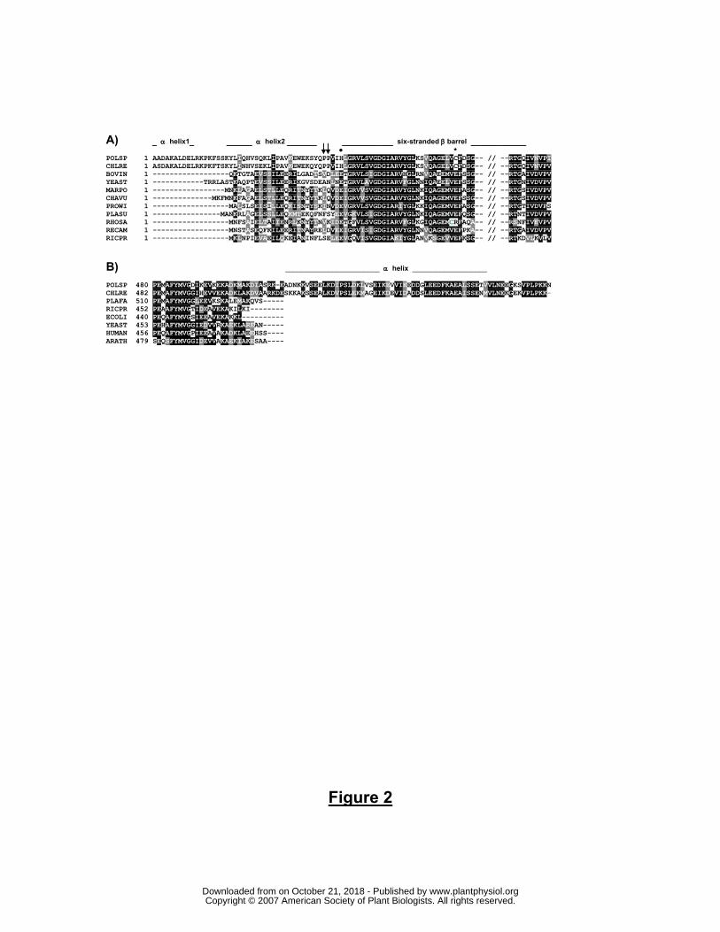
Figure 2. Subunits α and β in Chlamydomonad algae exhibit extensions. A) Multiple sequence
alignment of the N-terminal part of mature α subunits. Of the two predicted N-terminal α
helices, the second α helix is amphipathic and likely interacts with the N-terminus of the OSCP
(Weber et al., 2004). The arrows indicate the position of unique Pro residues in the algal
proteins; ●, conserved Glu residue substituted by a His residue in the algae; *, indicates the
presence of an unusual Cys residue in the algae. Sequences used are: BOVIN, Bos taurus
(P19483); CHAVU, Chara vulgaris (Q7YAN8); CHLRE, C. reinhardtii (Q96550); MARPO,
Marchantia polymorpha (P2685); PLASU, Platymonas subcordiformis (Q36517); POLSP,
Polytomella sp. (CAI3486); PROWI, Prototheca wickerhamii (Q37628); RECAM,
Reclinomonas americana (O21268); RICPR, Rickettsia prowazekii ( O50288); RHOSA,
Rhodomonas salina (Q9G8W0); YEAST, Saccharomyces cerevisiae (P07251). B) Multiple
sequence alignment of the C-terminal part of different β subunits. Sequences used are: CHLRE,
C. reinhardtii (P38482); POLSP, Polytomella sp. (CAI3487); PLAFA, Plasmodium falciparum
(Q810V2); RICPR, Rickettsia prowazekii (O50290); ECOLI, E. coli (P0ABB4); YEAST, S.
cerevisiae (P00830); HUMAN, H. sapiens (P06576); ARATH, A. thaliana (Q541W7). Note
that the presence of Pro residues in predicted α helices (as seen in the β extension) may in
reality disrupt the helices.
Figure 3. Localization of PsASA1 in the mitochondrial membrane and solubility of the
overexpressed protein. A) Mitochondrial membranes from Polytomella were subjected to the
indicated treatments. P, S correspond to the pellet and the soluble fractions after
ultracentrifugation. Immunoblotting shows the dissociation of ASA1 from the membrane with
www.plantphysiol.orgon October 21, 2018 - Published by Downloaded from Copyright © 2007 American Society of Plant Biologists. All rights reserved.

23
respect to the β subunit (extrinsic) and subunit COX2a of complex IV (intrinsic). B) Solubility
studies were done by sonicating E.coli cells overexpressing PsASA1 in Tris buffer, pH 8.0
(Control), in Na2CO3, pH 11.4 and in CAPS, pH 11.0, followed by centrifugation. The
supernatants were analyzed by Coomassie blue staining (CBB) and immunoblotting.
Figure 4. BN-PAGE analysis of heat-treated mitochondria from Polytomella and potato tubers.
Freshly isolated mitochondria were incubated at 55°C for 2 and 4 min. and solubilized with
dodecyl maltoside for BN-PAGE analysis. A) Coomassie blue stained blue native gel. B) In-gel
ATPase activity staining. The position of the OXPHOS (sub)complexes in untreated samples
were taken from previous works (Jänsch et al., 1996; Atteia et al., 2003). The roman numbers I
to V refer to the OXPHOS complexes; Vd, ATP synthase dimer; Vm, ATP synthase monomer;
Vi, intermediate form (uncharacterized); F1, dissociated F1-domain; ADHE, bifunctional
aldehyde/alcohol dehydrogenase; 0, untreated samples; 2, 2 min.; 4, 4min.
Figure 5. 2D BN/SDS-PAGE analysis of Polytomella mitochondria incubated at 55°C. For A,
B and C, a whole lane from a blue native gel was loaded on a Tricine denaturing gel (13%
acrylamide). A) No heat-treatment. B) 2 min. heat-treatment. Arrows indicate the subunits of
the F1-ATPase. C) Immunoblotting of proteins on 2D gels after 2 min. heat treatment. D) Pieces
from a blue native gel containing the different ATP synthase forms that were present after 2
min. of heat treatment were cut out and loaded on Tricine-SDS-PAGE. Dimer-specific subunits
ASA6 and ASA9 are boxed. Roman numbers I to V, Vd, Vm, as in Fig. 4; ?s, not identified and
only detected by silver staining; *, contaminating bands resulting from overlapping protein
components in the 2D protein profile. Subunit assignment on Tricine SDS-PAGE was
confirmed from Vázquez-Acevedo et al. (2006).
Figure 6. Working model of dimeric mitochondrial ATP synthase in Chlamydomonad algae,
based on our current knowledge of the enzyme from both C. reinhardtii and Polytomella sp.
The monomers are shown rotated 180° one from another (around a vertical axis), as proposed
previously for the yeast enzyme (Paumard et al., 2002). Arrows indicate the α and β extensions.
The α extension is shown interacting with the OSCP, and the β extension with ASA subunits of
the peripheral stalk. ASA1 is placed in the matrix because it is an extrinsic protein. ASA2 is a
hydrophobic protein and probably associates to the membrane. ASA3 is somewhat hydrophilic
but without specific features. ASA4 is a possible homodimer-forming subunit based on its
prediction for coiled coil formation (Table I). ASA5 may be anchored in the membrane and
protruding into the matrix. ASA6 and ASA9 are proposed to be dimer-specific and membrane-
bound, whereas ASA 7 and ASA8 may be associated to or traversing the membrane.
www.plantphysiol.orgon October 21, 2018 - Published by Downloaded from Copyright © 2007 American Society of Plant Biologists. All rights reserved.

24
Table I. Physico-chemical properties of the ASA subunits of the Chlamydomonad
mitochondrial ATP synthase. The presence of putative coiled coil-forming sequences may
indicate inter-subunit interactions or homodimerization. N-term., residue number marking start
of N-terminus; MM, molecular mass calculated from sequence; pI, isoelectric point; GRAVY,
grand average hydrophobicity score (higher scores indicate increasing hydrophobicity); TM
prediction, indicates the number of predicted TM segments using SOSUI (S), TMpred (T);
HMMTOP (H); Paircoil score, coiled coil domain prediction; Ps, Polytomella sp.; Cr., C.
reinhardtii.
Protein N-term. (res. no)
Protein ID1 MM (kDa)
pI GRAVY TM prediction
S T H
Max. Paircoil score
ASA1 (Ps)
23 CAD901582 66.2 5.47 -0.402 - - - 0.054 (K173-K202)
ASA1 (Cr)
24 78831 60.5 5.66 -0.377 - - - 0.390 (K17-K46) 0.743 (G424-C453)
ASA2 30 192142 45.6 9.08 0.346 - 2 - -
ASA3 33 152682 36.3 5.71 -0.053 - - - -
ASA4 29 182740 31.2 6.07 -0.184 - - - 0.383 (213A-E424) ASA7 27 192157 19.5 9.27 -0.064 - 1 - -
ASA5 1 184815 14.3 9.24 -0.370 - 1 - -
ASA6 28 186501 13.3 9.17 0.355 2 6 2 0.219 (E2-L31) ASA8 1 184537 9.9 9.43 -0.309 - 1 1 - ASA9 1 191034 12.1 9.08 -0.249 1 1 1 -
1Gene model numbers of JGI Chlamy v3.0 2Genbank accession number
www.plantphysiol.orgon October 21, 2018 - Published by Downloaded from Copyright © 2007 American Society of Plant Biologists. All rights reserved.

Figure 1
kDa
60
50
30
20
10
PsCr
ASA1�
�
�
a
OSCP
ASA2
ASA4ASA3
Cr
Glycine SDS-PAGE
60
50
30
20
10
kDa
ASA7
ASA6ASA5
c
�ASA8
Tricine SDS-PAGE
ASA1��
ASA4
�
ASA2
�
ASA7
aOSCP
ASA5
ASA3
ASA6*ASA8*ASA9*�*
c*
ASA9{ }
www.plantphysiol.orgon October 21, 2018 - Published by Downloaded from Copyright © 2007 American Society of Plant Biologists. All rights reserved.
A) _ α helix1_ ______ α helix2 _______ ____________ six-stranded β barrel _____________ ● * POLSP 1 AADAKALDELRKPKFSSKYLIQHVSQKLIPAVKEWEKSYQPPVIHLGRVLSVGDGIARVYGLKSVQAGELVCFDSG-- // --RTGQIVNVPI CHLRE 1 ASDAKALDELRKPKFTSKYLINHVSEKLIPAVKEWEKQYQPPVIHLGRVLSVGDGIARVYGLKSVQAGELVCFDSG-- // --RTGQIVNVPV BOVIN 1 ------------------QKTGTAEVSSILEERILGADTSVDLEETGRVLSIGDGIARVHGLRNVQAEEMVEFSSG-- // --RTGAIVDVPV YEAST 1 ------------TRRLASTKAQPTEVSSILEERIKGVSDEANLNETGRVLAVGDGIARVFGLNNIQAEELVEFSSG-- // --RTGNIVDVPV MARPO 1 -----------------MNKLAGAELSTLLEQRITNYYTKLQVDEIGRVVSVGDGIARVYGLNKIQAGEMVEFASG-- // --RTGSIVDVPV CHAVU 1 --------------MKFMNKFAGAELSTLLEQRITNYYTKLQVDEIGRVVSVGDGIARVYGLNKIQAGEMVEFSSG-- // --RTGSIVDVPV PROWI 1 ------------------MALSLSEISILLEQKISNTYSKINVDEVGRVLSVGDGIARIYGLKKIQAGEMVEFASG-- // --RTGTIVDVPS PLASU 1 ----------------MANKRLAGELSSLLEQKMTEKQFNFSYEEVGKVLSIGDGIARVYGLNKIQAGEMVEFQSG-- // --RTNTIVDVPV RHOSA 1 ------------------MNFSAIELTAIIENRLKNYYTNVKIDETGFVLSVGDGIARVFGLKGIQAGEMCRFAQA-- // --RSNFIVEVPV RECAM 1 ------------------MNSTASEQFKILEERITNAYRELDVEEIGRVISIGDGIARVYGLNNVQAGEMVEFPKA-- // --RTGAIVDVPV RICPR 1 ------------------MKLNPIEVAEILQKEIANINFLSELEEVGQVISVGDGIAKIYGLANVKSGEVVEFKSG-- // --RTKDVLKVLV
B) ________________________ α helix ___________________ POLSP 480 PEMAFYMVGDIKEVKEKADKMAKDIASRK-EADNKKVSEELKDIPSLDKLVSEIKEVVIEEDDGLEEDFKAEALSSETVVLNEEGKSVPLPKKN CHLRE 482 PEMAFYMVGGIHEVVEKADKLAKDVAARKDESKKAKSSEALKDVPSLEKMAGEIKDEVIDADDSLEEDFKAEAISSENMVLNEKGEKVPLPKK- PLAFA 510 PEMAFYMVGGLEEVKSKALEMAKQVS----- RICPR 452 PEAAFYMVGTIDEAVEKAKILKI-------- ECOLI 440 PEQAFYMVGSIEEAVEKAKKL---------- YEAST 453 PEHAFYMVGGIEDVVRKAEKLAREAN----- HUMAN 456 PEQAFYMVGPIEEAVAKADKLAEEHSS---- ARATH 479 SEQSFYMVGGIDEVVAKAEKIAKESAA----
Figure 2
www.plantphysiol.orgon October 21, 2018 - Published by Downloaded from Copyright © 2007 American Society of Plant Biologists. All rights reserved.

Anti-ASA1
Anti-
sub�
Anti-cox2a
Figure 3
P S P S P S P SCon
trol
55°C
Na
CO
2
3
55°C
+
Na
CO
2
3
Immunoblots
Anti-ASA1
CBB
A
B
Con
trol
Na
CO
2
3
pH11
ASA1 solubility
kDa
kDa
66
60
14
66
66
www.plantphysiol.orgon October 21, 2018 - Published by Downloaded from Copyright © 2007 American Society of Plant Biologists. All rights reserved.

Figure 4
Coomassie Blue ATPase staining
Polytomella Polytomellapotato potato
0 2 4 0 2 4 0 2 4 0 2 4
V
I
III
IV
kDa
1600
1000
500
200
V
I
III
IV
ADHE
F1
Vd
Vm
F1
Vm
BA
Vi
www.plantphysiol.orgon October 21, 2018 - Published by Downloaded from Copyright © 2007 American Society of Plant Biologists. All rights reserved.
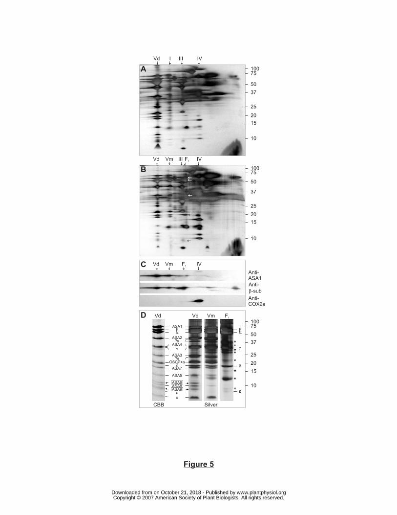
Figure 5
Vd I III IV
Vd Vm F1
III IV
10075
50
37
25
20
15
10
10075
50
37
25
20
15
10
A
B
Vd Vm F1D
10075
50
37
25
20
15
10
�
**
**
*
*
*�
�
��
* ��
CAnti-
ASA1
Anti-
-sub�
Anti-
COX2a
Vd Vm F1
IV
ASA1��
�
OSCP+a
ASA3
ASA2
ASA4
ASA7
ASA5
�
?s
?s
c
ASA6
ASA9�
ASA8
Vd
CBB Silver
www.plantphysiol.orgon October 21, 2018 - Published by Downloaded from Copyright © 2007 American Society of Plant Biologists. All rights reserved.

ccc
c
c
ε δγ
β
ccc c
γ
β
α
c
cc
ac
asa2
asa3
δε
c
asa4
asa1
OSCP
a
Figure 6
cc
asa1
Matrix
Intermem
branespace
OSCP
α
asa
8
asa
9
asa
6
asa7
asa5
www.plantphysiol.orgon October 21, 2018 - Published by Downloaded from Copyright © 2007 American Society of Plant Biologists. All rights reserved.