plasma membrane-associated scar complex ... · plasma membrane-associated scar complex...
TRANSCRIPT
Molecular Plant • Volume 1 • Number 6 • Pages 990–1006 • November 2008 RESEARCH ARTICLE
Plasma Membrane-Associated SCAR ComplexSubunitsPromoteCorticalF-ActinAccumulationandNormal Growth Characteristics in Arabidopsis Roots
Julia Dyachoka,d, Mon-Ray Shaoa, Kevin Vaughnb, Andrew Bowlingb, Michelle Facettec, Stevan Djakovica,e,Lauren Clarka and Laurie Smitha,1
a Section of Cell and Developmental Biology, University of California San Diego, 9500 Gilman Drive, La Jolla, CA 92093-0116, USAb Southern Weed Science Research Unit, USDA–ARS, 141 Experiment Station Road, Stoneville, MS 38776, USAc Carnegie Institution, Department of Plant Biology, 260 Panama Street, Stanford, CA 94305, USAd Present address: Division of Plant Biology, The Samuel Roberts Noble Foundation, 2510 Sam Noble Pkwy, Ardmore, OK 73401, USAe Present address: Section of Neurobiology, University of California San Diego, 9500 Gilman Drive, La Jolla, CA 92093-0116, USA
ABSTRACT The ARP2/3 complex, a highly conserved nucleator of F-actin polymerization, and its activator, the SCAR com-
plex, have been shown to play important roles in leaf epidermal cell morphogenesis in Arabidopsis. However, the intra-
cellular site(s) and function(s) of SCAR and ARP2/3 complex-dependent actin polymerization in plant cells remain unclear.
We demonstrate that putative SCAR complex subunits BRK1 and SCAR1 are localized to the plasma membrane at sites of
cell growth and wall deposition in expanding cells of leaves and roots. BRK1 localization is SCAR-dependent, providing
further evidence of an association between these proteins in vivo. Consistent with plasma membrane localization of SCAR
complex subunits, cortical F-actin accumulation in root tip cells is reduced in brk1 mutants. Moreover, mutations disrupting
the SCAR or ARP2/3 complex reduce the growth rate of roots and their ability to penetrate semi-solid medium, suggesting
reduced rigidity. Cell walls of mutant roots exhibit abnormal structure and composition at intercellular junctions where
BRK1 and SCAR1 are enriched in the adjacent plasma membrane. Taken together, our results suggest that SCAR and ARP2/3
complex-dependent actin polymerization promotes processes at the plasma membrane that are important for normal
growth and wall assembly.
Key words: cell expansion; cell morphogenesis; cytoskeleton; root biology; Arabidopsis.
INTRODUCTION
Plant cells acquire their shapes according to the patterns in
which their walls expand during cell growth (Mathur, 2004).
Actin filaments are thought to influence the expansion pat-
tern of the cell wall by helping to direct the delivery of secreted
wall material and membranes to sites of cell growth (Smith and
Oppenheimer, 2005; Hussey et al., 2006). Numerous studies
have established a role for the ARP2/3 complex in the spatial
regulation of epidermal cell growth in plants (Szymanski,
2005). The animal Arp2/3 complex, consisting of seven subunits
including the actin-related proteins Arp2 and Arp3, nucleates
actin polymerization mainly by initiating new branches on the
sides of existing actin filaments (Goley and Welch, 2006). Arp2/
3 complex activity depends on nucleation promoting factors
including members of the Scar/WAVE family (Stradal and Scita,
2005). In mammalian cells, Scar/WAVE is found in a complex
with four other proteins: Sra1, Nap1, Abi1, and Hspc300 (Eden
et al., 2002; Gautreau et al., 2004). In response to upstream reg-
ulators Rac and Nck, Scar/WAVE is activated via a mechanism
that has been proposed to involve either dissociation of the
complex or recruitment of the intact complex to sites of F-actin
nucleation (reviewed, Takenawa and Suetsugu, 2007). Compo-
nents of the Scar/WAVE complex localize to plasma mem-
branes at the leading edge of animal cells where they
nucleate cortical F-actin that promotes lamellipodial extension
(Miki et al., 1998; Nozumi et al., 2003; Kunda et al., 2003;
Rogers et al., 2003; Stovold et al., 2005).
Homologs of all mammalian Arp2/3 and Scar/WAVE complex
subunits have been identified in Arabidopsis, including a
family of four proteins distantly related to animal Scar/WAVE
proteins (AtSCAR1 to AtSCAR4; Szymanski, 2005). Three of the
1 To whom correspondence should be addressed. E-mail [email protected],
fax 858-534-7108, tel. 858-822-2531.
ª The Author 2008. Published by the Molecular Plant Shanghai Editorial
Office in association with Oxford University Press on behalf of CSPP and
IPPE, SIBS, CAS.
doi: 10.1093/mp/ssn059, Advance Access publication 8 October 2008
Received 17 June 2008; accepted 22 August 2008

four Arabidopsis SCAR proteins have been shown to stimulate
mammalian Arp2/3 complex-dependent actin polymerization
in vitro (Frank et al., 2004; Basu et al., 2005; Uhrig et al.,
2007; Zhang et al., 2008). Although neither ARP2/3 nor SCAR
complexes have been purified from plant extracts or reconsti-
tuted from plant subunits to date, numerous in-vitro and in-
vivo binding interactions between putative complex subunits
support the existence of ARP2/3 and SCAR complexes in plants
(Szymanski, 2005; Uhrig et al., 2007). Mutations in genes
encoding nine different subunits of the putative ARP2/3 or
SCAR complexes dramatically perturb the growth patterns
of epidermal hairs called trichomes and also reduce lobe out-
growth and intercellular adhesion in epidermal pavement cells
(reviewed, Szymanski, 2005; Smith and Oppenheimer, 2005;
also see more recent studies by Basu et al., 2005; Zhang
et al., 2005; Djakovic et al., 2006; Le et al., 2006). Plants lacking
all SCAR function are phenotypically equivalent to ARP2/3
complex subunit mutants (Zhang et al., 2008), and double
mutants lacking both SCAR and ARP2/3 complex function
are no more severe than either type of single mutant (e.g.
El-Assal et al., 2004; Djakovic et al., 2006), suggesting that
SCAR is the primary if not the sole activator of the ARP2/3
complex in Arabidopsis. Surprisingly, no severe loss of F-actin
has been observed in ARP2/3 or SCAR complex subunit
mutants. Instead, excessive bundling and spatial disorganiza-
tion of cytoplasmic actin strands have been observed in
expanding mutant trichomes, and alterations in cortical F-
actin distribution have been observed in expanding mutant
pavement cells (e.g. Mathur et al., 2003a; Le et al., 2003; Li
et al., 2003; Deeks et al., 2004; Brembu et al., 2004; Djakovic
et al., 2006).
In the moss Physcomitrella patens, ARP2/3 and SCAR com-
plex subunit mutants have severe defects in filament elonga-
tion associated with loss or disruption of the F-actin ‘cap’
structure normally found at the apical growth site (Harries
et al., 2005; Perroud and Quatrano, 2006, 2008; Finka et al.,
2007). Moreover, ARPC4 (a subunit of the putative ARP2/3
complex) and BRK1 (the plant homolog of the Hspc300 subunit
of the animal Scar/WAVE complex) are both specifically local-
ized at the filament apex (Perroud and Quatrano, 2006, 2008).
Thus, tip-localized activity of the SCAR–ARP2/3-dependent ac-
tin nucleation pathway plays a critical role in promotion of tip
growth in P. patens. The ARP2/3 complex also promotes tip
growth in elongating root hairs of Arabidopsis, but its role
is minor and largely redundant with those of other actin reg-
ulatory proteins (Mathur et al., 2003b; Deeks et al., 2007). Al-
though the genes encoding ARP2/3 and SCAR complex
subunits are ubiquitously expressed (e.g. El-Assal et al.,
2004; Basu et al., 2005; Uhrig et al., 2007), very little is known
about roles for this pathway in vascular plants outside the con-
text of expanding leaf epidermal cells and tip growing cells.
Even in these cells, it remains unclear where SCAR and
ARP2/3 complex-dependent actin nucleation occurs and what
cellular processes are disrupted to cause the cell shape and ad-
hesion defects observed in mutants.
Here, we use complementary methods to demonstrate that
SCAR complex subunits are associated with the plasma mem-
brane in various tissues of Arabidopsis. Analysis of roots of
SCAR complex subunit mutants demonstrates an important
role for SCAR and ARP2/3 complexes in accumulation of corti-
cal (plasma membrane-associated) F-actin and in certain
aspects of root growth and cell wall assembly. Together, these
results provide new perspectives on the role(s) played by SCAR
and ARP2/3 complex-dependent actin polymerization in plant
cell growth.
RESULTS
BRK1 and SCAR1 Are Localized to the Cell Periphery
To gain insight into where SCAR- and ARP2/3-complex-depen-
dent actin polymerization occurs in plant cells, we studied the
localization of BRK1 and SCAR1 fluorescent fusion proteins in
transgenic plants. BRK1::YFP expressed from the BRK1
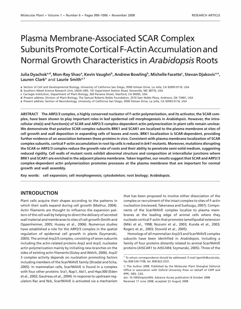
promoter is functional as indicated by its ability to comple-
ment the distorted trichome phenotype of brk1 mutants
(Figure 1C). In contrast to scar2 mutants, which display a mild
distorted trichome phenotype (Basu et al., 2005; Zhang et al.,
2005), scar1 mutants have normally shaped trichomes and lack
any obvious phenotypes, although a redundant role for SCAR1
in trichomome morphogenesis is suggested by a slight en-
hancement of the scar2 trichome phenotype in scar1;scar2
double mutants (Zhang et al., 2008). Thus, to determine
whether GFP::SCAR1 is functional in vivo, we tested its ability
to rescue the scar2 trichome phenotype when driven from the
SCAR2 promoter (since the wild-type SCAR1 gene is not suffi-
cient for normal trichome morphogenesis in scar2 mutants, we
reasoned that SCAR1 promoter-driven expression of
SCAR1::GFP would not rescue the scar2 trichome phenotype).
Indeed, normal trichome morphology was restored in scar2
mutants transformed with SCAR2p::GFP::SCAR1 (Figure 1E).
Thus, GFP::SCAR1 is functional because it can substitute for
SCAR2 and we reasoned that the localization of
SCAR2p::GFP::SCAR1 in complemented scar2 mutants could
provide information regarding the intracellular sites of SCAR1
and/or SCAR2 function.
Both BRK1::YFP and GFP::SCAR1 localized to the periphery
of expanding trichomes (Figure 2A–2H). In young trichomes,
BRK1::YFP and GFP::SCAR1 were enriched at the tips of newly
initiated branches (Figure 2A–2D), presumably corresponding
to sites of active cell growth. In partially expanded branches of
older trichomes, BRK1::YFP and GFP::SCAR1 both remained pe-
ripherally localized but were spread along the full length of
the elongating branch (Figure 2E–2H). At this stage, growth
is also distributed along the entire length of the branch
(Schwab et al., 2003). Thus, BRK1::YFP and GFP::SCAR1 appear
to be localized at growth sites in expanding trichomes.
GFP::SCAR1 was also associated with medium-sized intracellu-
lar organelles of unknown identity in expanding trichomes
(Figure 2D). However, this component of the GFP::SCAR1 local-
ization pattern was not observed for BRK1::YFP (Figure 2B).
Dyachok et al. d Localization of SCAR Complex Subunits in Arabidopsis | 991
Moreover, as described below, it was not observed for
GFP::SCAR1 in other leaf epidermal cell types, or for endoge-
nous SCAR1 detected by immunofluorescence in roots. Thus,
the significance of organelle-associated SCAR1::GFP in tri-
chomes is unknown, and may be artifactual.
BRK1::YFP and GFP::SCAR1 also localized to the cell periph-
ery in expanding leaf epidermal pavement cells (Figure 2M–2P).
Bright foci of higher fluorescence intensity were consistently
observed at three-way intercellular junctions (henceforth re-
ferred to as ‘cell corners’) both in unexpanded pavement cells
(arrows, Figure 2M and 2O) and in partially expanded ones
that have already attained their characteristically lobed shapes
(arrows, Figure 2N and 2P). These foci of BRK1::YFP and
GFP::SCAR1 presumably correspond to sites of increased depo-
sition of cell wall material, which fills the triangular-shaped
spaces found at three-way wall junctions. Partially expanded
pavement cells also exhibited more diffusely localized enrich-
ments of BRK1::YFP and GFP::SCAR1 at points of maximum cur-
vature (arrowheads, Figure 2N and 2P), which might
correspond to sites of lobe outgrowth.
We next examined the localization of BRK1 and SCAR1 pro-
teins in root tips. While no clear BRK1::YFP localization could
be detected in epidermal cells including elongating root hairs
(data not shown), cortical and endodermal cells of the root tip
showed peripherally localized BRK1::YFP predominantly at
transverse cell ends, which was markedly enriched at cell cor-
ners as observed in leaves (arrows, Figure 2Q) and persisted as
these cells elongated (Figure 2R). SCAR1 localization in roots
was investigated via whole mount immunofluorescent
labeling with a previously characterized anti-SCAR1 antibody
(Djakovic et al., 2006). Similar to localization results for
BRK1::YFP, anti-SCAR1 staining was observed at the periphery
of cells in internal layers of the root, and was often concen-
trated at cell corners (arrows, Figure 2T). This labeling could
be attributed to SCAR1 because it was absent in scar1 mutants
(Figure 2S), which were previously shown to lack SCAR1 protein
via Western blot analysis (Djakovic et al., 2006). Non-
specific cytoplasmic staining by the anti-SCAR1 antibody
obscures detection of SCAR1 in root and leaf epidermal cells, in-
cluding trichomes (data not shown). In summary, BRK1::YFP,
GFP::SCAR1, and endogenous SCAR1 showed very similar pat-
ternsof localizationatthecellperiphery inboth leavesandroots.
Consistent with previous analyses demonstrating ubiqui-
tous expression of BRK1 (Djakovic et al., 2006; Le et al.,
Figure 1. BRK1p::BRK1::YFP and SCAR2p::GFP::SCAR1 Restore Wild-Type TrichomeMorphology in brk1 and itb1-16/scar2 Mu-tant Plants, Respectively.
Light micrographs of leaves.(A) Columbia wild-type.(B) brk1 mutant (Columbia ecotype).(C)brk1mutantexpressingBRK1p::BRK1::YFP.(D) itb1-16 (scar2) mutant (RLD ecotype).(E) itb1-16 (scar2) mutant expressingSCAR2p::GFP::SCAR1.Trichome morphology in RLD wild-typeplants is indistinguishable from Columbiawild-type (Zhang et al., 2005) and is notshown. Scale bar 5 250 microns.
992 | Dyachok et al. d Localization of SCAR Complex Subunits in Arabidopsis

2006), we found that BRK1p::BRK1::YFP was present in a wide
variety of cell types surveyed in addition to those described
above, including expanding epidermal cells of hypocotyls,
cotyledons, gynoecia, anther filaments, petals, and sepals, as
well as fully expanded leaf epidermal pavement and guard
cells. Peripheral localization of BRK1::YFP was observed in
all of these cell types with foci at cell corners as described
for expanding leaf and root cells (data not shown). These
Figure 2. BRK1::YFP, GFP::SCAR1 and SCAR1 Localize to the Cell Periphery.
(A–L) Trichomes at early (A–D, I, J) and later (E–H, K, L) stages of expansion. Brightfield (A, E) and confocal fluorescence (B, F) images ofcomplemented brk1 mutants expressing BRK1p::BRK1::YFP. Brightfield (C, G) and confocal fluorescence (D, H) images of wild-type plantsexpressing SCAR2p::GFP::SCAR1. Arrowheads in (B) and (D) point to local enrichments of fluorescence seen at tips of newly emerged tri-chome branches. Brightfield (I, K) and confocal fluorescence (J, L) images of non-transgenic trichomes acquired under the same conditions(laser power, exposure time, etc.) employed for transgenic trichomes showed no detectable fluorescence, confirming that the fluorescenceshown in (B, D, F, H) is due to the presence of YFP/GFP and not to autofluorescence.(M–P) Leaf epidermal pavement cells that are unexpanded (M, O) or partially expanded (N, P). (M, N) Complemented brk1 mutants express-ing BRK1p::BRK1::YFP. (O, P) Complemented itb1-16/scar2 mutants expressing SCAR2p::GFP::SCAR1. Arrows in (M–P) point to cell cornersshowing local enrichments of fluorescence. Arrowheads in (N) and (P) point to broader, localized enrichments observed at points of max-imum curvature in partially expanded pavement cells.(Q–T) BRK1::YFP and SCAR1 localization in root cortical cells. (Q, R) Complemented brk1 mutant expressing BRK1p::BRK1::YFP. (S, T) Con-focal fluorescence images of scar1 mutant (S, negative control) and wild-type (T) roots fixed and stained with anti-SCAR1 antibody. Arrowsin (Q, R, T) point to cell corners showing local enrichments of fluorescence. Scale bar = 20 microns in (A–P); 10 microns in (Q–T).
Dyachok et al. d Localization of SCAR Complex Subunits in Arabidopsis | 993

observations suggest that BRK1’s function in expanding cells is
not restricted to those exhibiting shape abnormalities in brk1
mutants, and that it has a role to play even in cells that are no
longer growing.
BRK1 and SCAR1 Are Peripherally Associated with Plasma
Membrane
The plasma membrane is attached to the cell wall, and differ-
entiating plasma membrane versus cell wall localization is dif-
ficult at the light microscope level. To determine whether
BRK1::YFP is associated with the plasma membrane or cell wall,
we analyzed its localization in root cells after retraction of the
plasma membrane from the cell wall via plasmolysis with su-
crose. In plasmolyzed cells, BRK1::YFP (including the foci of
BRK1::YFP found at cell corners) retracted from the cell wall
with the plasma membrane, indicating its association with
the plasma membrane rather than the cell wall (Figure 3).
We further used a biochemical approach to determine
whether BRK1::YFP and/or SCAR1 are membrane-associated.
In Western blots of unfractionated seedling extracts,
BRK1::YFP and SCAR1 were barely detectable using anti-GFP
and anti-SCAR1 antibodies, respectively (data not shown).
Consistent with a membrane association for both proteins,
when extracts were separated into microsomal and soluble
fractions by ultracentrifugation, the majority of SCAR1 and
BRK1::YFP were found in the microsomal fraction with the
membrane marker protein a-1,2-Mannosidase I (Preuss
et al., 2004), whereas the cytoplasmic marker protein PUX1
(Rancour et al., 2004) was mostly found in the soluble fraction
(Figure 4). To investigate the nature of their association with
the microsomal fraction, we attempted to extract BRK1::YFP
and SCAR1 from microsomal pellets under various conditions.
Both BRK1::YFP and SCAR1 were partially solubilized with
detergents or 0.1 M Na2CO3 but not with 2 M NaCl, suggesting
a peripheral membrane association (Figure 5). Consistent with
these findings and with the lack of a signal peptide or trans-
membrane domain in BRK1, BRK1::YFP failed to accumulate
intracellularly upon treatment with brefeldin A, a secretion in-
hibitor, suggesting that BRK1::YFP is targeted to the plasma
membrane via a secretion-independent pathway (Figure 6).
SCARs Are Required for Plasma Membrane Localization of
BRK1::YFP
Arabidopsis SCARs bind to BRK1 in vitro (Frank et al., 2004;
Zhang et al., 2005), suggesting a possible role for one of
the proteins in localization of the other. To investigate this pos-
sibility, we analyzed the localization of transiently expressed
BRK1::YFP following bombardment into expanding epidermal
pavement cells of wild-type plants and mutants lacking SCAR
function. To avoid any possible ambiguity arising from func-
tional redundancy among members of the SCAR family (Zhang
et al., 2008), we constructed a scar1,2,3,4 quadruple mutant
for this experiment (see Methods for details). For comparison
with scar quadruple mutants, we also analyzed BRK1::YFP lo-
calization in arp2 mutants. However, we could not investigate
the dependence of SCAR1 localization on BRK1, because SCAR
proteins are degraded in the absence of BRK1 (Djakovic et al.,
2006; Le et al., 2006).
For unknown reasons, transiently expressed BRK1::YFP
showed more cytoplasmic localization than was observed in
stable BRK1::YFP transgenics, even when expressed at very
low levels. In highly vacuolated cells such as epidermal pave-
ment cells, plasma membrane-associated fluorescence is not
readily discernable in the presence of cytoplasmic fluores-
cence, which is concentrated at the cell periphery in a thin
layer of cortical cytoplasm. However, we observed that at junc-
tions between wild-type pavement cells and guard cells, tran-
siently expressed BRK1::YFP typically showed localized
accumulation at cell corners (arrow, Figure 7A)—a characteris-
tic feature of BRK1::YFP localization in stable BRK1::YFP trans-
genic plants that was clearly distinguishable from cytoplasmic
BRK1::YFP. Quantitative analysis showed that in wild-type
pavement cells, bright foci of BRK1::YFP were present at
75% of corners associated with junctions between pavement
Figure 3. BRK1::YFP and SCAR1 Remain Asso-ciated with Plasma Membranes after Plasmol-ysis.
Confocal fluorescence images of root tip cellsof complemented brk1 mutants illustratingBRK1p::BRK1::YFP fluorescence immediatelyafter partial plasmolysis with 0.5 M sucrose(monochrome in (A), green in (C)) in relationto plasma membranes labeled with FM4-64(monochrome in (B), red in (C)). Cells wereonly partially plasmolysed for these experi-ments because, for unknown reasons, plas-molysis caused loss of BRK1::YFP from thecell periphery. Even with only partial plas-molysis, some BRK1::YFP was lost from thecell periphery, causing the BRK1::YFP signalto appear fainter and more restricted to cellcorners than it does in Figure 2Q. Scale bar =10 microns.
994 | Dyachok et al. d Localization of SCAR Complex Subunits in Arabidopsis

and guard cells (n = 24). The proportion of such corners show-
ing bright foci of BRK1::YFP was not significantly different in
arp2 mutants (76.5%, n = 34; Figure 7B), but was reduced in
scar1,2,3,4 quadruple mutants to 11.5% (n = 26, significantly
different from wild-type at p , 0.0001 by Fisher’s Exact Test;
Figure 7C). Thus, localization of BRK1 at cell corners depends
on SCAR but not the ARP2/3 complex.
Cortical Actin Is Partially Depleted in Root Tip Cells of brk1
Mutants
The Scar/WAVE complex is localized to sites of actin nucleation
in animal cells, and its depletion markedly abolishes actin po-
lymerization at those sites (Miki et al., 1998; Rogers et al., 2003;
Kunda et al., 2003; Innocenti et al., 2004). Localization of
BRK1::YFP and GFP::SCAR1 at the plasma membrane suggests
a role for the SCAR complex in promotion of cortical F-actin
polymerization. Previous studies have not reported depletion
of cortical F-actin in expanding trichomes or pavement cells of
SCAR or ARP2/3 complex subunit mutants (Li et al., 2003; Dja-
kovic et al., 2006), but actin has not been previously examined
in roots of these mutants. Therefore, we examined F-actin in
root tips of brk1 mutants, focusing on internal tissue layers
where plasma membrane localization of BRK1::YFP and SCAR1
was observed.
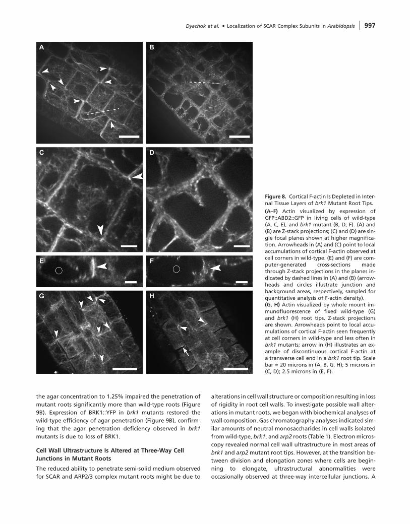
We visualized F-actin in plants expressing an actin binding do-
main of fimbrin tagged at both ends with GFP (GFP::ABD2::GFP)
(Wang et al., 2008). As illustrated in Figure 8A, GFP::ABD2::GFP
revealed cortical F-actin mainly at the transverse ends of wild-
type root cells. Interestingly, cortical F-actin was often enriched
at cell corners (arrowheads, Figure 8A and 8C) where BRK1::YFP
and SCAR1 are also enriched. Cortical F-actin visualized by
this method was markedly depleted in brk1 mutant root tips
(Figure 8B and 8D). We also visualized F-actin via whole-
mount immunolabeling of fixed root tips. This approach
also revealed cortical F-actin at the transverse ends of most
wild-type root tip cells, which often appeared enriched at
cell corners (Figure 8G), and depletion of cortical F-actin
at transverse cell ends and cell corners in brk1 mutants
(Figure 8H).
To further analyze the difference between wild-type and
brk1 mutant, F-actin density was analyzed quantitatively at
cell corners in GFP::ABD2::GFP transgenic plants where differ-
ences between wild-type and brk1 appeared most pro-
nounced. Computer-generated cross-sections were made
through Z-stack projections at cell corners (examples of section
planes are illustrated with dashed lines in Figure 8A and 8B)
and displayed in the X/Y plane as shown in Figure 8E and
8F. For each corner analyzed, we calculated the ratio of fluo-
rescence intensity at the intercellular junction (arrowheads,
Figures 8E and 8F) relative to background (sampled in the ad-
jacent vacuole as indicated by circles in Figure 8E and 8F).
The average ratio in brk1 mutants (1.47 6 0.15, n = 135 junc-
tions total from 15 roots) was significantly lower than that in
Figure 4. BRK1::YFP and SCAR1 Are Enriched in the MicrosomalFraction of Plant Tissue Extracts.
Extracts were ultra-centrifuged to separate microsomal membranes(‘pellet’) from supernatants (‘soluble’). Western blots were probedwith the indicated antibodies (see Methods for details regardingprocedures and antibodies). The bands shown are consistent withthe predicted molecular masses of SCAR1 and BRK1::YFP and con-firmed as SCAR1 and BRK1::YFP by their absence in samples derivedfrom scar1 mutants and non-transgenic plants, respectively, whichwere processed in parallel but are not shown in the figure.
Figure 5. BRK1::YFP and SCAR1 Behave Biochemically as PeripheralMembrane Proteins.
Microsomal membrane pellets were re-suspended in the originalextraction buffer alone (‘controls’) or with this buffer supple-mented with the indicated reagents, and subsequently ultra-centri-fuged to separate solubilized proteins (‘soluble’) from thoseremaining insoluble (‘pellets’). Western blots of each sample wereprobed with anti-GFP to detect BRK1::YFP (arrows, left panel) orwith anti-SCAR1 (right panel). BRK1::YFP and SCAR1 remain insol-uble in the buffer alone (control) and 2 M NaCl samples, but arepartially extracted from the microsomal pellet with 0.1 M Na2CO3
and 3% Triton X-100. Bands identified as BRK1::YFP and SCAR1 areconsistent with their predicted molecular masses and their identi-ties were confirmed by their absence from samples derived fromnon-transgenic plants and scar1 mutants, respectively, which wereprocessed in parallel but are not shown in the figure.
Dyachok et al. d Localization of SCAR Complex Subunits in Arabidopsis | 995

wild-type (2.36 6 0.4, n = 175 junctions total from 19 roots)
(p , 0.00001 by student’s t-test).
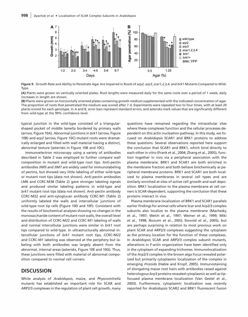
SCAR and ARP2/3 Complex Subunit Mutants Have Altered
Root Growth Characteristics
Our findings that SCAR complex subunits are plasma mem-
brane-localized in root cells and that the SCAR complex is re-
quired for normal cortical F-actin accumulation in these cells
raised the question of what actin-dependent processes might
be disrupted in roots of SCAR and ARP2/3 complex subunit
mutants, which has not been previously addressed. Several
aspects of root growth are known to be actin-dependent, in-
cluding gravitropism and elongation rate (Blancaflor et al.,
2006). We found that root gravitropic responses were not sig-
nificantly altered in ARP2/3 and SCAR complex subunit
mutants (data not shown). However, root growth rates were
significantly reduced in all mutants examined (Figure 9A).
Moreover, we observed that roots of all mutants were defi-
cient in their ability to penetrate semi-solid medium. This de-
ficiency did not appear to be due to the reduced growth rate
of mutant roots, since both wild-type and mutant roots effi-
ciently penetrated media containing 0.5% agar, but increasing
Figure 6. BRK1::YFP Localization in Root CellsIs Not Altered by Treatment with Brefeldin A(BFA) for 30 min.
As previously described (Geldner et al., 2001),PIN1::GFP is localized at the plasma membraneof root tip cells (A) but treatment with 50 mMBFA for 30 min caused partial loss of PIN1::GFPfrom the plasma membrane and its accumula-tion in BFA bodies (Royle and Murrell-Lagnado, 2003) (arrows, (B)). BRK1::YFP islocalized at the cell periphery both before(C) and after 30 min of treatment with50 mM BFA (D). Scale bar = 20 microns.
Figure 7. SCAR-Dependent Localization ofBRK1p::BRK1::YFP Transiently Expressed inExpanding Cotyledon Pavement Cells.
(A) Wild-type, (B) arp2 mutant, and (C) scar1,2,3,4 quadruple mutant. Arrows in (A)and (B) indicate bright foci of BRK1::YFPfound at cell corners associated with junc-tions between epidermal pavement andguard cells in wild-type and arp2 mutantleaves, but not in scar 1,2,3,4 quadruple mu-tant leaves. Peripheral YFP fluorescence out-lining transformed pavement cells in all threepanels is attributable to the cytoplasm basedon the presence of fluorescent transvacuolarcytoplasmic strands in focal planes not shownhere. Scale bar = 10 microns.
996 | Dyachok et al. d Localization of SCAR Complex Subunits in Arabidopsis
the agar concentration to 1.25% impaired the penetration of
mutant roots significantly more than wild-type roots (Figure
9B). Expression of BRK1::YFP in brk1 mutants restored the
wild-type efficiency of agar penetration (Figure 9B), confirm-
ing that the agar penetration deficiency observed in brk1
mutants is due to loss of BRK1.
Cell Wall Ultrastructure Is Altered at Three-Way Cell
Junctions in Mutant Roots
The reduced ability to penetrate semi-solid medium observed
for SCAR and ARP2/3 complex mutant roots might be due to
alterations in cell wall structure or composition resulting in loss
of rigidity in root cell walls. To investigate possible wall alter-
ations in mutant roots, we began with biochemical analyses of
wall composition. Gas chromatography analyses indicated sim-
ilar amounts of neutral monosaccharides in cell walls isolated
from wild-type, brk1, and arp2 roots (Table 1). Electron micros-
copy revealed normal cell wall ultrastructure in most areas of
brk1 and arp2 mutant root tips. However, at the transition be-
tween division and elongation zones where cells are begin-
ning to elongate, ultrastructural abnormalities were
occasionally observed at three-way intercellular junctions. A
Figure 8. Cortical F-actin Is Depleted in Inter-nal Tissue Layers of brk1 Mutant Root Tips.
(A–F) Actin visualized by expression ofGFP::ABD2::GFP in living cells of wild-type(A, C, E), and brk1 mutant (B, D, F). (A) and(B) are Z-stack projections; (C) and (D) are sin-gle focal planes shown at higher magnifica-tion. Arrowheads in (A) and (C) point to localaccumulations of cortical F-actin observed atcell corners in wild-type. (E) and (F) are com-puter-generated cross-sections madethrough Z-stack projections in the planes in-dicated by dashed lines in (A) and (B) (arrow-heads and circles illustrate junction andbackground areas, respectively, sampled forquantitative analysis of F-actin density).(G, H) Actin visualized by whole mount im-munofluorescence of fixed wild-type (G)and brk1 (H) root tips. Z-stack projectionsare shown. Arrowheads point to local accu-mulations of cortical F-actin seen frequentlyat cell corners in wild-type and less often inbrk1 mutants; arrow in (H) illustrates an ex-ample of discontinuous cortical F-actin ata transverse cell end in a brk1 root tip. Scalebar = 20 microns in (A, B, G, H); 5 microns in(C, D); 2.5 microns in (E, F).
Dyachok et al. d Localization of SCAR Complex Subunits in Arabidopsis | 997
typical junction in the wild-type consisted of a triangular-
shaped pocket of middle lamella bordered by primary walls
(arrow, Figure 10A). Abnormal junctions in brk1 (arrow, Figure
10B) and arp2 (arrow, Figure 10C) mutant roots were dramat-
ically enlarged and filled with wall material having a distinct,
abnormal texture (asterisks in Figure 10B and 10C).
Immunoelectron microscopy using a variety of antibodies
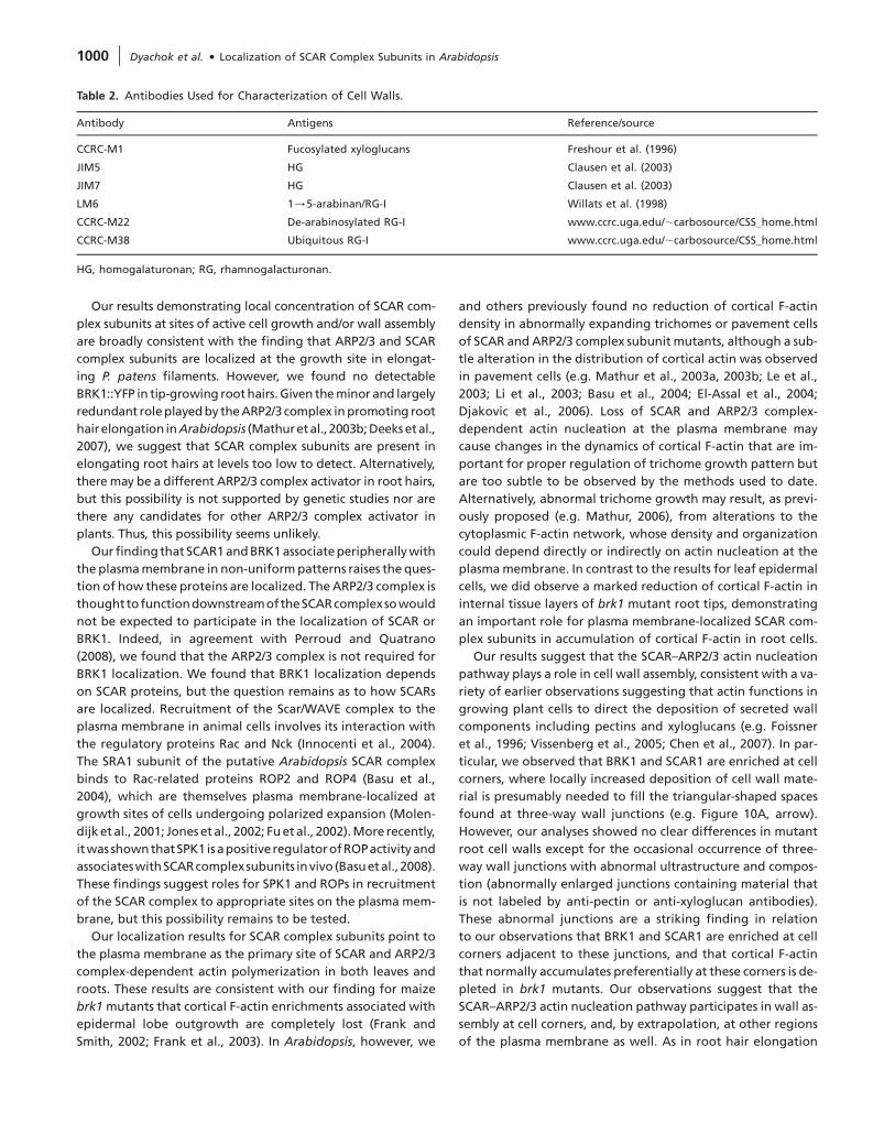
described in Table 2 was employed to further compare wall
composition in mutant and wild-type root tips. Anti-pectin
antibodies JIM5 and JIM7 are widely used for immunolabeling
of pectins, but showed very little labeling of either wild-type
or mutant root tips (data not shown). Anti-pectin antibodies
LM6 and CCRC-M38 (Table 2) gave stronger labeling signals
and produced similar labeling patterns in wild-type and
brk1 mutant root tips (data not shown). Anti-pectin antibody
CCRC-M22 and anti-xyloglucan antibody CCRC-M1 (Table 2)
uniformly labeled the walls and intercellular junctions of
wild-type root tip cells (Figure 10D and 10F). Consistent with
the results of biochemical analyses showing no changes in the
monosaccharide content of mutant root walls, the overall level
and distribution of CCRC-M22 and CCRC-M1 labeling of walls
and normal intercellular junctions were similar in brk1 root
tips compared to wild-type. In ultrastructurally abnormal in-
tercellular junctions of brk1 mutant root tips, CCRC-M22
and CCRC-M1 labeling was observed at the periphery but la-
beling with both antibodies was largely absent from the
abnormal, internal areas (asterisks, Figure 10E and 10G). Thus,
these junctions were filled with material of abnormal compo-
sition compared to normal cell corners.
DISCUSSION
While analysis of Arabidopsis, maize, and Physcomitrella
mutants has established an important role for SCAR and
ARP2/3 complexes in the regulation of plant cell growth, many
questions have remained regarding the intracellular sites
where these complexes function and the cellular processes de-
pendent on this actin nucleation pathway. In this study, we fo-
cused on Arabidopsis SCAR1 and BRK1 proteins to address
these questions. Several observations reported here support
the conclusion that SCAR1 and BRK1, which bind directly to
each other in vitro (Frank et al., 2004; Zhang et al., 2005), func-
tion together in vivo via a peripheral association with the
plasma membrane. BRK1 and SCAR1 are both enriched in
the membrane fraction and both behave biochemically as pe-
ripheral membrane proteins. BRK1 and SCAR1 are both local-
ized to plasma membranes in several cell types and are
similarly enriched at sites of active cell growth and wall depo-
sition. BRK1 localization to the plasma membrane at cell cor-
ners is SCAR-dependent, supporting the conclusion that these
proteins interact in vivo.
Plasma membrane localization of BRK1 and SCAR1 parallels
earlier findings for animal cells where Scar and Arp2/3 complex
subunits also localize to the plasma membrane (Machesky
et al., 1997; Welch et al., 1997; Weiner et al., 1999; Miki
et al., 1998; Nozumi et al., 2003; Stovold et al., 2005), but
are perhaps surprising in relation to most previous work on
plant SCAR and ARP2/3 complexes suggesting the cytoplasm
as the primary location for the function of these complexes.
In Arabidopsis SCAR and ARP2/3 complex subunit mutants,
alterations in F-actin organization have been identified only
in the cytoplasm of expanding trichomes. Immunolocalization
of the Arp2/3 complex in the brown alga Fucus revealed polar-
ized but primarily cytoplasmic localization of the complex in
emerging rhizoids (Hable and Kropf, 2005). Immunostaining
of elongating maize root hairs with antibodies raised against
heterologous Arp3 proteins revealed cytoplasmic as well as tip-
focused plasma membrane localization (Van Gestel et al.,
2003). Furthermore, cytoplasmic localization was recently
reported for Arabidopsis SCAR2 and BRK1 fluorescent fusion
Figure 9. Growth Rate and Ability to Penetrate Agar Are Impaired in Roots of arp2, arp3, scar1,2,3,4, and brk1 Mutants Compared to Wild-Type.
(A) Plants were grown on vertically oriented plates. Root lengths were measured daily for the same roots over a period of 1 week; dailyincreases in length are shown.(B) Plants were grown on horizontally oriented plates containing growth medium supplemented with the indicated concentration of agar.The proportion of roots that penetrated the medium was scored after 7 d. Experiments were repeated two to four times, with at least 20plants scored for each genotype. In A and B, error bars represent standard errors, and asterisks mark values that are significantly differentfrom wild-type at the 99% confidence level.
998 | Dyachok et al. d Localization of SCAR Complex Subunits in Arabidopsis

proteins transiently expressed in onion and Arabidopsis epi-
dermal cells (Uhrig et al., 2007), but overexpression of these
fusion proteins could account for the cytoplasmic localization
in this case. Indeed, we also observed cytoplasmic localization
of BRK1::YFP and GFP::SCAR1 when expressed from the 35S
promoter in stable transformants, or transiently following
bombardment into epidermal pavement cells (data not
shown). Thus, we believe that expression of fusion proteins
at low levels from their native promoters is needed to reveal
the normal localization patterns of SCAR complex subunits.
Table 1. Chemical Composition of Cell Walls from Roots from 5-Day-Old Seedlings from Wild-Type (wt), brk1, and arp2.
Neutral sugar composition (mol%)
Rha Fuc Ara Xyl Man Gal Glu
Wt 5.21 6 0.14 3.42 6 0.22 24.20 6 1.47 15.18 6 1.10 4.89 6 0.57 37.43 6 3.55 9.69 6 0.59
brk1 4.84 6 0.29 3.19 6 0.27 25.16 6 1.76 15.15 6 1.34 4.33 6 0.33 37.97 6 4.3 9.35 6 0.50
arp2 5.19 6 0.28 3.47 6 0.24 27.57 6 1.47 15.85 6 0.99 4.79 6 0.39 33.16 6 3.59 9.97 6 0.42
Chloroform/methanol-insoluble material was used for sugar analysis. Measurements represent means 6 standard errors (n = 4 for wt and arp2,n = 3 for brk1). a Rha, rhamnose; Fuc, fucose; Ara, arabinose; Xyl, xylose; Man, mannose; Gal, galactose; Glc, glucose.
Figure 10. Cell Wall Ultrastructure andComposition Are Occasionally Altered atThree-Way Junctions in Root Tips ofbrk1 and arp2 Mutants.
(A–C) Cell wall ultrastructure visualizedafter osmium tetroxide post-fixationand uranyl acetate staining of wild-type(A), brk1 mutant (B), and arp2 mutant(C) root tips.(D–G) Electron micrographs of wild-type(D, F) and brk1 mutant (E, G) root tips la-beled with CCRC-M1 anti-xyloglucan anti-body (D, E) and CCRC-M22 anti-pectinantibody (F, G).Arrows point to three-way wall junctions.Asterisks mark abnormally enlarged junc-tions seen only in mutant root tips, whichare deficient in CCRC-M1 and CCRC-M22labeling. Scale bar 5 1 micron in (A–C);0.5 micron in (D–G).
Dyachok et al. d Localization of SCAR Complex Subunits in Arabidopsis | 999
Our results demonstrating local concentration of SCAR com-
plex subunits at sites of active cell growth and/or wall assembly
are broadly consistent with the finding that ARP2/3 and SCAR
complex subunits are localized at the growth site in elongat-
ing P. patens filaments. However, we found no detectable
BRK1::YFP in tip-growing root hairs. Given the minor and largely
redundant role played by the ARP2/3 complex in promoting root
hairelongation inArabidopsis (Mathuretal., 2003b;Deeks etal.,
2007), we suggest that SCAR complex subunits are present in
elongating root hairs at levels too low to detect. Alternatively,
there may be a different ARP2/3 complex activator in root hairs,
but this possibility is not supported by genetic studies nor are
there any candidates for other ARP2/3 complex activator in
plants. Thus, this possibility seems unlikely.
Our finding that SCAR1 and BRK1 associate peripherally with
the plasma membrane in non-uniform patterns raises the ques-
tion of how these proteins are localized. The ARP2/3 complex is
thoughttofunctiondownstreamoftheSCARcomplexsowould
not be expected to participate in the localization of SCAR or
BRK1. Indeed, in agreement with Perroud and Quatrano
(2008), we found that the ARP2/3 complex is not required for
BRK1 localization. We found that BRK1 localization depends
on SCAR proteins, but the question remains as to how SCARs
are localized. Recruitment of the Scar/WAVE complex to the
plasma membrane in animal cells involves its interaction with
the regulatory proteins Rac and Nck (Innocenti et al., 2004).
The SRA1 subunit of the putative Arabidopsis SCAR complex
binds to Rac-related proteins ROP2 and ROP4 (Basu et al.,
2004), which are themselves plasma membrane-localized at
growth sites of cells undergoing polarized expansion (Molen-
dijk et al., 2001; Jones et al., 2002; Fu et al., 2002). More recently,
itwas shownthatSPK1 is apositive regulatorofROPactivityand
associateswithSCARcomplexsubunits invivo (Basuetal.,2008).
These findings suggest roles for SPK1 and ROPs in recruitment
of the SCAR complex to appropriate sites on the plasma mem-
brane, but this possibility remains to be tested.
Our localization results for SCAR complex subunits point to
the plasma membrane as the primary site of SCAR and ARP2/3
complex-dependent actin polymerization in both leaves and
roots. These results are consistent with our finding for maize
brk1 mutants that cortical F-actin enrichments associated with
epidermal lobe outgrowth are completely lost (Frank and
Smith, 2002; Frank et al., 2003). In Arabidopsis, however, we
and others previously found no reduction of cortical F-actin
density in abnormally expanding trichomes or pavement cells
of SCAR and ARP2/3 complex subunit mutants, although a sub-
tle alteration in the distribution of cortical actin was observed
in pavement cells (e.g. Mathur et al., 2003a, 2003b; Le et al.,
2003; Li et al., 2003; Basu et al., 2004; El-Assal et al., 2004;
Djakovic et al., 2006). Loss of SCAR and ARP2/3 complex-
dependent actin nucleation at the plasma membrane may
cause changes in the dynamics of cortical F-actin that are im-
portant for proper regulation of trichome growth pattern but
are too subtle to be observed by the methods used to date.
Alternatively, abnormal trichome growth may result, as previ-
ously proposed (e.g. Mathur, 2006), from alterations to the
cytoplasmic F-actin network, whose density and organization
could depend directly or indirectly on actin nucleation at the
plasma membrane. In contrast to the results for leaf epidermal
cells, we did observe a marked reduction of cortical F-actin in
internal tissue layers of brk1 mutant root tips, demonstrating
an important role for plasma membrane-localized SCAR com-
plex subunits in accumulation of cortical F-actin in root cells.
Our results suggest that the SCAR–ARP2/3 actin nucleation
pathway plays a role in cell wall assembly, consistent with a va-
riety of earlier observations suggesting that actin functions in
growing plant cells to direct the deposition of secreted wall
components including pectins and xyloglucans (e.g. Foissner
et al., 1996; Vissenberg et al., 2005; Chen et al., 2007). In par-
ticular, we observed that BRK1 and SCAR1 are enriched at cell
corners, where locally increased deposition of cell wall mate-
rial is presumably needed to fill the triangular-shaped spaces
found at three-way wall junctions (e.g. Figure 10A, arrow).
However, our analyses showed no clear differences in mutant
root cell walls except for the occasional occurrence of three-
way wall junctions with abnormal ultrastructure and compos-
tion (abnormally enlarged junctions containing material that
is not labeled by anti-pectin or anti-xyloglucan antibodies).
These abnormal junctions are a striking finding in relation
to our observations that BRK1 and SCAR1 are enriched at cell
corners adjacent to these junctions, and that cortical F-actin
that normally accumulates preferentially at these corners is de-
pleted in brk1 mutants. Our observations suggest that the
SCAR–ARP2/3 actin nucleation pathway participates in wall as-
sembly at cell corners, and, by extrapolation, at other regions
of the plasma membrane as well. As in root hair elongation
Table 2. Antibodies Used for Characterization of Cell Walls.
Antibody Antigens Reference/source
CCRC-M1 Fucosylated xyloglucans Freshour et al. (1996)
JIM5 HG Clausen et al. (2003)
JIM7 HG Clausen et al. (2003)
LM6 1/5-arabinan/RG-I Willats et al. (1998)
CCRC-M22 De-arabinosylated RG-I www.ccrc.uga.edu/;carbosource/CSS_home.html
CCRC-M38 Ubiquitous RG-I www.ccrc.uga.edu/;carbosource/CSS_home.html
HG, homogalaturonan; RG, rhamnogalacturonan.
1000 | Dyachok et al. d Localization of SCAR Complex Subunits in Arabidopsis

(Deeks et al., 2007), the role of the SCAR–ARP2/3 pathway in
wall assembly may be largely redundant such that carbohy-
drate composition and ultrastructure are largely normal in
SCAR and ARP2/3 complex mutants. However, more in-depth
analyses of wall composition might reveal differences that
could account for the reduced root rigidity we observed in
SCAR and ARP2/3 complex mutants along with other previ-
ously described characteristics of these mutants such as their
cell adhesion defects.
Taken together with results of earlier studies, our findings
implicate SCAR and ARP2/3 complex-dependent actin polymer-
ization in events occurring at the plasma membrane that pro-
mote normal cell growth and cell wall assembly. Regulation of
membrane dynamics seems to be the most obvious possibility
given the well established role played by the Arp2/3 complex
in endocytosis in yeast and mammalian cells (Kaksonen et al.,
2006; Sun et al., 2006; Merrifield et al., 2004, 2005). Endocytosis
in plants is actin-dependent and has been proposed to be im-
portant for removal of excess membrane at sites of cell growth
(e.g. Kang et al., 2003; Ovecka et al., 2005; Helling et al., 2006) as
well as for wall assembly based on the observation of pectin in
endosomes (Baluska et al., 2002; Dhonukshe et al., 2006). Thus,
defects in endocytosis could explain many features of SCAR and
ARP2/3 complex mutant phenotypes including reduced or ab-
errantly patterned cell growth in many tissues as well as the ab-
normal three-way wall junctions we observed in roots.
Although the transient foci of Arp2/3 complex-dependent cor-
tical F-actin associated with endocytosis in yeast and animal
cells have not been observed in plant cells, this could be due
to limitations of the imaging methods employed so far to visu-
alize cortical F-actin in plant cells. An alternative (or additional)
role for SCAR and ARP2/3 complex-dependent actin polymeri-
zation at the plasma membrane of plant cells could be to facil-
itate properly localized exocytosis. An exocytosis defect could
also explain the variety of growth defects observed in mutants,
and impairment in localized exocytosis of factors important for
wall assembly could explain the abnormal three-way wall junc-
tions we observed in brk1 roots. Interestingly, a role for Arp2/3
complex-dependent actin polymerization in exocytosis has re-
cently been suggested based on defects in myoblast fusion
resulting from mutations in the Scar-related Arp2/3 complex ac-
tivator WASP inDrosophila (Kim et al., 2007). Moreover, models
envisioning a possible role for Arp2/3 complex-dependent actin
polymerization in generating outward membrane curvature
that could drive early events in exocytosis have also been re-
cently proposed (Takenawa and Suetsugu, 2007). Further work
will be needed to directly investigate possible roles for SCAR
and ARP2/3 complex-dependent actin polymerization in endo-
cytosis or exocytosis in plant cells.
METHODS
Plant Material
Mutants used in this study are described in Table 3. scar1,2,3,4
quadruple mutants were constructed by crossing scar1
to scar2-DsLox and the scar3 and scar4 mutations described
in Table 3. SCAR genotypes were determined via PCR with
gene-specific (G) and T-DNA or Ds-specific (I) primers listed
in Table 4. The PIN1p::PIN1::GFP transgenic line (Heisler
et al., 2005) was a gift from Jeff Long, Salk Institute. Plants used
for phenotypic analysis were grown on MS medium without
sucrose, solidified with 0.8% agar, unless otherwise indicated,
and grown on a 16 h light/8 h dark cycle at 20–22�C.
Production Transgenic Plants Expressing Fluorescent
Fusion Proteins
YFP and GFP fusion constructs were created using pEZRK-LNY
and pEZS-CL, respectively, both gifts from David Ehrhardt,
Table 3. Mutants Used in this Study.
Stock ID (source) Genetic background Mutation RNA/protein levels Reference
brk1 CS_86544 (ABRC) Col (wild-typefor ERECTA)
Early prematurestop
Not tested Djakovic et al., 2006;Le et al., 2006
arp2 SALK_077920 (ABRC) Col T-DNA insertion No full-lengthmRNA by RT–PCR
Li et al., 2003
arp3 SALK_010045 (ABRC) Col T-DNA insertion No full-lengthmRNA by RT–PCR
Li et al., 2003
scar1 FST line 123F07(INRA–Versailles)
Ws T-DNA insertion Protein null Djakovic et al., 2006
itb1-16/ scar2 David Oppenheimer,Univ. Florida
RLD 529-bp deletion Not tested Zhang et al., 2005
scar2-DsLox Wisc–DsLox423A4(ABRC)
Col Modified Dsinsertion
No full-lengthmRNA by RT–PCR(data not shown)
This study
scar3 SALK_036994 Col T-DNA insertion No full-lengthmRNA by RT–PCR
Zhang et al., 2008
scar4 GK_605G03(GABI–Kat)
Col T-DNA insertion No full-lengthmRNA by RT–PCR(data not shown)
This study
Dyachok et al. d Localization of SCAR Complex Subunits in Arabidopsis | 1001
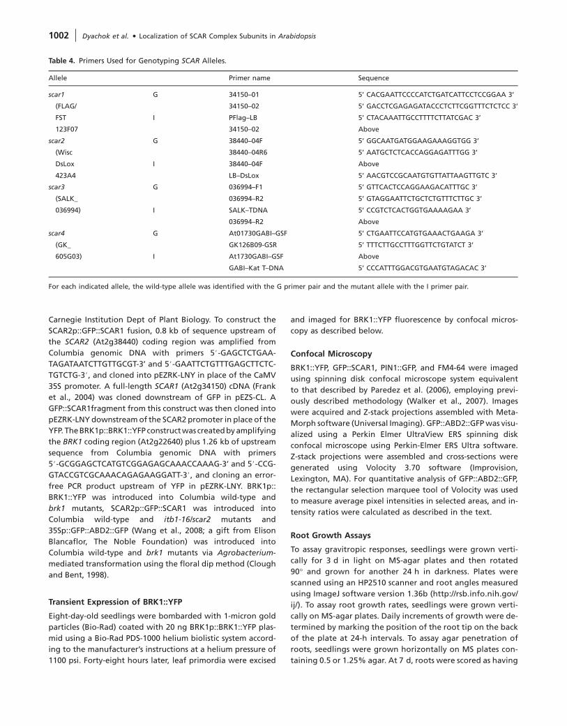
Carnegie Institution Dept of Plant Biology. To construct the
SCAR2p::GFP::SCAR1 fusion, 0.8 kb of sequence upstream of
the SCAR2 (At2g38440) coding region was amplified from
Columbia genomic DNA with primers 5#-GAGCTCTGAA-
TAGATAATCTTGTTGCGT-3’ and 5#-GAATTCTGTTTGAGCTTCTC-
TGTCTG-3#, and cloned into pEZRK-LNY in place of the CaMV
35S promoter. A full-length SCAR1 (At2g34150) cDNA (Frank
et al., 2004) was cloned downstream of GFP in pEZS-CL. A
GFP::SCAR1fragment from this construct was then cloned into
pEZRK-LNY downstream of the SCAR2 promoter in place of the
YFP. The BRK1p::BRK1::YFP construct was createdby amplifying
the BRK1 coding region (At2g22640) plus 1.26 kb of upstream
sequence from Columbia genomic DNA with primers
5#-GCGGAGCTCATGTCGGAGAGCAAACCAAAG-3’ and 5#-CCG-
GTACCGTCGCAAACAGAGAAGGATT-3#, and cloning an error-
free PCR product upstream of YFP in pEZRK-LNY. BRK1p::
BRK1::YFP was introduced into Columbia wild-type and
brk1 mutants, SCAR2p::GFP::SCAR1 was introduced into
Columbia wild-type and itb1-16/scar2 mutants and
35Sp::GFP::ABD2::GFP (Wang et al., 2008; a gift from Elison
Blancaflor, The Noble Foundation) was introduced into
Columbia wild-type and brk1 mutants via Agrobacterium-
mediated transformation using the floral dip method (Clough
and Bent, 1998).
Transient Expression of BRK1::YFP
Eight-day-old seedlings were bombarded with 1-micron gold
particles (Bio-Rad) coated with 20 ng BRK1p::BRK1::YFP plas-
mid using a Bio-Rad PDS-1000 helium biolistic system accord-
ing to the manufacturer’s instructions at a helium pressure of
1100 psi. Forty-eight hours later, leaf primordia were excised
and imaged for BRK1::YFP fluorescence by confocal micros-
copy as described below.
Confocal Microscopy
BRK1::YFP, GFP::SCAR1, PIN1::GFP, and FM4-64 were imaged
using spinning disk confocal microscope system equivalent
to that described by Paredez et al. (2006), employing previ-
ously described methodology (Walker et al., 2007). Images
were acquired and Z-stack projections assembled with Meta-
Morph software (Universal Imaging). GFP::ABD2::GFP was visu-
alized using a Perkin Elmer UltraView ERS spinning disk
confocal microscope using Perkin-Elmer ERS Ultra software.
Z-stack projections were assembled and cross-sections were
generated using Volocity 3.70 software (Improvision,
Lexington, MA). For quantitative analysis of GFP::ABD2::GFP,
the rectangular selection marquee tool of Volocity was used
to measure average pixel intensities in selected areas, and in-
tensity ratios were calculated as described in the text.
Root Growth Assays
To assay gravitropic responses, seedlings were grown verti-
cally for 3 d in light on MS-agar plates and then rotated
90� and grown for another 24 h in darkness. Plates were
scanned using an HP2510 scanner and root angles measured
using ImageJ software version 1.36b (http://rsb.info.nih.gov/
ij/). To assay root growth rates, seedlings were grown verti-
cally on MS-agar plates. Daily increments of growth were de-
termined by marking the position of the root tip on the back
of the plate at 24-h intervals. To assay agar penetration of
roots, seedlings were grown horizontally on MS plates con-
taining 0.5 or 1.25% agar. At 7 d, roots were scored as having
Table 4. Primers Used for Genotyping SCAR Alleles.
Allele Primer name Sequence
scar1 G 34150–01 5’ CACGAATTCCCCATCTGATCATTCCTCCGGAA 3’
(FLAG/ 34150–02 5’ GACCTCGAGAGATACCCTCTTCGGTTTCTCTCC 3’
FST I PFlag–LB 5’ CTACAAATTGCCTTTTCTTATCGAC 3’
123F07 34150–02 Above
scar2 G 38440–04F 5’ GGCAATGATGGAAGAAAGGTGG 3’
(Wisc 38440–04R6 5’ AATGCTCTCACCAGGAGATTTGG 3’
DsLox I 38440–04F Above
423A4 LB–DsLox 5’ AACGTCCGCAATGTGTTATTAAGTTGTC 3’
scar3 G 036994–F1 5’ GTTCACTCCAGGAAGACATTTGC 3’
(SALK_ 036994–R2 5’ GTAGGAATTCTGCTCTGTTTCTTGC 3’
036994) I SALK–TDNA 5’ CCGTCTCACTGGTGAAAAGAA 3’
036994–R2 Above
scar4 G At01730GABI–GSF 5’ CTGAATTCCATGTGAAACTGAAGA 3’
(GK_ GK126B09-GSR 5’ TTTCTTGCCTTTGGTTCTGTATCT 3’
605G03) I At1730GABI–GSF Above
GABI–Kat T–DNA 5’ CCCATTTGGACGTGAATGTAGACAC 3’
For each indicated allele, the wild-type allele was identified with the G primer pair and the mutant allele with the I primer pair.
1002 | Dyachok et al. d Localization of SCAR Complex Subunits in Arabidopsis

penetrated the agar if they had made contact with the bot-
tom of the plate.
BFA Treatment
Three-day-old PIN1p::PIN1::GFP and BRK1p::BRK1::YFP seedlings
grown on MS agar-plates were transferred onto Whatman
filter paper soaked with either 50 micromolar BFA (Sigma-
Aldrich, St Louis, MO) in 0.28% DMSO or with DMSO only
as a control. After 30 min, the seedlings were mounted onto
slides and the fluorescent fusion proteins were imaged via con-
focal microscopy as described above.
Plasmolysis
Three-day-old seedlings expressing BRK1p::BRK1::YFP were
mounted in water on slides and examined for YFP fluorescence
via confocal microscopy as described above. Water was then
carefully substituted by 5 micrograms ml�1 FM4-64 (Molecular
Probes/Invitrogen,Carlsbad,CA) in0.5 Msucrose,androotswere
immediately imaged again for YFP and FM4-64 fluorescence.
Whole Mount Immunofluorescence
Actin immunolocalization in roots of 4-day-old seedlings was
carried out as described by Rahman et al. (2007) with minor
modifications (fixation was for 1 h in 50 mM PIPES, pH 7.2,
with 20 mM EGTA and 20 mM MgSO4 containing 2% parafor-
maldehyde, 0.1% Triton X-100 and 400 mm Maleimidobenzoyl-
N-hydroxysuccinimide ester (Pierce/ThermoFisher Scientific,
Rockford, IL), and wall digestion was for 20 min in 0.05%
Pectolyase Y-23). To localize SCAR1, we used the protocol of
Sugimoto et al. (2000), except that the fixative contained
2% formaldehyde and no glutaraldehyde, and sodium borohy-
dride treatment was eliminated. Affinity-purified chicken anti-
SCAR1 (Djakovic et al., 2006) was used at 5 micrograms ml�1
and Genway Biotech (San Diego, CA) FITC-anti-chicken IgY
at 1:200. Samples were mounted in Vectashield (Vector Labo-
ratories, Burlingame, CA) and root tips imaged using a Leica
TCS SP2 AOBS confocal laser scanning microscope as described
previously (Wang et al., 2008).
Transmission Electron Microscopy
Ultrastructural analysis was carried out as described by
Meloche et al. (2007) except that seedlings were fixed initially
for 1 h on agar plates by addition of 3% glutaraldehyde/
0.05 M PIPES buffer (pH 7.4) directly to the plate and then
in scintillation vials with fresh fixative solution (same as above)
for another hour. For immunocytochemistry, tissue was em-
bedded in LR White and processed as described by Meloche
et al. (2007). A minimum of four grids were examined for each
antibody treatment.
Microsome Preparation and Analysis of Membrane
Association
Approximately 0.5 g of 14-day-old seedlings were homoge-
nized in 1 ml TBS with 1 mM DTT, 10% sucrose and 1/100 plant
protease inhibitor cocktail (Sigma) using an Omni TH homog-
enizer on ice. Supernantants following centrifugation at
16 000 g for 10 min were then centrifuged at 150 000 g for
40 min at 4�C to obtain a microsomal fraction. Analysis of con-
ditions required for extraction of SCAR1 and BRK1::YFP from
the microsomal was carried out as described by Boonsirichai
et al. (2003). Briefly, the microsomal fractions were re-
suspended in the buffer described above supplemented with
2 M NaCl, 0.1 M Na2CO3, or 3% Triton X-100, incubated for
40 min at room temperature and centrifuged at 200 000 g
for 60 min at 4�C. Supernatants were concentrated by precip-
itation using trichloroacetic acid.
Western Blot Analyses
Gel electrophoresis and Western blotting with anti-SCAR1
were carried out as previously described (Djakovic et al.,
2006). For detection of BRK1::YFP, mouse anti-GFP (Zymed/
Invitrogen, Carlsbad, CA) was used at 0.05 micrograms ml�1
followed by alkaline phosphatase-conjugated goat anti-
mouse IgG (Promega, Madison, WI) diluted 1:10 000. Anti-a-
1,2-Mannosidase I (Preuss et al., 2004) and anti-PUX1 (Rancour
et al., 2004) were used to detect membrane and cytoplasmic
proteins, respectively. Both antibodies were a gift from David
Rancour, University of Wisconsin.
Analysis of Wall Composition
Neutral monosaccharides were measured by gas chromatogra-
phy using a modification of the method of Blakeney et al.
(1983), as follows. Roots were excised from 5-day-old seedlings
and extracted in 10–12 changes of 1:1 chloroform:methanol
over 4 d. The roots were pulverized using a ball mill and ino-
sitol was added as an internal standard to approximately
400 mg of the resulting residue. Roots were hydrolyzed in
2 M trifluoracetic acid in an autoclave for 1 h, and the result-
ing monosaccharides were derivatized to their alditol acetates,
which were separated and measured using a Agilent 6890N
gas chromatograph equipped with a Supelco SP2330 capillary
column (30 m 3 0.25 mm, 0.2 micron film thickness).
FUNDING
This work was supported by NSF grant IOB–0544226 to L.G.S.
ACKNOWLEDGMENTS
Thanks to members of the Smith laboratory for helpful discussions
and comments on the manuscript, to James Ingham and Michael
Burke for help with construction of scar 1,2,3,4 quadruple mutants,
to Elison Blancaflor for invaluable input and support of this work,
and to ABRC and GABI-Kat for mutants. No conflict of interest de-
clared.
REFERENCES
Baluska, F., Hlavacka, A., Samaj, J., Palme, K., Robinson, D.G.,
Matoh, T., McCurdy, D.W., Menzel, D., and Volkmann, D.
Dyachok et al. d Localization of SCAR Complex Subunits in Arabidopsis | 1003
(2002). F-actin-dependent endocytosis of cell wall pectins in mer-
istematic root cells: insights from brefeldin A-induced compart-
ments. Plant Physiol. 130, 422–431.
Basu, D., El-Assal Sel, D., Le, J., Mallery, E.L., and Szymanski, D.B.
(2004). Interchangeable functions of Arabidopsis PIROGI and the
human WAVE complex subunit SRA1 during leaf epidermal de-
velopment. Development. 131, 4345–4355.
Basu, D., Le, J., El-Assal Sel, D., Huang, C., Zhang, C., Mallery, E.L.,
Koliantz, G., Staiger, C.J., and Szymanski, D.B. (2005). DIS-
TORTED3/SCAR2 is a putative Arabidopsis WAVE complex sub-
unit that activates the Arp2/3 complex and is required for
epidermal morphogenesis. Plant Cell. 17, 502–524.
Basu, D., Le, J., Zakharova, T., Mallery, E.L., and Szymanski, D.B.
(2008). A SPIKE1 signaling complex controls actin-dependent cell
morphogenesis through the heteromeric WAVE and ARP2/3
complexes. Proc. Natl Acad. Sci. U S A. 105, 4044–4049.
Blakeney, A.B., Harris, P.J., Henry, R.J., and Stone, B.A. (1983). A sim-
ple and rapid preparation of alditol acetates for monosaccharide
analysis. Carbohydrate Res. 113, 291–299.
Blancaflor, E.B., Wang, Y.S., and Motes, C.M. (2006). Organization
and function of the actin cytoskeleton in developing root cells.
Int. Rev. Cytol. 252, 219–264.
Boonsirichai, K., Sedbrook, J.C., Chen, R., Gilroy, S., and
Masson, P.H. (2003). ALTERED RESPONSE TO GRAVITY is a periph-
eral membrane protein that modulates gravity-induced cyto-
plasmic alkanilization and lateral auxin transport in plant
statocytes. Plant Cell. 15, 2612–2625.
Brembu, T., Winge, P., Seem, M., and Bones, A.M. (2004). NAPP and
PIRP encode subunits of a putative WAVE regulatory protein
complex involved in plant cell morphogenesis. Plant Cell. 16,
2335–2349.
Chen, T., Teng, N., Wu, X., Wang, Y., Tang, W., Samaj, J., Baluska, F.,
and Lin, J. (2007). Disruption of actin filaments by Latrunculin B
affects cell wall construction in Picea meyeri pollen tube by dis-
turbing vesicle trafficking. Plant Cell Physiol. 48, 19–30.
Clausen, M.H., Willats, W.G.T., and Knox, J.P. (2003). Synthetic
methyl hexagalacturonate hapten inhibitors of anti-homogalac-
turonan monoclonal antibodies LM7, JIM5 and JIM7. Carbohydr.
Res. 338, 1797–1800.
Clough, S.J., and Bent, A.F. (1998). Floral dip: a simplified method
for Agrobacterium-mediated transformation of Arabidopsis
thaliana. Plant J. 16, 735–743.
Deeks, M.J., Kaloriti, D., Davies, B., Malho, R., and Hussey, P.J.
(2004). Arabidopsis NAP1 is essential for Arp2/3-dependent tri-
chome morphogenesis. Curr. Biol. 14, 1410–1414.
Deeks, M.J., Rodrigues, C., Dimmock, S., Ketelaar, T., Maciver, S.K.,
Malho, R., and Hussey, P.J. (2007). Arabidopsis CAP1: a key reg-
ulator of actin organisation and development. J. Cell Sci. 120,
2609–2618.
Dhonukshe, P., Baluska, F., Schlicht, M., Hlavacka, A., Samaj, J.,
Friml, J., and Gadella, T.W., Jr (2006). Endocytosis of cell surface
material mediates cell plate formation during plant cytokinesis.
Dev. Cell. 10, 137–150.
Djakovic, S., Dyachok, J., Burke, M., Frank, M., and Smith, L.G.
(2006). ARP2/3 complex-dependent and -independent functions
for BRICK1 in the spatial regulation of epidermal cell morpho-
genesis in Arabidopsis. Development. 133, 1091–1100.
Eden, S., Rohtagi, R., Podtelejnikov, A.V., Mann, M., and
Kirschner, M. (2002). Mechanism of regulation of WAVE1-
induced actin nucleation by Rac1 and Nck. Nature. 418,
790–793.
El-Assal, S.D., Le, J., Basu, D., Mallery, E.L., and Szymanski, D.B.
(2004). Arabidopsis GNARLED encodes a NAP125 homolog that
positively regulates ARP2/3. Curr. Biol. 14, 1405–1409.
Finka, A., Schaefer, D.G., Saidi, Y., Goloubinoff, P., and Zyrd, J.P.
(2007). In vivo visualization of F-actin structures during the de-
velopment of the moss Physcomitrella patens. New Phytol. 174,
63–76.
Foissner, I., Lichtscheidl, I.K., and Wasteneys, G.O. (1996). Actin-
based vesicle dynamics and exocytosis during wound wall forma-
tion in Characean internodal cells. Cell Motil. Cytoskel. 35, 35–48.
Frank, M., Egile, C., Dyachok, J., Djakovic, S., Nolasco, M., Li, R., and
Smith, L.G. (2004). Activation of Arp2/3 complex-dependent ac-
tin polymerization by plant proteins distantly related to Scar/
WAVE. Proc. Natl Acad. Sci. U S A. 101, 16379–16384.
Frank, M.J., and Smith, L.G. (2002). A small, novel protein highly
conserved in plants and animals promotes the polarized growth
and division of maize leaf epidermal cells. Curr. Biol. 12, 849–853.
Frank, M.J., Cartwright, H.N., and Smith, L.G. (2003). Three brick
genes have distinct functions in a common pathway promoting
polarized cell division and cell morphogenesis in the maize leaf
epidermis. Development. 130, 753–762.
Freshour, G., Clay, R.P., Fuller, M.S., Albersheim, P., Darvill, A.G., and
Hahn, M.G. (1996). Development and tissue-specific structural
alterations of cell wall polysaccharides of the Arabidopsis thali-
ana root. Plant Physiol. 110, 1413–1429.
Fu, Y., Li, H., and Yang, Z. (2002). The ROP2 GTPase controls the for-
mation of cortical fine F-actin and the early phase of directional
cell expansion during Arabidopsis organogenesis. Plant Cell. 14,
777–794.
Gautreau, A., Ho, H.Y., Steen, H., Gygi, S.P., and Kirschner, M.W.
(2004). Purification and architecture of the ubiquitous WAVE
complex. Proc. Natl Acad. Sci. U S A. 101, 4379–4383.
Geldner, N., Friml, J., Stierhof, Y.D., Jurgens, G., and Palme, K.
(2001). Auxin transport inhibitors block PIN1 cycling and vesicle
trafficking. Nature. 413, 425–428.
Goley, E.D., and Welch, M.D. (2006). The ARP2/3 complex: an actin
nucleator comes of age. Nature Rev. Mol. Cell Biol. 7, 713–726.
Hable, W.E., and Kropf, D.L. (2005). The Arp2/3 complex nucleates
actin arrays during zygote polarity establishment and growth.
Cell Motil. Cytoskeleton. 61, 9–20.
Harries, P.A., Pan, A., and Quatrano, R.S. (2005). Arp2/3 complex
component ARPC1 is required for proper cell morphogenesis
and polarized cell growth in Physcomitrella patens. Plant Cell.
17, 2327–2339.
Heisler, M.G., Ohno, C., Das, P., Sieber, P., Reddy, G.V., Long, J.A.,
and Meyerowitz, E.M. (2005). Patterns of auxin transport and
gene expression during primordium development revealed by
live imaging of the Arabidopsis inflorescence meristem. Curr.
Biol. 15, 1899–1911.
Helling, D., Possart, A., Cottier, S., Klahre, U., and Kost, B. (2006).
Pollen tube tip growth depends on plasma membrane polariza-
tion mediated by tobacco PLC3 activity and endocytic membrane
recycling. Plant Cell. 18, 3519–3534.
1004 | Dyachok et al. d Localization of SCAR Complex Subunits in Arabidopsis
Hussey, P.J., Ketelaar, T., and Deeks, M.J. (2006). Control of the actin
cytoskeleton in plant cell growth. Annu. Rev. Plant Biol. 57,
109–125.
Innocenti, M., et al. (2004). Abi1 is essential for the formation and
activation of a WAVE2 signaling complex mediating Rac-depen-
dent actin remodeling. Nature Cell Biol. 6, 319–327.
Jones, M.A., Shen, J.-J., Fu, Y., Yang, Z., and Grierson, C.S. (2002).
The Arabidopsis Rop2 GTPase is a positive regulator of both root
hair initiation and tip growth. Plant Cell. 14, 763–776.
Kaksonen, M., Toret, C.P., and Drubin, D.G. (2006). Harnessing actin
dynamics for clathrin-mediated endocytosis. Nature Rev. Mol.
Cell Biol. 7, 404–414.
Kang, B.H., Busse, J.S., and Bednarek, S.Y. (2003). Members of the
Arabidopsis dynamin-like gene family, ADL1, are essential for
plant cytokinesis and polarized cell growth. Plant Cell. 15,
899–913.
Kim, S., Shilagardi, K., Zhang, S., Hong, S.N., Sens, K.L., Bo, J.,
Gonzalez, G.A., and Chen, E.H. (2007). A critical function for
the actin cytoskeleton in targeted exocytosis of prefusion
vesicles during myoblast fusion. Dev. Cell. 12, 571–586.
Kunda, P., Craig, G., Dominguez, V., and Baum, B. (2003). Abi, Sra1,
and Kette control the stability and localization of Scar/WAVE to
regulate the formation of actin-based protrusions. Curr. Biol. 13,
1867–1875.
Le, J., El-Assal Sel, D., Basu, D., Saad, M.E., and Szymanski, D.B.
(2003). Requirements for Arabidopsis ATARP2 and ATARP3 dur-
ing epidermal development. Curr. Biol. 13, 1341–1347.
Le, J., Mallery, E.L., Zhang, C., Brankle, S., and Szymanski, D.B.
(2006). Arabidopsis BRICK1/HSPC300 is an essential WAVE-
complex subunit that selectively stabilizes the Arp2/3 activator
SCAR2. Curr Biol. 16, 895–901.
Li, S., Blanchoin, L., Yang, Z., and Lord, E.M. (2003). The putative
Arabidopsis Arp2/3 complex controls leaf cell morphogenesis.
Plant Physiol. 132, 2034–2044.
Machesky, L.M., Reeves, E., Wientjes, F., Mattheyse, F.J., Grogan, A.,
Totty, N.F., Burlingame, A.L., Hsuan, J.J., and Segal, A.W. (1997).
Mammalian actin-related protein 2/3 complex localizes to
regions of lamellipodial protrusion and is composed of evolu-
tionarily conserved proteins. Biochem. J. 328, 105–112.
Mathur, J. (2004). Cell shape development in plants. Trends Plant
Sci. 12, 583–590.
Mathur, J. (2006). Local interaction shape plant cells. Curr. Opin. Cell
Biol. 18, 40–46.
Mathur, J., Mathur, N., Kernebeck, B., and Hulskamp, M. (2003b).
Mutations in actin-related proteins 2 and 3 affect cell shape de-
velopment in Arabidopsis. Plant Cell. 15, 1632–1645.
Mathur, J., Mathur, N., Kirik, V., Kernebeck, B., Srinivas, B.P., and
Hulskamp, M. (2003a). Arabidopsis CROOKED encodes for the
smallest subunit of the ARP2/3 complex and controls cell shape
by region specific fine F-actin formation. Development. 130,
3137–3146.
Meloche, C.G., Knox, J.P., and Vaughn, K.C. (2007). A cortical band
of gelatinous fibers causes the coiling of redvine tendrils: a model
based upon cytochemical and immunocytochemical studies.
Planta. 225, 485–498.
Merrifield, C.J., Perrais, D., and Zenisek, D. (2005). Coupling
between clathrin-coated-pit invagination, cortactin recruit-
ment, and membrane scission observed in live cells. Cell. 121,
593–606.
Merrifield, C.J., Qualmann, B., Kessels, M.M., and Almers, W.
(2004). Neural Wiskott–Aldrich syndrome protein (N-WASP)
and the Arp2/3 complex are recruited to sites of clathrin-
mediated endocytosis in cultured fibroblasts. Eur. J. Cell Biol.
83, 13–18.
Miki, H., Suetsugu, S., and Takenawa, T. (1998). WAVE, a novel
WASP-family protein involved in actin reorganization induced
by Rac. EMBO J. 17, 6932–6941.
Molendijk, A.J., Bischoff, F., Rajendrakumar, C.S.V., Friml, J.,
Braun, M., Gilroy, S., and Palme, K. (2001). Arabidopsis thaliana
Rop GTPases are localized to tips of root hairs and control polar
growth. EMBO J. 11, 2779–2788.
Nozumi, M., Nakagawa, H., Miki, H., Takenawa, T., and
Miyamoto, S. (2003). Differential localization of WAVE isoforms
in filopodia and lamellipodia of the neuronal growth cone.
J. Cell Sci. 116, 239–246.
Ovecka, M., Lang, I., Baluska, F., Ismail, A., Illes, P., and
Lichtscheidl, I.K. (2005). Endocytosis and vesicle trafficking dur-
ing tip growth of root hairs. Protoplasma. 226, 39–54.
Paredez, A.R., Somerville, C.R., and Ehrhardt, D.W. (2006). Visuali-
zation of cellulose synthase demonstrates functional association
with microtubules. Science. 312, 1491–1495.
Perroud, P.F., and Quatrano, R.S. (2006). The role of ARPC4 in tip
growth and alignment of the polar axis in filaments of Physco-
mitrella patens. Cell Motil. Cytoskeleton. 63, 162–171.
Perroud, P.F., and Quatrano, R.S. (2008). BRICK1 is required for api-
cal cell growth in filaments of the moss Physcomitrella patens
but not for gametophore morphology. Plant Cell. 20, 411–422.
Preuss, M.L., Serna, J., Falbel, T.G., Bednarek, S.Y., and Nielsen, E.
(2004). The Arabidopsis Rab GTPase RabA4b localizes to the tips
of growing root hair cells. Plant Cell. 16, 1589–1603.
Rahman, A., Bannigan, A., Sulaman, W., Pechter, P., Blancaflor, E.B.,
and Baskin, T.I. (2007). Auxin, actin, and growth of theArabidop-
sis thaliana primary root. Plant J. 50, 514–528.
Rancour, D.M., Park, S., Knight, S.D., and Bednarek, S.Y. (2004).
Plant UBX domain-containing protein 1, PUX1, regulates the
oligomeric structure and activity of Arabidopsis CDC48. J. Biol.
Chem. 279, 54264–54274.
Rogers, S.L., Wiedemann, U., Stuurman, N., and Vale, R.D. (2003).
Molecular requirements for actin-based lamella formation in
Drosophila S2 cells. J. Cell Biol. 162, 1079–1088.
Royle, S.J., and Murrell-Lagnado, R.D. (2003). Constitutive cycling:
a general mechanism to regulate cell surface proteins. Bioessays.
25, 39–46.
Schwab, B., Mathur, J., Saedler, R., Schwarz, H., and Frey, B. (2003).
Regulation of cell expansion by the DISTORTED genes in
Arabidopsis thaliana: actin controls the spatial organization of
microtubules. Mol. Genet. Genomics. 269, 350–360.
Smith, L.G., and Oppenheimer, D.G. (2005). Spatial control of cell
expansion by the plant cytoskeleton. Annu. Rev. Cell Dev. Biol.
21, 271–295.
Stovold, C.F., Millard, T.H., and Machesky, L.M. (2005). Inclusion of
Scar/WAVE3 in a similar complex to Scar/WAVE1 and 2. BMC Cell
Biol. 6, 11.
Dyachok et al. d Localization of SCAR Complex Subunits in Arabidopsis | 1005
Stradal, T.E.B., and Scita, G. (2005). Protein complexes regulating
Arp2/3-mediated actin assembly. Curr. Op. Cell Biol. 18, 4–10.
Sugimoto, K., Williamson, R.E., and Wasteneys, G.O. (2000). New
techniques enable comparative analysis of microtubule orienta-
tion, wall texture, and growth rate in intact roots of Arabidopsis.
Plant Physiol. 124, 1493–1506.
Sun, Y., Martin, A., and Drubin, D.G. (2006). Endocytic internaliza-
tion in budding yeast requires coordinated actin nucleation and
myosin motor activity. Dev. Cell. 11, 33–46.
Szymanski, D.B. (2005). Breaking the WAVE complex: the point of
Arabidopsis trichomes. Curr. Opin. Plant Biol. 8, 103–112.
Takenawa, T., and Suetsugu, S. (2007). The WASP–WAVE protein
network: connecting the membrane to the cytoskeleton. Nature
Rev. Mol. Cell Biol. 8, 37–48.
Uhrig, J.F., Mutondo, M., Zimmerman, I., Deeks, M.,
Machesky, L.M., Thomas, P., Uhrig, S., Hussey, P., and
Hulskamp, M. (2007). The role of Arabidopsis genes in ARP2-
ARP3-dependent cell morphogenesis. Development. 134,
967–977.
Van Gestel, K., Slegers, H., Von Witsch, M., Samaj, J., Baluska, F.,
and Verbelen, J.P. (2003). Immunological evidence for the pres-
ence of plant homologues of the actin-related protein Arp3 in
tobacco and maize: subcellular localization to actin-enriched
pit fields and emerging root hairs. Protoplasma. 222, 45–52.
Vissenberg, K., Fry, S.C., Pauly, M., Hofte, H., and Verbelen, J.-P.
(2005). XTH acts at the microfibril–matrix interface during cell
elongation. J. Exp. Bot. 56, 673–683.
Walker, K.L., Muller, S., Moss, D., Ehrhardt, D.W., and Smith, L.G.
(2007). Arabidopsis TANGLED identifies the division plane
throughout mitosis and cytokinesis. Curr. Biol. 17, 1827–1836.
Wang, Y.-S., Yoo, C.-M., and Blancaflor, E.B. (2008). Improved imag-
ing of actin filaments in transgenicArabidopsis plants expressing
a green fluorescent protein fusion to the C- and N-termini of the
fimbrin actin-binding domain 2. New Phytol. 177, 525–536.
Weiner, O.D., Servant, G., Welch, M.D., Mitchison, T.J., Sedat, J.W.,
and Bourne, H.R. (1999). Spatial control of actin polymerization
during neutrophil chemotaxis. Nature Cell Biol. 1, 75–81.
Welch, M.D., DePace, A.H., Verma, S., Iwamatsu, A., and
Mitchison, T.J. (1997). The human Arp2/3 complex is composed
of evolutionarily conserved subunits and is localized to cellular
regions of dynamic actin filament assembly. J. Cell Biol. 138,
375–384.
Willats, W.G.T., Marcus, S.E., and Knox, J.P. (1998). Generation of
a monoclonal antibody specific to (1/5) a-L-arabinan. Carbohy-
drate Res. 308, 149–152.
Zhang, C., Mallery, E.L., Schlueter, J., Huang, S., Fan, Y., Brankle, S.,
Staiger, C.J., and Szymanski, D.B. (2008).Arabidopsis SCARs func-
tion interchangeably to meet actin-related protein 2/3 activation
thresholds during morphogenesis. Plant Cell. 20, 995–1011.
Zhang, X., Dyachok, J., Krishnakumar, S., Smith, L.G., and
Oppenheimer, D.G. (2005). IRREGULAR TRICHOME BRANCH1
in Arabidopsis encodes a plant homolog of the actin related pro-
tein2/3 complex activator Scar/WAVE that regulates actin and mi-
crotubule organization. Plant Cell. 17, 2314–2326.
1006 | Dyachok et al. d Localization of SCAR Complex Subunits in Arabidopsis