plasmoblastic lymphoma associated with human ... · plasmoblastic lymphoma associated with human...
TRANSCRIPT

Romanian Journal of Morphology and Embryology 2008, 49(3):309–314
CCAASSEE RREEPPOORRTT
Plasmoblastic lymphoma associated with Human Immunodeficiency Virus EMŐKE HORVÁTH1), L. KRENÁCS2), ENIKŐ BAGDI2), Z. PÁVAI1),
I. MACARIE3), ELŐD-ERNŐ NAGY4), SMARANDA DEMIAN3)
1)Department of Pathology, University of Medicine and Pharmacy of Targu Mures, Romania
2)Laboratory of Tumor Pathology and Molecular Diagnostics, “Bay Zoltán” Foundation for Applied Research, Szeged, Hungary
3)Ist Internal Medicine Clinics 4)Pharmaceutical Biochemistry
University of Medicine and Pharmacy of Targu Mures, Romania
Abstract Plasmoblastic lymphoma (PBL) is a subtype of the diffuse large B-cell lymphoma, typically present as extranodal disease associated with human immune deficiency virus (HIV) infection. PBLs are often the initial manifestation of AIDS. Here we present a case of PBL concerning the oral cavity. A 34-year-old woman presented a tumor in the oral cavity that involved the maxilla and gingiva (confirmed by CT-scan). The gingival biopsy showed a massive infiltration by large lymphoid cells with round, vesicular nuclei, prominent nucleoli, fine chromatin and an significant amount of basophilic cytoplasm which express CD79a, CD138, cytoplasmic lambda light chain and LCA, without staining for CD20, CD38, CD3 and CTK. Serological analysis confirmed HIV positivity. PBLs lack most B-lineage markers, but many express CD79a in at least some of the cells, therefore generate difficulties in differential diagnosis. Overall assessment and correlation of the histopathological and immunohistochemical features with the clinical findings and serology investigation are the most helpful diagnostic tools and can lead to the final diagnosis. Keywords: extranodal PBL, immunohistochemistry, B-lineage markers, HIV.
Introduction
Patients with HIV infection are at a high risk for developing malignant lymphoma. It has been estimated that approximately 3% of AIDS patients develop non-Hodgkin’s lymphoma, and the risk of developing a lymphoma in this population is 60-fold greater than in the normal population. The majority of the cases present with sites of extranodal involvement are often the initial manifestations of AIDS, being a distinct entity with an aggressive presentation and a poor prognosis. The prognosis with the combination treatment anti-HIV and chemotherapy of the hematological disease in the seropositive patients’ case is similar to the one of seronegative patients.
The aim of this study is to present the clinicopathological features of a PBLs case concerning the oral cavity and discusses the histomorphological features in the mirror of an extensive immunohistochemistry panel, which helps us in setting the final diagnosis. All of the examinations presented above have been performed in the possession of the patient’s written consent.
Patient and methods
Our patient presented to her primary care physician in 2006 with fatigue, weight loss (20 kg in three months) night sweats and with a large tumor in the oral
cavity. The physical examination describes the swelling of the upper lip and left jaw region extending to the palatal area, with a 3.5/3 cm vegetant tumor mass, having a grown consistency. It was intensely hyperemic and not bleeding. The axillary and latero-cervical lymph nodes were bilaterally increased in their size, not aching. The liver overpasses costal edge with 3 cm in the medio-clavicular line. The spleen was palpable.
Number of the patient’s blood cell translated a moderate anemia with thrombocytopenia (hemoglobin 11.8 g/dL, platelet number 130000/cm3, white blood cell count 3090/cm3 with 2000/cm3 neutrophils), blood and bone marrow smear did not show remarkable modifications (small numbers of nucleated red cells, some of which showed dyserythropoetic features, 23% lymphocytes with normal aspect, granulopoiesis was active, with evidence of cell maturation, and number of the blast cells was not increased), ESR was 75/30 mm, serum LDH 400 U/l, other biochemical analyses had been confined to normal ranges. No abnormal protein was detected by serum or urine analysis. Our patient had negative tests for B and C hepatitis antigens and had positive anti-VCA IgG antibody titers.
The tumor mass was visualized by CT followed by performing the biopsy from the tumor mass. Following the histological diagnosis of PBL, a comprehensive metastatic workup was performed, including chest
Emőke Horváth et al.
310
X-ray, abdominal and pelvic CT-scan, bone marrow aspirate and biopsy, revealing evidence of disease in latero-cervical, retroperitoneal lymph nodes and bone marrow.
The tumor piece was represented from one biopsy fragment, fixed in 10% buffered formalin and embedded in paraffin. Sections were stained with Hematoxylin and Eosin, periodic acid Schiff and Giemsa for the routine histopathological evaluation.
Immunohistochemical stains was performed with LabVision and DAKO reagents, using: CD20cy (DAKO clone L26, dilution 1/200), CD138 (DAKO clone MI15, dilution 1/50), CD38 (LabVision, clone AT1, dilution 1/40), IRF4 (DAKO clone MUM1p, dilution 1/50), CD79a (DAKO clone JCB117, dilution 1/50), BCL-6 (DAKO clone PG-B6p, dilution 1/20), CD3 (LabVision, clone SP7, dilution 1/150), Ki67 (LabVision, clone SP6, dilution 1/200), LCA (DAKO clone 2B11, dilution 1/200), panCTK (LabVision, clone A1/A3, dilution 1/150), EB LAMP1 (DAKO clone CS.1–4, dilution 1/50), Ig M (LabVision, polyclonal dilution 1/1000), lambda light chain (LabVision, clone N/10/2, dilution 1/25), IgG (LabVision, polyclonal dilution 1/1000), TdT (LabVision, clone Ab-2, dilution 1/5), and CD68 (DAKO clone PG-M1, dilution 1/75).
Bound antibodies were visualized using heat induced epitope retrieval method by the Ultra Vision LP Large Volume Detection System HRP Polymer detection system and DAKO En VisionTM/HRP kit, followed by Hematoxylin counterstaining. For positive controls, sections from reactive lymph nodes and tonsils were tested parallel. For negative controls, the primary antibodies were omitted.
The bone marrow biopsy obtained from the right posterior iliac crest, was fixed in 10% buffered formalin and briefly decalcified in EDTA.
We did not evaluate the molecular aspect of this tumor, because the molecular biology studies may not resolve the differential diagnostic problems, because the cytogenetic aspect is not characteristic.
Results
Pathology findings Gross inspection of the gingival biopsy specimen
revealed a 15/10 mm tumor. The tumor tissue was consistent with grayish color and was covered with ulcerated mucosa.
Microscopic examination
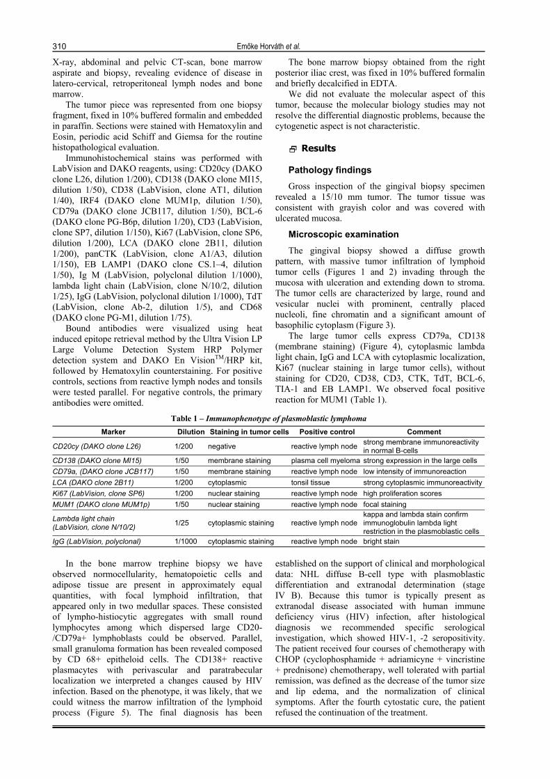
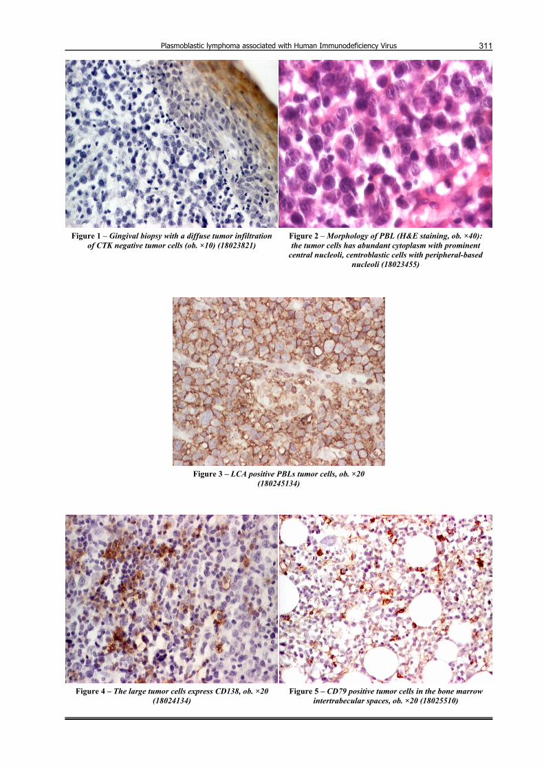
The gingival biopsy showed a diffuse growth pattern, with massive tumor infiltration of lymphoid tumor cells (Figures 1 and 2) invading through the mucosa with ulceration and extending down to stroma. The tumor cells are characterized by large, round and vesicular nuclei with prominent, centrally placed nucleoli, fine chromatin and a significant amount of basophilic cytoplasm (Figure 3).
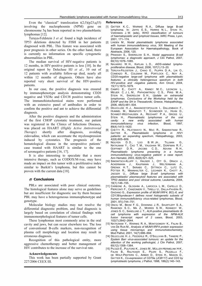
The large tumor cells express CD79a, CD138 (membrane staining) (Figure 4), cytoplasmic lambda light chain, IgG and LCA with cytoplasmic localization, Ki67 (nuclear staining in large tumor cells), without staining for CD20, CD38, CD3, CTK, TdT, BCL-6, TIA-1 and EB LAMP1. We observed focal positive reaction for MUM1 (Table 1).
Table 1 – Immunophenotype of plasmoblastic lymphoma Marker Dilution Staining in tumor cells Positive control Comment
CD20cy (DAKO clone L26) 1/200 negative reactive lymph node strong membrane immunoreactivity in normal B-cells
CD138 (DAKO clone MI15) 1/50 membrane staining plasma cell myeloma strong expression in the large cells CD79a, (DAKO clone JCB117) 1/50 membrane staining reactive lymph node low intensity of immunoreaction LCA (DAKO clone 2B11) 1/200 cytoplasmic tonsil tissue strong cytoplasmic immunoreactivityKi67 (LabVision, clone SP6) 1/200 nuclear staining reactive lymph node high proliferation scores MUM1 (DAKO clone MUM1p) 1/50 nuclear staining reactive lymph node focal staining
Lambda light chain (LabVision, clone N/10/2) 1/25 cytoplasmic staining reactive lymph node
kappa and lambda stain confirm immunoglobulin lambda light restriction in the plasmoblastic cells
IgG (LabVision, polyclonal) 1/1000 cytoplasmic staining reactive lymph node bright stain
In the bone marrow trephine biopsy we have observed normocellularity, hematopoietic cells and adipose tissue are present in approximately equal quantities, with focal lymphoid infiltration, that appeared only in two medullar spaces. These consisted of lympho-histiocytic aggregates with small round lymphocytes among which dispersed large CD20-/CD79a+ lymphoblasts could be observed. Parallel, small granuloma formation has been revealed composed by CD 68+ epitheloid cells. The CD138+ reactive plasmacytes with perivascular and paratrabecular localization we interpreted a changes caused by HIV infection. Based on the phenotype, it was likely, that we could witness the marrow infiltration of the lymphoid process (Figure 5). The final diagnosis has been
established on the support of clinical and morphological data: NHL diffuse B-cell type with plasmoblastic differentiation and extranodal determination (stage IV B). Because this tumor is typically present as extranodal disease associated with human immune deficiency virus (HIV) infection, after histological diagnosis we recommended specific serological investigation, which showed HIV-1, -2 seropositivity. The patient received four courses of chemotherapy with CHOP (cyclophosphamide + adriamicyne + vincristine + prednisone) chemotherapy, well tolerated with partial remission, was defined as the decrease of the tumor size and lip edema, and the normalization of clinical symptoms. After the fourth cytostatic cure, the patient refused the continuation of the treatment.
Plasmoblastic lymphoma associated with Human Immunodeficiency Virus
311
Figure 1 – Gingival biopsy with a diffuse tumor infiltration of CTK negative tumor cells (ob. ×10) (18023821)
Figure 2 – Morphology of PBL (H&E staining, ob. ×40): the tumor cells has abundant cytoplasm with prominent
central nucleoli, centroblastic cells with peripheral-based nucleoli (18023455)
Figure 3 – LCA positive PBLs tumor cells, ob. ×20
(180245134)
Figure 4 – The large tumor cells express CD138, ob. ×20 (18024134)
Figure 5 – CD79 positive tumor cells in the bone marrow intertrabecular spaces, ob. ×20 (18025510)

Emőke Horváth et al.
312
By morphology alone, the differential diagnosis of these tumors includes poorly differentiated carcinoma, anaplastic plasmocytoma, Burkitt’s lymphoma (plasmoblastic variant) and ALK1-positive large B-cell lymphoma. We excluded these tumors by using a large immunohistochemical panel in correlation with the serum examination (CTK negative with positive reaction in the epithelial cells of mucosa, CD20 negative, absence of monoclonal serum protein).
Discussion
In our case, we report an extranodal PBL and discuss the clinical and pathological aspects of the disease, including new means of diagnosis, differential diagnosis promising results of combined therapy.
Clinical features
Plasmoblastic lymphoma is a rapidly progressive and almost invariably fatal CD20-/VS38c+ DLBC, first described in the oral cavity. It is a relatively recently described a variant of diffuse large B-cell lymphoma (has been acknowledged in the WHO classification) [1], in which the neoplastic cells resemble large lymphoid blasts (immunoblasts or less commonly, centroblasts), but the phenotype closely resembles plasma cells. Most plasmoblastic lymphomas thus arise in the setting of viral infection and/or immune deficiency. PBL typically present as extranodal disease in immunosupressed patients, usually Human Immunodeficiency Virus (HIV) infection, involving the jaw and oral mucosa with an aggressive clinical course. PBL in HIV patients may also involve lymph nodes as a primary site [2–4] skin, gastro-intestinal tract and testis [5, 6]. Delecluse HJ et al. report a high incidence of KSHV infection in solid HIV-associated immunoblastic/plasmoblastic NHLs, in patients lacking effusions and without evidence of multicentric Castelman disease [7].
Patients with PBL have a tendency towards extranodal manifestation and B symptoms.
The term “plasmoblastic” seems to encompass a wide and ever-growing spectrum of lymphomas with different clinical and biological features [8].
The histological and immunophenotypical pattern raises several differential diagnosis problems. However, it has been noted that a number of DLBCL in HIV-negative patients also may have features of plasmocytoid or plasmoblastic differentiation [9, 10].
The immunophenotype of the tumor is characterized by positive/negative staining for CD45, CD19, CD20, CD79a (may be positive in a minority of cells), positive for VS38c, CD138, MUM1, p63 and cIg monoclonal, and negative for BCL-6.
In our case, we found a characteristic immunophenotype: tumor cells were CD138, CD79a and MUM1 positive with IgM and lambda light chain expression.
MUM1 protein (multiple myeloma oncogene-1) is a 50 kDa protein encoded by the MUM1 gene that was originally identified because of its involvement in the
rare t(6;14)(p25;q32) translocation observed in multiple myeloma (MM), causing juxtaposition of the MUM1 gene to the Ig heavy chain locus.
Expression of MUM1 protein is independent from the t(6;14)(p25;q32) translocation and has been detected in multiple myelomas, LPL, diffuse large B-cell lymphomas (DLBCL), and activated T-cells. MUM1 protein is not specific for plasmacytic differentiation, but it may be added to the panel of phenotypic markers available for characterization of B-cell lymphoma histogenesis [11].
The most common feature of plasmacytomas and certain non-Hodgkin’s lymphomas is the restricted expression of a single light and heavy chain class. Demonstration of clonality in lymphoid infiltrates indicates that the infiltrate is monoclonal and therefore malignant.
CD138 is a characteristic antigen for various epithelial tumor cells, the intensity of its expression is reduced during malignant transformation of various epithelia [12], and the antigen is rapidly lost by myeloma cells entering into apoptosis, thus CD138 is a marker of viable myeloma cells [13]. Together with BCL-6 and IRF4, CD138 may be of diagnostic value for AIDS-related non-Hodgkin lymphomas and HIV-related Hodgkin lymphomas [11].
Delecluse HJ et al. in they study (1997) found predominantly PBL cases with CD20 and IgM expression, since most PBLs already listed in the WHO classification are CD20-, and are composed of tumor cells related to plasma-cell stage A subset of cases were negative for Epstein-Barr virus (EBV) and the Kaposi sarcoma herpes virus 8 latent nuclear antigen (HHV8/LANA) [14].
The high growth fraction, absence of monoclonal serum protein, and the characteristic clinical features help to distinguish this variant from plasma cell myeloma with plasmoblastic features. In spite of the fact that CD3 antigen is a highly specific marker for T-cells, and is present in majority of T-cell tumors, we found in literature cases with coexpression CD3 and CD79a in the PBL [15].
DLBC with plasmoblastic features may not represent a B-cell lymphoma reflecting a narrow window of B-cell maturation, but seems to encompass a spectrum ranging from (late) germinal center stage to (early) plasma-cell stage of B-cell development. Conceptually, this transition is characterized morphologically by plasmoblastic cytology and increasing plasmacytic features and paralleled by the loss of germinal center vs. acquisition of post-germinal center immunophenotypic features, respectively.
Molecular biology studies may not resolve the differential diagnostic problems, because the cytogenetic aspect is not characteristic. Furthermore, no characteristic VH rearrangement is known in this disease entity, and mutation rate are in agreement with the findings in the general category of DLBCL, also indicating that PBLs cells have undergone somatic mutations and are derived from either germinal or postgerminal center B cells [10].
Plasmoblastic lymphoma associated with Human Immunodeficiency Virus
313
Even the “classical” translocation t(2;5)(p23;q35) involving the nucleophosmin (NPM) gene on chromosome 5q has been reported in two plasmoblastic lymphomas [12].
Teruya-Feldstein J et al. found a high incidence of TP53 deletions detected by FISH in her patients diagnosed with PBL. This feature was associated with poor prognosis in other series. On the other hand, there is currently no information on specific cytogenetic abnormalities in PBL.
The median survival of HIV-negative patients is 12 months, in HIV-positive patients is less [10]. In the original report by Delecluse HJ et al., 10 out of 12 patients with available follow-up died, nearly all within 12 months of diagnosis. Others have also reported very short survival of the HIV-positive patients.
In our case, the positive diagnosis was ensured by immunophenotype analysis demonstrating CD20 negative and VS38c and CD138 positive tumor cells. The immunohistochemical stains were performed with an extensive panel of antibodies in order to confirm the positive and ant to achieve the differential diagnosis.
After the positive diagnosis and the administration of the first CHOP cytostatic treatment, our patient was registered in the Clinics of Infections Diseases, was placed on HAART (Highly Active Antiretrovial Therapy) shortly after diagnosis, avoiding zidovudine, which can accentuate the myelosupression induced by cytostatics. The prognosis of the hematological disease in the seropositive patients’ case treated with HAART is similar to the one of seronegative patients [16, 17].
It is also interesting to speculate that a more intensive therapy, such as CODOX/M-ivac, may have made an impact on this tumor with a proliferative index similar to Burkitt’s lymphoma, but this cannot be proven with the current data [18].
Conclusions
PBLs are associated with poor clinical outcome. The histological features alone may serve as guidelines but are insufficient for diagnostic use by them because PBL may have a heterogeneous immunophenotype and genotype.
Molecular biology studies may not resolve the differential diagnostic problem, and final diagnosis is largely based on correlation of clinical findings with immunomorphological features of tumor cells.
These lymphomas most commonly arise in the oral cavity and jaws, but can occur elsewhere too. The loss of conventional B-cells markers, non-recognition of plasma cell morphology and location may result in erroneous diagnosis.
Recognition of this pathological entity, more aggressive chemotherapy and better management of HIV contributes to the increase of the survival rate.
Acknowledgements This work has been partially supported by Grant
227/2006 CEEX III.
References [1] GATTER K. C., WARNKE R. A., Diffuse large B-cell
lymphoma. In: JAFFE E. S., HARRIS N. L., STEIN H., VARDIMAN J. W. (eds), WHO classification of tumours of haematopoietic and lymphoid tissues, IARC Press, Lyon, 2001, 171–174.
[2] LINDEN M., Nodal plasmablastic lymphoma associated with human immunodeficiency virus, XIII Meeting of the European Association for Haematopathology, Book of Abstracts, 2006, 120.
[3] PRAKASH S., SWERDLOW S. H., Nodal aggressive B-cell lymphomas: a diagnostic approach, J Clin Pathol, 2007, 60(10):1076–1085.
[4] NAVARRO W. H., KAPLAN L. D., AIDS-related lympho-proliferative disease, Blood, 2006, 107(1):13–20.
[5] TERUYA-FELDSTEIN J., CHIAO E., FILIPPA D. A., LIN O., COMENZO R., COLEMAN M., PORTLOCK C., NOY A., CD20-negative large-cell lymphoma with plasmablastic features: a clinically heterogenous spectrum in both HIV-positive and -negative patients, Ann Oncol, 2004, 15(11):1673–1679.
[6] CAMPO E., CHOTT A., KINNEY M. C., LEONCINI L., MEIJER C. J. L. M., PAPADIMITRIOU C. S., PIRIS M. A., STEIN H., SWERDLOW S. H., Update on extranodal lymphomas. Conclusions of the Workshop held by the EAHP and the SH in Thessaloniki, Greece, Histopathology, 2006, 48(5):481–504.
[7] DELECLUSE H. J., ANAGNOSTOPOULOS I., DALLENBACH F., HUMMEL M., MARAFIOTI T., SCHNEIDER U., HUHN D., SCHMIDT-WESTHAUSEN A., REICHART P. A., GROSS U., STEIN H., Plasmablastic lymphomas of the oral cavity: a new entity associated with human immunodeficiency virus infection, Blood, 1997, 89(4):1413–1420.
[8] CHETTY R., HLATSWAYO N., MUC R., SABARATNAM R., GATTER K., Plasmablastic lymphoma in HIV+ patients: an expanding spectrum, Histopathology, 2003, 42(6):605–609.
[9] NGUYEN D. D., LOO B. W. JR., TILLMAN G., NATKUNAM Y., CAO T. M., VAUGHAN W., DORFMAN R. F., GOFFINET D. R., JACOBS C. D., ADVANI R. H., Plasmablastic lymphoma presenting in a human immunodeficiency virus-negative patient: a case report, Ann Hematol, 2003, 82(8):521–525.
[10] SIMONITSCH-KLUPP I., HAUSER I., OTT G., DRACH J., ACKERMANN J., KAUFMANN J., WELTERMANN A., GREINIX H. T., SKRABS C., DITTRICH C., LUTZ D., PÖTTER R., MANNHALTER C., LECHNER K., CHOTT A., JAEGER U., Diffuse large B-cell lymphomas with plasmablastic/ plasmacytoid features are associated with TP53 deletion and poor clinical outcome, Leukemia, 2004, 18(1):146–155.
[11] CARBONE A., GLOGHINI A., LAROCCA L. M., CAPELLO D., PIERCONTI F., CANZONIERI V., TIRELLI U., DALLA-FAVERA R., GAIDANO G., Expression profile of MUM1/IRF4, BCL-6, and CD138/syndecan-1 defines novel histogenetic subsets of human immunodeficiency virus-related lymphomas, Blood, 2001, 97():744–751.
[12] ONCIU M., BEHM F. G., DOWNING J. R., SHURTLEFF S. A., RAIMONDI S. C., MA Z., MORRIS S. W., KENNEDY W., JONES S. C., SANDLUND J. T., ALK-positive plasmablastic B-cell lymphoma with expression of the NPM-ALK fusion transcript: report of 2 cases, Blood, 2003, 102(7):2642–2644.
[13] NATKUNAM Y., WARNKE R. A., MONTGOMERY K., FALINI B., VAN DE RIJN M., Analysis of MUM1/IRF4 protein expression using tissue microarrays and immunohistochemistry, Mod Pathol, 2001, 14(7):686–694.
[14] DELECLUSE H. J., FEEDERLE R., O’SULLIVAN B., TANIERE P., Epstein Barr virus-associated tumours: an update for the attention of the working pathologist, J Clin Pathol, 2007, 60(12):1358–1364.
[15] PILOZZI E., PULFORD K., JONES M., MÜLLER-HERMELINK H-K., FALINI B., RALFKIAER E., PILERI S., PEZZELLA F., DE WOLF-PEETERS C., ARBER D., STEIN H., MASON D., GATTER K., Co-expression of CD79a (JCB117) and CD3 by lymphoblastic lymphoma, J Pathol, 1998, 186(2):140–143.

Emőke Horváth et al.
314
[16] GREIPP P. R., LEONG T., BENNETT J. M., GAILLARD J. P., KLEIN B., STEWART J. A., OKEN M. M., KAY N. E., VAN NESS B., KYLE R. A., Plasmablastic morphology – an independent prognostic factor with clinical and laboratory correlates: Eastern Cooperative Oncology Group (ECOG) myeloma trial E9486 report by the ECOG Myeloma Laboratory Group, Blood, 1998, 91(7):2501–2507.
[17] NASTA S. D., CARRUM G. M., SHAHAB I., HANANIA N. A., UDDEN M. M., Regression of plasmablastic lymphoma in a patient with HIV on highly active antiretroviral therapy, Leuk Lymphoma, 2002, 43(2):423–426.
[18] ANTINORI A., CINGOLANI A., ALBA L., AMMASSARI A., SERRAINO D., CIANCIO B. C., PALMIERI F., DE LUCA A., LAROCCA L. M., RUCO L., IPPOLITO G., CAUDA R., Better response to chemotherapy and prolonged survival in AIDS-related lymphomas responding to highly active antiretroviral therapy, AIDS, 2001, 15(12):1483–1491.
Corresponding author Emőke Horváth, Lecturer, MD, PhD, Department of Pathology, University of Medicine and Pharmacy of Târgu Mureş, 50 Gheorghe Marinescu Street, 450 000 Târgu Mureş, Romania; Phone +40265–212 111 / 250, Fax +40265–210 407, E-mail: [email protected] Received: May 9th, 2008
Accepted: June 15th, 2008