plastic and reconstructive surgery || physiology and wound healing
TRANSCRIPT
AbbreviationsbFGF Basic fi broblast growth factorGM-CSF Granulocyte monocyte colony stimu-
lating factor IFNγ Interferon-gammaIL InterleukinMMP Matrix metalloproteinasePDGF Platelet-derived growth factorPF Platelet factorTGF Transforming growth factor
TNF Tumor necrosis factortPA Tissue-type plasminogen activatoruPA Urokinase-type plasminogen activatorVEGF Vascular endothelial growth factor
IntroductionAll surgical specialties rely on a detailed knowl-edge of the mechanisms of wound healing and frequently encounter the challenge of the treat-ment of chronic wounds. The healing of a wound requires a sequence of processes to occur in a characteristic manner, with distinct roles for a large number of different types of cells, growth factors, cytokines, and other agents. Although countless experimental as well as clinical studies have identifi ed the key players and their role in wound repair, clinicians still face conditions in which the regular healing process is disturbed. Alterations of physiologic wound healing can occur in certain circumstances where dysregula-tion of the cellular processes can lead to exces-sive scarring, resulting in hypertrophic scars and keloids. On other occasions, abnormalities in wound repair result in defi cient wound healing, as can be seen in chronic, nonhealing wounds. The aim of this review is to summarize the current understanding of the wound healing process, mainly focusing on skin wound healing. Furthermore, current wound therapy is briefl y reviewed.
1Physiology and Wound HealingRaymund E. Horch, Oliver Bleiziffer, and Ulrich Kneser
Summary
Wound Healing is a complex and tightly regu-lated process involving different cell types and a large number of growth factors and cytokines which have their specifi c roles in the wound healing phases which are referred to as infl am-mation, proliferation and regeneration. Dysre-gu lation of the physiologic wound healing process may lead to disturbed wound healing such as scar formation or delayed healing. The aim of current wound therapy is to opti-mize conditions for the healing process and provide custom-tailored individual treatment for different wound healing problems by the use of adjunct therapies as well as innovative therapeutic strategies.
M.Z. Siemionow and M. Eisenmann-Klein (eds.), Plastic and Reconstructive Surgery, 3Springer Specialist Surgery Series, © Springer-Verlag London Limited 2010
PLASTIC AND RECONSTRUCTIVE SURGERY
4
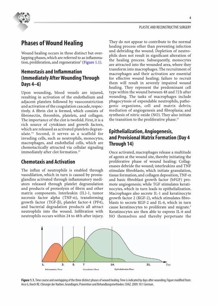
Phases of Wound HealingWound healing occurs in three distinct but over-lapping phases, which are referred to as infl amma-tion, proliferation, and regeneration5 (Figure 1.1).
Hemostasis and Infl ammation (Immediately After Wounding Through Days 4–6)
Upon wounding, blood vessels are injured, resulting in activation of the endothelium and adjacent platelets followed by vasoconstriction and activation of the coagulation cascade, respec-tively. A fi brin clot is formed, which consists of fi bronectin, thrombin, platelets, and collagen. The importance of the clot is twofold. First, it is a rich source of cytokines and growth factors, which are released as activated platelets degran-ulate.14 Second, it serves as a scaffold for invading cells, such as neutrophils, monocytes, macrophages, and endothelial cells, which are chemotactically attracted via cellular signaling immediately after clot formation.12
Chemotaxis and Activation
The infl ux of neutrophils is enabled through vasodilation, which in turn is caused by prosta-glandins activated through infl ammatory medi-ators released through platelet degranulation and products of proteolysis of fi brin and other matrix components. Interleukin (IL)-1, tumor necrosis factor alpha (TNF-α), transforming growth factor (TGF-β), platelet factor-4 (PF4), and bacterial degradation products all attract neutrophils into the wound. Infi ltration with neutrophils occurs within 24 to 48 h after injury.
They do not appear to contribute to the normal healing process other than preventing infection and debriding the wound. Depletion of neutro-phils does not result in signifi cant alteration of the healing process. Subsequently, monocytes are attracted into the wounded area, where they transform into macrophages. The recruitment of macrophages and their activation are essential for effective wound healing; failure to recruit them will result in severely impaired wound healing. They represent the predominant cell type within the wound between 48 and 72 h after wounding. The tasks of macrophages include phagocytosis of expendable neutrophils, patho-genic organisms, cell and matrix debris; mediation of angiogenesis and fi broplasia; and synthesis of nitric oxide (NO). They also initiate the transition to the proliferative phase.21
Epithelialization, Angiogenesis, and Provisional Matrix Formation (Day 4 Through 14)
Once activated, macrophages release a multitude of agents at the wound site, thereby initiating the proliferative phase of wound healing: Collag-enases debride the wound; interleukins and TNF stimulate fi broblasts, which initiate granulation, tissue formation, and collagen deposition, TNF-α and basic fi broblast growth factor (bFGF) pro-mote angiogenesis; while TGF stimulates kerati-nocytes, which in turn leads to epithelialization. Macrophages also secrete IL-1 and keratinocyte growth factor 2 (KGF-2), which stimulates fi bro-blasts to secrete KGF-2 and IL-6, which in turn cause keratinocytes to proliferate and migrate.5 Keratinocytes are then able to express IL-6 and NO themselves and thereby perpetuate the
Figure 1.1. Time course and overlapping of the three distinct phases of wound healing. Time is indicated by days after wounding. Figure modifi ed from: Arco G, Horch RE. Chirurgie der Narben. Grundlagen, Prävention und Behandlungsmethoden. CHAZ. 2009; 10:1 German.
PHYSIOLOGY AND WOUND HEALING
5
process. If the basement membrane has been destroyed, epidermal regeneration occurs from proliferating epithelial cells located on the skin edge of the wound. In order to restore the integ-rity of the epidermal layer, keratinocytes must migrate over the wound margin and therefore cut a path through the fi brin clot or along the inter-face between the clot and the healthy dermis. For this purpose, leading edge keratinocytes express particularly high levels of tissue-type plasmino-gen activator (tPA) or urokinase-type plasmino-gen activator (uPA), both of which activate plasmin, the chief fi brinolytic enzyme. Various members of the matrix metalloproteinase (MMP) family are also preferentially generated by lead-ing edge keratinocytes as well as fi broblasts, mac-rophages, and monocytes. In particular, MMPs-1, -9, and -10 facilitate migration of the above cells through the extracellular matrix.
The connective tissue in the wound is referred to as granulation tissue because of the granular appearance caused by the invading capillaries. Angiogenesis, that is, the process of forming new blood vessels, is ongoing throughout the previ-ously mentioned phases of wound healing. bFGF and vascular endothelial growth factor (VEGF) are released at the wound site by endothelial cells, macrophages, and keratinocytes. Endothelial cells also generate NO in response to hypoxia, and this in turn stimulates more VEGF production. NO causes vasodilation of the endothelium and has a protective effect on newly formed tissue with regard to ischemia and rep-erfusion injury.
The formation of granulation tissue is the fi nal part of the proliferative phase. Platelet-derived growth factor (PDGF) and EGF, which are generated by fi broblasts and macrophages, serve as the main signals for incoming fi bro-blasts to synthesize collagen. Fibroblasts them-selves perpetuate the process with autocrine and paracrine stimulation with PDGF.
Fibroblasts that are located directly at the site of injury not only synthesize collagen for granu-lation tissue formation but can also be stimu-lated by macrophages (through TGF-β1 and PDGF) to transform into a myofi broblast and contribute to wound contraction. By about a week after wounding, fi broblasts are the pre-dominant cell type in the wound. At the same time, the fi brin clot will have been remodeled toward a collagen-rich matrix, and wound con-traction will subsequently take place under the infl uence of the myofi broblast.5
Maturation and Remodeling
Maturation and remodeling of wound healing begins at around a week after wounding and con-tinues for over months for until a year after wounding. It encompasses collagen deposition in an organized manner toward a stable network. Clinically, maturation and remodeling is a par-ticularly relevant stage in wound healing, since problems will occur through matrix deposition defi cits (reducing wound strength) as well as through excessive matrix deposition (formation of hypertrophic scars and keloids). The collagen initially formed after wounding is thinner than that in unwounded skin. The increased rate of collagen deposition after wounding is the result of both a net increase in collagen production per fi broblast and an increase in the number of fi bro-blasts.7 TGF-β directs the construction of the col-lagen matrix, with growth factor levels peaking in the wound between day 7 and 14 after wounding. Over time, the initial collagen is replaced by col-lagen strands that are thicker and therefore more stable. This can be verifi ed by the increase in ten-sile strength of the wound over time. Nevertheless, the collagen in the resulting scar will never reach the stability of the collagen present in intact skin. Therefore, wound strength may reach up to only about 80% of that in uninjured skin, compared with 3% at 1 week and 30% at 3 weeks.19
Table 1.1 gives an overview of the several cell types involved in the different phases of wound healing as outlined here, the growth factors and cytokines they secrete, and their actions. A sum-mary of the growth factors involved and their actions can be found in Table 1.2.
Abnormal Wound HealingA multitude of local as well as systemic factors or conditions are associated with or will result in abnormal wound healing. Abnormal wound healing is often multifactorial.
Delayed Wound Healing
Numerous local and systemic factors can greatly infl uence wound healing. Adequate blood sup-ply to provide glucose and oxygen during the healing process, which is characterized by increased metabolism and protein synthesis, is of paramount importance. Hypoxia results in a delay of the healing process. Prolonged times of
PLASTIC AND RECONSTRUCTIVE SURGERY
6
hypoxia will result in endothelial cell apoptosis induced by TNF-α,13 while wound neutrophils show decreased activity. Their function is also impaired through low temperature, low pH, and elevated glucose concentrations.1 Fibroblasts
respond to hypoxia with a reduced formation of the extracellular matrix, resulting in delayed healing.20
Edema leads to increased interstitial pressure and thereby tissue ischemia. Clinically, tissue
Table 1.1. Growth factors, cytokines, and other mediators and their role in wound healing.
Growth factor Source cells Functions
PDGF Platelets, macrophages, monocytes, fi broblasts, smooth muscle cells, endothelial cells
Chemotaxis and activation of neutrophils and macrophages, fi broblast proliferation, chemotaxis and collagen metabolism, angiogenesis
VEGF Receptors found on endothelial cells only Expressed by most skin cells
Does not act on macrophages, fi broblasts, and smooth muscle cells
EGF Platelets, macrophages Mitogenic for keratinocytes and fi broblasts, stimulation of keratinocyte migration
TNF-α Macrophages, mast cells, T lymphocytes Activation of macrophages and stimulation of angiogenesis, mitogenic for fi broblasts
KGF Fibroblasts Stimulation of keratinocyte proliferation, migration, and differentiation
TGF-α Macrophages, T lymphocytes, keratinocytes Mitogenic for keratinocytes and fi broblasts, stimulates keratinocyte migration
TGF-β Platelets, T lymphocytes, macrophages, endothelial cells, keratinocytes
Chemotaxis of cells stimulating angiogenesis and fi broplasia
Interleukins Macrophages, mast cells, keratinocytes, lymphocytes IL-1: Induction of fever and adrenocorticotrophic hormone release, activation of granulocytes and endothelial cells, stimulation of hematopoiesis, enhances TNF-α and IFN-γIL-2: activates macrophages, T cells, natural killer cells, and lymphokine-activated killer cells, stimulates differentiation of activated B cells, stimulates proliferation of activated B and T cells, induces feverIL-6: induces fever and enhances release of hepatic acute-phase proteinsIL-8: enhances neutrophil adherence, chemotaxis, and granule release
FGF Macrophages, mast cells, T lymphocytes, endothelial cells
Chemotaxis and mitogenesis for keratinocytes and fi broblasts, stimulation of angiogenesis
Table 1.2. Commercially available growth factor products.
Name Growth factor Comments
Regranex (Ortho-McNeil Pharmaceutical)
PDGF-BB FDA-approved for diabetic foot ulcers but also appears to be effi cient in treatment of other types of wounds, such as pressure ulcers, pyoderma gangrenosum, ulcers of vasculitis, and acute surgical defects. First recombinant human growth factor to be used in clinical practice.
Procuren (Curative Health Services) PDGF The fi rst product to be commercially available. Platelet collected from patient’s blood. Therefore, other growth factors may be part of the preparation.
Leukine (Immunex Corp.) GM-CSF Primarily treatment for patients with AML and bone marrow rescue, but also tried in some chronic wounds with promising results, injectable around ulcerated areas, topically as aqueous solution.
Repifermin (Human Genom Sciences) KGF-2 Treatment for cancer therapy-induced mucositis, venous ulcers, and skin grafts. Shown to signifi cantly increase healing and epithelialization over the wound bed. Initial trial in venous ulcers with very promising results. Currently phase two clinical development.
PHYSIOLOGY AND WOUND HEALING
7
edema after ischemia-reperfusion injury in skel-etal muscle can lead to the compartment syndrome. Raised tissue pressure induces increased capil-lary closure, leading to severe hypoxia, which in turn results in cell death with necrosis of various tissues.15
Local wound infection exerts an inhibitory effect on wound healing, because bacteria prolong the infl ammatory phase and inhibit epithelialization, contraction, and collagen deposition. Collagen degradation is increased due to increased collage-nase levels. Bacterial infection is precipitated by foreign bodies. Moreover, they constitute a physical obstacle within the wound, preventing wound contraction and complete epithelialization.
Wound complications also occur when patients have characteristics on a systemic level that pre-dispose them to wound healing problems. Conditions such as old age, smoking, obesity, burns, steroid therapy, and diabetes have been associated with delayed wound healing for a long time. The impairment of healing in diabetic patients is due to several etiologies.5 So-called diabetic ulcers usually occur in diabetic patients suffering from neuropathy, leading to impaired sensibility and failure to release cutaneous pres-sure. The concomitant vasculopathy leads to isch-emia and reduced supply of oxygen and other nutrients. The risk for infections is increased due to impaired function and chemotaxis of granulo-cytes. Diabetic ulcers are also characterized by prolonged infl ammation, impairment of neovas-cularization, decreased collagens synthesis, increased levels of metalloproteinases, and defec-tive macrophage function.
Increased serum glucose has a major effect on wound healing. Traditionally, diabetic complica-tions were believed to be related mainly to micro-vascular occlusive disease, but recent research points to an additional direction.6 Accumulation of the toxic byproduct of glucose metabolism, sorbitol, appears to account for vascular, renal, and ocular complications associated with diabe-tes. Dermal vascular permeability is increased
and leads to pericapillary albumin deposition, resulting in impaired diffusion of oxygen and nutrients. The function of structural and enzy-matic proteins is impaired due to hyperglycemia-associated nonenzymatic glycosylation, the latter increasing collagen’s resistance to enzymatic degradation and rendering it less soluble.9
Experimental as well as clinical studies of dia-betic wound healing show decreases in granula-tion tissue formation and decreased collagen levels in granulation tissue along with defects in collagen maturation. Wound maturation is delayed and the number of dermal fi broblasts is decreased. The levels of several different growth factors were shown to be reduced as well.6
Adjuncts to Wound HealingAdjuncts to wound healing attempt to correct some of the described obstacles to wound heal-ing on several different levels. Some of the most promising adjuncts are presented in the following sections.
Bioengineered Skin
Skin replacement products such as bioengi-neered skin can be differentiated based on their composition and classifi ed based on their struc-ture as either epidermal, dermal, or composite and as living or nonliving (Table 1.3). Their pur-pose is to supply the wound with ingredients favorable to healing, such as growth factors, cytokines, a collagen matrix, and – depending on the product – cells.10 Apligraft, for example, is a composite consisting of neonatal fi broblasts, keratinocytes on a collagen allograft. The cells appear to act as a rich source of growth factors and collagens, which stimulate epithelialization, formation of granulation tissue, angiogenesis, and chemotaxis while they themselves prolifer-ate, thereby contributing to wound coverage in an autocrine and paracrine fashion.8
Table 1.3. Bioengineered skin substitutes and their features.
Composition Structure/living Trade name
Cultured keratinocyte autografts Epidermal/yes EpicelTreated cadaver skin allograft Dermal/no AlloDermBovine collagen/glycosaminoglycan/Silastic Dermal/no IntegraNeonatal fi broblast/polyglactin mesh allograft Dermal/yes DermagraftNeonatal fi broblast/keratinocyte collagen allografts Composite/yes Apligraf
PLASTIC AND RECONSTRUCTIVE SURGERY
8
Growth Factors
Numerous experimental studies have demon-strated the benefi cial effects of recombinant growth factors in different wound healing models in the past. This has prompted clinical studies in the course of which some of the initial hopes were disappointed but, nevertheless, resulted in clinical approval of several commercially available growth factor products5 (Table 1.2).
Negative Pressure Therapy
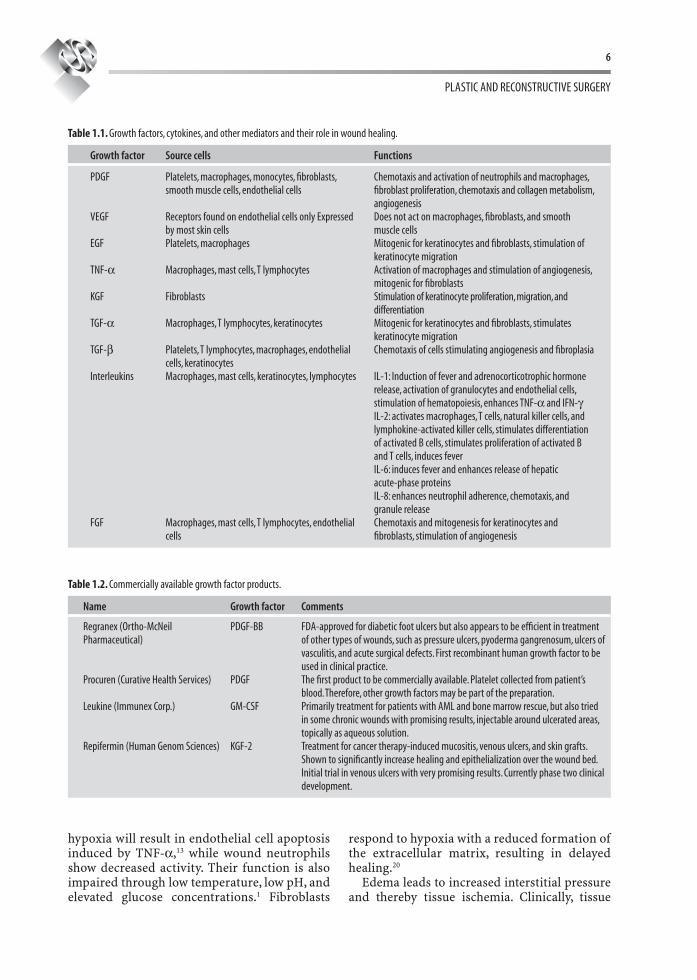
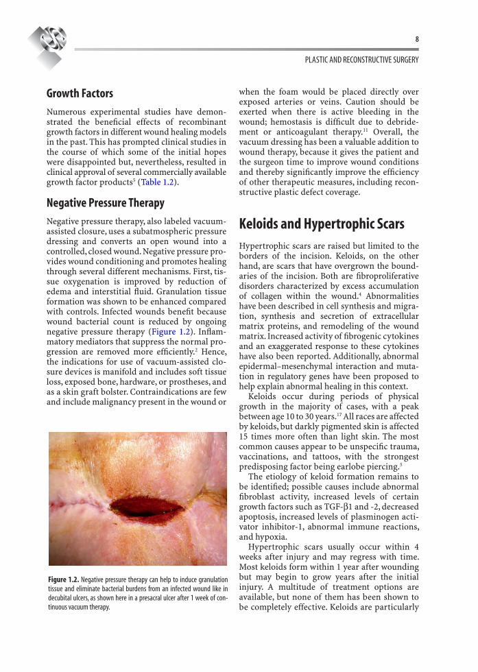
Negative pressure therapy, also labeled vacuum-assisted closure, uses a subatmospheric pressure dressing and converts an open wound into a controlled, closed wound. Negative pressure pro-vides wound conditioning and promotes healing through several different mechanisms. First, tis-sue oxygenation is improved by reduction of edema and interstitial fl uid. Granulation tissue formation was shown to be enhanced compared with controls. Infected wounds benefi t because wound bacterial count is reduced by ongoing negative pressure therapy (Figure 1.2). Infl am-matory mediators that suppress the normal pro-gression are removed more effi ciently.2 Hence, the indications for use of vacuum-assisted clo-sure devices is manifold and includes soft tissue loss, exposed bone, hardware, or prostheses, and as a skin graft bolster. Contraindications are few and include malignancy present in the wound or
when the foam would be placed directly over exposed arteries or veins. Caution should be exerted when there is active bleeding in the wound; hemostasis is diffi cult due to debride-ment or anticoagulant therapy.11 Overall, the vacuum dressing has been a valuable addition to wound therapy, because it gives the patient and the surgeon time to improve wound conditions and thereby signifi cantly improve the effi ciency of other therapeutic measures, including recon-structive plastic defect coverage.
Keloids and Hypertrophic ScarsHypertrophic scars are raised but limited to the borders of the incision. Keloids, on the other hand, are scars that have overgrown the bound-aries of the incision. Both are fi broproliferative disorders characterized by excess accumulation of collagen within the wound.4 Abnormalities have been described in cell synthesis and migra-tion, synthesis and secretion of extracellular matrix proteins, and remodeling of the wound matrix. Increased activity of fi brogenic cytokines and an exaggerated response to these cytokines have also been reported. Additionally, abnormal epidermal–mesenchymal interaction and muta-tion in regulatory genes have been proposed to help explain abnormal healing in this context.
Keloids occur during periods of physical growth in the majority of cases, with a peak between age 10 to 30 years.17 All races are affected by keloids, but darkly pigmented skin is affected 15 times more often than light skin. The most common causes appear to be unspecifi c trauma, vaccinations, and tattoos, with the strongest predisposing factor being earlobe piercing.5
The etiology of keloid formation remains to be identifi ed; possible causes include abnormal fi broblast activity, increased levels of certain growth factors such as TGF-β1 and -2, decreased apoptosis, increased levels of plasminogen acti-vator inhibitor-1, abnormal immune reactions, and hypoxia.
Hypertrophic scars usually occur within 4 weeks after injury and may regress with time. Most keloids form within 1 year after wounding but may begin to grow years after the initial injury. A multitude of treatment options are available, but none of them has been shown to be completely effective. Keloids are particularly
Figure 1.2. Negative pressure therapy can help to induce granulation tissue and eliminate bacterial burdens from an infected wound like in decubital ulcers, as shown here in a presacral ulcer after 1 week of con-tinuous vacuum therapy.

PHYSIOLOGY AND WOUND HEALING
9
notorious for high recurrence rates. Surgical excision or excision using a laser, steroid injec-tions, radiation therapy, magnetic discs, cryosur-gery, and application of silicone gel sheets have all been used and shown to be benefi cial to a certain extent. However, no universally effective treatment has emerged so far. Corticosteroid injections using triamcinolone are commonly regarded as an effi cacious fi rst-line therapy. Silicone gel sheets are often a good recommen-dation in children and those who do not tolerate the pain associated with other therapies.3
Future Wound Healing TherapiesSkin is an easily accessible tissue, and its most superfi cial part, the epidermis, is characterized by a high turnover rate. During wound healing, a multitude of cytokines and growth factors undergo short-term up- and downregulation. All these facts render skin wound healing an ideal setting for gene therapy approaches, which are currently believed to be the most promising tool to enhance wound healing in the future. Short-term gene expression, which is often a drawback of many gene therapy vectors in other circumstances, is desirable when it comes to wound healing. Induction of the gene into the wound can be carried out either directly or indi-rectly by keratinocytes or fi broblasts, which can be harvested and cultured in vitro, followed by transduction with a gene of interest, for example, a gene encoding a growth factor, and fi nally transplanted into the wound. Many different protocols and vectors have been investigated,18 and the most common and promising are pre-sented in the following section.
Viral Techniques
The most common viral vectors in gene therapy for wound healing have been retroviruses, aden-oviruses, and adeno-associated viruses. Recombinant viral vectors are generally created by deletion of certain parts of their genome, thereby disabling viral replication, while at the same time a gene of interest is inserted, most commonly encoding for a growth factor. The packaging capacity of the vector limits the size of the gene that can be inserted and is dependent on the type of virus. The gene of interest is usually
cloned under the control of a particularly pow-erful promoter such as the cytomegalovirus pro-moter to optimize the expression of the desired gene. Retroviruses have a high effi ciency in ex vivo transduction but carry the risk of inser-tional mutagenesis and subsequent tumorigenic transformation. Adenoviruses also attain good transfection effi ciency in vivo but can induce an immune response and allow only small DNA inserts up to 8 kb. Adeno-associated viruses can provide particularly long-lasting gene expres-sion, while they are diffi cult to grow to high titers and also carry the risk for insertional mutagen-esis. Herpes simplex virus allows for particularly large DNA inserts but is diffi cult to manipulate due to its complex life cycle and carries the risk of potential wild-type breakthrough. Generally, nonviral gene transfer techniques are consid-ered safer but often less effi cient in terms of effi ciency.16
Naked DNA
Transduction using naked DNA is probably the safest method for gene delivery. Simple injection using hypodermic needles was shown to deliver and express genes in clinically relevant concen-trations, even though the transduction effi ciency is very low. The “gene gun” approach where DNA-coated gold particles are employed or micro-seeding, which employs a set of oscillating needles to which DNA is delivered via an infu-sion pump, provided superior gene transfer to wounds by increase in the surface area and induction of microtrauma of the treated tissue, thereby improving DNA uptake.
In electroporation, brief electric impulses tran-siently create pores in the plasma membrane of the cell to allow for DNA diffusion into the cell.
Cationic Liposomes
These positively charged lipid vesicles form a complex with negatively charged DNA. Transfer of the DNA across the cell membrane appears to occur through an endocytosis-like process. Due to their lack of immunogenicity, repeated deliv-eries in vivo are possible. Another advantage is the potential to deliver large amounts of DNA and to incorporate large transgenes. The limiting factor, however, is their low transfection effi ciency in vivo compared with that of viral vectors.

PLASTIC AND RECONSTRUCTIVE SURGERY
10
ConclusionAdequate wound management relies on a multi-tude of factors and requires a profound knowl-edge of the mechanisms of wound healing and the factors that infl uence it.
References 1. Allen DB, Maguire JJ, Mahdavian M, et al. Wound hypoxia
and acidosis limit neutrophil bacterial killing mecha-nisms. Arch Surg. 1997;132:991–996.
2. Argenta LC, Morykwas MJ. Vacuum-assisted closure: a new method for wound control and treatment: clinical experience. Ann Plast Surg. 1997;38:563–576.
3. Berger A, Hierner R. Plastische Chirurgie. Grundlagen, Prinzipien, Techniken. 1st ed. Berlin: Springer; 2003.
4. Blackburn WR, Cosman B. Histologic basis of keloid and hypertrophic scar differentiation: Clinicopathologic cor-relation. Arch Pathol. 1966;82:65–71.
5. Broughton G, Janis JE, Attinger C. Wound healing: an overview. Plast Reconstr Surg. 2006;117:1e–S.
6. Bucalo B, Eaglstein WH, Falanga V. Inhibition of cell pro-liferation by chronic wound fl uid. Wound Repair Regen. 1993;1:181–186.
7. Diegelmann R. Analysis of collagen synthesis. Methods Mol Med. 2003;78:349–358.
8. Falanga V, Sabolinski M. A bilayered living skin construct (Apligraf) accelerates complete closure of hard-to-heal venous ulcers. Wound Repair Regen. 7:201–207.
9. He Z, King GL. Microvascular complications of diabetes. Endocrinol Metab Clin North Am. 2004;33:215–238.
10. Horch RE, Kopp J, Kneser U, et al. Tissue engineering of cultured skin substitutes. J Cell Mol Med. 2005;9:592–608.
11. Kloth LC. 5 questions-and answers-about negative pressure wound therapy. Adv Skin Wound Care. 2002;15:226–229.
12. Kurkinen M, Vaheri A, Roberts P, et al. Sequential appear-ance of fi bronectin and collagen in experimental granu-lation tissue. Lab Invest. 1980;43:47–51.
13. Longaker MT, Adzick NS, Hall J, et al. Studies in fetal wound healing: VII. Fetal wound healing may be modu-lated by hyaluronic acid stimulating activity in amnionic fl uid. J Pediatr Surg. 1990;25:430–433.
14. Martin P. Wound Healing-aiming for perfect skin regen-eration. Science. 1997;276:75–81.
15. Massberg S, Messmer K. The nature of ischemia/reperfu-sion injury. Transplant Proc. 1998;30:4217–4223.
16. Mulligan RC. The basic science of gene therapy. Science. 1993;260:926–932.
17. Niessen FB, Spauwen PH, Schalkwijk J, et al. On the nature of hypertrophic scars and keloids: a review. Plast Reconstr Surg. 1999;104:1435–1458.
18. Petrie NC, Yao F, Eriksson E. Gene therapy in wound healing. Surg Clin North Am. 2003;83:597–616.
19. Sabiston D. Textbook of Surgery: The Biological Basis of Modern Surgical Practice. 15th ed. St Louis, MO: Saunders; 1997.
20. Steinbrech DS, Longaker MT, Mehrara B, et al. Fibroblast response to hypoxia: the relationship between angiogen-esis and matrix regulation. J Surg Res. 1999;84:127–133.
21. Witte M, Barbul A. Role of nitric oxide in wound repair. Am J Surg. 2002;183:406.