plastination of macroparasites: an eco-friendly method of ... · nemathelminthes-class nematoda...
TRANSCRIPT

Veterinary World, EISSN: 2231-0916 1394
Veterinary World, EISSN: 2231-0916Available at www.veterinaryworld.org/Vol.10/November-2017/19.pdf
RESEARCH ARTICLEOpen Access
Plastination of macroparasites: An eco-friendly method of long-term preservation
Niranjan Kumar1, Bhupamani Das1, Jayesh B. Solanki1, Mehul M. Jadav1 and Ramasamy Menaka2
1. Department of Parasitology, College of Veterinary Science and Animal Husbandry, Navsari Agricultural University, Navsari - 396 450, Gujarat, India; 2. Department of Anatomy, College of Veterinary Science and Animal Husbandry,
Navsari Agricultural University, Navsari-396 450, Gujarat, India.Corresponding author: Niranjan Kumar, e-mail: [email protected]
Co-authors: BD: [email protected], JBS: [email protected], MMJ: [email protected], RM: [email protected]
Received: 30-06-2017, Accepted: 25-10-2017, Published online: 29-11-2017
doi: 10.14202/vetworld.2017.1394-1400 How to cite this article: Kumar N, Das B, Solanki JB, Jadav MM, Menaka R (2017) Plastination of macroparasites: An eco-friendly method of long-term preservation, Veterinary World, 10(11): 1394-1400.
AbstractAim: Preservation of macroparasites by infiltrating the polymer in the tissues can defy the inherited shortcoming of classical wet preservation method.
Materials and Methods: Preservation was done by infiltrating the melamine alone or with xylene (MX)/chloroform (MC)/turpentine oil (MT) in 1:1 and hardener (MH) in 9:1 ratio in the tissues of the gross specimen of the animal parasites.
Results: The plastinated models withstand the process of microbial decomposition, and remain intact in the environmental conditions. The polymer mixture resists the entry of the water molecule, and model dried just after taking out it from the water tank. Overall, the plastinated parasites were dry, non-sticky, glossy, odorless, chemical free, and harmless, to some extent flexible, with detectable morphological structure, and retain their natural form but lost their natural color. Full marks were assigned to the degree of dryness, non-stickiness, and odorlessness to the model plastinated in different solutions on a five-point scale. For flexibility, the score was 1.2, 2.2, and 2.4 for the plastinated model in melamine/MH, MX/MC, and MT solutions, respectively. The average score of glossiness was 4.6 and 5 for the specimen plastinated in melamine/MH and MX/MC/MT solutions, respectively. The degree of dryness, glossiness, stickiness, and flexibility varies non-significantly, with the polymer mixtures used.
Conclusion: The prepared model can be used to educate the students/general mass population.
Keywords: macroparasites, melamine, plastination, preservation.
Introduction
The macroparasites occupy all the ecological niche of the world and exerts ill effects on the health of the hosts (animals, birds, and human) [1,2]. There are different techniques to preserve the parasites for educational purposes in the academic institutions. The most widely accepted method of preservation is immersions and storage of the biological specimens in 10% formalin or 70% ethyl alcohol [3]. Although these materials are well-known fixative but the stored specimens are having some inherent shortcomings such as wet, with noxious odors, hazardous to the handlers, and difficult to transport. The continuously emitting noxious gas can harm the respiratory system, eyes, and skin of the handlers. Some study highlighted the role of formaldehyde as carcinogenic and neuro-toxic agents [4,5]. Histological cross sections are also utilized in some teaching laboratories [6,7]. The
disadvantage of histological specimens is their limita-tion in scope as definitive identification of the para-sites requires integration with other factors [8]. The preservatives can also destroy the normal morpholog-ical features of the macroparasites, and thus, impede their identification [9-11].
Plastination technique, a method of dry preser-vation, is a delicate method of forced impregnation by replacing water and lipid tissues with the curable polymers, melamine or others [12-15]. Melamine is a nitrogen-rich heterocyclic triazine used primarily in the synthesis of melamine-formaldehyde resins for the manufacture of laminates, plastics, coatings, com-mercial filters, glues or adhesives, and molding com-pounds (dishware and kitchenware) [16,17]. There is a rare report of the inhalation toxicity of the melamine, but its oral exposure may cause the formation of renal calculi [18,19].
The purpose of this study was to develop alter-native eco-friendly method of preservation of the macroparasites describing their natural identifying features by infiltrating the melamine polymer.Materials and MethodsEthical approval
As the study was conducted utilizing the gross specimen of the animal parasites stored in 10%
Copyright: Kumar, et al. Open Access. This article is distributed under the terms of the Creative Commons Attribution 4.0 International License (http://creativecommons.org/licenses/by/4.0/), which permits unrestricted use, distribution, and reproduction in any medium, provided you give appropriate credit to the original author(s) and the source, provide a link to the Creative Commons license, and indicate if changes were made. The Creative Commons Public Domain Dedication waiver (http://creativecommons.org/publicdomain/zero/1.0/) applies to the data made available in this article, unless otherwise stated.

Veterinary World, EISSN: 2231-0916 1395
Available at www.veterinaryworld.org/Vol.10/November-2017/19.pdf
formalin in the departmental museum, so the ethical committee approval was not required.Parasites specimens
The whole or segments of parasites belong-ing to Phylum: Platyhelminthes- Class Trematoda (Fasciola gigantica) and Cestoda (Moniezia spp.); Nemathelminthes- Class Nematoda (Ascaris suum, Parascaris equorum, Toxocara vitulorum, Toxocara canis, Haemonchus contortus, Oesophagostomum spp., Bunostomum spp., Trichuris ovis, and Oxyuris equi), Arthropoda- Class Insecta (Oestrus ovis and blow fly) and Arachnida (soft and hard ticks) have been plastinated.
To measure the extent of shrinkage, the dimen-sion and weight of the plastinating materials were taken before and after impregnation of the polymer.Plastinating materials
The melamine polymer, hardener, and touch-wood of Asian Paints along with turpentine oil were procured from the local distributors. The acetone, xylene, and chloroform were of Merck make.Plastination technique
In the present investigation, plastination tech-nique was performed as per the method described by Menaka et al. [20] with certain modification. The selected parasites specimens were washed 24 h in running cold tap water (5°C), to remove as much as possible formalin [21]. For dehydration, the samples were placed in 100% acetone in the ratio 10:1 (approx-imately) at -20°C. The parasites were dehydrated in 3 changes of dehydrating agents at 1 week interval. When the dehydrating agent’s concentration remained at 99% (approximately after 3 weeks), dehydration was deemed complete [22]. The 3:1 volume ratio of polymer/polymer mixture to the macroparasites spec-imens was incubated at −20°C for 30 days. To create an ideal plastinated model of the macroparasites, the reagents such as xylene, chloroform, turpentine oil, and hardener were added in different permutation and combination in the melamine. The polymer mixtures were prepared by mixing melamine and xylene (MX), melamine and chloroform (MC), and melamine and turpentine oil (MT) in 1:1 while melamine and hardner (MH) in 9:1 ratio. Following the forced impregnation, the specimens were removed from the polymer/poly-mer mixture, and kept in a Petri dish for 2-3 days to drain the excess polymer. To impart glossy appearance, the specimens were brushed with colorless varnish.Hydrophobicity testing
To check the power of wettability, the plastinated specimen was dipped in water for 30 minutes, and then allowed to stand at the environmental condition.Quality evaluation
Five technical personnel were allocated to mea-sure the extent of dryness, non-stickiness, odorless-ness, glossiness, and flexibility of the prepared model on a five-point scale.
Statistical analysisResults were compiled systematically, and data
were analyzed using IBM SPSS Statistics 20.00 for Windows (SPSS Inc., Chicago, USA) to perform Chi-square tests and/or Student’s t-test and/or one-way ANOVA using Duncan test (2-sided) for the determi-nation of statistical significance. The p>0.05 was con-sidered as statistically non-significant.Results
The plastinated models withstand the process of microbial decomposition, and remain intact at environmental condition till the acceptance of the manuscript. The polymer mixture resists the entry of water molecules inside the specimens, and the model becomes dry just after taking out it from the water tank; thus, the plastinated model can be maintained even in the environment with high level of humidity.
Overall, the plastinated parasites were dry, non-sticky, glossy, odorless, chemical free, and harmless, to some extent flexible, with detectable morphological structure, and retain their natural form but lost their natural color. On a five-point scale, all five personnel assigned full marks for dryness, non-stickiness, and odorlessness to the model plastinated in different solu-tions. For flexibility, the score was 1.2, 2.2, and 2.4 for the plastinated model in melamine/MH, MX/MC, and MT solutions, respectively. The average score of glossiness was 4.6 and 5 for the specimen plastinated in melamine/MH and MX/MC/MT solutions, respec-tively. The degree of dryness, glossiness, stickiness, and flexibility of the prepared model varies non-sig-nificantly with the different polymer mixtures.
The parasites plastinated solely in melamine were dry, non-sticky, and odorless but less-glossy and brittle in nature (Figure-1).
The parasites plastinated in MX solution were dry, non-sticky, odorless, and glossy but less flex-ible (Figure-2a-e). The plastinated insect larvae clearly depicted the feature of immature/mature lar-vae of O. ovis, large, around 2.0 cm long (Table-1) with black oral hooks (Figure-2b), small rows of rose
Figure-1: Parascaris equorum plastinated in melamine (M) solution.

Veterinary World, EISSN: 2231-0916 1396
Available at www.veterinaryworld.org/Vol.10/November-2017/19.pdf
Pla
stin
atio
n
mix
ture
Par
asit
e
Typ
eN
ame
Dim
ensi
onW
eig
ht
(mg
)
Len
gth
(m
m)
Wid
th (
mm
)B
efor
eA
fter
Sh
rin
kag
e
(%)
Bef
ore
Aft
erS
hri
nka
ge
(%
)B
efor
eA
fter
Sh
rin
kag
e (%
)
MX
Roun
d w
orm
Asc
aris
suu
m17
417
11.
73
233
.327
018
1.3
32.9
Asc
aris
suu
m16
116
00.
63
233
.322
014
3.2
34.9
Toxo
cara
can
is75
715.
31
0.9
1013
091
30M
ean±
SE
136.
66±
31.0
6a13
4±31
.65a
2.33
±0.
67b
1.63
±0.
37b
206.
67±
40.9
6c13
8.5±
26.1
7cFl
at w
orm
Mon
iezi
a sp
p. s
egm
ents
3833
13.2
65
16.7
270
201
25.5
Mon
iezi
a sp
p. s
egm
ents
4340
7.0
65
16.7
290
203.
729
.8Fa
scio
la g
igan
tica
4640
13.0
75
28.6
160
135.
615
.3Fa
scio
lops
is b
uski
2820
28.6
1915
21.1
330
261.
220
.9M
ean±
SE
38.7
5±3.
94d
33.2
5±4.
71d
9.5±
3.18
e7.
5±2.
5e26
2.5±
36.3
7f20
0.38
±25
.67f
Inse
ct’s
la
rva
Oes
trus
ovis
larv
a19
1615
.87
614
.338
030
9.8
18.5
Blo
w fly
larv
ae15
146.
72
20
5041
.616
.8M
ean±
SE
17±
2g15
±1g
4.5±
2.5h
4±2h
215±
165i
175.
7±13
4.1i
Sof
t tic
kO
rnith
odor
os s
pp.
1010
06
60
680
582.
714
.3M
CRo
und
wor
mAsc
aris
suu
m19
018
81.
14
325
310
221
28.7
Toxo
cara
vitu
loru
m16
516
21.
87
614
.311
5098
414
.4To
xasc
aris
leon
ina
43.
512
.50.
80.
625
3010
.565
Mea
n±SE
119.
67±
58.2
8j11
7.83
±57
.66j
3.93
±1.
79k
3.2±
1.56
k49
6.67
±33
6.52
l40
5.17
±29
5.73
l
Flat
wor
mM
onie
zia
spp.
seg
men
ts32
306.
256
516
.726
018
1.6
30.2
Fasc
iola
gig
antic
a32
2812
.56
350
150
109.
626
.9M
ean±
SE
32±
0m29
±1m
6±0n
4±1n
205±
55o
145.
6±36
o
Inse
ct’s
la
rva
Oes
trus
ovi
s la
rva
1614
12.5
64
33.3
220
151
31.4
Oes
trus
ovi
s la
rva
1711
35.3
75
28.6
180
145.
918
.9Blo
w fly
larv
ae18
175.
62
1.6
2052
47.1
9.4
Mea
n±SE
17±
0.58
p14
±1.
73p
5±1.
53q
3.53
±1.
01q
150.
67±
50.6
7r11
4.67
±33
.82r
Inse
ctTa
banu
s sp
p.18
180
44
060
2165
Har
d tic
kIx
odes
spp
.9
90
44
090
90.8
-0.9
MT
Roun
d w
orm
Asc
aris
suu
m13
413
12.
22
150
160
101.
436
.6To
xoca
ra c
anis
4.5
411
.10.
90.
722
.232
12.5
60.9
Mea
n±SE
69.2
5±64
.75s
67.5
±63
.5s
1.45
±0.
55t
0.85
±0.
15t
96±
64u
56.9
5±44
.45u
Flat
wor
mM
onie
zia
spp.
seg
men
ts30
2710
65
16.7
220
195.
511
.1Fa
scio
la g
igan
tica
4741
12.8
63
5016
012
6.4
21M
ean±
SE
38.5
±8.
5v34
±7v
6±0w
4±1w
190±
30x
160.
95±
34.5
5xIn
sect
’s
larv
aO
estr
usov
is la
rva
1813
27.8
75
28.6
190
156.
717
.5
1815
16.7
76
14.3
360
283.
521
.3M
ean±
SE
18±
0y14
±1y
7±0z
5.5±
0.5z
275±
85aa
220.
1±63
.4aa
Tab
le-1
: Shr
inka
ge o
f m
acro
para
site
s in
ter
ms
of d
imen
sion
and
bod
y w
eigh
t.
(Con
td...
)

Veterinary World, EISSN: 2231-0916 1397
Available at www.veterinaryworld.org/Vol.10/November-2017/19.pdf
thorn-shaped spines on the ventral aspect (Figures-2b) dark black bands on all the segments on the dorsal aspect (Figure-2c), and truncated and large posterior end with clearly visible brown color “D-” shaped pos-terior spiracles with radiating slits (Figure-2d). Blow fly larva plastinated in MX solution was elongated (15 mm long×2 mm width), glossy, and creamy color (Table-1 and Figure-2e).
The parasites plastinated in MC solution were dry, non-sticky, odorless, and glossy while to some extent flexible (Figure-3a-k). The plastinated T. vitulorum was up to 16.5 cm×7 mm with translu-cent cuticle, and the body does not taper toward the extremities (Table-1 and Figure-3a). The large thick-set fluke plastinated in the MC solution described the overall morphological features of Fasciolopsis buski like elongate-oval body slightly broader posteriorly than anteriorly without shoulders, measuring about 30 mm by 20 mm, much larger ventral sucker than the oral one (Figure-3b and c). Moniezia spp. segments plastinated in MC solution were transparent, about 6 mm in width, broader than long, with two sets of genital organs with the marginal genital pores (Table 1 and Figure-3d). The plastinated immature larvae of O. ovis was around 1.6 cm long (Table-1), with truncated and large posterior end (Figure-3e), small rows of rose thorn-shaped spines on the ventral aspect (Figure-3f and g), and black oral hooks (Figure-3h). Tabanus spp. fly plastinated in MC solution depicted the important morphological structure, large compound eyes, longi-tudinal white stripes on the thorax, clear wings with the 4th radial vein being forked at the apex, hexago-nal discal cell, and brown color abdomen (Figure-3i). The plastinated soft (Ornithodoros moubata) and hard [Rhipicephalus (Boophilus) microplus] tick in MC solution had described their common morphological features (Figure-3j and k).
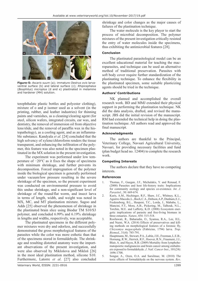
The parasites plastinated in MT solu-tion were dry, non-sticky, odorless, glossy, and flexible (Figure-4). The parasites plastinated in MH solution were hard, brittle, and difficult to handle during the plastination process (Figure-5a-e). The
Pla
stin
atio
n
mix
ture
Par
asit
e
Typ
eN
ame
Dim
ensi
onW
eig
ht
(mg
)
Len
gth
(m
m)
Wid
th (
mm
)B
efor
eA
fter
Sh
rin
kag
e
(%)
Bef
ore
Aft
erS
hri
nka
ge
(%
)B
efor
eA
fter
Sh
rin
kag
e (%
)
Har
d tic
kIx
odes
spp
.10
100
66
013
013
1.2
-0.9
MH
Inse
ct’s
la
rva
Oes
trus
ovi
s la
rva
2117
19.0
85
37.5
200
176
12
Har
d tic
kIx
odes
spp
.10
100
66
019
019
2-1
.1
SE=
Sta
ndar
d er
ror;
Sig
nific
ant
at p≤
0.05
; p
valu
e -
a=0.
95,
b=0.
41,
c=0.
23,
d=0.
41,
e=0.
64,
f=0.
21,
g=0.
47,
h=0.
89,
i=0.
87,
j=0.
98,
k=0.
77,
l=0.
85,
m=
0.10
, n=
0.18
, o=
0.46
, p=
0.18
, q=
0.47
, r=
0.59
, s=
0.99
, t=
0.40
, u=
0.67
, v=
0.72
, w
=0.
18,
x=0.
59,
y=0.
06,
z=0.
10,
aa=
0.66
; M
X=
Mel
amin
e an
d xy
lene
, M
C=
Mel
amin
e an
d ch
loro
form
, M
T=M
elam
ine
and
turp
entin
e oi
l, M
H=
Mel
amin
e an
d ha
rden
er
Tab
le-1
: (C
onti
nu
ed)
Figure-2: Ascaris suum (a); Oestrus ovis larva-ventral surface (b); dorsal surface (c), and spiracles (d); blowfly larva-dorsal surface (e) plastinated in melamine and xylene (MX) solution.
d
cba
e

Veterinary World, EISSN: 2231-0916 1398
Available at www.veterinaryworld.org/Vol.10/November-2017/19.pdf
set protocol and plastinating agent failed to yield the plastinated model of small size nematodes such as H. contortus, Oesophagostomum spp., Bunostomum spp., T. ovis, and O. equi.
Shrinkage of macroparasites in terms of dimen-sion and body weight is summarized in Table-1. The parasites with soft body had received more shrinkage than the parasites with tough body. The tough chitin-ous layer (exoskeleton) of the arthropods withstands the process of shrinkage in various plastinating poly-mer solutions except MH. The specimens plastinated in MH polymer solution had received drastic overall shrinkage, and become brittle in nature (Figure-5a). Shrinkage of the round/flat worm and insect larva in terms of length, width, and weight was statistically non-significant in MX, MC, and MT plastination mix-ture (Table-1).Discussion
Plastination technique was originally developed for the preservation of biological specimens in the medical world by Gunther von Hagens in 1977 [23]. The macroparasite, Ascaris lumbricoides, was first time plastinated by Asadi and Mahmodzaeh [22] through Sl0 Techniques. The alcohol or formaldehyde persevered parasite samples have many disadvantages such as being less permanent, having regular needs of changing the immersion solution, the unpleasant smell, and having hardly recognizable parts of the par-asites [14]. Plastinated parasites can be an excellent alternative as it lowers the risk of undue exposure to the formaldehyde with higher health and safety reg-ulations. Furthermore, the plastinated model is easy
Figure-3: Toxocara vitulorum (a); Fasciolopsis buski-ventral surface (b); and dorsal surface (c); Moniezia expansa (d); immature Oestrus ovis larva-ventral surface (e); dorsal surface (f); ventral surface close view (g); and anterior end close view (h); Tabanus spp. fly (i); Ornithodoros moubata soft tick (j); Rhipicephalus (Boophilus) microplus hard tick (k) plastinated in melamine and chloroform (MC) solution.
d
h
i j k
c
g
b
f
a
e
Figure-4: Ascaris suum plastinated in melamine and turpentine oil (MT) solution.
to carry, palpable, with clearly visible structure, and can be stored for an infinite period at room tempera-ture [12].
The present study dealt with the preparation of a plastinated model of macroparasites of animal origin using melamine polymer first time in India. There is a very limited report of using melamine polymer as plastinating agents to preserve the biological speci-men in native condition [20].
Laboratory grade xylene is a fair mixture of o, p, m, and p isomer and ethylbenzene (6-20%) with the traces of toluene, trimethylbenzene, phenol, thio-phene, pyridine, and hydrogen sulfide. The xylene has multivalent function, as a plasticizing agent (p-iso-mer is the precursor to terephthalic acid and dimethyl terephthalate, used in the production of polyethylene
Veterinary World, EISSN: 2231-0916 1399
Available at www.veterinaryworld.org/Vol.10/November-2017/19.pdf
terephthalate plastic bottles and polyester clothing), mixture of o and p isomer used as a solvent (in the printing, rubber, and leather industries) for thinning paints and varnishes, as a cleaning/clearing agent (for steel, silicon wafers, integrated circuits, ear wax, and dentistry, the removal of immersion oil from objective lens/slide, and the removal of paraffin wax in the his-topathology), as a cooling agent, and as an inflamma-ble substance. Kandyala et al. [24] concluded that the high solvency of xylene/chloroform renders the tissue transparent, and enhancing the infiltration of the poly-mer, this feature was also noted in the specimen plas-tinated in the MX solution in the present investigation.
The experiment was performed under low tem-perature of −20°C as it fixes the shape of specimens with minimum shrinkage, and further prevents its decomposition. Forced impregnation of the polymer inside the biological specimen is generally performed under vacuum/low pressure resulting in the severe shrinkage of the specimen, so the present experiment was conducted on environmental pressure to avoid this undue shrinkage, and a non-significant level of shrinkage of the round/flat worm, and insect larva in terms of length, width, and weight was noted in MX, MC, and MT plastination mixture. Sagoo and Adds [25] observed the phenomenon of shrinkage in the plastinated brain slice using Biodur TM S10/S3 polymer, and concluded 6.99% and 6.19% shrinkage in lengths and widths, respectively, was acceptable.
The plastinated specimens in the different poly-mer mixtures were dry and odorless, and successfully demonstrated the gross morphological features of the parasites while the color was more esthetic than that of the specimens stored in formaldehyde. The shrink-age and resulting distorted anatomy were the import-ant observations of the present investigation, and were also observed by Miklošova and Mikloš [26] in the most ideal plastination method, silicone S10. Furthermore, Latorre et al. [27] also concluded
shrinkage and color changes as the major causes of failures of the plastination technique.
The water molecule is the key player to start the process of microbial decomposition. The polymer mixtures of the present investigation critically resisted the entry of water molecules inside the specimens, thus exhibiting the antimicrobial features [28].Conclusion
The plastinated parasitological model can be an excellent educational material for teaching the mac-roparasites, and technique can be used an alternative method of traditional preservation. Parasites with soft body cover require further standardization of the plastinating technique. To enhance the flexibility in the plastinated specimen, some suitable plasticizing agents should be tried in the technique.Authors’ Contributions
NK planned and accomplished the overall research work. BD and MMJ extended their physical support in performing the plastination technique. NK did the data analysis, drafted, and revised the manu-script. JBS did the initial revision of the manuscript. RM had extended the technical help in doing the plas-tination technique. All authors read and approved the final manuscript.Acknowledgments
The authors are thankful to the Principal, Veterinary College, Navsari Agricultural University, Navsari, for providing necessary facilities and fund (plan budget head no. 12404) to complete the research work.Competing Interests
The authors declare that they have no competing interests.References1. Thomas, F., Guegan, J.F., Michalakis, Y. and Renaud, F.
(2000) Parasites and host life-history traits: Implications for community ecology and species co-existence. Int. J. Parasitol., 30: 669-674.
2. Kuris, A.M., Hechinger, R.F., Shaw, J.C., Whitney, K.L., Aguirre-Macedo, L., Boch, C.A., Dobson, A.P., Dunham, E.J., Fredensborg, B.L., Huspeni, T.C., Lorda, J., Mababa, L., Mancini, F.T., Mora, A.B., Pickering, M., Talhouk, N.L., Torchin, M.E. and Lafferty, K.D. (2008) Ecosystem ener-getic implications of parasite and free-living biomass in three estuaries. Nature, 454: 515-518.
3. Rosilawati, R., Baharudin, O., Syamsa, R.A., Lee, H.L. and Nazni, W.A. (2014) Effects of preservatives and kill-ing methods on morphological features of a forensic fly, Chrysomya megacephala (Fabricius, 1794) larva. Trop. Biomed., 31(4): 785-791.
4. Hauptmann, M., Stewart, P.A., Lubin, J.H., Freeman, L.E.B., Hornung, R.W., Herrick, R.F., Hoover, R.N., Fraumeni, J.J., Blair, A. and Hayes, R.B. (2009) Mortality from lymphohe-matopoietic malignancies and brain cancer among embalm-ers exposed to formaldehyde. J. Natl. Cancer Inst., 101(24): 1696-1708.
5. Songur, A., Ozen, O.A. and Sarsilmaz, M. (2010) The toxic effects of formaldehyde on the nervous system. Rev.
Figure-5: Ascaris suum (a); immature Oestrus ovis larva-ventral surface (b) and lateral surface (c); Rhipicephalus (Boophilus) microplus (d and e) plastinated in melamine and hardener (MH) solution.
dc
ba
e

Veterinary World, EISSN: 2231-0916 1400
Available at www.veterinaryworld.org/Vol.10/November-2017/19.pdf
Environ. Contam. Toxicol., 203: 105-118.6. Panyarachun, B., Ngamniyom, A., Sobhon, P. and
Anuracpreeda, P. (2013) Morphology and histology of the adult Paramphistomum gracile Fischoeder, 1901. J. Vet. Sci., 14(4): 425-432.
7. Hanna, R. (2015) Fasciola hepatica: Histology of the repro-ductive organs and differential effects of triclabendazole on drug-sensitive and drug-resistant fluke isolates and on flukes from selected field cases. Pathogens, 4: 431-456.
8. Galant, C., Malghem, J., Sibille, C., Docquier, P.L. and Delloye, C. (2008) Current limitations to the histopatho-logical diagnosis of some frequently encountered bone tumours. Acta. Orthop. Belg., 74: 1-6.
9. Day, D.M. and Wallma, J.F. (2008) Effect of preservative solutions on preservation of Calliphora augur and Lucilia cuprina larvae (Diptera: Calliphoridae) with implications for post-mortem interval estimates. Forensic Sci. Int., 179: 1-10.
10. Midgley, J.M. and Villet, M.H. (2009) Effect of killing method on post-mortem change of larvae of Thanatophilus micans (Fabricus, 1794) (Coleoptera: Silphidae) stored in 70% ethanol. Int. J. Legal Med., 123: 103-108.
11. Villet, M.H., Richards, C.S. and Midgley, J.M. (2010) Contemporary precision, bias and accuracy of minimum post-mortem interval estimated using development of car-rion feeding insetcs. In: Amendt, J., Campobasso, C.P., Goff, M.L., and Grassberger, M., editors. Current Concepts in Forensic Entomology. Springer, Heidelberg. p109-137.
12. Singh, O., Mishra, B.K., Pandit, S., Maheshwari, T.P. and Hasan, S. (2013) Plastination: A promising method for pre-serving: A review article. Int. J. Sci. Res., 3(6): 1-3.
13. Ravikumar, C. (2014) Plastination. J. Pharm. Sci. Res., 6(8): 271-273.
14. Menaka, R., Kelawala, N.H. and Vyas, K.N. (2015) Plastination technique represents a life in biological spec-imens-An overview. Vet. Res. Int., 3(2): 20-23.
15. Prasad, G., Karkera, B., Pandit, S., Desai, D. and Tonse, R.G. (2015) Preservation of tissue by plastination: A Review. Int. J. Adv. Health Sci., 1(11): 27-31.
16. Wu, C.F., Hsieh, T.J., Chen, B.H., Liu, C.C. and Wu, M.T. (2013) A crossover study of noodle soup consumption in melamine bowls and total melamine excretion in urine. JAMA Intern. Med., 173(4): 317-319.
17. Poorjafari, N., Zamani, A., Mohseni, M. and Parizanganeh, A. (2015) Assessment of residue melamine in dairy products exhibited in Zanjan market, Iran by high-performance liq-uid chromatography method. Int. J. Environ. Sci. Technol.,
12: 1003-1010.18. Dobson, R.L., Motlagh, S., Quijano, M., Cambron, R.T.,
Baker, T.R., Pullen, A.M., Regg, B.T., Bigalow-Kern, A.S., Vennard, T., Fix, A., Reimschuessel, R., Overmann, G., Shan, Y. and Daston, G.P. (2008) Identification and charac-terization of toxicity of contaminants in pet food leading to an outbreak of renal toxicity in cats and dogs. Toxicol. Sci., 106(1): 251-262.
19. Reimschuessel, R., Gieseker, C.M., Miller, R.A., Ward, J., Boehmer, J., Rummel, N., Heller, D.N., Nochetto, C., de Alwis, G.K., Bataller, N., Andersen, W.C., Turnipseed, S.B., Karbiwnyk, C.M., Satzger, R.D., Crowe, J.B., Wilber, N.R., Reinhard, M.K., Roberts, J.F. and Witkowski, M.R. (2008) Evaluation of the renal effects of experimental feeding of melamine and cyanuric acid to fish and pigs. Am. J. Vet. Res., 69: 1217-1228.
20. Menaka, R., Chaurasia, S. and Kelawala, N.H. (2010) Plastination of goat (kid) cadaver-a teaching model. Indian J. Vet. Anat. 22(1): 50-51.
21. Kocevski, Z., Stefanovska, J., Ilieski, V., Pendovski, L. and Atanaskova, E. (2010) Improved determination of macro-scopic parasite preparations using S10 modified plastina-tion procedure. Mac. Vet. Rev., 33(2): 7-14.
22. Asadi, M.H. and Mahmodzaeh, A. (2004) Ascaris plasti-nation through Sl0 techniques. J. Int. Soc. Plastination, 19: 20-21.
23. von Hagens, G.,Tiedmann, K. and Kriz, W. (1987) The current potential of plastination. Anat. Embryol. (Berlin), 175(4): 411-421.
24. Kandyala, R., Phani, S., Raghavendra, C. and Rajasekharan, S.T. (2010) Xylene: An overview of its health hazards and preventive measures. J. Oral. Maxillofac. Pathol., 14(1): 1-5.
25. Sagoo, M.G. and Adds, P.J. (2013) Low-temperature dehy-dration and room-temperature impregnation of brain slices using Biodur TM S10/S3. J. Plastination, 25(1): 3-8.
26. Miklošova, M. and Mikloš, V. (2004) Plastination with sil-icone method S10- monitoring and analysis causes of fail-ure. Biomed. Pap., 148(2): 237-238.
27. Latorre, R.M., Reed, R.B., Gil, F., Azla, M.D., Martinez-Gomariz, F. and Henry, R.W. (2002) Epoxy impregnation without hardener: To decrease yellowing, to delay cast-ing, and to aid bubble removal. J. Int. Soc. Plastination, 17: 17-22.
28. Rahangdale, S.S. (2012) Synthesis, characterization and antimicrobial activity of resorcinol-melamine-formalde-hyde resin. J. Chem. Pharm. Res., 4(10): 4451-4458.
********