platelet-rich fibrin in bone regenerative strategies in
TRANSCRIPT
materials
Review
Platelet-Rich Fibrin in Bone Regenerative Strategiesin Orthodontics: A Systematic Review
Inês Francisco 1, Maria Helena Fernandes 2,3,* and Francisco Vale 1,*1 Institute of Orthodontics, Faculty of Medicine of the University of Coimbra, 3000-075 Coimbra, Portugal;
[email protected] Faculty of Dental Medicine, University of Porto, 4200-393 Porto, Portugal3 LAQV/REQUIMTE, U. Porto, 4160-007 Porto, Portugal* Correspondence: [email protected] (M.H.F.); [email protected] (F.V.)
Received: 5 April 2020; Accepted: 13 April 2020; Published: 16 April 2020�����������������
Abstract: Preservation of the alveolar bone is a determinant in the outcome of orthodontic treatment.Alveolar bone defects or a decrease of their height and width may occur due to common reasonssuch as inflammation, tooth extraction, or cleft lip and palate. The aim of this systematic reviewwas to investigate and appraise the quality of the most up to date available evidence regarding theapplications and effects of platelet-rich fibrin (PRF) in orthodontics. This study was carried outaccording to preferred reporting items for systematic reviews and meta-analyses guidelines using thefollowing databases: Medline via PubMed, Cochrane Library, Web of Science Core Collection andEMBASE. The qualitative assessment of the included studies was performed using Cochrane Risk ofBias tool and ROBINS-I guidelines. Results: From a total of 489 studies, nine studies were selected.The majority of the included studies demonstrate that autogenous anterior iliac graft with PRF had ahigher amount of newly formed bone. Furthermore, this review also suggests that the application ofplatelet derivatives in the extraction socket can accelerate orthodontic tooth movement. Despite thelimitations in the included studies, this systematic review suggested that PRF can improve alveolarcleft reconstruction and orthodontic tooth movement.
Keywords: orthodontics; bone regeneration; platelet rich fibrin; platelet concentrate
1. Introduction
Regenerative therapy in oro-dental and maxillo-facial defects is challenging because the oral cavityhas several tissues with distinct cell populations (ectodermal and mesodermal), making regenerativeprocedures more complex [1]. Bone and soft tissue regeneration may be indicated for managing defectssubsequent from several conditions, such as congenital defects (cleft lip and palate), alveolar boneresorption, periodontal defects (recession coverage and furcation defects), cystic cavities, bone infection(osteomyelitis), and traumatic bone destruction [1–4]. Nowadays, the current clinical approaches haveseveral limitations, namely limited self-renewal capacity and/or limited donor supply, risk of immuneresponse, operative time, and costs and donor site morbidity. As a consequence, new biomaterialshave been developed to modulate inflammation and enhance the healing process [5].
Platelet derivatives are increasingly used in regenerative dentistry, particularly in implantology,oral surgery, and periodontology. Platelets, 2–3 µm blood corpuscles, are cytoplasm fragments from themegakaryocytes in the bone marrow that then enter the circulation. Following tissue injury, activatedplatelets have a key role in soft and hard tissue regeneration. Platelet concentrates release a varietyof cytokines and growth factors that promote the regenerative capacity of periosteum and improvebone and tissue healing and regeneration. Choukroun et al. reported that the platelet-rich fibrin (PRF)improves tissue repair and regeneration. PRF is prepared from centrifuged autologous blood with
Materials 2020, 13, 1866; doi:10.3390/ma13081866 www.mdpi.com/journal/materials

Materials 2020, 13, 1866 2 of 15
no addition of bovine thrombin or anticoagulants [6]. During blood centrifugation, two processesoccur: (1) blood coagulation and (2) separation of blood elements due to the centrifugation force.Subsequently, three distinct layers are formed: platelet-poor plasma (top), PRF (middle zone), and redblood cells (bottom) [7].
This fibrin matrix contains platelets, leukocytes, growth factors and cytokines, such as interleukin(IL)-1β, IL-4, and IL-6, transforming growth factor-beta1 (TGF-β1), platelet-derived growth factor(PDGF), and vascular endothelial growth factor (VEGF) [6,7]. These factors can promote theproliferation/differentiation pathways of osteoblasts, endothelial cells, chondrocytes, and varioussources of fibroblasts, which can stimulate the regenerative capacity of periosteum and enhancebone and tissue repair and regeneration [8]. Furthermore, the fibrous structure of PRF acts as athree-dimensional fibrin scaffold for cell migration [9]. Thus, PRF may be used with bone substitutes,which allows wound sealing, hemostasis, and improves bone maturation and graft stabilization.Furthermore, PRF membrane can be used for guided bone regeneration [2].
Tissue regeneration is a new emerging approach in orthodontics because a high percentage ofpatients need both regeneration and orthodontic treatment. Orthodontic treatment can be performedon children, young adults, and adults. All of these patients may need regenerative approaches due todifferent indications (e.g., children with cleft lip and palate who need closure of alveolar cleft; olderpatients who need an orthodontic treatment due to bone defect as a result of tooth loss). Moreover,the application of mechanical force on the teeth affects the periodontal ligament and the alveolarbone, which allows orthodontic tooth movement (OTM) [10]. Thus, a change in support structuresmay interfere with orthodontic success. Therefore, the use of PRF can improve orthodontic treatmentresults, since it promotes a biological response involving a minimally invasive procedure. Moreover,PRF is completely autologous, requires minimal biochemical handling of blood, provides releaseof growth factors over time, and it is easy to prepare and cost effective [11]. During recent years,clinical applications and effects of PRF in regenerative dentistry have been reviewed, but studies onthe application of PRF in orthodontics are sparse.
Objective
The purpose of this review was to systematically investigate and appraise the quality of themost up to date available evidence from human studies regarding the applications and effects of PRFin orthodontics.
2. Materials and Methods
2.1. Protocol
This systematic review was designed and reported according to the Preferred Reporting Items forSystematic Reviews and Meta-Analyses (PRISMA) guidelines and Cochrane guidelines for SystematicReviews [12,13]. The PICO (Population, Intervention, Comparison and Outcome) research questionwas: “What is the application and effects of Platelet-Rich Fibrin in orthodontic treatment?”
The protocol for this systematic review was registered on PROSPERO and waiting forregistration number.
2.2. Eligibility Criteria
Table 1 describes the PICO research question.
2.3. Search Strategy and Study Selection
Four electronic databases (Medline via PubMed, Cochrane Library, Web of Science Core Collection,and EMBASE) were searched until 16 December 2019 independently by two reviewers (I.F., F.V.).
To conduct the research, a combination of medical subject headings (MeSH) with relevant freetext words was used in each database. Table A1 summarises the search strategies. The following
Materials 2020, 13, 1866 3 of 15
language filters were applied: English, Portuguese, and Spanish. Furthermore, no restrictions ofpublication date were applied. A manual search of the references lists of the retrieved full text articleswas also conducted.
Table 1. Research question according to the PICO format.
Parameter Assessment
Population (P) Orthodontic Patients of any gender or age
Intervention (I) Participants who underwent treatments approaches with the use of PRF with/without a combined biomaterial.
Comparison (C) The control group consisted of participants that underwent treatments approaches without PRF.
Outcome (O)
Outcome were:
- hard tissue reconstruction of alveolar bone—assessed by volume of the newly formed bone (measured incubic centimeter or percentage of newly formed bone);
- rate of tooth movement—assessed by the change in horizontal linear distance between the mid-marginalridges of the adjacent teeth (measures in millimeters).
Articles were screened based on the titles and abstracts according to the eligibility criteria by twoindependent reviewers, in duplicate. Subsequently, full texts were screened for potential inclusion anddisagreements were resolved through mediation with a third reviewer (M.F.).
The following inclusion criteria were considered: (i) randomised controlled trials (RCTs), controlledclinical trials (CCTs) and cohort studies; (ii) studies in humans; (iii) orthodontic patients; (iv) reportedhard tissue reconstruction or rate of tooth movement as outcome (v) the study should evaluate theapplications and effects of PRF on orthodontics. The exclusion criteria were as follows: considered:(i) non-clinical studies and all other research types (for example, editorials, textbooks, and technicalreports); (ii) edentulous patients; (iii) animal studies; (iv) case reports or descriptive studies; (v) repeatedpublications; (vi) studies with missing data.
2.4. Data Extraction
For data extraction, a standard form was developed. The information that was extracted fromeach article included: field of study, first author and year of publication, study design, aim of study,number of participants in experimental and control group, PRF protocol, results and main conclusions.In the case of uncertainty or discrepancies between the reviewers (I.F., F.V.), a third reviewer wasconsulted (M.F.).
2.5. Risk of Bias
Two reviewers assessed the methodological quality of recruited studies independently. For bothRCTs and CCTs, the Cochrane Risk of Bias tool was used [14]. The domains evaluated were: (1) randomsequence generation, (2) allocation concealment, (3) blinding of participants and personnel, (4) blindingof outcome assessment, (5) incomplete outcome data, (6) selective reporting, and (7) other bias. Risk ofbias is detailed in Table A2. The overall risk of bias of individual studies was categorized as low (if alldomains were considered as having a low risk of bias), unclear risk (if one or more domains were atunclear risk of bias) and high (if at least one domain was at high risk of bias).
For cohort studies, the qualitative assessment of the selected studies was performed using the riskof bias in non-randomized studies of interventions (ROBINS-I) assessment tool [15]. The domainsevaluated were: (1) Bias due to confounding; (2) Bias in selection of participants into the study; (3) Biasin classification of interventions; (4) Bias due to deviations from intended intervention; (5) Bias due tomissing data; (6) Bias in measurement of outcomes; (7) Bias in selection of the reported result. Thisinformation is summarized in Table A3. The overall risk of bias of individual studies was categorizedas low (if all domains were considered as having a low risk of bias), moderate (if low or moderate riskof bias for all domains), serious (if at least one domain was at serious risk of bias), critical (if at leastone domain was at critical risk of bias) and no information (if no clear indication that the study is atserious or critical risk of bias and there is a lack of information in one or more key domains of bias).
Materials 2020, 13, 1866 4 of 15
3. Results
3.1. Selection of the Studies
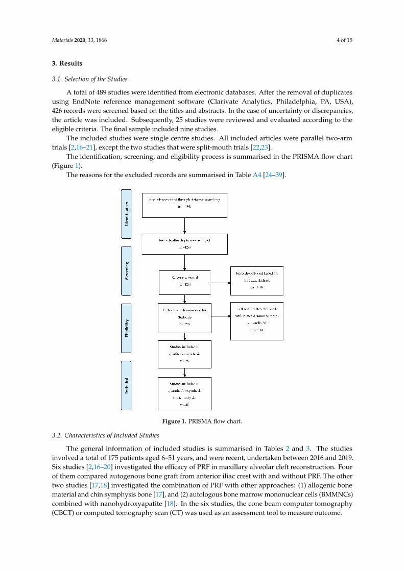
A total of 489 studies were identified from electronic databases. After the removal of duplicatesusing EndNote reference management software (Clarivate Analytics, Philadelphia, PA, USA),426 records were screened based on the titles and abstracts. In the case of uncertainty or discrepancies,the article was included. Subsequently, 25 studies were reviewed and evaluated according to theeligible criteria. The final sample included nine studies.
The included studies were single centre studies. All included articles were parallel two-armtrials [2,16–21], except the two studies that were split-mouth trials [22,23].
The identification, screening, and eligibility process is summarised in the PRISMA flow chart(Figure 1).
The reasons for the excluded records are summarised in Table A4 [24–39].Materials 2020, 13, x FOR PEER REVIEW 5 of 18
Figure 1. PRISMA flow chart.
3.2. Characteristics of Included Studies
The general information of included studies is summarised in Tables 2 and 3. The studies involved a total of 175 patients aged 6–51 years, and were recent, undertaken between 2016 and 2019. Six studies [2,16–20] investigated the efficacy of PRF in maxillary alveolar cleft reconstruction. Four of them compared autogenous bone graft from anterior iliac crest with and without PRF. The other two studies [17,18] investigated the combination of PRF with other approaches: (1) allogenic bone material and chin symphysis bone [17], and (2) autologous bone marrow mononuclear cells (BMMNCs) combined with nanohydroxyapatite [18]. In the six studies, the cone beam computer tomography (CBCT) or computed tomography scan (CT) was used as an assessment tool to measure outcome.
Three articles investigated tooth movement and post-orthodontic stability [21–23]. Two studies evaluated the amount of OTM by decreasing the horizontal linear distance between the mid-marginal ridges of the adjacent teeth [22,23]. Only one study considered clinical parameters and patient feedback to evaluate pain, post-surgical inflammation, and infection [21].
Figure 1. PRISMA flow chart.
3.2. Characteristics of Included Studies
The general information of included studies is summarised in Tables 2 and 3. The studiesinvolved a total of 175 patients aged 6–51 years, and were recent, undertaken between 2016 and 2019.Six studies [2,16–20] investigated the efficacy of PRF in maxillary alveolar cleft reconstruction. Fourof them compared autogenous bone graft from anterior iliac crest with and without PRF. The othertwo studies [17,18] investigated the combination of PRF with other approaches: (1) allogenic bonematerial and chin symphysis bone [17], and (2) autologous bone marrow mononuclear cells (BMMNCs)combined with nanohydroxyapatite [18]. In the six studies, the cone beam computer tomography(CBCT) or computed tomography scan (CT) was used as an assessment tool to measure outcome.

Materials 2020, 13, 1866 5 of 15
Table 2. Characteristics of included studies on alveolar cleft reconstruction.
Alveolar Cleft Reconstruction
Study Omidkhoda et al. [16] Movahedian Attar et al. [17] El-Ahmady et al. [18] Saruhan et al. [2] Shawky et al. [19] Desai et al. [20]
Year 2018 2017 2018 2018 2016 2019
Study design Parallel-group RCT Parallel-group RCT Parallel-group RCT Parallel-group RCT Parallel-group RCT Parallel-group RCT
Aim of studyEfficacy of PRF in the quality
and quantity of maxillaryalveolar cleft repair.
Efficacy of (i) combination of symphysisbone, allograft and PRF, and (ii) iliac
bone graft, in the regeneration of cleftdefects.
Use of autologous BMMNCs combinedwith PRF and nanohydroxyapatite as an
effective technique for alveolar cleftrepair.
Effect of PRF in alveolar bonegrafting using volumetric
analysis.
Effect of PRF in the quality andquantity of unilateral maxillary
alveolar cleft repair.
Efficacy of PRF for secondaryalveolar bone grafting.
Interventions Autogenous anterior iliac graftwith PRF (n = 5)
Bone graft from allogenic bone material,chin symphysis bone and L-PRF (n = 10)
Autologous BMMNCs combined withnanohydroxyapatite and autologous
PRF (n = 10)
Autogenous bone graft fromanterior iliac crest with PRF
(n = 17 alveolar cleft segment)
Autogenous bone graft fromanterior iliac crest with PRF
(n = 12)
Autogenous bone graft fromanterior iliac crest with PRF
(n = 20)
Control Autogenous anterior iliac graft(n = 5)
Autogenous bone graft from anterioriliac crest (n = 10)
Autogenous bone graft from anterioriliac crest (n = 10)
Autogenous bone graft fromanterior iliac crest (n = 14
alveolar cleft segment)
Autogenous bone graft fromanterior iliac crest (n = 12)
Autogenous bone graft fromanterior iliac crest (n = 20)
Sample Size(females/males) 10 (4/6) 20 (9/11) 20 (12/8) 31 alveolar cleft segments in 22
patients (13/9) 24 (8/16) 40 (19/21)
Participant age(mean ± SD)
9–12(11.3 ± 0.83)
8–14(9.7 ± 1.7)
8–15(11.50 ± 7.55)
6–28(17.71 ± 5.4)
9–14(10.92 ± 2.75)
9–18(15.29 ± 4.79)
PRFProtocol 3000 rpm, 10 min 3000 rpm, 10 min 3000 rpm, 20 min 3000 rpm, 10 min 3000 rpm, 10 min 2900 rpm, 10 min
Outcome assessed
CBCT images (Planmeca,Finland, 2009). Exposure
parameters: field of view of 90× 100 mm, voxel size of
200 µm, X-ray tube kilovoltageof 88 kVp, and 8 mA.
CBCT Images (Cranex 3D, Sordex,Helsinki, Finland). Exposure
parameters: 0.5 mm scan thickness foraxial cuts.
Panoramic radiographs and CBCTimages. Pain was measured with a
numerical scale score reporting painintensity.
CBCT Images (NewTom FP,Quantitative Radiology,Verona, Italy). Exposureparameters: 0.5 mm scanthickness for axial cuts.
CT scan (Philips Brilliance 32Slice.Cardiac MDCT, Philips
Healthcare, city, Netherlands)of upper jaw. The axial cuts
were 0.625 mm thick.
Orthopantomogram, upperocclusal view, and CT scan
(KODAK 9000C and KODAK9000C 3D extra oral Imaging
System, 2016; Carestream Inc.,New York, USA).
Follow up3 months. CBCT images:
immediate postoperative and 3months after surgery.
12 months. CBCT images: beforesurgery and 12 months after. Clinical
controls: 1week, 1, 3, 6 and 12 months.
12 months. CBCT images: 6 and 12months after surgery. Clinical Controls:1 day, 1 and 3 weeks, 6 and 12 months
after surgery.
6 months. CBCT images:preoperative and 6 months
after surgery. Clinical controls:every week during the firstmonth; every month for the
next 5 months.
6 months. CBCT images:preoperative and 6 months
after surgery.
9 months. Radiographicassessment: preoperative;
immediate, 3, 6 and 9 monthspostoperative.
Conclusion
PRF group did not have asignificant increase in the
thickness, height, and densityof alveolar bone graft.
Averagely 69.5% of alveolar defects wereregenerated with bone in experimentalgroup and 73.8% on control group (Pvalue = 0.156). Chin symphysis bone
and allogenic bone material combinedwith L-PRF was an appropriate graft
material.
Experimental group demonstrated 90%of complete alveolar bone union verses
70% in control group. AutologousBMMNCs in combination with
autologous PRF andnanohydroxyapatite promote bone
regeneration in alveolar clefts defects.
Postoperative newly formedbone volume was better in theexperimental group (68.21%)
than in control group (64.62%).Although, no statisticallysignificant difference was
found.
The mean amount andpercentage of newly formed
bone volume was higher in theexperimental (0.78 cm3; 82.6%)than control group (0.62 cm3;
68.38%). Bone density does notincrease, but the difference was
not statistically significant.
PRF in combination withautogenous bone results in
higher osteogenic effect whichincreases new bone
regeneration and better woundhealing.
RCT, randomized controlled trial. PRF, platelet-rich fibrin. SD, standard deviation. CBCT, cone beam computer tomography. L-PRF, leucocyte- and platelet-rich fibrin. BMMNCs, bonemarrow mononuclear cells. CT, computed tomography scan.

Materials 2020, 13, 1866 6 of 15
Table 3. Characteristics of included studies on tooth movement and post-orthodontic stability.
Tooth Movement and Post-Orthodontic Stability
Study Muñoz et al. [21] Tehranchi et al. [22] Nemtoi et al. [23]
Year 2016 2018 2018
Study design Cohort Split mouth clinical trial RCT Split mouth clinical trial CCT
Aim of study Effect of L-PRF in PAOO concerning post-operative pain,inflammation, infection and post-orthodontic stability. Effect of LPRF on OTM in extraction cases. Efficacy of PRF in accelerating bone regeneration and
orthodontic tooth movement.
Interventions Wilcko’s modified PAOO technique combined with L-PRF Extraction socket with LPRF (n = 15) Extraction socket with LPRF (n = 20)
Control NA Extraction socket with secondary healing (n = 15) Extraction socket with secondary healing (n = 20)
Sample Size (females/males) 11 (8/3) Thirty extraction sockets in 8 patients (3/5) Forty extraction sockets in 20 patients (11/9)
Participant age (mean ± SD) 15–51 12–25 (17.37 ± 12.48) 12–20 (16.43)
PRF Protocol 3000 rpm, 10 min 2700 rpm, 12 min 2700 rpm, 12 min
Outcome assessed Clinical parameters and patient feedback were used toevaluate pain, post-surgical inflammation and infection.
OTM was measured by comparing the change in horizontallinear distance between the mid-marginal ridges of the adjacent
teeth on a regular basis.
CBCT images (PlanmecaPromax 3D Mid, Planmeca OY,Helsinki, Finland). Exposure conditions: 90 kV, 12 mA, andexposure time of 18.3 s. OTM was measured by comparing
the change in horizontal linear distance between themid-marginal ridges of the adjacent teeth on a regular basis.
Follow up Clinical evaluation: 1, 2, 4, 8 and 10 days post-operative. 16 weeks. OTM measurements: 2, 4, 6, 8, 10, 12, 14 and 16 weeks. 24 weeks. OTM measurements: before placement of PRF; 4,8, 12, 16, 20 and 24 weeks after placement of PRF.
Conclusion
(1) No severe pain; (2) Edema resolution begun by day 4with most patients (72.7%); (3) Orthodontic treatmentaverage time was 9.3 months; (4) All cases maintained
stability for at least 2 years.
LPRF group: decreased horizontal linear measurement betweenthe mid marginal ridges of teeth (p = 0.006). Therefore, LPRF
may accelerate OTM, particularly in extraction cases.
PRF group: decreased horizontal linear measurementbetween the mid marginal ridges of teeth. Therefore, LPRF
may accelerate OTM, particularly in extraction cases.
RCT, randomized controlled trial. PRF, platelet-rich fibrin. SD, standard deviation. CBCT, cone beam computer tomography. L-PRF, leucocyte- and platelet-rich fibrin. PAOO, periodontallyaccelerated osteogenic orthodontics. OTM, orthodontic tooth movement. CCT, controlled clinical trial.

Materials 2020, 13, 1866 7 of 15
Three articles investigated tooth movement and post-orthodontic stability [21–23]. Two studiesevaluated the amount of OTM by decreasing the horizontal linear distance between the mid-marginalridges of the adjacent teeth [22,23]. Only one study considered clinical parameters and patient feedbackto evaluate pain, post-surgical inflammation, and infection [21].
3.3. Risk of Bias
The results of the quality assessment of the RCTs and CCTs studies are summarized in Figure 2.Three studies were judged as having high risk of bias, mostly due to deviations from the randomizationprocess [2,22,23]. Two trials were judged as low risk of bias [17,19]. The remaining studies wereconsidered as unclear risk due to deviations from the randomization process [16,18,20] and bias inselection of the reported results [16,20].
Materials 2020, 13, x FOR PEER REVIEW 9 of 18
3.3. Risk of Bias
The results of the quality assessment of the RCTs and CCTs studies are summarized in Figure 2. Three studies were judged as having high risk of bias, mostly due to deviations from the randomization process [2,22,23]. Two trials were judged as low risk of bias [17,19]. The remaining studies were considered as unclear risk due to deviations from the randomization process [16,18,20] and bias in selection of the reported results [16,20].
Cohort study was considered as a moderate risk of bias due to deviations from the selection of participants into the study, the measurement of outcomes and the selection of the reported results [21]. The remaining domains showed a low risk of bias. The limited number of trials did not allow risk of bias assessment across studies.
Figure 2. Risk of bias of the RCTs and CCTs studies. + low risk of bias. ? unclear risk of bias. – high risk of bias.
3.4. Quantitative Synthesis of the Results
The heterogeneous interventions and treatment performed in the included articles did not allow a quantitative synthesis of the results. Furthermore, the heterogeneity in the design and methodologies precludes the quantitative analysis of results.
3.5. Results of included studies
There was a range of different PRF protocol preparation in the included studies. Five studies [2,16,17,19,21] adjusted centrifugation to 3000 rpm for 10 min, while others used 3000 rpm for 20 min [18], 2900 rpm for 10 min [20], and 2700 rpm for 12 min [22,23].
The nine studies were grouped under two categories: (1) alveolar cleft reconstruction and (2) tooth movement and post-orthodontic stability.
3.5.1. Alveolar Cleft Reconstruction
All the works that evaluated alveolar cleft reconstruction comprised a control group in which patients went through maxillary alveolar cleft reconstruction with autogenous anterior iliac crest bone graft [2,16–19]. Four studies compared control group with autogenous anterior iliac graft only with PRF [2,16,19,20]. Omidkhoda et al. showed that PRF combined with autogenous bone did not
Figure 2. Risk of bias of the RCTs and CCTs studies. + low risk of bias. ? unclear risk of bias. – highrisk of bias.
Cohort study was considered as a moderate risk of bias due to deviations from the selection ofparticipants into the study, the measurement of outcomes and the selection of the reported results [21].The remaining domains showed a low risk of bias. The limited number of trials did not allow risk ofbias assessment across studies.
3.4. Quantitative Synthesis of the Results
The heterogeneous interventions and treatment performed in the included articles did not allow aquantitative synthesis of the results. Furthermore, the heterogeneity in the design and methodologiesprecludes the quantitative analysis of results.
3.5. Results of Included studies
There was a range of different PRF protocol preparation in the included studies. Fivestudies [2,16,17,19,21] adjusted centrifugation to 3000 rpm for 10 min, while others used 3000 rpm for20 min [18], 2900 rpm for 10 min [20], and 2700 rpm for 12 min [22,23].
The nine studies were grouped under two categories: (1) alveolar cleft reconstruction and (2)tooth movement and post-orthodontic stability.
3.5.1. Alveolar Cleft Reconstruction
All the works that evaluated alveolar cleft reconstruction comprised a control group in whichpatients went through maxillary alveolar cleft reconstruction with autogenous anterior iliac crest bonegraft [2,16–19]. Four studies compared control group with autogenous anterior iliac graft only withPRF [2,16,19,20]. Omidkhoda et al. showed that PRF combined with autogenous bone did not have a

Materials 2020, 13, 1866 8 of 15
significant increase in the thickness, height, and density of alveolar bone graft [16]. The other threestudies suggested that experimental group had higher amount of newly formed bone [2,19,20].
Saruhan et al. reported that the mean percentage of newly formed bone was 68.21 ± 10.80% and64.62 ± 9.49% in experimental and control group respectively [2]. Desai et al. evaluated vertical boneheight with a four-point scale (type 1—0%–25% resorption; type 2—25%–50%; type 3—50%–75%; type4—75%–100%). In the experimental group, 18 patients had grade 1 resorption and two patients hadgrade 2 at nine months. In the control group, 12 patients had grade 1 resorption and eight patients hadgrade 2 at nine months [20]. Shawky et al. verified that the experimental group had higher percentageof newly formed bone (82.60%) compared with control group (68.38%). This study is the only one withstatistically significant results assessing the percentage of newly formed bone [19].
Concerning the mean bone density, experimental group demonstrated lower values than controlgroup with no statistically significant differences. Shawky et al. reported values of 384.03 HU and360.82 HU, respectively for control and experimental groups, at six months follow-up [19]. Omidkhodaet al. also verified that the mean bone density was lower in experimental group (302.83 HU) than oncontrol group (349.58) at three months after surgery [16].
The two studies that included other materials [17,18] have found that PRF combined withother regenerative materials was an appropriate graft material for reconstruction in alveolar cleftsdefects. Movahedian et al. evaluated the efficiency of the combination of bone graft from allogenicbone material, chin symphysis bone and leucocyte- and platelet-rich fibrin (L-PRF) and verified thatthe percentages of bone reconstruction were lower in experimental group (69.57 ± 10.13%) than incontrol group (73.86 ± 6.93%), without statistical differences between the two groups [17]. Otherwise,El-Ahmady et al. showed that 70% of the experimental group (using PRF with autologous BMMNCsand nanohydroxyapatite) presented bone tissue at the cementoenamel junction of the teeth next to thecleft covering at least 75% of both roots against 30% of the control group, at 12 months follow up [18].
The most frequent outcome measures were volumetric measurements and percentage ratiossuch as height, thickness and length. Four studies evaluated augmentation, bone reconstruction andgraft ratios by comparing pre and postoperative 3D X-rays [2,17,19,20]. The evaluation of alveolarresorption or residual bone ratio and postoperative follow up was performed using 3D X-rays [16,18].The follow–up ranged from 3 to 12 months across the studies. During the follow-up period, differentoutcomes were reported. Two studies did not verify complications (dehiscence, flap necrosis andinfection or persistent oro-nasal fistula) [17,19], whereas another two studies identified persistentoro-nasal fistula in 30% of the control group [18] and dehiscence in four patients in the experimentalgroup (n = 20) and eight patients in control group (n = 20) [19]. The other two included studies [2,16]did not report any information.
3.5.2. Tooth Movement and Post-Orthodontic Stability
Two trials evaluated the effect of L-PRF or PRF on orthodontic tooth movement [22,23]. Bothstudies showed that application of these platelets’ derivatives in the extraction socket can accelerateorthodontic tooth movement (p = 0.006) [22,23], and it has been shown by Tehranchi et al. that therewas no statistical difference between teeth in the maxillary and mandibular arch on OTM rate [22].
The study by Muñoz et al. was the only one that evaluated the effects of L-PRF consideringinflammation and post-orthodontic stability. They demonstrated that, in a high percentage of patients(72.7%), edema resolution was set about day 4, and orthodontic stability was preserved for two ormore years post-surgery in all patients [21].
4. Discussion
Alveolar cleft reconstruction and accelerated orthodontic tooth movement are matters of concernin contemporary orthodontics [28,40]. However, few studies were found assessing application of PRFon orthodontics compared to other dentistry areas.

Materials 2020, 13, 1866 9 of 15
All included articles in this systematic review studied the advantages of PRF in the orthodonticsfield. Although a quantitative report of the findings was not possible, qualitative systematic reviewsstill improve the current understanding and provide a critical appraisal of research relevant toregenerative orthodontics.
To evaluate the postoperative newly formed bone, 2D or 3D X-rays are required in additionto clinical assessment. Three-dimensional radiological studies had some benefits compared withtwo-dimensional ones, namely the three-dimensional location of the bone graft and the assessment ofteeth eruption process on alveolar graft [41]. The advantages of CBCT over CT include the possibilityto scan small regions for specify diagnosis, minimal scanning time (10–70 s), low radiation dose,and reduced image artefact [42]. In the present study, CBCT scan was used in five trials [2,16–18,23]and CT scan in two trials [19,20]. Due to its high performance, CBCT should be a standard treatmentoutcome method for the assessment of newly formed bone in further studies [28]. According toThuaksuban et al., the remodelling process with cortical maturation occurs after six months, becomingstable until month 24 [43]. Therefore, CBCT should be carried out six months after the bone graft.Regarding the included studies, Omidkhoda et al. only evaluated a three month time-point [16],and thus their results should be carefully analysed as the remodelling process may not be completed.
Orthodontic treatment combined with surgical approaches is a common procedure in cleft lip andpalate patients. In these patients, the treatment begins at birth and continues into adulthood usuallyrequiring prosthesis in an anterior region or mesial movement of the posterior teeth to space closureof agenesis, mainly of the upper lateral incisor [44]. Alveolar cleft reconstruction with bone graftallows for an adequate volume of alveolar bone, which is fundamental for the dental movement in themaxillary aesthetic zone throughout the orthodontic treatment. Thus, the orthodontist can performmore stable and aesthetic treatments. In patients with alveolar cleft, autogenous iliac crest bone isthe gold standard [45]. Autogenous bone graft is osteoconductive, osteoinductive and a source ofosteogenic precursor cells [19]. However, new strategies, such as the use of PRF, have been advancedto speed up bone formation, reduce bone resorption and enhance soft tissue healing. PRF is a plateletconcentrate without addition of thrombin or anticoagulants [6]. The physiologic polymerization inPRF allows the cytokines and growth factors to be stored and then slowly released, ensuring bioactivelevels for a long time-period (up to 28 days) [46]. Besides protecting the surgical site, PRF membranespromote soft tissue healing functioning like a matrix to support neoangiogenesis, and migration of stemcells and osteoprogenitor cells into the graft [6,19]. In line with this, PRF decreases bone resorption andhasten wound healing in soft and hard tissues [11], which might contribute to the lower prevalence ofcomplications during the follow-up period observed in the PRF group compared to the control group,reported by El-Ahmady et al. [18] and, also, to the increased new bone regeneration and better woundhealing observed by Desai et al. [20]. These results are in line with other stating that PRF increasedsignificantly root coverage [47,48].
Regarding orthodontic tooth movement, several non-invasive or invasive techniques have beenproposed for accelerating this process. Most non-invasive techniques need more studies to prove theirclinical effectiveness [49]. Invasive techniques appear to be more effective in promoting orthodontictooth movement. The bone injury associated to the surgical procedure triggers a tissue reactionthat enhances normal molecular and cellular events involved in tissue healing [50]. Somehow, theapplication of PRF mimics the surgical-induced healing capabilities, also inducing tissue regeneration.Being a physiologic polymerized fibrin matrix, PRF incorporates platelets, leucocytes, bioactivemolecules and trapped circulating stem cells and progenitors to promote local tissue healing [6].The two trials included in the present systematic review showed that the use of PRF or L-PRF inthe extraction socket could accelerate OTM (p = 0.006), specifically in the beginning of orthodontictreatment (alignment and leveling) [22,23]. These results are in line with previous ones reporting thatthe application of several bioactive grafts can increase the bone maturation without interfering withthe natural healing process [51]. However, cytokines and growth factors levels are maintained for along time-period (up to 28 days). Liou demonstrated that the clinical effect of application of platelet

Materials 2020, 13, 1866 10 of 15
derivatives could last 5–6 months with the faster rate of orthodontic movement in 2–4 months [52].Although no conclusion of the potential effect of PRF on this process could be drawn based on thesetwo trials, there was a trend that PRF has the ability to increase tooth movement. Thus, the applicationof PRF may shorten the orthodontic treatment time reducing associated costs, which nowadays is aconcern in orthodontic patients, specifically in adults and patients with longer treatments such as thoseneeding tooth extractions. Nevertheless, PRF has some disadvantages, namely the limited volume thatcan be produced and used, and tissue banks are impracticable, as it is specific to the donor and cannotbe used as an allogenic graft tissue [6].
4.1. Limitations in this Review
The methodological and clinical heterogeneity among studies only allowed to qualitatively accountthe findings of this systematic review. Newly formed bone measurement tools were inconsistent acrossstudies. Furthermore, most of the selected studies were classified as having a high or unclear risk ofbias, which may decrease the certainty of the results. The heterogeneity of studies can be justified bythe methodological differences across the studies, such as sample sizes, intervention protocols andfollow-up times. Furthermore, several factors can affect local bone remodeling, namely age at surgery,width of the cleft defect, volume of grafted bone, and position of teeth on bone graft.
The authors recognize that the expertise of the clinician and support team, as well as the scientificproficiency of the all research group, influence the outcome evaluation. Some of the selected studiesdid not assess this factor, which should be considered when figuring out the results of this review.
4.2. Recommendations for Future Research
Given the inconsistent results presented in the limited literature, it is recommended to performfurther research with standardized methodologies, a larger sample size, and longer follow-up toevaluate the applications and effects of PRF in the orthodontics field. Possible sources of bias shouldbe controlled, such as randomization procedure, PRF protocols preparations, measurement tools ofnewly formed bone, and follow-up periods. Further studies should also investigate the cost–benefitanalysis of using PRF in orthodontics for patients and clinicians.
5. Conclusions
Despite the limitations in the included studies, this systematic review suggested that PRF canimprove alveolar cleft reconstruction. Concerning orthodontic tooth movement, the results highlightthe positive effects of PRF, since it may shorten the orthodontic treatment time, thereby reducingassociated costs.
Further, high-quality randomised controlled trials with identical methodologies, larger samplesize, and longer follow-up periods are required.
Author Contributions: Conceptualization, I.F.; methodology, I.F. and F.V; investigation, all authors; data curation,all authors; writing—original draft preparation, I.F.; writing—review and editing, F.V. and M.H.F.; supervision,M.H.F. All authors have read and agreed to the published version of the manuscript.
Funding: This research received no external funding.
Conflicts of Interest: The authors declare no conflict of interest.

Materials 2020, 13, 1866 11 of 15
Appendix A
Table A1. Search Strategy for each of the databases
Database Search Equation
Medline (via PubMed)
(“Orthodontics”[Mesh] OR “Tooth Movement Techniques”[Mesh] OR “Orthodontic Brackets”[Mesh] OR “ Tooth Movement”OR “Accelerating Orthodontic” OR “Surgery, Oral”[Mesh] OR “Alveolar Bone Grafting”[Mesh] OR “Post extraction Socket” OR“Socket Preservation” OR “Guided Tissue Regeneration”[Mesh] OR “Bone Regeneration”[Mesh] OR “Tissue Scaffolds”[Mesh]
OR “Bone Transplantation”[Mesh] OR “Bone Remodeling”[Mesh] OR “Bone Substitutes”[Mesh]) AND (“Platelet-RichFibrin”[Mesh] OR “Platelet Rich Fibrin” OR “Fibrin rich in growth factors” OR “Platelet concentrate” OR “PRF” OR “Second
generation platelet rich fibrina” OR “osteoinductive biomaterials”)
Cochrane Library (Mesh descriptor: [Orthodontics] OR Mesh descriptor: [Alveolar Bone Grafting] OR Mesh descriptor: [Bone Regeneration] ORtooth movement OR alveolar bone grafting) AND (Mesh descriptor: [Platelet-Rich Fibrin] OR PRF OR Platelet rich fibrin)
Web of Science CoreCollection
TS = (platelet rich fibrin* OR platelet-rich fibrin * OR PRF* OR second generation platelet concentrate* OR platelet concentrate*)AND TS = (orthodontics* OR tooth movement* OR alveolar bone grafting* OR orthodontic brackets*)
EMBASE (“Orthodontics” OR “Orthodontic tooth movement” OR “Orthodontic device” OR “Alveolar Bone Grafting”) AND (“Tissueregeneration” OR “bone regeneration”) AND (“Platelet-rich fibrin” OR “platelet AND concentrate”)
Table A2. Version 2 of the Cochrane tool for assessing risk of bias (RoB 2) [14].
Response OptionsBias Domain and Signalling Question *
Lower Risk of Bias Higher Risk of Bias Other
Bias arising from the randomisation process
1.1 Was the allocation sequence random? Y/PY N/PN NI
1.2 Was the allocation sequence concealed until participants were enrolled and assigned to interventions? Y/PY N/PN NI
1.3 Did baseline differences between intervention groups suggest a problem with the randomisation process? N/PN Y/PY NI
Risk-of-bias judgment (low/high/some concerns)
Optional: What is the predicted direction of bias arising from the randomisation process?
Bias due to deviations from intended interventions
2.1 Were participants aware of their assigned intervention during the trial? N/PN Y/PY NI
2.2 Were carers and people delivering the interventions aware of participants’ assigned intervention duringthe trial? N/PN Y/PY NI
2.3 If Y/PY/NI to 2.1 or 2.2: Were there deviations from the intended intervention that arose because of thetrial context? N/PN Y/PY NA/NI
2.4 If Y/PY/NI to 2.3: Were these deviations likely to have affected the outcome? N/PN Y/PY NA/NI
2.5 If Y/PY to 2.4: Were these deviations from intended intervention balanced between groups? Y/PY N/PN NA/NI
2.6 Was an appropriate analysis used to estimate the effect of assignment to intervention? Y/PY N/PN NI
2.7 If N/PN/NI to 2.6: Was there potential for a substantial impact (on the result) of the failure to analyseparticipants in the group to which they were randomised? N/PN Y/PY NA/NI
Risk-of-bias judgment (low/high/some concerns)
Optional: What is the predicted direction of bias due to deviations from intended interventions?
Bias due to missing outcome data
3.1 Were data for this outcome available for all, or nearly all, participants randomised? Y/PY N/PN NI
3.2 If N/PN/NI to 3.1: Is there evidence that the result was not biased by missing outcome data? Y/PY N/PN NA
3.3 If N/PN to 3.2: Could missingness in the outcome depend on its true value? N/PN Y/PY NA/NI
3.4 If Y/PY/NI to 3.3: Is it likely that missingness in the outcome depended on its true value? N/PN Y/PY NA/NI
Risk-of-bias judgment (low/high/some concerns)
Optional: What is the predicted direction of bias due to missing outcome data?
Bias in measurement of the outcome
4.1 Was the method of measuring the outcome inappropriate? N/PN Y/PY NI
4.2 Could measurement or ascertainment of the outcome have differed between intervention groups? N/PN Y/PY NI
4.3 If N/PN/NI to 4.1 and 4.2: Were outcome assessors aware of the intervention received by study participants? N/PN Y/PY NI
4.4 If Y/PY/NI to 4.3: Could assessment of the outcome have been influenced by knowledge ofintervention received? N/PN Y/PY NA/NI
4.5 If Y/PY/NI to 4.4: Is it likely that assessment of the outcome was influenced by knowledge ofintervention received? N/PN Y/PY NA/NI
Risk-of-bias judgment (low/high/some concerns)
Optional: What is the predicted direction of bias in measurement of the outcome?
Bias in selection of the reported result
5.1 Were the data that produced this result analysed in accordance with a prespecified analysis plan that wasfinalised before unblinded outcome data were available for analysis? Y/PY N/PN NI
Is the numerical result being assessed likely to have been selected, on the basis of the results, from:
5.2 ... multiple eligible outcome measurements (e.g., scales, definitions, time points) within the outcome domain? N/PN Y/PY NI
5.3 ... multiple eligible analyses of the data? N/PN Y/PY NI
Risk-of-bias judgment (low/high/some concerns)
Optional: What is the predicted direction bias due to selection of the reported results?
Overall bias
Risk-of-bias judgment (low/high/some concerns)
Optional: What is the overall predicted direction of bias for this outcome?
Y = yes; PY = probably yes; PN = probably no; N = no; NA = not applicable; NI = no information

Materials 2020, 13, 1866 12 of 15
Table A3. Robins-I tool for assessing risk of bias for non-randomised studies [15].
Domain Explanation
Pre-intervention Risk of bias assessment is mainly distinct from assessments of randomised trials
Bias due to confounding
Baseline confounding occurs when one or more prognostic variables (factors that predict the outcome of interest) also predictsthe intervention received at baseline.ROBINS-I can also address time-varying confounding, which occurs when individuals switch between the interventions beingcompared and when post-baseline prognostic factors affect the intervention received after baseline.
Bias in selection ofparticipants into the
study
When exclusion of some eligible participants, or the initial follow-up time of some participants, or some outcome events isrelated to both intervention and outcome, there will be an association between interventions and outcome even if the effects ofthe interventions are identical.This form of selection bias is distinct from confounding—A specific example is bias due to the inclusion of prevalent users,rather than new users, of an intervention.
At intervention Risk of bias assessment is mainly distinct from assessments of randomised trials
Bias in classification ofinterventions
Bias introduced by either differential or non-differential misclassification of intervention status.Non-differential misclassification is unrelated to the outcome and will usually bias the estimated effect of intervention towardsthe null.Differential misclassification occurs when misclassification of intervention status is related to the outcome or the risk of theoutcome, and is likely to lead to bias.
Post-intervention Risk of bias assessment has substantial overlap with assessments of randomised trials
Bias due to deviationsfrom intendedinterventions
Bias that arises when there are systematic differences between experimental intervention and comparator groups in the careprovided, which represent a deviation from the intended intervention(s).Assessment of bias in this domain will depend on the type of effect of interest (either the effect of assignment to intervention orthe effect of starting and adhering to intervention).
Bias due to missing dataBias that arises when later follow-up is missing for individuals initially included and followed (such as differential loss tofollow-up that is affected by prognostic factors); bias due to exclusion of individuals with missing information aboutintervention status or other variables such as confounders.
Bias in measurement ofoutcomes
Bias introduced by either differential or non-differential errors in measurement of outcome data. Such bias can arise whenoutcome assessors are aware of intervention status, if different methods are used to assess outcomes in different interventiongroups, or if measurement errors are related to intervention status or effects.
Bias in selection of thereported result
Selective reporting of results in a way that depends on the findings and prevents the estimate from being included in ameta-analysis (or other synthesis).
Table A4. List of excluded studies.
Study Reason for Exclusion
Alhasyimi et al. [24] Animal studyChe et al. [25] Review article
Dukka et al. [26] Case ReportJanssen et al. [27] Evaluates PRP and not PRFStasiak et al. [28] Systematic reviewShetty et al. [29] Trial registration
Subbalekha et al. [30] Trial registrationAvinash et al. [31] Trial registration
Tehranchi et al. [32] Trial registrationMazzone et al. [33] Not orthodontic field
Shah et al. [34] Technical noteIskenderoglu et al. [35] Case Report
Dimofte et al. [36] Review articleNadon et al. [37] Evaluate a derivate of PRFAras et al. [38] Case Report
Findik et al. [39] Case Report
References
1. Miron, R.J.; Zucchelli, G.; Pikos, M.A.; Salama, M.; Lee, S.; Guillemette, V.; Fujioka-Kobayashi, M.; Bishara, M.;Zhang, Y.; Wang, H.L.; et al. Use of platelet-rich fibrin in regenerative dentistry: A systematic review.Clin. Oral Investig. 2017, 21, 1913–1927. [CrossRef] [PubMed]
2. Saruhan, N.; Ertas, U. Evaluating of platelet-rich fibrin in the treatment of alveolar cleft with iliac bone graftby means of volumetric analysis. J. Craniofac. Surg. 2018, 29, 322–326. [CrossRef] [PubMed]
3. Meshram, V.S.; Lambade, P.N.; Meshram, P.V.; Kadu, A.; Tiwari, M.S. The autologous platelet rich fibrin:A novel approach in osseous regeneration after cystic enucleation: A pilot study. Indian J. Dent. Res. 2015, 26,560–564. [CrossRef]
4. Crisci, A.; Marotta, G.; Licito, A.; Serra, E.; Benincasa, G.; Crisci, M. Use of leukocyte platelet (l-prf) richfibrin in diabetic foot ulcer with osteomyelitis (three clinical cases report). Diseases 2018, 6, 30. [CrossRef]

Materials 2020, 13, 1866 13 of 15
5. Zumarán, C.C.; Parra, M.V.; Olate, S.A.; Fernández, E.G.; Muñoz, F.T.; Haidar, Z.S. The 3 R’s for platelet-richfibrin: A “super” tri-dimensional biomaterial for contemporary naturally-guided oro-maxillo-facial soft andhard tissue repair, reconstruction and regeneration. Materials 2018, 11, 1293. [CrossRef]
6. Choukroun, J.; Diss, A.; Simonpieri, A.; Girard, M.O.; Schoeffler, C.; Dohan, S.L.; Dohan, A.J.J.; Mouhyi, J.;Dohan, D.M. Platelet-rich fibrin (PRF): A second-generation platelet concentrate. Part IV: Clinical effects ontissue healing. Oral Surg. Oral Med. Oral Pathol. Oral Radiol. Endod. 2006, 101, e56–e60. [CrossRef] [PubMed]
7. Dohan, D.M.; Choukroun, J.; Diss, A.; Dohan, S.L.; Dohan, A.J.; Mouhyi, J.; Gogly, B. Platelet-rich fibrin(PRF): A second-generation platelet concentrate. Part II: Platelet-related biologic features. Oral Surg. OralMed. Oral Pathol. Oral Radiol. Endod. 2006, 101, e45–e50. [CrossRef]
8. Dohan Ehrenfest, D.M.; Diss, A.; Odin, G.; Doglioli, P.; Hippolyte, M.P.; Charrier, J.B. In vitro effects ofChoukroun’s PRF (plateletrich fibrin) on human gingival fibroblasts, dermal prekeratinocytes, preadipocytes,and maxillofacial osteoblasts in primary cultures. Oral Surg. Oral Med. Oral Pathol. Oral Radiol. Endod. 2009,108, 341–352. [CrossRef] [PubMed]
9. Toffler, M.; Toscano, N.; Holtzclaw, D.; Corso, M.; Dohan, D. Introducing Choukroun’s platelet rich fibrin(PRF) to the reconstructive surgery milieu. J. Implant Adv. Clin. Dent. 2009, 1, 22–31.
10. Limeback, H.F.; Sodek, J. Procollagen synthesis and processing in periodontal ligament in vivo and in vitro.A comparative study using slab-gel fluorography. Eur. J. Biochem. 1979, 100, 541–550. [CrossRef]
11. Shah, R.; Triveni, M.G.; Thomas, R.; Mehta, D.S. An update on the protocols and biologic actions of plateletrich fibrin in dentistry. Eur. J. Prosthodont. Restor. Dent. 2017, 25, 64–72. [PubMed]
12. Swartz, M.K. The PRISMA statement: A guideline for systematic reviews and meta-analyses. J. Pediatr.Health Care 2011, 25, 1–2. [CrossRef] [PubMed]
13. Higgins, J.P.; Altman, D.G.; Gøtzsche, P.C.; Jüni, P.; Moher, D.; Oxman, A.D.; Savovic, J.; Schulz, K.F.;Weeks, L.; Sterne, J.A. The Cochrane Collaboration’s tool for assessing risk of bias in randomised trials. BMJ2011, 343, d5928. [CrossRef] [PubMed]
14. Sterne, J.A.C.; Savovic, J.; Page, M.J.; Elbers, R.G.; Blencowe, N.S.; Boutron, I.; Cates, C.J.; Cheng, H.Y.;Corbett, M.S.; Eldridge, S.M.; et al. RoB 2: A revised tool for assessing risk of bias in randomised trials. BMJ2019, 366, l4898. [CrossRef] [PubMed]
15. Sterne, J.A.; Hernán, M.A.; Reeves, B.C.; Savovic, J.; Berkman, N.D.; Viswanathan, M.; Henry, D.; Altman, D.G.;Ansari, M.T.; Boutron, I.; et al. ROBINS-I: A tool for assessing risk of bias in non-randomized studies ofinterventions. BMJ 2016, 355, i4919. [CrossRef]
16. Omidkhoda, M.; Jahnabin, A.; Khoshandam, F.; Eslami, F.; Zarch, S.H.H.; Afshari, J.T.; Kermani, H. Efficacyof platelet-rich fibrin combined with autogenous bone graft in the quality and quantity of maxillary alveolarcleft reconstruction. Iran J. Otorhinolaryngol. 2018, 30, 329–334.
17. Movahedian Attar, B.; Naghdi, N.; Etemadi, S.M.; Mehdizadeh, M. Chin symphysis bone, allograft, andplatelet rich fibrin: Whether the combination is effective in repair of alveolar cleft? J. Oral Maxillofac. Surg.2017, 75, 1026–1035. [CrossRef]
18. El-Ahmady, H.H.; Abd Elazeem, A.F.; Bellah Ahmed, N.E.; Shawkat, W.M.; Elmasry, M.; Abdelrahman, M.A.;Abderazik, M.A. Combining autologous bone marrow mononuclear cells seeded on collagen sponge withnano hydroxyapatite, and platelet-rich fibrin: Reporting a novel strategy for alveolar cleft bone regeneration.J. Craniomaxillofac. Surg. 2018, 46, 1593–1600. [CrossRef]
19. Shawky, H.; Seifeldin, S.A. Does platelet-rich fibrin enhance bone quality and quantity of alveolar cleftreconstruction? Cleft Palate Craniofac. J. 2016, 53, 597–606. [CrossRef]
20. Desai, A.K.; Kumar, N.; Dikhit, P.; Koikude, S.B.; Bhaduri, S. Efficacy of platelet-rich fibrin in secondary cleftalveolar bone grafting. Craniomaxillofacial Trauma Reconstr. Open 2019, 3, e43–e50. [CrossRef]
21. Munoz, F.; Jiménez, C.; Espinoza, D.; Vervelle, A.; Beugnet, J.; Haidar, Z. Use of leukocyte and platelet-richfibrin (L-PRF) in periodontally accelerated osteogenic orthodontics (PAOO): Clinical effects on edema andpain. J. Clin. Exp. Dent. 2016, 8, e119–e124. [CrossRef] [PubMed]
22. Tehranchi, A.; Behnia, H.; Pourdanesh, F.; Behnia, P.; Pinto, N.; Younessian, F. The effect of autologousleukocyte platelet rich fibrin on the rate of orthodontic tooth movement: A prospective randomized clinicaltrial. Eur. J. Dent. 2018, 12, 350–357. [CrossRef] [PubMed]
23. Nemtoi, A.; Sirghe, A.; Nemtoi, A.; Haba, D. The effect of a plasma with platelet-rich fibrin in boneregeneration and on rate of orthodontic tooth movement in adolescents. Rev. Chim. 2018, 69, 3727–3730.[CrossRef]
Materials 2020, 13, 1866 14 of 15
24. Alhasyimi, A.A.; Pudyani, P.S.; Asmara, W.; Ana, I.D. Effect of carbonated hydroxyapatite incorporatedadvanced platelet rich fibrin intrasulcular injection on the alkaline phosphatase level during orthodonticrelapse. AIP Conf. Proc. 1933, 2018, 030006. [CrossRef]
25. Che, Y.; Li, P.; Tian, L.; Wang, M.; Xiong, R.; Lei, X.; Li, X.; Xu, N.; Ge, Z. The Research Progress of Platelet-RichFibrin Applications in the Orthodontic Treatment. Adv. Comput. Sci. Res. 2017, 59, 524–529.
26. Dukka, H.; Gossweiler, M.; Kishimoto, T.; Blanchard, S. Periodontally accelerated osteogenicorthodontics (paoo) using platelet rich fibrin (prf) alone: A modified approach with a 3-year follow-up.Clin. Adv. Periodontics 2018, 8, 177–181. [CrossRef]
27. Janssen, N.G.; Weijs, W.L.; Koole, R.; Rosenberg, A.J.; Meijer, G.J. Tissue engineering strategies for alveolarcleft reconstruction: A systematic review of the literature. Clin. Oral Investig. 2014, 18, 219–226. [CrossRef]
28. Stasiak, M.; Wojtaszek-Słominska, A.; Racka-Pilszak, B. Current methods for secondary alveolar bone graftingassessment in cleft lip and palate patients—A systematic review. J. Craniomaxillofac. Surg. 2019, 47, 578–585.[CrossRef]
29. Shetty, N.; Chatterjee, S. Evaluation of Bone Density Before and After Surgical Orthodontic Tooth Movement.Available online: http://www.ctri.nic.in/Clinicaltrials/pmaindet2.php?trialid=24036 (accessed on 10 December2019).
30. Subbalekha, K. Study of the Platelet Rich Fibrin in Alveolar Cleft Bone Graft by Using Cone Beam ComputedTomography. Available online: http://www.clinicaltrials.in.th/index.php?tp=regtrials&menu=trialsearch&smenu=fulltext&task=search&task2=view1&id=4073 (accessed on 10 December 2019).
31. Avinash, B.S.; Nair, M.B. A Study to Help in Accelerating Tooth Movement in Patients UndergoingClip Treatment and to Check the Bone Activity of the Tooth During the Procedure. Available online:http://www.ctri.nic.in/Clinicaltrials/pmaindet2.php?trialid=29183 (accessed on 10 December 2019).
32. Tehranchi, A. The Effect of Autologous Leukocyte Platelet Rich Fibrin (LPRF) on the Rate of OrthodonticTooth Movement—A Prospective Randomized Clinical Trial. Available online: http://en.irct.ir/trial/20620(accessed on 10 December 2019).
33. Mazzone, N.; Mici, E.; Calvo, A.; Runci, M.; Crimi, S.; Lauritano, F.; Belli, E. Preliminary results of boneregeneration in oromaxillomandibular surgery using synthetic granular graft. BioMed Res. Int. 2018, 2018,8503427. [CrossRef]
34. Shah, R.; Gowda, T.M.; Thomas, R.; Kumar, T.; Mehta, D.S. Biological activation of bone grafts using injectableplatelet-rich fibrin. J. Prosthet. Dent. 2019, 121, 391–393. [CrossRef]
35. Iskenderoglu, N.S.; Choi, B.J.; Seo, K.W.; Lee, Y.J.; Lee, B.S.; Kim, S.H. Single-tooth osteotomy combined widelinear corticotomy under local anesthesia for correcting anterior protrusion with ectopically erupted canine.J. Craniofac. Surg. 2017, 28, e30–e33. [CrossRef] [PubMed]
36. Dimofte, M.; Geletu, G.L.; Costan, V.; Benghiac, A.G.; Moscalu, M.; Popescu, E. Considerations of platelet-richfibrin use in oral surgery. Med. Surg. J. 2016, 120, 920–925.
37. Nadon, F.; Chaput, B.; Périssé, J.; de Bérail, A.; Lauwers, F.; Lopez, R. Interest of mineralized plasmaticmatrix in secondary autogenous bone graft for the treatment of alveolar clefts. J. Craniofac. Surg. 2015, 26,2148–2151. [CrossRef] [PubMed]
38. Aras, I.; Olmez, S.; Akay, M.C.; Oztürk, V.O.; Aras, A. Treatment of lateral open bite with vertical dentoalveolardistraction osteogenesis. Am. J. Orthod. Dentofac. Orthop. 2015, 148, 321–331. [CrossRef] [PubMed]
39. Findik, Y.; Baykul, T. Secondary closure of alveolar clefts with mandibular symphyseal bone grafts and withplatelet-rich fibrin under local anesthesia: Three case reports. J. Contemp. Dent. Pract. 2013, 14, 751–753.[CrossRef] [PubMed]
40. Bagga, D.K. Adult orthodontics versus adolescent orthodontics: An overview. J. Oral Health Comm. Dent.2010, 4, 42–47. [CrossRef]
41. Oberoi, S.; Chigurupati, R.; Gill, P.; Hoffman, W.Y.; Vargervik, K. Volumetric assessment of secondary alveolarbone grafting using cone beam computed tomography. Cleft Palate Craniofac. J. 2009, 46, 503–511. [CrossRef]
42. Scarfe, W.C.; Farman, A.G.; Sukovic, P. Clinical applications of cone-beam computed tomography in dentalpractice. J. Can. Dent. Assoc. 2006, 72, 75–80.
43. Thuaksuban, N.; Nuntanaranont, T.; Pripatnanont, P. A comparison of autogenous bone graft combinedwith deproteinized bovine bone and autogenous bone graft alonefor treatment of alveolar cleft. Int. J. OralMaxillofac. Surg. 2010, 39, 1175–1180. [CrossRef]
Materials 2020, 13, 1866 15 of 15
44. Halpern, R.M.; Noble, J. Location and presence of permanent teeth in a complete bilateral cleft lip and palatepopulation. Angle Orthod. 2010, 80, 591–596. [CrossRef]
45. Swan, M.C.; Goodacre, T.E. Morbidity at the iliac crest donor site following bone grafting of the cleft alveolus.Br. J. Oral Maxillofac. Surg. 2006, 44, 129–133. [CrossRef] [PubMed]
46. Del Corso, M.; Vervelle, A.; Simonpieri, A.; Jimbo, R.; Inchingolo, F.; Sammartino, G.; Dohan Ehrenfest, D.M.Current knowledge and perspectives for the use of platelet-rich plasma (PRP) and platelet-rich fibrin (PRF)in oral and maxillofacial surgery part 1: Periodontal and dentoalveolar surgery. Curr. Pharm. Biotechnol.2012, 13, 1207–1230. [CrossRef] [PubMed]
47. Agarwal, S.K.; Jhingran, R.; Bains, V.K.; Srivastava, R.; Madan, R.; Rizvi, I. Patient-centered evaluation ofmicrosurgical management of gingival recession using coronally advanced flap with platelet-rich fibrin oramnion membrane: A comparative analysis. Eur. J. Dent. 2016, 10, 121–133. [CrossRef]
48. Aroca, S.; Keglevich, T.; Barbieri, B.; Gera, I.; Etienne, D. Clinical evaluation of a modified coronally advancedflap alone or in combination with a platelet-rich fibrin membrane for the treatment of adjacent multiplegingival recessions: A 6-month study. J. Periodontol. 2009, 80, 244–252. [CrossRef]
49. Gkantidis, N.; Mistakidis, I.; Kouskoura, T.; Pandis, N. Effectiveness of non-conventional methods foraccelerated orthodontic tooth movement: A systematic review and meta-analysis. J. Dent. 2014, 42,1300–1319. [CrossRef] [PubMed]
50. Kalemaj, Z.; DebernardI, C.L.; Buti, J. Efficacy of surgical and non-surgical interventions on acceleratingorthodontic tooth movement: A systematic review. Eur. J. Oral Implantol. 2015, 8, 9–24. [PubMed]
51. Dohan Ehrenfest, D.M.; Bielecki, T.; Jimbo, R.; Barbé, G.; Del Corso, M.; Inchingolo, F.; Sammartino, G.Do the fibrin architecture and leukocyte content influence the growth factor release of platelet concentrates?An evidence-based answer comparing a pure platelet-rich plasma (P-PRP) gel and a leukocyte- andplatelet-rich fibrin (L-PRF). Curr. Pharm. Biotechnol. 2012, 13, 1145–1152. [CrossRef]
52. Liou, E.J. The development of submucosal injection of platelet rich plasma for accelerating orthodontic toothmovement and preserving pressure side alveolar bone. APOS Trends Orthod. 2016, 6, 5–11. [CrossRef]
© 2020 by the authors. Licensee MDPI, Basel, Switzerland. This article is an open accessarticle distributed under the terms and conditions of the Creative Commons Attribution(CC BY) license (http://creativecommons.org/licenses/by/4.0/).