platelets therapy
TRANSCRIPT
PLATELETS ACTIVATION IN NORMAL CONDITIONS AND DURING ACUTE
THROMBOEMBOLIC DISEASES
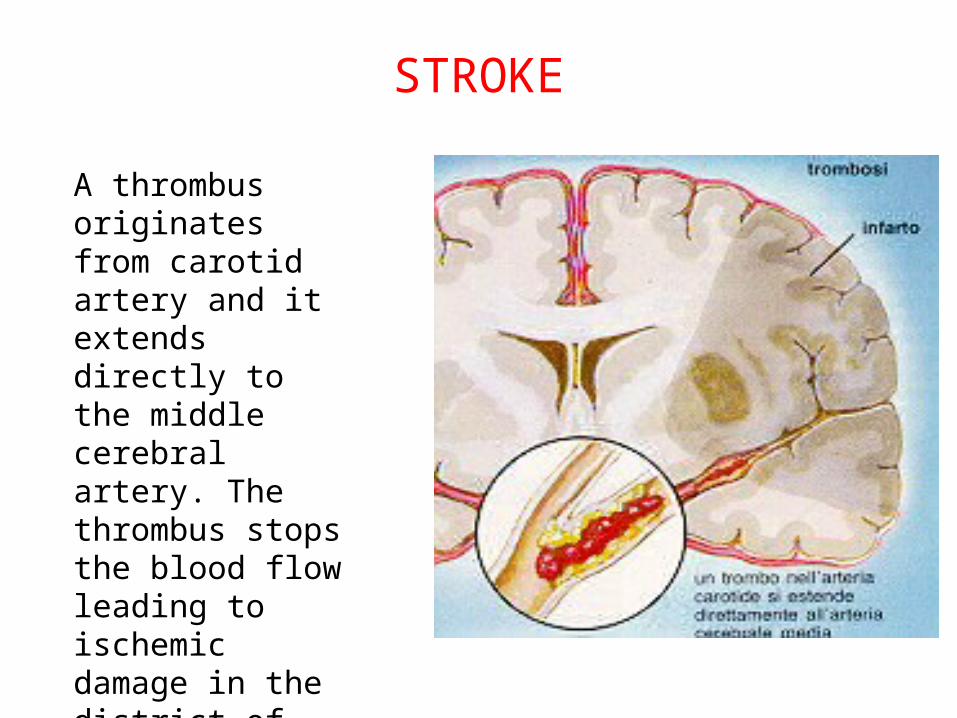
STROKE
A thrombus originates from carotid artery and it extends directly to the middle cerebral artery. The thrombus stops the blood flow leading to ischemic damage in the district of brain tissue that receives arterial blood from the blocked artery.
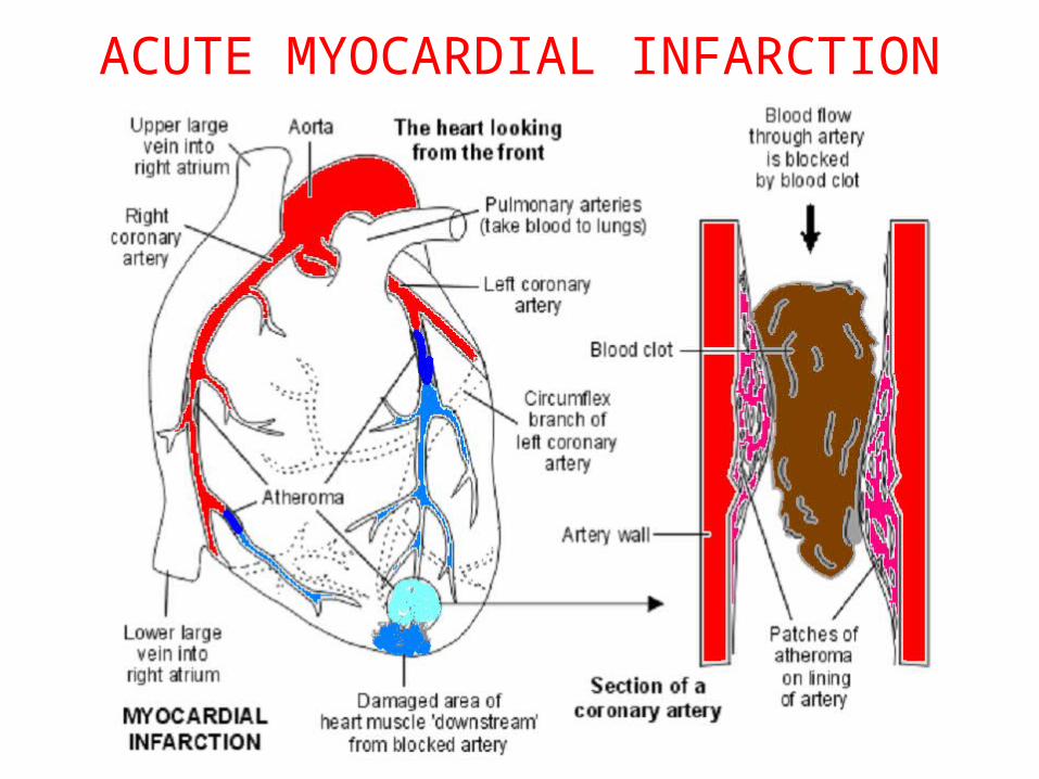
ACUTE MYOCARDIAL INFARCTION
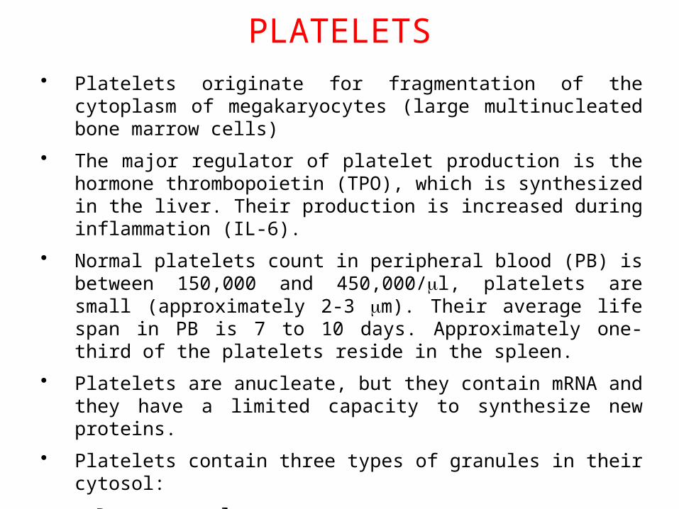
PLATELETS• Platelets originate for fragmentation of the cytoplasm of megakaryocytes
(large multinucleated bone marrow cells)
• The major regulator of platelet production is the hormone thrombopoietin (TPO), which is synthesized in the liver. Their production is increased during inflammation (IL-6).
• Normal platelets count in peripheral blood (PB) is between 150,000 and 450,000/ml, platelets are small (approximately 2-3 mm). Their average life span in PB is 7 to 10 days. Approximately one-third of the platelets reside in the spleen.
• Platelets are anucleate, but they contain mRNA and they have a limited capacity to synthesize new proteins.
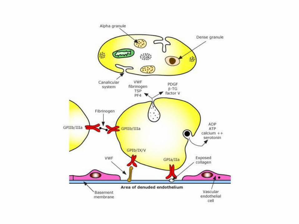
• Platelets contain three types of granules in their cytosol:
Dense granules
a-granules
Lisosomial granules
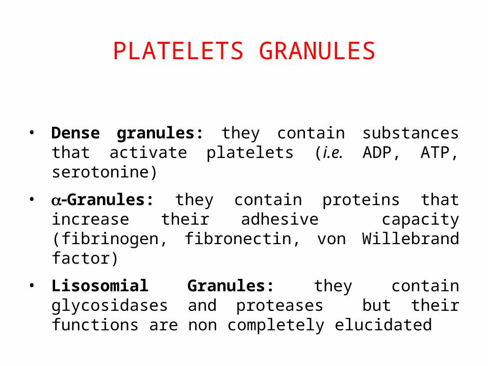
PLATELETS GRANULES
• Dense granules: they contain substances that activate platelets (i.e. ADP, ATP, serotonine)
• -a Granules: they contain proteins that increase their adhesive capacity (fibrinogen, fibronectin, von Willebrand factor)
• Lisosomial Granules: they contain glycosidases and proteases but their functions are non completely elucidated
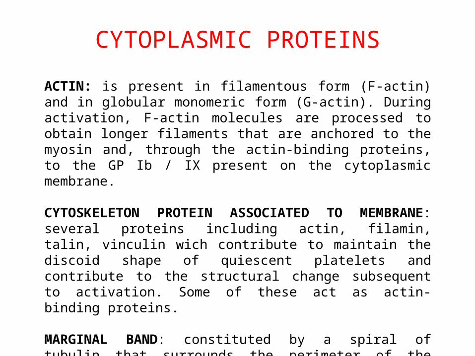
CYTOPLASMIC PROTEINS
ACTIN: is present in filamentous form (F-actin) and in globular monomeric form (G-actin). During activation, F-actin molecules are processed to obtain longer filaments that are anchored to the myosin and, through the actin-binding proteins, to the GP Ib / IX present on the cytoplasmic membrane.
CYTOSKELETON PROTEIN ASSOCIATED TO MEMBRANE: several proteins including actin, filamin, talin, vinculin wich contribute to maintain the discoid shape of quiescent platelets and contribute to the structural change subsequent to activation. Some of these act as actin-binding proteins.
MARGINAL BAND: constituted by a spiral of tubulin that surrounds the perimeter of the platelet; during platelet activation the tubulin spiral contracts and contributes to morphological changes.
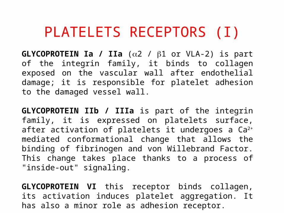
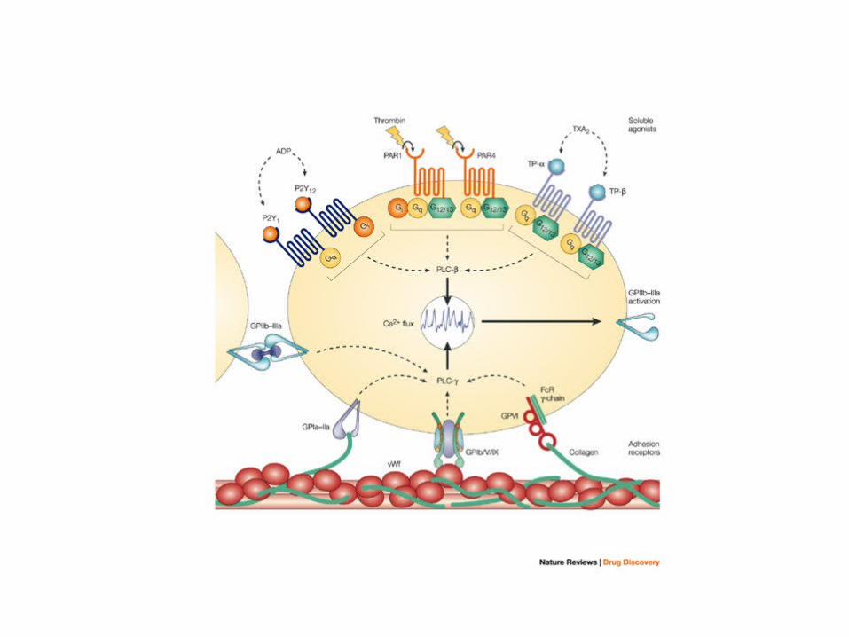
PLATELETS RECEPTORS (I)
GLYCOPROTEIN Ia / IIa (a2 / b1 or VLA-2) is part of the integrin family, it binds to collagen exposed on the vascular wall after endothelial damage; it is responsible for platelet adhesion to the damaged vessel wall.
GLYCOPROTEIN IIb / IIIa is part of the integrin family, it is expressed on platelets surface, after activation of platelets it undergoes a Ca2+ mediated conformational change that allows the binding of fibrinogen and von Willebrand Factor. This change takes place thanks to a process of "inside-out" signaling.
GLYCOPROTEIN VI this receptor binds collagen, its activation induces platelet aggregation. It has also a minor role as adhesion receptor.
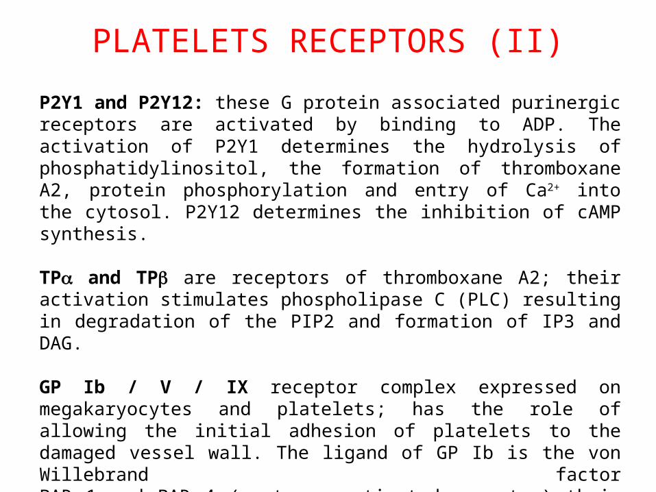
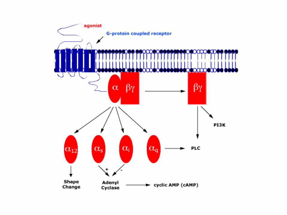
P2Y1 and P2Y12: these G protein associated purinergic receptors are activated by binding to ADP. The activation of P2Y1 determines the hydrolysis of phosphatidylinositol, the formation of thromboxane A2, protein phosphorylation and entry of Ca2+ into the cytosol. P2Y12 determines the inhibition of cAMP synthesis.
TPa and TPb are receptors of thromboxane A2; their activation stimulates phospholipase C (PLC) resulting in degradation of the PIP2 and formation of IP3 and DAG.
GP Ib / V / IX receptor complex expressed on megakaryocytes and platelets; has the role of allowing the initial adhesion of platelets to the damaged vessel wall. The ligand of GP Ib is the von Willebrand factorPAR 1 and PAR 4 (protease activated receptor) their ligand is thrombin, the most potent activator of platelets. Their activation, started by fibrin, leads to activation of the G protein.
PLATELETS RECEPTORS (II)
• Adhesion
• Activation and secretion
• Aggregation
• Interaction with coagulation factors
PLATELETS FUNCTIONS
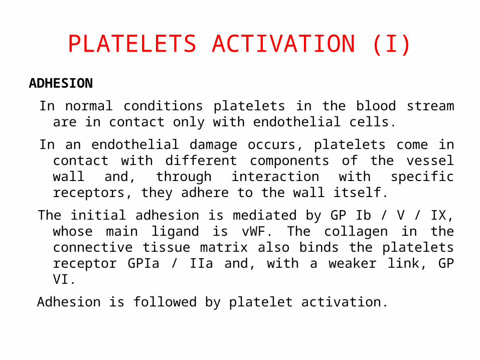
PLATELETS ACTIVATION (I)
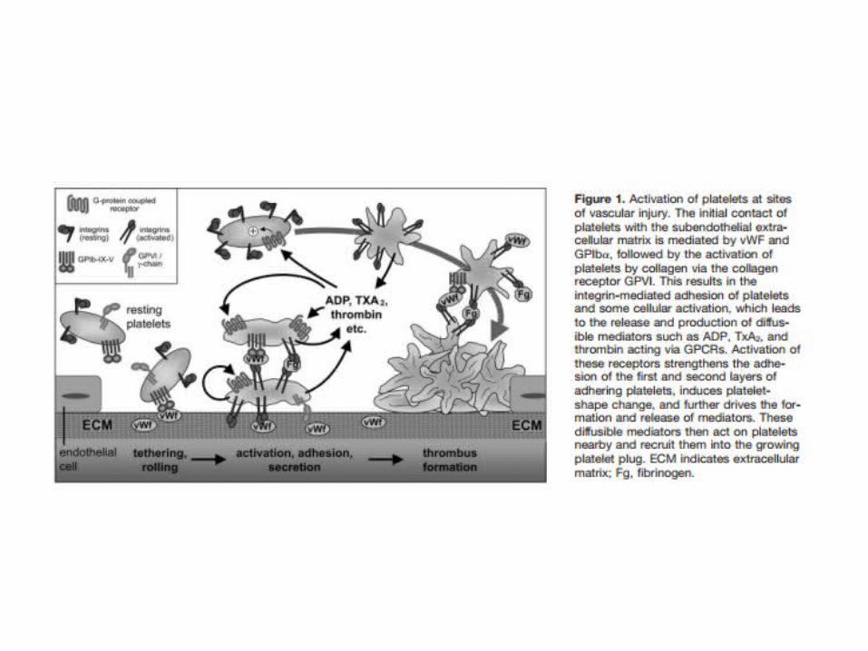
ADHESION
In normal conditions platelets in the blood stream are in contact only with endothelial cells.
In an endothelial damage occurs, platelets come in contact with different components of the vessel wall and, through interaction with specific receptors, they adhere to the wall itself.
The initial adhesion is mediated by GP Ib / V / IX, whose main ligand is vWF. The collagen in the connective tissue matrix also binds the platelets receptor GPIa / IIa and, with a weaker link, GP VI.
Adhesion is followed by platelet activation.
ACTIVATION AND SECRETION
The first step of is the conformational change (platelets become flat, their surface increases and they assume a dendritic shape) These changes allows platelets to adhere more closely to collagen fibers. In addition platelets degranulation leads to the release of ADP and thromboxane A2 into the circulation. These two substances amplify the reaction because they are potent activators of circulating platelets enhancing the recruitment of other platelets to be activated.
Platelets contain pre-mRNA of tissue factor (TF). Platelet activation leads to the splicing of pre-mRNA and the synthesis of TF which activates the coagulation cascade leading to the conversion of prothrombin to thrombin and, finally, of fibrinogen to fibrin. These events determine the stabilization of the thrombus.
PLATELETS ACTIVATION (II)
AGGREGATION
Activated platelets are able to bind fibrinogen. This protein, in fact, has two binding sites for platelet integrin GPIIb / IIIa (also known as AIIb / b3), abundantly expressed on the surface of platelets (40000-80000 receptors per platelet). Fibrinogen represents a bridge between the platelets wich form large aggregates.
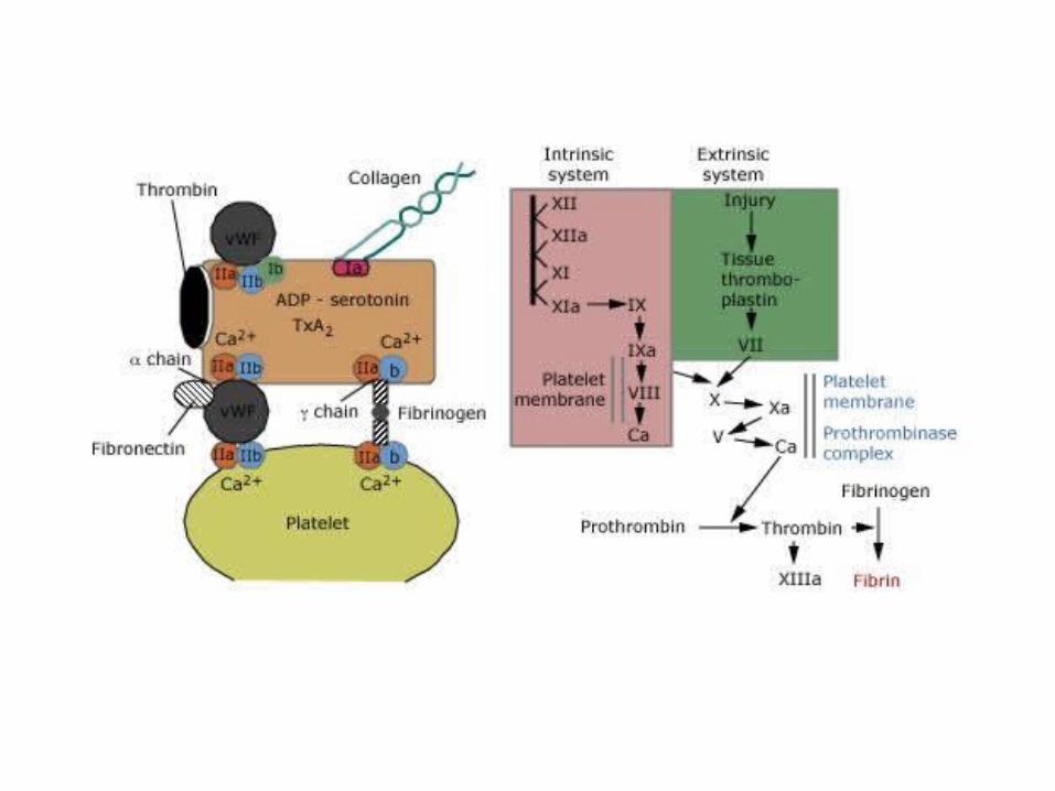
INTERACTION WITH COAGULATION FACTORS
The platelet clot is stabilized by the formation of a fibrin network, the result of the coagulation cascade. There is a synergistic action between platelets and coagulation factors: the formation of thrombin, in fact, is due in large part to the TF released from activated platelets.
PLATELETS ACTIVATION (III)
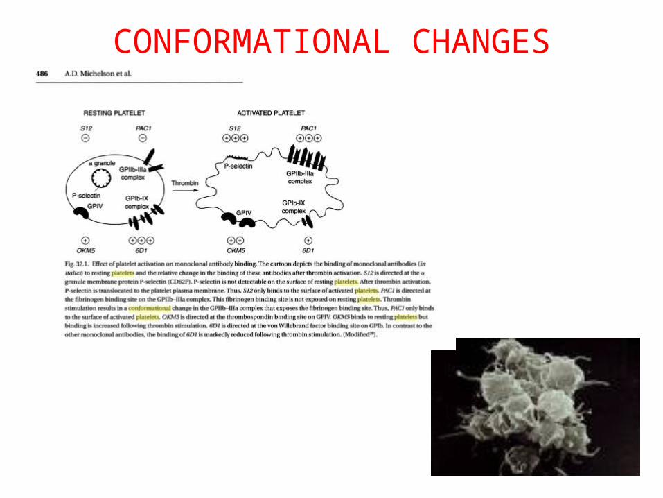
CONFORMATIONAL CHANGES
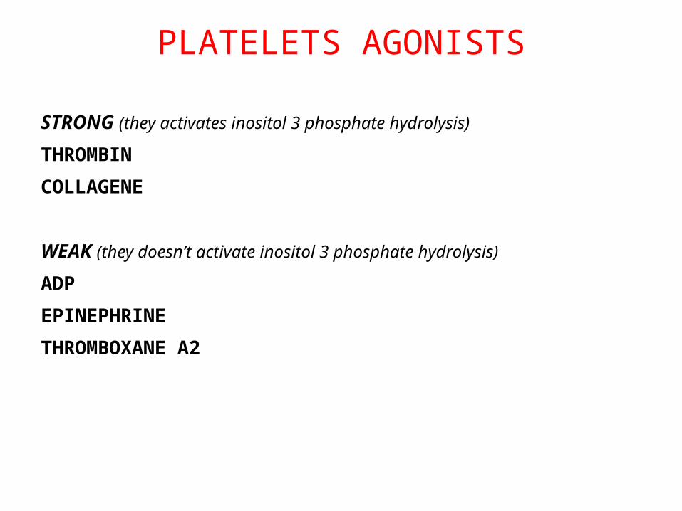
PLATELETS AGONISTS
STRONG (they activates inositol 3 phosphate hydrolysis)
THROMBIN
COLLAGENE
WEAK (they doesn’t activate inositol 3 phosphate hydrolysis)
ADP
EPINEPHRINE
THROMBOXANE A2
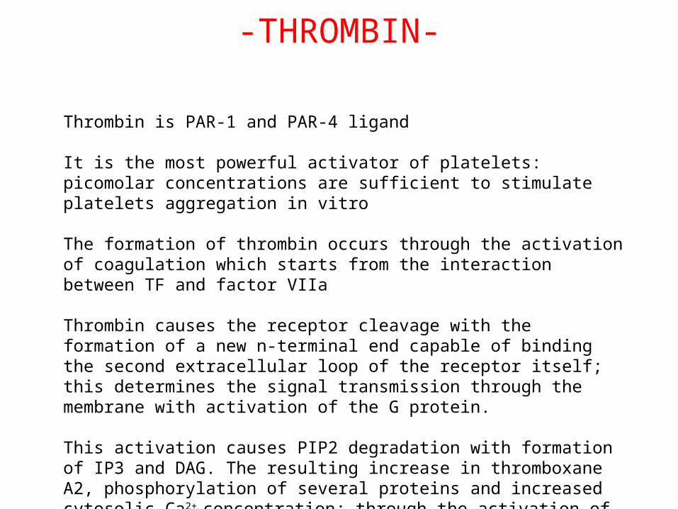
-THROMBIN-
Thrombin is PAR-1 and PAR-4 ligand
It is the most powerful activator of platelets: picomolar concentrations are sufficient to stimulate platelets aggregation in vitro
The formation of thrombin occurs through the activation of coagulation which starts from the interaction between TF and factor VIIa
Thrombin causes the receptor cleavage with the formation of a new n-terminal end capable of binding the second extracellular loop of the receptor itself; this determines the signal transmission through the membrane with activation of the G protein.
This activation causes PIP2 degradation with formation of IP3 and DAG. The resulting increase in thromboxane A2, phosphorylation of several proteins and increased cytosolic Ca2+ concentration; through the activation of Rho also are induced cytoskeletal changes that determines the conformational changes.
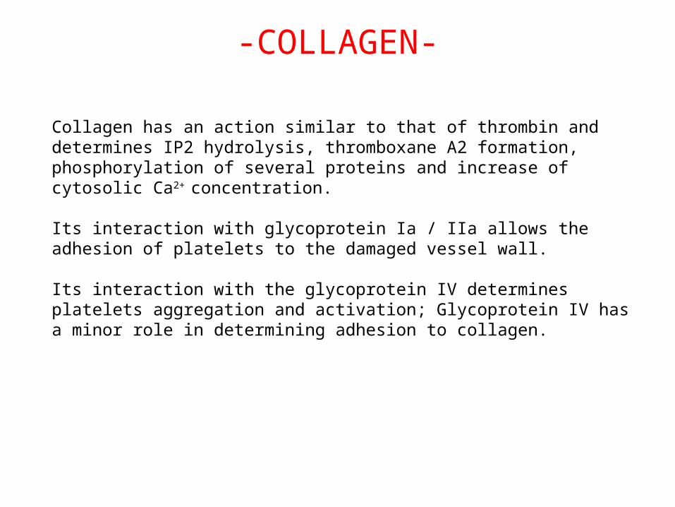
-COLLAGEN-
Collagen has an action similar to that of thrombin and determines IP2 hydrolysis, thromboxane A2 formation, phosphorylation of several proteins and increase of cytosolic Ca2+ concentration.
Its interaction with glycoprotein Ia / IIa allows the adhesion of platelets to the damaged vessel wall.
Its interaction with the glycoprotein IV determines platelets aggregation and activation; Glycoprotein IV has a minor role in determining adhesion to collagen.
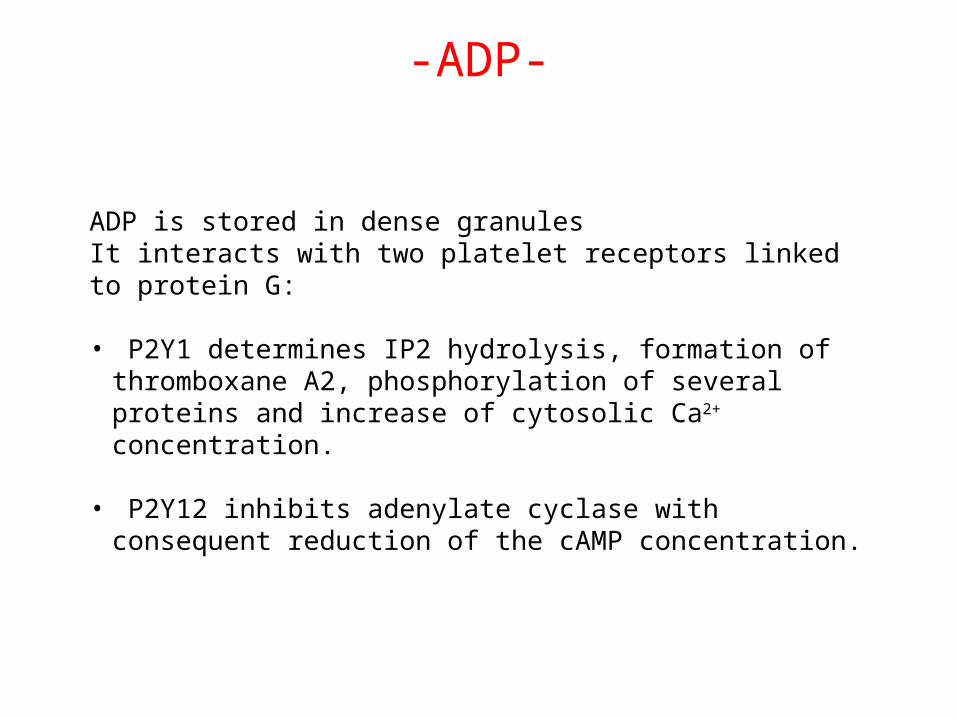
-ADP-
ADP is stored in dense granulesIt interacts with two platelet receptors linked to protein G:
• P2Y1 determines IP2 hydrolysis, formation of thromboxane A2, phosphorylation of several proteins and increase of cytosolic Ca2+
concentration.
• P2Y12 inhibits adenylate cyclase with consequent reduction of the cAMP concentration.
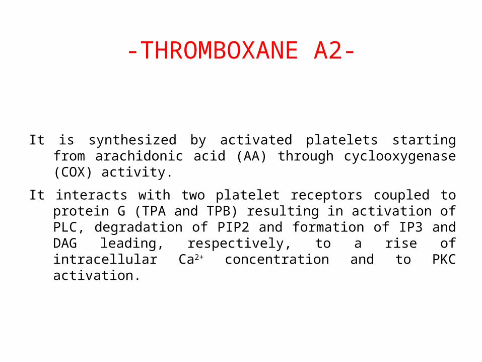
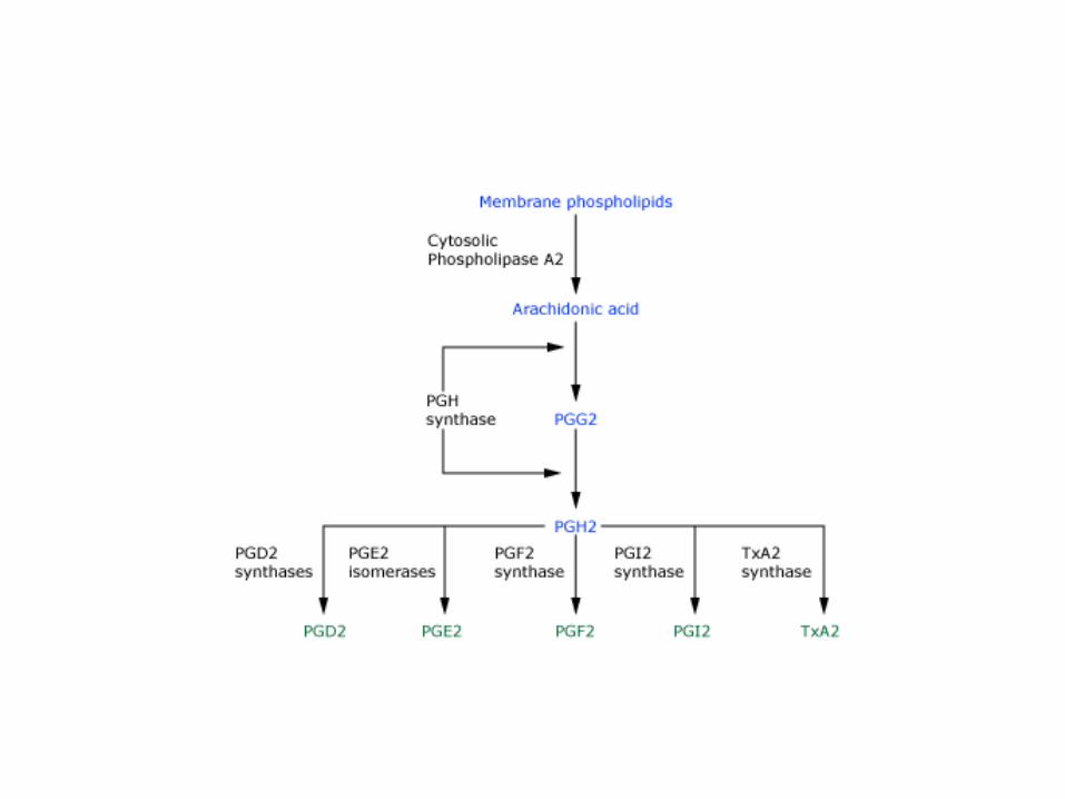
-THROMBOXANE A2-
It is synthesized by activated platelets starting from arachidonic acid (AA) through cyclooxygenase (COX) activity.
It interacts with two platelet receptors coupled to protein G (TPA and TPB) resulting in activation of PLC, degradation of PIP2 and formation of IP3 and DAG leading, respectively, to a rise of intracellular Ca2+ concentration and to PKC activation.
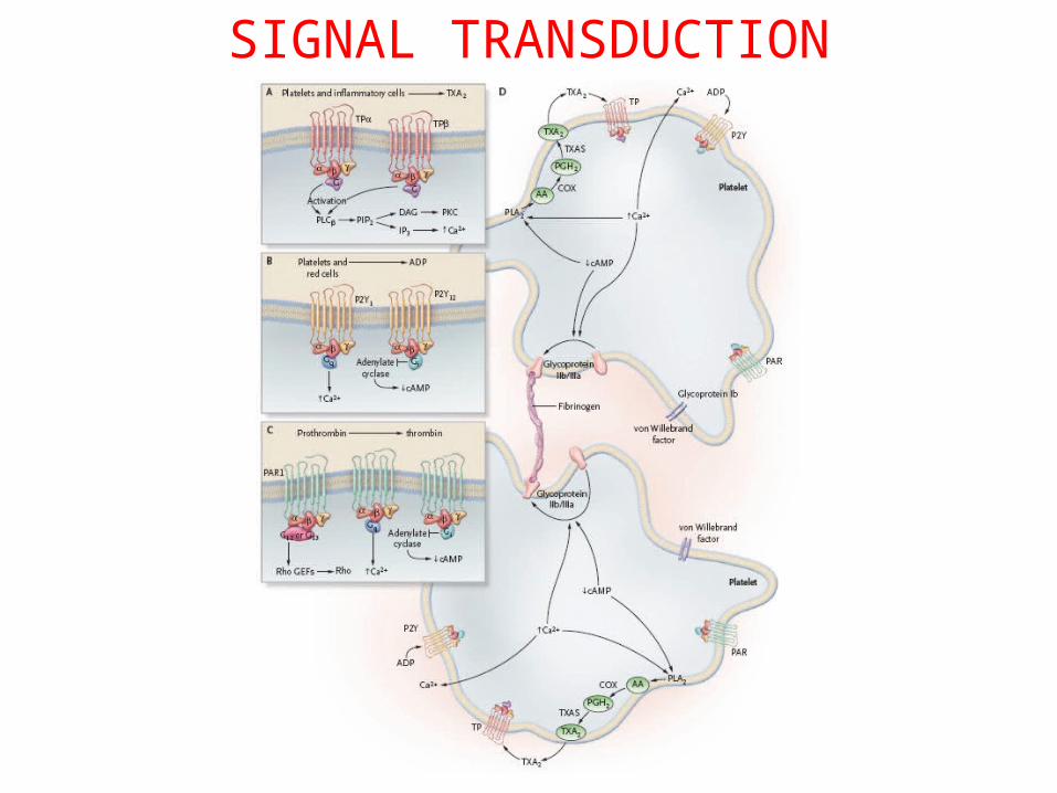
SIGNAL TRANSDUCTION
INFLAMMATORY MEDIATORS
Platelets are able to produce pro-inflammatory substances that determine alterations of the endothelial cells with consequent recall and chemotactic activation of monocytes
CD40 ligand: a member of the TNF family, is stored in the cytoplasm of quiescent platelets; following activation it is translocated on the platelet membrane, and thereafter, the soluble active form is released; when it reaches the endothelial cells it stimulates the production of peroxides, free radicals, adhesion molecules, cytokines and TF.
Interleukin 1b: it is not stored in platelet granules, its synthesis is induced by splicing of the mRNA content in platelets. It determines endothelial activation with increased expression of adhesion molecules for monocytes and polymorphonuclear cells and increased production of chemokines.
PF4, P-selectin, metalloproteinases, pro-angiogenic factors are other mediators of inflammation whose production increases during platelet activation.
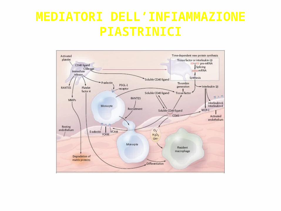
Figura 2 NEJM, Davì 2482
MEDIATORI DELL’INFIAMMAZIONE PIASTRINICI
ANTIPLATELET DRUGS
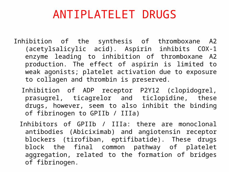
Inhibition of the synthesis of thromboxane A2 (acetylsalicylic acid). Aspirin inhibits COX-1 enzyme leading to inhibition of thromboxane A2 production. The effect of aspirin is limited to weak agonists; platelet activation due to exposure to collagen and thrombin is preserved.
Inhibition of ADP receptor P2Y12 (clopidogrel, prasugrel, ticagrelor and ticlopidine, these drugs, however, seem to also inhibit the binding of fibrinogen to GPIIb / IIIa)
Inhibitors of GPIIb / IIIa: there are monoclonal antibodies (Abiciximab) and angiotensin receptor blockers (tirofiban, eptifibatide). These drugs block the final common pathway of platelet aggregation, related to the formation of bridges of fibrinogen.
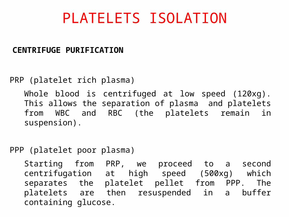
PLATELETS ISOLATION
PRP (platelet rich plasma)
Whole blood is centrifuged at low speed (120xg). This allows the separation of plasma and platelets from WBC and RBC (the platelets remain in suspension).
PPP (platelet poor plasma)
Starting from PRP, we proceed to a second centrifugation at high speed (500xg) which separates the platelet pellet from PPP. The platelets are then resuspended in a buffer containing glucose.
• PURIFICAZIONE PER GEL FILTRAZIONE
CENTRIFUGE PURIFICATION
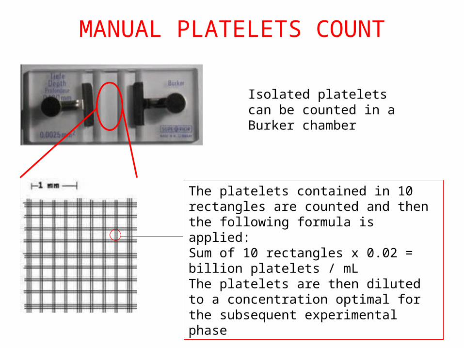
MANUAL PLATELETS COUNT
Isolated platelets can be counted in a Burker chamber
The platelets contained in 10 rectangles are counted and then the following formula is applied:Sum of 10 rectangles x 0.02 = billion platelets / mLThe platelets are then diluted to a concentration optimal for the subsequent experimental phase
PRP
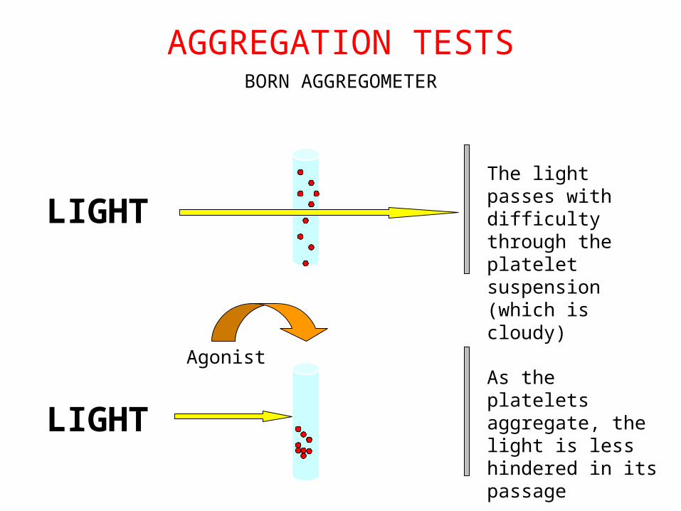
AGGREGATION TESTSBORN AGGREGOMETER
LIGHTThe light passes with difficulty through the platelet suspension (which is cloudy)
LIGHT
AgonistAs the platelets aggregate, the light is less hindered in its passage
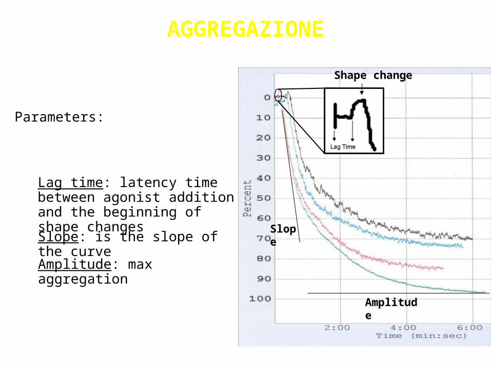
AGGREGAZIONE
Parameters:
Amplitude: max aggregation
Slope: is the slope of the curve
Lag time: latency time between agonist addition and the beginning of shape changes
Slope
Amplitude
Shape change
Platelet Function TestingPlatelet Function Testing
PFA-100® System
Clinical Indications
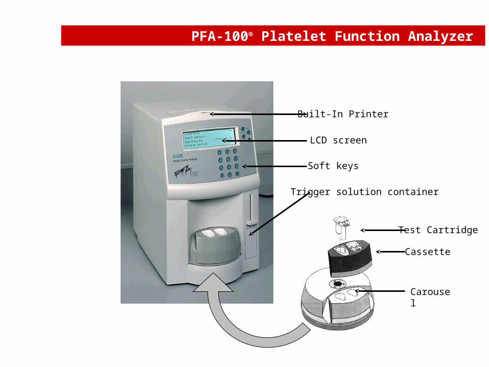
PFA-100® Platelet Function Analyzer
Trigger solution container
Soft keys
LCD screen
Built-In Printer
Carousel
Cassette
Test Cartridge
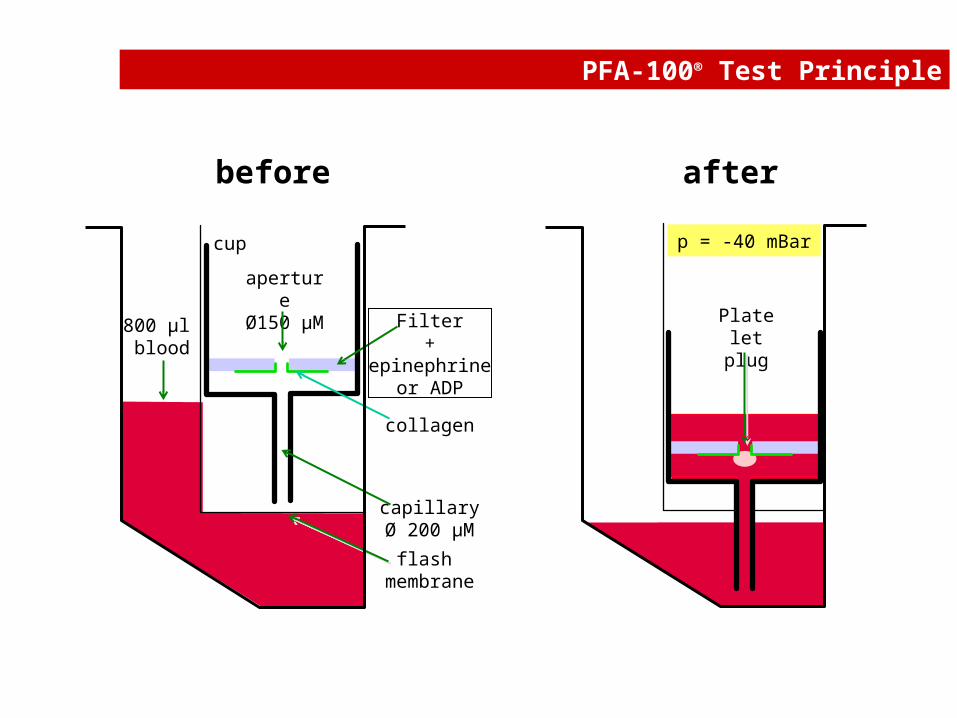
PFA-100® Test Principle
before after
apertureØ150 µM
cup
Platelet plug
Filter+
epinephrineor ADP
flash membrane
800 µl blood
capillaryØ 200 µM
collagen
p = -40 mBar
high shear rate>5000 /s
high shear rate>5000 /s
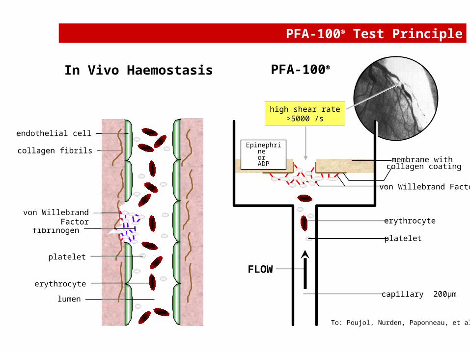
PFA-100® Test Principle
PFA-100®
capillary 200µm
Epinephrine or
ADP membrane with
platelet
von Willebrand Factor
erythrocyte
FLOW
collagen coating
To: Poujol, Nurden, Paponneau, et al.
In Vivo Haemostasis
lumen
fibrinogen
platelet
collagen fibrils
erythrocyte
von Willebrand Factor
endothelial cell
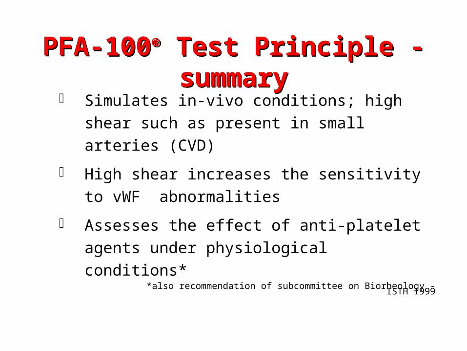
Simulates in-vivo conditions; high shear such as
present in small arteries (CVD)
High shear increases the sensitivity to vWF
abnormalities
Assesses the effect of anti-platelet agents under
physiological conditions*
*also recommendation of subcommittee on Biorheology - ISTH 1999
PFA-100® Test Principle - summary
PFA-100® Test Principle - summary
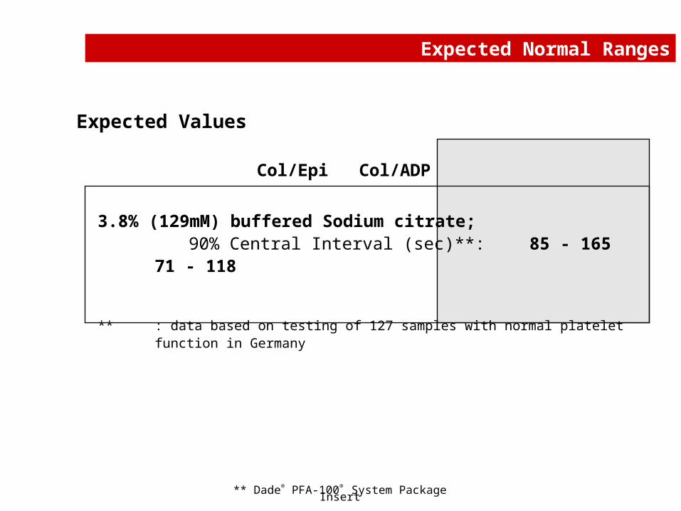
Col/Epi Col/ADP
3.8% (129mM) buffered Sodium citrate;90% Central Interval (sec)**: 85 - 165 71
- 118
** : data based on testing of 127 samples with normal platelet function in Germany
** Dade® PFA-100® System Package Insert
Expected Normal Ranges
Expected Values
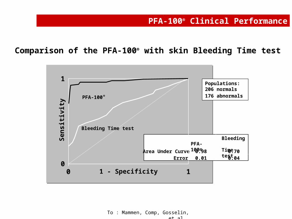
Comparison of the PFA-100® with skin Bleeding Time test
To : Mammen, Comp, Gosselin, et al..
0
1
0 1
Sen
siti
vity
1 - Specificity
PFA-100 ®
Bleeding Time test
PFA-100®
Area Under Curve 0.98 0.70Error 0.01 0.04
Bleeding Time test
Populations:206 normals176 abnormals
PFA-100® Clinical Performance
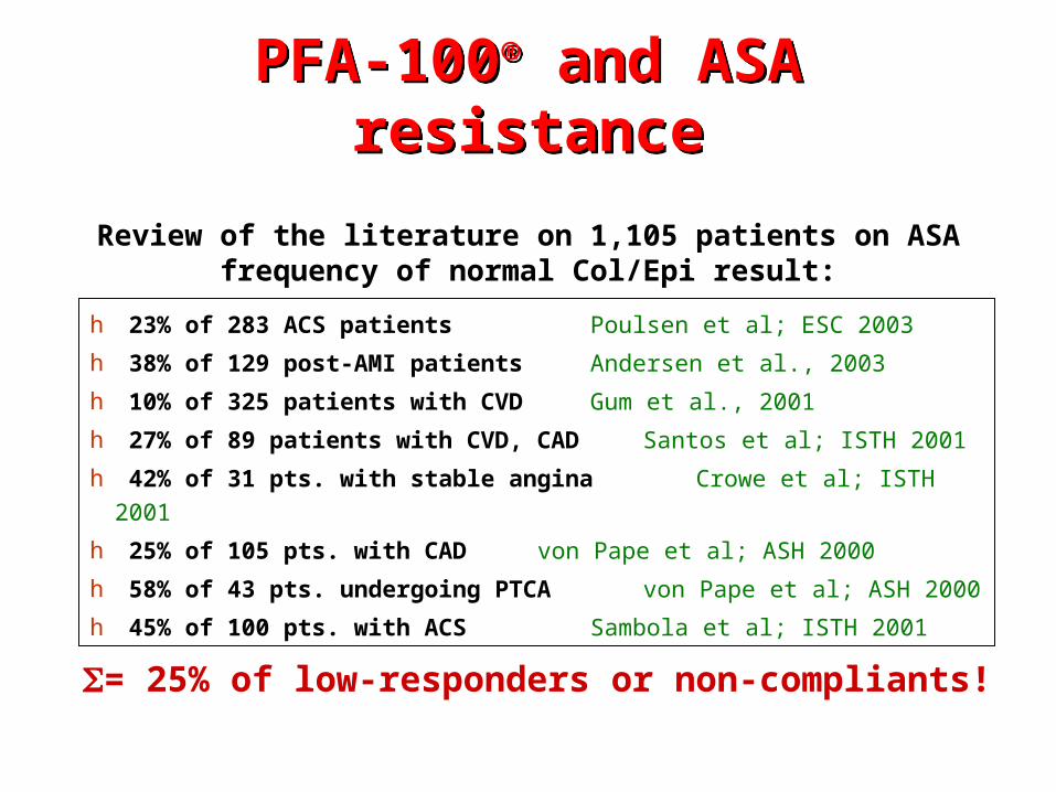
h 23% of 283 ACS patients Poulsen et al; ESC 2003
h 38% of 129 post-AMI patients Andersen et al., 2003
h 10% of 325 patients with CVD Gum et al., 2001
h 27% of 89 patients with CVD, CAD Santos et al; ISTH 2001
h 42% of 31 pts. with stable angina Crowe et al; ISTH 2001
h 25% of 105 pts. with CAD von Pape et al; ASH 2000
h 58% of 43 pts. undergoing PTCA von Pape et al; ASH 2000
h 45% of 100 pts. with ACS Sambola et al; ISTH 2001
= 25% of low-responders or non-compliants!
Review of the literature on 1,105 patients on ASAfrequency of normal Col/Epi result:
PFA-100® and ASA resistancePFA-100® and ASA resistance