platinum(ii) complexes of indaphyrin and...
TRANSCRIPT
Subscriber access provided by Columbia Univ Libraries
Inorganic Chemistry is published by the American Chemical Society. 1155Sixteenth Street N.W., Washington, DC 20036
Article
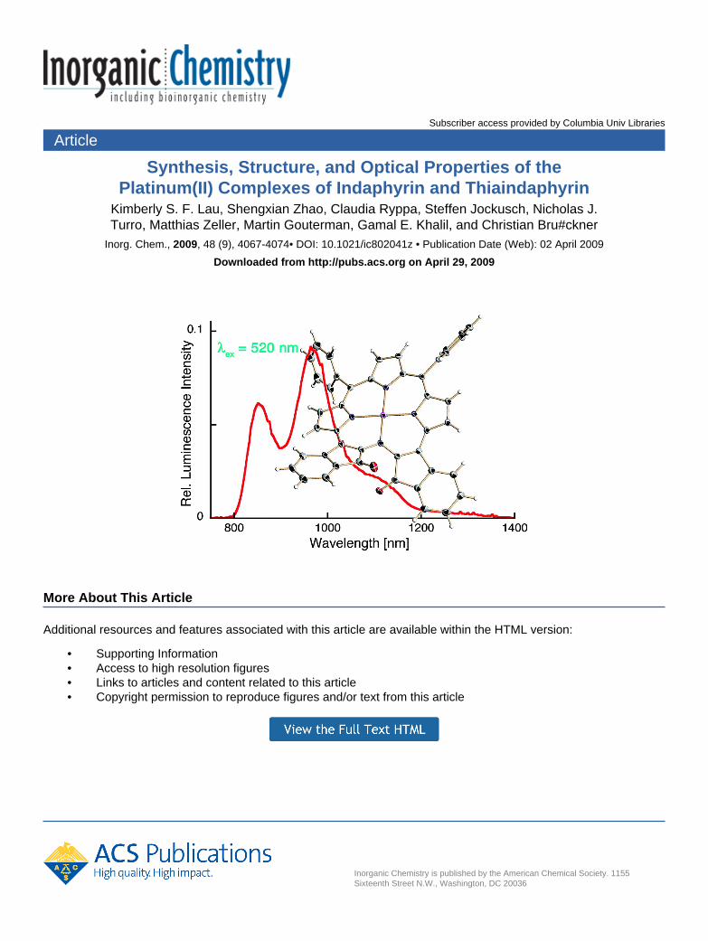
Synthesis, Structure, and Optical Properties of thePlatinum(II) Complexes of Indaphyrin and Thiaindaphyrin
Kimberly S. F. Lau, Shengxian Zhao, Claudia Ryppa, Steffen Jockusch, Nicholas J.Turro, Matthias Zeller, Martin Gouterman, Gamal E. Khalil, and Christian Bru#ckner
Inorg. Chem., 2009, 48 (9), 4067-4074• DOI: 10.1021/ic802041z • Publication Date (Web): 02 April 2009
Downloaded from http://pubs.acs.org on April 29, 2009
More About This Article
Additional resources and features associated with this article are available within the HTML version:
• Supporting Information• Access to high resolution figures• Links to articles and content related to this article• Copyright permission to reproduce figures and/or text from this article

Synthesis, Structure, and Optical Properties of the Platinum(II)Complexes of Indaphyrin and Thiaindaphyrin
Kimberly S. F. Lau,†,‡ Shengxian Zhao,‡ Claudia Ryppa,‡ Steffen Jockusch,§ Nicholas J. Turro,§
Matthias Zeller,| Martin Gouterman,† Gamal E. Khalil,† and Christian Bruckner*,‡
Department of Chemistry, UniVersity of Washington, Box 351700,Seattle, Washington 98195-1700, Department of Chemistry, UniVersity of Connecticut,Unit 3060, Storrs, Connecticut 06269-3060, Department of Chemistry,Columbia UniVersity, New York, New York 10027, and Department of Chemistry,Youngstown State UniVersity, One UniVersity Plaza,Youngstown, Ohio 44555-3663
Received October 24, 2008
The novel free base meso-di(5′-methylthien-2′-yl)thiaindaphyrin, 10, was prepared from the corresponding meso-tetra(thien-2-yl)porphyrin using a methodology analogous to that for the preparation of known meso-diphenylindaphyrin,5: ,′-Dihydroxylation of the porphyrin is followed by oxidative diol cleavage. The resulting aldehyde moietiesundergo an acid-catalyzed intramolecular Friedel-Crafts alkylation of the adjacent meso-thienyl groups withconcomitant oxidation. Insertion of Pt(II) into either of the chromophores is facile, producing 5Pt and 10Pt. Thecrystal structure of 5Pt, the first for any indaphyrin, shows that the conformation of the indaphyrinato ligand isstrongly ruffled, while the N4 donor set that coordinates the central Pt(II) maintains a near-perfect square-planarcoordination geometry around the central metal ion (crystal data for C44H24N4O2Pt: triclinic space group P1 with a) 8.8735(4) Å, b ) 12.9285(6) Å, c ) 14.3297(6) Å, R ) 88.785(1)°, ) 82.248(1)°, γ ) 72.422(1)°; Z ) 2).The UV-vis and emission spectra, triplet yields, and lifetimes of the Pt(II) complexes 5Pt and 10Pt were determined.Both complexes luminesce (in EtOH at 77 K) in the NIR (5Pt: λmax-emission ) 864, 974 nm, lifetime 2 µs; 10Pt:λmax-emission ) 990, 1112, 1276 nm) with modest to low quantum yields (Φp ∼ 1% and ∼ 6 × 10⁻³ %,respectively).
Introduction
The triplet oxygen-mediated quenching of the tripletexcited-state of many chromophores (Stern-Volmerquenching) allows a correlation between the degree ofquenching and a particular oxygen partial pressure.1 Thisgives rise to the development of optical oxygen sensorsthat find applications in medicine, engineering, andchemical and environmental analyses.1-3
One particularly well-studied class of compounds foroxygen sensing applications is that of the Pt(II) porphyrins,such as [meso-tetraphenylporphyrinato]Pt(II) (1Pt) and re-lated compounds.1,4-7 In Pt(II) and Pd(II) porphyrins, ligand-based emissions are observed (λmax-emission for 1Pt ) 650, 712
* To whom correspondence should be addressed. Phone: (860) 486-2743. Fax: (860) 486-2981. E-mail: [email protected].
† University of Washington.‡ University of Connecticut.§ Columbia University.| Youngstown State University.
(1) Lakowicz, J. R. Principles of Fluorescence Spectroscopy, 3rd ed.;Springer: New York, 2006.
(2) Gouterman, M. J. Chem. Educ. 1997, 74, 697–702.(3) (a) Leiner, M. J. P. Anal. Chim. Acta 1991, 255, 209–222. (b)
Papkovsky, D. B.; O’Riordan, T.; Soini, A. Biochem. Soc. Trans. 2000,28, 74–77.
(4) Xiang, H.-F.; Li, C.-N.; Yu, S. C.; Che, C.-M.; Lai, P. T.; Chui, P. C.Proc. SPIE Int. Soc Opt. Eng. 2004, 5519, 218–225.
(5) Vinogradov, S. A.; Lo, L.-W.; Wilson, D. F. Eur. J. Chem. 1999, 5,1338–1347.
(6) Brinas, R. P.; Troxler, T.; Hochstrasser, R. M.; Vinogradov, S. A.J. Am. Chem. Soc. 2005, 127, 11851–11862.
(7) Huo, C.; Zhang, H.; Zhang, H.; Zhang, H.; Yang, B.; Zhang, P.; Wang,Y. Inorg. Chem. 2006, 45, 4735–4742.
(8) Gouterman, M.; Hall, R. J.; Khalil, G.-E.; Martin, P. C.; Shankland,E. G.; Cerny, R. L. J. Am. Chem. Soc. 1989, 111, 3702–3707.
(9) Zelelow, B.; Khalil, G. E.; Phelan, G.; Carlson, B.; Gouterman, M.;Callis, J. B.; Dalton, L. R. Sens. Actuators, B 2003, 96, 304–314.
(10) Gouterman, M.; Callis, J.; Dalton, L.; Khalil, G.; Mebarki, Y.; Cooper,K. R.; Grenier, M. Meas. Sci. Technol. 2004, 15, 1986–1994.
(11) Khalil, G. E.; Costin, C.; Crafton, J.; Jones, G.; Grenoble, S.;Gouterman, M.; Callis, J. B.; Dalton, L. R. Sens. Actuators, B 2004,97, 13–21.
Inorg. Chem. 2009, 48, 4067-4074
10.1021/ic802041z CCC: $40.75 2009 American Chemical Society Inorganic Chemistry, Vol. 48, No. 9, 2009 4067Published on Web 04/02/2009
nm).2 This allows a modulation of the excitation andemission wavelengths through alterations of the porphyrinicchromophore. Examples include the use of the Pd(II) andPt(II) complexes of porpholactones, 2Pt,8-11 -oxo-porphy-rins,12 or benzoporphyrins, such as complex 3Pt.13
A red shift of the excitation or luminescence spectra intothe red or NIR range (λmax-emission > 750 nm) is desirable fora number of applications, particularly in the life sciences.14
By utilizing red to near-infrared (NIR) wavelengths, anincrease in the sensitivity of luminescence-based assays inbiological systems measurements can be achieved becauseno natural chromophore emits in this region. The NIR is alsothe region of the “spectroscopic window” in tissue, andchromophores emitting in this range may allow the detectionof events deep in the tissue. Last, longer wavelengths arescattered less in opaque media than shorter ones, increasingthe resolution of the emission images obtained.
This emission red shift was the driving force behindthe development of π-extended porphyrins such as 3Pt(λmax-emission ) 745 nm)15 or the bimetallic complex of anN-confused calyx[6]phyrin 4Pt2 (λmax-emission ) 1010 nm).16
An ideal sensor molecule should fulfill other key require-ments such as high extinction coefficients and highemission quantum yields (i.e., possessing high brightness),and excellent photostability. Ancillary properties such asnonaggregation in a polymer matrix, good processability,and a large Stokes shift further improve their applicability.
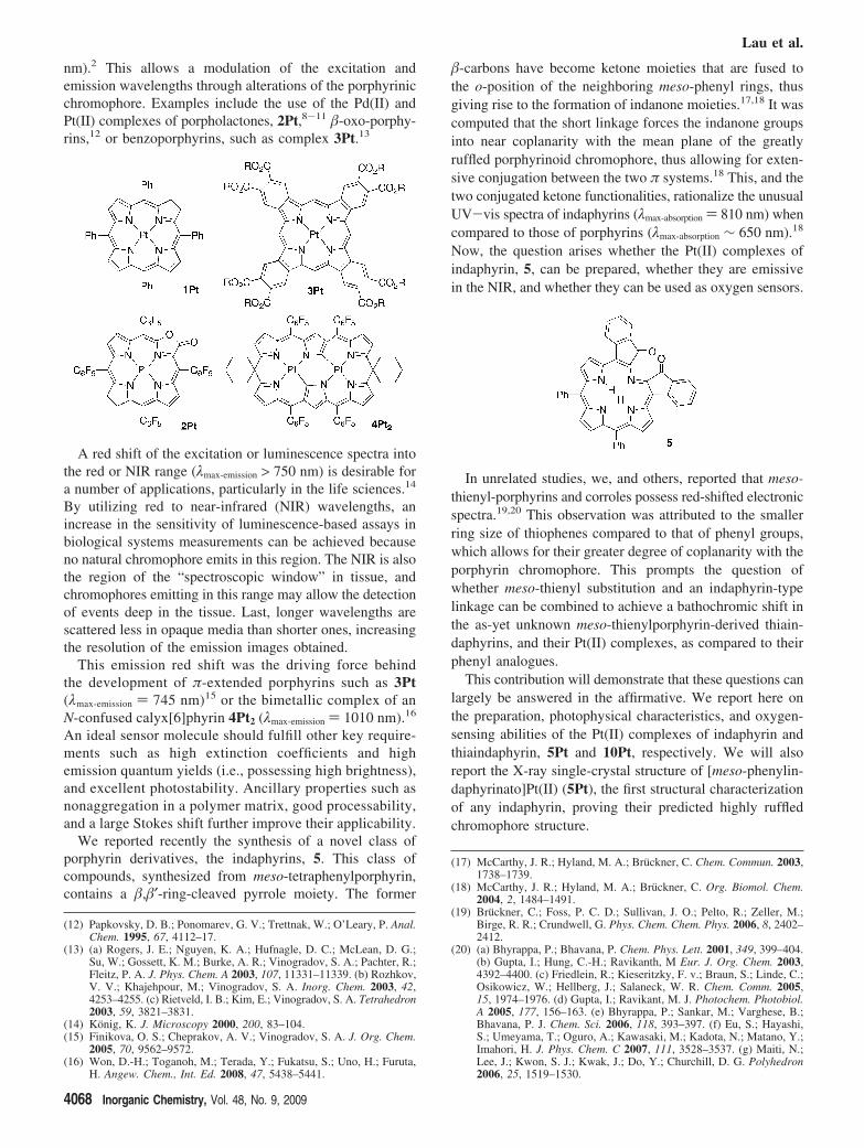
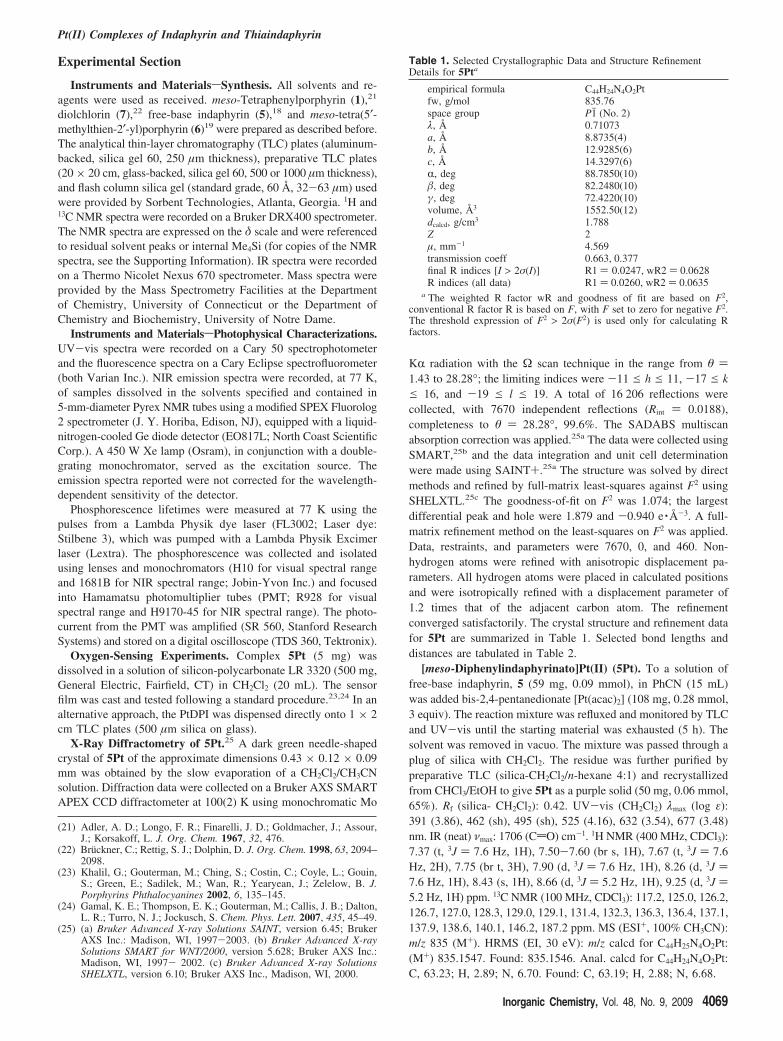
We reported recently the synthesis of a novel class ofporphyrin derivatives, the indaphyrins, 5. This class ofcompounds, synthesized from meso-tetraphenylporphyrin,contains a ,′-ring-cleaved pyrrole moiety. The former
-carbons have become ketone moieties that are fused tothe o-position of the neighboring meso-phenyl rings, thusgiving rise to the formation of indanone moieties.17,18 It wascomputed that the short linkage forces the indanone groupsinto near coplanarity with the mean plane of the greatlyruffled porphyrinoid chromophore, thus allowing for exten-sive conjugation between the two π systems.18 This, and thetwo conjugated ketone functionalities, rationalize the unusualUV-vis spectra of indaphyrins (λmax-absorption ) 810 nm) whencompared to those of porphyrins (λmax-absorption ∼ 650 nm).18
Now, the question arises whether the Pt(II) complexes ofindaphyrin, 5, can be prepared, whether they are emissivein the NIR, and whether they can be used as oxygen sensors.
In unrelated studies, we, and others, reported that meso-thienyl-porphyrins and corroles possess red-shifted electronicspectra.19,20 This observation was attributed to the smallerring size of thiophenes compared to that of phenyl groups,which allows for their greater degree of coplanarity with theporphyrin chromophore. This prompts the question ofwhether meso-thienyl substitution and an indaphyrin-typelinkage can be combined to achieve a bathochromic shift inthe as-yet unknown meso-thienylporphyrin-derived thiain-daphyrins, and their Pt(II) complexes, as compared to theirphenyl analogues.
This contribution will demonstrate that these questions canlargely be answered in the affirmative. We report here onthe preparation, photophysical characteristics, and oxygen-sensing abilities of the Pt(II) complexes of indaphyrin andthiaindaphyrin, 5Pt and 10Pt, respectively. We will alsoreport the X-ray single-crystal structure of [meso-phenylin-daphyrinato]Pt(II) (5Pt), the first structural characterizationof any indaphyrin, proving their predicted highly ruffledchromophore structure.
(12) Papkovsky, D. B.; Ponomarev, G. V.; Trettnak, W.; O’Leary, P. Anal.Chem. 1995, 67, 4112–17.
(13) (a) Rogers, J. E.; Nguyen, K. A.; Hufnagle, D. C.; McLean, D. G.;Su, W.; Gossett, K. M.; Burke, A. R.; Vinogradov, S. A.; Pachter, R.;Fleitz, P. A. J. Phys. Chem. A 2003, 107, 11331–11339. (b) Rozhkov,V. V.; Khajehpour, M.; Vinogradov, S. A. Inorg. Chem. 2003, 42,4253–4255. (c) Rietveld, I. B.; Kim, E.; Vinogradov, S. A. Tetrahedron2003, 59, 3821–3831.
(14) Konig, K. J. Microscopy 2000, 200, 83–104.(15) Finikova, O. S.; Cheprakov, A. V.; Vinogradov, S. A. J. Org. Chem.
2005, 70, 9562–9572.(16) Won, D.-H.; Toganoh, M.; Terada, Y.; Fukatsu, S.; Uno, H.; Furuta,
H. Angew. Chem., Int. Ed. 2008, 47, 5438–5441.
(17) McCarthy, J. R.; Hyland, M. A.; Bruckner, C. Chem. Commun. 2003,1738–1739.
(18) McCarthy, J. R.; Hyland, M. A.; Bruckner, C. Org. Biomol. Chem.2004, 2, 1484–1491.
(19) Bruckner, C.; Foss, P. C. D.; Sullivan, J. O.; Pelto, R.; Zeller, M.;Birge, R. R.; Crundwell, G. Phys. Chem. Chem. Phys. 2006, 8, 2402–2412.
(20) (a) Bhyrappa, P.; Bhavana, P. Chem. Phys. Lett. 2001, 349, 399–404.(b) Gupta, I.; Hung, C.-H.; Ravikanth, M Eur. J. Org. Chem. 2003,4392–4400. (c) Friedlein, R.; Kieseritzky, F. v.; Braun, S.; Linde, C.;Osikowicz, W.; Hellberg, J.; Salaneck, W. R. Chem. Comm. 2005,15, 1974–1976. (d) Gupta, I.; Ravikant, M. J. Photochem. Photobiol.A 2005, 177, 156–163. (e) Bhyrappa, P.; Sankar, M.; Varghese, B.;Bhavana, P. J. Chem. Sci. 2006, 118, 393–397. (f) Eu, S.; Hayashi,S.; Umeyama, T.; Oguro, A.; Kawasaki, M.; Kadota, N.; Matano, Y.;Imahori, H. J. Phys. Chem. C 2007, 111, 3528–3537. (g) Maiti, N.;Lee, J.; Kwon, S. J.; Kwak, J.; Do, Y.; Churchill, D. G. Polyhedron2006, 25, 1519–1530.
Lau et al.
4068 Inorganic Chemistry, Vol. 48, No. 9, 2009
Experimental Section
Instruments and MaterialssSynthesis. All solvents and re-agents were used as received. meso-Tetraphenylporphyrin (1),21
diolchlorin (7),22 free-base indaphyrin (5),18 and meso-tetra(5′-methylthien-2′-yl)porphyrin (6)19 were prepared as described before.The analytical thin-layer chromatography (TLC) plates (aluminum-backed, silica gel 60, 250 µm thickness), preparative TLC plates(20 × 20 cm, glass-backed, silica gel 60, 500 or 1000 µm thickness),and flash column silica gel (standard grade, 60 Å, 32-63 µm) usedwere provided by Sorbent Technologies, Atlanta, Georgia. 1H and13C NMR spectra were recorded on a Bruker DRX400 spectrometer.The NMR spectra are expressed on the δ scale and were referencedto residual solvent peaks or internal Me4Si (for copies of the NMRspectra, see the Supporting Information). IR spectra were recordedon a Thermo Nicolet Nexus 670 spectrometer. Mass spectra wereprovided by the Mass Spectrometry Facilities at the Departmentof Chemistry, University of Connecticut or the Department ofChemistry and Biochemistry, University of Notre Dame.
Instruments and MaterialssPhotophysical Characterizations.UV-vis spectra were recorded on a Cary 50 spectrophotometerand the fluorescence spectra on a Cary Eclipse spectrofluorometer(both Varian Inc.). NIR emission spectra were recorded, at 77 K,of samples dissolved in the solvents specified and contained in5-mm-diameter Pyrex NMR tubes using a modified SPEX Fluorolog2 spectrometer (J. Y. Horiba, Edison, NJ), equipped with a liquid-nitrogen-cooled Ge diode detector (EO817L; North Coast ScientificCorp.). A 450 W Xe lamp (Osram), in conjunction with a double-grating monochromator, served as the excitation source. Theemission spectra reported were not corrected for the wavelength-dependent sensitivity of the detector.
Phosphorescence lifetimes were measured at 77 K using thepulses from a Lambda Physik dye laser (FL3002; Laser dye:Stilbene 3), which was pumped with a Lambda Physik Excimerlaser (Lextra). The phosphorescence was collected and isolatedusing lenses and monochromators (H10 for visual spectral rangeand 1681B for NIR spectral range; Jobin-Yvon Inc.) and focusedinto Hamamatsu photomultiplier tubes (PMT; R928 for visualspectral range and H9170-45 for NIR spectral range). The photo-current from the PMT was amplified (SR 560, Stanford ResearchSystems) and stored on a digital oscilloscope (TDS 360, Tektronix).
Oxygen-Sensing Experiments. Complex 5Pt (5 mg) wasdissolved in a solution of silicon-polycarbonate LR 3320 (500 mg,General Electric, Fairfield, CT) in CH2Cl2 (20 mL). The sensorfilm was cast and tested following a standard procedure.23,24 In analternative approach, the PtDPI was dispensed directly onto 1 × 2cm TLC plates (500 µm silica on glass).
X-Ray Diffractometry of 5Pt.25 A dark green needle-shapedcrystal of 5Pt of the approximate dimensions 0.43 × 0.12 × 0.09mm was obtained by the slow evaporation of a CH2Cl2/CH3CNsolution. Diffraction data were collected on a Bruker AXS SMARTAPEX CCD diffractometer at 100(2) K using monochromatic Mo
KR radiation with the Ω scan technique in the range from θ )1.43 to 28.28°; the limiting indices were -11 e h e 11, -17 e ke 16, and -19 e l e 19. A total of 16 206 reflections werecollected, with 7670 independent reflections (Rint ) 0.0188),completeness to θ ) 28.28°, 99.6%. The SADABS multiscanabsorption correction was applied.25a The data were collected usingSMART,25b and the data integration and unit cell determinationwere made using SAINT+.25a The structure was solved by directmethods and refined by full-matrix least-squares against F2 usingSHELXTL.25c The goodness-of-fit on F2 was 1.074; the largestdifferential peak and hole were 1.879 and -0.940 e ·Å-3. A full-matrix refinement method on the least-squares on F2 was applied.Data, restraints, and parameters were 7670, 0, and 460. Non-hydrogen atoms were refined with anisotropic displacement pa-rameters. All hydrogen atoms were placed in calculated positionsand were isotropically refined with a displacement parameter of1.2 times that of the adjacent carbon atom. The refinementconverged satisfactorily. The crystal structure and refinement datafor 5Pt are summarized in Table 1. Selected bond lengths anddistances are tabulated in Table 2.
[meso-Diphenylindaphyrinato]Pt(II) (5Pt). To a solution offree-base indaphyrin, 5 (59 mg, 0.09 mmol), in PhCN (15 mL)was added bis-2,4-pentanedionate [Pt(acac)2] (108 mg, 0.28 mmol,3 equiv). The reaction mixture was refluxed and monitored by TLCand UV-vis until the starting material was exhausted (5 h). Thesolvent was removed in vacuo. The mixture was passed through aplug of silica with CH2Cl2. The residue was further purified bypreparative TLC (silica-CH2Cl2/n-hexane 4:1) and recrystallizedfrom CHCl3/EtOH to give 5Pt as a purple solid (50 mg, 0.06 mmol,65%). Rf (silica- CH2Cl2): 0.42. UV-vis (CH2Cl2) λmax (log ε):391 (3.86), 462 (sh), 495 (sh), 525 (4.16), 632 (3.54), 677 (3.48)nm. IR (neat) νmax: 1706 (CdO) cm-1. 1H NMR (400 MHz, CDCl3):7.37 (t, 3J ) 7.6 Hz, 1H), 7.50-7.60 (br s, 1H), 7.67 (t, 3J ) 7.6Hz, 2H), 7.75 (br t, 3H), 7.90 (d, 3J ) 7.6 Hz, 1H), 8.26 (d, 3J )7.6 Hz, 1H), 8.43 (s, 1H), 8.66 (d, 3J ) 5.2 Hz, 1H), 9.25 (d, 3J )5.2 Hz, 1H) ppm. 13C NMR (100 MHz, CDCl3): 117.2, 125.0, 126.2,126.7, 127.0, 128.3, 129.0, 129.1, 131.4, 132.3, 136.3, 136.4, 137.1,137.9, 138.6, 140.1, 146.2, 187.2 ppm. MS (ESI+, 100% CH3CN):m/z 835 (M+). HRMS (EI, 30 eV): m/z calcd for C44H25N4O2Pt:(M+) 835.1547. Found: 835.1546. Anal. calcd for C44H24N4O2Pt:C, 63.23; H, 2.89; N, 6.70. Found: C, 63.19; H, 2.88; N, 6.68.
(21) Adler, A. D.; Longo, F. R.; Finarelli, J. D.; Goldmacher, J.; Assour,J.; Korsakoff, L. J. Org. Chem. 1967, 32, 476.
(22) Bruckner, C.; Rettig, S. J.; Dolphin, D. J. Org. Chem. 1998, 63, 2094–2098.
(23) Khalil, G.; Gouterman, M.; Ching, S.; Costin, C.; Coyle, L.; Gouin,S.; Green, E.; Sadilek, M.; Wan, R.; Yearyean, J.; Zelelow, B. J.Porphyrins Phthalocyanines 2002, 6, 135–145.
(24) Gamal, K. E.; Thompson, E. K.; Gouterman, M.; Callis, J. B.; Dalton,L. R.; Turro, N. J.; Jockusch, S. Chem. Phys. Lett. 2007, 435, 45–49.
(25) (a) Bruker AdVanced X-ray Solutions SAINT, version 6.45; BrukerAXS Inc.: Madison, WI, 1997-2003. (b) Bruker AdVanced X-raySolutions SMART for WNT/2000, version 5.628; Bruker AXS Inc.:Madison, WI, 1997- 2002. (c) Bruker AdVanced X-ray SolutionsSHELXTL, version 6.10; Bruker AXS Inc., Madison, WI, 2000.
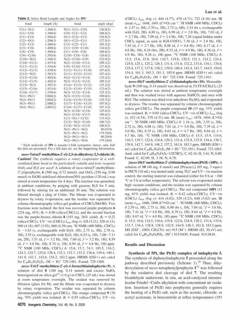
Table 1. Selected Crystallographic Data and Structure RefinementDetails for 5Pta
empirical formula C44H24N4O2Ptfw, g/mol 835.76space group Pj1 (No. 2)λ, Å 0.71073a, Å 8.8735(4)b, Å 12.9285(6)c, Å 14.3297(6)R, deg 88.7850(10), deg 82.2480(10)γ, deg 72.4220(10)volume, Å3 1552.50(12)dcalcd, g/cm3 1.788Z 2µ, mm-1 4.569transmission coeff 0.663, 0.377final R indices [I > 2σ(I)] R1 ) 0.0247, wR2 ) 0.0628R indices (all data) R1 ) 0.0260, wR2 ) 0.0635
a The weighted R factor wR and goodness of fit are based on F2,conventional R factor R is based on F, with F set to zero for negative F2.The threshold expression of F2 > 2σ(F2) is used only for calculating Rfactors.
Pt(II) Complexes of Indaphyrin and Thiaindaphyrin
Inorganic Chemistry, Vol. 48, No. 9, 2009 4069
meso-Tetra(5′-methylthien-2′-yl)-2,3-cis-dihydroxy-2,3-chlorin (8).Caution! The synthesis requires a rotary eVaporator in a well-Ventilated fume hood as the particularly Volatile and toxic reagentsOsO4 and H2S are used! A solution of meso-tetra(5′-methylthien-2′-yl)porphyrin, 6 (500 mg, 0.72 mmol), and OsO4 (250 mg, 0.98mmol) in EtOH-stabilized chloroform/50% pyridine (120 mL) wasstirred at room temperature for 4 days. The reaction was quenched,at ambient conditions, by purging with gaseous H2S for 5 min,followed by stirring for an additional 30 min. The solution wasfiltered through a plug of Celite. The filtrate was evaporated todryness by rotary evaporation, and the residue was separated bycolumn chromatography (silica-gel gradient of CHCl3/MeOH). Twofractions were isolated: the first was recovered purple porphyrin 6(224 mg, 45%; Rf ) 0.90 (silica-CHCl3)), and the second fractionwas the purple-brown chlorin 8 (193 mg, 26% yield). Rf ) 0.22(silica-CHCl3). UV-vis (CHCl3) λmax (log ε): 428 (5.21), 528 (4.13),560 (4.18), 607 (3.92), 660 (4.39) nm. 1H NMR (400 MHz, CDCl3)δ: -1.63 (s, exchangeable with D2O, 1H), 2.75 (s, 3H), 2.78 (s,3H), 3.55 (s, exchangeable with D2O, 1H), 6.53 (s, 1H), 7.08-7.11(m, 2H), 7.51 (d, J ) 3.2 Hz, 1H), 7.60 (d, J ) 3.2 Hz, 1H), 8.59(d, J ) 4.8 Hz, 1H), 8.74 (s, 1H), 8.94 (d, J ) 4.8 Hz, 1H) ppm.13C NMR (100 MHz, CDCl3) δ: 15.6, 15.7, 74.3, 105.5, 116.2,124.3, 124.7, 125.0, 128.4, 132.1, 132.7, 133.2, 136.6, 139.4, 140.1,141.9, 142.1, 142.6, 154.2, 163.2 ppm. HRMS (ESI+) m/z calcdfor C40H33N4O2S4: (M + H)+ 729.1481. Found: 729.1488.
meso-Tri(5′-methylthien-2′-yl)-1-formylthiaindaphyrin (9). Asolution of diol 8 (100 mg, 0.14 mmol) and excess NaIO4
heterogenized on silica gel18 (1.0 g) in CHCl3 (25 mL) was stirredat room temperature overnight. The oxidant was removed byfiltration (glass frit M), and the filtrate was evaporated to drynessby rotary evaporation. The residue was separated by columnchromatography (silica gel-CHCl3). The orange compound 9 (70mg, 70% yield) was isolated. Rf ) 0.55 (silica-CHCl3). UV-vis
(CHCl3) λmax (log ε): 444 (4.77), 478 (4.71), 722 (4.16) nm. IR(neat) νmax: 1648, 1691 (CdO) cm-1. 1H NMR (400 MHz, CDCl3)δ: 2.57 (s, 3H), 2.70 (s, 3H), 2.72 (s, 6H), 3.34 (br s, exchangeablewith D2O, 2H), 6.89 (s, 1H), 6.99 (d, J ) 2.8 Hz, 1H), 7.03 (d, J) 2.7 Hz, 1H), 7.09 (d, J ) 2.4 Hz, 1H), 7.26 (signal hidden underCHCl3 signal, as seen in H,H-COSY), 7.30 (d, J ) 2.8 Hz, 1H),7.45 (d, J ) 2.7 Hz, 1H), 8.08 (d, J ) 4.6 Hz, 1H), 8.17 (d, J )4.6 Hz, 1H), 8.24 (brs, 2H), 8.32 (d, J ) 4.9 Hz, 1H), 8.36 (d, J )4.9 Hz, 1H), 9.28 (s, 1H) ppm. 13C NMR (100 MHz, CDCl3) δ:15.5, 15.6, 15.9, 16.0, 110.7, 119.6, 120.5, 121.1, 124.2, 124.4,125.0, 125.1, 125.2, 126.3, 131.4, 131.6, 132.2, 133.4, 134.1, 134.8,135.4, 137.2, 137.8, 138.2, 139.0, 140.4, 142.4, 143.7, 144.9, 154.0,154.4, 161.3, 165.3, 181.3, 185.0 ppm. HRMS (ESI+) m/z calcdfor C40H29N4O2S4: (M + H)+ 725.1168. Found: 725.1163.
meso-Di(5′-methylthien-2′-yl)thiaindaphyrin (10). Monoalde-hyde 9 (100 mg, 0.14 mmol) was dissolved in 2%TFA/CH2Cl2 (25mL). The solution was stirred at ambient temperature overnightand then was washed twice with aqueous NaHCO3 and once withH2O. The solution was dried over anhydrous Na2SO4 and evaporatedto dryness. The residue was separated by column chromatography(silica gel-CHCl3). The purple compound 10 (33 mg, 33% yield)was isolated. Rf ) 0.65 (silica-CHCl3). UV-vis (CHCl3) λmax (logε): 432 (4.54), 570 (4.31) nm. IR (neat) νmax: 1678, 1694 (CdO)cm-1. 1H NMR (400 MHz, CDCl3) δ: 1.16 (s, 1H), 2.55 (s, 3H),2.72 (s, 3H), 6.88 (s, 1H), 7.01 (d, J ) 3.0 Hz, 1H), 7.39 (d, J )3.0 Hz, 1H), 8.35 (s, 1H), 8.61 (d, J ) 4.7 Hz, 1H), 8.64 (d, J )4.7 Hz, 1H). 13C NMR (100 MHz, CDCl3) δ: 15.5, 15.9, 114.6,116.3, 119.7, 122.6, 124.8, 129.2, 131.4, 132.9, 133.9, 134.4, 138.7,138.8, 142.7, 144.9, 148.2, 157.2, 161.8, 183.3 ppm. HRMS (ESI+)m/z calcd for C40H27N4O2S4 (M + H)+ 723.1011. Found: 723.1003.Anal. calcd for C40H26N4O2S4 ·1/2CHCl3: C, 62.16; H, 3.41; N, 6.80.Found: C, 62.09; H, 3.38; N, 6.70.
[meso-Di(5′-methylthien-2′-yl)thiaindaphyrinato]Pt(II) (10Pt). Asolution of 10 (40 mg, 6 mmol) and [Pt(acac)2] (65 mg, 3 equiv)in PhCN (10 mL) was heated until, using TLC and UV-vis reactioncontrol, the starting material was exhausted (either for 8 h at ∼160°C or 5 h at reflux temperature). The solvent was evaporated underhigh vacuum conditions, and the residue was separated by columnchromatography (silica gel-CHCl3). The red compound 10Pt (31mg, 61% yield) was isolated. Rf ) 0.42 (silica-CHCl3). UV-vis(CHCl3) λmax (log ε): 414 (4.02), 529 (4.23), 648 (3.62) nm. IR(neat) νmax: 1680, 1696 (CdO) cm-1. 1H NMR (400 MHz, CDCl3):δ 2.59 (s, 3H), 2.75 (s, 3H), 6.90 (br s, 1H), 7.04 (d, 3J ) 3.6 Hz,1H), 7.41 (d, 3J ) 4.0 Hz, 1H), 8.39 (s, 1H), 8.64 (d, 3J ) 4.0 Hz,1H), 8.67 (d, 3J ) 4.0 Hz, 1H) ppm. 13C NMR (100 MHz, CDCl3)δ: 15.5, 16.0, 114.5, 116.6, 119.8, 122.6, 124.9, 129.2, 131.6, 133.0,133.7, 134.4, 138.8, 138.9, 142.9, 144.9, 148.3, 161.8, 183.2 ppm.MS (ESI+, 100% CH3CN): m/z 915 (M+). HRMS (EI, 30 eV) m/zcalcd for C40H24N4O2PtS4: (M+) 915.0430. Found: 915.0418.
Results and Discussion
Synthesis of 5Pt, the Pt(II) complex of indaphyrin 5.The synthesis of diphenylindaphyrin 5 proceeded along thepathway described previously (Scheme 1).18 Thus, dihy-droxylation of meso-tetraphenylporphyrin 122 was followedby the oxidative diol cleavage of diol 7. The resultingbisaldehyde underwent, in situ, an acid-catalyzed intramo-lecular Friedel-Crafts alkylation with concomitant air oxida-tion. Insertion of Pt(II) into porphyrins generally requiresthe heating of a Pt(II) salt, such as the acetate, chloride, oracetyl acetonate, in benzonitrile at reflux temperatures (191
Table 2. Select Bond Lengths and Angles for 5Pta
bond length [Å] bond angle [deg]
C(1)-N(1) 1.360(3) N(1)-C(1)-C(9) 126.9(2)C(1)-C(9) 1.389(4) C(9)-C(1)-C(2) 108.8(2)C(1)-C(2) 1.504(4) O(1)-C(2)-C(3) 126.9(3)C(2)-O(1) 1.208(3) O(1)-C(2)-C(1) 127.5(3)C(2)-C(3) 1.486(4) C(3)-C(2)-C(1) 105.3(2)C(3)-C(8) 1.409(4) C(1)-C(9)-C(10) 124.6(2)C(8)-C(9) 1.489(4) C(1)-C(9)-C(8) 108.6(2)C(9)-C(10) 1.392(4) C(10)-C(9)-C(8) 126.8(2)C(10)-N(2) 1.384(3) N(2)-C(10)-C(9) 122.9(2)C(10)-C(11) 1.437(4) N(2)-C(10)-C(11) 109.1(2)C(11)-C(12) 1.354(4) C(9)-C(10)-C(11) 127.9(2)C(12)-C(13) 1.439(4) C(12)-C(11)-C(10) 107.6(2)C(13)-N(2) 1.383(3) C(11)-C(12)-C(13) 107.4(2)C(13)-C(14) 1.402(4) N(2)-C(13)-C(14) 125.2(2)C(14)-C(15) 1.402(4) N(2)-C(13)-C(12) 109.1(2)C(15)-N(3) 1.387(3) C(14)-C(13)-C(12) 125.2(2)C(15)-C(16) 1.431(4) C(13)-C(14)-C(15) 123.6(2)C(16)-C(17) 1.359(4) N(3)-C(15)-C(14) 125.3(2)N(1)-Pt(1) 2.004(2) N(3)-C(15)-C(16) 109.0(2)N(2)-Pt(1) 2.002(2) C(14)-C(15)-C(16) 125.7(2)N(3)-Pt(1) 2.000(2) C(17)-C(16)-C(15) 107.9(2)N(4)-Pt(1) 2.007(2) C(16)-C(17)-C(18) 107.1(2)
N(3)-C(18)-C(19) 125.6(2)N(3)-C(18)-C(17) 109.4(2)C(1)-N(1)-C(32) 115.5(2)C(13)-N(2)-C(10) 106.7(2)C(18)-N(3)-C(15) 106.5(2)N(3)-Pt(1)-N(2) 89.67(9)N(3)-Pt(1)-N(1) 179.76(8)N(2)-Pt(1)-N(1) 90.29(9)N(2)-Pt(1)-N(4) 179.24(8)
a Each molecule of 5Pt is pseudo-2-fold symmetric; hence, only halfthe data are presented. For a full data set, see the Supporting Information.
Lau et al.
4070 Inorganic Chemistry, Vol. 48, No. 9, 2009
°C) for extended periods of time (typically 12-24 h).26,27
Appreciable Pt(II) insertion into indaphyrin 5 already takesplace in benzonitrile at 140-160 °C. At reflux temperatures,the reaction is completed within 3-5 h. We attribute this tothe much greater conformational flexibility of the indaphyrinchromophore as compared to that of a porphyrin. Chroma-tography, followed by recrystallization, produced Pt(II)indaphyrin 5Pt in good yields.
The spectroscopic and analytical data were all consistentwith the structure of 5Pt. In particular, its 1H and 13C NMRspectra were very similar to those of free-base 5, anindication that the insertion of Pt(II) did not lead to dramaticstructural changes. As detailed below, a single-crystalstructure analysis of 5Pt confirms its connectivity andhighlights its conformation. The optical properties of allchromophores synthesized are discussed below.
Metal complexes of indaphyrins were described before.18
However, the Ni(II) complexes of indaphyrins are unstableand form ring-opened products within days in solution,though the larger ions Cu(II) and Zn(II) form stable inda-phyrinato complexes.18 Complex 5Pt is stable in solutionfor days and as a solid for at least 2 years.
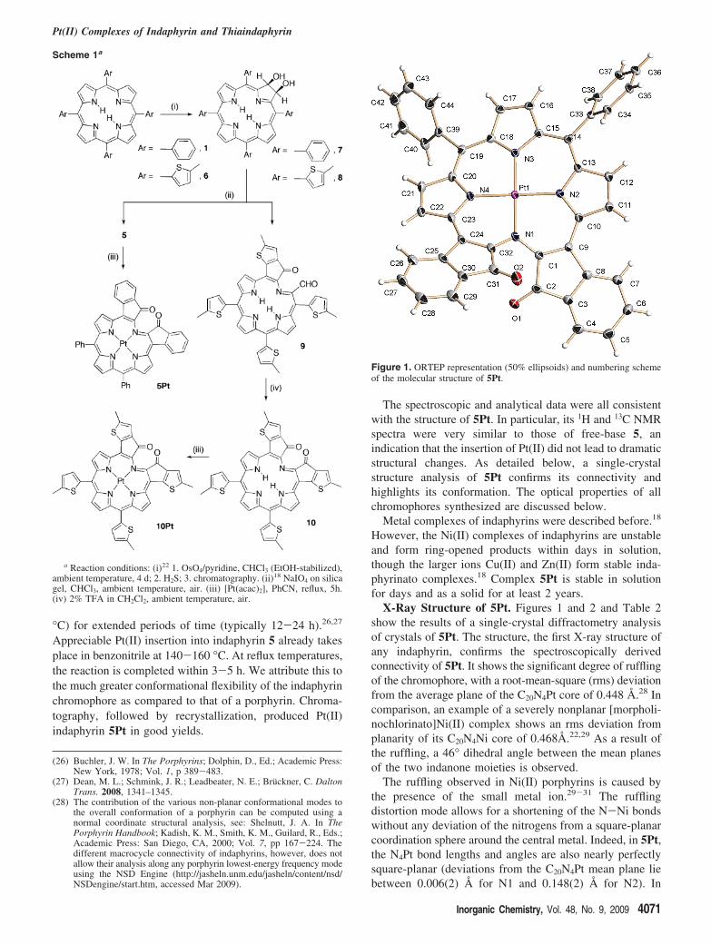
X-Ray Structure of 5Pt. Figures 1 and 2 and Table 2show the results of a single-crystal diffractometry analysisof crystals of 5Pt. The structure, the first X-ray structure ofany indaphyrin, confirms the spectroscopically derivedconnectivity of 5Pt. It shows the significant degree of rufflingof the chromophore, with a root-mean-square (rms) deviationfrom the average plane of the C20N4Pt core of 0.448 Å.28 Incomparison, an example of a severely nonplanar [morpholi-nochlorinato]Ni(II) complex shows an rms deviation fromplanarity of its C20N4Ni core of 0.468Å.22,29 As a result ofthe ruffling, a 46° dihedral angle between the mean planesof the two indanone moieties is observed.
The ruffling observed in Ni(II) porphyrins is caused bythe presence of the small metal ion.29-31 The rufflingdistortion mode allows for a shortening of the N-Ni bondswithout any deviation of the nitrogens from a square-planarcoordination sphere around the central metal. Indeed, in 5Pt,the N4Pt bond lengths and angles are also nearly perfectlysquare-planar (deviations from the C20N4Pt mean plane liebetween 0.006(2) Å for N1 and 0.148(2) Å for N2). In
(26) Buchler, J. W. In The Porphyrins; Dolphin, D., Ed.; Academic Press:New York, 1978; Vol. 1, p 389-483.
(27) Dean, M. L.; Schmink, J. R.; Leadbeater, N. E.; Bruckner, C. DaltonTrans. 2008, 1341–1345.
(28) The contribution of the various non-planar conformational modes tothe overall conformation of a porphyrin can be computed using anormal coordinate structural analysis, see: Shelnutt, J. A. In ThePorphyrin Handbook; Kadish, K. M., Smith, K. M., Guilard, R., Eds.;Academic Press: San Diego, CA, 2000; Vol. 7, pp 167-224. Thedifferent macrocycle connectivity of indaphyrins, however, does notallow their analysis along any porphyrin lowest-energy frequency modeusing the NSD Engine (http://jasheln.unm.edu/jasheln/content/nsd/NSDengine/start.htm, accessed Mar 2009).
Scheme 1a
a Reaction conditions: (i)22 1. OsO4/pyridine, CHCl3 (EtOH-stabilized),ambient temperature, 4 d; 2. H2S; 3. chromatography. (ii)18 NaIO4 on silicagel, CHCl3, ambient temperature, air. (iii) [Pt(acac)2], PhCN, reflux, 5h.(iv) 2% TFA in CH2Cl2, ambient temperature, air.
Figure 1. ORTEP representation (50% ellipsoids) and numbering schemeof the molecular structure of 5Pt.
Pt(II) Complexes of Indaphyrin and Thiaindaphyrin
Inorganic Chemistry, Vol. 48, No. 9, 2009 4071

contrast to the Ni(II) porphyrins, however, the distortion isintrinsic to the indaphyrin ligand. This is because theconformation of the ligand in 5Pt is, albeit slightly morepronounced, very similar to that computed for free-base 5(rms deviation of the C20N4 core from planarity wascomputed to be 0.39 Å).18 Interestingly, the observed N-Ptbond lengths (average of 2.003(2) Å) are identical to thoseobserved in the slightly ruffled [porphyrinato]Pt(II) com-plexes (2.005 Å in 1Pt at 295 K).32
The cleavage of the pyrrole and fusion of two indanoneunits into the macrocycle results in a significant dissymmetryof the bond lengths and angles within the porphyrinoidmacrocycle (Table 2). For instance, the N(1)imine-C(1)indanone
and the C(1)indanone-C(9)indanone bond lengths are significantlyshorter (1.360(3) and 1.389(4) Å, respectively) compared tothe corresponding and unusually long N(2 or 3)pyrrole-C(11/13 or 15/18)pyrrole (average of 1.385(3) Å) and C(13/15)pyrrole-C(14)meso bond lengths (1.402(4) Å), respectively.The corresponding bond lengths in metalloporphyrin 1Pt lie,with an average N-CR length of 1.376 Å and an averageCR-Cmeso length of 1.388 Å, between the two extreme valuesobserved in 5Pt. The C-N-C bond angles within the pyrroleunits are with 107° within the range expected in a porphyrin,whereas the bond angle of the imine-type linkage betweenthe indanone units is widened to 115°.
The crystal is composed of offset columns of π-stackedpairs of 5Pt that are held together by π-π interactionsbetween meso-phenyl groups. No major channels or cavitiesare discernible. Complex 5Pt is chiral and crystallizes as aracemate in the nonchiral space group Pj1.
Synthesis of the Dithienylthiaindaphyrin 10 and ItsPt(II) Complex 10Pt. The synthetic methodology towardindaphyrin 5 is perceivably transferable to any meso-arylporphyrin with an ortho position on the meso-aryl groupthat is susceptible to electrophilic aromatic substitution,including also meso-thien-2-ylporphyrin, 6 (Scheme 1). Thus,osmylation/reduction of the meso-tetra(4-methyl-thien-2-yl)porphyrin,33 6, resulted in the formation of the corre-sponding diol chlorin 8. Its HRMS (ESI+, m/z ) 729.1488for [M + H]+) spectrum confirms its composition(C40H33N4O2S4), and diagnostic peaks in the 1H and 13C NMRspectrum confirm its structure. Moreover, the UV-visspectrum of 8 is a typical chlorin spectrum, with λmax at 658nm the most intense of all side bands. It is, as expected,based on the comparison of thienylporphyrins with phe-nylporphyrins,19 ∼12 nm bathochromically shifted comparedto that of meso-phenyl-based chlorin 7 (Figure 3).22,34
Oxidative diol cleavage of diol 8 using NaIO4 hetero-genized on silica gel results in the formation of a putativebisaldehyde intermediate that, analogous to the reactivity ofits meso-phenyl analogue,17,18 spontaneously undergoes aFriedel-Crafts alkylation with concomitant oxidation to formmonothiaindanone monoaldehyde 9. It is identified by thecharacteristic loss of symmetry in the 1H and 13C NMRspectra; an ESI+ HRMS indicative of the expected composi-tion for MH+ (C40H29N4O2S4); and the appearance of onealdehyde signal in the 1H NMR (9.28 ppm, s, 1H) and twocarbonyl signals in the 13C NMR (181.1, 185.0 ppm) andIR (ν ) 1691, 1648 cm-1) spectra. Product 9 possesses,however, only limited stability in solution. Using 2% TFAin CH2Cl2, this orange product is converted to a brown-purpleand slightly less polar product that could be identified asthe meso-di(5′-methylthien-2-yl)-substituted thiaindaphyrin10. Its HR-MS spectrum indicates the loss of two hydrogenscompared to the composition of its precursor 9. Its 1H and13C NMR spectra suggest a molecule of 2-fold symmetrywith diagnostic signals for the presence of an ortho-linked(29) Bruckner, C.; Hyland, M. A.; Sternberg, E. D.; MacAlpine, J.; Rettig,
S. J.; Patrick, B. O.; Dolphin, D. Inorg. Chim. Acta 2005, 358, 2943–2953.
(30) Kratky, C.; Waditschatka, R.; Angst, C.; Johansen, J. E.; Plaquevent,J. C.; Schreiber, J.; Eschenmoser, A. HelV. Chim. Acta 1985, 68, 1313–1337.
(31) Barkigia, K. M.; Berber, M. D.; Fajer, J.; Medforth, C. J.; Renner,M. W.; Smith, K. M. J. Am. Chem. Soc. 1990, 112, 8851–8857.
(32) Hazell, A Acta Crystallogr., Sect. C: Cryst. Struct. Commun. 1984,40, 751. CSD code: CEZKEX.
(33) This particular thiophen-2-yl porphyrin derivative was chosen, as itwas characterized by a significantly red-shifted UV-vis spectrumcompared to the corresponding non-methylated compound, see ref 19.
(34) Interestingly, the unique acid-base dependency of the optical proper-ties of chlorin 8 vs chlorin 7 is similar to the shifts observed for theporphyrins, see ref 19. The spectra are shown in the SupportingInformation.
Figure 2. Front view (top) along the N(1)-Pt(1)-N(3) bond axis and(bottom) side view along the N(4)-Pt(1)-N(2) bond axis of the molecularstructure of 5Pt. Hydrogen atoms are omitted for clarity.
Figure 3. Normalized UV-vis spectra of 7 (broken trace) and 8 (solidtrace) in CH2Cl2 + 0.1% of Et3N. For the spectra of these species in thepresence of TFA, see the Supporting Information.
Lau et al.
4072 Inorganic Chemistry, Vol. 48, No. 9, 2009

aryl group,18 only three types of thienyl -protons, and onetype of ketone functionality (183.3 ppm in the 13C NMR).Further, a comparatively high-field-shifted signal assignedto the NH protons can be discerned (1.16 ppm, br s, 1H),another characteristic of indaphyrins that is likely a reflectionof the great distortion from planarity in these porphyrinoids.18
Taken together, the spectroscopic data confirm the structureof 10. It is interesting to note that the formation of athiaindanone35 moiety in the target compound 10 appearsto be similarly facile as in the phenyl derivative 5, despitethe fact that the steric requirements of forming the indanonemoiety (one five- and six-membered fused ring) are differentfrom those for the formation of a thiaindanone unit (twofused five-membered rings).
The insertion of Pt(II) into 10 also proceeded smoothly,and 10Pt could be isolated as a microcrystalline powder in65% yield. It possesses all of the expected spectroscopic andanalytical properties. Aside from the disappearance of thesignals for the NH protons, only minor shifts in the 1H NMRspectrum are observed upon the insertion of Pt(II) into free-base 10. This is likely a testimony to the preservation of theconformation of the free ligand in its complex. A Pt(II)insertion into thiaindaphyrin monoaldehyde 9 failed due tothe instability of the ligand.
Photophysical Properties of 5 and 10, and Their Pt(II)Complexes. The UV-vis spectra of free-base meso-tri(thien-2-yl)formylthiaindaphyrin, 9, and meso-di(thien-2-yl)thiain-daphyrin, 10, in comparison to that of known meso-diphenylindaphyrin, 5,18 are shown in Figure 4. The spectrumof thienyl derivative 10 is, as hoped for, red-shifted comparedto that of the phenyl analogue 5, though the number andrelative intensities of the bands also vary slightly. This is areflection of the differing π systems of the two chromophoresand the differing conformation and conformational flexibility.The relatively broad and far-into-the-red-reaching bands of5 and 10 allow a prediction for ligand-based emissions inthe NIR for their Pt(II) complexes. The spectrum of theformylated “half-thiaindaphyrin” 9 possesses larger extinctioncoefficients and appears to be more “porphyrin-like”, anobservation also made for its phenyl analogue.18
Figure 5 shows the absorption spectra of the Pt(II)complexes 5Pt and 10Pt in comparison to that of 1Pt. Themain absorption bands of 5Pt and 10Pt are a Soret-like band(for 5Pt at ∼ 525 nm; ε ) 14 500 M-1 cm-1) and two sidebands (at 632 and 677 nm, ε ) 3500 and 3000 M-1 cm-1,respectively) with a vibrational spacing of 1070 cm-1,assuming that the origin of the bands is similar to that of1Pt. The absorption spectrum of 5Pt has little resemblanceto that of a platinum porphyrin complex (1Pt). It is muchbroader, spanning almost the entire visible spectral regionup to ∼680 nm, albeit the extinction coefficients of the bandsare only ∼10% of those of 1Pt. A broad absorption spectrumhas the advantage that it allows for the use of many readilyavailable excitation light sources. As predicted, the absorptionspectrum of 10Pt is slightly bathochromically shifted com-pared to that of 5Pt. Also reflecting the trend seen in thefree base thiaindaphyrin, the spectrum of 10Pt is relativelybroad.
Spin-orbit interactions with the coordinated 5d8 ion Pt(II)increase the intersystem crossing rate of photoexcited Pt(II)porphyrin, resulting in very high triplet yields.2 Subsequentphosphorescence emissions are on the nanosecond to mi-crosecond scale. Ideally, the observed lifetimes of the 3T1
state are above ∼10 µs, allowing ample opportunity for thequenching of the triplet state by 3O2. In this triplet lifetimeregime, high sensitivity with respect to the oxygen-pressuremeasurement can also be achieved through a time-resolvedrecording of the luminescence.36
The phosphorescence emission spectra of 5Pt and 10Ptin EtOH at 77 K are shown in Figure 5. The emission spectraare two-band spectra that are strongly solvochromic (5Pt:λmax-emission ) 850 and 965 nm in EtOH and λmax-emission )880 and 995 nm in methylcyclohexane, see SupportingInformation) with a minimum Stokes shift of 2880 cm-1.No detectable spectral sharpening was detected for the 10-6
M solution of 5Pt in n-octane, a Shopl’skii-type matrix. Theemissions around 1000 nm are several hundreds of nanom-eters red-shifted compared to those observed in Pt porphyrinsand related derivatives and are comparable to that of the
(35) The formal name for the resulting moiety is cyclopenta[b]thiophen-4-one, also referred to as thiaindanone: Aparajithan, K.; Thompson,A. C.; Sam, J. J. Heterocycl. Chem. 1966, 3, 466–469.
(36) Lippitsch, M. E.; Draxler, S.; Kieslinger, D. Sens. Actuators B 1997,B38, 96–102.
Figure 4. UV-vis spectra of 5 (green trace), 9 (blue trace), and 10 (redtrace) in CH2Cl2 + 0.1% of Et3N.
Figure 5. UV-vis absorption spectra (CH2Cl2, r.t.) of 1Pt (solid blacktrace), 5Pt (solid green trace), and 10Pt (solid red trace) in CH2Cl2;phosphorescence emission spectra (EtOH glass, 77 K) of 5Pt (dotted greentrace; λexcitation ) 524 nm) and 10Pt (dotted red trace; λexcitation ) 529 nm).The emission spectra were confirmed using excitation emission scans.
Pt(II) Complexes of Indaphyrin and Thiaindaphyrin
Inorganic Chemistry, Vol. 48, No. 9, 2009 4073

structurally more complex calyx[6]phyrin Pt2 complex 4Pt2.16
While the phosphorescence quantum yield for the phenylderivative 5Pt, at 77 K in an deoxygenated EtOH glass, is∼1%, it is about 3 orders of magnitude less in the thioanalogue 10Pt (∼6 × 10⁻³ %). In comparison, the quantumyield for 1Pt is near unity, and that of the 2Pt is 70%.23
This low quantum yield (and short triplet lifetime, see below)are likely caused by a larger conformational flexibility ofthe indaphyrin and the conjugated ketone oxygens. Consider-ing the very low reported fluorescence yields for thienylpor-phyrins, the low emission yield for 10Pt is not a surprise.22
The triplet lifetime of 5Pt was determined to be a relativelyshort 2 µs. In comparison, the triplet lifetime for 1Pt is 120µs, and that for 2Pt is 70 µs,9 while the Pt2 complexes ofcalix[6]phyrin are characterized by two-digit nanosecondlifetimes (at r.t.).16
Testing of 5Pt as Oxygen Sensor. One notable applicationof oxygen sensors is in air flow visualization. In thisapplication, also referred to as phosphorescence barometry,a sensor is incorporated into an oxygen-permeable polymermatrix that is applied to a surface. The resulting thin layerserves as a two-dimensional air pressure profile sensor forobjects exposed to wind flow.2,10,11
The oxygen sensitivity of 5Pt was first tested by incor-porating it into a gas-permeable silicon-polycarbonatecopolymer matrix. Disappointingly, no oxygen sensitivitywas detected. Subsequently, 5Pt was dispensed directly ontoa silica TLC plate. TLC plates provide a chemically neutralflat surface that is highly reflective, and serves as an openconfiguration with a highly oxygen-permeable surface. Uponirradiation with light of 520 nm, the oxygen partial pressuredependency of the phosphorescence emission of 5Pt in therange from 700 to 1400 nm was recorded. In the range from0 to 100% O2, the intensity of the emission was quenchedby only 15% (see Supporting Information), indicating thatthis dye is not a practical dye for O2 sensing. The lowsensitivity of 5Pt towards a variation of [O2] can likely be
attributed to the short phosphorescence lifetime of the Pt(II)indaphyrin.
Summary and Conclusions
The Pt(II) complex of indaphyrin 5, 5Pt, forms readilyand is stable. Its nonplanar conformation was proven to bemost similar to the computed conformation of the free-baseligand 5. Their meso-thienyl analogues, free-base thiainda-phyrin 10 and its Pt(II) complex 10Pt, can also be formedin acceptable yields. They are the first examples of non-meso-phenylporphyrin-derived indaphyrins. Significantly, 5Ptand 10Pt possess the expected NIR emission spectra, buttheir short triplet lifetimes, relatively low emission yields,and impractically low sensitivity of the emission intensityvariation with respect to changing oxygen partial pressurerender these complexes unsuitable as practical oxygensensors.
Acknowledgment. We thank Paul Foss for preliminarywork on the preparation of 8 and Pedro Daddario fortechnical assistance. C.B. thanks the National ScienceFoundation for financial support through grants NSF CHE-0517782 and NSF CMMI-0730826. This work was alsosupported by the Department of Defense Multi-DisciplinaryUniversity Research Initiative (MURI) Center on PolymericSmart Skin Materials through the Air Force Office ofScientific Research contract F49620-01-1-0364 (to G.E.K.).The diffractometer was funded by NSF grant 0087210, byOhio Board of Regents grant CAP-491, and by YSU. S.J.and N.J.T. thank the National Science Foundation forfinancial support through grant NSF CHE-0717518.
Supporting Information Available: The X-ray crystallographicfile in CIF format for the reported structure 5Pt. 1H, 13C NMR,and IR spectra of 5Pt, 8, 9, 10, and 10Pt and more details on thephotophysical investigations. This material is available free ofcharge via the Internet at http://pubs.acs.org.
IC802041Z
Lau et al.
4074 Inorganic Chemistry, Vol. 48, No. 9, 2009