please stand up. - amazon web...
TRANSCRIPT

1
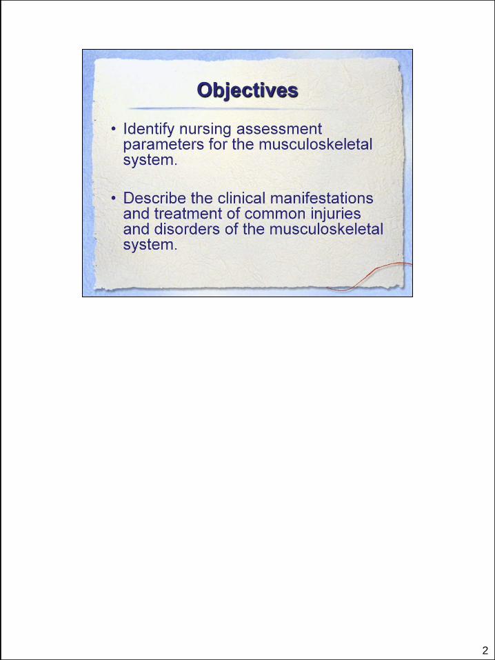
2

Please stand up.
Raise your arms over your head.
Bend to the side…the other side…
Now say, Thank you Musculoskeletal system!

4

5
The skeleton is made up of the
Axial Skeleton: Skull, Vertebral column, sternum, ribs 80 bones
Appendicular Skeleton: The extremities, Pectoral Girdle
(shoulder), Pelvic Girdle (Hip area)
20% of our bones weight is water
80% of our bones weight is made up of minerals
99% body’s calcium is in bone, a calcium reserve. Phosphorous also stored
in bone.

6
Among the Soft Tissues are connective Tissues:
Cartilage is found on the articular surface of the joints as well as in the nose,
ear.
Tendons attach muscle to bone and facilitate movement. More elastic
Ligaments attach bone to bone and provide stability or limit undesired
movement. Not very elastic.
Fascia = sheet or band of connective tissue can withstand limited stretching.
Not very elastic
Fascia surrounds the muscle, minimizes friction by allowing one muscle to
glide over another.
Tendon sheath surrounds the tendons in much the same way.

7

8
Cancellous Bone:
includes the epiphyses: this allows for longitudinal bone growth.
Being more porous and with a larger surface area,allows
diffusion of forces/weight distribution.
This bone has higher rates of metabolic and remodeling
processes.
Cortical Bone:
the symmetrical, cylindrical shape allows for dissipation of
pressure as well, protecting it from injury during activity…
Includes the Haversian System: It is within the cortical bone.
System of interstitial communication among mature bone cells/ osteocytes.
Pereostium is a dense connective tissue that covers the outer surface of bone.
It contains nocioceptors and feels our pain, with bone injuries.
Bone Marrow:
Red: Hematopoiesis = blood cell formation
Yellow marrow as well.
Birth, nearly all marrow is red and hematopoietically active. As need
decreases, red marrow is gradually replaced by yellow. Only Vertebrae, ribs,
sternum, and iliac bones have red marrow in the adult.
Bone biopsies/ bone grafts are usually taken from iliac crest…why do you think
that is?

9
Osteoblasts = basic bone-forming cells, participate in bone remodeling by
synthesizing bone matrix and new cells.
Osteoclasts = participate in bone remodeling by breaking down bone tissue
Through the release of enzymes and acids the osteoclasts
hydrolyze the bone matrix and dissolve bone minerals. This is part of the
healing, remodeling process. But just like everything, it could get out of control,
as with Osteoporosis. (which we will discuss later)
Osteocytes are mature bone cells.
Fracture healing…these cells all have a place.
Osteogenesis imperfecta
Bones are not forming correctly. They are very weak and susceptible to
breakage. Have you seen the movie “Unbreakable”? If he really had OGI, he
would have been short and deformed, not tall and normal looking.
10
.
11
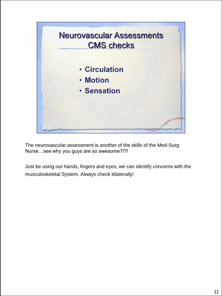
The neurovascular assessment is another of the skills of the Med-Surg
Nurse…see why you guys are so awesome?!?!
Just be using our hands, fingers and eyes, we can identify concerns with the
musculoskeletal System. Always check bilaterally!

12
Need to know normal!
Always check bilaterally!
If you cannot palpate a dorsalis pedis pulse, use a doppler
For diffucult pulses, may use doppler…mark it with an X for
comparison each shift.
Post-operatively:
NV assess at least every 4 hrs.
Operative leg checks: bilateral pedal pulses, skin color, temperature,
sensation and movement of toes.

13
Check for pitting edema/ swelling…over the anterior tibia/shin bone
Always assess bilaterally If possible, compare the pre and post op assessment.

14
Be sure to check everything bilaterally!!
If an injury or surgery, check distally to the site.
15
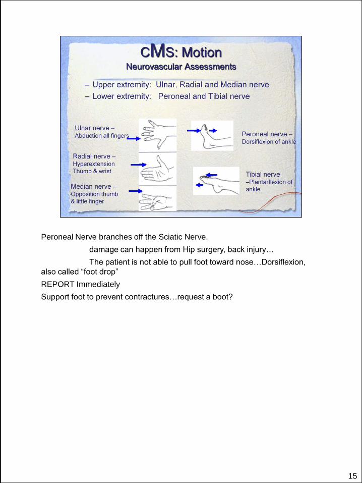
Peroneal Nerve branches off the Sciatic Nerve.
damage can happen from Hip surgery, back injury…
The patient is not able to pull foot toward nose…Dorsiflexion,
also called “foot drop”
REPORT Immediately
Support foot to prevent contractures…request a boot?

16
17
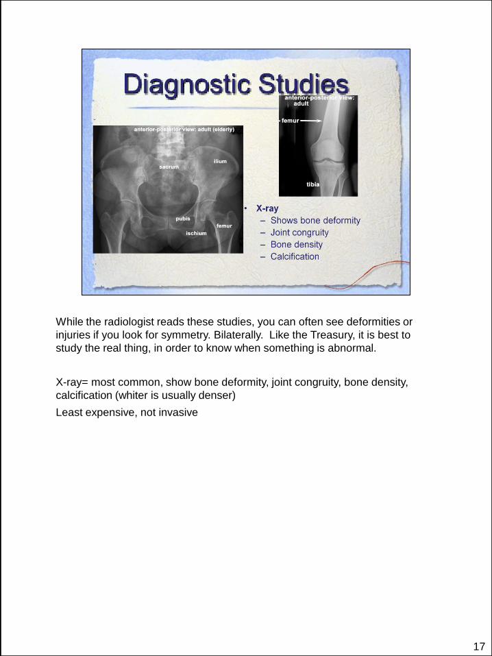
While the radiologist reads these studies, you can often see deformities or
injuries if you look for symmetry. Bilaterally. Like the Treasury, it is best to
study the real thing, in order to know when something is abnormal.
X-ray= most common, show bone deformity, joint congruity, bone density,
calcification (whiter is usually denser)
Least expensive, not invasive
18
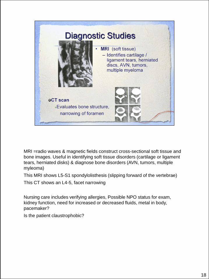
MRI =radio waves & magnetic fields construct cross-sectional soft tissue and
bone images. Useful in identifying soft tissue disorders (cartilage or ligament
tears, herniated disks) & diagnose bone disorders (AVN, tumors, multiple
myleoma)
This MRI shows L5-S1 spondylolisthesis (slipping forward of the vertebrae)
This CT shows an L4-5, facet narrowing
Nursing care includes verifying allergies, Possible NPO status for exam,
kidney function, need for increased or decreased fluids, metal in body,
pacemaker?
Is the patient claustrophobic?

19
Bone scan: osteomyelitis = increased uptake; AVN = decreased uptake.
Myeloma= tumor in cells of hematopoietic portion of bone marrow (the red
marrow).
Cancer usually shows in increased uptake, since the vasculature is increased
for these fast growing cells.

20
Arthro = joint
This myleogram shows a Rt L4-5 herniated disc – with compression of the
spinal column
Nursing Indications:
Consent signed
Pain controlled. Many tests require patient to lie still for extended periods of
time.
Some studies are painful. Apply ice afterwards

21
Arthro = joint
This myleogram shows a Rt L4-5 herniated disc – with compression of the
spinal column
Nursing Indications:
Consent signed
Pain controlled. Many tests require pat to lie still for extended periods of time.
Some studies are painful. Apply ice afterwards
Is the patient pregnant?

22
Aldolase: Muscular dystrophy = muscle atrophy
Dermatomyositis = inflammation muscles, rash, weakness
Alkaline Phosphatase: An enzyme produced by osteoblasts (responsible for
bone-forming, synthesize bone matrix and new cells.
Osteomalacia = bone softening, Vitamin D deficiency
Paget’s disease=inflammation of bones, thickening and hypertrophy of long
bones.
ANA:
Calcium: Bone is the primary storage site for calcium.
23
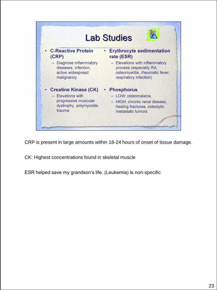
CRP is present in large amounts within 18-24 hours of onset of tissue damage.
CK: Highest concentrations found in skeletal muscle
ESR helped save my grandson’s life. (Leukemia) Is non-specific

24
Aplastic Anemia: The white blood cells attack the bone marrow
Leukemia: immature white blood cells created in bone marrow (blasts)
Not always specific…help diagnose and rule out things.

25
Some of the diseases of the MS system…
26
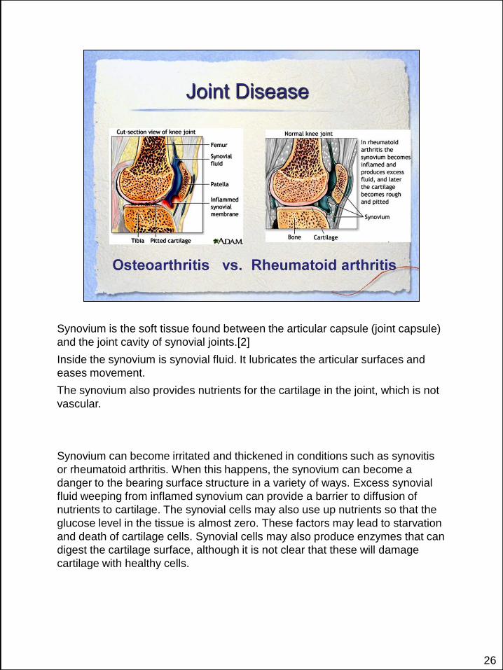
Synovium is the soft tissue found between the articular capsule (joint capsule)
and the joint cavity of synovial joints.[2]
Inside the synovium is synovial fluid. It lubricates the articular surfaces and
eases movement.
The synovium also provides nutrients for the cartilage in the joint, which is not
vascular.
Synovium can become irritated and thickened in conditions such as synovitis
or rheumatoid arthritis. When this happens, the synovium can become a
danger to the bearing surface structure in a variety of ways. Excess synovial
fluid weeping from inflamed synovium can provide a barrier to diffusion of
nutrients to cartilage. The synovial cells may also use up nutrients so that the
glucose level in the tissue is almost zero. These factors may lead to starvation
and death of cartilage cells. Synovial cells may also produce enzymes that can
digest the cartilage surface, although it is not clear that these will damage
cartilage with healthy cells.

27
OA high incidence in weight bearing joints – hips, knees, C & L spine, DIP, PIP,
MCP, MTP joints.
Deterioration of cartilage. Carilage becomes softer, more susceptible to
friction, looses its elasticity, and its ability to resist wear. Becomes yellow and
dull.
Osteophytes: small pieces of cartilage shear off surface….a real MESS!
Weight bearing joints, hands…assymmetrical
Swelling and stiffness. Eased with rest in early stages. There are some genetic
factors…my dad had osteoarthritis…I have some in my hands.
90% qffected by age 40. more women than men over age 50. Crepitus
(rough due to loosening of joint cartilage.
Joint deformity or instability = Heberden’s nodes (DIP), Bouchard’s nodes
(PIP), leg length discrepancy, varus (bow-legged) deformity due to cartilage
loss in medial compartment.
DGX: x-ray, bone scan, MRI.

28
Women more than men, peaks between fourth and sixth decade.
Can happen precipitously;;; STORY: a friend went camping…tic bite…within
days, she had swollen tender joints, fatigue, malaise….ravaged all her joints
immediately
Wonder if it is attached to the Sweet, kind soft-spoken gene…I have known
/cared for so many women with RA that are so sweet and kind.
Pregnancy is a stressor that could trigger RA as well.
Common deformity: Ulnar Shift: actor in Maverick…
BIONIC Woman story
Test: anemia, 75% positive RA factor, ESR and CRP high due to
inflammatory process. X-ray.
Complications: MS: contractures, deformity, loss of function. Resp =
laryngeal pain, difficulty speaking, increased mortality pulmonary disease.
Cardiac= abnormal pericardium. Neuro= cervical spine instability. Optical =
scleritis
Affects joints bilaterally
Enbrel and Humira…see on TV…They affect the Immune system: Nursing
Implications: No live Flu vaccine, Check for TB prior to initiation of treatment.
Watch for signs of infection: viral, fungal, yeast, …Increases risks of some
cancers
29
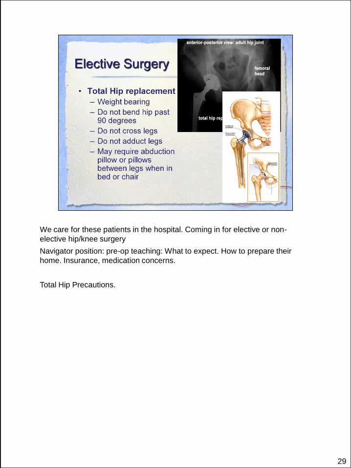
We care for these patients in the hospital. Coming in for elective or non-
elective hip/knee surgery
Navigator position: pre-op teaching: What to expect. How to prepare their
home. Insurance, medication concerns.
Total Hip Precautions.

30
They use jogs to cut the bone to the correct angles and size for the prosthesis
to fit.
Nursing Care:
The newer knees allow more normal movements of the knee. CPMs aren’t
used as much as they used to be.
Monitor CMS/ compartment syndrome
Hemovac. S/S low H/H
Fat embolism…watch O2 Sat
Blood transfusions via Autotransfusion system…salvaged blood given back to
patient
Weight bearing status. Precautions. Should not rest leg in bent position.
Harder to get straight than to bend, if it should get stiff.
Check CMS bilaterally.

31
DVT: 55% with THA, 80% with TKA with no prophylactic treatment. Risk due
to venous stasis and vessel injury and hypercoagulability. Mobilize,
anticoagulate, SCD, TED hose
PE 20-40% incidence.
32

33
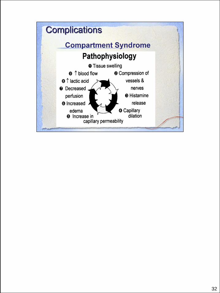
CS = high pressure in a muscle compartment in the closed fascial space
related to swelling. Can occur 6 hrs after insult or up to 2 days later.
Elevation compromises blood flow

34

35
Initially, you will only reinforce dressing. Notify surgeon if continues to
drain/saturate dressings.
With SCIP: timed antibiotics
Remove foley
Prevent superficial infections by keeping site clean and dry.
Early deep infection is from surgery.
Late deep infection is from hematogenous seeding…an infection travels to the
hip/ joint…
Increasing pain could be a sign of infection

36
37
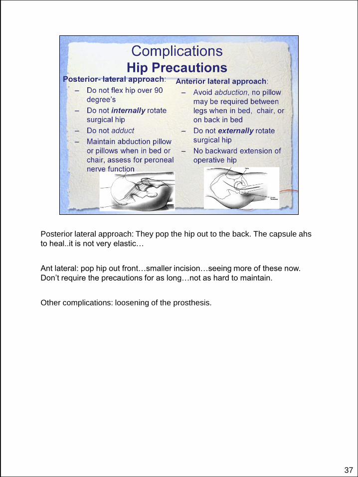
Posterior lateral approach: They pop the hip out to the back. The capsule ahs
to heal..it is not very elastic…
Ant lateral: pop hip out front…smaller incision…seeing more of these now.
Don’t require the precautions for as long…not as hard to maintain.
Other complications: loosening of the prosthesis.
38
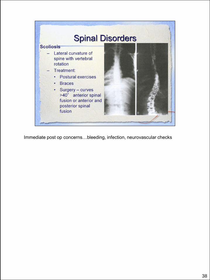
Immediate post op concerns…bleeding, infection, neurovascular checks

39

40
HNP – annulus fibrosis – fibrocartilaginous layer of disc weakens and cracks,
becomes unable to contain the disc material (nucleus pulpous).
Spondylolysis – defect or break in pars interarticularis L4-5 or L5- S1.
Spondylolisthesis – forward slippage of one vertebrae over another. Most L4-5
or L5-S1.
Spinal stenosis – narrowing of the spinal canal – esp. cervical and lumbar
area. Gradual shift in spinal alignment (kyphosis (humpback or posterior
curve), lordosis (anterior curve) , stenosis (narrowing).

41
C4-C5 = Quadriplegia - Above T1; T1-L2 = Paraplegia – below T1

42

43
44

45
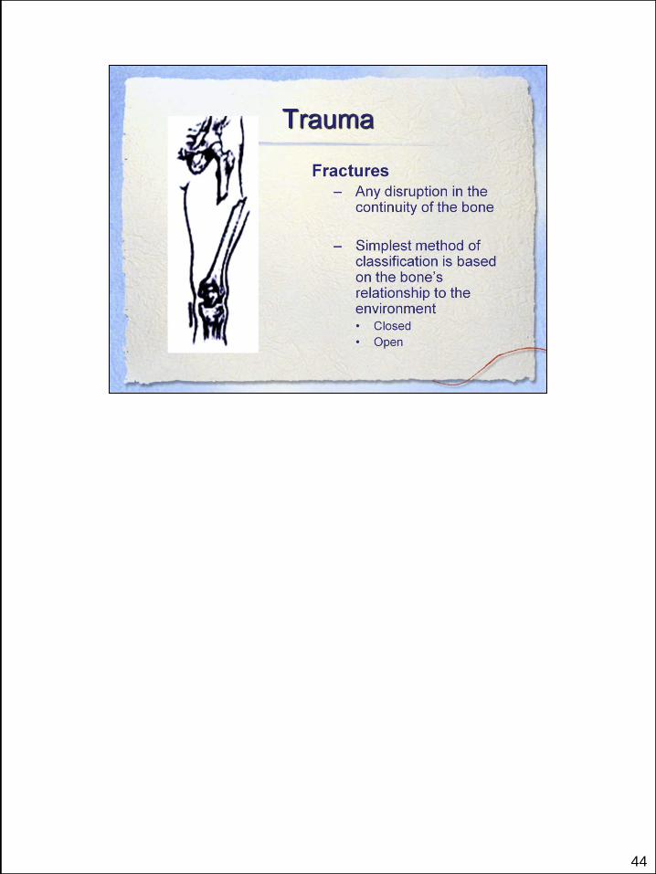
Comminuted
Spiral
Impacted…Distal femur
Tansverse
Obliqe

46
Osteoclasts group at fracture site. Fibroblasts from outer layer periosteum
and damaged connective tissue are involved. Blood is supplied by medullary
circulation.
Granulation tissue matures into a callus
Callus converts the loosely woven network of bone, cartilage and fibrous
tissue which forms a bridge over the bone fragments, uniting the fractured
bone (occification).
Consolidation and remodeling: bone is reshaped to meet its mechanical
demands.

47

48
Plaster and Fiberglass Casts: Nursing Implications: OTA 48-72 hours to dry
completely. Avoid creating indentations.
Elevate: Marble theory
CMS checks distal to injury
May need to bivalve cast
Often will switch ot a boot or brace, after a certain period of time. Avoid putting
things inside cast to scratch
Fiberglass casts: harden more quickly, can tolerate more abuse.

49
ORIF
External fixators: scary looking…pin care. Watch for infection.

50
Pulse Ox: we think it is usually used for respiratory depression form narcotics,
but hypoxia can also be an early sign of fat embolism (24-72 hrs after trauma)
ABGs indicate respiratory alkalosis. Fat emboli are more flexible
Rash/ Petechiae…is a sign…chest axilla, neck…due to the fat emboli float and
embolize in non-dependent areas.
TREAT FAT EMBOLI: O2, IVF, Mech Ventilation

51

52
Optimal bone mass at age 30…then we start losing bone…the question is how
rapidly?
Get a DEXA scan

53
Remember Wallie, the movie?
The people that were living in the space ship…no gravity…caused shrinking of
bones
Use it or lose it! Just like muscles that atrophy…
Remember, our bones were created to withstand pressure…they expect it and
need it.

54
Loss of stage 4 REM sleep is believed to cause ineffective tissue restoration
and pain.
Abnormal serotonin metabolism can alter pain perception in tissues
55

56

57