pontifÍcia universidade catÓlica do rio … can influence mechanisms regulating hydroelectrolytic...
TRANSCRIPT

1
PONTIFÍCIA UNIVERSIDADE CATÓLICA DO RIO GRANDE DO SUL
FACULDADE DE BIOCIÊNCIAS
PROGRAMA DE PÓS-GRADUAÇÃO EM BIOLOGIA CELULAR E MOLECULAR
MESTRADO
DANIELA LIVINALLI RODRIGUEZ
AVALIAÇÃO DO SISTEMA RENINA-ANGIOTENSINA E DO
ESTRESSE OXIDATIVO NO SISTEMA RENAL DE RATOS
MANIPULADOS NO PERÍODO NEONATAL
Prof. Dr. Jarbas Rodrigues de Oliveira
Orientador
Porto Alegre, 2010.
2
DANIELA LIVINALLI RODRIGUEZ
AVALIAÇÃO DO SISTEMA RENINA-ANGIOTENSINA E DO ESTRESSE
OXIDATIVO NO SISTEMA RENAL DE RATOS MANIPULADOS NO PERÍODO
NEONATAL
Dissertação apresentada ao Programa de
Pós-Graduação em Biologia Celular e
Molecular da Faculdade de Biociências da
Pontifícia Universidade Católica do Rio
Grande do Sul como requisito para obtenção
do título de Mestre em Biologia Celular e
Molecular.
Orientador: Prof. Dr. Jarbas Rodrigues de Oliveira
Porto Alegre, 2010.
3
Agradecimentos
Ao CNPq pela bolsa concedida.
Ao professor Jarbas Rodrigues de Oliveira pela oportunidade, dedicação e
conhecimento durante todo o trabalho.
Ao Professor Márcio F. Donadio pela co-orientação. Sempre presente para
esclarecer dúvidas.
A professora Virgínia Minghelli Schmitt que sempre esteve disposta a ajudar nas
dúvidas no decorrer do trabalho.
A todos do Laboratório de Biofísica Celular e Inflamação, que sempre estiveram
presentes para tirar dúvidas, pela amizade, pelo carinho e também nos muitos
momentos de descontração. Em especial aos colegas da Biologia Molecular que
junto comigo realizaram o primeiro trabalho do laboratório nesta área.
A minha família, em especial meus pais Adriana e Heber Daniel que sempre me
apoiaram para chegar aonde eu cheguei, sem eles nada seria possível. Aos meus
irmãos Pablo Vicente e Mariana que de alguma maneira me confortaram nas
dificuldades e entenderam a minha ausência.
Aos meus amigos, minha segunda família, sempre presentes para comemorar as
minhas conquistas e ajudar nos momentos mais difíceis. Em especial a colega
Bárbara Scherer que esteve ao meu lado desde a prova até a conclusão do
mestrado.
A todos aqueles que direta ou indiretamente contribuíram para a realização deste
trabalho.

4
Resumo
A manipulação neonatal é considerada uma intervenção que ocasiona efeitos
duradouros no comportamento emocional e na reatividade ao estresse em animais
adultos. O período imediatamente após o nascimento é um período crítico quando o
cérebro imaturo é permanentemente alterado por hormônios esteróides gonadais e
da supra-renal. Estudos prévios demonstraram que a manipulação neonatal pode
influenciar mecanismos de regulação do equilíbrio hidroeletrolítico e na função renal.
Os componentes do sistema renina-angiostensina (SRA) no rim são
altamente expressos durante o desenvolvimento renal e estão ligados a estágios
específicos da nefrogênese e vascularização, tendo maior expressão em ratos
recém-nascidos. Esse sistema tem papel fundamental nos mecanismos de
inflamação e defesa de células e tecidos do organismo. A angiotensina II é o
principal hormônio efetor nessa cascata e é também considerada um hormônio pró-
oxidante, e liga-se a dois tipos de receptores: AT1 e AT2, que têm funções opostas.
O estresse oxidativo (EO) é definido como um desequilíbrio entre a formação de
espécies reativas de oxigênio (ERO) e mecanismos de defesa antioxidante. Essas
EROs quando em excesso causam dano a vários componentes celulares. O objetivo
dessa pesquisa foi avaliar o efeito da manipulação neonatal sobre o SRA e o
balanço oxidativo renal de ratos
Verificou-se que os animais manipulados apresentaram aumento na expressão
de renina e do receptor AT2 e uma diminuição do receptor AT1. Foi encontrado um
aumento de ANG II plasmática e também de TBARS no rim. Estes resultados
indicam que a manipulação neonatal altera o sistema renina-angiotensina e
justificam as alterações renais já descritas.
Palavras-chave: Sistema Renina-Angiotensina; Estresse oxidativo; Manipulação
neonatal; receptores de angiotensina II.

5
Abstract
Neonatal handling is considered an intervention that causes lasting effects on
emotional behavior and reactivity to stress in adult animals. The period immediately
after birth is a critical period when the immature brain is permanently altered by
gonadal steroid hormones and adrenal. Previous studies have shown that neonatal
handling can influence mechanisms regulating hydroelectrolytic balance and renal
function.
The components of the renin-angiostensina (RAS) are highly expressed in the
kidney during renal development and are linked to specific stages of nephrogenesis
and vascularization is higher in newborn rats. This system has a fundamental role in
the mechanisms of inflammation and protection of cells and tissues. Angiotensin II is
the main effector hormone in this cascade, and binds to two types of receptors: AT1
and AT2, which have opposite functions. The aim of this study was to evaluate the
effect of neonatal handling on the renal RAS and oxidative balance in rats.
Handled animals presented an increase in AT2 receptor expression and a
decrease in AT1 receptor expression. An increase in plasmatic ANG II and in kidney
TBARS was also found. These results indicate that neonatal handling changes the
renin-angiotensin system, justifying the renal alterations described.
Key words: Renin-angiotensin system; oxidative stress; neonatal handling;
angiotensin II receptors.

6
SUMÁRIO
CAPÍTULO 1 __________________________________________________ 7
1.1 INTRODUÇÃO ______________________________________________________ 7
1.1.1 MANIPULAÇÃO NEONATAL ________________________________________________ 7
1.1.2 ANGIOTÊNSINA II E O DESENVOLVIMENTO RENAL __________________________ 12
1.1.3 ESTRESSE OXIDATIVO __________________________________________________ 14
1.2 JUSTIFICATIVA ____________________________________________________ 17
1.3 OBJETIVOS _______________________________________________________ 18
1.3.1 GERAL ________________________________________________________________ 18
1.3.2 ESPECÍFICOS __________________________________________________________ 18
1.4 ASPECTOS ÉTICOS _________________________________________________ 19
CAPÍTULO 2 _________________________________________________ 20
2.1 MANUSCRITO DO TRABALHO EXPERIMENTAL _________________________ 20
CAPÍTULO 3 _________________________________________________ 44
3.1 CONSIDERAÇÕES FINAIS ___________________________________________ 44
REFERÊNCIAS _______________________________________________ 46
ANEXO A ____________________________________________________ 50

7
CAPÍTULO 1
1.1 INTRODUÇÃO
1.1.1 MANIPULAÇÃO NEONATAL
A manipulação neonatal é considerada uma intervenção durante o período
neonatal que tem efeitos duradouros no comportamento emocional e na reatividade
ao estresse em animais adultos (Donadio et al., 2009). A manipulação neonatal
altera permanentemente o eixo hipotálamo-hipófise-adrenal (HPA) em resposta a
estímulos estressantes (Gomes et al., 2005) e tem sido usada como um modelo
experimental para examinar os mecanismos pelos quais mudanças ambientais
precoces podem afetar o sistema neural, conduzindo a alterações comportamentais
e mudanças neuroendócrinas. Como nos adultos, ratos manipulados no período
neonatal sintetizam e secretam menor quantidade de hormônio liberador de
corticotropina (CRH) e corticosterona em resposta a uma variedade de estressores,
e mostra um rápido retorno desses hormônios aos níveis basais após o término do
estímulo (Gomes et al., 2005). O modelo de manipulação neonatal consiste na
retirada dos filhotes de perto da mãe e da manipulação dos mesmos diariamente por
1 minuto durante os primeiros 10 dias de vida (Donadio et al., 2009; Gomes et al.,
2006, Gomes et al., 1999; Gomes et al., 2005, Hermel et al., 2001; Lucion, et al.
2003, Severino et al., 2004; Winkelmann-Duarte et al.; 2007). Este procedimento
altera a diferenciação do eixo HPA promovendo mudanças nos padrões
comportamentais e de resposta ao estresse em animais adultos (Levine, 2001;
Meerlo et al., 1999; Núñez et al., 1996).
A atividade do eixo HPA é governada pela secreção de CRH e de vasopressina
(AVP) pelo hipotálamo, os quais, por sua vez, ativam o hormônio
adrenocorticotrópico (ACTH) pela pituitária, que finalmente estimula a secreção de
glicocorticóides pelo córtex adrenal como ilustra a figura 1. Os glicocorticóides,
então, interagem com seus receptores em múltiplos tecidos-alvo, incluindo o eixo
HPA, onde são responsáveis pela retro-alimentação da secreção do ACTH pela
pituitária e do HLC a partir do hipotálamo. Embora os glicocorticóides regulem a

8
função de quase todos os tecidos do corpo, o efeito fisiológico mais conhecido
desses hormônios é a regulação do metabolismo energético (Juruena et al., 2004).
Figura 1 - Eixo HPA (Rang, H.P., Dale, M.M., Ritter, J.M, 2008)
O estresse é considerado por muitos cientistas como uma situação de ativação
do eixo HPA, representado principalmente pela elevação dos níveis de hormônio
ACTH (Moal, 2007). Outros autores sugerem que a ativação de outros sistemas com
ou sem elevação de ACTH pode refletir em distúrbios da homeostasia induzidos pelo
estresse (Pacák e Palkovits, 2001). A homeostasia foi descrita primeiramente como
processos coordenados que mantêm constante a fisiologia do organismo (Cannon,
1941), que pode ser constantemente ameaçada por forças ou estressores
intrínsecos e extrínsecos (Moal, 2007). Dentre os estressores intrínsecos estão
polimorfismos genéticos bem como alterações na expressão de genes e estressores
extrínsecos como fatores ambientais, são importantes na determinação de respostas
individuais ao estresse (Pacák e Palkovits, 2001).
O sistema de estresse coordena respostas adaptativas do organismo a
qualquer tipo de estressor (Tsigos e Chrousos 2002). A ativação do sistema de

9
estresse leva a mudanças comportamentais e periféricas para melhorar a habilidade
do organismo a ajustar a homeostase e aumentar sua chance de sobrevivência.
Durante uma situação de estresse agudo, a amplitude e a sincronização de CRH e
AVP na hipófise aumenta, levando a um aumento de ACTH e secreção de cortisol.
Dependendo do tipo de estressor, outros fatores como AVP de origem de neurônios
magnocelulares, angiotensina II (ANG II), várias citocinas e mediadores lipídicos da
inflamação são secretados e atuam em componentes hipotálamicos, hipofisários ou
adrenais do eixo HPA potencializando sua atividade (Tsigos e Chrousos 2002). O
ACTH circulante é o principal regulador da secreção de glicocorticóides pelo córtex
adrenal. Os glicocorticóides são os efetores finais do eixo HPA e participam no
controle da homeostasia corporal e na resposta ao estresse do organismo. O
feedback negativo dos glicocorticóides na resposta secretória de ACTH atua como
limite na duração da exposição dos glicocorticóides aos tecidos, assim, minimizando
os efeitos catabólicos, antireprodutivos e imunossupressores desses hormônios
(Ritter, J.M, 2008). Além disso, os glicocorticóides alteram a estabilidade de RNAs
mensageiros, portanto a tradução de muitas proteínas dependentes de
glicocorticóides, bem como o potencial elétrico das células neuronais (Tsigos e
Chrousos 2002).
O desenvolvimento normal do eixo HPA é essencial para o funcionamento
adequado do organismo durante a vida adulta. Muitos estudos em humanos
demonstraram, que um trauma nos primeiros dias de vida, como abuso ou maltratato
de crianças, tem efeitos duradouros na hipófise-adrenal e representa um risco maior
para o desenvolvimento de transtornos de humor e de ansiedade (Schmidt et al.,
2003). No rato uma das características do desenvolvimento pós-natal do sistema de
estresse é chamada de período hiporresponsivo ao estresse. É o período que
compreende do 4° ao 14° dia pós-natal, e é caracterizado por níveis basais muito
baixos de corticosterona devido a incapacidade dos estressores induzirem um
aumento de ACTH e uma liberação de corticosterona (Schmidt, et al. 2003, Levine
2001).
Esse período logo após o nascimento é um período crítico em que o cérebro é
permanentemente alterado por hormônios esteróides gonodais e adrenais (Gomes
et al., 1999), deste modo é de grande importância que o animal mantenha o eixo
HPA sob supressão durante o período neonatal, característica padão no seu
10
desenvolvimento (Martin et al., 1977). Durante o período hiporresponsivo ao
estresse, tanto estimulações aparentemente inofensivas, bem como estímulos como
frio e choque elétrico induzem alterações comportamentais e endócrinas na vida
adulta (Levine, 1994). Os efeitos de intervenções ambientais durante o período
neonatal pode constituir a formação de vulnerabilidades individuais a doenças
relacionadas ao estresse ao longo da vida (Lucion, et al. 2003).
Variações no cuidado maternal têm sido amplamente consideradas como uma
influência crítica no desenvolvimento (Caldji et al., 2000; Champagne, et al. 2003).
No rato, variações no comportamento maternal, particularmente o ato de lamber o
filhote, regula o desenvolvimento de respostas endócrinas, emocionais e cognitivas
ao estresse (Champagne, et al. 2003) e esses efeitos são mediados por variações
no cuidado maternal de tal forma que o comportamento da mãe altera o
desenvolvimento de respostas comportamentais e endócrinas em resposta ao
estresse na prole através de efeitos na expressão gênica de tecidos específicos
(Weaver et al., 2006). O desenvolvimento do eixo HPA em resposta ao estresse é
modificado por eventos no ambiente neonatal, incluindo a manipulação neonatal
(Liu, et al. 1997).
Quando analisados os comportamentos de mães de animais manipulados e
não manipulados durante os 10 primeiros dias de vida, um período crítico para os
efeitos da manipulação no desenvolvimento do eixo HPA, as mães de animais
manipulados mostraram um aumento no ato de lamber os filhotes comparados com
mães de filhotes não manipulados (Liu, et al. 1997). Quando adultas, a prole das
mães que lamberam mais mostraram redução significativa no ACTH plasmático e
respostas limitadas de corticosterona ao estresse comparado com a prole das mães
que lamberam menos (Caldji et al.,2000; Liu et al., 1997). Animais adultos,
manipulados quando recém-nascidos, demonstraram diminuição do medo sob
condições de estresse (Caldji et al.,2000; Hsu et al., 2003; Weaver et al., 2006) e
respostas diminuidas do eixo HPA em resposta ao estresse (Weaver et al., 2006).
Diferenças individuais no comportamento (Caldji et al.,2000; Francis e Meaney
1999), no eixo HPA (Caldji et al.,2000) e nas respostas ao estresse (Francis e
Meaney, 1999) são, em parte, derivadas de variações no cuidado maternal pós-natal
(Caldji et al.,2000; Francis e Meaney, 1999).
11
Esse procedimento aparentemente inofensivo na infância causa efeitos
comportamentais e neuroendócrinos estáveis, expressos pela redução de respostas
emocionais na vida adulta, como um aumento da atividade exploratória, que é
interpretada pela diminuição do medo em ambientes novos, o que foi comprovado,
pois ratos manipulados adultos machos e fêmeas exibem uma redução do medo
frente a um predador (Severino, et al. 2004). Também foram relatas diminuição da
secreção de corticosterona e adrenocorticotropina em resposta a estressores na
vida adulta (Núñez, et al. 1996).
Em ratos, a manipulação neonatal altera o eixo HPA, bem como o eixo
hipotálamo-hipófise-gônadas (HPG) (Gomes et al., 2005). Fêmeas manipuladas
mostraram diminuição nos níveis de prolactina 2 e 5 minutos após o estresse,
quando comparadas com o grupo não manipulado no mesmo período de tempo
após o estresse (Severino, et al. 2004), também apresentaram redução no número
de oócitos confirmando uma diminuição da ovulação causada pela manipulação
(Gomes et al., 2005). Outro estudo também demonstrou que o número de fêmeas
manipuladas que apresentou ciclos anovulatórios foi significativamente maior que as
fêmeas do grupo não manipulado (Gomes et al., 1999). A manipulação neonatal
também reduz a atividade sexual em ratos machos e fêmeas e os níveis plasmáticos
de estradiol, hormônio luteinizante (LH), hormônio folículo estimulante (FSH) e
prolactina (PRL) (Gomes et al., 2006).
A manipulação aumenta a densidade de receptores de glicocorticóides no
hipocampo e no córtex frontal (Liu et al., 1997; Winkelmann-Duarte et al., 2007) e
mudanças em receptores para o neurotransmissor ácido gama-aminobutírico
(GABAA) no hipocampo (Hsu et al., 2003; Winkelmann-Duarte, et al. 2007). Além
disso, resultados mostraram uma diminuição na densidade de receptores de
angiotensina II (ANG II) na área pré-optica (MPOA) e no núcleo paraventricular do
hipotálamo (PVN) em animais manipulados (Gomes et al., 2006) e também
mostraram redução significativamente no número de células e no volume do locus
coeruleus em ratos machos e fêmeas manipulados comparados com ratos não
manipulados. Isso sugere que a estimulação precoce no ambiente neonatal induz a
mudanças estruturais no núcleo central noradrenérgico, que pode ser um dos fatores
responsáveis pelas alterações comportamentais e hormonais observadas nesses
animais na vida adulta (Lucion et al., 2003).

12
Estudos prévios em nosso laboratório mostraram uma redução no peso total
renal, na ingestão hídrica, no volume urinário, no clearance de creatinina, na ANG II
plasmática, corticosterona e aldosterona e um aumento da osmolaridade urinária e
da fração excretada de sódio comparando animais manipulados com animais não
manipulados demostrando que a manipulação neonatal pode influenciar
mecanismos de regulação do equilíbrio hidroeletrolítico e no processo de maturação
renal, considerando que o período neonatal é crucial para o desenvolvimento renal e
do trato urinário (Donadio et al., 2009).
1.1.2 ANGIOTÊNSINA II E O DESENVOLVIMENTO RENAL
O sistema renina-angiotensina (SRA), que é um importante modulador da
função do eixo HPA através da estimulação da secreção de CRH durante a resposta
ao estresse, tem um papel crucial no desenvolvimento renal (nefrogênese) incluindo
desenvolvimento vascular e estruturas tubulares (Chen et al., 2004; Lasaitiene, et al.
2006). No rim, todos os componentes do SRA (renina, angiotensinogênio, enzima
conversora de angiotensina (ECA) e os receptores de angiotensina tipo I e II) são
sintetizados localmente. Além disso, a expressão de todos os componentes do SRA
no rim pode ser detectada a partir do 12° ao 17° dias de gestação, sendo maior em
ratos fetos e neonatais do que em ratos adultos (Chen et al., 2004). Apesar dos
componenetes do SRA estarem expressos em níveis altos na fase gestacional e
neonatal, uma hiperativação deste sistema tem sido considerada como tendo um
papel central na progressão da doença renal crônica (Lin et al., 2009; Nistala et al.,
2009)
A renina é a enzima que cliva o angiotensinogênio, uma substância produzida
pelo fígado, em um polipeptídeo chamado angiotensina I. A angiotensina I circula no
sangue, e uma segunda enzima, chamada enzima conversora de angiotensina
(ECA), converte angiotensina I em angiotensina II (ANG II) pela remoção de um
único polipeptídeo. Dentre os componentes desta cascata, é a ANG II que tem
atividade sobre o rim. Os primeiros efeitos da ANG II no rim é a retenção de água e
sódio; no sistema arterial, a ANG II causa vasoconstrição, que leva a um aumento
na pressão arterial. Esse sistema tem um papel fundamental nos mecanismos de
13
inflamação e na defesa de células e tecidos de organismos (Serrano, et al. 2009). A
ANG II é o principal hormônio efetor na cascata da SRA (Lasaitiene, et al. 2006), e é
considerado um hormônio com atividade próoxidante, pró-fibrogênica (Wei et al.,
2009) e pró-inflamatória (Serrano et al., 2009) que pode influenciar todos os
estágios da resposta inflamatória (Marchesi et al.,2008). A ANG II é conhecida por
aumentar a pressão arterial, induzir hipertrofia celular renal, e promover a ativação
do fator nuclear κB (NFκB) e acumulação de matriz protéica, eventos que estão
ligados com a progressão da doença renal (Vaziri, et al. 2007).
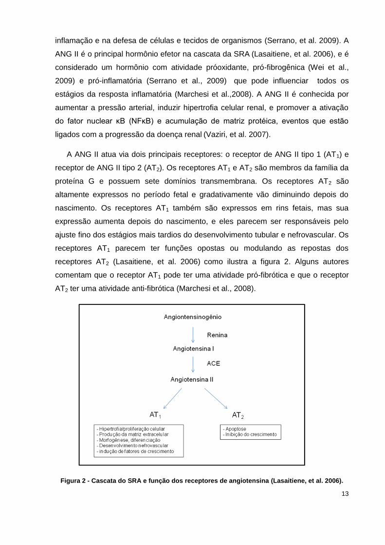
A ANG II atua via dois principais receptores: o receptor de ANG II tipo 1 (AT1) e
receptor de ANG II tipo 2 (AT2). Os receptores AT1 e AT2 são membros da família da
proteína G e possuem sete domínios transmembrana. Os receptores AT2 são
altamente expressos no período fetal e gradativamente vão diminuindo depois do
nascimento. Os receptores AT1 também são expressos em rins fetais, mas sua
expressão aumenta depois do nascimento, e eles parecem ser responsáveis pelo
ajuste fino dos estágios mais tardios do desenvolvimento tubular e nefrovascular. Os
receptores AT1 parecem ter funções opostas ou modulando as repostas dos
receptores AT2 (Lasaitiene, et al. 2006) como ilustra a figura 2. Alguns autores
comentam que o receptor AT1 pode ter uma atividade pró-fibrótica e que o receptor
AT2 ter uma atividade anti-fibrótica (Marchesi et al., 2008).
Figura 2 - Cascata do SRA e função dos receptores de angiotensina (Lasaitiene, et al. 2006).

14
A ligação da ANG II ao receptor AT1 leva a ativação de NAD(P)H oxidase
causando um aumento na produção de espécies reativas de oxigênio (ERO)
(Lenarczyk et al., 2009; Nistala et al., 2009; Severino, et al. 2004). A produção de
ERO leva a ativação de NFκB e a transcrição de fatores pró-inflamatórios (Marchesi
et al.,2008; Serrano, et al. 2009), levando em um aumento nos fatores de
crescimento, citocinas e quimiocinas (Serrano, et al. 2009). A inibição farmacológica
ou genética da produção de ANG II ou a ativação do receptor de ANG II tipo I (AT1)
durante a nefrogênese resultam em anormalidades no desenvolvimento estrutural e
funcional dos rins em várias espécies animais (Chen et al., 2004; Lasaitiene et al.,
2006) e o bloqueio desse receptor causa diminuição na pressão sanguínea,
desacelera a progressão de doenças renais e atenua a super regulação do sistema
pró-oxidante e pró-inflamatório (Vaziri, et al. 2007). Em humanos, o uso incorreto de
inibidores da enzima conversora de angiotensina (ECA) ou de bloqueadores dos
receptores AT1 durante a gravidez causa anúria neonatal e displasia tubular renal,
confirmando o papel do SRA na nefrogênese humana (Lasaitiene, et al. 2006).
Outros estudos mostraram um marcado aumento na produção de ANG II e na
expressão do receptor AT1 na região tubulointersticial com redução na massa renal e
também outras nefropatias (Vaziri, et al. 2007). Considerando que o processo de
desenvolvimento e maturação dos órgãos ocorre continuamente ao longo dos
períodos pré e pós natais, modificações no desenvolvimento podem levar a redução
dos néfrons, hipertensão e doenças renais, e fatores genéticos e condições
ambientais parecem contribuir nessas alterações (Puddu, et al. 2009).
1.1.3 ESTRESSE OXIDATIVO
O estresse oxidativo (EO) é definido como um desequilíbrio entre a formação
de ERO e mecanismos de defesa anti-oxidante (Galle 2001). É um processo
biológico que causa acúmulo de espécies reativas de oxigênio no plasma, tecido,
interior das células e nas mitocôndrias, acúmulo esse que causa danos à estrutura
das biomoléculas de DNA, lipídios, carboidratos e proteínas, além de outros
componentes celulares (Serrano et al., 2009; Núñez, et al., 1996) e que envolve

15
ativação de vias de sinalização específicas (Serrano et al., 2009). Os oxidantes são
gerados como resultado do metabolismo normal intracelular na mitocôndria e nos
peroxissomos e influenciam numerosos processos celulares que estão ligados ao
envelhecimento e doenças relacionadas ao envelhecimento (Finkel e Holbrook,
2000) e também com atividade física intensa, apoptose, câncer, diabetes mellitus e
arteriosclerose (Núñez, et al. 1996). Mas os oxidantes também podem ser gerados
de forma exógena, como, por exemplo, por exposição ao álcool, fumo, drogas, raios
ultravioleta, quimioterápicos, citocinas inflamatórias, toxinas ambientais, entre outros
fatores (Finkel e Holbrook, 2000; Núñez et al., 1996) como ilustra a figura 3.
As ERO necessitam estar em níveis fisiológicos normais, pois uma diminuição
das mesmas abaixo do ponto homeostático pode interromper o papel fisiológico dos
oxidantes na proliferação celular e na defesa do hospedeiro (Finkel e Holbrook,
2000). O termo EO é utilizado em circunstâncias nas quais os radicais livres
promovem dano tecidual ou na produção de compostos tóxicos ou danosos aos
tecidos (Finkel e Holbrook, 2000; Schneider e Oliveira, 2004). Dentre os diversos
sistemas que podem ser afetados por intervenções precoces e que possuem grande
importância nos mecanismos de controle da homeostase está o sistema de defesa
antioxidante (Finkel e Holbrook, 2000). Este sistema é composto basicamente por
proteínas enzimáticas, não enzimáticas e algumas vitaminas, capazes de inibir ou
reduzir os danos causados por espécies reativas de oxigênio e nitrogênio (Ferreira e
Matsubara, 1997; Finkel e Holbrook, 2000; Lenarczyk et al., 2009). O sistema de
defesa antioxidante enzimático inclui a catalase (CAT), superóxido dismutase (SOD)
e a glutationa peroxidase (GPx), e o sistema de defesa não-enzimático inclui a
glutationa reduzida (GSH) e algumas vitaminas como as vitaminas A, C e E (Finkel e
Holbrook, 2000).

16
Figura 3 - As fontes e as respostas celulares a espécies reativas de oxigênio (ERO) (Finkel e
Holbrook 2000).
Pode-se dizer que um organismo encontra-se sob EO quando ocorre um
desequilíbrio entre os sistemas pro-oxidantes favorecendo o dano oxidativo e
antioxidantes, de maneira que os primeiros sejam predominantes (Finkel e Holbrook,
2000; Galle, 2001; Schneider e Oliveira, 2004). Um dos principais mecanismos de
lesão é a lipoperoxidação (LPO), ou seja, a oxidação da camada lipídica da
membrana celular e um dos métodos capazes de avaliar a LPO é a medida de
substâncias que reagem ao ácido tiobarbitúrico (TBARS), embora seja um método
indireto é bastante utilizado (Schneider e Oliveira, 2004).

17
1.2 JUSTIFICATIVA
Levando-se em consideração a importância das alterações comportamentais e
neuroendócrinas provocadas pela manipulação no período neonatal e as evidências
funcionais nos rins de animais adultos que foram manipulados ao nascimento, criou-
se a hipótese de que os componentes do SRA, como a renina e os receptores AT1 e
AT2 da ANG II, e o estresse oxidativo possam estar alterados nesses animais. Dessa
forma, decidiu-se investigar os efeitos da manipulação neonatal sobre a expressão
de diferentes componentes do SRA e de parâmetros de estresse oxidativo.

18
1.3 OBJETIVOS
1.3.1 GERAL
Avaliar o efeito da manipulação neonatal sobre o SRA e o balanço oxidativo
no sistema renal de ratos.
1.3.2 ESPECÍFICOS
- Avaliar a expressão do RNAm da renina e dos receptores AT1 e AT2 no
sistema renal;
- Avaliar o dano oxidativo (TBARS) e os parâmetros antioxidantes (CAT,
SOD) no sistema renal dos animais em estudo;
- Avaliar a concentração plasmática de ANG II.

19
1.4 ASPECTOS ÉTICOS
Este projeto foi aprovado pelo Comitê Científico da Faculdade de Biociências
da Pontifícia Universidade Católica do Rio Grande do Sul, protocolado sob o no
100/09.
20
CAPÍTULO 2
2.1 MANUSCRITO DO TRABALHO EXPERIMENTAL
Evaluation of the kidney renin-angiotensin system and oxidative stress in
neonatal handled rats
Submetido ao periódico: Developmental Biology

21
Evaluation of the kidney renin-angiotensin system and oxidative stress in
neonatal handled rats
Daniela Livinalli Rodriguez1,2, Márcio Vinícius Fagundes Donadio1,3, Fernanda
Cristina de Mesquita1, Débora Attolini1, Bruna Borba1, Patrícia da Silva Scherer1,
Priscilla Heberle Almeida1, Vinícius Lorini da Costa1, Bárbara de Souza Scherer1,2,
Virgínia Minguelli Schmitt4 and Jarbas Rodrigues de Oliveira1,2
1Faculdade de Biociências e Laboratório de Pesquisa em Biofísica Celular e
Inflamação, Pontifícia Universidade Católica do Rio Grande do Sul (PUCRS).
2Programa de Pós-graduação em Biologia Celular e Molecular (PPGBCM), Pontifícia
Universidade Católica do Rio Grande do Sul (PUCRS). 3Faculdade de Enfermagem,
Nutrição e Fisioterapia, Pontifícia Universidade Católica do Rio Grande do Sul
(PUCRS). 4Faculdade de Farmácia, Pontifícia Universidade Católica do Rio Grande
do Sul (PUCRS).
Adress correspondence to: Márcio Vinícius F. Donadio, Av. Ipiranga 6681 – Prédio
12 – 8° andar. Porto Alegre, Rio Grande do Sul 90619-900 (Brazil). CEP90619-900.
Tel. +55 51 3320 3646, Fax +55 51 3320 3647.
E-mail:[email protected]
22
ABSTRACT
Neonatal handling induces behavioral, hormonal and hydroelectrolytic balance
changes. In the kidney, all renin-angiotensin system components, including
angiotensin II type 1 (AT1) and type 2 (AT2) receptors, are highly expressed during
nephrogenesis. This study aimed to evaluate the effects of neonatal handling on the
kidney renin-angiotensin system and oxidative stress balance. The pups were
divided into two groups: nonhandled and handled. The procedure consisted of
handling the pups for 1 min/day in the first 10 days of life. On day 1, 5 and 10,
animals were killed by decapitation. Blood samples were collected and the kidneys
were removed. Renin, AT1 and AT2 mRNA expression were evaluated through RT-
PCR. Angiotensin II (ANG II) plasma levels were also measured. Results have shown
an increase in ANG II plasma concentration and kidney AT2 expression mRNA. The
kidney mRNA AT1 expression was decreased and, when the oxidative stress balance
was evaluated, a TBARS increase was shown. The results indicate that handling in
the neonatal period induces the activation of the angiotensinergic system, as well as
modulates its receptors, indicating that it may play an important role in the
development of some of the renal alterations.
Key words: Neonatal handling, renin-angiotensin system, oxidative stress,
angiotensin II receptors.

23
Introduction
Environmental stimuli during the neonatal period have lasting effects on
emocional behavioral and reactivity to stress in adult animals (Meerlo et al., 1999;
Padoin et al., 2001). Neonatal handling has been used as an experimental model to
examine the mechanisms by which early environmental changes could affect neural
systems, leading to stable behavioral and neuroendocrine changes (Levine, 1994).
Adult male and female rats handled for 1 minute during the first 10 days exhibited
reduction in emotional responses in adulthood, expressed by incresed exploratory
activity, which is interpreted as attenuated fearfulness to novel environments. This
apparently harmless procedure in infancy reduces the secretion of corticosterone,
adrenocorticotrophin and prolactin in response to stressors in adulthood (Meerlo et
al., 1999). Other results showed that neonatal handling may affect the regulation of
angiotensin II (ANG II) receptors in the central nervous system (Gomes et al., 2006),
as well as regulatory mechanisms of hydroelectrolyte balance and renal function in
adult rats. Neonatal handling significantly reduced urinary volume, water intake,
creatinine clearance, plasma aldosterone, angiotensin II (ANG II) and corticosterone
concentrations (Donadio et al., 2009). This long-lasting effects could be related to a
possible disruption in the system’s maturation process, considering that neonatal
period is crucial for kidney and urinary tract development.
The renin-angiotensin system (RAS) is a coordinated hormonal cascade that
culminates in the production of ANG II and are involved in the control of
cardiovascular, renal and adrenal function, controlling fluid, electrolyte balance and
arterial pressure (Carey and Siragy, 2003). In the kidney, all components of the RAS,
including angiotensin II type 1 (AT1) and type 2 (AT2) receptors, are highly expressed
during nephrogenesis (Mesquita et al., 2010), and evidence shows that this local
system can work independentely through paracrine mechanisms (Siragy, 2010). AT2
receptors are highly expressed in fetal kidneys but are markedly downregulated after
birth, suggesting a key role for AT2 receptors in fetal nephrogenesis. AT1 receptors
are also expressed by fetal kidneys but their expression is markedly upregulated at
birth, and they appear to be responsible for the fine-tuning of later stages of tubular
and nephrovascular development. In general, AT1 receptors appear to have functions
24
opposite to and perhaps balacing those of AT2 receptors (Lasaitiene et al., 2006).
Furthermore ANG II causes production of reactive oxygen species (ROS) through
activation of NADPH oxidase (Lenarczyk et al., 2009).
Oxidative stress is defined as an imbalance between formation of reactive
oxygen species (ROS) and antioxidative defence mechanisms (Galle, 2001). The
balance between ROS production and antioxidant defences determines the degree of
oxidative stress. Consequences of this stress include modification to cellular protein,
lipids and DNA. One of the main mechanisms of injury is the lipid peroxidation, which
is the oxidation of cell membrane, and can be mensured through thiobarbituric acid
reactive substances (TBARS). On the other hand, the main enzimatic antioxidant
defence system includes catalase (CAT), superoxide dismutase (SOD) and
glutathione peroxidase (GPx) (Finkel and Holbrook, 2000).
Considering that early-life environmental changes can induce stable long-lasting
effects upon a variety of systems, including the renal function, and the importance of
the RAS in nephrogenesis, this study aimed to evaluate the effects of neonatal
handling on the expression of renin, AT1 and AT2 receptors in the kidney. The renal
oxidant defence system balance and the lipid peroxidation were also determined.
The possible influence of early handling on the RAS, as well as on the oxidative
stress system, could contribute to the understanding of long-term renal function
impairment seen in this model.
Materials and Methods
Animals
We have used male and female Wistar rats that were maintained on a 12-hour
light-dark cycle (lights on from 06:00 to 18:00), room temperature was 24±2°C, and
water and food were available at all times. On the day of birth (day 0), the number of
pups was randomly culled to 8 per dam.

25
Neonatal Handling
The pups were divided into two groups: nonhandled (control) and handled
during the neonatal period. First, the mother was placed in another cage next to the
home cage, and then all the pups gently handled at the same time using both hands,
covered with fine latex gloves, for 1 minute. After handling, all pups were returned to
the nest at the same time and then the mother was placed back in the home cage.
This procedure was repeated from the 1st to the 10th postnatal day, during the light
period of the daily photoperiod cycle (Donadio et al., 2009).
Experimental Design
Firstly, all animals were divided in two groups (nonhandled and handled), as
previously described. After that, all experiments were performed in 3 different days:
first, fifth and tenth day after birth. For all animals included in the handled group,
experiments were also conducted in two different moments in each of the 3 days:
before and after (30 min) the handling procedure. The complete description of all
experimental groups is summarized in table 1. In all cases, 6 animals per group were
used.
All animals were killed by decapitation. Kidney were collected and stored in -
80°C to evaluate renin and ANG II receptors (AT1 and AT2) expression by RT-PCR,
as well as antioxidative and lipid peroxidation parameters. Blood samples were also
collected in order to measure ANG II serum concentration.
Reverse transcriptase-polymerase chain reaction (RT-PCR)
Total cellular RNA from all experimental groups were isolated from preparations
using TRIzol Reagent ® (Invitrogen, Carlsbad, California, USA), following
manufacturer’s instructions. After the total RNA was extracted from each kidney, the
cDNA synthesis was performed using the SuperScriptTM First-Strand Synthesis
System for RT-PCR ® kit (Invitrogen, Carlsbad, California, USA).
26
β-actin amplification was performed using 2.5 mM MgCl2, 0.2 mM dNTP, 1U
Taq polymerase and the following primers: forward was 5’–ACC TTC AAC ACC CCA
GCC ATG-3’ and reverse was 5’-GGC CAT CTC TTG CTC GAA GTC-3’. The
amplification was carried out using an initial denaturing cycle at 94°C for 2 min and
the subsequent cycles as follows: denaturation, 1 min at 94°C; anneling 1 min at
60°C; and extension, 1 min at 72°C and a final extension, 5 min at 72°C. In the renin
amplification we have used: 2.5 mM MgCl2, 0.2 mM dNTP, 1U Taq polymerase. The
forward primer was 5’–CTA CGT GAG CAT CAG CAA GG-3’ and the reverse was 5’-
AGG TAG AAG GAG ATG TCG G-3’. The amplification was carried out using an
initial denaturing cycle at 95°C for 5 min and the subsequent cycles as follows:
denaturation, 40 s at 95°C; anneling 40 s at 57°C; and extension, 30 s at 72°C and a
final extension, 5 min at 72°C. AT1 receptor amplification was performed using 2.5
mM MgCl2, 0.45 mM dNTP, 2,5 U Taq polymerase and the following primers: forward
was 5’-TGA AAC GCG CAC ACT GTG ATA T-3’ and the reverse was 5’-ACT TTG
CCC CTG TGG GCA G-3’. The amplification was carried out using an initial
denaturing cycle at 94°C for 5 min and the subsequent cycles as follows:
denaturation, 30 s at 94°C; anneling 30 s at 60°C; and extension, 45 s at 72°C and a
final extension, 3 min at 72°C. In the AT2 receptor amplification we have used: 1.5
mM MgCl2, 0.45 mM dNTP, 2,5 U Taq polymerase. The forward primer was 5’-CCT
TCT TGG ATG CTC TGA CC-3’ and the reverse was 5’-TGG AGC CAA GTA ATG
GGA AC-3’. The amplification was carried out using an initial denaturing cycle at
94°C for 5 min and the subsequent cycles as follows: denaturation, 30 s at 94°C;
anneling 30 s at 65°C; and extension, 45 s at 72°C and a final extension, 3 min at
72°C.
PCR products were electrophoresed using 1.5% agarose gel containing
ethidium bromide 0.5 µg/mL. The gel was subjected to ultraviolet light and
photographed. The band intensities were measured using the public domain National
Institutes of Health Image program (Image J) and the signals were expressed
relatively to the intensity of the β-actin amplicon in each coamplified sample.
27
Oxidative stress
The kidney were homogenized and the supernatant was used to evaluate the
following parameters in all groups.
Lipid peroxidation (TBARS)
Lipid peroxidation resulting from lesions in the cell membrane causes the
formation of malondialdehyde and other substances that heated in the presence of
thiobarbituric acid form a pink compound measured spectrophotometrically at 535
nm. The reagents used were thiobarbituric acid (TBA) 0.67 %, trichloroacetic acid
(TCA) 10 % butyl alcohol (Draper, et al., 1993).
Catalase
CAT activity is based on the conversion of H2O2 into water and oxygen. The
measurement of CAT levels was determined according to the principle that the
absorbance at 240 nm will decrease due to dismutation of H2O2. The amount of H2O2
converted in 60 seconds is accepted as the enzyme-reaction velocity.
Superoxide dismutase
The method used to determine the SOD activity is based on the inhibition of
superoxide radical (O2-) reaction with adrenaline, which is a compound that self-
oxidizes with pH changes. The auto-oxidation of adrenaline, in an alkaline medium,
generates O2-. SOD in the sample competes with the detection system for O2
- radical.
The oxidation of adrenaline leads to the formation of a colored product, the
adrenochrome, detected spectrophotometrically. The SOD activity is determined by
measuring the rate of formation of adrenochrome, observed at 480 nm in a reaction
medium containing glycine-NaOH (50mM at pH 10.2) and adrenaline (60 mM at pH
2) (Boveris et al., 1983).
28
Protein Measurement
Protein was measured using a commercial kit (Labtest Diagnóstica S.A., Minas
Gerais, Brazil), according to the manufacturer’s instructions.
Angiotensin II Measurement
Plasma angiotensina II levels were measured using commercially available
ELISA kits (Peninsula Laboratories Inc., San Carlos, Calif., USA). The lower limit for
detection was 0.02-0.04 ng/ml and all procedures were conducted according to the
manufacturer’s instructions.
Statistical analysis
The normality of the data was assessed by the Kolmogorov-Smirnov test and
the results were presented by descriptive statistics as average and standard error of
the mean. Differences between means were evaluated by two way analysis of
variance - ANOVA and Bonferroni post hoc test was used. The level of significance
was p≤0.05 and all data analysis were performed using the SPSS (Statistical
Package for Social Sciences software) 17.0.

29
Results
Renin expression
The results demonstrate an increased in the kidney renin mRNA in the AHd1
group when compared to the NHd1 group (p=0.01), indicating that neonatal handling
induced renin mRNA expression in the first day. When the expression was analyzed
on day 5, it have found an increase in the AHd5 group when compared to the NHd5
group (p=0.038), showing that neonatal handling also increased renin expression in
day 5. We have also demonstrated an increased expression when the BHd5 group
was compared to the NHd1 (p=0.002), indicating that the high renin mRNA
expression induced in the first day was still present in day 5 before animals were
handled (Figure 1).
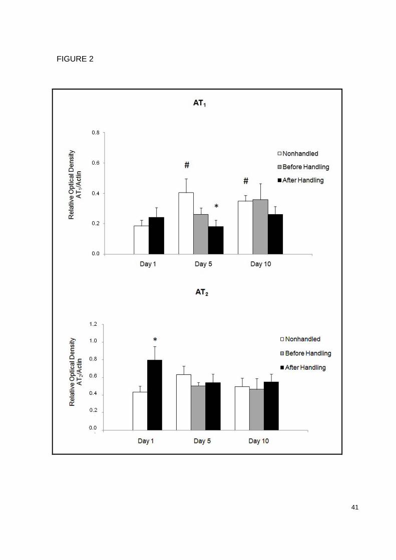
AT1 receptor expression
When the kidney AT1 receptor mRNA expression was analyzed, a decrease in
the AHd5 group was found when compared to the NHd5 group (p=0.05), indicating
that neonatal handling can also act modulating the receptor expression. We have
also found an increased expression in both NHd5 (p=0.04) and NHd10 (p=0.05)
groups when compared to the NHd1 group, demonstrating that mRNA AT1 receptor
expression increases during the neonatal period (Figure 2a).
AT2 receptor expression
The results show an increase in the kidney AT2 receptor mRNA expression in
the AHd1 group when compared to the NHd1 group (p=0.05). No other significant
differences were found. This also indicates that neonatal handling can act modulating
not only AT1 receptors but also AT2 receptor mRNA expression (Figure 2b).
Angiotensin II evaluation
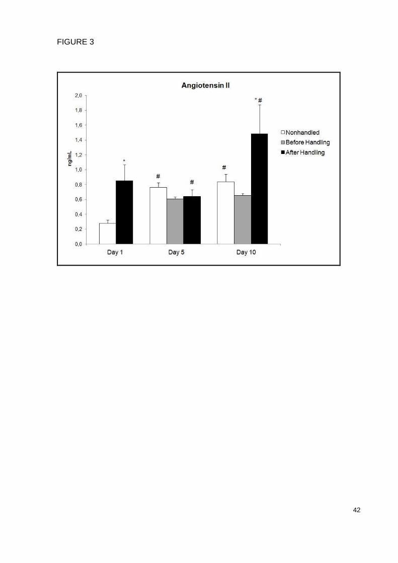
When ANG II plasma concentration was analyzed, a significant increase was
found after the handling procedure on day 1 (p=0.01) and day 10 (p=0.002),
demonstrating that the neonatal handling intervention can act inducing a RAS
activation. Moreover, we have also found an increased ANG II plasma concentration
in the NHd5 (p=0.05) and NHd10 (p=0.03) groups compared to the NHd1, showing
that ANG II levels also increase during the neonatal period (Figure 3).

30
Oxidative Stress Evaluation
Lipid peroxidation was accessed through the TBARS assay. Results show an
increase in the AHd10 group compared to both NHd10 (p=0.05) and BHd10 (p=0.01)
groups. However, no other significant differences were found when both CAT and
SOD antioxidant defense enzymes were analyzed, indicating that neonatal handling
induces very limited effects on the kidney oxidative stress balance (Figure 4).
31
Discussion
The present study shows that neonatal handling induces alterations in some of
the kidney RAS components mRNA expression, as well as increases ANG II plasma
concentration. On the other hand, the neonatal handling as an early life intervention
seems not to have major effects on the oxidative stress balance.
The period right after birth is called hyporesponsive stress period, which
comprehends approximately between the fourth and the 14th day after birth, and it is
characterized by very low glucocorticoid levels due to the inability of several stressors
in inducing an ACTH and corticosterone plasma increase (Levine, 2001; Schmidt et
al., 2003). In spite of the characteristic HPA axis hyporesponsivity of this period, our
results demonstrate that neonatal handling induced an increase in the kidney mRNA
renin expression and ANG II plasma concentration. Considering that the neonatal
period is crucial for the development of several systems (Lucion et al., 2003; Schmidt
et al., 2003) and the evidences that the RAS may act modulating these functions
through its’ receptors (Chen et al., 2004), the RAS activation in response to the
neonatal handling could be one of the mechanisms responsible for important
changes previously described in this experimental model. It is well known that stress
increases circulating ANG II production by increasing renin secretion through
sympathetic stimulation and beta-adrenergic receptor activation (Saavedra, et al.
2004; Yang et al., 1996), potencializing the HPA axis activity by acting over
hypothalamic, pituitary and adrenal components (Tsigos and Chrousos, 2002). Thus,
in spite of the neonatal handling model being considered a stress model or not, our
results demonstrate that this early life intervention was capable of inducing a RAS
activation at different levels.
Regardless being a stress hormone, ANG II plays a key hole in regulating the
kidney development, since it is the main RAS hormone. Indeed, all kidney RAS
components are highly expressed in the neonatal period (Puddu et al., 2009). ANG II
effects are mainly mediated by AT1 and AT2 receptors. AT2 receptors are highly
expressed in the fetal kidney and progressively decrease after birth, while AT1
receptors increase its expression. AT1 receptors seem to have functions opposite to
and perhaps balancing those of AT2 receptors, since AT1 receptors are involved in
the cellular proliferation, nefrovascular development and growth factors induction,

32
while AT2 receptors have apoptotic and growth inhibition functions (Lasaitiene et al.,
2006). A previous study has demonstrated that animals handled in the neonatal
period presented a decrease in the kidney function, evaluated through the glomerular
filtration rate, and an alteration in the hydroelectrolytic regulatory mechanisms, when
compared to the nonhandled animals (Donadio et al., 2009). Our results show an
increase in the kidney AT2 mRNA expression in the first day of handling, a decrease
in the kidney AT1 mRNA expression in the fifth day of handling and an increase in the
circulating levels of ANG II. Taken together, these results may help explaining the
previously described renal alterations induced by handling the animals in the
neonatal period, since a greater AT2 receptor expression, associated to an increase
in both renin expression and circulating ANG II levels, could be involved in inhibitory
mechanisms regulating kidney growth, considering the well described AT2 receptor
apoptotic effects. However, in spite of the fact that previous results have failed to
demonstrate a significant decrease in the number of nephrons, a kidney weight
decrease was shown (Donadio et al., 2009), indicating that growth inhibition
mechanisms are likely to be involved and that other morphological parameters
changes that were not addressed couldn’t be ruled out.
The production of circulating and local ANG II, as well as the expression of AT1
receptors, increases during stress situations (Saavedra and Benincky 2007). We
have demonstrated here that neonatal handling increased plasma ANG II and kidney
renin mRNA expression. Evidence shows that neonatal handling affects
sympathoadrenal activity (Young, 2000). On the other hand, in spite of several
physiological situations that influence mechanisms regulating the renin secretion, it is
known that stress also increases renin activity (Jindra and Kvetnansky 1982). Thus, it
is possible that an increased sympathetic activity could play a role in the increased
renin expression and plasma ANG II seen in our results. Enhanced AT1 receptor
activity, which is also influenced by stress, has been reported to modulate AT2
receptor expression. However, regulation of the AT2 receptor expression is still poorly
understood. The AT2 receptor is upregulated by sodium depletion, insulin and insulin-
like growth factor 1, and is downregulated by ANG II and growth factors such as
platelet-derived growth factor and epidermal growth factor, and in diabetes (Siragy,
2010). However, in spite of the possible origin of the RAS activation and mechanisms
underlying receptor alterations, to our knowledge, this is the first study showing that
33
the main components of the RAS, for instance plasma ANG II, renin and both
receptors (AT1 and AT2) mRNA expression are influenced by early environmental
changes such as neonatal handling in rats.
On the other hand, oxidative stress is defined as an imbalance between
reactive oxygen species (ROS) formation and antioxidative defense mechanisms
(Galle, 2001). This imbalance can cause injuries to DNA molecules, lipids,
carbohydrates and proteins, in addition to other cellular components (Lenarczyk et
al., 2009). Our results have showed an increase in the TBARS levels in the day 10,
after animals were handled, although no significant changes were demonstrated in
the antioxidant defense enzimes analyzed. This TBARS increase seen in the 10th
day, although isolated, could be contributing to increase renal tissue cellular injury.
Besides that, it is known that ANG II is also a pro-oxidative hormone through its
binding to AT1 receptors, which will activate NAD(P)H oxidase and then increase the
ROS (Lenarczyk et al., 2009; Serrano et al., 2009). Thus, it is possible that the
increased TBARS in our results could be a consequence of the RAS activation.
Nevertheless, the hypothesis that oxidative stress balance changes could influence
the kidney development and be involved in the long-lasting alterations previously
described seems very unlikely, considering present results that demonstrate no
reliable changes in the oxidative stress balance after neonatal handling in rats.
Conclusion
Taken together, the results indicate that handling in the neonatal period induces
the activation of the angiotensinergic system, as well as modulates its receptors,
indicating that it may play an important role in the development of some of the renal
alterations previously described. On the other hand, the mechanisms regulating the
oxidative stress balance system seems not to be involved. The precise mechanisms
by which neonatal handling induces long-lasting effects are still not completely
understood and futures studies addressing this issue should be conducted.
34
Acknowledgements
The authors an indebted to CNPq for financial support.

35
REFERENCES
Boveris, A., Fraga, C.G., Vassavsks, A.I., Koch, O.R, 1983. Archives of Biochemistry
and Biophysics. 534-541.
Carey, R.M., Siragy, H.M., 2003. Newly Recognized Components of the Renin-
Angiotensin System: Potential Roles in Cardiovascular and Renal Regulation.
Endocrine Reviews. 24, 261-271.
Chen, Y., Lasaitiene, D., Frieberg, P., 2004. The renin-angiotensin system in kidney
development. Acta Physiological Scandinavian. 181, 529-535.
Donadio, M.V., Jacobs, S., Corezola, K.L., Melo, D.M., Dias, H.M., Reichel, C.L.,
Franci, C.R., Jeckel-Neto, E.M., Lulhier, F., Oliveira, J.R., Sanvitto, G.L., 2009.
Neonatal Handling Reduces Renal Function in Adult Rats. Kidney & Blood Pressure
Research. 32, 286-292.
Draper, H.H., Squires, E.J., Mahmoodi, H., Agarwae, S., Hadley, M., 1993. A
comparative evaluation of thiobarbituric acid methods for the determination of
malondialdehyde in biological materials. Free Radical Biology and Medicine. 15, 353-
363.
Finkel, T., Holbrook, N., 2000. Oxidants, oxidative stress and the biology of ageing.
Nature. 408, 239-247.
Galle, J., 2001. Oxidative stress in chronic renal failure. Nephrology Dialysis
Transplantation 16, 2135-2137.
Gomes, C.M., Donadio, M.V., Franskoviaki, I., Anselmo-Franci, J.A., Franci, C.R.,
Lucion, A.L., Sanvitto, G.L., 2006. Neonatal handling reduces angiotensin II receptor
density in the medial preoptic area and paraventricular nucleus but not in arcuate
nucleus and locus coeruleus of female rats. Brain Research 1067, 177-180.
Jindra, A., Kvetnansky, R., 1982. Stress-induced activation of inactive renin. Journal
of Biological Chemistry 257, 5997– 5999.
Lasaitiene, D., Chen, Y., Adams, M.A., Frieberg, P., 2006. Further insights into role of
angiotensin II and kidney development. Clinical Physiology and Functional Imaging
26, 197-204.
Lenarczyk, M., Cohen, E. P., Fish, B. L., Irving, A. A., Sharma, M., Driscoll, C. D.,
Moulder, J. E., 2009. Chronic Oxidative Stress as a Mechanism for Radiation
Nephropaty. Radiation Research. 171, 164-172.
Levine, S., 2001. Primary social relationships influence the development of the
hypothalamic-pituitary-adrenal axis in the rat. Physiology & Behavior. 73, 255-260.
36
Levine, S., 1994. The ontogeny of the hypothalamic-pituitary-adrenal axis. The
influence os maternal factors. Annual New York Academy of Sciences. 746, 275-293.
Lucion, A. B., Pereira, F. M., Winkelman, E. C., Sanvitto, G. L., & Anselmo-Franci, J.
A., 2003. Neonatal handling reduces the number of cells in the locus coeruleus of
rats. Behavior Neuroscience. 117, 894-903.
Meerlo, P., Horvath, K.M., Nagy, G.M., Bohus, B., Koolhaas, J.M., 1999. The
Influence of Postnatal Handling on Adult Neuroendocrine and Behavioural Stress
Reactivity. Journal of Neuroendocrinology. 11, 925-933.
Mesquita, F.F., Gontijo, J.A.R, Boer, P.A., 2010. Expression od renin-angiotensin
system signalling compounds in maternal protein restricted rats: effect on renal
sodium excretion and blood pressure. Nephrology Dialysis Transplantation 25, 380-
388.
Padoin, M.J., Barros, H.M.T, Cadore, L.P., Gomes, C.M., Lucion, A.B., 2001. Long-
lasting effects of neonatal stimulation on the behavior of rats.” Behavioral
neuroscience. 115, 1332-1340.
Puddu, M., Fanos, V., Podda, F., Zaffanello. The kidney from Prenatal to Adult Life:
Perinatal Programming and Reduction of Number of Nephrons during Development.
American Journal of Nephrology. 30, 162-170.
Saavedra, J.M., Benicky, J., 2007. Brain and peripheral angiotensin II play a major
role in stress. Stress. 10, 185-189.
Saavedra, J.M., Ando, H., Armando, I., Baiardi, G., Bregonzio, C., Jezova, M., Zhou,
J., 2004. Brain Angiotensin II, an Important Stress Hormone: Regulatory Sites and
Therapeutic Opportunities. Annals of the New York Academy of Sciences. 1018, 76-
84.
Schmidt, M., Enthoven, L., Van der Mark, M., Levine, S., Kloet, E.R., Oitzl, M.S.,
2003. The postnatal development of the hypothalamic–pituitary–adrenal axis in the
mouse. International Journal of Developmental Neuroscience. 21, 125-132.
Serrano, G.L., Ritchie, B., Hoffman, D., Ferder, L., 2009. A new concept for an old
system: The anti-inflammatory paradigm of the renin-angiotensin system. Medical
Hypotheses 72, 584-588.
Siragy, H.M., 2010. The angiotensin II type 2 receptor and the kidney. Journal of the
Renin-Angiotensin-Aldosterone System 11, 33-36.
Tsigos, C., Chrousos, G.P., 2002. Hypothalamic-pituitary-adrenal axis,
neuroendocrine factors and stress. Journal of Psychosomatic Research. 53, 865-
871.

37
Yang, H., Wan, Y., & Zhu, Y., 1996. Angiotensin II: an important stress hormone.
Biology Signals. 5, 1-8.
Young, J.B., 2000. Effects of neonatal handling on sympathoadrenal activity and
body composition in adult male rats. American Journal of Physiology - Regulatory,
Integrative and Comparative Physiology. 279, R1745-1752.

38
Table 1 - Experimental design used in the study.
Group Day after birth Time of euthanasia
Nonhandled
Day 1 (NHd1) -
Day 5 (NHd5) -
Day 10 (NHd10) -
Handled
Day 1
Before Handling* (BHd1)
30 min After Handling (AHd1)
Day 5
Before Handling(BHd5)
30 min After Handling (AHd5)
Day 10
Before Handling (BHd10)
30 min After Handling (AHd10)
*Considering that in the first day the nonhandled group (NHd1) would be the same as the group
before handling (BHd1), since no intervention was performed so far, this group (BHd1) was not used
in the experiments. In all cases n = 6 per group.

39
FIGURE LEGENDS
FIGURE 1. Effects of neonatal handling on the kidney mRNA renin expression
evaluated through RT-PCR. Each bar represents the mean ± SEM (n=6 per group).
Data were analyzed by two way analysis of variance - ANOVA and Bonferroni post
hoc test was used. Significant differences accepted at p≤0.05. * Indicates a
significant difference between groups in the same day and # indicates a significant
difference between days in the same group.
FIGURE 2. Effects of neonatal handling on the kidney mRNA AT1 (a) and AT2
receptors (b) expression evaluated through RT-PCR. Each bar represents the mean
± SEM (n=6 per group). Data were analyzed by two way analysis of variance -
ANOVA and Bonferroni post hoc test was used. Significant differences accepted at
p≤0.05. * Indicates a significant difference between groups in the same day and #
indicates a significant difference between days in the same group.
FIGURE 3 – Effects of neonatal handling on the angiotensin II plasma concentrations
evaluated through an immunoassay (ELISA). Each bar represents the mean ± SEM
(n=6 per group). Data were analyzed by two way analysis of variance - ANOVA and
Bonferroni post hoc test was used. Significant differences accepted at p≤0.05. *
Indicates a significant difference between groups in the same day and # indicates a
significant difference between days in the same group.
FIGURE 4 – Effects of neonatal handling on the oxidative stress system: TBARS (a),
SOD (b) and CAT (c). Each bar represents the mean ± SEM (n=6 per group). Data
were analyzed by two way analysis of variance - ANOVA and Bonferroni post hoc
test was used. Significant differences accepted at p≤0.05. * Indicates a significant
difference between groups in the same day and # indicates a significant difference
between days in the same group.

40
FIGURE 1
41
FIGURE 2
42
FIGURE 3

43
FIGURE 4
44
CAPÍTULO 3
3.1 CONSIDERAÇÕES FINAIS
Este trabalho demonstrou que a manipulação neonatal afeta o sistema renina-
angiotensina (SRA). Estudos demonstram que o período que compreende do 4° ao
14° dia pós-natal é chamado de período hiporresponsivo ao estresse, por ser um
período com níveis basais muito baixos de corticosterona (Levine, 2001). Isso se
deve a uma dificuldade dos estressores induzirem um aumento de ACTH e
conseqüente liberação de corticosterona (Levine, 2001; Schmidt et al., 2003).
Apesar da hiporresponsividade do eixo HPA, os resultados obtidos neste trabalho
demonstraram que o SRA responde em situações de estresse nessa fase do
desenvolvimento.
O estresse causado pela manipulação nos animais, aumentou a expressão de
mRNA de renina no primeiro e no quinto dia de manipulação, mostrando que existe
um efeito estimulatório sobre o SRA. Este sistema tem papel importante na
nefrogênese e todos os seus componentes são altamente expressos durante o
desenvolvimento renal (Lasaitiene et al., 2006; Puddu et al., 2009). Alguns estudos
já demonstraram que a hiperativação tem um papel central na progressão da doença
renal (Lin et al., 2009). E desta forma os resultados demonstram um aumento da
atividade do SRA até o quinto dia da manipulação quando comparado ao primeiro
dia.
Os efeitos da angiotensina são mediados principalmente pelos receptores AT1
e AT2. O receptor AT1 parece ter uma função oposta ao receptor AT2 ou modulando
sua resposta, uma vez que os receptores AT1 são responsáveis por proliferação
celular, desenvolvimento nefrocelular e induzem fatores de crescimento e os
receptores AT2 tem funções apoptóticas e de inibição do crescimento (Lasaitiene et
al., 2006). Foi encontrado um aumento na expressão do receptor AT2 e uma
diminuição na expressão do receptor AT1 induzido pela manipulação. Foi encontrado
também um aumento na concentração plasmática de ANG II. Este aumento do
receptor de AT2, junto com o aumento de renina e ANG II pode indicar que a
ativação do SRA esteja induzindo efeitos de apoptose.
45
Estudos mostraram que a função renal está alterada em animais adultos que
foram manipulados no período neonatal, pois esses animais apresentaram uma
redução no peso total do rim, na ingestão hídrica, no volume urinário e no clearance
de creatinina (Donadio, et al. 2009). O SRA no período neonatal contribui de forma
importante para o desenvolvimento estrutural e funcional dos rins, o que foi
demonstrado através de estudos que utilizaram bloqueio farmacológico ou genético
dos seus componentes e foi encontrado diminuição (Lasaitiene, et al. 2006, Chen, et
al. 2004).
O estresse oxidativo (EO) é definido como um desequilíbrio entre a formação
de espécies reativas de oxigênio (ERO) e mecanismos de defesa anti-oxidante
(Galle 2001). Este desbalanço gera acúmulo de EROs que causa danos à estrutura
das biomoléculas de DNA, lipídios, carboidratos e proteínas, além de outros
componentes celulares (Lenarczyk, et al. 2009). Nossos resultados mostraram um
aumento de TBARS no décimo dia após a manipulação, porém não mostraram
nenhuma alteração das enzimas de defesa antioxidante. Este aumento de TBARS
isolado no décimo dia poderia estar contribuindo para um aumento do dano celular
no tecido renal. Além disso sabe-se que a ANG II é também um hormônio com
atividade pró-oxidante pela sua ligação ao receptor AT1 que leva a ativação de
NAD(P)H oxidase causando um aumento na produção de espécies reativas de
oxigênio (ERO) (Serrano, et al. 2009; Lenarczyk, et al. 2009). A hipótese de que
alterações no balanço oxidativo pudessem influenciar o desenvolvimento de
sistemas e ter envolvimento nas alterações estáveis já descritas parece não se
confirmar, considerando os resultados do presente estudo que demonstram que a
manipuação neonatal praticamente não altera o estresse oxidativo.
Em conjunto, os resultados indicam que a manipulação no período neonatal
provoca uma ativação do SRA, além da modulação de seus receptores, podendo ter
um papel importante no desenvolvimento das alterações renais já descritas. Por
outro lado, os mecanismos de regulação do sistema de balanço oxidativo parecem
não estar envolvidos. Os mecanismos precisos pelos quais essas alterações
ocorrem ainda não são claros e devem ser avaliados em estudos futuros.

46
REFERÊNCIAS
Caldji, C., Diorio, J., & Meaney, M. J. (2000). Variations in Maternal Care in Infancy
Regulate the Development of Stress Reactivity. Society of Biological Psychiatry , 48,
1164-1174.
Cannon, W. B. (1941). The body physiologic and the body politic. Science , 93.
Champagne, F. A., Francis, D. D., Mar, A., & Meaney, M. J. (2003). Variations in
maternal care in the rat as a mediating influence for the effects of environment on
development. Physiology & Behavior , 79, 359-371.
Chen, Y., Lasaitiene, D., & Frieberg, P. (2004). The renin-angiotensin system in
kidney development. Acta Physiological Scandinavian , 181, 529-535.
Donadio, M. V., Jacobs, S., Corezola, K. L., Melo, D. A., Dias, H. B., Reichel, C. L., et
al. (2009). Neonatal Handling Reduces Renal Function in Adult Rats. Kidney & Blood
Pressure Research , 32, 286-292.
Ferreira, A. L., & Matsubara, L. S. (1997). Radicais livres: conceitos, doenças
relacionadas, sistema de defesa e estresse oxidativo. Revista da Associação Médica
Brasileira , 43, 61-68.
Finkel, T., & Holbrook, N. (2000). Oxidants, oxidative stress and the biology of
ageing. Nature , 408, 239-247.
Francis, D. D., & Meaney, M. J. (1999). Maternal care and the development of stress
responses. Current Opinion in Neurobiology , 9, 128-134.
Galle, J. (2001). Oxidative stress in chronic renal failure. Nephrology Dialysis
Transplantation , 16, 2135-2137.
Gomes, C. M., Donadio, M. V., Franskoviaki, I., Anselmo-Franci, J. A., Franci, C. R.,
Lucion, A. B., et al. (2006). Neonatal handling reduces angiotensin II receptor density
in the medial preoptic area and paraventricular nucleus but not in arcuate nucleus
and locus coeruleus of female rats. Brain Research , 1067, 177-180.
Gomes, C. M., Frantz, P. J., Sanvitto, G. L., Anselmo-Franci, J. A., & Lucion, A. B.
(1999). Neonatal handling induces anovulatory estrous cycles in rats. Brazilian
Journal of Medical and Biological Research , 32, 1239-1242.
Gomes, C. M., Raineski, C., Paula, P. R., Severino, G. S., Helena, C. V., Anselmo-
Franci, J. A., et al. (2005). Neonatal handling and reproductive function in female
rats. Journal of Endocrinology , 184, 435-445.
Hsu, F.-C., Zhang, G.-j., Raol, Y. S., Valentino, R. J., & Coulter, D. A. (2003).
Repeated neonatal handling with maternal separation permanently alters

47
hippocampal GABAa receptors and behavioral stress responses. PNAS , 100 (21),
12213-12218.
Juruena, M. F., Cleare, A. J., & Pariante, C. M. (2004). Eixo Hipotálamo-pituitária-
adrenal, a função dos receptores de glicocorticóides e sua importância na
depressão. Revista Brasileira de Psiquiatria , 26, 189-201.
Lasaitiene, D., Chen, Y., Adams, M. A., & Frieberg, P. (2006). Further insights into
role of angiotensin II and kidney development. Clinical Physiology and Functional
Imaging , 26, 197-204.
Lenarczyk, M., Cohen, E. P., Fish, B. L., Irving, A. A., Sharma, M., Driscoll, C. D., et
al. (2009). Chronic Oxidative Stress as a Mechanism for Radiation Nephropaty.
Radiation Research , 171, 164-172.
Levine, S. (2001). Primary social relationships influence the development of the
hypothalamic-pituitary-adrenal axis in the rat. Physiology & Behavior , 73, 255-260.
Levine, S. (1994). The ontogeny of the hypothalamic-pituitary-adrenal axis. The
influence os maternal factors. Annual New York Academy of Sciences , 746, 275-
293.
Lin, J., Hu, F. B., Qi, L., & Curhan, G. C. (2009). Genetic polymorphisms of
angiotensin-2 type 1 receptor and angiotensinogen and risk of renal dysfunction and
coronary heart disease in type 2 diabetes mellitus. Biomed Central Nephrology , 10,
1-8.
Liu, D., Diorio, J., Tannenbaum, B., Caldj, C., Francis, D., Freedman, A., et al.
(1997). Maternal Care, Hippocampal Glucocorticoid Receptors, and Hypothalamic-
Pituitary-Adrenal Responses to Stress. Science , 277, 1659-1661.
Lucion, A. B., Pereira, F. M., Winkelman, E. C., Sanvitto, G. L., & Anselmo-Franci, J.
A. (2003). Neonatal handling reduces the number of cells in the locus coeruleus of
rats. Behavior Neuroscience , 117, 894-903.
Marchesi, C., Paradis, P., & Schiffrin, E. L. (2008). Role of the renin-angiotensin
system in vascular inflammation. Cell Press , 367-374.
Martin, C. A., Cake, M., & Holbrook, N. j. (1977). Relationship between foetal
corticosteroids, maternal progesterone and parturition in the rat. Acta Endocrinology ,
84, 167-176.
Meerlo, P., Horvath, K. M., Nagy, G. M., Bohus, B., & Koolhaas, J. M. (1999). The
Influence of Postnatal Handling on Adult Neuroendocrine and Behavioural Stress
Reactivity. Journal of Neuroendocrinology , 11, 925-933.
Moal, M. L. (2007). Historical approach and evolution of the stress concept: A
personal account. Psychoneuroendocrinology , 32, 53-59.

48
Nistala, R., Wei, Y., Sowers, J. R., & Whaley-Connel, A. (2009). Renin-angiotensin-
aldosterone system-mediated redox effects in chronic disease. Subspeciality in
translational medicine , 153, 102-113.
Núñez, J. F., Ferré, P., Escorihuela, R. M., Tobeña, A., & Fernández-Teruel, A.
(1996). Effects of Postnatal Handling of Rats on Emotional, HPA-Axis, and Prolactin
Reactivity to Novelty and Conflict. Physiology & Behavior , 60, 1355-1359.
Pacák, k., Palkovits, M. (2001). Stressor Specificity of Central Neuroendocrine
Responses: Implications for Stress-Related Disorders. Endocrine Reviews , 22, 502-
548.
Puddu, M., Fanos, V., Podda, F., & Zaffanello. (2009). The kidney from Prenatal to
Adult Life: Perinatal Programming and Reduction of Number of Nephrons during
Development. American Journal of Nephrology , 30, 162-170.
Schmidt, M., Enthoven, L., Van der Mark, M., Levine, S., Kloet, E. R., & Oitzl, M. S.
(2003). The postnatal development of the hypothalamic–pituitary–adrenal axis in the
mouse. International Journal of Developmental Neuroscience , 21, 125-132.
Schneider, C. D., & Oliveira, A. R. (2004). Radicais livres de oxigênio e exercício:
mecanismos de formação e adaptação ao treinamento físico. Revista Brasileira de
Medicina do Esporte , 10, 308-313.
Serrano, G. L., Ritchie, B., Hoffman, D., & Ferder, L. (2009). A new concept for an
old system: The anti-inflammatory paradigm of the renin-angiotensin system. Medical
Hypotheses , 72, 584-588.
Severino, G. S., Fossati, I. A., Padoin, M. J., Gomes, C. M., Trevizan, L., Sanvitto, G.
L., et al. (2004). Effects of neonatal on the behavior and prolactin stress response in
male and female rats at various ages and estrous cycle phases of females.
Physiology & Behavior , 81, 489-498.
Tsigos, C., & Chrousos, G. P. (2002). Hypothalamic-pituitary-adrenal axis,
neuroendocrine factors and stress. Journal of Psychosomatic Research , 53, 865-
871.
Vaziri, N. D., Bai, Y., Ni, Z., Quiroz, Y., Pandian, R., & Rodriguez-Iturbe, B. (2007).
Intra-renal angiotensin II/AT1 receptor, oxidative stress, inflammation, and
progressive injury in renal mass reduction. The Journal of Pharmacology and
Experimental Therapeutics , 323, 85-93.
Weaver, I. C., Meaney, M. J., & Szyf, M. (2006). Maternal care effects on the
hippocampal transcriptome and anxiety-mediated behaviors in the offspring that are
reversible in adulthood. PNAS , 103 (9), 3480-3485.
Wei, Y., Clark, S. E., Thyfault, J. P., Uptergrove, G. M., Whaley-Connell, A. T.,
Ferrario, C. M., et al. (2009). Oxidative Stress-Mediated Mitochondrial Dysfunction

49
Contributes to Angiotensin II-Induced Nonalcoholic Fatty Liver Disease in Transgenic
Ren2 Rats. The American Journal of Pathology , 174.
Winkelmann-Duarte, E. C., Todeschin, A. S., Fernandes, M. C., Bittencourt, L. C.,
Pereira, G. A., Samios, V. N., et al. (2007). Plastic changes induced by neonatal
handling in the hypothalamus of female rats. Brain Research , 1170, 20-30.
50
ANEXO A
DOCUMENTO DE CONFIRMAÇÃO DE SUBMISSÃO
From: [email protected] on behalf
of Developmental Biology
Subject: Submission Received
Title: Evaluation of the kidney renin-angiotensin system and oxidative stress in
neonatal handled rats
Corresponding Author: Dr Marcio V Donadio
Authors: Daniela L Rodriguez; Fernanda C Mesquita; Débora Attolini; Bruna S
Borba; Patrícia S Scherer; Priscilla H Almeida; Vinícius L Costa; Bárbara S
Scherer; Virgínia M Schmitt; Jarbas R de Oliveira
Dear Dr Donadio,
This is to confirm that the above-mentioned manuscript has been received for
consideration in Developmental Biology.
You will be able to check on the progress of your manuscript by logging on to the
Elsevier Editorial System for Developmental Biology as an author:
http://ees.elsevier.com/developmentalbiology/
Your paper will be given a manuscript number shortly and you will receive an e-mail
with this number for your reference.
Thank you for submitting your manuscript to Developmental Biology. Should you
have any questions, please feel free to contact our office.
Kind regards,
Developmental Biology
Elsevier Science
525 B St., Ste. 1900
San Diego, CA 92101-4495 USA
tel: 619-699-6351
fax: 619-699-6211