porphyrin metabolism & porphyrias hmim224. objectives understand the structure of...
TRANSCRIPT

Porphyrin metabolism & Porphyrin metabolism & porphyriasporphyrias
HMIM224HMIM224

ObjectivesObjectives
• Understand the structure of metalloprphyrins Understand the structure of metalloprphyrins • Apply that the role of heme is dictated by the environment created Apply that the role of heme is dictated by the environment created
by the three dimensional structure relation to proteinby the three dimensional structure relation to protein• Identify the rate limiting step and the iso-enzymes sites of Identify the rate limiting step and the iso-enzymes sites of
productionproduction• Describe the site of effect of certain drugs on heme biosynthesis Describe the site of effect of certain drugs on heme biosynthesis
and its clinical importanceand its clinical importance• Identify how blocking in one of the enzyme involved in heme Identify how blocking in one of the enzyme involved in heme
biosynthesis will affect the mode of presentation of the diseasebiosynthesis will affect the mode of presentation of the disease• Identify the most common type of porphyrias & its cause Identify the most common type of porphyrias & its cause • Identify the heme degradation productIdentify the heme degradation product

What are porphyrinsWhat are porphyrins? ?
• PorphyrinsPorphyrins are cyclic compounds that bind metal ions (usually Fe2+ or Fe3+)
• Porphyrin + Metal = MetalloporphyrinMetalloporphyrin• Most prevalent metalloporphyrin in humans is hemeheme (metal here is iron
ion)
Heme consists ofHeme consists of:• One ferrous ion One ferrous ion (Fe2+) in the centre• Protoporphyrin IX Protoporphyrin IX (a tetrapyrrole ring)• The role of heme is dictated by the enviroment created by the three The role of heme is dictated by the enviroment created by the three
dimensional structure of proteindimensional structure of protein Heme is the prosthetic group Heme is the prosthetic group of hemoglobin, myoglobin, cytochromes, catalase, tryptophan pyrrolase So, heme + globin protein = hemoglobin

Structure of porphyrinsStructure of porphyrins
• PorphyrinsPorphyrins are cyclic molecules formed by 4 pyrrole (tetrapyrrole) rings linked by methenyl bridges.
• Different porphyrins vary in the nature of side chains that are attached to each of the 4 pyrrole rings Protoporphyrin IX contains vinyl, methyl & propionate
Distribution of side chains - Side chains can be ordered around tetrapyrrole nucleus in 4 different ways designated I, II, III & IV series. Only type III porphyrinstype III porphyrins are physiologically important in humans - Protoporphyrin IX Protoporphyrin IX is a member of type III series
• Porphyrinogens are porphyrin precursors intermediate between porphobilinogen & protoporphyrin Porphobilinogen Porphyrinogens Protoporphyrins

Methenyl bridge
Pyrrole ringSide
Chains
Structure of PorphyrinStructure of Porphyrin
Determine the type of porphyrinDetermine the type of porphyrin

Derived from protoporphyrin IX
Binds metals: Heme- Fe2+ (ferrous)Hemin- Fe3+ (ferric)
Extended conjugation across ring systemZinc protoporphyrin (ZnPP)- Zn2+
Pattern of side chains defines isomer
Structure and Properties of Iron Protoporphyrin IX
propionate
methyl
vinyl
pyrrole ring

Uroporphyrinogen I Coproporphyrinogen I
Overview of Heme SynthesisOverview of Heme Synthesis
Heme synthesis occurs in all cells due to the requirement for heme as a prosthetic group on enzymes and electron transport chain. By weight, the major locations of heme synthesis are
the liver and the erythroid progenitor cells of the bone marrow.
Succinyl CoA + Glycine
-aminolevulinic acid
-aminolevulinic acid
Porphobilinogen Uroporphyrinogen III Coproporphyrinogen III
Coproporphyrinogen III
Protoporphyrinogen IX
Protoporphyrin IX
Heme
ALA synthase
cytoplasm
mitochondrial matrix

First & First & second steps second steps
of heme of heme synthesissynthesis
Biosynthesis of heme Biosynthesis of heme (cont.)

Biosynthesis of hemeBiosynthesis of heme
Site of biosynthesisSite of biosynthesis: Liver & Bone Marrow (erythryoid producing cells)
• There are 2 isozymes ALA S1 (liver) & ALAS2 (erythroid)
StepsSteps:
Step 1Step 1: : Formation of Formation of -amino levulinic acid (ALA)-amino levulinic acid (ALA): in mitochondria (Rate Controlling Step)
GlycineGlycine + Succinyl CoASuccinyl CoA (nonessential amino acid) (intermediate of citric acid cycle)
Enzyme: ALA Synthase Coenzyme: Pyridoxal Phosphate (PLP)
-Amino levulinic acid -Amino levulinic acid (ALA)(ALA)

• Cytochrome P450s (CYPs) are actually a superfamily of related, heme-containing monooxygenase enzymes that participate in abroad variety of reactions.This system performs different functions in two separate locations in cells.
• The over-all reaction catalyzed by a cytochrome P450 enzyme is:
• R-H + O2+ NADPH + H+→R-OH + H2O NADP+ • where R may be a steroid, drug, or other chemical.
The name P450 reflects the absorbance at 450 nm by the protein.
These modifications is two-fold:These modifications is two-fold:• First, it may itself activate or inactivate a drug or
• Second, make a toxic compound more soluble, thus facilitating its excretion in the urine or feces.
Cytochrome P450 Monooxygenase Cytochrome P450 Monooxygenase SystemSystem

Clinical importance of first stepClinical importance of first step::
When heme (end product) is produced in excessive amounts, heme is converted to heminhemin. Hemin decreases action of ALA synthase in liver. (end product inhibition).The reverse occurs when heme biosynthesis is reduced.
DrugsDrugs as grisofulvin (antifungal), hydantoin & phenobarbital (anticonvulsant) increase ALA synthase activity:as these drugs are metabolized by cytochrome p450 in liver resulting in more consumption of heme (component of cytochrome). Accordingly, heme concentration is reduced resulting in stimulation of action of ALA synthase.
Biosynthesis of heme Biosynthesis of heme (cont.)

First & First & second steps second steps
of heme of heme synthesissynthesis
Biosynthesis of heme Biosynthesis of heme (cont.)

Step 2Step 2: : Formation of porphobilinogenFormation of porphobilinogen:
2 molecules of -Amino levulinic acid (ALA) condense to formporphobilinogenporphobilinogen by the enzyme ALA dehydrataseALA dehydratase.
Clinical importanceClinical importance:
ALA dehydratase enzyme is inhibited by heavy metals as leadlead that results in anemia. (lead poisoning). In this case: ALA in blood is elevated (lab investigation)
Biosynthesis of heme Biosynthesis of heme (cont.)
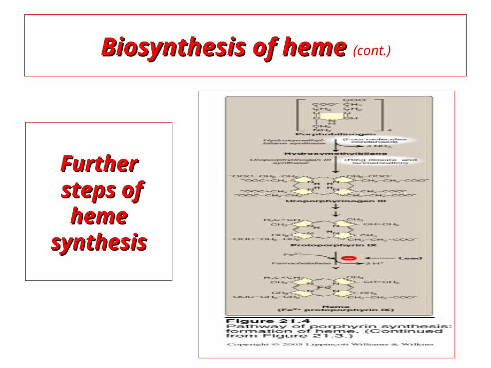
Biosynthesis of heme Biosynthesis of heme (cont.)
FurtherFurthersteps ofsteps of heme heme
synthesissynthesis

Further steps: Further steps: (in mitochondria) (in mitochondria)
Formation of protoporphyrin IXprotoporphyrin IX
Then, ferrous ions (Fe2+) are introduced into protoporphyrin IXferrous ions (Fe2+) are introduced into protoporphyrin IX, either:
simultaneously or: enhanced by ferrochelatase ferrochelatase
Clinical importanceClinical importance: Ferrochelatase enzyme is inhibited by lead
Biosynthesis of heme Biosynthesis of heme (cont.)

GlycineGlycine + Succinyl CoASuccinyl CoA
Enzyme: ALA Synthase Enzyme: ALA Synthase STEP 1STEP 1 PLP PLP
-Amino levulinic acid (ALA)-Amino levulinic acid (ALA) Enzyme: ALA dehydrataseEnzyme: ALA dehydratase. STEP 2STEP 2 porphobilinogenporphobilinogen
FURTHER STEPSFURTHER STEPS
Protoporphyrin IXProtoporphyrin IX
Ferrous ion (Fe2+ ) Ferrous ion (Fe2+ ) introduction of ironintroduction of iron Enzyme: ferrochelataseEnzyme: ferrochelatase
hemeheme
Summary of biosynthesis of hemeSummary of biosynthesis of heme

PorphyriasPorphyrias
PorphyriaPorphyria are rare inherited defects in heme synthesis.
An inherited defect in an enzyme of heme synthesis results in accumulation of one or more of porphyrin precursors depending on location of block of the heme synthesis pathway.
These precursors increase in blood & appear in urine of patients.
PorphyriaPorphyria means purple colour caused by pigment-like porphyrins in urine of patients. (Diagnosed by lab investigation) Most porphyrias show a prevalent autosomal dominant patternautosomal dominant pattern, except congenital eythropoietic porphyria, which is recessive

Clinical manifestations of porphyriasClinical manifestations of porphyrias: Two types of porphyriasTwo types of porphyrias: erythropoietic (bone marrow) & hepatic Hepatic porphyrias areHepatic porphyrias are: acute & chronic porrphyrias
Generally, individuals with an enzyme defect priorprior to the synthesis of the tetrapyrrolestetrapyrroles manifest abdominal and neuropsychiatric signs
Those with with tetrapyrrole intermediatestetrapyrrole intermediates show photosensitivity with formation of reactive oxygen species (ROS) that damage membranes by oxidation
resulting in the following effects :
- Skin blisters, itches (pruritis) - Skin may darken, grow hair (hypertrichosis)
PorphyriasPorphyrias (cont.)(cont.)

Porphyria Cutanea TardaPorphyria Cutanea Tarda
• Chronic hepatic porphyria• The most common type of porphyria
• a deficiency in uroporphyrinogen decarboxylaseuroporphyrinogen decarboxylase
• Clinical expression of the enzyme deficiency is influenced by various factors, such as exposure to sunlight, the presence of hepatitis B or C
• Clinical onset is during the fourth or fifth decade of life.
• Porphyrin accumulation leads to cutaneous symptomscutaneous symptoms and urineurine that is red to brown in natural light and pink to red in fluorescent light

PorphyriasPorphyrias (cont.)(cont.)

Acute Hepatic PorphyriasAcute Hepatic Porphyrias
e.g. Acute Intermittent Porphyria • Porphyrias leading to accumulation of ALA and porphobilinogen cause
abdominal pain and neuropsychiatric disturbances, ranging from anxiety to delirium.
• Symptoms of the acute hepatic porphyrias are often precipitated by administration of drugs such as barbiturates and ethanol.

Types of PorphyriasTypes of Porphyrias

Degradation of HemeDegradation of Heme


Most heme from RBCs (85%) - rest from turnover of cytochromes, p450s, immature erythrocytes.
RBCs last 120 days, degraded by reticuloendothelial (RE) system [liver and spleen].
Microsomal heme oxygenase hydroxylates methenyl bridge carbon and oxidizes Fe2+ to Fe3+. Second reaction open ring and release methenyl carbon as CO.
CO has a vasodilator effect while bilirubin has an antioxidant effect
Serum albumin carries bilirubin in circulation, ligandin in hepatocytes.
Bilirubin & its derivative is collectively known as bile pigments


ALAALA
Synthesis of Synthesis of
ALASALAS 11
Decreased hemeDecreased heme
Synthesis of CYP 450 is increasedSynthesis of CYP 450 is increased
Large Amount of Drug intakeLarge Amount of Drug intake
Metabolised by CYP 450Metabolised by CYP 450