portulacastrum l. extracts mediate the radioprotective
TRANSCRIPT
Page 1/21
Antioxidant and Anti-in�ammatory ActivitiesMediate the Radioprotective Effect of TrianthemaPortulacastrum L. ExtractsUttam Das
West Bengal University of Health Sciences College of Medicine and JNM HospitalTanmay Saha
West Bengal University of Health Sciences College of Medicine and JNM HospitalReshma Kumari Sharma
IISER-K: Indian Institute of Science Education and Research KolkataDharmendra Kumar Maurya
Bhabha Atomic Research CentrePartho Sarothi Ray
IISER-K: Indian Institute of Science Education and Research KolkataSubir Kumar Das ( [email protected] )
West Bengal University of Health Sciences College of Medicine and JNM Hospitalhttps://orcid.org/0000-0003-0908-5437
Research Article
Keywords: Trianthema portulacastrum, Scavenging properties, Ionizing radiation, Anti-in�ammatoryactivity, RAW 264.7 cells, WRL 68cells.
Posted Date: August 20th, 2021
DOI: https://doi.org/10.21203/rs.3.rs-804074/v1
License: This work is licensed under a Creative Commons Attribution 4.0 International License. Read Full License
Page 2/21
AbstractIonizing radiation (IR) generates reactive oxygen species (ROS) which leads to oxidative stress and oftenleads to in�ammatory responses in organisms. Trianthema portulacastrum L., a plant commonly growingin India, is rich in antioxidant phytochemicals which is responsible for scavenging free radicals, and mayprovide radio-protective and anti-in�ammatory effects in response to ionizing radiation. The effect of T.portulacastrum extracts was studied in hepatic cells, which are susceptible to radiation-induced damage,and in macrophages which are the primary in�ammatory cells of the body. T. portulacastrum stemextracts showed e�cient free radical scavenging activity in hepatocytes and reduction of radiation-induced lipid peroxidation in cell and mitochondrial membranes. Treatment of irradiated cells with T.portulacastrum stem extracts enhanced cell viability, although at higher concentrations there wasreduction in cell viability. Treatment with low concentration of T. portulacastrum stem extract alsoreduced cellular ROS generation and increased cellular concentration of the anti-oxidant glutathione. T.portulacastrum extracts also showed a marked anti-in�ammatory effect in macrophages activated by thein�ammatory agonist bacterial lipopolysaccharide (LPS) by reducing in�ammatory gene expression andnitric oxide (NO) production, and increasing glutathione content. LPS treatment lowered expression ofNrf2, a transcription factor involved in regulation of multiple anti-oxidant genes, while treatment with lowconcentration of T. portulacastrum stem extract signi�cantly restored it. Together, these observationsdemonstrated a potential radioprotective role of T. portulacastrum extract mediated by both itsantioxidant activity on hepatic epithelial cells and its anti-in�ammatory activity on immune cells
IntroductionExposure to ionizing radiation (IR) causes cellular damage either by direct impairment of biomolecules orindirectly by generation of free radicals. Water radiolysis leads to generation of reactive oxygen species(ROS) which causes lipid peroxidation in membrane, DNA strand breaks and oxidation of cellular protein(Wang et al. 2018). Radiation not only affects the irradiated cells but also non-irradiated cells and tissuesundesirably due to bystander effect through the activation of in�ammatory responses (Shemetun andPilins’ka 2007). IR sensitizes lymphocytes, macrophages, monocytes and other immune cell (Carvalhoand Villar 2018). Radiation exposure shows immune-modulatory properties through the production ofreactive oxygen and nitrogen species (RONS), and release of in�ammatory cytokines such as tumornecrosis factor-alpha (TNF-α), tumor growth factor-beta (TGF-β), interleukins, nucleoside, high mobilitygroup box-1 molecule (HMGB1) and heat shock proteins (HSPs) (Carvalho and Villar 2018). As ionizingradiation is a mainstay of therapy in many cancers, methods of protection against radiation-induceddamage are an important requisite for cancer radiotherapy.
Radioprotectors may be naturally occurring antioxidants that can protect normal cells and tissues fromradiation-induced damage. Sulfhydryl agents such as glutathione, cysteine, cystamine and otherantioxidants have shown protective activity against the lethal effect of radiation and also increased thesurvival rate of irradiated mice (Obrador et al. 2020). Sulfhydryl group containing erdosteine protect ratsagainst gamma radiation through antioxidants and anti-in�ammatory properties (Elkady and Ibrahim
Page 3/21
2016). Sulfhydryl compounds protect cellular DNA through a combination of free radical scavenging,modulation of repair process, and hydrogen donation ability (Kumar et al. 2002).
Natural products are rich in antioxidants and are possible sources of various dietary supplements withtherapeutic importance. Phenolic compounds present in plant-based natural products have signi�cantpharmacological properties. They are reported as antioxidants observed by metal chelation and freeradicals scavenging activities (Godlewska-Żyłkiewicz et al. 2020) and have signi�cant anti-in�ammatoryproperties (Farhood et al. 2019). Curcumin and epigallocatechin-3-gallate, are well-known naturalcompounds having both radioprotective as well as anti-in�ammatory properties (Azab et al. 2016;Farhood et al. 2019). However, depending on several factors, an antioxidant may act as prooxidants in aconcentration-dependent manner. This possibly involves the reduction of transition metal ions from Fe3+
and Cu2+ to Fe2+ and Cu+ respectively and inducing Fenton reaction (Maurya and Devasagayam 2010).Therefore, further exploration of naturally occurring compounds in plants with antioxidant properties iswarranted.
Trianthema porulacastrum L., a well-known medicinal plant from the family of Aizoaceae, is a naturalsource of antioxidant and phytochemicals and has been used for treatment of numerous diseaseconditions in Indian and African traditional medicine (Shivhare et al. 2012; Das et al. 2020). T.portulacastrum is also well-known for its hepatoprotective activity against chemical-induced toxicity suchas carbon tetrachloride (CCl4) (Sarkar et al. 1999), paracetamol and thioacetamide (Kumar et al. 2004).The hepatoprotective activity of T. portulacastrum was marked by enhancement of antioxidant enzymes,suggesting that the protection of liver cells from oxidative damage may be a mode of hepatoprotectionby T. portulacastrum extract. However, no studies have been performed on the anti-in�ammatory role of T.portulacastrum, although in�ammatory responses are known to be a major contributor to hepaticdamage. Therefore, the objective of this study was to evaluate the radioprotective activity of T.portulacastrum in hepatocytes and its anti-in�ammatory effects using murine macrophages.
Materials And MethodsChemicals
2,2-diphenyl-1-picrylhydrazyl (DPPH), 2’,7’-dichloro�uorescin diacetate (H2DCFDA), 2,2'-azino-bis(3-ethylbenzothiazoline-6-sulfonicacid) (ABTS), monochlorobimane (MCB), Lipopolysaccharides (LPS) andprimers from Sigma; DMEM and Fetal bovine serum (FBS) from HiMedia; FBS from Invitrogen; RPMI-1640, L-Glutamine, oligo-(dT) primer, M-MLV reverse transcriptase from Thermo Fisher Scienti�c and allother chemicals of AR grade were procured from SRL India Ltd and Merck India LTD.
Plant materials
T. portulacastrum L. plants were collected from �elds in Kalyani, Dist. Nadia, West Bengal, India and wereauthenticated from the Department of Botany, University of Kalyani, Kalyani, Nadia (Voucher No. UD-101).
Page 4/21
Preparation of TP extracts: Dried powder of different parts of T. portulacastrum such as leaves, stem andwhole plant (100 g) was extracted with 500 ml petroleum ether for 24 h with constant shaking and�ltered. This process was repeated twice. Ethyl acetate, acetone and ethanol solvent were used twicesequentially followed by petroleum ether. All the solvents were evaporated and dried. Further studies werecarried out with ethanolic fractions.
Antioxidant capacity study
Antioxidant capacity of the different extracts of the TP was measured using 2,2'-azino-bis(3-ethylbenzothiazoline-6-sulfonic acid) (ABTS•+) (Maurya and Devasagayam 2010) and 2,2-diphenyl-1-picrylhydrazyl (DPPH) (Maurya and Devasagayam 2010) radical scavenging assays, whereas metalreducing power was evaluated using ferric reducing power assay (FRPA) (Maurya and Devasagayam2010) and molybdenum reduction assay (MRA) (Saxena et al. 2016).
Cell Lines:Human hepatic cells (WRL 68) were cultured in Dulbecco’s modi�ed Eagle’s Medium (DMEM) and RAW264.7 cells in RPMI-1640 medium with L-Glutamine. For culturing both the cell line media weresupplemented with 10% FBS (Thermo Fisher Scienti�c, 10500064) and 1% Pen-Strep (Thermo FisherScienti�c, 15140-122).
Evaluation of radioprotective property of T. portulacastrum extract
Radioprotective property of T. portulacastrum extract was evaluated using sub-cellular and cellular modelsystems. For sub-cellular assay, we have used murine mitochondrial membrane whereas for cellularassay we have employed human hepatic cells (WRL 68) as a model system.
Evaluation IR-induced lipid peroxidation
For evaluation of lipid peroxidation mouse mitochondrial membrane and human hepatic cells were used.Mitochondrial membrane fractions were isolated from the liver of male Swiss mice as described (Checkeret al. 2010). Damage to the mitochondrial membrane fraction after radiation exposure was assessed interms of lipid peroxidation (Maurya and Devasagayam 2010). Mitochondrial membrane fraction (aprotein equivalent of 300 µg) was suspended in 300 µl of 10 mM potassium phosphate buffer, pH 7.4,and exposed to 50 Gy radiations in the absence and presence of different concentrations of TP extracts(pre-treated for 30 min at 37°C). After treatment, 900 µl TBA reagent (0.375% TBA, 0.25 M HCl, 15%trichloroacetic acid (TCA) and 6 mM Na2-EDTA) was added. The reaction mixture was incubated at 950C
for 20 min, cooled to ambient temperature and centrifuged at 12,000 g for 5 min at 250C.Malondialdehyde (MDA) equivalents in the supernatant were estimated by measuring the �uorescence(as �uorescence provide more sensitivity) with excitation at 530 nm and emission at 590 nm using amicroplate reader.
Page 5/21
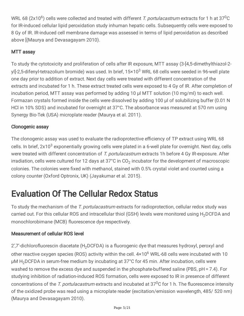
WRL 68 (2x10⁶) cells were collected and treated with different T. portulacastrum extracts for 1 h at 370Cfor IR-induced cellular lipid peroxidation study inhuman hepatic cells. Subsequently cells were exposed to8 Gy of IR. IR-induced cell membrane damage was assessed in terms of lipid peroxidation as describedabove [(Maurya and Devasagayam 2010).
MTT assay
To study the cytotoxicity and proliferation of cells after IR exposure, MTT assay (3-[4,5-dimethylthiazol-2-yl]-2,5-difenyl-tetrazolium bromide) was used. In brief, 15×103 WRL 68 cells were seeded in 96-well plateone day prior to addition of extract. Next day cells were treated with different concentration of theextracts and incubated for 1 h. These extract treated cells were exposed to 4 Gy of IR. After completion ofincubation period, MTT assay was performed by adding 10 µl MTT solution (10 mg/ml) to each well.Formazan crystals formed inside the cells were dissolved by adding 100 µl of solubilizing buffer (0.01 NHCl in 10% SDS) and incubated for overnight at 37°C. The absorbance was measured at 570 nm usingSynergy Bio-Tek (USA) microplate reader (Maurya et al. 2011).
Clonogenic assay
The clonogenic assay was used to evaluate the radioprotective e�ciency of TP extract using WRL 68cells. In brief, 2x10³ exponentially growing cells were plated in a 6-well plate for overnight. Next day, cellswere treated with different concentration of T. portulacastrum extracts 1h before 4 Gy IR-exposure. Afterirradiation, cells were cultured for 12 days at 37°C in CO2 incubator for the development of macroscopiccolonies. The colonies were �xed with methanol, stained with 0.5% crystal violet and counted using acolony counter (Oxford Optronix, UK) (Jayakumar et al. 2015).
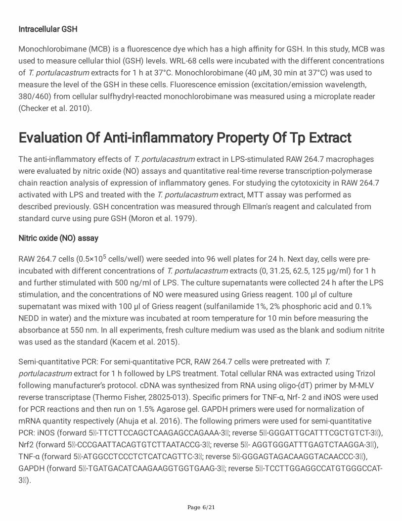
Evaluation Of The Cellular Redox StatusTo study the mechanism of the T. portulacastrum extracts for radioprotection, cellular redox study wascarried out. For this cellular ROS and intracellular thiol (GSH) levels were monitored using H2DCFDA andmonochlorobimane (MCB) �uorescence dye respectively.
Measurement of cellular ROS level
2’,7’-dichloro�uorescin diacetate (H2DCFDA) is a �uorogenic dye that measures hydroxyl, peroxyl and
other reactive oxygen species (ROS) activity within the cell. 4×10⁶ WRL-68 cells were incubated with 10µM H2DCFDA in serum-free medium by incubating at 37°C for 45 min. After incubation, cells werewashed to remove the excess dye and suspended in the phosphate-buffered saline (PBS, pH = 7.4). Forstudying inhibition of radiation-induced ROS formation, cells were exposed to IR in presence of differentconcentrations of the T. portulacastrum extracts and incubated at 370C for 1 h. The �uorescence intensityof the oxidized probe was read using a microplate reader (excitation/emission wavelength, 485/ 520 nm)(Maurya and Devasagayam 2010).
Page 6/21
Intracellular GSH
Monochlorobimane (MCB) is a �uorescence dye which has a high a�nity for GSH. In this study, MCB wasused to measure cellular thiol (GSH) levels. WRL-68 cells were incubated with the different concentrationsof T. portulacastrum extracts for 1 h at 37°C. Monochlorobimane (40 µM, 30 min at 37°C) was used tomeasure the level of the GSH in these cells. Fluorescence emission (excitation/emission wavelength,380/460) from cellular sulfhydryl-reacted monochlorobimane was measured using a microplate reader(Checker et al. 2010).
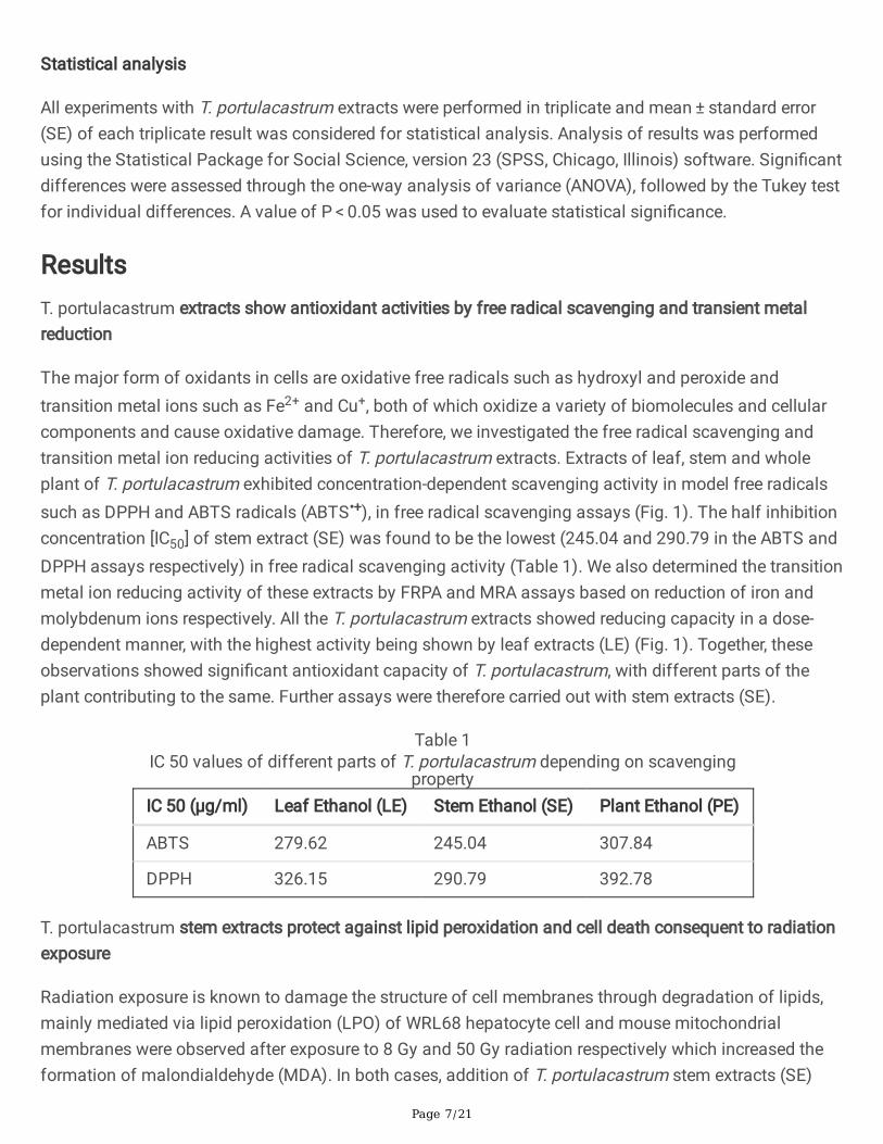
Evaluation Of Anti-in�ammatory Property Of Tp ExtractThe anti-in�ammatory effects of T. portulacastrum extract in LPS-stimulated RAW 264.7 macrophageswere evaluated by nitric oxide (NO) assays and quantitative real-time reverse transcription-polymerasechain reaction analysis of expression of in�ammatory genes. For studying the cytotoxicity in RAW 264.7activated with LPS and treated with the T. portulacastrum extract, MTT assay was performed asdescribed previously. GSH concentration was measured through Ellman's reagent and calculated fromstandard curve using pure GSH (Moron et al. 1979).
Nitric oxide (NO) assay
RAW 264.7 cells (0.5×10⁵ cells/well) were seeded into 96 well plates for 24 h. Next day, cells were pre-incubated with different concentrations of T. portulacastrum extracts (0, 31.25, 62.5, 125 µg/ml) for 1 hand further stimulated with 500 ng/ml of LPS. The culture supernatants were collected 24 h after the LPSstimulation, and the concentrations of NO were measured using Griess reagent. 100 µl of culturesupernatant was mixed with 100 µl of Griess reagent (sulfanilamide 1%, 2% phosphoric acid and 0.1%NEDD in water) and the mixture was incubated at room temperature for 10 min before measuring theabsorbance at 550 nm. In all experiments, fresh culture medium was used as the blank and sodium nitritewas used as the standard (Kacem et al. 2015).
Semi-quantitative PCR: For semi-quantitative PCR, RAW 264.7 cells were pretreated with T.portulacastrum extract for 1 h followed by LPS treatment. Total cellular RNA was extracted using Trizolfollowing manufacturer’s protocol. cDNA was synthesized from RNA using oligo-(dT) primer by M-MLVreverse transcriptase (Thermo Fisher, 28025-013). Speci�c primers for TNF-α, Nrf- 2 and iNOS were usedfor PCR reactions and then run on 1.5% Agarose gel. GAPDH primers were used for normalization ofmRNA quantity respectively (Ahuja et al. 2016). The following primers were used for semi-quantitativePCR: iNOS (forward 5 -TTCTTCCAGCTCAAGAGCCAGAAA-3 ; reverse 5 -GGGATTGCATTTCGCTGTCT-3 ),Nrf2 (forward 5 -CCCGAATTACAGTGTCTTAATACCG-3 ; reverse 5 - AGGTGGGATTTGAGTCTAAGGA-3 ),TNF-α (forward 5 -ATGGCCTCCCTCTCATCAGTTC-3 ; reverse 5 -GGGAGTAGACAAGGTACAACCC-3 ),GAPDH (forward 5 -TGATGACATCAAGAAGGTGGTGAAG-3 ; reverse 5 -TCCTTGGAGGCCATGTGGGCCAT-3 ).
Page 7/21
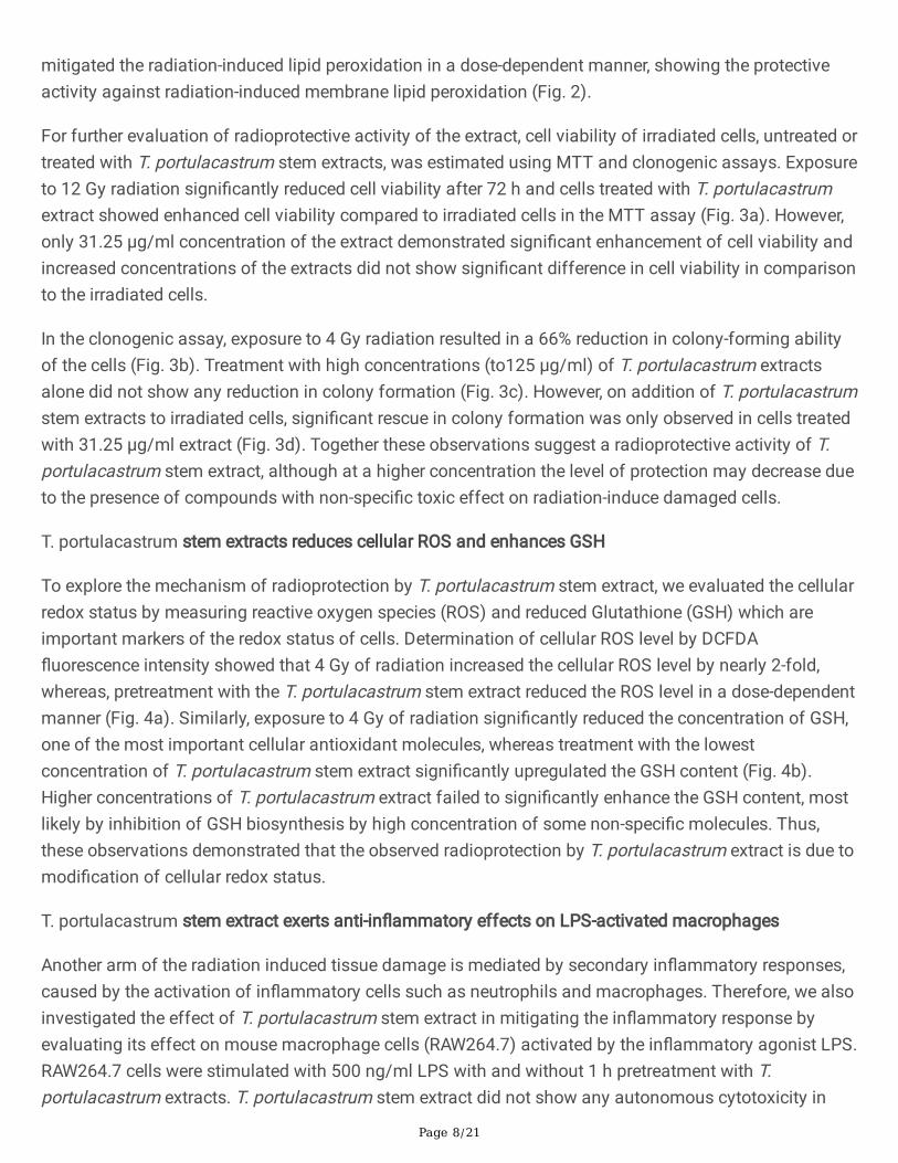
Statistical analysis
All experiments with T. portulacastrum extracts were performed in triplicate and mean ± standard error(SE) of each triplicate result was considered for statistical analysis. Analysis of results was performedusing the Statistical Package for Social Science, version 23 (SPSS, Chicago, Illinois) software. Signi�cantdifferences were assessed through the one-way analysis of variance (ANOVA), followed by the Tukey testfor individual differences. A value of P < 0.05 was used to evaluate statistical signi�cance.
ResultsT. portulacastrum extracts show antioxidant activities by free radical scavenging and transient metalreduction
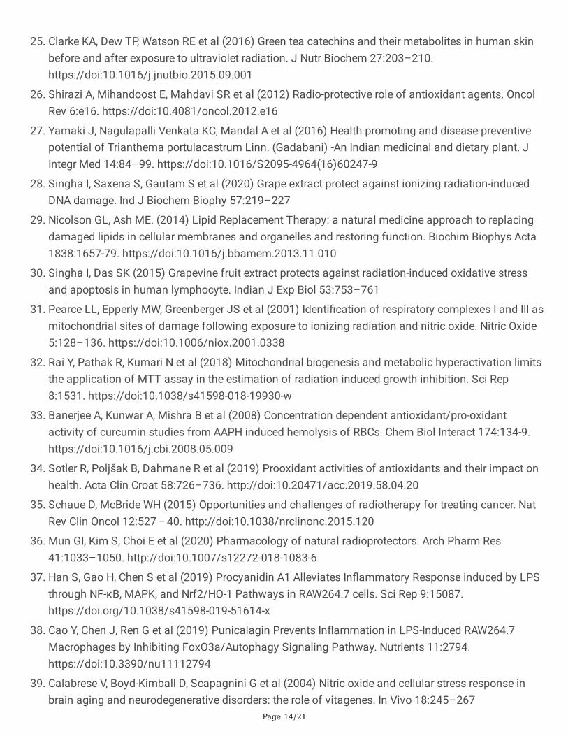
The major form of oxidants in cells are oxidative free radicals such as hydroxyl and peroxide andtransition metal ions such as Fe2+ and Cu+, both of which oxidize a variety of biomolecules and cellularcomponents and cause oxidative damage. Therefore, we investigated the free radical scavenging andtransition metal ion reducing activities of T. portulacastrum extracts. Extracts of leaf, stem and wholeplant of T. portulacastrum exhibited concentration-dependent scavenging activity in model free radicalssuch as DPPH and ABTS radicals (ABTS•+), in free radical scavenging assays (Fig. 1). The half inhibitionconcentration [IC50] of stem extract (SE) was found to be the lowest (245.04 and 290.79 in the ABTS andDPPH assays respectively) in free radical scavenging activity (Table 1). We also determined the transitionmetal ion reducing activity of these extracts by FRPA and MRA assays based on reduction of iron andmolybdenum ions respectively. All the T. portulacastrum extracts showed reducing capacity in a dose-dependent manner, with the highest activity being shown by leaf extracts (LE) (Fig. 1). Together, theseobservations showed signi�cant antioxidant capacity of T. portulacastrum, with different parts of theplant contributing to the same. Further assays were therefore carried out with stem extracts (SE).
Table 1IC 50 values of different parts of T. portulacastrum depending on scavenging
propertyIC 50 (µg/ml) Leaf Ethanol (LE) Stem Ethanol (SE) Plant Ethanol (PE)
ABTS 279.62 245.04 307.84
DPPH 326.15 290.79 392.78
T. portulacastrum stem extracts protect against lipid peroxidation and cell death consequent to radiationexposure
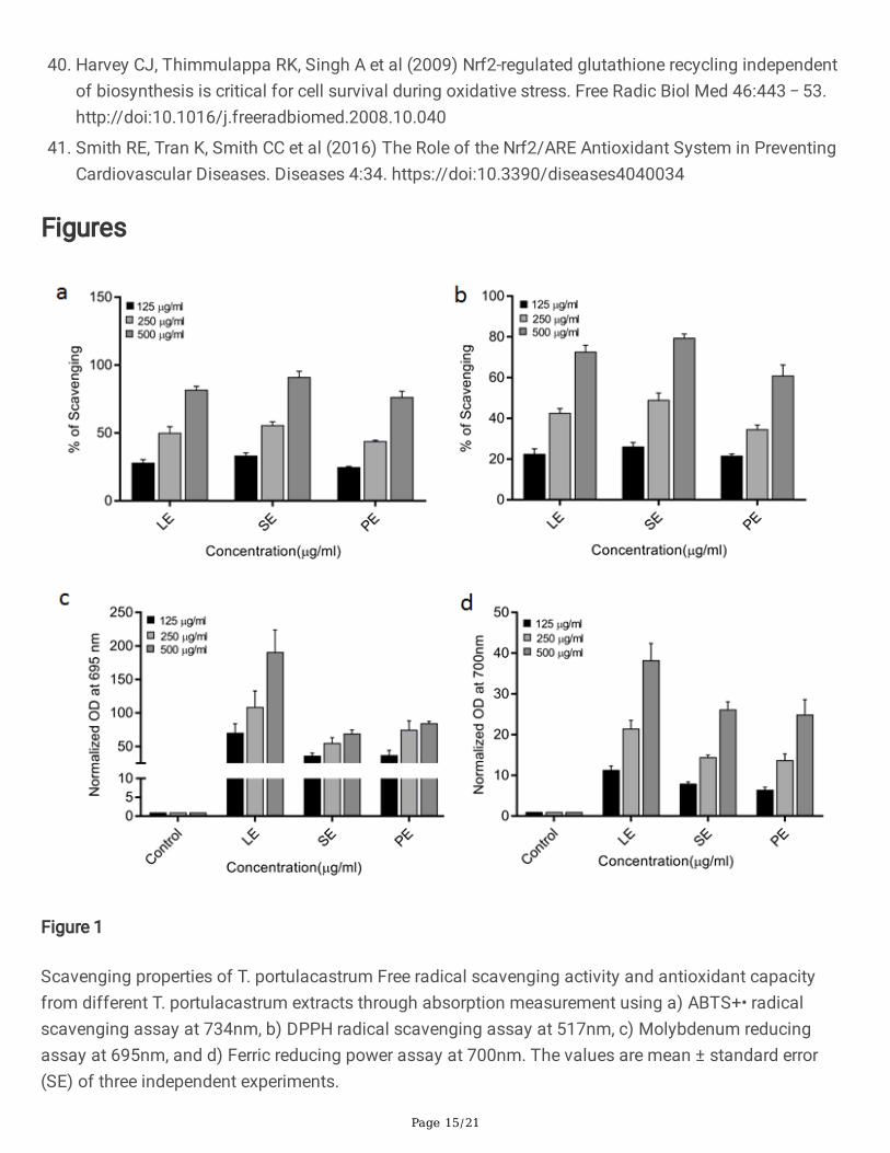
Radiation exposure is known to damage the structure of cell membranes through degradation of lipids,mainly mediated via lipid peroxidation (LPO) of WRL68 hepatocyte cell and mouse mitochondrialmembranes were observed after exposure to 8 Gy and 50 Gy radiation respectively which increased theformation of malondialdehyde (MDA). In both cases, addition of T. portulacastrum stem extracts (SE)
Page 8/21
mitigated the radiation-induced lipid peroxidation in a dose-dependent manner, showing the protectiveactivity against radiation-induced membrane lipid peroxidation (Fig. 2).
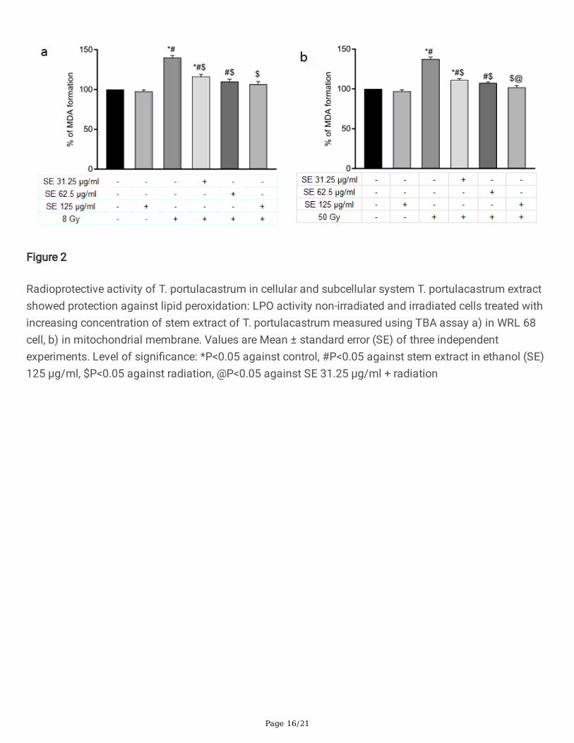
For further evaluation of radioprotective activity of the extract, cell viability of irradiated cells, untreated ortreated with T. portulacastrum stem extracts, was estimated using MTT and clonogenic assays. Exposureto 12 Gy radiation signi�cantly reduced cell viability after 72 h and cells treated with T. portulacastrumextract showed enhanced cell viability compared to irradiated cells in the MTT assay (Fig. 3a). However,only 31.25 µg/ml concentration of the extract demonstrated signi�cant enhancement of cell viability andincreased concentrations of the extracts did not show signi�cant difference in cell viability in comparisonto the irradiated cells.
In the clonogenic assay, exposure to 4 Gy radiation resulted in a 66% reduction in colony-forming abilityof the cells (Fig. 3b). Treatment with high concentrations (to125 µg/ml) of T. portulacastrum extractsalone did not show any reduction in colony formation (Fig. 3c). However, on addition of T. portulacastrumstem extracts to irradiated cells, signi�cant rescue in colony formation was only observed in cells treatedwith 31.25 µg/ml extract (Fig. 3d). Together these observations suggest a radioprotective activity of T.portulacastrum stem extract, although at a higher concentration the level of protection may decrease dueto the presence of compounds with non-speci�c toxic effect on radiation-induce damaged cells.
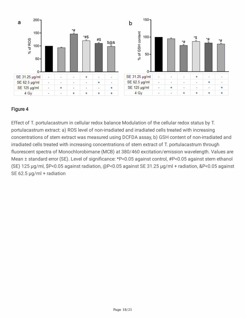
T. portulacastrum stem extracts reduces cellular ROS and enhances GSH
To explore the mechanism of radioprotection by T. portulacastrum stem extract, we evaluated the cellularredox status by measuring reactive oxygen species (ROS) and reduced Glutathione (GSH) which areimportant markers of the redox status of cells. Determination of cellular ROS level by DCFDA�uorescence intensity showed that 4 Gy of radiation increased the cellular ROS level by nearly 2-fold,whereas, pretreatment with the T. portulacastrum stem extract reduced the ROS level in a dose-dependentmanner (Fig. 4a). Similarly, exposure to 4 Gy of radiation signi�cantly reduced the concentration of GSH,one of the most important cellular antioxidant molecules, whereas treatment with the lowestconcentration of T. portulacastrum stem extract signi�cantly upregulated the GSH content (Fig. 4b).Higher concentrations of T. portulacastrum extract failed to signi�cantly enhance the GSH content, mostlikely by inhibition of GSH biosynthesis by high concentration of some non-speci�c molecules. Thus,these observations demonstrated that the observed radioprotection by T. portulacastrum extract is due tomodi�cation of cellular redox status.
T. portulacastrum stem extract exerts anti-in�ammatory effects on LPS-activated macrophages
Another arm of the radiation induced tissue damage is mediated by secondary in�ammatory responses,caused by the activation of in�ammatory cells such as neutrophils and macrophages. Therefore, we alsoinvestigated the effect of T. portulacastrum stem extract in mitigating the in�ammatory response byevaluating its effect on mouse macrophage cells (RAW264.7) activated by the in�ammatory agonist LPS.RAW264.7 cells were stimulated with 500 ng/ml LPS with and without 1 h pretreatment with T.portulacastrum extracts. T. portulacastrum stem extract did not show any autonomous cytotoxicity in

Page 9/21
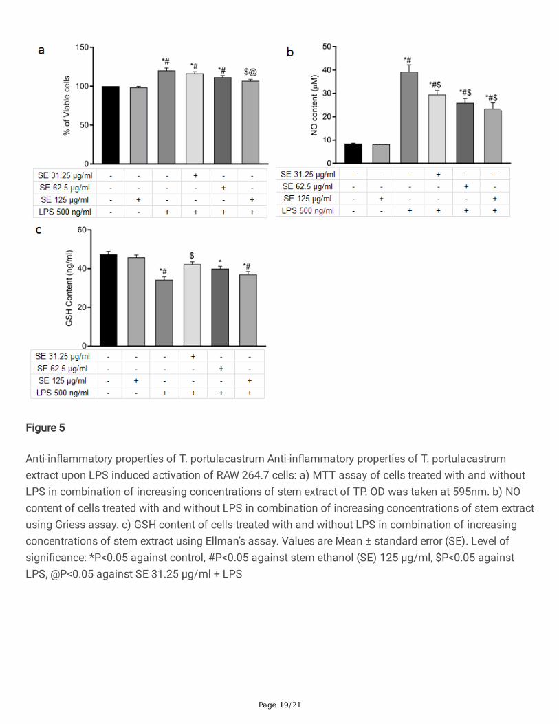
RAW264.7 cells at the highest concentration (125 µg/ml). However, MTT assays showed that treatmentwith TP stem extract reduced the LPS-stimulated proliferation of RAW 264.7 cells in a dose-dependentmanner (Fig. 5a).
One of the major in�ammatory mediators released by macrophages after LPS stimulation is nitric oxide(NO) generated by upregulation of inducible nitric oxide synthase (iNOS). We therefore determined NOgeneration by LPS-stimulated RAW264.7 cells in presence and absence of treatment with T.portulacastrum stem extract. LPS stimulation increased the secreted NO level by 4.6 fold while treatmentwith T. portulacastrum stem extract dose-dependently reduced the secreted NO level (Fig. 5b). LPSinduced activation of macrophages not only elevates NO level but also downregulated GSHconcentration. Treatment with T. portulacastrum stem extract also enhanced GSH level in LPS-treatedRAW264.7 cells, but the highest enhancement was observed at the lowest concentration of T.portulacastrum stem extract as in the case of hepatocytes (Fig. 5c).
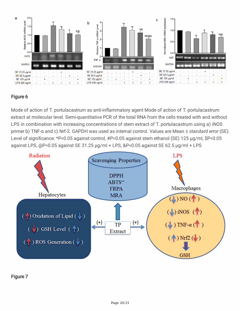
We then investigated the expression of some of the genes responsible for the synthesis of the majorin�ammatory mediators in LPS stimulated macrophages in presence and absence of treatment with T.portulacastrum extract. LPS stimulation increased the mRNA level of iNOS, the enzyme responsible forNO synthesis, by 1.5 fold while treatment with T. portulacastrum stem extract brought down the iNOSmRNA to basal level of expression (Fig. 6a). iNOS expression is mediated through activation of TNF-α, apro-in�ammatory cytokine. TNF-α mRNA level was increased signi�cantly after 24 h of LPS treatmentand while treatment with T. portulacastrum stem extract also signi�cantly decreased the TNF-α level in adose-dependent manner (Fig. 6b).
We also checked the level of Nrf2 mRNA as Nrf2 is a well-known transcription factor which regulates anumber of antioxidant genes in cells, including GSH, and is involved in protection against oxidative stressand in�ammation. Treatment of the cells with LPS lowers the Nrf2 mRNA level while treatment with T.portulacastrum stem extract signi�cantly restored it although the mRNA level reduced with increasingconcentration of the T. portulacastrum extract (Fig. 6c), re�ecting the effect on GSH level as observedbefore.
Together, these observations demonstrate a potential radioprotective role of T. portulacastrum extractmediated by both its antioxidant activity on epithelial cells and its anti-in�ammatory activity on immunecells.
DiscussionExposure of cells to ionizing radiation induces reactive oxygen species (ROS) and nitrogen species (RNS)together with alteration in cellular antioxidant status and resulting cell damage (Reisz et al. 2014).Supplements of antioxidants during radiotherapy have been shown to decrease the damaging effects(Singh et al. 2018). Plants are one of the main natural sources of antioxidants. Radioprotective activityhas been observed from plant antioxidants such as shigoka extract, green tea polyphenols andcurcumins (Seong et al. 2015; Clarke et al. 2016; Shirazi et al. 2012). T. portulacastrum has been shown
Page 10/21
to exhibit hepatoprotective activity against chemical-induced toxicity (Yamaki et al. 2016). Therefore, inthis study we have demonstrated the radioprotective role of TP extract on hepatocytes, mediated via itsactivity of reducing oxidative damage to cells. Moreover, we have demonstrated an anti-in�ammatory roleof T. portulacastrum extract in the case of activated macrophages, which is likely to enhance itsradioprotective function by mitigating the in�ammatory response induced by radiation damage.
A major mode of antioxidant function is via scavenging of oxidative free radicals. Radical scavengingcapacity of an antioxidant lies on its proton donating or accepting ability (Singha et al. 2020). T.portulacastrum extracts showed e�cient free radical scavenging activity in both DPPH and ABTS•+
assays which are based on electron transfer ability used to measure antioxidant capacity. Moreover, T.portulacastrum extracts showed e�cient reducing ability to reduce Mo (VI) to Mo (V) and Fe (III) to Fe (II)which is a marker of its antioxidant activity.
Exposure to radiation causes damage to different biomolecules such as lipids, proteins and nucleic acids.Cellular membranes are one of the major targets of the oxidative free radicals, generated due to radiationexposure. Damage of the lipids present in the cell membranes changes its �uidity status and alsoactivates several critical signaling pathways (Nicolson and Ash 2014). ROS is responsible for thioloxidation which initiates lipid peroxidation. Radiation induced free radicals react with macromoleculesand damage membranes of intracellular organelles (Singha and Das 2015). It has been reported that 50Gy of gamma radiation impairs mitochondrial membrane function by damaging complex I (NADHdehydrogenase) and III (cytochrome c reductase) (Pearce et al. 2001). In our study T. portulacastrumstem extract showed e�cient protection against lipid peroxidation by ionizing radiation in a MDAformation assay, further substantiating its role in radioprotection.
The �nal effect to check in case of radioprotection is the reduction of cell death in response to radiationexposure. Treatment with T. portulacastrum extract showed a survival advantage of irradiated cells inboth short term (MTT) and long term (colony formation) cell viability assays. As it has been reported thatlower radiation dose gives false-positive result in MTT as high formazan is deposited in irradiated cellscompared to control (Rai et al. 2018), the clonogenic assay is a good measure to study the ability of T.portulacastrum extract to protect against radiation-induced cell damage. However, interestingly, higherconcentration of T. portulacastrum extract failed to show this survival advantage and whether this is dueto a pro-oxidant activity demonstrated at a higher concentration as reported for other natural products(Banerjee et al. 2008; Sotler wt al. 2019) or due to an unknown effect of other compounds present in theextract remains to be investigated. A similar concentration-dependent effect was observed in the case ofthe effect of T. portulacastrum extract on GSH concentration of cells, which suggests that this effectmight be mediated by the in�uence of T. portulacastrum extract on the level of GSH, one of the primaryanti-oxidant molecules in the cell.
Radiation-induced cellular damage is not only due to the alteration of the redox and antioxidant balancebut also due to the activation of in�ammatory responses (Sachaue and McBrid 2015). Ionizing radiation-induced activation of the immune system results in in�ammation through enhancing the release of
Page 11/21
growth factors and pro-in�ammatory cytokines (Mun et al. 2018). Changes in the cellular oxidative stresslevel play a pivotal role in in�ammation (Han et al. 2019). However, the effect of natural products onradiation-induced in�ammation has not been explored su�ciently. Therefore, we investigated the effectof TP extract on the in�ammatory response in LPS-treated macrophages, one of the major immune cellsinvolved in radiation-induced in�ammation. Treatment with T. portulacastrum extract was able to reducethe secretion of NO from these cells, as well as reduce the expression, of inducible nitric oxide synthase(iNOS) gene, which are important mediators of the in�ammatory response (Cao et al. 2019). Excess NOinduces in�ammation and nitrosative stress (Calabrese et al. 2004). These results showed that T.portulacastrum extract may protect from radiation damage by modulating the in�ammatory response inthe body.
In cells LPS binds with Toll-like receptor 4 (TLR-4) which further activates pro-in�ammatory cytokines liketumor necrosis factor alpha (TNF-α) and interleukin (IL)-6 and IL-1β which further activates anin�ammatory signaling mechanism (Cao et al. 2019). In our study, LPS stimulation up regulated the levelof TNF-α but TP extract downregulated the TNF-α expression level and thus reduced the TNF-α mediatedin�ammatory signaling cascade.
The nuclear factor erythroid 2-related factor (Nrf2) is a potent antioxidant marker responsible for thereduction of oxidative stress (Han et al. 2019). Nrf2 protects cell from oxidative stress after dissociationfrom Keap1 and binds with antioxidant-response elements (AREs) which ultimately promotes theexpression of several genes including GSH and GSH dependent antioxidant enzymes (Harvey et al. 2009;Smith et al. 2016). GSH, a well-known antioxidant regulates redox status and signaling, death and cellproliferation (Harvey et al. 2009). LPS stimulation of macrophages decreased the Nrf2 mRNA level aswell as the concentration of GSH but treatment with T. portulacastrum extract markedly upregulated themRNA expression of Nrf2 and GSH concentration, suggesting a molecular mechanism for upregulation ofGSH in cells treated with TP extract. Interestingly, the same effect of higher dose of T. portulacastrumextract failing to enhance Nrf2 expression and GSH concentration was noted, re�ecting the similarity withradiation induced GSH concentration, and cell viability in hepatocytes. This warrants further investigationinto the concentration dependent effect of T. portulacastrum extract, and its bioactive molecules, onregulation of expression of anti-oxidant genes in cells.
Conclusion
This study for the �rst time shows a radioprotective activity of extracts from the plant Trianthemaportulacastrum, mediated via its dual effect in modifying the redox status of irradiated cells and thein�ammatory response of immune cells activated by the in�ammatory agonist LPS. Both these effectstogether may strongly support the role of T. portulacastrum extract as a natural product with signi�cantradioprotective ability.
DeclarationsCon�ict of Interest Statement:
Page 12/21
There is no con�ict of Interest
AcknowledgementFinancial assistance received from the Department of Atomic Energy-Board of Research in NuclearStudies (35/14/39/2016-BRNS/35174) is gratefully acknowledged. Research in the laboratory of PSR issupported by SERB grant EMR/2016/003525.
References1. Wang Js, Wang Hj, Qian, Hl (2018) Biological effects of radiation on cancer cells. Military Med Res
5:20. https://doi.org/10.1186/s40779-018-0167-4
2. Shemetun OV, Pilins’ka MA (2007) Radiation-induced “bystander” effect. Cytol Genet 41:251–255.https://doi.org/10.3103/S0095452707040111
3. Carvalho HA, Villar RC (2018) Radiotherapy and immune response: the systemic effects of a localtreatment. Clinics (Sao Paulo) 73:e557s. https://doi:10.6061/clinics/2018/e557s
4. Obrador E, Salvador R, Villaescusa JI et al (2020) Radioprotection and Radiomitigation: From theBench to Clinical Practice. Biomedicines 8:461. https://doi:10.3390/biomedicines8110461
5. Elkady AA, Ibrahim IM (2016) Protective effects of erdosteine against nephrotoxicity caused bygamma radiation in male albino rats. Hum Exp Toxicol 35:21 − 8.https://doi:10.1177/0960327115574919
�. Kumar KS, Srinivasan V, Toles R et al (2002) Nutritional approaches to radioprotection: vitamin E. MilMed 167:57–59
7. Godlewska-Żyłkiewicz B, Świsłocka R, Kalinowska M et al (2020) Biologically Active Compounds ofPlants: Structure-Related Antioxidant, Microbiological and Cytotoxic Activity of Selected CarboxylicAcids. Materials (Basel) 13:4454. https://doi:10.3390/ma13194454
�. Farhood B, Mortezaee K, Goradel NH et al (2019) Curcumin as an anti-in�ammatory agent:Implications to radiotherapy and chemotherapy. J Cell Physiol 234:5728–5740.https://doi:10.1002/jcp.27442
9. Azab A, Nassar A, Azab AN (2016) Anti-In�ammatory Activity of Natural Products. Molecules21:1321. https://doi:10.3390/molecules21101321
10. Maurya DK, Devasagayam TP (2010) Antioxidant and prooxidant nature of hydroxycinnamic acidderivatives ferulic and caffeic acids. Food Chem Toxicol 48:3369–3373.https://doi:10.1016/j.fct.2010.09.006
11. Shivhare MK, Singour PK, Chaurasiya PK et al (2012) Trianthema portulacastrum Linn. (Bishkhapra).Pharmacogn Rev 6:132–140. https://doi:10.4103/0973-7847.99947
12. Das U, Saha T, Ghosh R et al (2020) Trianthema portulacastrum L.: Traditional medicine inhealthcare and biology. Ind J Biochem Biophy 57:127–145
Page 13/21
13. Sarkar A, Pradhan S, Mukhopadhyay I et al (1999) Inhibition of early DNA-damage and chromosomalaberrations by Trianthema portulacastruml. In carbon tetrachloride-induced mouse liver damage. CellBiol Int 23:703–708. https://doi:10.1006/cbir.1999.0439
14. Kumar G, Banu GS, Pappa PV et al (2004) Hepatoprotective activity of Trianthema portulacastrum L.against paracetamol and thioacetamide intoxication in albino rats. J Ethnopharmacol 92:37–40.https://doi:10.1016/j.jep.2003.12.009
15. Saxena S, Verma J, Gautam S (2016) Potential Prophylactic Properties of Apple and Characterizationof Potent Bioactive from cv. "Granny Smith" Displaying Strong Antimutagenicity in Models IncludingHuman Lymphoblast TK6(+/-) Cell Line. J Food Sci 81:H508-H518. https://doi:10.1111/1750-3841.13190
1�. Checker R, Sharma D, Sandur SK et al (2010) Plumbagin inhibits proliferative and in�ammatoryresponses of T cells independent of ROS generation but by modulating intracellular thiols. J CellBiochem 110:1082–1093. https://doi:10.1002/jcb.22620
17. Maurya DK, Nandakumar N, Devasagayam TP (2011) Anticancer property of gallic acid in A549, ahuman lung adenocarcinoma cell line, and possible mechanisms. J Clin Biochem Nutr 48:85–90.https://doi:10.3164/jcbn.11-004FR
1�. Jayakumar S, Pal D, Sandur SK (2015) Nrf2 facilitates repair of radiation induced DNA damagethrough homologous recombination repair pathway in a ROS independent manner in cancer cells.Mutat Res 779:33–45. https://doi:10.1016/j.mrfmmm.2015.06.007
19. Moron MS, Depierre JW, Mannervik B (1979) Levels of glutathione, glutathione reductase andglutathione S-transferase activities in rat lung and liver. Biochim Biophys Acta 582:67–78.https://doi:10.1016/0304-4165(79)90289-7
20. Kacem M, Simon G, Leschiera R et al (2015) Antioxidant and anti-in�ammatory effects of Rutachalepensis L. extracts on LPS-stimulated RAW 264.7 cells. In Vitro Cell Dev Biol Anim 51:128–141.https://doi:10.1007/s11626-014-9813-7
21. Ahuja D, Goyal A, Ray PS (2016) Interplay between RNA-binding protein HuR and microRNA-125bregulates p53 mRNA translation in response to genotoxic stress. RNA Biol 13:1152–1165.https://doi:10.1080/15476286.2016.1229734
22. Reisz JA, Bansal N, Qian J et al (2014) Effects of ionizing radiation on biological molecules–mechanisms of damage and emerging methods of detection. Antioxid Redox Signal 21:260–292.https://doi:10.1089/ars.2013.5489
23. Singh K, Bhori M, Kasu YA et al (2018) Antioxidants as precision weapons in war against cancerchemotherapy induced toxicity - Exploring the armoury of obscurity. Saudi Pharm J 26:177–190.http://doi:10.1016/j.jsps.2017.12.013
24. Seong KM, Yu M, Lee KS et al (2015) Curcumin mitigates accelerated aging after irradiation inDrosophila by reducing oxidative stress. Biomed Res Int 2015:425380.https://doi:10.1155/2015/425380
Page 14/21
25. Clarke KA, Dew TP, Watson RE et al (2016) Green tea catechins and their metabolites in human skinbefore and after exposure to ultraviolet radiation. J Nutr Biochem 27:203–210.https://doi:10.1016/j.jnutbio.2015.09.001
2�. Shirazi A, Mihandoost E, Mahdavi SR et al (2012) Radio-protective role of antioxidant agents. OncolRev 6:e16. https://doi:10.4081/oncol.2012.e16
27. Yamaki J, Nagulapalli Venkata KC, Mandal A et al (2016) Health-promoting and disease-preventivepotential of Trianthema portulacastrum Linn. (Gadabani) -An Indian medicinal and dietary plant. JIntegr Med 14:84–99. https://doi:10.1016/S2095-4964(16)60247-9
2�. Singha I, Saxena S, Gautam S et al (2020) Grape extract protect against ionizing radiation-inducedDNA damage. Ind J Biochem Biophy 57:219–227
29. Nicolson GL, Ash ME. (2014) Lipid Replacement Therapy: a natural medicine approach to replacingdamaged lipids in cellular membranes and organelles and restoring function. Biochim Biophys Acta1838:1657-79. https://doi:10.1016/j.bbamem.2013.11.010
30. Singha I, Das SK (2015) Grapevine fruit extract protects against radiation-induced oxidative stressand apoptosis in human lymphocyte. Indian J Exp Biol 53:753–761
31. Pearce LL, Epperly MW, Greenberger JS et al (2001) Identi�cation of respiratory complexes I and III asmitochondrial sites of damage following exposure to ionizing radiation and nitric oxide. Nitric Oxide5:128–136. https://doi:10.1006/niox.2001.0338
32. Rai Y, Pathak R, Kumari N et al (2018) Mitochondrial biogenesis and metabolic hyperactivation limitsthe application of MTT assay in the estimation of radiation induced growth inhibition. Sci Rep8:1531. https://doi:10.1038/s41598-018-19930-w
33. Banerjee A, Kunwar A, Mishra B et al (2008) Concentration dependent antioxidant/pro-oxidantactivity of curcumin studies from AAPH induced hemolysis of RBCs. Chem Biol Interact 174:134-9.https://doi:10.1016/j.cbi.2008.05.009
34. Sotler R, Poljšak B, Dahmane R et al (2019) Prooxidant activities of antioxidants and their impact onhealth. Acta Clin Croat 58:726–736. http://doi:10.20471/acc.2019.58.04.20
35. Schaue D, McBride WH (2015) Opportunities and challenges of radiotherapy for treating cancer. NatRev Clin Oncol 12:527 − 40. http://doi:10.1038/nrclinonc.2015.120
3�. Mun GI, Kim S, Choi E et al (2020) Pharmacology of natural radioprotectors. Arch Pharm Res41:1033–1050. http://doi:10.1007/s12272-018-1083-6
37. Han S, Gao H, Chen S et al (2019) Procyanidin A1 Alleviates In�ammatory Response induced by LPSthrough NF-κB, MAPK, and Nrf2/HO-1 Pathways in RAW264.7 cells. Sci Rep 9:15087.https://doi.org/10.1038/s41598-019-51614-x
3�. Cao Y, Chen J, Ren G et al (2019) Punicalagin Prevents In�ammation in LPS-Induced RAW264.7Macrophages by Inhibiting FoxO3a/Autophagy Signaling Pathway. Nutrients 11:2794.https://doi:10.3390/nu11112794
39. Calabrese V, Boyd-Kimball D, Scapagnini G et al (2004) Nitric oxide and cellular stress response inbrain aging and neurodegenerative disorders: the role of vitagenes. In Vivo 18:245–267
Page 15/21
40. Harvey CJ, Thimmulappa RK, Singh A et al (2009) Nrf2-regulated glutathione recycling independentof biosynthesis is critical for cell survival during oxidative stress. Free Radic Biol Med 46:443 − 53.http://doi:10.1016/j.freeradbiomed.2008.10.040
41. Smith RE, Tran K, Smith CC et al (2016) The Role of the Nrf2/ARE Antioxidant System in PreventingCardiovascular Diseases. Diseases 4:34. https://doi:10.3390/diseases4040034
Figures
Figure 1
Scavenging properties of T. portulacastrum Free radical scavenging activity and antioxidant capacityfrom different T. portulacastrum extracts through absorption measurement using a) ABTS+• radicalscavenging assay at 734nm, b) DPPH radical scavenging assay at 517nm, c) Molybdenum reducingassay at 695nm, and d) Ferric reducing power assay at 700nm. The values are mean ± standard error(SE) of three independent experiments.
Page 16/21
Figure 2
Radioprotective activity of T. portulacastrum in cellular and subcellular system T. portulacastrum extractshowed protection against lipid peroxidation: LPO activity non-irradiated and irradiated cells treated withincreasing concentration of stem extract of T. portulacastrum measured using TBA assay a) in WRL 68cell, b) in mitochondrial membrane. Values are Mean ± standard error (SE) of three independentexperiments. Level of signi�cance: *P<0.05 against control, #P<0.05 against stem extract in ethanol (SE)125 µg/ml, $P<0.05 against radiation, @P<0.05 against SE 31.25 µg/ml + radiation
Page 17/21
Figure 3
Validation of Radioprotective activity Radioprotective property of T. portulacastrum extract in WRL 68 cellsystem: a) MTT assay of non-irradiated and irradiated cells treated with increasing concentrations ofstem extract of T. portulacastrum. OD was read at 595 nm. Clonogenic assay was performed 12 dayspost irradiation where cells were allowed to form colonies and then counted by crystal violet staining. b)control groups c) different concentration of T. portulacastrum extracts in presence of radiation and e)graphical representation of CFUs from three independent experiments. Values are Mean ± standard error(SE). Level of signi�cance: *P<0.05 against control, #P<0.05 against stem ethanol (SE) 125 µg/ml,$P<0.05 against radiation, @P<0.05 against SE 31.25 µg/ml + radiation
Page 18/21
Figure 4
Effect of T. portulacastrum in cellular redox balance Modulation of the cellular redox status by T.portulacastrum extract: a) ROS level of non-irradiated and irradiated cells treated with increasingconcentrations of stem extract was measured using DCFDA assay, b) GSH content of non-irradiated andirradiated cells treated with increasing concentrations of stem extract of T. portulacastrum through�uorescent spectra of Monochlorobimane (MCB) at 380/460 excitation/emission wavelength. Values areMean ± standard error (SE). Level of signi�cance: *P<0.05 against control, #P<0.05 against stem ethanol(SE) 125 µg/ml, $P<0.05 against radiation, @P<0.05 against SE 31.25 µg/ml + radiation, &P<0.05 againstSE 62.5 µg/ml + radiation
Page 19/21
Figure 5
Anti-in�ammatory properties of T. portulacastrum Anti-in�ammatory properties of T. portulacastrumextract upon LPS induced activation of RAW 264.7 cells: a) MTT assay of cells treated with and withoutLPS in combination of increasing concentrations of stem extract of TP. OD was taken at 595nm. b) NOcontent of cells treated with and without LPS in combination of increasing concentrations of stem extractusing Griess assay. c) GSH content of cells treated with and without LPS in combination of increasingconcentrations of stem extract using Ellman’s assay. Values are Mean ± standard error (SE). Level ofsigni�cance: *P<0.05 against control, #P<0.05 against stem ethanol (SE) 125 µg/ml, $P<0.05 againstLPS, @P<0.05 against SE 31.25 µg/ml + LPS
Page 20/21
Figure 6
Mode of action of T. portulacastrum as anti-in�ammatory agent Mode of action of T. portulacastrumextract at molecular level. Semi-quantitative PCR of the total RNA from the cells treated with and withoutLPS in combination with increasing concentrations of stem extract of T. portulacastrum using a) iNOSprimer b) TNF-α and c) Nrf-2. GAPDH was used as internal control. Values are Mean ± standard error (SE).Level of signi�cance: *P<0.05 against control, #P<0.05 against stem ethanol (SE) 125 µg/ml, $P<0.05against LPS, @P<0.05 against SE 31.25 µg/ml + LPS, &P<0.05 against SE 62.5 µg/ml + LPS
Figure 7
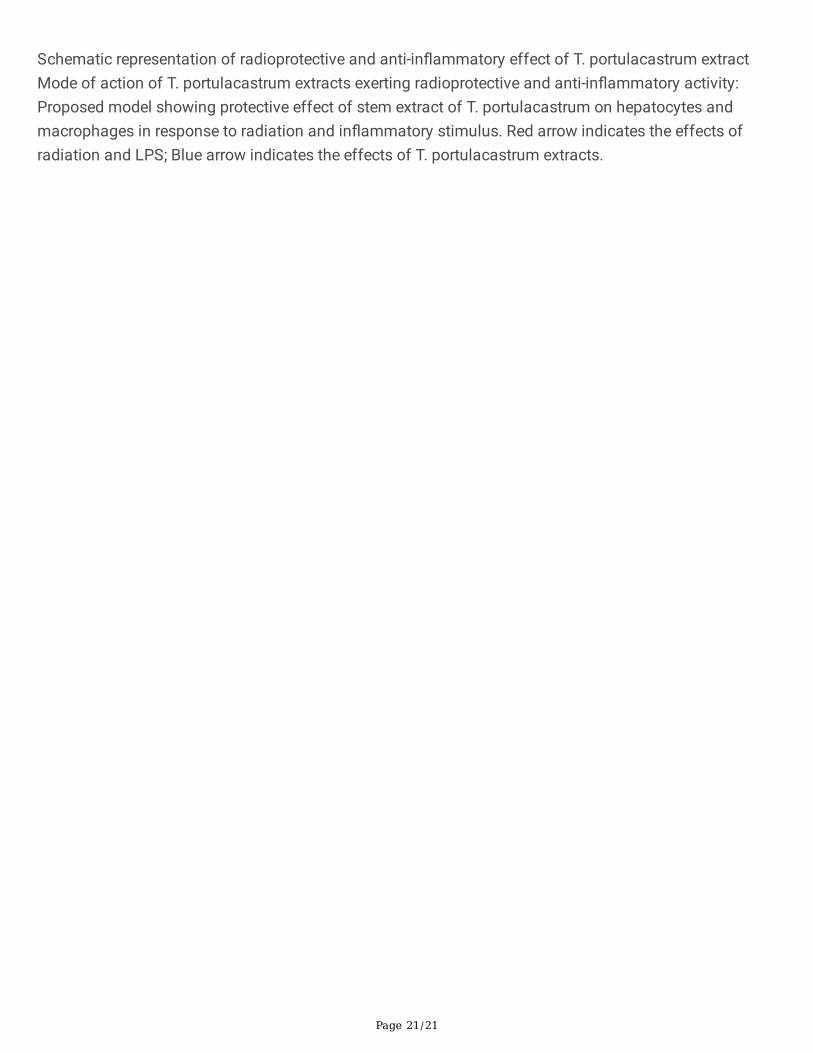
Page 21/21
Schematic representation of radioprotective and anti-in�ammatory effect of T. portulacastrum extractMode of action of T. portulacastrum extracts exerting radioprotective and anti-in�ammatory activity:Proposed model showing protective effect of stem extract of T. portulacastrum on hepatocytes andmacrophages in response to radiation and in�ammatory stimulus. Red arrow indicates the effects ofradiation and LPS; Blue arrow indicates the effects of T. portulacastrum extracts.