positive and negative functions of the saga complex mediated
TRANSCRIPT

Positive and negative functionsof the SAGA complex mediatedthrough interaction of Spt8 with TBPand the N-terminal domain of TFIIALinda Warfield,1 Jeffrey A. Ranish,2 and Steven Hahn1,3
1Fred Hutchinson Cancer Research Center, and Howard Hughes Medical Institute, Seattle, Washington 98109, USA;and 2Institute for Systems Biology, Seattle, Washington 98103, USA
A surface that is required for rapid formation of preinitiation complexes (PICs) was identified on theN-terminal domain (NTD) of the RNA Pol II general transcription factor TFIIA. Site-specificphotocross-linkers and tethered protein cleavage reagents positioned on the NTD of TFIIA and assembled inPICs identified the SAGA subunit Spt8 and the TFIID subunit Taf4 as located near this surface. In agreementwith these findings, mutations in Spt8 and the TFIIA NTD interact genetically. Using purified proteins, it wasfound that TFIIA and Spt8 do not stably bind to each other, but rather both compete for binding to TBP.Consistent with this competition, Spt8 inhibits the binding of SAGA to PICs in the absence of activator. Inthe presence of activator, Spt8 enhances transcription in vitro, and the positive function of the TFIIA NTD islargely mediated through Spt8. Our results suggest a mechanism for the previously observed positive andnegative effects of Spt8 on transcription observed in vivo.
[Keywords: Transcription; SAGA; TFIIA; coactivator; repression]
Supplemental material is available at http://www.genesdev.org.
Received February 6, 2004; revised version accepted March 22, 2004.
Binding of early-acting gene-specific factors to gene regu-latory sequences and recruitment of the chromatin re-modeling machinery initiates a cascade of events inwhich transcription coactivators and the general tran-scription machinery are recruited to promoters, forminga preinitiation complex (PIC; Kuras and Struhl 1999; Liet al. 1999; Agalioti et al. 2000; Cosma 2002). The re-cruitment of TBP, a DNA sequence-specific bindingcomponent of PICs, is regulated both positively andnegatively (Hampsey 1998). In many cases, activators in-crease the binding of TBP to promoters in direct propor-tion to their activity in transcription activation (Kurasand Struhl 1999; Li et al. 1999). In contrast, factors suchas Mot1 or the Taf1 N-terminal domain (TAND) inhibitthe DNA binding activity of TBP (Auble et al. 1994;Kokubo et al. 1998; Cang et al. 1999). The general factorTFIIA interacts directly with TBP and stabilizes TBP–DNA interactions (Weideman et al. 1997; Hampsey1998; Liu et al. 1999). TFIIA also stimulates and stabi-lizes the binding of TFIID to DNA as part of an activa-tor–TFIID–TFIIA–DNA complex, a rate-limiting inter-
mediate in the transcription of certain promoters (Wanget al. 1992; Lieberman and Berk 1994; Chi et al. 1995).Finally, TFIIA can compete with negative factors such asNC2, Mot1, and the Taf1 TAND domain for binding toTBP (Hampsey 1998; Kokubo et al. 1998; Ozer et al.1998b; Cang et al. 1999). TFIIA has also been found tointeract with at least two Tafs and some transcriptionactivators, using two-hybrid and affinity chromatogra-phy analysis (Yokomori et al. 1993; Ozer et al. 1994;Kobayashi et al. 1998; Kraemer et al. 2001).
TFIIA is unique in that it is the only Pol II generalfactor that is not absolutely required for transcriptionunder standard conditions. In vitro, TFIIA stimulatesboth basal and activated transcription from 2- to 10-fold,but generally only when the TFIID form of TBP is used(Orphanides et al. 1996; Hampsey 1998). Several ap-proaches have been taken to determine which genes re-quire TFIIA for expression in vivo. These include TFIIAsubunit depletion, mutation of TFIIA subunits to impairinteraction with TBP, or mutation of TBP to inhibit in-teraction with TFIIA (Kang et al. 1995; Ozer et al. 1998a;Liu et al. 1999; Stargell et al. 2000). These studies gen-erally agree that TFIIA is important for transcription of aspecific subset of genes, although there is not yet agree-ment on the exact subset of genes requiring TFIIA fornormal expression.
3Corresponding author.E-MAIL [email protected]; FAX (206) 667-6497.Article and publication are at http://www.genesdev.org/cgi/doi/10.1101/gad.1192204.
1022 GENES & DEVELOPMENT 18:1022–1034 © 2004 by Cold Spring Harbor Laboratory Press ISSN 0890-9369/04; www.genesdev.org
Cold Spring Harbor Laboratory Press on April 13, 2018 - Published by genesdev.cshlp.orgDownloaded from

The SAGA complex is a cofactor required for normalexpression of some genes in yeast (Sterner et al. 1999;Lee et al. 2000). On gene activation in vivo, SAGA can berecruited to promoters independently of the general tran-scription factors (Bhaumik and Green 2001; Larschanand Winston 2001). The SAGA complex contains a corecomprising Ada and Spt subunits, a subset of Tafs, acet-yltransferase and ubiquitin protease activities, the es-sential factor Tra1, and two factors related to TBP func-tion, Spt3 and Spt8 (Grant et al. 1998; Sterner et al. 1999;Pray-Grant et al. 2002; Sanders et al. 2002). SAGA isclosely related to the SLIK/SALSA complex, which lacksSpt8 but which contains at least one unique subunit,Rtg2 (Pray-Grant et al. 2002; Sterner et al. 2002; Wu andWinston 2002). It is not yet known how redundantlythese two related complexes function in vivo. Only asmall percentage of promoters seem to be exclusivelySAGA- or TFIID-dependent in vivo (Lee et al. 2000). Ingeneral, the requirement of a particular promoter forSAGA or TFIID is determined by the core promoter se-quence, and the particular activator determines whetherSAGA or TFIID is recruited to a promoter (Shen andGreen 1997; Cheng et al. 2002; Mencia et al. 2002). Itseems that many yeast promoters can use either SAGAor TFIID, because a double mutation of SAGA and TFIIDsubunits or a mutation of a shared TFIID/SAGA subunitaffects expression from many more yeast genes than doesa single mutation of either TFIID or SAGA specific sub-units (Lee et al. 2000).
At SAGA-dependent promoters, SAGA is required forrecruitment of the transcription machinery (Dudley etal. 1999; Bhaumik and Green 2001; Larschan and Win-ston 2001). Although recruitment of SAGA to promotersis independent of Spt3 and Spt8, these subunits are re-quired for general factor recruitment at most SAGA-de-pendent promoters tested (Bhaumik and Green 2001,2002; Larschan and Winston 2001). Taken together,these observations suggest that SAGA is a coactivatorthat links some activators to the general transcriptionmachinery. The mechanism whereby SAGA interactswith the general transcription machinery is not well un-derstood. It has been proposed that the Spt3 subunitinteracts directly with TBP (Bhaumik and Green 2001;Larschan and Winston 2001). Mutations on oppositesides of the TBP surface have been found to suppress anSpt3 mutation, and a mutation in Spt3 was found tosuppress TBP mutations (Eisenmann et al. 1992). Spt3was found to coimmune precipitate with TBP, but onlywhen overexpressed. This may have been due to nonspe-cific interaction because Spt8, another SAGA subunit,did not coimmune precipitate with TBP (Eisenmann etal. 1992, 1994). Protein–protein interaction studies andtwo-hybrid analysis have not shown any direct interac-tion between Spt3 and TBP (Eisenmann et al. 1994;Madison and Winston 1997). In contrast, intact SAGAwas found to interact with TBP and this interaction re-quired the Spt8 and Ada3 subunits but was independentof Spt3 (Saleh et al. 1997; Sterner et al. 1999; Sanderset al. 2002).
In this work, we examine the function of the TFIIA
N-terminal domain (NTD), an essential region of TFIIAlocated opposite of TBP and DNA in the TFIIA–TBP–DNA complex structure (Geiger et al. 1996; Tan et al.1996). Using site-specific protein cross-linking and atethered protein cleavage reagent positioned near thefunctional surface of the NTD, we determine the iden-tity of polypeptides located near this surface in PICs andshow surprising positive and negative interactionsamong the SAGA subunit Spt8, TBP, and the NTD ofTFIIA.
Results
A surface on the TFIIA N-terminal domainrequired for rapid PIC formation
Previously, alanine-scanning mutagenesis of the twoTFIIA subunits, Toa1 and Toa2, identified a double ala-nine substitution of Toa2 residues D21 and D24 affect-ing the function of TFIIA in vivo (Kang et al. 1995). Thisdouble mutation, located on the surface of the four-helixbundle of the TFIIA NTD, was temperature sensitive invivo and defective for both basal and activated transcrip-tion in vitro. As expected from the structure of the TFI-IA–TBP–DNA complex (Geiger et al. 1996; Tan et al.1996), these mutations did not affect interaction ofTFIIA with TBP, formation of the TBP–TFIIA–DNAcomplex, TFIIA dimerization, or in vivo stability ofTFIIA, suggesting this TFIIA mutant was defective inprotein–protein interaction with some other componentof the transcription machinery. Later work also showedthe importance of other nearby residues on this TFIIANTD surface. Protein–protein interaction studies usingthe two-hybrid and GST-pulldown assays suggestedthat the TFIIA NTD interacts with the TFIID-specificsubunit Taf11 (Kraemer et al. 2001). This interactionis blocked by mutation of Toa2 residue I27, which alsocauses a defect in transcription from a subset of genes invivo (Kraemer et al. 2001). In other work, it was pro-posed that this TFIIA NTD surface is important for tran-scription activation because mutation of Toa2 residueG30 suppresses the TBP mutation P109A, which is de-fective in activation of specific promoters (Liu et al.1999).
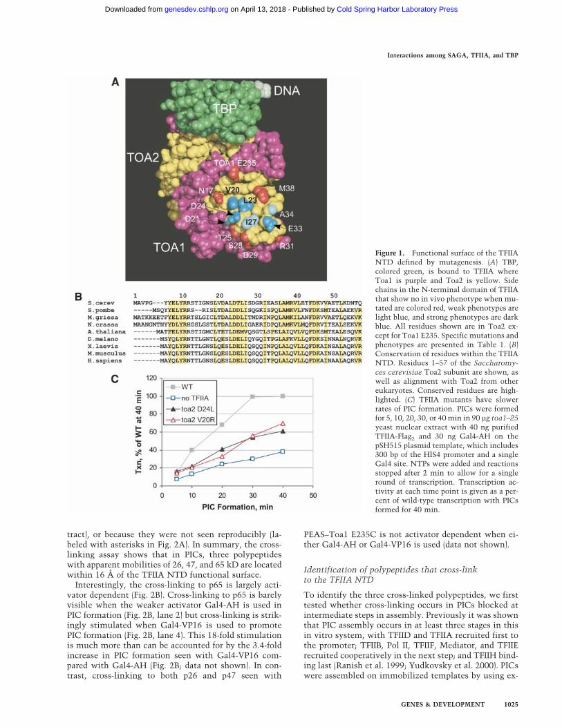
To more precisely define the function of the TFIIANTD, we used site-directed mutagenesis to define thefunctional surface of the TFIIA NTD. Mutations weremade in plasmids containing TOA1 or TOA2 and as-sayed for viability and in vivo phenotype by plasmidshuffle (Table 1). Of the 14 residues mutagenized, 7 dis-played in vivo phenotypes and are clustered on a con-served surface of the NTD near the tip of the four-helixbundle (Fig. 1A), with side chains conferring an in vivophenotype colored blue and those with no phenotypecolored red. Residues I27, L23, and D24, all of whichshow an in vivo phenotype when truncated to alanine,are conserved from yeast to human (Fig. 1B). Othernearby nonconserved residues such as V20 and E33 showan in vivo phenotype only when substituted with a largedissimilar side chain. This functional surface is bounded
Interactions among SAGA, TFIIA, and TBP
GENES & DEVELOPMENT 1023
Cold Spring Harbor Laboratory Press on April 13, 2018 - Published by genesdev.cshlp.orgDownloaded from

by seven Toa1 and Toa2 residues that show no pheno-type when mutated. Six of the Toa2 mutations with adefective in vivo phenotype on plasmids were integratedinto the chromosomal TOA2 locus, and in all casesshowed similar or, in most cases, more severe in vivophenotypes (Table 1). An interactive 3D structure high-lighting functionally important residues in the TFIIANTD can be viewed at http://www.fhcrc.org/labs/hahn/chime_pages/3dstruct_index.html.
To determine the biochemical defect of these muta-tions, we assayed the Toa2 mutations V20R and D24Lfor the ability to form PICs and to support transcriptionin vitro. TFIIA function was measured by supplementinga nuclear extract defective in TFIIA activity with recom-binant TFIIA and assaying PIC formation on the yeastHIS4 promoter. The rate of PIC formation was measuredby incubation of the TFIIA supplemented extract withDNA for various times, followed by nucleotide additionfor a limited time to permit a single round of transcrip-tion initiation (Fig. 1C). By this assay, the absence ofTFIIA resulted in about a threefold slower rate of PICformation. Similarly, extracts containing Toa2 muta-tions D24L or V20R showed a defect in the rate of PIC
formation with a rate twofold slower than that of wildtype. Western blot analysis of PICs formed with eitherwild-type or mutant TFIIA showed similar relativeamounts of all general transcription factors tested, sug-gesting that PICs formed with the TFIIA mutants werenot specifically depleted in any one general factor (datanot shown).
Site-specific photocross-linking of PICs identifiesthree polypeptides near the TFIIA NTD
Because these results, along with previous work, sug-gested that the TFIIA NTD mutants are defective in pro-tein–protein interaction with some component of thetranscription machinery, we set out to determine whatpolypeptides are located near this surface when TFIIA isassembled in PICs. In an initial approach, we isolatedsuppressors of Toa2 L23A and D24A by using EMS mu-tagenesis. Genetic analysis of these suppressors proveddifficult because the phenotype of the original mutationsreverted with a relatively high frequency and because thephenotype of most suppressors was weak semidominantsuppression and sporulation defective. Because this ge-netic approach proved difficult, we directly identifiedpolypeptides near this surface by using site-specific pho-tocross-linking. In this strategy, TFIIA was engineered tocontain unique surface accessible cysteine residues nearthe TFIIA functional surface, and these residues wereused to attach the 125I-labeled photocross-linker PEAS,which has a probe length of 16Å (Chen et al. 1994). PICscontaining this modified recombinant TFIIA were iso-lated and treated with UV light to activate the cross-linker. Reducing the product of this reaction with DTTtransfers the labeled cross-linker from TFIIA to thecross-linked polypeptide.
Two residues near the TFIIA functional surface thatshowed no in vivo phenotype when mutated were indi-vidually changed to cysteine (Toa1 E235C and Toa2M38C). Two surface cysteines on Toa2 were also altered(C72V and C116S) to eliminate other potential sites ofcross-linker attachment. None of these changes led to anin vivo or in vitro phenotype. The photocross-linker 125I-PEAS was conjugated to modified Toa1 or Toa2 at posi-tion 235 or 38. These TFIIA derivatives were active for invitro transcription, with activity comparable to that ofwild-type TFIIA (Supplementary Fig. 1A). When PICswere formed with purified 125I-PEAS–TFIIA, activated byUV light, and reduced by DTT, the 125I-PEAS was trans-ferred to several polypeptides (Fig. 2A, lanes 2,4). WhenPEAS is attached to Toa2 M38C, labeled cross-linker istransferred to a protein with apparent mobility of 65 kD(p65) as well as to Toa1 and intramolecularly to Toa2,remaining stably bound to Toa2 after UV and DTT treat-ment. When cross-linker is attached to Toa1 E235C, UVand DTT treatment causes transfer to Toa2, Toa1, andproteins of ∼26, 47, and 65 kD. Other labeled polypep-tides were not analyzed further because they were ob-served to be labeled in the absence of UV treatment (pre-sumably due to the result of cys–PEAS reaction with thecysteine side chain of some other polypeptide in the ex-
Table 1. TFIIA mutant summary
Mutant Phenotype on plasmid Phenotype in genome
toa2N17A wtN17L wtV20A wt wtV20R ts —D21A wtD21L slightly ts tsL23A ts dead at 36°D24A ts tsD24L severe ts severe tsT25A wtT25L wtI27A slightly tsI27L wtS28A wtS28L wtD29A wtD29L wtR31A wtE33A wt slowE33L ts —A34L slightly tsM38A wtM38L wtM38R wt
toa1E235A wtE235L wt
Phenotypes of TFIIA NTD mutants. Yeast strains containingeither plasmid or integrated copies of TOA1 or TOA2 pointmutants were checked for phenotypes at 18°C, 25°C, 30°C, and36°C by streaking onto glucose complete–Leu media orYPD + Ade media respectively. Two of the eight mutationswere unable to integrate into the genome, indicated by —.
Warfield et al.
1024 GENES & DEVELOPMENT
Cold Spring Harbor Laboratory Press on April 13, 2018 - Published by genesdev.cshlp.orgDownloaded from
tract), or because they were not seen reproducibly (la-beled with asterisks in Fig. 2A). In summary, the cross-linking assay shows that in PICs, three polypeptideswith apparent mobilities of 26, 47, and 65 kD are locatedwithin 16 Å of the TFIIA NTD functional surface.
Interestingly, the cross-linking to p65 is largely acti-vator dependent (Fig. 2B). Cross-linking to p65 is barelyvisible when the weaker activator Gal4-AH is used inPIC formation (Fig. 2B, lane 2) but cross-linking is strik-ingly stimulated when Gal4-VP16 is used to promotePIC formation (Fig. 2B, lane 4). This 18-fold stimulationis much more than can be accounted for by the 3.4-foldincrease in PIC formation seen with Gal4-VP16 com-pared with Gal4-AH (Fig. 2B; data not shown). In con-trast, cross-linking to both p26 and p47 seen with
PEAS–Toa1 E235C is not activator dependent when ei-ther Gal4-AH or Gal4-VP16 is used (data not shown).
Identification of polypeptides that cross-linkto the TFIIA NTD
To identify the three cross-linked polypeptides, we firsttested whether cross-linking occurs in PICs blocked atintermediate steps in assembly. Previously it was shownthat PIC assembly occurs in at least three stages in thisin vitro system, with TFIID and TFIIA recruited first tothe promoter; TFIIB, Pol II, TFIIF, Mediator, and TFIIErecruited cooperatively in the next step; and TFIIH bind-ing last (Ranish et al. 1999; Yudkovsky et al. 2000). PICswere assembled on immobilized templates by using ex-
Figure 1. Functional surface of the TFIIANTD defined by mutagenesis. (A) TBP,colored green, is bound to TFIIA whereToa1 is purple and Toa2 is yellow. Sidechains in the N-terminal domain of TFIIAthat show no in vivo phenotype when mu-tated are colored red, weak phenotypes arelight blue, and strong phenotypes are darkblue. All residues shown are in Toa2 ex-cept for Toa1 E235. Specific mutations andphenotypes are presented in Table 1. (B)Conservation of residues within the TFIIANTD. Residues 1–57 of the Saccharomy-ces cerevisiae Toa2 subunit are shown, aswell as alignment with Toa2 from othereukaryotes. Conserved residues are high-lighted. (C) TFIIA mutants have slowerrates of PIC formation. PICs were formedfor 5, 10, 20, 30, or 40 min in 90 µg toa1–25yeast nuclear extract with 40 ng purifiedTFIIA-Flag2 and 30 ng Gal4-AH on thepSH515 plasmid template, which includes300 bp of the HIS4 promoter and a singleGal4 site. NTPs were added and reactionsstopped after 2 min to allow for a singleround of transcription. Transcription ac-tivity at each time point is given as a per-cent of wild-type transcription with PICsformed for 40 min.
Interactions among SAGA, TFIIA, and TBP
GENES & DEVELOPMENT 1025
Cold Spring Harbor Laboratory Press on April 13, 2018 - Published by genesdev.cshlp.orgDownloaded from
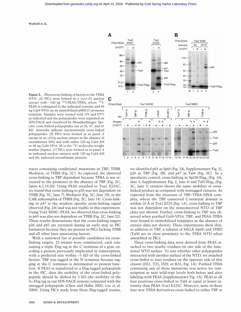
tracts containing conditional mutations in TBP, TFIIB,Mediator, or TFIIH (Fig. 2C). As expected, the observedcross-linking is TBP dependent because TFIIA is not re-cruited to the promoter in the absence of TBP (Fig. 2C,lanes 4,5,19,20). Using PEAS attached to Toa1 E235C,we found that cross-linking to p26 was not dependent onTFIIB (Fig. 2C, lane 7), Mediator (Fig. 2C, lane 10), or theCAK subcomplex of TFIIH (Fig. 2C, lane 14). Cross-link-ing to p47 is the weakest specific cross-linking signalobserved (Fig. 2A) and was not visible in this experiment.Using Toa2 M38C–PEAS, we observed that cross-linkingto p65 was also not dependent on TFIIB (Fig. 2C, lane 22).These results demonstrate that the cross-linking targetsp26 and p65 are recruited during an early step in PICformation because they are present in PICs lacking TFIIBand all other later associating factors.
With a narrowed list of possible candidates for cross-linking targets, 22 strains were constructed, each con-taining a triple Flag tag at the C terminus of a gene en-coding a protein previously implicated in transcription,with a predicted size within ∼5 kD of the cross-linkedfactors. TBP was tagged at the N terminus because tag-ging at the C terminus is detrimental to protein func-tion. If PEAS is transferred to a Flag-tagged polypeptidein the PIC, then the mobility of the cross-linked poly-peptide should be shifted by 5 kD (the mobility of the3×-Flag tag in our SDS-PAGE system) compared with theuntagged polypeptide (Chen and Hahn 2003; Liu et al.2004). Using PICs made from these Flag-tagged strains,
we identified p65 as Spt8 (Fig. 3A; Supplementary Fig. 2),p26 as TBP (Fig. 3B), and p47 as Taf4 (Fig. 3C). As aspecificity control, cross-linking in Spt20-Flag3 (Fig. 3A,lane 3; Supplementary Fig. 2, lane 4) and Taf5-Flag3 (Fig.3C, lane 3) extracts shows the same mobility of cross-linked product as compared with nontagged extracts. Asexpected from the structure of TBP–TFIIA–DNA com-plex, where the TBP conserved C-terminal domain iswithin 16 Å of Toa1 E235 (Fig. 1A), cross-linking to TBPwas not dependent on the nonconserved NTD of TBP(data not shown). Further, cross-linking to TBP was ob-served when purified Gal4-VP16, TBP, and PEAS–TFIIAwere bound to immobilized templates in the absence ofextract (data not shown). These experiments show that,in addition to TBP, a subunit of SAGA (Spt8) and TFIID(Taf4) are in close proximity to the TFIIA NTD whenassembled in PICs.
These cross-linking data were derived from PEAS at-tached to two nearby residues on one side of the func-tional NTD surface. To test whether other polypeptidesinteracted with another surface of the NTD, we attachedcross-linker to four residues on the opposite side of thiscluster (D21, T25, D29, or R31; Fig. 1A). Purified TFIIAcontaining any of these mutations was active for tran-scription at near wild-type levels both before and afterlabeling with PEAS (Supplementary Fig. 1A). PEAS at allfour positions cross-linked to Taf4 at equal or lesser in-tensity than PEAS–Toa1 E235C. However, none of thesefour new TFIIA derivatives cross-linked to either TBP or
Figure 2. Photocross-linking of factors to the TFIIANTD. (A) PICs were formed in a toa1–25 nuclearextract with ∼160 ng 125I-PEAS–TFIIA, where 125I-PEAS is conjugated to the indicated cysteine and 96ng Gal4-VP16 on an immobilized pSH515 promotertemplate. Samples were treated with UV and DTTas indicated and the polypeptides were separated onSDS-PAGE and visualized by PhosphorImager. Spe-cific cross-linked polypeptides run at 26, 47, and 65kD. Asterisks indicate inconsistently cross-linkedpolypeptides. (B) PICs were formed as in panel Aexcept in an srb2� nuclear extract in the absence ofrecombinant Srb2 and with either 120 ng Gal4-AHor 96 ng Gal4-VP16. M is the 14C molecular weightmarker (Sigma). (C) PICs were formed as in panel Ain indicated nuclear extracts with 120 ng Gal4-AHand the indicated recombinant proteins.
Warfield et al.
1026 GENES & DEVELOPMENT
Cold Spring Harbor Laboratory Press on April 13, 2018 - Published by genesdev.cshlp.orgDownloaded from

Spt8, or consistently to any other polypeptide (data notshown). These data approximate the position of Spt8 andTaf4 in the PIC. Numerous other Flag-tagged nuclearextracts were also tested in the cross-linking assay, noneof which displayed cross-linked polypeptides of shiftedmobility. Proteins of note that did not cross-link to theTFIIA NTD include Taf11, previously shown to interactwith the TFIIA NTD by two-hybrid assay (Kraemer et al.2001), and Spt3, which genetically interacts with Spt8and TBP in the SAGA complex (Eisenmann et al. 1994).
Mapping TFIIA interaction to Spt8by hydroxyl radical cleavage
To further explore the interaction of TFIIA and Spt8, wefirst mapped the region of Spt8 located near the TFIIANTD. To accomplish this, we used the site-specific pro-tein cleavage reagent FeBABE (Datwyler and Meares2000; Chen and Hahn 2003). In this strategy, the 12Å-long FeBABE reagent is attached to surface-exposed cys-teines, and these TFIIA derivatives are used to assemblePICs, as was described for the PEAS cross-linker. AfterPIC assembly, hydroxyl radicals are generated by addi-tion of sodium ascorbate and hydrogen peroxide, leadingto cleavage of the polypeptide backbone in nearby pro-teins. Because the hydroxyl radical can diffuse to a lim-ited extent, this reagent probes for proteins locatedwithin ∼15–20 Å of the Fe-EDTA center (Datwyler andMeares 2000; Chen and Hahn 2003).
FeBABE was conjugated to the same purified TFIIAcysteine mutants used in the cross-linking experiments.These TFIIA–FeBABE conjugates showed transcriptionactivity similar to that of wild-type TFIIA (Supplemen-tary Fig. 1B). PICs were formed by using an extract madefrom a strain containing N-terminal triple Flag-taggedSpt8. Cleavage products were visualized by Western blot,probing for the Flag epitope. Both full-length Spt8 andcleavage products containing an intact N terminus are
detected. Figure 4A shows that attempted FeBABE con-jugation to TFIIA with no surface-accessible cysteinesgenerates only full-length Flag3-Spt8 with no cleavageproducts (Fig. 4A, lanes 1,2). In contrast, FeBABE linkedto six positions on either Toa1 or Toa2 all give uniqueN-terminal cleavage products only when ascorbate andH2O2 are added (Fig. 4A, lanes 3–14; indicated by brack-ets). Although the cross-linking data showed that PEASat Toa2 M38C and Toa1 E235C cross-links to Spt8 butPEAS–Toa2 D21C, T25C, D29C, and R31C do not, thecleavage data show that FeBABE attachment at all sixTFIIA sites leads to Spt8 cleavage. This variation islikely due to the difference in the effective lengths of thetwo probes.
To accurately determine the location of the proteincleavage sites, we expressed a set of Flag3-Spt8 fragmentsby in vitro transcription and translation (SupplementaryFig. 3). The mobility of these in vitro translated productswas determined by SDS-PAGE, and this was used to cal-culate the sizes of the Flag3-Spt8 cleavage products fromFigure 4A. From multiple mapping experiments andfrom mapping some cleavage sites from both ends ofSpt8, we estimate that these cleavage sites are accurateto within ±5 amino acids. The Spt8 cleavage sites deter-mined by this method are shown in Figure 4B, with thelength of the line representing relative cleavage effi-ciency. The cleavage products from Figure 4A map to theC-terminal half of Spt8, and the more intense cleavageproducts are clustered near the C terminus. Supplemen-tary Table 1 summarizes the Toa1 and Toa2 cysteinemutations used in the FeBABE mapping, the residues onSpt8 cleaved by these mutants, and the intensity of theFlag3-Spt8 cleavage products produced. Although thesecleavage sites are spread over almost 300 residues, theirpositions are likely in close proximity in the 3D struc-ture of Spt8 (Chen and Hahn 2003).
We also attempted to map Taf4 interaction with TFIIAby the FeBABE cleavage assay; however, no cleavage
Figure 3. Spt8, Taf4, and TBP are identified as cross-linked polypeptides by using Flag-tagged nuclear extracts. (A–C) PICs formed asin Figure 2A except in the indicated nuclear extracts. Asterisks indicate inconsistently cross-linked polypeptides.
Interactions among SAGA, TFIIA, and TBP
GENES & DEVELOPMENT 1027
Cold Spring Harbor Laboratory Press on April 13, 2018 - Published by genesdev.cshlp.orgDownloaded from

products were observed (data not shown). One possibleexplanation for this result is that FeBABE cleavage ofFlag3-Taf4 occurs very near the N or C terminus of Taf4.Cleavage very near the N terminus would generate frag-ments too small to observe by SDS-PAGE, and cleavagevery near the C terminus would generate fragments toolarge to separate from full-length Flag3-Taf4. Addition-ally, because cross-linking of TFIIA to Spt8 is muchstronger than to Taf4, the NTD of TFIIA is almost cer-tainly closer to Spt8 in the PIC compared with Taf4.
Genetic interaction between TFIIA, Spt8, and Spt3
To independently confirm the interaction of TFIIA andSpt8, we tested whether these two factors interact ge-netically. In this strategy, a strain containing a doublemutation in both the TFIIA NTD and Spt8 was tested foran in vivo phenotype that was stronger than the pheno-type of either single mutation. We used a similar assay totest for genetic interaction between Spt3 (genetically re-lated to Spt8) and the TFIIA NTD. Yeast strains contain-ing an spt8� and either wild-type TOA2, toa2 L23A, or
toa2 D24L were constructed and tested for growth onrich media at 25°C and 37°C. Figure 5 shows that neitherSPT3 nor SPT8 deletions display a growth phenotype un-der these conditions. In contrast, both TFIIA NTD mu-tants show temperature-sensitive growth at 37°C. Whenthese TFIIA NTD mutations are combined with eitherspt3� or spt8� deletions, the strains display a mild slowgrowth phenotype at 25°C and a stronger temperature-sensitive phenotype at 37°C compared with the NTDmutations alone. These results demonstrate a geneticinteraction between the NTD of TFIIA and the twoSAGA subunits Spt3 and Spt8 and are consistent withprevious findings of a genetic interaction between TFIIAand Spt3 (Madison and Winston 1997).
Spt8 and TFIIA compete for binding to TBP
One model to explain these data is that the C terminusof Spt8 binds to the TFIIA NTD, providing a direct con-nection between SAGA and the general transcriptionfactors. This model was tested directly by using recom-binant TFIIA, TBP, and Spt8 (Fig. 6A). Purified GST orGST-Spt8 was bound to glutathione Sepharose beads andincubated with TBP, TFIIA, or both TBP and TFIIA. Nosignificant amount of TBP or TFIIA bound to GST alone(Fig. 6A, lanes 5,7,9). TBP bound strongly to GST-Spt8,consistent with previous findings that SAGA binding toTBP was dependent on Spt8 (Fig. 6A, lanes 6,8; Sterner etal. 1999). TBP also bound a C-terminal construct of Spt8(residues 265–602) to the same extent as full-length Spt8(data not shown). Surprisingly, TFIIA did not bind to ei-ther full-length GST-Spt8 or GST-Spt8 (265–602) (Fig. 6A,lanes 8,10; data not shown). Strikingly, when both TBP
Figure 5. Genetic interaction between Spt8, Spt3, and TFIIA.Strains were grown in YPD + adenine overnight at 30°C anddiluted to 5 × 10⁷ cells/mL, and 3.5 µL of 10-fold dilutions werespotted to YPD + adenine plates and grown for 2 d at 25°C or37°C.
Figure 4. Determination of TFIIA–FeBABE cleavage sites inSpt8. (A) Western blot analysis of TFIIA–FeBABE cleavage ofFlag3-Spt8 probed with �-Flag. PICs were formed as in Figure 2Aexcept in a Flag3-Spt8 nuclear extract with ∼200 ng TFIIA con-jugated with FeBABE on the indicated cysteines. TFIIA with nosurface cysteines after attempted FeBABE conjugation is indi-cated by “WT”. Hydroxyl radical cleavage was initiated withthe addition of Na Ascorbate and H2O2 where indicated. Brack-ets indicate cleavage products and asterisks indicate nonspecificsignals. (B) Map of Spt8 with the TFIIA–FeBABE cleavage siteslisted in Supplementary Table 1. The length of the line indicat-ing each cleavage site corresponds to the intensity of the bandfrom panel A. Positions of the acidic domain and the WD-40repeat are indicated.
Warfield et al.
1028 GENES & DEVELOPMENT
Cold Spring Harbor Laboratory Press on April 13, 2018 - Published by genesdev.cshlp.orgDownloaded from

and either a twofold or sevenfold excess of TFIIA werepresent in the reaction, TFIIA was found to inhibit TBPbinding to GST-Spt8 (Fig. 6A, lanes 6,8; data not shown).Using known amounts of TBP to quantitate TBP binding(Fig. 6A, lanes 1–3), we found that TFIIA inhibited TBPbinding to Spt8 13-fold under these in vitro conditions.In similar assays, no binding was detected when Spt3-His6 was tested for binding to GST-Spt8, suggesting Spt3and Spt8 do not interact directly under these conditions(data not shown).
To test whether the competition between Spt8 andTFIIA was specific for TFIIA, we tested whether TFIIB,another TBP-interacting factor, would compete withSpt8 for TBP binding (Fig. 6B). TBP with or without asevenfold molar excess of yeast TFIIB was incubatedwith GST-Spt8 beads as described earlier, washed, andanalyzed for factors binding to the immobilized Spt8beads. TBP interacting with Spt8 clearly bound TFIIB,and this Spt8 binding was not inhibited by TFIIB (Fig. 6B,lanes 2,3). In contrast, a sevenfold excess of TFIIAstrongly inhibited TBP binding to Spt8 and this inhibi-
tion was not affected by mutations in the TFIIA NTDthat inhibit the positive function of the NTD. Fromthese experiments, we conclude that inhibition of Spt8binding is specific to TFIIA and that the inhibitory sur-face of TFIIA is not limited to one face of the TFIIANTD.
Although these results show that purified TFIIA andSpt8 compete for binding to TBP, cross-linking and otherbiochemical results shown earlier demonstrate that TBP,Spt8, and TFIIA are simultaneously present and are inclose proximity within the PIC. Together, these resultssuggest that the presence of Spt8 in the SAGA complexmay reduce the affinity of SAGA binding to the generaltranscription factors because of competition with TFIIA.To test this model, we assayed whether deletion of Spt8results in more efficient binding of SAGA to the PIC.One complication in testing this model is that in vivostudies have shown that SAGA can be recruited to pro-moters via interaction with the activators GAL4 andVP16 independently of the transcription machinery(Bhaumik and Green 2001). Binding to VP16 is also in-dependent of the general factors in the in vitro transcrip-tion system (Fig. 7A). A low amount of SAGA is found inPICs formed in the absence of any activator (Fig. 7A, lane1), and recruitment of SAGA is greatly stimulated byGal4-VP16 binding to a single site upstream of the core
Figure 6. Spt8 competes with TFIIA for binding to TBP. (A)Purified GST or GST-Spt8 bound to glutathione sepharose beadswas incubated with recombinant full-length TBP and/or a sev-enfold excess of TFIIA. After separation by SDS-PAGE, theWestern blot was probed with �-TBP and �-Toa2. Knownamounts of purified TBP and TFIIA were run in lanes 1–4. GSTand GST-Spt8 were eluted, separated by SDS-PAGE, and Coo-massie stained to confirm that similar amounts of GST andGST-Spt8 were bound to the beads (data not shown). (B) Purifiedfull-length TBP and a sevenfold excess of either TFIIB, wild-typeTFIIA, or TFIIA NTD mutants were incubated with GST orGST-Spt8 as indicated earlier, then bound protein was analyzedby Western blot with �-TBP and �-TFIIB as described in panel A.The TFIIA point mutations indicated are each in the Toa2 sub-unit. TFIIB does not significantly bind to GST beads (notshown).
Figure 7. Interaction of SAGA with activator and the basaltranscription machinery. (A) Western blot of PICs formed in WTnuclear extracts on either immobilized pSH515 template orpSH515�P template, which contains 150 bp of nonpromotersequence and a single Gal4 binding site. Either no activator orGal4-VP16 was added as indicated. (B) Western blot of PICsformed in the indicated nuclear extracts with no activator.
Interactions among SAGA, TFIIA, and TBP
GENES & DEVELOPMENT 1029
Cold Spring Harbor Laboratory Press on April 13, 2018 - Published by genesdev.cshlp.orgDownloaded from

promoter (Fig. 7A, lane 2). Consistent with in vivo re-sults, the stimulation of recruitment by VP16 does notrequire an intact core promoter. When an immobilizedtemplate containing the activator binding site but lack-ing the core promoter is used (515�P), the TFIID-specificsubunits Taf1 and Taf4, as well as TBP and TFIIA, arefound at lower levels compared with the intact promoter(Fig. 7A, lanes 2,3). In contrast, SAGA-specific subunitsAda1, Ada2, or Taf12 (shared between TFIID and SAGA)are insensitive to promoter deletion, suggesting thatSAGA binds to the activator independently of both thegeneral factors and promoter DNA. Consistent with thisconclusion, SAGA is recruited to the immobilized tem-plate in an activator-dependent fashion in extracts lack-ing functional TBP that are blocked in the first step inPIC assembly (Yudkovsky 2001).
To assay the direct interaction of SAGA with the gen-eral factors, we monitored SAGA binding to PICs in theabsence of any added activator. Levels of the SAGA-spe-cific subunit Ada1 and the TFIID/SAGA subunit Taf12present in the PIC were compared in wild-type, spt3�,and spt8� extracts by quantitation of Western blot sig-nals (Fig. 7B). As predicted earlier, SAGA lacking theSpt8 subunit was recruited to the PIC an average of 3.3-fold better compared with wild-type extracts in threeseparate experiments. The level of Taf12, a shared sub-unit between SAGA and TFIID, was also increased in thespt8� extract. In contrast, all other non-SAGA subunitstested showed no reproducible change when all threeextracts were compared. These results demonstrate thatwithin the SAGA complex, Spt8 has a negative effect onSAGA interaction with the general factors under basaltranscription conditions. This is consistent with the pre-vious finding that Spt8 inhibits basal expression fromthe HIS3 and TRP3 promoters in vivo (Belotserkovskayaet al. 2000). In contrast, SAGA lacking Spt3 was re-cruited slightly less well, with an average twofold lowerAda1 level in PICs compared with wild type (Fig. 7B,lanes 1,2).
The positive function of the TFIIA NTDis mediated through Spt8
In addition to repression of basal expression, Spt8 alsopositively regulates transcription at some SAGA-depen-dent promoters (Bhaumik and Green 2001; Larschan andWinston 2001). To test whether the positive function ofthe TFIIA NTD is mediated through Spt8, we deter-mined the effect of the TFIIA NTD mutations in a strainlacking Spt8. Extracts from a spt8� strain were found tobe about threefold deficient for multiround transcription(Fig. 8A, lanes 21,22), confirming the positive role of Spt8in the in vitro system. If the positive function of theTFIIA NTD is mediated through Spt8, then the activityof the NTD mutants should be comparable to wild-typeTFIIA in a spt8� extract. TFIIA was depleted from thespt8� extract and this depleted extract was supple-mented by adding recombinant wild-type TFIIA or theTFIIA mutants Toa2 D24L or V20R. As shown in Figure1C, these TFIIA mutations result in a twofold decreasein the rate of PIC formation in an otherwise wild-typeextract. In contrast, both TFIIA mutants have activity ator near the level of wild-type TFIIA in the spt8� extract(Fig. 8). Comparing the initial rate of PIC formation dur-ing the first 20 min, we found that the TFIIA mutantsToa2 D24L and V20R support a rate of PIC formationequal to or within 80% of wild-type, respectively. Thisdemonstrates that the positive function of the TFIIANTD is largely mediated through Spt8 in the in vitrosystem. In contrast, complete lack of TFIIA still resultedin a low rate of PIC formation in the spt8� strain. Thisresult is consistent with the importance of the TFIIAC-terminal domain in other functions such as stabiliza-tion of TBP–DNA binding and competition with inhibi-tors of TBP function.
Discussion
SAGA is an important cofactor for activation of a subsetof genes in yeast. At SAGA-dependent promoters, SAGA
Figure 8. The function of the TFIIA NTD in enhanc-ing the rate of PIC formation is mediated largelythrough Spt8. (A, left panel) Single-round transcriptionassayed by primer extension. PICs were formed for theindicated times by using 90 µg of spt8�, Toa2-Flag3
nuclear extract, which was depleted of TFIIA by using�-Flag M2 agarose (Sigma) and supplemented with asaturating amount (20 ng) of recombinant TFIIA. NTPswere added and reactions stopped after 2 min to allowfor a single round of transcription. (Right panel) Multi-round transcription assayed by primer extension by us-ing 90 µg of either wild-type extract or the spt8� extractdepleted for TFIIA as described earlier and supple-mented with 20 ng purified “wild-type” TFIIA. All pu-rified TFIIA proteins used in this experiment have thesurface cysteines eliminated (Toa2 C72V and C116S),and display wild-type in vitro transcription activity(Supplementary Fig. 1A). (B) Quantitation of data pre-sented in the left side of panel A. Transcription activityat each time point is given as a percent of wild-typetranscription with PICs formed for 40 min.
Warfield et al.
1030 GENES & DEVELOPMENT
Cold Spring Harbor Laboratory Press on April 13, 2018 - Published by genesdev.cshlp.orgDownloaded from

appears to be recruited directly to the activator, indepen-dently of the transcription machinery. Mutations inSAGA subunits such as Spt3 or Spt8 do not affect therecruitment of SAGA to the activator, but can block sub-sequent recruitment of the general transcription factors(Bhaumik and Green 2001; Larschan and Winston 2001).SAGA was also shown to repress basal expression fromspecific promoters in yeast, mediated by the Spt3 andSpt8 subunits. The mechanism by which SAGA inter-acts with the general factors has been unclear because ofconflicting genetic and biochemical data. Here, we havedemonstrated an unexpected connection between Spt8,TBP, and the NTD of TFIIA. When assembled into PICs,site-specific photocross-linking shows that Spt8 is clos-est to the functional surface of the TFIIA NTD. ThisNTD surface is also near the TFIID-specific subunitTaf4, but the observed weak cross-linking signal sug-gests that Taf4 is located near the limit of the 16Å-probedistance and therefore may not directly interact with theTFIIA NTD. The dependence of Spt8-NTD cross-linkingon TBP demonstrates that in PICs, TBP, TFIIA, and Spt8are all in close proximity. No cross-linking was observedbetween the TFIIA NTD and Taf11, which were previ-ously proposed to interact on the basis of the two-hybridassay and two mutations in the TFIIA NTD that blockedthis two-hybrid interaction (Kraemer et al. 2001). In con-trast, our results suggest that the TFIIA NTD and Taf11do not normally interact when assembled into PICs.
FeBABE mapping showed that at least 16 residuesspread throughout the C-terminal half of Spt8 are nearthe TFIIA NTD when assembled in PICs. Although thestructure of Spt8 is not yet known, it is likely that theseFeBABE reactive residues are located in close proximityin the native Spt8 structure. The C-terminal half of Spt8also bound TBP with the same affinity as full-lengthSpt8. Together, these findings suggest that the C-termi-nal half of Spt8 forms a domain that interacts with bothTBP and the TFIIA NTD.
Given the finding that TFIIA, TBP, and Spt8 are all inclose proximity in PICs, it was surprising that experi-ments that used purified proteins showed that Spt8 andTFIIA do not stably bind. In contrast, it was observedthat both Spt8 and TFIIA compete for binding to TBP. Onthe basis of the cross-linking results showing that TBP,Spt8, and TFIIA are simultaneously present in PICs, wepropose that Spt8 and TFIIA do not bind to the samelocation on the surface of TBP, but rather sterically in-terfere with each other when each interacts with itsbinding site on TBP. This work using purified proteins issupported by studies using the intact SAGA complex inwhich SAGA lacking Spt8 bound more efficiently thanwild-type SAGA to a PIC. Also consistent with our re-sults is the finding that deletion of SPT8 (or SPT3) cancause derepression of basal expression from a subset ofgenes in vivo such as HIS3 and TRP3 (Belotserkovskayaet al. 2000).
We have also shown that the positive function of theTFIIA NTD surface is largely mediated through Spt8 invitro. The TFIIA NTD was demonstrated to play a role inthe rate of PIC formation, because extracts with TFIIA
NTD mutations were found to form PICs with a twofoldlower rate compared with wild-type extracts. Extractslacking Spt8 were found to have about a threefold defectfor transcription in vitro, and in these extracts TFIIANTD mutants and wild-type TFIIA gave nearly equiva-lent rates of PIC formation. This result is consistentwith a mechanism where the function of the TFIIA NTDis dependent on Spt8 activity. Together, our findingsshow that Spt8 can have both a positive and a negativefunction on transcription, mediated through TFIIA.
How can the competitive interaction between TFIIAand Spt8 lead to positive effects on gene expression? Onemodel to explain these results is that during activatedtranscription, the interaction between the TFIIA NTDand Spt8 causes a conformational change leading to amore active state (Fig. 9). This conformational changewould be driven by the binding energy derived from in-teraction of TFIIA and Spt8 with TBP as well as by otherpresumed protein–protein interactions between SAGA,activator, and the transcription machinery. It is proposedthat Spt8 and/or TFIIA adopts a structure that is moreefficient at promoting transcription in this altered con-formation. Under basal or unactivated conditions atSpt8-dependent promoters, this proposed conforma-tional change would not occur efficiently, leading to therepression of basal expression observed in vivo. Such amodel would explain the observed genetic interactionbetween TFIIA and Spt8 where mutation in the TFIIANTD combined with an spt8� has a more severe pheno-type than either single mutation. We postulate that eachfactor alone has an independent positive function suchas promotion of TBP/TFIID binding or coactivator func-tion. Together, both factors cooperate to generate a moreactive state of the transcription machinery, explainingwhy the phenotype of the double mutation is the mostsevere. Although this model is unusual, the direct photo-cross-linking assay can rule out more conventional com-
Figure 9. Proposed model of interactions among TFIIA, Spt8,and TBP. TFIIA and Spt8 each bind individually to TBP; how-ever, they compete for TBP binding under basal transcriptionconditions. Because the photocross-linking results indicate thatall three proteins are simultaneously present in the PIC underactivated transcription conditions, a conformational changemay occur in Spt8 and/or TFIIA, allowing both Spt8 and TFIIAto stably bind TBP and leading to transcriptional stimulation.
Interactions among SAGA, TFIIA, and TBP
GENES & DEVELOPMENT 1031
Cold Spring Harbor Laboratory Press on April 13, 2018 - Published by genesdev.cshlp.orgDownloaded from

petition mechanisms whereby PICs are assembled invitro with either TFIIA or Spt8.
The regulation of transcription by Spt8 almost cer-tainly does not happen at all promoters, because only asubset of promoters is SAGA dependent (Lee et al. 2000).In addition, SLIK/SALSA, a SAGA-related complex,lacks the Spt8 subunit altogether and contains at leastone unique subunit, Rtg2 (Pray-Grant et al. 2002; Sterneret al. 2002; Wu and Winston 2002). Under our in vitroconditions, we do not detect significant levels of Rtg2 inPICs formed at the HIS4 promoter (G. Rani and S. Hahn,unpubl.). At some promoters in vivo, it is also possiblethat Spt8 plays a strictly negative role (Belotserkovskayaet al. 2000), an issue that could be addressed by genome-wide expression assays. Finally, similar mechanisms in-volving both positive and negative protein–protein inter-actions may partly explain why other TBP interactingfactors, such as NC2 and Mot1, inhibit assembly of thePIC with purified factors but have been shown to actpositively on a subset of genes in vivo (Pereira et al.2003).
Materials and methods
Yeast strains and nuclear extracts
Strains used in this study were as follows: BY4705 (wild type;Brachmann et al. 1998); SHY193 Mata ura3 lys2 ade2 trp1 his3leu2 toa2��LYS2/pSH342 (ARS CEN URA3 TOA2); SHY194,same as SHY193 but Mat�; SHY195 Mata ura3 lys2 ade2 trp1his3 leu2 toa1��LYS2/pSH325 (ARS CEN URA3 TOA1);SHY196, same as SHY195 but Mat�; SHY109 (toa1–25ts; Kanget al. 1995); SHY70 (TBP I143N ts; Reddy and Hahn 1991);SHY245 (sua7 G41E cs; Ranish et al. 1999); SHY202 (Z525)srb2� (Ranish et al. 1999); kin28-ts16 (Cismowski et al. 1995).LHY78, LHY79, SHY519, and SHY512 contain triple Flag tags atthe C terminus of Spt20, Spt8, Taf5, and Taf4, respectively.LHY99 contains a triple Flag tag at the N terminus of TBP.spt8�, spt3�, and ada1� strains are in the BY4741 backgroundfrom Research Genetics; LHY4 and LHY10, respectively, con-tain toa2 D24L and toa2 L23A integrated at the TOA2 locus inBY4705. LHY117 (toa2 D24L, spt8�), LHY118 (toa2 D24L,spt3�), LHY119 (toa2 L23A, spt8�), and LHY120 (toa2 L23A,spt3�) were constructed by mating and sporulation of LHY4 orLHY10 with spt8� or spt3� strains. Yeast nuclear extracts wereprepared according to the method described at http://www.fhcrc.org/labs/hahn.
TFIIA mutagenesis and purification
TFIIA mutations for the phenotype screen were generated bysite-directed mutagenesis of pSH343 (2 kb PstI TOA2 fragmentin pRS315) or pSH363 (∼2.2 kb BamHI/XhoI TOA1 fragment inpRS315). TOA1 and TOA2 mutants were checked for pheno-type on minimal media by plasmid shuffle assay. Cysteine sub-stitutions in Toa2 and Toa1 for PEAS and FeBABE attachmentwere made in pSH343 or pSH363. All TFIIA cysteine mutantsalso had two naturally occurring surface cysteines on Toa2eliminated (C72V and C116S). Phenotypes of all mutations werechecked by plasmid shuffle assay. TFIIA expression and purifi-cation are described at http://www.fhcrc.org/labs/hahn.
PEAS and FeBABE attachment to TFIIA
One hundred fifty micrograms (200–400 µL) of purified TFIIAwas exchanged into labeling buffer (30 mM Tris, 10% glycerol,
200 mM KCl at pH 7.4) by using NAP-5 columns (Amersham)and concentrated to ∼30 µL by using Ultrafree Biomax4-10Kconcentrators (Millipore). The photocross-linker PEAS {N-[(2-pyridyldithio)ethyl]-4-azidosalicylamide; Molecular Probes} waslabeled with 125I (350–600 mCi/mL; Amersham) in Iodogentubes (Pierce) containing 110 µL 0.1 M Na Phosphate (pH 7.4),11 nmole PEAS, and ∼1.5 mCi 125I per 150 µg TFIIA. After 5min, the reaction was transferred to a tube containing 100nmole tyrosine per TFIIA labeling to scavenge unreacted 125I.This mixture was supplemented with 150 µg TFIIA and allowedto react for 30–45 min at 23°C. 125I-PEAS–TFIIA was purifiedfrom unreacted 125I-PEAS by buffer exchange on a NAP-5 col-umn into 1 mL fresh labeling buffer. Recovery was ∼100 µg withan activity of 6–11 µCi/µg TFIIA, whereas control TFIIA withno surface cysteines had an activity of ∼0.5 µCi/µg TFIIA afterattempted labeling. Labeled TFIIA was immediately aliquotedand stored at −70°C. TFIIA was shown to be labeled on thecorrect subunit by SDS-PAGE followed by autoradiography. Asexpected, PEAS was cleaved from TFIIA by addition of 50 mMDTT.
FeBABE [iron(S)-1-(p-bromoacetamido-benzyl) EDTA; Do-jindo] was attached to TFIIA in the same manner as I25I-PEAS,except 135 µg TFIIA was exchanged into conjugation buffer (10mM MOPS, 200 mM NaCl, 2 mM EDTA, 5% glycerol at pH8.0), concentrated to ∼100 µL, and reacted with a 20-fold molarexcess of FeBABE for 4 h at 23°C. The TFIIA–FeBABE was pu-rified from unreacted FeBABE by exchange into TFIIA storagebuffer (30 mM Tris, 200 mM KCl, 2 mM EDTA, 10% glycerol atpH 7.5) and immediately aliquoted and stored at −70°C.
Pol II immobilized template, photocross-linking,and FeBABE cleavage assays
Immobilized template assays for PIC formation, photocross-linking, and FeBABE cleavage reactions were performed as pre-viously described (Chen and Hahn 2003) except that 240–480 µgyeast nuclear extract was used depending on the extract (ex-tracts were dialyzed to remove DTT for photocross-linking)and ∼160 ng of either 125I-PEAS–TFIIA or FeBABE–TFIIA wasadded. The molecular sizes of Flag3-Spt8 fragments generated byFeBABE cleavage were determined as previously described(Chen and Hahn 2003), using a set of in vitro translated Flag3-Spt8 constructs. Proteins were visualized and quantitated byWestern blot by using the LI-COR Bioscience Odyssey Infraredimaging system.
GST pull-down assay
One microgram GST or GST-Spt8 protein was bound per 5 µLglutathione Sepharose beads in acetate transcription buffer (100mM KOAc, 100 mM HEPES, 1 mM EDTA, 5 mM MgOAc at pH7.6), 160 µg/ml PMSF, 50 µg/ml BSA, 0.05% NP-40 for 1 h at4°C, then washed three times. Each pull-down reaction con-tained 25 µL GST or GST-Spt8 beads, 1 µg full-length TBP,and/or 25 µg TFIIA in a total volume of 400 µL of the buffer.Reactions were rotated for 1.5 h at 4°C, washed three times, andthen resuspended in 20 µL 1× NuPAGE LDS sample buffer (In-vitrogen) and proteins were eluted at 95°C for 5 min. Proteinswere visualized by Western blot.
Acknowledgments
We thank S. Hurst for initial work on mutagenesis of TFIIA, A.Movius and H-T. Chen for help with development of photo-cross-linking and FeBABE assays, and B. Moorefield for help
Warfield et al.
1032 GENES & DEVELOPMENT
Cold Spring Harbor Laboratory Press on April 13, 2018 - Published by genesdev.cshlp.orgDownloaded from

with in vitro transcription/translation. We thank H-T. Chen, N.Mohibullah, and G. Rani for nuclear extracts; F. Winston, T.Weil, M. Green, and J. Reese for plasmids and antibodies; D.Gottschling for discussion and advice on genetic analysis ofTFIIA; and B. Moorefield and W. Reeves for comments on themanuscript. This work was supported by a grant from the NIH.S.H. is an associate investigator of the Howard Hughes MedicalInstitute.
The publication costs of this article were defrayed in part bypayment of page charges. This article must therefore be herebymarked “advertisement” in accordance with 18 USC section1734 solely to indicate this fact.
References
Agalioti, T., Lomvardas, S., Parekh, B., Yie, J., Maniatis, T., andThanos, D. 2000. Ordered recruitment of chromatin modi-fying and general transcription factors to the IFN-beta pro-moter. Cell 103: 667–678.
Auble, D.T., Hansen, K.E., Mueller, C.G.F., Lane, W.S., Thor-ner, J., and Hahn, S. 1994. Mot1, a global repressor of RNApolymerase II transcription, inhibits TBP binding to DNA byan ATP-dependent mechanism. Genes & Dev. 8: 1920–1934.
Belotserkovskaya, R., Sterner, D.E., Deng, M., Sayre, M.H., Lie-berman, P.M., and Berger, S.L. 2000. Inhibition of TATA-binding protein function by SAGA subunits Spt3 and Spt8 atGcn4-activated promoters. Mol. Cell. Biol. 20: 634–647.
Bhaumik, S.R. and Green, M.R. 2001. SAGA is an essential invivo target of the yeast acidic activator Gal4p. Genes & Dev.15: 1935–1945.
———. 2002. Differential requirement of SAGA components forrecruitment of TATA-box-binding protein to promoters invivo. Mol. Cell. Biol. 22: 7365–7371.
Brachmann, C.B., Davies, A., Cost, G.J., Caputo, E., Li, J., Hie-ter, P., and Boeke, J.D. 1998. Designer deletion strains de-rived from Saccharomyces cerevisiae S288C: A useful set ofstrains and plasmids for PCR-mediated gene disruption andother applications. Yeast 14: 115–132.
Cang, Y., Auble, D.T., and Prelich, G. 1999. A new regulatorydomain on the TATA-binding protein. EMBO J. 18: 6662–6671.
Chen, H.T. and Hahn, S. 2003. Binding of TFIIB to RNA poly-merase II: Mapping the binding site for the TFIIB zinc ribbondomain within the preinitiation complex. Mol. Cell 12: 437–447.
Chen, Y., Ebright, Y.W., and Ebright, R.H. 1994. Identificationof the target of a transcription activator protein by protein–protein photocrosslinking. Science 265: 90–92.
Cheng, J.X., Floer, M., Ononaji, P., Bryant, G., and Ptashne, M.2002. Responses of four yeast genes to changes in the tran-scriptional machinery are determined by their promoters.Curr. Biol. 12: 1828–1832.
Chi, T., Lieberman, P., Ellwood, K., and Carey, M. 1995. A gen-eral mechanism for transcriptional synergy by eukaryoticactivators. Nature 377: 254–257.
Cismowski, M.J., Laff, G.M., Solomon, M.J., and Reed, S.I. 1995.KIN28 encodes a C-terminal domain kinase that controlsmRNA transcription in S. cerevisiae but lacks cyclin-depen-dent kinase-activating kinase (CAK) activity. Mol. Cell. Biol.15: 2983–2992.
Cosma, M.P. 2002. Ordered recruitment: Gene-specific mecha-nism of transcription activation. Mol. Cell 10: 227–236.
Datwyler, S.A. and Meares, C.F. 2000. Protein–protein interac-tions mapped by artificial proteases: Where � factors bind toRNA polymerase. Trends Biochem. Sci. 25: 408–414.
Dudley, A.M., Rougeulle, C., and Winston, F. 1999. The Sptcomponents of SAGA facilitate TBP binding to a promoterat a post-activator-binding step in vivo. Genes & Dev. 13:2940–2945.
Eisenmann, D.M., Arndt, K.M., Ricupero, S.L., Rooney, J.W.,and Winston, F. 1992. SPT3 interacts with TFIID to allownormal transcription in S. cerevisiae. Genes & Dev. 6: 1319–1331.
Eisenmann, D.M., Chapon, C., Roberts, S.M., Dollard, C., andWinston, F. 1994. The Saccharomyces cerevisiae SPT8 geneencodes a very acidic protein that is functionally related toSPT3 and TATA-binding protein. Genetics 137: 647–657.
Geiger, J.H., Hahn, S., Lee, S., and Sigler, P.B. 1996. Crystalstructure of the yeast TFIIA/TBP/DNA complex. Science272: 830–836.
Grant, P.A., Schieltz, D., Pray-Grant, M.G., Steger, D.J., Reese,J.C., Yates III, J.R., and Workman, J.L. 1998. A subset ofTaf(II)s are integral components of the SAGA complex re-quired for nucleosome acetylation and transcriptionalstimulation. Cell 94: 45–53.
Hampsey, M. 1998. Molecular genetics of the RNA polymeraseII general transcriptional machinery. Microbiol. Mol. Biol.Rev. 62: 465–503.
Kang, J.J., Auble, D.T., Ranish, J.A., and Hahn, S. 1995. Analysisof the yeast transcription factor TFIIA: Distinct functionalregions and a polymerase II-specific role in basal and acti-vated transcription. Mol. Cell. Biol. 15: 1234–1243.
Kobayashi, N., Horn, P.J., Sullivan, S.M., Triezenberg, S.J.,Boyer, T.G., and Berk, A.J. 1998. DA-complex assembly ac-tivity required for VP16C transcriptional activation. Mol.Cell. Biol. 18: 4023–4031.
Kokubo, T., Swanson, M.J., Nishikawa, J.I., Hinnebusch, A.G.,and Nakatani, Y. 1998. The yeast Taf145 inhibitory domainand TFIIA competitively bind to TATA-binding protein.Mol. Cell. Biol. 18: 1003–1012.
Kraemer, S.M., Ranallo, R.T., Ogg, R.C., and Stargell, L.A. 2001.TFIIA interacts with TFIID via association with TATA-bind-ing protein and Taf40. Mol. Cell. Biol. 21: 1737–1746.
Kuras, L. and Struhl, K. 1999. Binding of TBP to promoters invivo is stimulated by activators and requires Pol II holoen-zyme. Nature 399: 609–613.
Larschan, E. and Winston, F. 2001. The S. cerevisiae SAGAcomplex functions in vivo as a coactivator for transcriptionalactivation by Gal4. Genes & Dev. 15: 1946–1956.
Lee, T.I., Causton, H.C., Holstege, F.C., Shen, W.C., Hannett,N., Jennings, E.G., Winston, F., Green, M.R., and Young,R.A. 2000. Redundant roles for the TFIID and SAGA com-plexes in global transcription. Nature 405: 701–704.
Li, X.Y., Virbasius, A., Zhu, X., and Green, M.R. 1999. Enhance-ment of TBP binding by activators and general transcriptionfactors. Nature 399: 605–609.
Lieberman, P.M. and Berk, A.J. 1994. A mechanism for Tafs intranscriptional activation: Activation domain enhancementof TFIID–TFIIA–promoter DNA complex formation. Genes& Dev. 9: 995–1006.
Liu, Q., Gabriel, S.E., Roinick, K.L., Ward, R.D., and Arndt,K.M. 1999. Analysis of TFIIA function in vivo: Evidence fora role in TATA-binding protein recruitment and gene-spe-cific activation. Mol. Cell. Biol. 19: 8673–8685.
Liu, Y., Kung, C., Fishburn, J., Ansari, A.Z., Shokat, K.M., andHahn, S. 2004. Two cyclin-dependent kinases promote RNApolymerase II transcription and formation of the ScaffoldComplex. Mol. Cell. Biol. 24: 1721–1735.
Madison, J.M. and Winston, F. 1997. Evidence that Spt3 func-tionally interacts with Mot1, TFIIA, and TBP to confer pro-moter-specific transcriptional control in S. cerevisiae. Mol.
Interactions among SAGA, TFIIA, and TBP
GENES & DEVELOPMENT 1033
Cold Spring Harbor Laboratory Press on April 13, 2018 - Published by genesdev.cshlp.orgDownloaded from

Cell. Biol. 17: 287–295.Mencia, M., Moqtaderi, Z., Geisberg, J.V., Kuras, L., and Struhl,
K. 2002. Activator-specific recruitment of TFIID and regula-tion of ribosomal protein genes in yeast. Mol. Cell 9: 823–833.
Orphanides, G., Lagrange, T., and Reinberg, D. 1996. The gen-eral transcription factors of RNA polymerase II. Genes &Dev. 10: 2657–2683.
Ozer, J., Moore, P.A., Bolden, A.H., Lee, A., Rosen, C.A., andLieberman, P.M. 1994. Molecular cloning of the small (g)subunit of human TFIIA reveals functions critical for acti-vated transcription. Genes & Dev. 8: 2324–2335.
Ozer, J., Lezina, L.E., Ewing, J., Audi, S., and Lieberman, P.M.1998a. Association of transcription factor IIA with TBP isrequired for transcriptional activation of a subset of promot-ers and cell cycle progression in S. cerevisiae. Mol. Cell. Biol.18: 2559–2570.
Ozer, J., Mitsouras, K., Zerby, D., Carey, M., and Lieberman,P.M. 1998b. Transcription factor IIA derepresses TATA-binding protein (TBP)-associated factor inhibition of TBP–DNA binding. J. Biol. Chem. 273: 14293–14300.
Pereira, L.A., Klejman, M.P., and Timmers, H.T. 2003. Roles forBTaf1 and Mot1p in dynamics of TATA-binding protein andregulation of RNA polymerase II transcription. Gene 315: 1–13.
Pray-Grant, M.G., Schieltz, D., McMahon, S.J., Wood, J.M.,Kennedy, E.L., Cook, R.G., Workman, J.L., Yates III, J.L., andGrant, P.A. 2002. The novel SLIK histone acetyltransferasecomplex functions in the yeast retrograde response pathway.Mol. Cell. Biol. 22: 8774–8786.
Ranish, J.A., Yudkovsky, N., and Hahn, S. 1999. Intermediatesin formation and activity of the RNA polymerase II preini-tiation complex: Holoenzyme recruitment and a postrecruit-ment role for the TATA box and TFIIB. Genes & Dev.13: 49–63.
Reddy, P. and Hahn, S. 1991. Dominant negative mutations inyeast TFIID define a bipartite DNA-binding region. Cell65: 349–357.
Saleh, A., Lang, V., Cook, R., and Brandl, C.J. 1997. Identifica-tion of native complexes containing the yeast coactivator/repressor proteins NGG1/ADA3 and ADA2. J. Biol. Chem.272: 5571–5578.
Sanders, S.L., Jennings, J., Canutescu, A., Link, A.J., and Weil,P.A. 2002. Proteomics of the eukaryotic transcription ma-chinery: Identification of proteins associated with compo-nents of yeast TFIID by multidimensional mass spectrom-etry. Mol. Cell. Biol. 22: 4723–4738.
Shen, W.-C. and Green, M.R. 1997. Yeast TafII145 functions asa core promoter selectivity factor, not a general coactivator.Cell 90: 615–624.
Stargell, L.A., Moqtaderi, Z., Dorris, D.R., Ogg, R.C., and Struhl,K. 2000. TFIIA has activator-dependent and core promoterfunctions in vivo. J. Biol. Chem. 275: 12374–12380.
Sterner, D.E., Grant, P.A., Roberts, S.M., Duggan, L.J., Belot-serkovskaya, R., Pacella, L.A., Winston, F., Workman, J.L.,and Berger, S.L. 1999. Functional organization of the yeastSAGA complex: Distinct components involved in structuralintegrity, nucleosome acetylation, and TATA-binding pro-tein interaction. Mol. Cell. Biol. 19: 86–98.
Sterner, D.E., Belotserkovskaya, R., and Berger, S.L. 2002.SALSA, a variant of yeast SAGA, contains truncated Spt7,which correlates with activated transcription. Proc. Natl.Acad. Sci. 99: 11622–11627.
Tan, S., Hunziker, Y., Sargent, D.F., and Richmond, T.J. 1996.Crystal structure of a yeast TFIIA/TBP/DNA complex. Na-ture 381: 127–134.
Wang, W., Gralla, J.D., and Carey, M. 1992. The acidic activatorGAL4-AH can stimulate polymerase II transcription by pro-moting assembly of a closed complex requiring TFIID andTFIIA. Genes & Dev. 6: 1716–1727.
Weideman, C.A., Netter, R.C., Benjamin, L.R., McAllister, J.J.,Schmiedekamp, L.A., Coleman, R.A., and Pugh, B.F. 1997.Dynamic interplay of TFIIA, TBP, and TATA DNA. J. Mol.Biol. 271: 61–75.
Wu, P.Y. and Winston, F. 2002. Analysis of Spt7 function in theSaccharomyces cerevisiae SAGA coactivator complex. Mol.Cell. Biol. 22: 5367–5379.
Yokomori, K., Admon, A., Goodrich, J.A., Chen, J.L., and Tjian,R. 1993. Drosophila TFIIA-L is processed into two subunitsthat are associated with the TBP/Taf complex. Genes & Dev.7: 2235–2245.
Yudkovsky, N. 2001. “Mechanisms of factor recruitment at pro-moters during RNA polymerase II transcription.” Ph.D. the-sis, University of Washington and Fred Hutchinson CancerCenter, Seattle.
Yudkovsky, N., Ranish, J.A., and Hahn, S. 2000. A transcriptionreinitiation intermediate that is stabilized by activator. Na-ture 408: 225–229.
Warfield et al.
1034 GENES & DEVELOPMENT
Cold Spring Harbor Laboratory Press on April 13, 2018 - Published by genesdev.cshlp.orgDownloaded from

10.1101/gad.1192204Access the most recent version at doi: 18:2004, Genes Dev.
Linda Warfield, Jeffrey A. Ranish and Steven Hahn TFIIAthrough interaction of Spt8 with TBP and the N-terminal domain of Positive and negative functions of the SAGA complex mediated
Material
Supplemental
http://genesdev.cshlp.org/content/suppl/2004/05/07/18.9.1022.DC1
References
http://genesdev.cshlp.org/content/18/9/1022.full.html#ref-list-1
This article cites 53 articles, 33 of which can be accessed free at:
License
ServiceEmail Alerting
click here.right corner of the article or
Receive free email alerts when new articles cite this article - sign up in the box at the top
Cold Spring Harbor Laboratory Press
Cold Spring Harbor Laboratory Press on April 13, 2018 - Published by genesdev.cshlp.orgDownloaded from