post-assembly functionalization of supramolecular nanostructures
TRANSCRIPT
Post-Assembly Functionalization of Supramolecular Nanostructureswith Bioactive Peptides and Fluorescent Proteins by Native ChemicalLigationSaahir Khan,†,‡,§ Shantanu Sur,† Patricia Y. W. Dankers,† Ricardo M. P. da Silva,† Job Boekhoven,†,#
Taylor A. Poor,§,⊥ and Samuel I. Stupp*,†,∥,#,¶
†Institute for BioNanotechnology in Medicine, Northwestern University 303 East Superior Avenue, Rm. 11-123, Chicago, Illinois60611, United States‡Department of Biomedical Engineering, ⊥Department of Molecular Biosciences, and #Department of Chemistry, NorthwesternUniversity Tech Building, 2145 Sheridan Avenue, Evanston, Illinois 60208, United States§Medical Scientist Training Program, Feinberg School of Medicine, Morton Building, R. 1-670, 303 East Chicago Avenue, Chicago,Illinois 60611, United States∥Department of Materials Science and Engineering, Northwestern University Cook Hall, Rm. 1-3002, 2220 Campus Drive, Evanston,Illinois 60208, United States¶Department of Medicine, Feinberg School of Medicine Galter Pavilion, Suite 3-150, 251 East Huron Street, Chicago, Illinois 60611,United States
*S Supporting Information
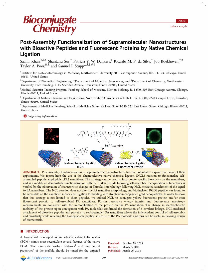
ABSTRACT: Post-assembly functionalization of supramolecular nanostructures has the potential to expand the range of theirapplications. We report here the use of the chemoselective native chemical ligation (NCL) reaction to functionalize self-assembled peptide amphiphile (PA) nanofibers. This strategy can be used to incorporate specific bioactivity on the nanofibers,and as a model, we demonstrate functionalization with the RGDS peptide following self-assembly. Incorporation of bioactivity isverified by the observation of characteristic changes in fibroblast morphology following NCL-mediated attachment of the signalto PA nanofibers. The NCL reaction does not alter the PA nanofiber morphology, and biotinylated RGDS peptide was found tobe accessible on the nanofiber surface after ligation for binding with streptavidin-conjugated gold nanoparticles. In order to showthat this strategy is not limited to short peptides, we utilized NCL to conjugate yellow fluorescent protein and/or cyanfluorescent protein to self-assembled PA nanofibers. Forster resonance energy transfer and fluorescence anisotropymeasurements are consistent with the immobilization of the protein on the PA nanofibers. The change in electrophoreticmobility of the protein upon conjugation with PA molecules confirmed the formation of a covalent linkage. NCL-mediatedattachment of bioactive peptides and proteins to self-assembled PA nanofibers allows the independent control of self-assemblyand bioactivity while retaining the biodegradable peptide structure of the PA molecule and thus can be useful in tailoring designof biomaterials.
■ INTRODUCTION
A biomaterial developed as an artificial extracellular matrix(ECM) mimic must recapitulate several features of the nativeECM. The nanoscale surface features1 and mechanicalproperties2 of the scaffold should be tuned for the targeted
Received: October 29, 2013Revised: March 5, 2014Published: March 26, 2014
Article
pubs.acs.org/bc
© 2014 American Chemical Society 707 dx.doi.org/10.1021/bc400507v | Bioconjugate Chem. 2014, 25, 707−717
tissue in order to achieve precise control of cellular behavior.3
The chemical structure of the biomaterial should ideally containchemical bonds that allow biodegradation in vivo and thuseventually be replaced by native ECM.4 Finally, the method forincorporating biological functionality into the material shouldbe applicable to a broad array of bioactive molecules, fromsmall molecules to peptides to proteins.Biomaterials based on peptide amphiphiles (PAs) meet many
of these criteria. PA molecules, composed of an oligopeptideconjugated to a lipid tail, self-assemble into supramolecularnanostructures.5 The chemical structure of the PA moleculedoes not contain any non-natural components or linkages, andPA nanostructures are biodegradable and biocompatible invivo.6 Our group pioneered the use of peptide sequences thatlead to the self-assembly of high aspect ratio cylindricalnanofibers and at the same time effectively display bioactivecues on their surfaces.7 This self-assembly process is mediatedby hydrophobic collapse of the lipid tails and β-sheet formationamong oligopeptides.8 The highly entangled PA nanofiber gelmimics the fibrillar nature of native ECM7b and confers tunablemechanical rigidity and supramolecular cohesiveness.9 Bio-logical activity is provided by inclusion of bioactive peptidesequences that can bind soluble ligands or cell surfacereceptors.10 The presentation of multiple bioactive cues on asingle PA nanofiber at high surface densities11 that maximizesignaling capability can be achieved through coassembly ofdifferent PA molecules.12 Through the inclusion of differentbioactive cues, PA nanofibers have demonstrated the capacityto signal for differentiation,13 proliferation,14 biologicaladhesion,15 angiogenesis,16 and insulin secretion.17 Previousstrategies to incorporate bioactivity into PA nanostructureshave generally been limited to short peptide sequences thatallow for the solid-phase peptide synthesis of the entire PAmolecule, including the bioactive sequence. However, thepresence of a bioactive cue can potentially influence the self-assembly process of the PA molecules, resulting in theformation of an altered nanostructure.18 Post-assemblyfunctionalization of the PA nanostructures with bioactivegroups would allow independent control of self-assembly andincorporation of bioactivity. Furthermore, in many cases thecomplexity of ligand−receptor binding interactions cannot befully recapitulated with short peptides. The ability to attachlarger peptides and proteins to the self-assembled PAnanostructure would therefore expand the scope of possiblebiological applications.One approach for conjugating larger peptides and proteins to
the self-assembled PA nanostructure is native chemical ligation(NCL). Originally developed by Dawson et al. in 1994, NCL
achieves the chemoselective formation of a native peptide bondbetween peptides or protein fragments.19 This reaction occursat neutral pH upon simple mixing of two reactive precursors,one containing an N-terminal cysteine residue and the othercontaining a C-terminal thioester moiety.20 In contrast withconjugation strategies such as carbodiimide-mediated amidechemistry or Schiff base formation, amine-containing lysineresidues and carboxylic acid-containing glutamate and aspartateresidues do not interfere with the NCL reaction. Moreover, theNCL reaction occurs under biocompatible conditions, unlikeother chemoselective conjugation strategies such as copper-catalyzed click chemistry.21 In the context of peptide-based self-assembled systems, the NCL reaction has been utilized toenhance the mechanical strength of a hydrogel composed offibrillar β-sheet peptides via terminal cross-linking22 and forsurface PEGylation of self-assembled monolayers.23 Thebiocompatibility of the NCL reaction has been demonstratedby its use in a FRET-based probe for in vivo cellular imaging24
and by its use for cross-linking soluble polymers into hydrogelsfor three-dimensional cell culture.25 In addition, the NCLreaction has been used to conjugate relatively large bioactivemoieties, including proteins and other macromolecules.26
In this work, we investigate the use of post-assemblymodification of PA nanostructures using NCL as an approachto add bioactivity while retaining the basic one-dimensionalmorphology. As a model, our objective has been to carry outfunctionalization of fibers with the fibronectin derived epitopeRGDS27 as a bioactive cue. We have also investigated theattachment of proteins to the fibers using thioester-terminatedfluorescent proteins.
■ RESULTS AND DISCUSSION
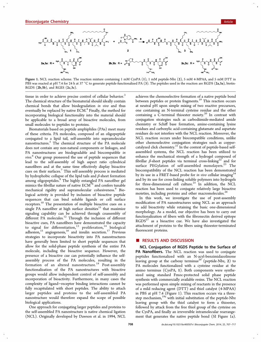
NCL Conjugation of RGDS Peptide to the Surface ofPA Nanofibers. The NCL reaction was used to conjugatepeptides functionalized with an N-acyl-benzimidazolinoneleaving group at the carboxy terminus20 (peptide-Nbz, 2) toPA molecules functionalized with a cysteine residue at theamino terminus (CysPA, 1). Both components were synthe-sized using standard Fmoc-protected solid phase peptidesynthesis with commercially available resins. The NCL reactionwas performed upon simple mixing of reactants in the presenceof a mild reducing agent (DTT) and thiol catalyst (4-MPAA)in PBS at pH 7.4 (Figure 1). This reaction occurs via a three-step mechanism,19a with initial substitution of the peptide-Nbzleaving group with the thiol catalyst to form a thioester,followed by attack from the free thiol group of the cysteine onthe CysPA, and finally an irreversible intramolecular rearrange-ment that generates the native peptide bond (SI Figure 1a).
Figure 1. NCL reaction scheme. The reaction mixture containing 1 mM CysPA (1), 1 mM peptide-Nbz (2), 5 mM 4-MPAA, and 5 mM DTT inPBS was reacted at pH 7.4 for 24 h at 37 °C to generate peptide-functionalized PA (3). The peptides used in the reaction are RGDS (2a,3a), biotin-RGDS (2b,3b), and RGES (2c,3c).
Bioconjugate Chemistry Article
dx.doi.org/10.1021/bc400507v | Bioconjugate Chem. 2014, 25, 707−717708
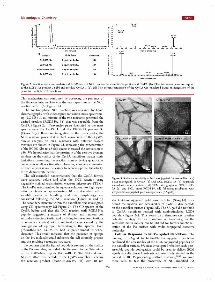
This mechanism was confirmed by observing the presence ofthe thioester intermediate 4 in the mass spectrum of the NCLreaction at 3 h (SI Figure 1b).The solution-phase NCL reaction was analyzed by liquid
chromatography with electrospray ionization mass spectrome-try (LC-MS). A 1:1 mixture of the two reactants generated thedesired product (RGDS-PA, 3a) that was separable from theCysPA (Figure 2a). Two major peaks identified in the massspectra were the CysPA 1 and the RGDS-PA product 3a(Figure 2b,c). Based on integration of the major peaks, theNCL reaction proceeded to 46% conversion of the CysPA.Similar analyses on NCL reactions with different reagentmixtures are shown in Figure 2d. Increasing the concentrationof the RGDS-Nbz to a 5-fold excess increased the conversion to68%. We hypothesize that the proximity of the reactive cysteineresidues on the surface of the CysPA nanofibers creates stericlimitations preventing the reaction from achieving quantitativeconversion of all reactive sites. However, complete conversionof reactive sites is not necessary to achieve optimal bioactivity,as we demonstrate below.The self-assembled nanostructures that the CysPA formed
were analyzed before and after the NCL reaction usingnegatively stained transmission electron microscopy (TEM).The CysPA self-assembled in aqueous solution into high aspectratio nanofibers of approximately 10 nm diameters with avariable degree of bundling, and this morphology wasconserved following the NCL reaction (Figure 3a and b).The secondary structure within the nanofibers was investigatedusing CD spectroscopy (SI Figure 2). The CD spectra of theCysPA before and after the NCL reaction with RGDS-Nbzpeptide suggested a mixture of β-sheet and random coilsecondary structure (estimated by fitting to linear combinationsof reference spectra) with a minimal change caused by theconjugation of epitope. Interestingly, the spectra frompresynthesized RGDS-PA had a predominant α-helicalcharacter. This result indicates that the presence of epitopeon the PA molecule could influence the self-assembly processand the resulting secondary structure.To confirm that the ligated peptide is present on the surface
of the PA nanofiber, we added a biotin group to the N-terminusof the RGDS-Nbz peptide (biotin-RGDS-Nbz, 2b) and usedNCL to attach this peptide to the CysPA nanofiber. Labelingthe reaction product (biotin-RGDS-PA, 3b) with 10 nm
streptavidin-conjugated gold nanoparticles (SA-gold) con-firmed the ligation and accessibility of biotin-RGDS peptideon the nanofiber surface (Figure 3d). The SA-gold did not bindto CysPA nanofibers reacted with nonbiotinylated RGDSpeptide (Figure 3c). This result also demonstrates anotherpotential strategy for incorporation of bioactivity, as theaccessible biotin moiety can be utilized for further functional-ization of the PA surface with avidin-conjugated bioactivemolecules.
Cellular Response to RGDS-Ligated Nanofibers. Thebinding of SA-gold to biotin-RGDS-conjugated nanofibersconfirmed the accessibility of the NCL-conjugated peptides onthe nanofiber surface. We next investigated whether such post-assembly peptide conjugation could be used to deliver ECMsignals to cells. Since fibroblasts are extensively studied in thecontext of RGDS presenting scaffold materials,15,28 we usedthese cells to test the bioactivity of NCL-modified PA
Figure 2. Reaction yields and analysis. (a) LCMS trace of NCL reaction between RGDS peptide and CysPA. (b,c) The two major peaks correspondto the RGDS-PA product 3a (b) and residual CysPA 1 (c). (d) The percent conversion of the CysPA was calculated based on integration of thepeaks for multiple NCL reactions.
Figure 3. Surface accessibility of NCL-conjugated PA assemblies. (a,b)TEM micrograph of CysPA (a) and NCL RGDS-PA (b) negativelystained with uranyl acetate. (c,d) TEM micrographs of NCL RGDS-PA (c) and NCL biotin-RGDS-PA (d) following incubation withstreptavidin-conjugated gold nanoparticles (SA-gold).
Bioconjugate Chemistry Article
dx.doi.org/10.1021/bc400507v | Bioconjugate Chem. 2014, 25, 707−717709

nanofibers. Cell response was measured to PAs coated on glasscoverslips using a method previously established in our group.29
Sterile coverslips were first coated with cationic poly(D-lysine)(PDL) and subsequently with a layer of anionic alginate, andfinally with the cationic PA nanofibers (SI Figure 3). Not onlydoes this method of sequential coating help to achieve auniform PA layer on the surface, but the intermediatenonfouling alginate layer also prevents any confounding cellresponse resulting from attachment to underlying glass or PDL.NIH/3T3 mouse embryonic fibroblasts were seeded on thesePA-coated surfaces and maintained under serum-free con-ditions to minimize nonspecific adsorption of proteins to thenanofibers.30
Fibroblast morphology was evaluated after 5 h of culture, assuch short incubation was found to be suitable by our groupand others for comparison of their response to sub-strates.28,29,31 Fibroblasts were found to attach to CysPAsurfaces but displayed a rounded morphology indicating a lackof cell spreading and adhesion (Figure 4a). This observationcan be explained by the ability of PA nanofibers to provide cellattachment; however, an absence of an integrin-binding cuelimits their spreading. In contrast, on the CysPA surfacesfunctionalized with the RGDS peptide using the NCL reaction(NCL RGDS-PA), cells appeared to be more spread with thepresence of prominent cytoplasmic processes giving the cells astellate morphology, similar to that found on presynthesizedRGDS-PA (Figure 4c,e). The observed change in cellmorphology was further quantified by measurement ofcircularity, calculated as 4π × area/perimeter so that a perfectlyround cell will have a value of 1.32 The circularity of fibroblastson the NCL RGDS-PA surface was 0.38 ± 0.03, significantlylower than the value of 0.65 ± 0.04 found on the CysPA surface
(Figure 4f). Cell viability was found to be higher than 95% ineach of these conditions (SI Figure 4), indicating that a toxicresponse to the underlying substrate is not attributable to thedifference in the cell morphology. To further evaluate whetherthe observed change in the cell morphology was specific to theRGDS sequence, we conjugated a mutated non-bioactive RGESpeptide to CysPA nanofibers using the NCL reaction (NCLRGES-PA). Fibroblasts on the NCL RGES-PA surface (Figure4d) had a circularity of 0.56 ± 0.04, which was not significantlydifferent from the circularity on the CysPA surface, indicatingthat the change in cell morphology is due to the specificbioactivity of the RGDS peptide and not due to the conditionsof the NCL reaction. Furthermore, addition of soluble RGDSpeptide into the culture medium did not alter the morphologyof cells on the CysPA surface (Figure 4b), confirming the needfor tethering of the RGDS peptide to nanofibers to provide forcell signaling.Although NCL did not achieve full conjugation efficiency,
several reports from our group and others have shown that thepresence of 10% bioactive molecules in a peptide nanofibersystem is sufficient to demonstrate the maximal cellresponse.15,22 Consistent with those findings, we observed acomparable cell response between the NCL RGDS-PA surfacesand the presynthesized RGDS-PA surfaces, which have a cuepresent on every molecule. It has been suggested that thecritical RGD sequence density required for fibroblast spreadingis determined by the steric factor separating two integrinmolecules.33 Even with a submaximal functionalization,following NCL reaction, RGDS sequence availability on thenanofiber surface would be sufficient for maximal integrinbinding.
Figure 4. Fibroblast morphology on PA-coated coverslips. (a−e) Fluorescent micrographs of NIH/3T3 fibroblasts cultured for 5 h and stained byphalloidin for F-actin on coverslips coated with CysPA (a), CysPA and soluble RGDS peptide (b), NCL RGDS-PA (c), NCL RGES-PA (d), andpresynthesized RGDS-PA not formed by NCL (e). (f) Comparison of cell circularity (4π × area/perimeter2) on various PA-coated surfaces (** p <0.01 vs CysPA, n = 100 cells).
Bioconjugate Chemistry Article
dx.doi.org/10.1021/bc400507v | Bioconjugate Chem. 2014, 25, 707−717710

Apart from the possibility of post-assembly functionalizationof PA nanofibers, the NCL conjugation strategy allowssimultaneous covalent functionalization of the PA nanofiberwith multiple different bioactive ligands. This approach couldtake advantage of potential synergies among different classes ofbioactive ligands without the need for independent chemicalconjugation schemes for each component. Furthermore, therelative biocompatibility of the NCL reaction mixture createsthe opportunity for temporal control over ligand conjugation inlive cell culture, potentially allowing the sequentially timedpresentation of bioactive cues to influence cell behaviors suchas differentiation.
Protein Ligation to PA Nanofibers in Solution and onCoated Surfaces. Next, we sought to extend the scope of theNCL method for biofunctionalization of PA nanofibers tolarger molecules such as proteins. To demonstrate that theNCL reaction can be used to ligate proteins to the PAnanofiber, we utilized YFP that was expressed with a self-cleavable intein domain and purified with 2-mercaptoethanesulfonate sodium salt (MESNA) to yield a thioester at the C-terminus. This YFP-MESNA was ligated to the CysPA via NCLwith dilute reactant concentrations (Figure 5a). The overallnanofiber morphology of the CysPA was retained followingconjugation with YFP, although the nanofibers appear wider
Figure 5. Protein ligation to PA nanostructures via NCL. (a) Schematic of NCL conjugation of YFP to CysPA using YFP-MESNA. (b,c) TEMmicrograph of CysPA assemblies prior to (b) and following NCL with YFP (c). (d) Emission spectra of YFP following NCL conjugation to CysPAcoassembled with different concentrations of TAMRA-labeled PA.
Figure 6. Characterization of PA−protein conjugates formed by NCL. (a,b) SDS-PAGE visualized by silver staining (a) and Western blot againstGFP (b) were used to analyze the NCL reaction between YFP and PA (Lane 1: YFP only, Lane 2: YFP conjugated to CysPA, Lane 3: CysPA only,Lanes L1, L2, L3: protein standards with indicated molecular weights in kDa). (c) YFP fluorescence intensity after conjugation to CysPA-coatedsurface (CysPA-YFP NCL). An identical PA lacking cysteine (GlyPA-YFP Control) or CysPA treated with reaction buffer alone (CysPA Control)was used as a control (** p < 0.01 vs CysPA-YFP NCL). (d) Confocal fluorescence microscopy of CysPA nanofiber-coated alginate microparticlessimultaneously reacted with YFP and CFP. (e) Fluorescence anisotropy of YFP following NCL reaction with CysPA. GlyPA was used as a control (*p < 0.05 vs YFP; # p < 0.05 vs GlyPA).
Bioconjugate Chemistry Article
dx.doi.org/10.1021/bc400507v | Bioconjugate Chem. 2014, 25, 707−717711
and less bundled (Figure 5b,c). These changes would beexpected upon ligation of large proteins onto the nanofibersurface.The CysPA-YFP NCL reaction mixture was analyzed by
SDS-PAGE to identify the reaction product. Two distinct bandswere detected on silver staining, one corresponding to the YFPand a second for a product that appears to have higherelectrophoretic mobility than the YFP (Figure 6a). Westernblot analysis showed both bands to be positively stained withanti-GFP antibody (Figure 6b). We attribute the band withhigher electrophoretic mobility to YFP conjugated with PA;although conventionally, the electrophoretic mobility would beexpected to decrease following conjugation due to an increasein molecular mass, we believe the character of the PA moleculeto be responsible for the increased electrophoretic mobility.The conjugation of the PA molecule adds a highly hydrophobicaliphatic chain and three positively charged lysine residues tothe YFP protein, and due to this aliphatic chain and thesepositive charges, the PA molecule can potentially bind a highernumber of SDS molecules relative to its molecular mass thanthe protein alone, resulting in an increase in surface charge thatis disproportionately higher than the increase in mass. This “gelshifting” effect has been observed for other proteins modifiedwith hydrophobic groups,34 as well as other peptide amphiphilesystems35 analyzed by electrophoresis with SDS. This effectwould increase the apparent electrophoretic mobility of theproduct on SDS-PAGE, consistent with the new band observedin the reaction mixture. The possibility of the second bandbeing a YFP degradation product can be excluded because theYFP control was treated under exactly the same conditions asthe NCL reaction except for the absence of PA. Taken together,the new band observed in the reaction mixture likelycorresponds to the CysPA-YFP ligation product.In order to confirm the YFP immobilization on the CysPA
nanofibers, we performed FRET experiments. The CysPA wascoassembled with TAMRA-labeled PA (SI Figure 3a), whichhas an excitation spectrum that overlaps the YFP emissionspectrum, forming a FRET pair. The proximity of YFP toTAMRA fluorophores on the nanofiber surface shouldtherefore induce FRET and quench the YFP emission. Fordistances in the range of the Forster radius, closer proximity ofthe fluorophores will induce a greater degree of YFP emissionquenching. This effect was observed as shown in Figure 5d,where all spectra were normalized using the maximum intensityobserved in the absence of TAMRA (YFP emission maximumat 525 nm). Increasing the molar concentration of theTAMRA-labeled PA increases the surface density of acceptors.A higher density of TAMRA on the nanofiber surface effectivelyreduces the average distance from YFP attached via NCL,resulting in greater quenching. In fact, increasing theproportion of TAMRA-labeled PA from 1 to 2 mol %generated an increase in the quenching of YFP fluorescencefrom 20% to 80%. For single donor−acceptor FRET pairs, thedecrease in intensity of the donor is accompanied by anincrease in intensity of the acceptor. However, at 2 mol %TAMRA fluorophore density on the nanofiber surface was highenough to induce self-quenching due to homo-FRETphenomena. Consequently, we did not observe an increasedintensity of TAMRA signal with increasing energy transferbetween YFP and TAMRA-PA.The specificity of YFP immobilization on CysPA nanofibers
via NCL was further confirmed by fluorescence anisotropy (r)measurements. In the absence of depolarization phenomena,
such as homo-FRET, light scattering, or reabsorption, theanisotropy only depends on the rotational diffusion of thefluorophore. If the rotational diffusion rate of a fluorophore isof similar magnitude to the fluorescence decay rate, the slowerrotational diffusion rate expected upon binding will be reflectedas an increase in anisotropy. As shown in Figure 6e, at less than10 nM, YFP anisotropy increased after NCL with CysPA butremained essentially the same in the presence of an identical PAlacking the N-terminal cysteine (GlyPA). Interestingly, theanisotropy decreased steadily as the concentration increasedabove a threshold of 10 nM YFP. At higher concentration ofYFP, the likelihood for the distance between two or more YFPmolecules to be shorter than the Forster radius for homo-FRETcan no longer be neglected. Therefore, the anisotropy decreasesdue to energy transfer between fluorophores with differentorientations. A similar behavior was observed previously forYFP-labeled protein homodimers and used to detect proteinreorientation upon binding.36 These results provide evidencenot only that the YFP was immobilized on the CysPAnanofibers by NCL, but also that the surface density ofimmobilized YFP increased as the YFP to CysPA molar ratioincreased in the reaction mixture.Finally, we sought to demonstrate that protein ligation via
NCL can be achieved on PA nanofiber-coated surfaces. Higherfluorescence intensity was observed following the NCL-mediated conjugation of YFP to CysPA nanofibers coated onglass coverslips (Figure 6c). Conversely, adding the same YFP-containing reaction mixture to an identical PA lacking the N-terminal cysteine (GlyPA) produced significantly lowerfluorescence intensity, indicating that the observed fluorescencewas due to retention of the YFP by a specific reaction withCysPA and not a nonspecific adsorption to PA nanofibers. Todemonstrate a potential application of protein ligation to PAnanofiber-coated surfaces via NCL, we utilized cell-size alginatemicroparticles electrostatically conjugated with a coating ofCysPA nanofibers by a process similar to the formation of theCysPA-coated glass coverslips. We simultaneously ligated YFPand cyan fluorescent protein (CFP) to the surfaces of theseCysPA-coated microparticles and visualized their surfacefluorescence by confocal microscopy. The surfaces of themicroparticles showed colocalized YFP and CFP fluorescence(Figure 5d), indicating that the two proteins were coligated tothe PA-coated microparticles. A full view of the field showsseparate areas of high intensity for each fluorophore, indicatingthat colocalization of fluorescence was not due to spectralbleed-through (SI Figure 3d).The approach of conjugating proteins to PA nanofibers by
NCL as described here can be applied to any protein expressedwith a C-terminal intein domain and purified by thiol-mediatedcleavage. The conjugated protein can potentially fulfill multiplefunctions, including targeting, adhesion, and cell signaling.These functions are especially useful when combined with theadvantages offered by PA nanofibers, including the ability toencapsulate hydrophobic drugs, form a scaffold that mimicsECM, and coat polymer surfaces. For example, PA nanofibersencapsulating therapeutic agents37 or drug loaded alginateparticles coated with PA nanofibers could be targeted to specificcells and tissues via proteins conjugated by NCL. Theconjugation of proteins to PA nanofibers via NCL wouldfacilitate these potential applications while maintaining thedesirable properties of biodegradability and biocompatibilityinherent to the PA molecule.
Bioconjugate Chemistry Article
dx.doi.org/10.1021/bc400507v | Bioconjugate Chem. 2014, 25, 707−717712
■ CONCLUSIONS
We have demonstrated here the use of native chemical ligationto conjugate both bioactive peptides and fluorescent proteins toself-assembled peptide amphiphile nanofibers. The ligatedpeptides and proteins were displayed on the nanofiber surfacewithout compromising the morphology of the supramolecularnanostructures. Evidence for bioactivity was established by theobservation of fibroblast spreading following conjugation of abiological signal to nanofibers using the ligation reaction. Ourresults suggest that post-assembly chemical ligation could be auseful tool to tailor the bioactivity of supramolecularnanostructures.
■ EXPERIMENTAL PROCEDURES
Peptide Synthesis and Purification. All PAs and peptideswere synthesized by fluorenylmethoxycarbonyl (Fmoc) pro-tected solid-phase peptide synthesis as previously reported byour group7a using materials purchased from EMD ChemicalsInc. (Merck KGaA, Darmstadt, Germany) unless otherwiseindicated. Briefly, the PAs were synthesized at 0.25 mmol scaleon Rink Amide MBHA resin, and peptides functionalized at thecarboxy-terminus with an N-acyl-benzimidazolinone for nativechemical ligation (peptide-Nbz) were synthesized at 0.25 mmolscale on 3-(Fmoc-amino)-4-aminobenzoyl AM resin. For eachamino acid addition, the resin was deprotected using 2% 1,8-diazabicycloundec-7-ene (DBU) and 2% piperidine in N,N-dimethylformamide (DMF), and the amino acid was coupledusing 4 equiv of protected amino acid functionalized with 4equiv of 2-(1h-benzotriazole-1-yl)-1,1,3,3- tetramethyluroniumhexafluorophosphate (HBTU) and 6 equiv of N,N-diisopropy-lethylamine (DIPEA) in DMF. The dodecanoic acid tail wassimilarly functionalized and coupled to a lysine side chainfollowing selective deprotection of the 4-methyltrityl (Mtt)group using a 92:5:3 mixture of dichloromethane (DCM),triisopropylsilane (TIPS), and trifluoroacetic acid (TFA). Forsynthesis of peptide-Nbz, a Boc-protected amino acid wasadded at the N-terminus, and the resin was treated with 5 equivp-nitrophenylchloroformate in DCM followed by 0.5 M DIPEAin DMF for activation. Resin cleavage and amino aciddeprotection was performed using a 95:2.5:2.5 mixture ofTFA, TIPS, and water for all PAs and peptides. Followingremoval of solvent by rotary evaporation, the crude PAs/peptides were precipitated by addition of cold diethyl ether.The precipitate was collected and dried in vacuo to generatecrude product with identity confirmed by electrosprayionization (ESI) mass spectrometry. All PAs and peptideswere purified by reverse-phase high performance liquidchromatography (HPLC) as previously reported by ourgroup.7a Briefly, the crude product was dissolved in 0.1%TFA in water, filtered, injected onto a Gemini-NX 5 μm C18column, and eluted using a water-acetonitrile solvent gradientfor separation. The purified product was lyophilized and storedat −20 °C. To conjugate biotin, 10 mM RGDS-Nbz peptide inPBS was mixed 1:1 with 20 mM sulfo-NHS-biotin (ThermoScientific, Rockford, IL) in PBS and reacted for 4 h at roomtemperature and overnight at 4 °C. The biotinylated peptidewas purified from the reaction mixture by HPLC, lyophilized,and stored at −20 °C.Protein Expression and Purification. The pTXB1-EYFP
and pTXB1-ECFP plasmids (see Supporting Information) weretransformed, according to the manufacturer’s protocol, into E.coli BL21 (DE3) (Novagen) competent cells by electro-
poration.38 The transformed cells were plated on LysogenyBroth (LB)-agar plates containing ampicillin (Amp) and grownovernight at 37 °C. Single colonies were picked and grown in30 mL LB medium (10 g peptone, 5 g yeast extract, 10 gsodium chloride, 1 L water) containing Amp overnight at 37 °Cin shaker at 250 rpm. Cells were stored in 1 mL aliquots at −80°C in the presence of 15% (w/v) glycerol. For overnightcultures, 10 mL of Amp-containing LB was inoculated with ∼1μL of the glycerol stock and grown overnight at 37 °C and 250rpm. For small-scale expression, volumes of 30 mL were used,while large-scale expression was performed in 500 mL LBmedium. Cell culturing was started with 5 mL overnight cultureand cells were grown at 37 °C, 250 rpm in the presence ofAmp. When an optical density (OD600) of 0.6−0.8 was reached,protein expression was induced by the addition of isopropyl-β-D-thiogalatopyranoside (IPTG; Duchefa Biochemie). Large-scale expression was induced by 0.5 mM IPTG and grownovernight at 15 °C. Cell harvesting was performed bycentrifuging the cell culture for 5 min at 10,000 g at 4 °C.The supernatant was removed and the cell pellet wasresuspended in 5 mL Bugbuster (Novagen) and 5 μLbenzonase (Novagen) per gram of cell pellet. After incubationfor 20 min at 21 °C, the cell suspension was centrifuged at16,000 g for 20 min at 4 °C. The supernatant was directlyapplied to 10 mL chitin beads (New England Biolabs),equilibrated with 10 column volumes of column buffer (20mM sodium phosphate, 0.5 M sodium chloride, pH 6). Theloaded column was washed with 10 volumes of column bufferafter which the column was quickly flushed with 2 columnvolumes of cleavage buffer (20 mM sodium phosphate, 0.5 Msodium chloride, 100 mM 2-mercaptoethane sulfonic acid(MESNA), pH 6). Subsequently, the flow was stopped and thecolumn with the cleavage buffer was incubated 2 × 20 h atroom temperature (using fresh cleavage buffer for the secondtime). Elution fractions containing the cleaved proteins with aC-terminal thioester were collected and pooled. The proteinswere buffer-exchanged (into 20 mM Tris, 150 mM sodiumchloride, pH 7.8) using Amicon ultra centrifuge tubes(MWCO: 10 kDa), after which the concentration wasmeasured using UV−vis. Protein solutions were stored at−80 °C.
Native Chemical Ligation Reaction in Solution. The N-terminal cysteine PA (CysPA) was mixed with 5 equiv ofdithiothreitol (DTT) and dissolved in phosphate-bufferedsaline (PBS) at pH 7.4, and the peptide-Nbz or YFP-MESNAwas mixed with 5 equiv of 4-mercaptophenylacetic acid (4-MPAA) and dissolved in PBS at pH 7.4. The two reagents weremixed at the desired ratio, and the reaction was subsequentlyanalyzed by liquid chromatography mass spectrometry, gelelectrophoresis, transmission electron microscopy, circulardichroism (CD) spectroscopy, Forster resonance energytransfer (FRET), or fluorescence anisotropy as indicated.
Liquid Chromatography Mass Spectroscopy. Analyticalliquid chromatography mass spectroscopy (LC-MS) wasperformed using an Agilent 1200 system with an Agilent6250 quadrupole-time-of-flight mass spectrometer using aPhenomenex Gemini C18 column (5 μm particle size, 150 ×1.0 mm) eluting with a gradient of 5% ACN to 95% ACN inwater, with each solvent containing 0.1% TFA. UV absorbancewas monitored at 220 nm.
Transmission Electron Microscopy. The nanostructuremorphology of each PA was characterized using conventionaltransmission electron microscopy (TEM). Each PA or NCL
Bioconjugate Chemistry Article
dx.doi.org/10.1021/bc400507v | Bioconjugate Chem. 2014, 25, 707−717713

reaction mixture was diluted in water to 0.1 mM PA, and 7.5 μLof this solution was deposited on a copper grid with 300 meshcarbon support film for 5 min, washed with water twice for 1min each, negatively stained with 2% (w/v) uranyl acetate twicefor 30 s each, and dried at room temperature overnight.For labeling, streptavidin-conjugated 10 nm gold nano-
particles in suspension (Sigma-Aldrich, St. Louis, MO) weremixed at 1:1 volume ratio with 20 mM dithiothreitol (DTT) inPBS and incubated for 30 min to prevent nonspecific thiolbinding to the gold. This suspension was mixed at a 1:4 volumeratio with the PA or NCL reaction mixture and incubated for30 min to allow streptavidin binding to biotin. This mixture wasdiluted to 0.1 mM PA and deposited onto the copper grid asdescribed above, except with four water washes to ensurecomplete removal of unbound gold nanoparticles. All imageswere acquired using an FEI Tecnai Spirit G2 microscopeworking at 120 kV.Circular Dichroism Spectroscopy. Circular dichroism
(CD) spectra of PA (1 mM) in PBS or NCL reaction mixturewere acquired on a JASCO J-715 CD spectrophotometer atroom temperature using quartz plates with a 0.05 mm pathlength. The CD spectra were fit to linear combinations ofreference spectra for known secondary structures using thePEPFIT algorithm39 to estimate secondary structure.Gel Electrophoresis and Western Blotting. Polyacryla-
mide gel electrophoresis with sodium dodecyl sulfate (SDS-PAGE) was performed using 17.5% polyacrylamide gels.Samples were prepared in SDS sample buffer with 4 M ureaand heated for 5 min at 95 °C. The reaction mixture andcontrols were diluted 15-fold resulting in consistent proteinloadings of approximately 5.6 μg per well for the YFP and 0.5μg per well for the CysPA across all samples. As reference, Bio-Rad Precision Plus Unstained Protein Standard was used forboth silver stain and Western blot, while Bio-Rad Broad-Range15% Protein Standard was used specifically for the silver stainand Magic Mark XP Western Protein Standard (Invitrogen)was used specifically for the Western blot. Electrophoresis wasperformed at 65 V for 21 h in Tris-Glycine running buffer usingthe Pharmacia EPS 600 power supply. Gels were rinsed withwater and developed by either silver nitrate exposure orWestern blot. For the Western blot, the protein was transferredto an immobilin-P membrane using a Bio-Rad Trans-Blot cellwith model 200/2.0 power supply at 35 V for 2 h in Tris-Glycine transfer buffer. The membrane was blocked using 5%milk in PBS + 0.3% Tween for 1 h. The membrane was thenstained with anti-GFP rabbit polyclonal primary antibody(ab290, Abcam) at 1:5000 dilution for 1 h, washed with PBS +0.3% Tween, further stained with goat anti-rabbit polyclonalsecondary antibody conjugated to FITC (Jackson ImmunoR-esearch) at 1:5000 dilution for 1 h, and washed again with PBS+ 0.3% Tween. The membrane was then read on a FujifilmFLA-5100 imager.Forster Resonance Energy Transfer and Fluorescence
Anisotropy. CysPA was mixed in hexafluoroisopropanol(HFIP) with 5-carboxytetramethylrhodamine (TAMRA) la-beled PA at different ratios (0, 1, and 2 mol %) and lyophilizedto ensure homogeneous mixing of coassembled nanofibers. Thelyophilized powder was redissolved in 10 mM DTT aqueoussolution, diluted in PBS at pH 7.4, and subsequently mixed withYFP-MESNA solution (supplemented with 5 equiv 4-MPAA),to yield final concentrations of YFP and PA of 1 and 100 μM,respectively. This mixture was reacted for around 24 h beforemeasurements. TAMRA was used as a FRET acceptor for YFP.
The same procedure was used to prepare solutions of anidentical PA lacking the N-terminal cysteine (GlyPA). Thesesolutions were used as control to rule out the contribution toFRET signal of potential nonspecific adsorption of YFP to PAnanofibers. Emission intensity spectra were recorded on aHoriba Nanolog fluorimeter, using an excitation wavelength of485 nm (bandpass of 2 nm). Emission intensity was scannedbetween 495 and 680 nm (increment of 1 nm), encompassingboth YFP and TAMRA emission ranges, using a bandpass of 2nm, integration time of 0.1 s, and PMT detector set at 950 kV.A similar procedure was used to prepare solutions forfluorescence anisotropy (FA), but without TAMRA labeledPA, and at variable YFP concentration (1−500 nM). FA wasrecorded at an emission wavelength of 535 nm (excitation at495 nm), 1 s integration time, and using variable mono-chromators’ bandpasses, which were set to guarantee signalintensities over 10 kCPS. Intensity was recorded at the fourpossible combinations for the orientations of excitation andemission polarizers: both along the vertical direction (IVV), bothoriented horizontally (IHH), the excitation polarizer orientedvertically and the emission horizontally (IVH), or vice versa(IHV). FA (r) was calculated according to the followingequation:
= − +r I I I I I I I I( . . )/( . 2 . )VV HH VH HV VV HH VH HV
The correct alignment of the polarizers was verified using adiluted colloidal silica suspension (r ≥ 0.97).
Native Chemical Ligation Reaction on PA-CoatedSurfaces and Microparticles. All PAs, peptides, and othermaterials were UV-sterilized prior to use to maintain sterility.12 mm glass coverslips were coated with 0.01% poly(D-lysine)(PDL) overnight and then washed with water and driedovernight. The PDL-coated coverslips were then coated with0.25% alginate dissolved in water for 1 h, after which thealginate was cross-linked with 10 mM calcium chloride in water.The alginate-coated coverslips were then coated with 0.5 mMPA dissolved in water with 5 equiv DTT overnight. Alginatemicroparticles were similarly coated with 0.5 mM PA dissolvedin water with 5 equiv DTT for 30 min. Following wash stepswith water and PBS, the PA-coated coverslips or microparticleswere immersed in NCL solution, which contained either 0.5mM peptide-Nbz or 5 μM YFP-MESNA and 5 μM CFP-MESNA dissolved in PBS with 5 equiv of 4-MPAA and DTT.Following a 24 h incubation at 37 °C, the coverslips ormicroparticles were washed with PBS, and the coverslips wereeither used for cell morphology and viability studies or assayedfor YFP fluorescence on a SpectraMax M5 microplate reader,while the microparticles were visualized by confocal microscopyfor both YFP and CFP fluorescence.
Cell Culture. NIH/3T3 mouse embryonic fibroblasts(American Type Culture Collection, Manassas, VA) weremaintained in monolayer culture in T25 culture flasks. Growthmedium consisted of Dulbecco’s Modified Eagle Medium(DMEM) with high glucose supplemented with 10% fetalbovine serum, 100 U/mL penicillin, and 100 μg/mLstreptomycin. Assay medium consisted of growth mediumwithout serum.
Cell Morphology and Viability Assays. The PA-coatedcoverslips functionalized using the NCL reaction were placed in24-well plates and seeded with 5,000 NIH/3T3 fibroblasts in 1mL assay medium per well. Following a 5 h incubation at 5%CO2 and 37 °C, the coverslips were washed with PBScontaining 1 mM CaCl2 and fixed for subsequent analysis.
Bioconjugate Chemistry Article
dx.doi.org/10.1021/bc400507v | Bioconjugate Chem. 2014, 25, 707−717714
For quantification of cell spreading, fibroblasts were stainedwith phalloidin and visualized by light microscopy. Coverslipswere fixed in 4% paraformaldehyde in PBS for 30 min at roomtemperature, then blocked with blocking buffer (10% normalgoat serum, 2% bovine serum albumin, and 0.4% Triton X100)for 30 min at 4 °C, then stained for F-actin with rhodamine-phalloidin (Invitrogen, Grand Island, NY) at 1:500 dilution for2 h at room temperature, and finally dried and mounted onslides for light microscopy. Solutions used in fixing, staining,and washing steps were supplemented with 1 mM CaCl2 tomaintain alginate cross-linking. The cells were imaged using aNikon Eclipse TE2000-U inverted fluorescence microscope,and images were analyzed using the Shape Descriptor 1u pluginon ImageJ software (NIH) to determine the circularity of thecells (calculated as 4π × area/perimeter2).Cell viability was assessed using commercially available
LIVE/DEAD cell viability assay kit (Life Technologies).Fibroblasts were cultured on PA coating for 5 h and stainedwith 1 μg/mL calcein AM and 1 μg/mL ethidium homodimer-1 following manufacturer’s protocol. Live cell (calcein positive)and dead cell (ethidium homodimer positive) populations werecounted manually under an inverted fluorescence microscope(Nikon Eclipse TE2000-U).Statistics and Data Analysis. For the determination of
form factor, five images from each of two replicates of eachcondition were analyzed, and the mean and standard deviationof the form factor of cells were calculated across all ten images.To establish statistical significance, each condition wascompared to CysPA alone using the Student’s t test. Forcomparison of cell viability approximately 100 cells werecounted from at least two replicates per condition. The cellviability index was defined as the ratio of calcein positive cells tothe total number of cells. Statistical significance was measuredagainst CysPA alone. For the determination of YFPfluorescence on PA coatings, two independent measurementsof the fluorescence emission at 535 nm following excitation at495 nm were taken from each of four replicates of eachcondition, and the mean and standard error were calculatedbased on these four replicates for each condition followingbackground subtraction of wells with alginate-coated coverslipswithout PA nanofibers. To establish statistical significance, theCysPA-YFP NCL condition was compared to each controlusing the Student’s t test. For the statistical comparison offluorescence anisotropy, three independent replicates weremeasured for each condition, and the CysPA condition wascompared to the GlyPA and YFP only conditions using theStudent’s t test.
■ ASSOCIATED CONTENT*S Supporting InformationDescription of the construction of the expression plasmids forEYFP-MESNA and ECFP-MESNA is provided in Methods. Adetailed mechanism for the NCL reaction and confirmation ofthis mechanism by mass spectroscopy is provided in Figure 1.CD spectra of PAs with analysis of secondary structure analysisis provided in Figure 2. A schematic of the protocol for coatingsurfaces with PA nanofibers for assessment of cell adhesion andspreading is provided in Figure 3. The cell viability on PAcoatings is provided in Figure 4. The structure of the TAMRA-labeled PA, full pictures of the protein gels, and a full field ofthe fluorescent protein-ligated PA-coated microparticles areshown in Figure 5. This material is available free of charge viathe Internet at http://pubs.acs.org.
■ AUTHOR INFORMATIONCorresponding Author*E-mail: [email protected]; Phone 312-503-0807.Present AddressPatricia Y. W. Yankers and Ricardo M. P. da Silva, Institute forComplex Molecular Systems, Eindhoven University ofTechnology Den Dolech 2, 5612 AZ Eindhoven, Netherlands;+31 (0)40 247 9111, [email protected], [email protected] authors declare no competing financial interest.
■ ACKNOWLEDGMENTSThis work was funded by a grant from the National Institutes ofHealth−National Institute of Biomedical Imaging and Bio-engineering (2 R01 EB003806-06A2, NIH/NIBIB). S.K.acknowledges support from the National Institutes ofHealth−National Institute on Drug Abuse (5 T90 DA022881NIH/NIDA). P.Y.W.D. acknowledges the Council for Chem-ical Sciences of The Netherlands Organization for ScientificResearch (CW-NWO) and J.B. acknowledges the NWO for aRubicon grant. The authors would like to acknowledge thefollowing core facilities at Northwestern University: CellImaging Facility, Electron Probe Instrumentation Center andInstitute for BioNanotechnology and Medicine. Finally, theauthors would like to thank Xuan Yue for performing the LC-MS analysis, Liam Palmer for editing this manuscript, MariekeRensen, Maarten Merkx, and Joost van Dongen for their inputwith respect to the fluorescent protein experiments and MarkSeniw for graphic designs used in this contribution.
■ REFERENCES(1) Petty, R. T., Li, H. W., Maduram, J. H., Ismagilov, R., andMrksich, M. (2007) Attachment of cells to islands presenting gradientsof adhesion ligands. J. Am. Chem. Soc. 129 (29), 8966−8967.(2) Engler, A. J., Sweeney, H. L., Discher, D. E., and Schwarzbauer, J.E. (2007) Extracellular matrix elasticity directs stem cell differentiation.J. Musculoskelet. Neuronal Interact. 7 (4), 335.(3) Gilbert, P. M., Havenstrite, K. L., Magnusson, K. E., Sacco, A.,Leonardi, N. A., Kraft, P., Nguyen, N. K., Thrun, S., Lutolf, M. P., andBlau, H. M. (2010) Substrate elasticity regulates skeletal muscle stemcell self-renewal in culture. Science 329 (5995), 1078−1081.(4) Kim, B. S., and Mooney, D. J. (1998) Development ofbiocompatible synthetic extracellular matrices for tissue engineering.Trends Biotechnol. 16 (5), 224−230.(5) Fields, G. B., Lauer, J. L., Dori, Y., Forns, P., Yu, Y. C., and Tirrell,M. (1998) Protein-like molecular architecture: biomaterial applicationsfor inducing cellular receptor binding and signal transduction.Biopolymers 47 (2), 143−51.(6) Ghanaati, S., Webber, M. J., Unger, R. E., Orth, C., Hulvat, J. F.,Kiehna, S. E., Barbeck, M., Rasic, A., Stupp, S. I., and Kirkpatrick, C. J.(2009) Dynamic in vivo biocompatibility of angiogenic peptideamphiphile nanofibers. Biomaterials 30 (31), 6202−6212.(7) (a) Hartgerink, J. D., Beniash, E., and Stupp, S. I. (2001) Self-assembly and mineralization of peptide-amphiphile nanofibers. Science294 (5547), 1684−1688. (b) Hartgerink, J. D., Beniash, E., and Stupp,S. I. (2002) Peptide-amphiphile nanofibers: a versatile scaffold for thepreparation of self-assembling materials. Proc. Natl. Acad. Sci. U.S.A. 99(8), 5133−5138.(8) (a) Palmer, L. C., and Stupp, S. I. (2008) Molecular self-assemblyinto one-dimensional nanostructures. Acc. Chem. Res. 41 (12), 1674−1684. (b) Palmer, L. C., Velichko, Y. S., de la Cruz, M. O., and Stupp,S. I. (2007) Supramolecular self-assembly codes for functionalstructures. Philos. Trans. R. Soc., A 365 (1855), 1417−1433.(c) Paramonov, S. E., Jun, H.-W., and Hartgerink, J. D. (2006) Self-assembly of peptide-amphiphile nanofibers: the roles of hydrogen
Bioconjugate Chemistry Article
dx.doi.org/10.1021/bc400507v | Bioconjugate Chem. 2014, 25, 707−717715

bonding and amphiphilic packing. J. Am. Chem. Soc. 128 (22), 7291−7298. (d) Tovar, J. D., Claussen, R. C., and Stupp, S. I. (2005) Probingthe interior of peptide amphiphile supramolecular aggregates. J. Am.Chem. Soc. 127 (20), 7337−7345. (e) Velichko, Y. S., Stupp, S. I., andde la Cruz, M. O. (2008) Molecular simulation study of peptideamphiphile self-assembly. J. Phys. Chem. B 112 (8), 2326−2334.(9) (a) Pashuck, E. T., Cui, H., and Stupp, S. I. (2010) Tuningsupramolecular rigidity of peptide fibers through molecular structure. J.Am. Chem. Soc. 132 (17), 6041−6046. (b) Greenfield, M. A., Hoffman,J. R., de la Cruz, M. O., and Stupp, S. I. (2010) Tunable mechanics ofpeptide nanofiber gels. Langmuir 26 (5), 3641−3647. (c) Newcomb,C. J., Sur, S., Ortony, J. H., Lee, O. S., Matson, J. B., Boekhoven, J., Yu,J. M., Schatz, G. C., and Stupp, S. I. (2014) Cell death versus cellsurvival instructed by supramolecular cohesion of nanostructures. Nat.Commun. 5, 3321.(10) (a) Malkar, N. B., Lauer-Fields, J. L., Juska, D., and Fields, G. B.(2003) Characterization of peptide-amphiphiles possessing cellularactivation sequences. Biomacromolecules 4 (3), 518−528. (b) Mardilo-vich, A., Craig, J. A., McCammon, M. Q., Garg, A., and Kokkoli, E.(2006) Design of a novel fibronectin-mimetic peptide-amphiphile forfunctionalized biomaterials. Langmuir 22 (7), 3259−3264. (c) Rajan-gam, K., Arnold, M. S., Rocco, M. A., and Stupp, S. I. (2008) Peptideamphiphile nanostructure-heparin interactions and their relationshipto bioactivity. Biomaterials 29 (23), 3298−3305. (d) Rajangam, K.,Behanna, H. A., Hui, M. J., Han, X., Hulvat, J. F., Lomasney, J. W., andStupp, S. I. (2006) Heparin binding nanostructures to promote growthof blood vessels. Nano Lett. 6 (9), 2086−2090. (e) Shah, R. N., Shah,N. A., Del Rosario Lim, M. M., Hsieh, C., Nuber, G., and Stupp, S. I.(2010) Supramolecular design of self-assembling nanofibers forcartilage regeneration. Proc. Natl. Acad. Sci. U.S.A. 107 (8), 3293−3298. (f) Boekhoven, J., and Stupp, S. I. (2014) Supramolecularmaterials for regenerative medicine. Adv. Mater. 26 (11), 1642−1659.(11) Guler, M. O., Soukasene, S., Hulvat, J. F., and Stupp, S. I. (2005)Presentation and recognition of biotin on nanofibers formed bybranched peptide amphiphiles. Nano Lett. 5 (2), 249−252.(12) (a) Behanna, H. A., Donners, J. J. J. M., Gordon, A. C., andStupp, S. I. (2005) Coassembly of amphiphiles with opposite peptidepolarities into nanofibers. J. Am. Chem. Soc. 127 (4), 1193−1200.(b) Behanna, H. A., Rajangam, K., and Stupp, S. I. (2007) Modulationof fluorescence through coassembly of molecules in organicnanostructures. J. Am. Chem. Soc. 129 (2), 321−327. (c) Niece, K.L., Hartgerink, J. D., Donners, J. J. J. M., and Stupp, S. I. (2003) Self-assembly combining two bioactive peptide-amphiphile molecules intonanofibers by electrostatic attraction. J. Am. Chem. Soc. 125 (24),7146−7147.(13) Silva, G. A., Czeisler, C., Niece, K. L., Beniash, E., Harrington,D. A., Kessler, J. A., and Stupp, S. I. (2004) Selective differentiation ofneural progenitor cells by high-epitope density nanofibers. Science 303(5662), 1352−1355.(14) Webber, M., Tongers, J., Renault, M., Roncalli, J., Losordo, D.,and Stupp, S. (2009) Development of bioactive peptide amphiphilesfor therapeutic cell delivery. Acta Biomater. 6 (1), 3−11.(15) Storrie, H., Guler, M. O., Abu-Amara, S. N., Volberg, T., Rao,M., Geiger, B., and Stupp, S. I. (2007) Supramolecular crafting of celladhesion. Biomaterials 28 (31), 4608−4618.(16) Webber, M. J., Tongers, J., Newcomb, C. J., Marquardt, K. T.,Bauersachs, J., Losordo, D. W., and Stupp, S. I. (2011) Supramolecularnanostructures that mimic VEGF as a strategy for ischemic tissuerepair. Proc. Natl. Acad. Sci. U.S.A. 108 (33), 13438−13443.(17) Khan, S., Sur, S., Newcomb, C. J., Appelt, E. A., and Stupp, S. I.(2012) Self-assembling glucagon-like peptide 1-mimetic peptideamphiphiles for enhanced activity and proliferation of insulin-secretingcells. Acta. Biomater. 8 (5), 1685−1692.(18) Goldberger, J. E., Berns, E. J., Bitton, R., Newcomb, C. J., andStupp, S. I. (2011) Electrostatic control of bioactivity. Angew. Chem.,Int. Ed. Engl. 50 (28), 6292−6295.(19) (a) Dawson, P. E., Muir, T. W., Clark-Lewis, I., and Kent, S. B.(1994) Synthesis of proteins by native chemical ligation. Science 266(5186), 776−779. (b) Dawson, P. E., and Kent, S. B. (2000) Synthesis
of native proteins by chemical ligation. Annu. Rev. Biochem. 69, 923−960.(20) Blanco-Canosa, J. B., and Dawson, P. E. (2008) An efficientFmoc-SPPS approach for the generation of thioester peptideprecursors for use in native chemical ligation. Angew. Chem., Int. Ed.Engl. 47 (36), 6851−6855.(21) Franke, R., Doll, C., and Eichler, J. (2005) Peptide ligationthrough click chemistry for the generation of assembled and scaffoldedpeptides. Tetrahedron Lett. 46 (26), 4479−4482.(22) Jung, J. P., Jones, J. L., Cronier, S. A., and Collier, J. H. (2008)Modulating the mechanical properties of self-assembled peptidehydrogels via native chemical ligation. Biomaterials 29 (13), 2143−2151.(23) Byun, E., Kim, J., Kang, S. M., Lee, H., Bang, D., and Lee, H.(2011) Surface PEGylation via native chemical ligation. BioconjugateChem. 22 (1), 4−8.(24) Long, L., Lin, W., Chen, B., Gao, W., and Yuan, L. (2011)Construction of a FRET-based ratiometric fluorescent thiol probe.Chem. Commun. 47 (3), 893−895.(25) Hu, B. H., Su, J., and Messersmith, P. B. (2009) Hydrogelscross-linked by native chemical ligation. Biomacromolecules 10 (8),2194−2200.(26) (a) Reulen, S. W. A., Brusselaars, W. W. T., Langereis, S.,Mulder, W. J. M., Breurken, M., and Merkx, M. (2007) Protein-liposome conjugates using cysteine-lipids and native chemical ligation.Bioconjugate Chem. 18 (2), 590−596. (b) Reulen, S. W. A., van Baal, I.,Raats, J. M. H., and Merkx, M. (2009) Efficient, chemoselectivesynthesis of immunomicelles using single-domain antibodies with a C-terminal thioester. BMC Biotechnol. 9, 66. (c) Venter, P. A., Dirksen,A., Thomas, D., Manchester, M., Dawson, P. E., and Schneemann, A.(2011) Multivalent display of proteins on viral nanoparticles usingmolecular recognition and chemical ligation strategies. Biomacromole-cules 12 (6), 2293−2301.(27) Ruoslahti, E., and Pierschbacher, M. D. (1987) Newperspectives in cell-adhesion - Rgd and Integrins. Science 238(4826), 491−497.(28) Massia, S. P., and Hubbell, J. A. (1991) An RGD spacing of 440nm is sufficient for integrin alpha V beta 3-mediated fibroblastspreading and 140 nm for focal contact and stress fiber formation. J.Cell Biol. 114 (5), 1089−1100.(29) Sur, S., Matson, J. B., Webber, M. J., Newcomb, C. J., and Stupp,S. I. (2012) Photodynamic control of bioactivity in a nanofiber matrix.ACS Nano 6 (12), 10776−10785.(30) Grafahrend, D., Heffels, K. H., Beer, M. V., Gasteier, P., Moller,M., Boehm, G., Dalton, P. D., and Groll, J. (2011) Degradablepolyester scaffolds with controlled surface chemistry combiningminimal protein adsorption with specific bioactivation. Nat. Mater.10 (1), 67−73.(31) Boekhoven, J., Rubert Perez, C. M., Sur, S., Worthy, A., andStupp, S. I. (2013) Dynamic display of bioactivity through host-guestchemistry. Angew. Chem., Int. Ed. Engl. 52 (46), 12077−12080.(32) Sung, K. E., Yang, N., Pehlke, C., Keely, P. J., Eliceiri, K. W.,Friedl, A., and Beebe, D. J. (2011) Transition to invasion in breastcancer: a microfluidic in vitro model enables examination of spatial andtemporal effects. Integr. Biol. 3 (4), 439−450.(33) Schvartzman, M., Palma, M., Sable, J., Abramson, J., Hu, X.,Sheetz, M. P., and Wind, S. J. (2011) Nanolithographic control of thespatial organization of cellular adhesion receptors at the single-molecule level. Nano Lett. 11 (3), 1306−1312.(34) (a) Shi, Y., Mowery, R. A., Ashley, J., Hentz, M., Ramirez, A. J.,Bilgicer, B., Slunt-Brown, H., Borchelt, D. R., and Shaw, B. F. (2012)Abnormal SDS-PAGE migration of cytosolic proteins can identifydomains and mechanisms that control surfactant binding. Protein Sci.21 (8), 1197−1209. (b) Khosravi-Far, R., Lutz, R. J., Cox, A. D.,Conroy, L., Bourne, J. R., Sinensky, M., Balch, W. E., Buss, J. E., andDer, C. J. (1991) Isoprenoid modification of rab proteins terminatingin CC or CXC motifs. Proc. Natl. Acad. Sci. U.S.A. 88 (14), 6264−6268.
Bioconjugate Chemistry Article
dx.doi.org/10.1021/bc400507v | Bioconjugate Chem. 2014, 25, 707−717716

(35) Lau, C., Bitton, R., Bianco-Peled, H., Schultz, D. G., Cookson,D. J., Grosser, S. T., and Schneider, J. W. (2006) Morphologicalcharacterization of self-assembled peptide nucleic acid amphiphiles. J.Phys. Chem. B 110 (18), 9027−9033.(36) Vaknin, A., and Berg, H. C. (2008) Direct evidence for couplingbetween bacterial chemoreceptors. J. Mol. Biol. 382 (3), 573−577.(37) Soukasene, S., Toft, D. J., Moyer, T. J., Lu, H., Lee, H. K.,Standley, S. M., Cryns, V. L., and Stupp, S. I. (2011) Antitumor activityof peptide amphiphile nanofiber-encapsulated camptothecin. ACSNano 5 (11), 9113−9121.(38) Sambrook, J.; Russell, D. W. Molecular Cloning - A laboratorymanual; Cold Spring Harbor Laboratory Press: New York, 2001.(39) Reed, J., and Reed, T. A. (1997) A set of constructed typespectra for the practical estimation of peptide secondary structure fromcircular dichroism. Anal. Biochem. 254 (1), 36−40.
Bioconjugate Chemistry Article
dx.doi.org/10.1021/bc400507v | Bioconjugate Chem. 2014, 25, 707−717717