post-infarct muscular vsd occluder · 2 indications and usage the amplatzer™ post-infarct...
TRANSCRIPT

Post-infarct Muscular VSD Occluder
™
CAUTION: Humanitarian device. Authorized by federal law for use in the treatment of muscular ventricular septal defects (VSDs) in patients with acute myocardial infarction. The effectiveness of this device for this use has not been demonstrated.
Device DescriptionThe AMPLATZER™ Post-infarct Muscular VSD Occluder is a self-expanding, double-disc device made from nitinol wire mesh and designed to facilitate occlusion of muscular VSDs that occur post-myocardial infarction. The discs are linked together by a waist corresponding to the size of the VSD. To increase its closing ability, the discs and waist are filled with polyester fabric that is sewn securely to the device with polyester thread. Radiopaque marker bands at each end of the device provide visualization under fluoroscopy.
Table 1. AMPLATZER™ Post-infarct Muscular VSD Occluder Specifications
Figure 1. AMPLATZER™ Post-infarct Muscular VSD Occluder
Catalog Number
ADisc
diameter(mm)
BDevice size
(waist diameter)(mm)
DWaist length
(mm)
Minimumrecommended
delivery sheath size(French)
9-VSDMPIHDE-016 26 16 10 9
9-VSDMPIHDE-018 28 18 10 9
9-VSDMPIHDE-020 30 20 10 10
9-VSDMPIHDE-022 32 22 10 10
9-VSDMPIHDE-024 34 24 10 10
A. Disc diameter
B. Device size (at waist)
C. Marker bands
D. Waist length
E. Delivery cable connector
F. Distal (left ventricular) disc
G. Proximal (right ventricular) disc
D
A B
C
F G
E
Instructions for Use
1

Indications and UsageThe AMPLATZER™ Post-infarct Muscular VSD Occluder is a percutaneous transcatheter occlusion device intended for closure of post-myocardial infarct muscular VSDs in patients who are not satisfactory surgical candidates.
ContraindicationsThe AMPLATZER™ Post-infarct Muscular VSD Occluder is contraindicated for the following:
• Patients with perimembranous VSDs or a VSDs close to the aortic or mitral valve
• Patients with congenital muscular VSDs
• Patients with the presence of thrombus at the intended site of implant, or documented evidence of venous thrombus in the vessels through which access to the defect is gained
• Patients with active endocarditis or other infections producing bacteremia
• Patients whose vasculature, through which access to the defect is gained, is inadequate to accommodate the appropriate sheath size
• Patients known to have active sepsis or any systemic infection that cannot be successfully treated prior to device placement
• Any patient known to have a bleeding disorder, untreated ulcer, or any other contraindications to aspirin therapy, unless another antiplatelet agent can be administered for 6 months
Warnings• The AMPLATZER™ Post-infarct Muscular VSD Occluder should only be used by physicians trained in transcatheter defect
closure techniques.
• Physicians must be prepared to deal with urgent situations, such as device embolization, which require removal of the device. This includes the availability of an on-site surgeon.
CAUTION: Embolized devices must be removed. Embolized devices should not be withdrawn through intracardiac structures unless they have been adequately collapsed within a sheath.
• Do not use the device if the packaging sterile barrier is open or damaged.
• Use on or before the last day of the expiration month noted on the product packaging.
• The device is sterilized using ethylene oxide and is for single use only. Do not reuse or resterilize. Attempts to resterilize the device may result in device malfunction, inadequate sterilization, or patient harm.
• Do not release the AMPLATZER™ Post-infarct Muscular VSD Occluder from the delivery cable if the device does not conform to its original configuration or if the device position is unstable. Recapture the device and redeploy. If still unsatisfactory, recapture the device and replace it with a new device.
• Remove the dilator and sheath from the patient slowly to prevent an ingress of air.
Precautions• This device contains nickel-titanium alloy, which is generally considered safe. However, in vitro testing has demonstrated
that nickel is released from this device for a minimum of 120 days. Patients who are allergic to nickel may have an allergic reaction to this device, especially those with a history of metal allergies. Certain allergic reactions can be serious; and therefore, patients should be instructed to seek medical assistance immediately if they suspect they are experiencing an allergic reaction. Symptoms may include difficulty in breathing or swelling of the face or throat. While data are currently limited, it is possible that some patients may develop an allergy to nickel if this device is implanted.
Handling
Store in a dry place.
Procedural
• Use standard interventional cardiovascular catheterization techniques when using AMPLATZER™ products.
• This device should only be used by physicians who have been trained in transcatheter techniques. The physician should determine which patients are suitable candidates for procedures using this device.
• The physician should exercise clinical judgment in situations that involve the use of anticoagulants or antiplatelet drugs before, during, and/or after the use of this device.
• Aspirin (3–5 mg/kg/day) is to be started at least 24 hours prior to the procedure. Cephalosporin therapy is optional.
• Transesophageal echocardiography (TEE) or similar imaging equipment is recommended as an aid in placing the device. If TEE is used, the patient's esophageal anatomy must be adequate for placement and manipulation of the TEE probe.
Use in Specific Populations
• Pregnancy - Care should be taken to minimize the radiation exposure to the fetus and the mother.
• Nursing mothers - There has been no quantitative assessment of the presence of leachables in breast milk.
2

MR Safety Information
Non-clinical testing has demonstrated the AMPLATZER™ Post-Infarct Muscular VSD Occluder is MR Conditional. A patient with this device can be safely scanned immediately after implantation in an MR system meeting the following conditions:
• Static magnetic field of 3T
• Maximum spatial field gradient of 720 gauss/cm
• Maximum MR system reported, whole body averaged specific absorption rate (SAR) of 2 W/kg (Normal Operating Mode)
Under the scan conditions defined above, the AMPLATZER™ Post-Infarct Muscular VSD Occluder is expected to produce a maximum temperature rise of less than 1.7ºC after 15 minutes of continuous scanning.
In non-clinical testing, the image artifact caused by the device extends approximately 20 mm from the AMPLATZER™ Post-Infarct Muscular VSD Occluder when imaged with a gradient echo pulse sequence and a 3T MRI system.
Adverse EventsPotential adverse events that may occur during or after a procedure placing this device include, but are not limited to:
Clinical Studies
Population Overview
A total of 44 post-myocardial infarction VSD patients treated with the AMPLATZER™ Post-infarct Muscular VSD Occluder are summarized, including 15 Registry, 6 High-risk protocol, and 23 Emergency/Compassionate-use patients.
Investigational Sites
A total of 15 patients from 5 investigational study sites contributed to the Registry cohort, 6 patients from 4 investigational sites contributed to the High-risk cohort, and 23 patients from 17 institutions contributed to the Emergency/Compassionate-use cohort.
Patient Demographics
Table 2 summarizes the overall demographics of the patient cohorts.
Table 2. Baseline Demographics
• Air embolus • Chest pain • Peripheral embolism
• Allergic reaction • Death • Renal insufficiency
• Anemia • Device embolization • Respiratory arrest
• Anesthesia reaction • Device fracture • Sepsis
• Apnea • Fever • Stridor
• Arrhythmia • Headache/migraine • Stroke/TIA
• Arterial pulse loss • Heart block • Sub-aortic stenosis
• Atelectasis • Heart failure • Thrombocytopenia
• Bacterial endocarditis • Hemolysis • Thrombus
• Bleeding • Hypertension • Valvular regurgitation/insufficiency
• Brachial plexus injury • Hypotension • Vascular access site complications
• Cardiac arrest • Left ventricular aneurysm • Venous thrombosis
• Cardiomyopathy • Myocardial infarction • Vomiting
• Cyanosis • Perforation of vessel or myocardium
Baseline characteristic RegistryN = 15
High-riskN = 6
Emergency/Compassionate
TotalN = 44
Age (years)72.8 ±10.0
(15)[54.1, 86.1]
77.2 ± 10.6(6)
[56.4, 83.1]
72.4 ± 10.7(23)
[49.3, 91.0]
73.2 ± 10.4(44)
[49.3, 91.0]
Gender Male Female
9/15 (60.0%)6/15 (40.0%)
3/6 (50.0%)3/6 (50.0%)
11/23 (47.8%)12/23 (52.2%)
23/44 (52.3%)21/44 (47.7%)
3

Baseline Measurements and Pre-procedure Characteristics
Table 3 summarizes the baseline cardiac history for the Registry and the High-risk patients. These cohorts presented with several cardiac-related concomitant conditions.
Table 3. Baseline Cardiac History Parameters
The Registry and High-risk patients also underwent a baseline physical exam and an ECG, results of which are shown in Table 4. All 21 patients showed some signs of heart murmur, with 16 (76.2%) of the patients experiencing a holosystolic heart murmur.
Post-infarct VSD Closure Evaluation
The probable benefit endpoint for VSD closure using the AMPLATZER™ Post-infarct Muscular VSD Occluder was evaluated using the 1-month success criteria below. Other outcome success criteria are also presented.
Height (cm) 169.3 ± 11.5 (14)[155.0, 188.0]
168.2 ± 12.3 (6)[152.0, 185.0]
168.7 ± 11.4 (22)[150.0, 185.0]
168.8 ± 11.3 (42)[150.0, 188.0]
Weight (kg) 76.2 ± 20.2 (15)[50.0, 125.0]
90.5 ± 13.7 (6)[69.2, 106.3]
79.8 ± 22.9 (22)[53.0, 170.0]
80.0 ± 21.0 (43)[50.0, 170.0]
Cardiac history parameters RegistryN = 15
High-riskN = 6
TotalN = 21
Congestive heart failure 10/15 (66.7%) 2/6 (33.3%) 12/21 (57.1%)
History of cardiac arrhythmia 5/15 (33.3%) 3/6 (50.0%) 8/21 (38.1%)
Shortness of breath 8/15 (53.3%) nc 8/15 (53.3%)
Contraindication to aspirin nc 1/6 (16.7%) 1/6 (16.7%)
Pulmonary hypertension 1/15 (6.7%) nc 1/15 (6.7%)
Other cardiac anomalies 11/15 (73.3%) nc 11/15 (73.3%)
Previous cardiac surgery nc 4/6 (66.7%) 4/6 (66.7%)
Other 12/15 (80.0%) 4/6 (66.7%) 16/21 (76.2%)
Table 4. Baseline Physical Exam/ECG Parameters
Parameter RegistryN = 15
High-riskN = 6
TotalN = 21
Heart murmur (not mutually exclusive)
None 0/15 (0.0%) 0/6 (0.0%) 0/21 (0.0%)
Holosystolic 11/15 (73.3%) 5/6 (83.3%) 16/21 (76.2%)
Mid-diastolic 0/15 (0.0%) 0/6 (0.0%) 0/21 (0.0%)
Systolic 5/15 (33.3%) nc 5/15 (33.3%)
Continuous 0/15 (0.0%) nc 0/15 (0.0%)
Other 3/15 (20.0%) 1/6 (16.7%) 4/21 (19.1%)
ECG (not mutually exclusive)
Left-ventricle hypertrophy 2/15 (13.3%) 0/5 (0.0%) 2/20 (10.0%)
Right-ventricle hypertrophy 1/15 (6.7%) 0/5 (0.0%) 1/20 (5.0%)
Biventricular hypertrophy 1/15 (6.7%) 0/5 (0.0%) 1/20 (5.0%)
Cardiac arrhythmia nc 3/6 (50.0%) 3/6 (50.0%)
RA hypertrophy 0/15 (0.0%) nc 0/15 (0.0%)
LA hypertrophy 1/15 (6.7%) nc 1/15 (6.7%)
Baseline characteristic RegistryN = 15
High-riskN = 6
Emergency/Compassionate
TotalN = 44
4

Technical Success
• Per Procedure
Per-procedure technical success was calculated as procedures during which device placement was attempted and a device was successfully deployed in a defect during a catheterization.
• Per Patient
Per-patient technical success was calculated as patients in which device placement was attempted and a device was successfully deployed in a defect during a catheterization at some point during the study.
Technical Failure
• Per Procedure
Per-procedure technical failure was calculated as procedures during which the device or delivery system was inserted, recaptured or embolized and the procedure aborted.
• Per Patient
Per-patient technical failure was calculated as patients in which the device or delivery system was inserted, recaptured or embolized and the procedure aborted, with no other attempt of device placement at a later date.
Acute Procedure Success
• Per Procedure
Per-procedure acute procedure success was calculated as procedures that resulted in a successful closure of the ventricular septal defect immediately following the procedure. Successful closure of the defect was defined as less than or equal to a 2.9-mm residual shunt. Technical failure procedures were considered acute procedure failures.
• Per Patient
Per-patient acute procedure success was calculated as patients who had successful closure of the ventricular septal defect immediately following the procedure. Successful closure of the defect was defined as less than or equal to a 2-mm residual shunt. However, given the corrections to the case report form (CRF), closure success for the acute procedure may include shunts up to 2.9 mm. If a patient underwent multiple procedures, acute procedure results were determined by the first successful procedure. Technical failure patients were considered acute procedure failures.
One-month Success
• Patients Seen
One-month success for patients seen was calculated as patients whose shunt status was evaluated at the 1-month follow-up visit and had successful closure of the ventricular septal defect. Successful closure of the defect was defined as less than or equal to a 2-mm residual shunt. Patients with a residual shunt greater than 2 mm were considered 1-month failures.
• All Patients
One-month success for all patients was calculated as patients whose shunt status was evaluated at the 1-month follow-up visit and had successful closure of the ventricular septal defect. Successful closure of the defect was defined as less than or equal to a 2-mm residual shunt. Patients who failed for any reason prior to or at the 1-month follow-up visit (e.g., technical failure, discontinued, or moderate or large shunt at the 1-month follow-up visit) were considered 1-month failures.
Technical Success Results
The technical success rate for the combined Registry and High-risk cohorts was 90.5% (19/21 patients).
Specifically, of the 15 Registry patients in whom deployment of the device was attempted, deployment was technically successful for 13 (86.7%) patients. Of the 6 High-risk patients in whom deployment of a device was attempted, deployment was technically successful for all 6 (100.0%) patients. In addition, of the 23 Emergency/Compassionate-use patients, all 23 (100.0%) were considered technical successes. Table 5 displays the technical success results both per patient and per procedure. Per-patient technical success is achieved when at least 1 procedure for that patient can be considered a technical success.
Table 5. Technical Success Results
Success criterion Registry High-risk Emergency/ Compassionate Total
Per-procedure results N = 19 N = 6 N = 26 N = 51
Technical success 16/19 (84.2%) 6/6 (100.0%) 26/26 (100.0%) 48/51 (94.1%)
Technical failure 3/19 (15.8%) 0/6 (0.0%) 0/26 (0.0%) 3/51 (5.9%)
95% CI for success (60.4%, 96.6%) (54.1%, 100.0%) (86.8%, 100.0%) (83.8%, 98.8%)
Per-patient results N = 15 N = 6 N = 23 N = 44
5

Acute Procedure Success Results
The residual shunt was evaluated immediately following the implant procedure. The categorization of the shunt sizes was slightly different for different subgroups of patients.
Table 6 displays the distribution of the shunt sizes per patient and per procedure. Post-procedure shunt success, classified as small residual shunt or less, was achieved for 65.0% (26/40) patients.
Table 6. Post-Procedure Shunt Status
Acute procedure success, as determined by the investigator, was based off the echocardiogram results. Residual shunt status was evaluated during angiographic post-device placement following a technically successful procedure, although the acute procedure success is calculated based on all patients.
The acute procedure success rate for the combined Registry and High-risk cohorts was 76.2% (16/21 patients).
Specifically, 12 (80.0%) of the 15 Registry patients and 4 (66.7%) of the 6 High-risk patients were determined to be acute procedure successes. In addition, 10 (43.5%) of the 23 Emergency/Compassionate-use patients were determined to be acute procedure successes.
Table 7 summarizes acute procedure results per procedure and per patient. Per-patient acute procedure success is achieved when at least 1 procedure for the patient can be considered an acute procedure success.
Table 7. Acute Procedure Success Results
Technical success 13/15 (86.7%) 6/6 (100.0%) 23/23 (100.0%) 42/44 (95.5%)
Technical failure 2/15 (13.3%) 0/6 (0.0%) 0/23 (0.0%) 2/44 (4.6%)
95% CI for success (59.5%, 98.3%) (54.1%, 100.0%) (85.2%, 100.0%) (84.5%, 99.4%)
Shunt sizea
a. Trivial shunt size was less than 1 mm; small shunt size was 1.0–2.9 mm; moderate shunt size was 2.0–4.0 mm; large shunt size was greater than 4 mm.
Registry High-risk Emergency/ Compassionate
Total
Per-procedure results N = 16 N = 6 N = 23b
b. Shunt was not evaluated following 3 procedures in the Emergency/Compassionate-use cohort.
N = 45
None/closed 0/16 (0.0%) 1/6 (16.7%) 2/23 (8.7%) 3/45 (6.7%)
Trivial 5/16 (31.3%) 1/6 (16.7%) 5/23 (21.7%) 11/45 (24.4%)
Small 8/16 (50.0%) 2/6 (33.3%) 4/23 (17.4%) 14/45 (31.1%)
Moderate 3/16 (18.8%) 2/6 (33.3%) 10/23 (43.5%) 15/45 (33.3%)
Large 0/16 (0.0%) 0/6 (0.0%) 2/23 (8.7%) 2/45 (4.4%)
Per-patient results N = 13 N = 6 N = 21c
c. Shunt was not evaluated following the procedures for 2 Emergency/Compassionate-cohort patients.
N = 40
None/closed 0/13 (0.0%) 1/6 (16.7%) 2/21 (9.5%) 3/40 (7.5%)
Trivial 5/13 (38.5%) 1/6 (16.7%) 5/21 (23.8%) 11/40 (27.5%)
Small 7/13 (53.9%) 2/6 (33.3%) 3/21 (14.3%) 12/40 (30.0%)
Moderate 1/13 (7.7%) 2/6 (33.3%) 10/21 (47.6%) 13/40 (32.5%)
Large 0/13 (0.0%) 0/6 (0.0%) 1/21 (4.8%) 1/40 (2.5%)
Success criterion Registry High-risk Emergency/ Compassionate
Total
Per-procedure results N = 19 N = 6 N = 26 N = 51
Acute procedure success 13/19 (68.4%) 4/6 (66.7%) 11/26 (42.3%) 28/51 (54.9%)
Acute procedure failure 6/19 (31.6%) 2/6 (33.3%) 15/26 (57.7%) 23/51 (45.1%)
Table 5. Technical Success Results
Success criterion Registry High-risk Emergency/ Compassionate Total
6

One-month Follow-up Success Results
One-month follow-up data was available for 8 (61.5%) of the 13 technical success Registry patients, 2 (33.3%) of the 6 High-risk patients, and 9 (39.1%) of the 23 Emergency/Compassionate-use patients.
Table 8 presents the results of the available data for tests completed at the 1-month exam for the Registry and High-risk cohorts.
Table 8. Distribution of 1-month Follow-up Parameters
One-month VSD success results were based off the echocardiogram results for the Registry and High-risk patients.
Among the combined Registry and High-risk cohorts, 10 patients expired and 1 discontinued between implant and the 1-month follow-up visit. Of the remaining 10 combined Registry and High-risk cohorts, the VSD residual shunt success rate at 1 month was 70.0% (7/10 patients).
Table 9 summarizes 1-month success results.
Table 9. One-month Success Results
95% CI for success (43.5%, 87.4%) (22.3%, 95.7%) (23.4%, 63.1%) (40.3%, 68.9%)
Per-patient results N = 15 N = 6 N = 23a N = 44
Acute procedure success 12/15 (80.0%) 4/6 (66.7%) 10/23 (43.5%) 26/44 (59.1%)
Acute procedure failure 3/15 (20.0%) 2/6 (33.3%) 13/23 (56.5%) 18/44 (40.9%)
95% CI for success (51.9%, 95.7%) (22.3%, 95.7%) (23.2%, 65.5%) (43.3%, 73.7%)
a. Two patients in the Emergency/Compassionate-use cohort did not have shunt status evaluated post-procedure and were considered to be failures.
Success criterion RegistryN = 8
High-riskN = 2
Emergency/ Compassionate
N = 9
TotalN = 17
Heart murmur
None 0/8 (0.0%) 0/2 (0.0%) nc 0/10 (0.0%)
Holosystolic 4/8 (50.0%) 1/2 (50.0%) nc 5/10 (50.0%)
Mid-diastolic 0/8 (0.0%) 0/2 (0.0%) nc 0/10 (0.0%)
Other 3/8 (37.5%) 1/2 (50.0%) nc 4/10 (40.0%)
ECG (not mutually exclusive)
LV hypertrophy 0/7 (0.0%) 0/2 (0.0%) nc 0/9 (0.0%)
RV hypertrophy 1/7 (14.3%) 0/2 (0.0%) nc 1/9 (11.1%)
Biventricular hypertrophy 0/7 (0.0%) 0/2 (0.0%) nc 0/9 (0.0%)
Cardiac arrhythmia 3/7 (42.9%) 0/2 (0.0%) nc 3/9 (33.3%)
Chest x-ray
Device position changed 0/7 (0.0%) 0/2 (0.0%) nc 0/9 (0.0%)
Evidence of nitinol wire breakage 0/6 (0.0%) 0/2 (0.0%) nc 0/8 (0.0%)
Residual shunt
None/closed 2/8 (25.0%) 1/2 (50.0%) 0/7 (0.0%) 3/17 (17.7%)
Trivial 1/8 (12.5%) 0/2 (0.0%) 1/7 (14.3%) 2/17 (11.8%)
Small 3/8 (37.5%) 0/2 (0.0%) 4/7 (57.1%) 7/17 (41.2%)
Moderate 2/8 (25.0%) 1/2 (50.0%) 2/7 (28.6%) 5/17 (29.4%)
Large 0/8 (0.0%) 0/2 (0.0%) 0/7 (0.0%) 0/17 (0.0%)
Success criterion Registry High-risk Emergency/ Compassionate Total
Patients seen results N = 8 N = 2 N = 8 N = 18
Success criterion Registry High-risk Emergency/ Compassionate
Total
7
One-month Survival Rate
The 1-month survival among the combined Registry and High-risk cohorts, calculated as “patients seen” and “assumed survival,” was found to be 50.0% (10/20 patients) and 47.6% (10/21 patients), respectively.
Table 10 displays the 1-month survival rate and a 95% exact binomial confidence interval.
Table 10. One-month Survival
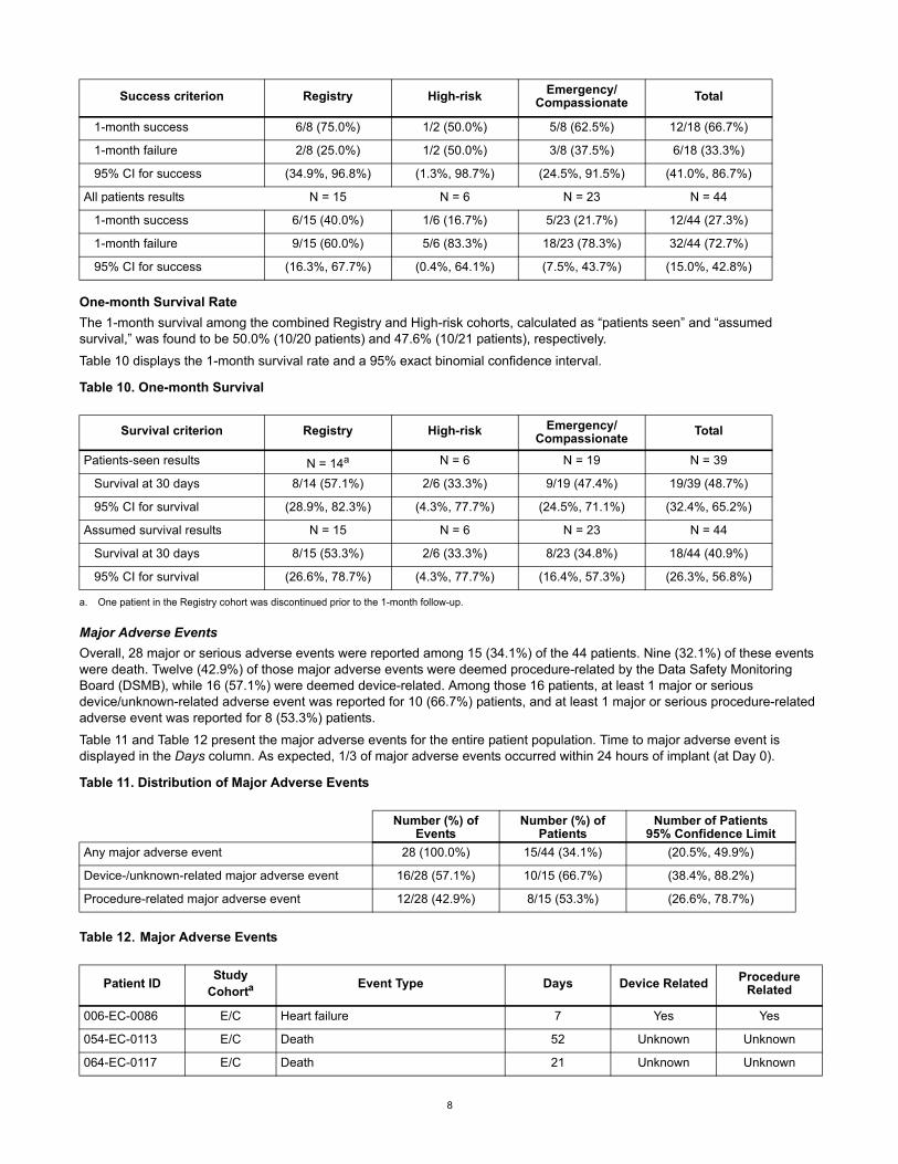
Major Adverse Events
Overall, 28 major or serious adverse events were reported among 15 (34.1%) of the 44 patients. Nine (32.1%) of these events were death. Twelve (42.9%) of those major adverse events were deemed procedure-related by the Data Safety Monitoring Board (DSMB), while 16 (57.1%) were deemed device-related. Among those 16 patients, at least 1 major or serious device/unknown-related adverse event was reported for 10 (66.7%) patients, and at least 1 major or serious procedure-related adverse event was reported for 8 (53.3%) patients.
Table 11 and Table 12 present the major adverse events for the entire patient population. Time to major adverse event is displayed in the Days column. As expected, 1/3 of major adverse events occurred within 24 hours of implant (at Day 0).
Table 11. Distribution of Major Adverse Events
Table 12. Major Adverse Events
1-month success 6/8 (75.0%) 1/2 (50.0%) 5/8 (62.5%) 12/18 (66.7%)
1-month failure 2/8 (25.0%) 1/2 (50.0%) 3/8 (37.5%) 6/18 (33.3%)
95% CI for success (34.9%, 96.8%) (1.3%, 98.7%) (24.5%, 91.5%) (41.0%, 86.7%)
All patients results N = 15 N = 6 N = 23 N = 44
1-month success 6/15 (40.0%) 1/6 (16.7%) 5/23 (21.7%) 12/44 (27.3%)
1-month failure 9/15 (60.0%) 5/6 (83.3%) 18/23 (78.3%) 32/44 (72.7%)
95% CI for success (16.3%, 67.7%) (0.4%, 64.1%) (7.5%, 43.7%) (15.0%, 42.8%)
Survival criterion Registry High-risk Emergency/ Compassionate
Total
Patients-seen results N = 14a
a. One patient in the Registry cohort was discontinued prior to the 1-month follow-up.
N = 6 N = 19 N = 39
Survival at 30 days 8/14 (57.1%) 2/6 (33.3%) 9/19 (47.4%) 19/39 (48.7%)
95% CI for survival (28.9%, 82.3%) (4.3%, 77.7%) (24.5%, 71.1%) (32.4%, 65.2%)
Assumed survival results N = 15 N = 6 N = 23 N = 44
Survival at 30 days 8/15 (53.3%) 2/6 (33.3%) 8/23 (34.8%) 18/44 (40.9%)
95% CI for survival (26.6%, 78.7%) (4.3%, 77.7%) (16.4%, 57.3%) (26.3%, 56.8%)
Number (%) of Events
Number (%) of Patients
Number of Patients 95% Confidence Limit
Any major adverse event 28 (100.0%) 15/44 (34.1%) (20.5%, 49.9%)
Device-/unknown-related major adverse event 16/28 (57.1%) 10/15 (66.7%) (38.4%, 88.2%)
Procedure-related major adverse event 12/28 (42.9%) 8/15 (53.3%) (26.6%, 78.7%)
Patient IDStudy
CohortaEvent Type Days Device Related Procedure
Related
006-EC-0086 E/C Heart failure 7 Yes Yes
054-EC-0113 E/C Death 52 Unknown Unknown
064-EC-0117 E/C Death 21 Unknown Unknown
Success criterion Registry High-risk Emergency/ Compassionate Total
8

Supplemental Clinical InformationUpon completion of the clinical evaluation summarized above, the AMPLATZER™ Post Infarct VSD Occluder continued to be implanted on an emergency and compassionate use basis. Data gathered from these cases was not collected in accordance with the original study protocol, but is summarized here as supplemental information in the interest of providing all available clinical experience regarding use of the AMPLATZER™ Post Infarct VSD Occluder. Data for 247 AMPLATZER™ Post Infarct VSD Occluder emergency or compassionate use cases performed from March 2002 through July 2016 in 243 patients are summarized in Tables 13, Figure 2, and Table 14. Four of these patients each had two procedures which results in the number of cases being more than the number of patients. The data provide an overview of case demographics, patient status, and procedural outcomes.
064-EC-0117 E/C Acute tubular necrosis 4 No Unknown
170-EC-0114 E/C Death 1 Unknown No
004-V-008 REG Blood loss 0 No Yes
020-V-006 REG Device kinking 0 No Yes
020-V-006 REG Cardiac perforation 0 No Yes
020-V-006 REG Death 0 No Yes
025-V-026 REG Aneurysm 68 No Yes
025-V-027 REG Respiratory arrest 1 No Unknown
025-V-027 REG Death 4 No Unknown
025-V-048 REG Arterial pulse loss 84 No Yes
025-V-048 REG Other – thrombocytopeniab 0 Unknown Yes
026-V-005 REG Bradycardia 0 Unknown Unknown
007-VHR-013 HR Multiple organ failure 2 Unknown Unknown
007-VHR-013 HR Sepsis 7 Unknown Unknown
007-VHR-013 HR Death 16 Unknown Unknown
016-VHR-034 HR Death 3 No Unknown
016-VHR-034 HR Cardiogenic shock 1 Unknown Unknown
016-VHR-034 HR Tricuspid regurgitation/tricuspid insufficiency/valvular regurgitation
0 No Yes
025-VHR-001 HR Death 10 Unknown Unknown
169-VHR-008 HR Blood loss 0 No Yes
169-VHR-055 HR Death 4 Unknown Unknown
169-VHR-055 HR Tricuspid regurgitation/tricuspid insufficiency/valvular regurgitation
0 Unknown Yes
169-VHR-055 HR Hypotension 0 Unknown Yes
169-VHR-055 HR Renal insufficiency 2 Unknown Unknown
169-VHR-055 HR Hemolysis 1 Yes No
a. Study cohorts: E/C – Emergency/Compassionate; REG – Registry; HR – High Riskb. No event type was reported on the DSMB CRF. Thrombocytopenia was determined by the investigator.
Patient IDStudy
CohortaEvent Type Days Device Related Procedure
Related
9

Table 13 provides a demographic summary, incidence rate of myocardial infarction, and cardiogenic shock by case type. Requests for emergency use account for 79% (196/247) and requests for compassionate use account for 21% (51/247) of the cases. Patients’ ages range from five to ninety-three years and the overall population was evenly divided between female and male patients. Ninety-two percent (92%) of the patient population presented following an acute myocardial infarction and fifty-five percent (55%) were in cardiogenic shock. Over half (54%) of the patients experienced both acute myocardial infarction and cardiogenic shock.
Patient Status
The status of the 243 patients is provided in Figure 2.
One hundred and ninety-four (194) patients (80%) were successfully implanted with the device. One hundred and twenty-two of the implanted patients (63%) survived and seventy-two (37%) died post-implant. Early death is defined as occurring ≤30 days post procedure and late death is defined as occurring >30 days post procedure. Early death occurred in sixty-seven implanted patients (35%) and late death occurred in five implanted patients (3%).
Forty-nine (49) patients (20%) underwent an unsuccessful attempt to implant the device. Within this group of patients undergoing an implant attempt, the survival rate was 49% and the death rate was 51%.
Among patients implanted with the device, there were 16 patients who underwent a device explant. Ten patients (63%) survived following device explant.
Table 13. Demographics and Medical History by Case
Mean ± SD [Min, Max] or n/N (%)
Variable Emergency Compassionate Overall
Number of Cases 196 51 247
Age (years) 69.2 ± 14.1 [5, 93] 64.5 ± 16.6 [16, 89] 68.2 ± 14.8 [5, 93]
Under 40 years of age 6 (3%) 5 (10%) 11 (4%)
Sex
Female 97 (49%) 26 (51%) 123 (50%)
Male 99 (51%) 25 (49%) 124 (50%)
Acute Myocardial Infarction (MI) 184 (94%) 43 (84%) 227 (92%)
Cardiogenic Shock 120 (61%) 16 (31%) 136 (55%)
Acute MI and Cardiogenic Shock 118 (60%) 16 (31%) 134 (54%)
Figure 2. Patient Status
10

All available procedure outcome data are presented in Table 14. Successful implant occurred in 80% (198/247) of the cases. Among patients implanted with the device, device embolization or migration occurred in 5% (9/198) of the cases.
Patients’ death rate was 40% (97/243), of which the majority (92/97) occurred within 30 days of the procedure.
Patient InformationRefer to the AMPLATZER™ Post-infarct Muscular VSD Occluder patient guide.
Directions for Use
Materials recommended for use with the device
• 0.035-inch AMPLATZER™ Noodlewire guidewire
• AMPLATZER™ TorqVue™ Delivery System - Refer to the AMPLATZER™ TorqVue™ Delivery System Instructions for Use.
Note: The device is packaged separately from the delivery system. Refer to Table 1 for the recommended delivery sheath size for this device.
• AMPLATZER™ TorqVue™ Exchange System - Refer to the AMPLATZER™ TorqVue™ Exchange System Instructions for Use.
Procedure
The AMPLATZER™ Post-infarct Muscular VSD Occluder is implanted percutaneously via right-or left-sided transcatheter approach. The approach depends on the location and the direction of the muscular defect.
This procedure is carried out under anesthesia and transesophageal echocardiography (TEE) guidance.
1. Prepare the patient for a standard transcatheter procedure, including administration of oral antiplatelet medication. Effective anticoagulation therapy should be maintained during and after the procedure as determined by the physician.
2. Define the location, size, and number of VSDs using angiography.
3. Use TEE to provide additional imaging of the VSD.
4. Select a device 3–4 mm larger than the largest VSD measurement obtained by TEE and angiography at end-diastole.
5. Select a delivery system based on the device size as indicated in Table 1.
6. Prepare the delivery system for use according to the manufacturer's instructions for use.
Note: Accurate defect sizing is crucial and mandatory for device selection.
Catheter Technique (retrograde)
1. Establish a stable arteriovenous loop.
CAUTION: During formation of the arteriovenous loop the guidewire should be covered by the catheter to prevent entanglement or damage to the tricuspid valve apparatus.
CAUTION: Do not advance the delivery sheath if resistance is met while crossing the tricuspid valve to gain access to the VSD. This may be an indication that the tricuspid valve chordae may be entangled. Continuing to advance when the delivery sheath meets resistance may damage the tricuspid valve resulting in tricuspid valve regurgitation.
2. Advance the dilator and sheath over the guidewire until the tip of the dilator meets the catheter in the right atrium.
3. Move the system as a unit until the sheath is within the ascending aorta.
Table 14. Procedure Outcomes (Attempts and Implants)
n/N (%)
Variable Emergency Compassionate Overall
Successful Implanta
a. The successful implant denominator is the number of procedures. Four patients had 2 procedures each.
154/196 (79%) 44/51 (86%) 198/247 (80%)
Device Embolization / Migration inImplanted Patients 6/154 (4%) 3/44 (7%) 9/198 (5%)
Deathsb
b. The deaths denominator is the number of patients (243), not the number of procedures (247).
86/193 (45%) 11/51 (22%) 97/243 (40%)
Early Deaths (≤ 30 Days) 83 9 92
Late Deaths (> 30 Days) 3 2 5
11

4. Remove the dilator.
5. Slowly retract the sheath from the ascending aorta until the tip reaches the left ventricular outflow tract.
6. Advance the guidewire and catheter until the tip of the catheter reaches the left ventricular apex.
7. Position the sheath in the apex using the catheter as an anchor.
Catheter Technique (antegrade)
1. Access the VSD with a guidewire.
2. Advance the dilator and sheath over the guidewire until the tip of the dilator crosses the VSD.
3. Remove the dilator.
Device Preparation
1. Pass the delivery cable through the loader and attach the AMPLATZER™ Post-infarct Muscular VSD Occluder to the delivery cable by rotating the device clockwise approximately 5 turns until the occluder is secure. Then rotate the device counterclockwise 1/8 of one turn to facilitate release of the device.
2. Immerse the device and loader in sterile saline and retract the device into the loader.
3. Advance the hemostasis valve over the delivery cable and attach to hub. Tighten the hemostasis valve over the delivery cable.
4. Attach a syringe to the hemostasis valve and flush with sterile saline to remove air. Close stopcock and remove syringe.
Device Placement
1. Slowly remove the guidewire and allow for blood backflow to purge air from the system.
2. Connect the loader to the sheath.
3. Advance the device from the loader into the sheath and, without rotation, advance the device to the tip of the sheath.
4. The loader must be attached firmly to the sheath to completely close the gap between the internal surfaces of the 2 components.
5. Retract the sheath slowly until the distal disc is deployed.
6. Use TEE and angiography as a guide during each step of device deployment.
7. Pull the entire assembly (delivery cable and delivery sheath) into the VSD.
8. Retract the sheath to deploy the waist of the device in the defect.
9. Confirm disc configuration and positioning.
- If the device does not conform to its original configuration or if the position of the device is unstable:
1. Slowly retract the delivery cable while advancing the sheath until the device is completely within the sheath.
2. Redeploy or remove the device from the patient.
- If the device position is satisfactory:
CAUTION: Use caution during deployment of the proximal (right ventricular) disc to avoid entanglement of the tricuspid valve apparatus. Entanglement of the valve apparatus could lead to complications of tricuspid valve regurgitation.
1. Retract the sheath to deploy the proximal disc.
2. Attach the plastic vise to the delivery cable, then release the device by rotating the delivery cable counterclockwise until it separates from the device.
3. Retract the delivery cable into the sheath.
- Remove the delivery cable and sheath from the patient.
Post-implant Imaging
• Complete a TEE evaluation to confirm device placement and assess for residual shunting.
• Perform an angiogram to assess for residual flow through the device.
Post-procedure Instructions• Endocarditis prophylaxis should be followed according to American Heart Association recommendations.
• Any patient who has a residual shunt should undergo an echocardiographic evaluation of the residual shunt every 6 months until complete closure of the defect has been confirmed.
• If the patient would like to receive a permanent patient identification card, complete the implant registration form and send the completed form to AGA Medical.
• MedicAlert™ service – Recommend the patient become a MedicAlert™ service member by calling +1.888.633.4298 or enrolling online at www.medicalert.org. The patient will be asked to provide his condition, implanted device name, and restrictions concerning the use of an MRI.
12

Disposal• The carton and IFU are recyclable. Dispose of all packaging materials as appropriate.
• Devices may be disposed of following standard solid biohazard waste procedures.
WarrantyAGA Medical Corporation warrants to buyer that, for a period equal to the validated shelf life of the product, this product shall meet the product specifications established by the manufacturer when used in accordance with the manufacturer's instructions for use and shall be free from defects in materials and workmanship. AGA Medical Corporation's obligation under this warranty is limited to replacing or repairing at its option, at its factory, this product if returned within the warranty period to AGA Medical Corporation and after confirmed to be defective by the manufacturer.
EXCEPT AS EXPRESSLY PROVIDED IN THIS WARRANTY, AGA MEDICAL CORPORATION DISCLAIMS ANY REPRESENTATION OR WARRANTY OF ANY KIND, EXPRESS OR IMPLIED, INCLUDING ANY WARRANTY AS TO MERCHANTABILITY OR FITNESS FOR A PARTICULAR PURPOSE.
See the Terms and Conditions of Sale for further information.
State of California (USA) Only:WARNING: This product and its packaging have been sterilized with ethylene oxide. The packaging may expose you to ethylene oxide, a chemical known to the State of California to cause cancer or birth defects or other reproductive harm.
13

Symbol DefinitionsThe following symbols may appear on the device packaging.
Symbol Definition
Manufacturer
EU authorized representative
Catalog number
Serial number
Lot number
Use by date
Date of manufacture
Quantity
Do not reuse
Sterilized using ethylene oxide
Consult instructions for use
Keep dry
Do not use if package is damaged
Not made with natural rubber latex
Inner diameter
Outer diameter
Length
Usable length
Recommended delivery sheath/catheter dimensions
14

MR conditional
Indication of conformity with the essential health and safety requirements set out in European Directives
Federal law (USA) restricts this device to sale by or on the order of a physician (or properly licensed practitioner).
Do not resterilize
15

Unless otherwise noted, ™ indicates that the name is a trademark of, or licensedto, St. Jude Medical or one of its subsidiaries. ST. JUDE MEDICAL and the nine-squares symbol are trademarks and services marks of St. Jude Medical, Inc. and
its related companies. AGA Medical Corporation is an indirectly wholly-ownedsubsidiary of St. Jude Medical, Inc.
MedicAlert is a trademark of MedicAlert Foundation United States, Inc.
Pat. http://patents.sjm.com
© 2017 St. Jude Medical, Inc. All Rights Reserved.
AGA Medical Corporation 5050 Nathan Lane North
Plymouth, MN
55442 USA
+1 888 478 5833
+1 651 756 5883
sjm.com
ARTUS600007173 A
2017-01
*600007173*