post-infection cognitive impairments in a cohort of
TRANSCRIPT

RESEARCH ARTICLE Open Access
Post-infection cognitive impairments in acohort of elderly patients with COVID-19Yu-Hui Liu1†, Ye-Ran Wang1†, Qing-Hua Wang1†, Yang Chen1, Xian Chen2, Ying Li3, Yuan Cen4, Cheng Xu5,Tian Hu2, Xu-Dong Liu2, Ling-Li Yang4, Si-Jing Li4, Xue-Fei Liu3, Chun-Mei Liu3, Jie Zhu1, Wei Li1, Li-Li Zhang1,Juan Liu1* and Yan-Jiang Wang1*
Abstract
Background: Understanding the long-term effects of coronavirus disease 2019 (COVID-19) on cognitive function isessential for monitoring the cognitive decline in the elderly population. This study aims to assess the currentcognitive status and the longitudinal cognitive decline in elderly patients recovered from COVID-19.
Methods: This cross-sectional study recruited 1539 COVID-19 inpatients aged over 60 years who were dischargedfrom three COVID-19-designated hospitals in Wuhan, China, from February 10 to April 10, 2020. In total, 466uninfected spouses of COVID-19 patients were selected as controls. The current cognitive status was assessed usinga Chinese version of the Telephone Interview of Cognitive Status-40 (TICS-40) and the longitudinal cognitivedecline was assessed using an Informant Questionnaire on Cognitive Decline in the Elderly (IQCODE). Cognitiveassessments were performed 6 months after patient discharge.
Results: Compared with controls, COVID-19 patients had lower TICS-40 scores and higher IQCODE scores [TICS-40median (IQR): 29 (25 to 32) vs. 30 (26 to 33), p < 0.001; IQCODE median (IQR): 3.19 (3.00 to 3.63) vs. 3.06 (3.00 to3.38), p < 0.001]. Severe COVID-19 patients had lower TICS-40 scores and higher IQCODE scores than non-severeCOVID-19 patients [TICS-40 median (IQR): 24 (18 to 28) vs. 30 (26 to 33), p < 0.001; IQCODE median (IQR): 3.63 (3.13to 4.31) vs. 3.13 (3.00 to 3.56), p < 0.001] and controls [TICS-40 median (IQR): 24 (18 to 28) vs. 30 (26 to 33), p < 0.001;IQCODE median (IQR) 3.63 (3.13 to 4.31) vs. 3.06 (3.00 to 3.38), p < 0.001]. Severe COVID-19 patients had a higherproportion of cases with current cognitive impairment and longitudinal cognitive decline than non-severe COVID-19 patients [dementia: 25 (10.50 %) vs. 9 (0.69 %), p < 0.001; Mild cognitive impairment (MCI): 60 (25.21 %) vs. 63(4.84 %), p < 0.001] and controls [dementia: 25 (10.50 %) vs. 0 (0 %), p < 0.001; MCI: 60 (25.21 %) vs. 20 (4.29 %), p <0.001)]. COVID-19 severity, delirium and COPD were risk factors of current cognitive impairment. Low educationlevel, severe COVID-19, delirium, hypertension and COPD were risk factors of longitudinal cognitive decline.
Conclusions: Severe acute respiratory syndrome coronavirus 2 (SARS-CoV-2) infection is associated with anincreased risk of long-term cognitive decline in elderly population. COVID-19 patients, especially severe patients,should be intensively monitored for post-infection cognitive decline.
Keywords: COVID-19, Coronavirus, Cognitive impairment, Post infection
© The Author(s). 2021 Open Access This article is licensed under a Creative Commons Attribution 4.0 International License,which permits use, sharing, adaptation, distribution and reproduction in any medium or format, as long as you giveappropriate credit to the original author(s) and the source, provide a link to the Creative Commons licence, and indicate ifchanges were made. The images or other third party material in this article are included in the article's Creative Commonslicence, unless indicated otherwise in a credit line to the material. If material is not included in the article's Creative Commonslicence and your intended use is not permitted by statutory regulation or exceeds the permitted use, you will need to obtainpermission directly from the copyright holder. To view a copy of this licence, visit http://creativecommons.org/licenses/by/4.0/.The Creative Commons Public Domain Dedication waiver (http://creativecommons.org/publicdomain/zero/1.0/) applies to thedata made available in this article, unless otherwise stated in a credit line to the data.
* Correspondence: [email protected]; [email protected]†Yu-Hui Liu, Ye-Ran Wang and Qing-Hua Wang contributed equally to thiswork.1Department of Neurology and Centre for Clinical Neuroscience, DapingHospital, Third Military Medical University, Chongqing, ChinaFull list of author information is available at the end of the article
Liu et al. Molecular Neurodegeneration (2021) 16:48 https://doi.org/10.1186/s13024-021-00469-w

BackgroundThe coronavirus disease 2019 (COVID-19) pandemichas affected over 180 million patients thus far, and thenumber is still increasing [1]. Understanding the short-and long-term health consequences of COVID-19 istherefore critical. Severe acute respiratory syndromecoronavirus 2 (SARS-CoV-2) infection causes damagesto multiple systems, including the respiratory, digestive,cardiovascular, renal, immune and nervous systems [2].More than one-third of hospitalized patients withCOVID-19 experience a variety of neurologic manifesta-tions at the acute stage of the infection, including alteredcognitive and mental status, cerebrovascular diseases,headache, vertigo, anosmia and ageusia [3], and neuro-logical sequelae are also reported [4–7]. Acute cognitivecomplications are common [8]; however, long-term ef-fects of COVID-19 on cognition are not clear yet. Thefirst bulk of hospitalized COVID-19 patients in Wuhan,China, has recovered for half a year. This study aimed toinvestigate the long-term impact of SARS-CoV-2 infec-tion on cognitive changes 6 months after recovery, andto determine risk factors of cognitive impairment in eld-erly patients recovered from COVID-19.
MethodsParticipantsParticipants in this study were inpatients who were dis-charged between February 10 and April 10, 2020, fromthree hospitals, including Huoshenshan Hospital, TongjiTaikang Hospital and General Hospital of the CentralTheatre Command of the People’s Liberation Army,which were designated to treat COVID-19 patients inWuhan during the pandemic in early 2020. Eligibility in-cluded the following: (1) aged 60 years and older; (2)agreed to participate in this study. Subjects were ex-cluded if they had the following conditions: (1) did notagree to participate, did not understand the questions inthe questionnaires, or had communicative obstacles dueto language or hearing reasons; (2) pre-existing subject-ive or diagnosed dementia; (3) a family history of de-mentia which may increase the risk of cognitiveimpairment; (4) a concomitant neurologic disorder po-tentially affecting cognitive function; and (5) severe car-diac, hepatic, renal diseases or any kind of tumour.Uninfected spouses that co-lived with the patients in thesame environment were selected as controls. In total,3233 patients discharged from the hospitals during thedesignated study period were screened.Ultimately, 1539 patients, including 238 severe cases
and 1301 non-severe cases, were eligible for this survey,and 466 spouses were recruited as controls. COVID-19designated hospitals including Huoshenshan and TongjiTaikang Hospital were disbanded after the crisis. There-fore, the protocols were approved by the institutional
review boards of Daping Hospital, Third Military Med-ical University, which launched this study. Since thisstudy is conducted based on telephone interviews, writ-ten consents were waived but verbal informed consentswere obtained from all participants or their legal guard-ians prior to the survey.
Clinical and cognitive assessmentThe diagnosis of COVID-19 was based on the WorldHealth Organization interim guidance [9]. The severityof COVID-19 was defined as severe or non-severe fol-lowing the American Thoracic Society guidelines forcommunity-acquired pneumonia [10]. Accordingly, se-vere cases with COVID-19 were defined as: fever or sus-pected respiratory infection, plus at least one of thefollowing conditions: respiratory rate > 30 breaths/min,severe respiratory distress, or SpO2 < 90 % on room air.Uninfected spouses were confirmed to be uninfected byhigh-throughput sequencing or real-time reverse-transcriptase polymerase-chain-reaction assay (RT-PCR)for nasal and pharyngeal swab specimens.The survey was conducted 6 months after patient dis-
charge. Due to the emerging infection risk, participantswere interviewed by a telephone survey. The telephoneinterview was conducted by a group of trained raters. Par-ticipants were allowed to terminate the survey at any time.The following information was collected from medical
records and a knowledgeable family member for each par-ticipant: demographics, including age, sex, education level,body mass index (BMI), comorbidities, including hyper-tension, diabetes mellitus, coronary heart disease,hyperlipidaemia, a history of cardiovascular diseases orstroke, and chronic obstructive pulmonary disease(COPD). The current cognitive status of participants wasassessed using a Chinese version of the Telephone Inter-view of Cognitive Status-40 (TICS-40, SupplementaryTable 1), which was previously validated [11]. The TICS-40 includes 10 variables and has a maximum of 40 points.Score ≤ 20 was determined as mild cognitive impairment(MCI), and score ≤ 12 was determined as dementia [11].Subjects’ family informants were interviewed to report thecognitive decline of patients and their spouses over theprevious 6 months using the Chinese version of the shortform Informant Questionnaire on Cognitive Decline inthe Elderly (IQCODE, Supplementary Table 2) [12], whichcontains 16 items that rate changes in memory and othercognitive domains and was previously validated [13]. Cog-nitive decline was defined as an IQCODE score ≥ 3.5,which provided a sensitivity of 92 % and a specificity of80 % [12].
Quality control of cognitive assessmentA group of raters consisting of six experienced nurseswere trained with the questionnaires by a neurologist
Liu et al. Molecular Neurodegeneration (2021) 16:48 Page 2 of 10

experienced in cognitive assessments. Seven subjectswith dementia, five subjects with MCI and nine cogni-tively normal subjects were recruited for the interraterreliability assessment. The intraclass correlation coeffi-cient (ICC) was 0.990 for TICS and 0.934 for IQCODE,reflecting a high interrater reliability for the assessments.
Statistical analysisContinuous variables, including age, education, BMI,TICS-40 and IQCODE, were not normally distributed,thus Mann-Whitney U tests were used to compare thesevariables between groups, and Kruskal Wallis tests wereused for multigroup comparison. Two-sample tests ofproportions (for categorical data) were used to compareproportions. As for comparison of TICS-40 andIQCODE, variables including sex, age, education, BMIand comorbidities that were significantly differentamong groups were adjusted for potential confoundingeffects.Linear and logistic regression models were utilized to
assess the association between COVID-19 and cognitiveoutcomes. Mechanical ventilation was excluded for re-gression analyses to reduce collinearity with COVID-19severity. In linear regression models, cognitive outcomes(TICS and IQCODE) were fitted as continuous variables.The linear regression models were adjusted for age andsex, and in the next step, variables including educationlevel, BMI, COVID-19 severity, intensive care unit (ICU)admission, high flow oxygen therapy, delirium, hyperten-sion, diabetes mellitus, hyperlipidaemia, stroke history,coronary heart disease, and COPD were added into theadjusted models. In logistical regression models, we firstfitted univariate models with a single candidate variableat one time, with the cross-sectional cognitive status(TICS-40 ≤ 20 indicates cognitive impairment) or longi-tudinal cognitive change (IQCODE ≥ 3.5 indicates cogni-tive decline) as the dependent variable and educationlevel, BMI, COVID-19 severity, ICU admission history,delirium, hypertension, diabetes mellitus, hyperlipid-aemia, COPD, or history of stroke and coronary heartdisease, as the independent variable. Potential risk fac-tors with a P value less than 0.2 in univariate analyseswere included in the final multivariate regression modelwith adjustment for age and sex. Statistical analyses wereconducted using SPSS statistical package version 24(IBM SPSS Statistics for Windows, Armonk, NY, USA).
ResultsDemographics of subjectsIn total, 3233 COVID-19 patients aged 60 years or abovewere screened for eligibility for this study. 1694 patientsfailed screening, among which 235 patients did not meetthe inclusion criteria, 1405 patients declined to partici-pate and 54 patients died after discharge. Finally, 1539
patients were enrolled in this study. Among 1317 co-living spouses, 851 spouses failed screening due to thefollowing reasons: 592 spouses were infected with SARS-CoV-2 and not admitted in the three designated hospi-tals, thus their demographic and medical informationwere not obtained. 183 spouses declined to participate,63 spouses did not meet the inclusion criteria and 13spouses died (Fig. 1). Finally, 466 spouses were includedin this study.There were no significant differences in the mean age,
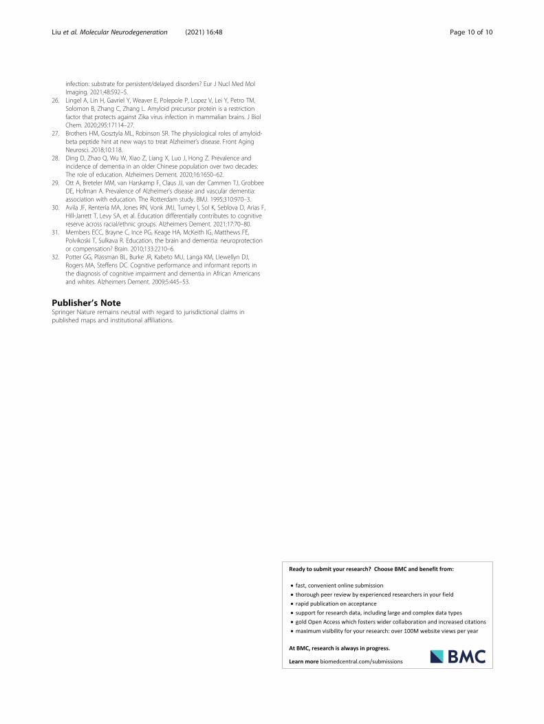
frequencies of males, median education level and BMIbetween COVID-19 patients and controls. Moreover,frequencies of hypertension, diabetes mellitus, hyperlip-idaemia, COPD, stroke, and coronary heart disease inCOVID-19 patients were comparable to those in con-trols. Compared with non-severe COVID-19 patients,severe COVID-19 patients were older, had a higher pro-portion of males and a higher BMI, and more frequentlyhad hypertension, a history of stroke, coronary heartdisease, and COPD. Severe COVID-19 patients hadcomparable frequencies of diabetes mellitus and hyper-lipidaemia with controls. Compared with non-severepatients, severe cases had higher education levels. More-over, severe cases had higher frequencies of ICUadmission, receiving mechanical ventilation, high flowoxygen therapy and incidences of delirium duringhospitalization than non-severe cases (Table 1). Further-more, significant differences were not found in thedemographic characteristics, including age, sex,education levels, BMI, incidences of severe COVID-19,frequencies of hypertension, diabetes mellitus, hyperlip-idaemia, stroke history, coronary heart disease andCOPD, frequencies of ICU admission, receiving mechan-ical ventilation, high flow oxygen therapy and incidencesof delirium, between patients included and those not in-cluded in this study (Table 2).
Current cognitive impairment of COVID-19 patientsTICS-40 was utilized to determine the current cognitivestatus of participants. COVID-19 patients had lowerTICS-40 scores than controls [median (IQR): 29 (25 to32) vs. 30 (26 to 33), p < 0.001] (Fig. 2A). SevereCOVID-19 patients had lower TICS-40 scores than non-severe patients [median (IQR): 24 (18 to 28) vs. 30 (26to 33), p < 0.001] and controls [median (IQR): 24 (18 to28) vs. 30 (26 to 33), p < 0.001]. TICS-40 scores werecomparable between non-severe COVID-19 cases andcontrols (p = 0.648) (Fig. 2B). Furthermore, the differ-ences remained significant after adjusted for potentialconfounding factors (Supplementary Tables 3 and 4).A cut-off value of 20 was defined for MCI and 12 for
dementia [13]. Severe COVID-19 patients more fre-quently reported to have dementia and MCI than non-severe COVID-19 patients [dementia: 25 (10.50 %) vs. 9
Liu et al. Molecular Neurodegeneration (2021) 16:48 Page 3 of 10
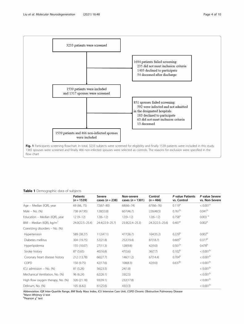
Fig. 1 Participants screening flowchart. In total, 3233 subjects were screened for eligibility and finally 1539 patients were included in this study.1365 spouses were screened and finally 466 non-infected spouses were selected as controls. The reasons for exclusion were specified in theflow chart
Table 1 Demographic data of subjects
Patients(n = 1539)
Severecases (n = 238)
Non-severecases (n = 1301)
Control(n = 466)
P value Patientsvs. Control
P value Severevs. Non-Severe
Age – Median (IQR), year 69 (66, 75) 72(67–80) 68(66–74) 67(66–76) 0.119a < 0.001a
Male – No. (%) 738 (47.95) 128(53.8) 607(46.7) 226(48.5) 0.761b 0.041b
Education – Median (IQR), year 12 (9–12) 12(6–12) 12(9–12) 12(6–12) 0.758a 0.003 a
BMI – Median (IQR), kg/m2 24.0(22.5–25.4) 24.4(22.9–25.7) 23.9(22.4–25.3) 24.2(22.5–25.6) 0.407a 0.002a
Coexisting disorders – No. (%)
Hypertension 589 (38.27) 112(47.1) 477(36.7) 164(35.2) 0.229b 0.002b
Diabetes mellitus 304 (19.75) 52(21.8) 252(19.4) 87(18.7) 0.605b 0.377b
Hyperlipidemia 155 (10.07) 27(11.3) 128(9.8) 42(9.0) 0.501b 0.478b
Stroke history 87 (5.65) 40(16.8) 47(3.6) 36(7.7) 0.102b < 0.001b
Coronary heart disease history 212 (13.78) 66(27.7) 146(11.2) 67(14.4) 0.704b < 0.001b
COPD 150 (9.75) 42(17.6) 108(8.3) 42(9.0) 0.637b < 0.001b
ICU admission – No. (%) 81 (5.26) 56(23.5) 24(1.8) < 0.001b
Mechanical Ventilation, No. (%) 96 (6.24) 62(26.1) 33(2.5) < 0.001b
High flow oxygen therapy, No. (%) 326 (21.18) 93(39.1) 232(17.8) < 0.001b
Delirium, No. (%) 105 (6.82) 61(25.6) 43(3.3) < 0.001b
Abbreviation: IQR Inter-Quartile Range, BMI Body Mass Index, ICU Intensive Care Unit, COPD Chronic Obstructive Pulmonary DiseaseaMann-Whitney U testbPearson χ2 test
Liu et al. Molecular Neurodegeneration (2021) 16:48 Page 4 of 10

(0.69 %), p < 0.001; MCI: 60 (25.21 %) vs. 63 (4.84 %), p <0.001] and controls [dementia: 25 (10.50 %) vs. 0 (0 %),p < 0.001; MCI: 60 (25.21 %) vs. 20 (4.29 %), p < 0.001)].However, no difference was found in the proportion ofcases with dementia or MCI between non-severeCOVID-19 patients and controls (dementia: p = 0.703;MCI: p = 0.123) (Fig. 2C). These findings indicate thatcurrent cognitive impairment was associated with bothSARS-CoV-2 infection and severity of COVID-19. Fur-thermore, differences were still significant after adjustedfor potential confounding factors.
Longitudinal cognitive decline of COVID-19 patientsAt 6 months after discharge, the cognitive decline ofCOVID-19 patients and their non-infected spouses wasassessed with the IQCODE, with higher IQCODE scoresindicating larger cognitive decline. COVID-19 patientshad higher IQCODE scores than controls [median(IQR): 3.19 (3.00 to 3.63) vs. 3.06 (3.00 to 3.38), p <0.001] (Fig. 2D). Moreover, severe COVID-19 cases hadhigher IQCODE scores than non-severe cases [median(IQR): 3.63 (3.13 to 4.31) vs. 3.13 (3.00 to 3.56), p <0.001] and controls [median (IQR): 3.63 (3.13 to 4.31)vs. 3.06 (3.00 to 3.38), p < 0.001]. Non-severe cases hadhigher IQCODE scores than controls (p < 0.001) (Fig. 2E).Furthermore, differences remained significant after ad-justed for potential confounding factors (SupplementaryTables 5 and 6).
Based on a cut-off value of 3.5 for clinically meaningfulcognitive decline [12], severe COVID-19 patients morefrequently showed cognitive decline than non-severeCOVID-19 patients [141 (59.24 %) vs. 373 (28.67 %), p <0.001] and controls [141 (59.24 %) vs. 100 (21.46 %), p <0.001]. Meanwhile, non-severe COVID-19 patients alsomore frequently showed cognitive decline than controls[373 (28.67 %) vs. 100 (21.46 %), p = 0.003] (Fig. 2F).These findings indicate that both SARS-CoV-2 infectionand severity of COVID-19 are associated with longitu-dinal cognitive decline. Furthermore, differences werestill significant after adjusted for potential confoundingfactors.
Risk factors for cognitive impairment and decline inCOVID-19 patientsRegression models were used to investigate potential riskfactors of current cognitive impairment or post-infectioncognitive decline in COVID-19 patients. In univariate lo-gistical regression analyses, age, severe COVID-19, ICUadmission, delirium, stroke history, coronary heart dis-ease and COPD were associated with cognitive impair-ment. Severe of COVID-19, delirium and COPDremained associated with current cognitive impairmentin the multivariate model (Table 3). In the linear regres-sion model with adjustment for age and sex, COVID-19severity, ICU admission, delirium, and COPD were asso-ciated with lower TICS-40 scores. Higher education level
Table 2 Demographic data of subjects included and not included in this study
Patients included (n = 1539) Patients not included (n = 1694) P value
Age – Median (IQR), year 69 (66, 75) 70 (66, 75) 0.165a
Male – No. (%) 738 (47.95) 826 (48.76) 0.647b
Education – Median (IQR), year 12 (9–12) 12 (9–12) 0.226a
BMI – Median (IQR) 23.97 (22.52, 25.38) 23.92 (22.42, 25.41) 0.284a
Severe cases -No. (%) 238 (15.46) 257 (15.17) 0.845b
Coexisting disorders – No. (%)
Hypertension 589 (38.27) 640 (37.78) 0.800b
Diabetes mellitus 304 (19.75) 348 (20.54) 0.599b
Hyperlipidaemia 155 (10.07) 176 (10.39) 0.772b
Stroke history 87 (5.65) 91 (5.37) 0.758b
Coronary heart disease history 212 (13.78) 231 (13.64) 0.919b
COPD 150 (9.75) 163 (9.62) 0.905b
ICU admission – No. (%) 81 (5.26) 94 (5.55) 0.756b
Mechanical Ventilation, No. (%) 96 (6.24) 106 (6.26) 1.000b
High flow oxygen therapy, No. (%) 326 (21.18) 355 (20.96) 0.897b
Delirium, No. (%) 105 (6.82) 117 (6.91) 0.945b
Abbreviation: IQR Inter-Quartile Range, BMI Body Mass Index, ICU intensive care unit, COPD Chronic Obstructive Pulmonary DiseaseaMann-Whitney U testbPearson χ2 test
Liu et al. Molecular Neurodegeneration (2021) 16:48 Page 5 of 10

and high flow oxygen therapy were associated withhigher TICS-40 scores (Supplementary Table 7).Consistently, in univariate logistical regression ana-
lyses, age, lower education level, severe COVID-19, ICUadmission, delirium, hypertension, diabetes, stroke his-tory, coronary heart disease and COPD were associatedwith longitudinal cognitive decline. In the multivariatemodel with adjustment for age and sex, the associationremained significant for lower education level, se-vere COVID-19, delirium, hypertension and COPD.However, high flow oxygen therapy was protectiveagainst longitudinal cognitive decline (Table 4). In thelinear regression mode adjusted for age and sex,COVID-19 severity, ICU admission, delirium, hyperten-sion, hyperlipidaemia and COPD were found to be asso-ciated with higher IQCODE scores. However, high flowoxygen therapy was found to be associated with lowerIQCODE scores (Supplementary Table 8).
DiscussionCurrently, the association between COVID-19 and long-term cognitive change post-infection has rarely been in-vestigated. We found that COVID-19 patients, includingboth severe and non-severe cases, had worse cognitiveoutcomes 6 months after recovery, indicating thatSARS-CoV-2 infection may affect long-term cognitiveperformance, particularly in severe patients, amongwhich 35.71 % of patients had current cognitive impair-ment, and 59.24 % reported longitudinal cognitive de-cline. This study identified several risk factors for post-infection cognitive impairment in COVID-19 patients,including older age, lower education level, comorbidities,severe COVID-19, ICU admission and delirium.While the exact mechanism underlying this association
remains to be elucidated, the aetiology of cognitive de-cline post-SARS-CoV-2 infection may be multifactorial.First, a potent mechanism underlying cognitive decline
Fig. 2 Current cognitive status and longitudinal cognitive decline in COVID-19 patients and controls. A TICS-40 scores of COVID-19 patients andcontrols. B TICS-40 scores of severe and non-severe cases with COVID-19 and controls. C Composition of subjects with different TICS-40 scores insevere and non-severe cases with COVID-19 and controls. D IQCODE scores of COVID-19 patients and controls. E IQCODE scores of severe andnon-severe cases with COVID-19 and controls. F Composition of subjects with different IQCODE scores in severe and non-severe cases withCOVID-19 and controls. A and D, Wilcoxon-Mann Whitney test. B and E, Kruskal Wallis test. C, Fisher exact test. F, χ2 test
Liu et al. Molecular Neurodegeneration (2021) 16:48 Page 6 of 10

after SARS-CoV-2 infection is hypoxia [2], because brainregions associated with cognitive functions, such as thehippocampus, are susceptible to hypoxia induced neur-onal damage [14, 15]. Oxygen deficiency at the acutedisease stage and after recovery can cause damages to
neurons, which are sensitive to hypoxia [16]. This hy-pothesis is evidenced by our finding that severe COVID-19 patients had worse cognitive impairment, as these pa-tients were in a more hypoxic state even months afterrecovery [1, 17]. Moreover, the severity of COVID-19
Table 3 Logistic regression models to evaluate risk factors for cognitive impairment as indicated by TICS-40≤ 20 in COVID-19patients
Variables Univariable ORs(95 %CI) P value Multivariable ORs(95 %CI) P value
Age, year 1.030(1.009–1.052) 0.005
Sex, male 1.235(0.887–1.721) 0.212
Education, year 1.000(0.971–1.030) 0.987
BMI, kg/m2 1.055(0.957–1.163) 0.279
Severe, vs. non-severe 9.311(6.518–13.300) < 0.001 6.507(4.411–9.599) < 0.001
ICU admission, vs. no 8.601(5.326–13.890) < 0.001
High flow oxygen therapy, vs. no 1.901(1.324–2.727) < 0.001
Delirium, vs. no 7.850(5.076–12.139) < 0.001 3.714(2.247–6.136) < 0.001
Coexisting disorder, vs. no
Hypertension 1.278(0.915–1.787) 0.150
Diabetes 1.149(0.768–1.718) 0.500
Hyperlipidaemia 1.023(0.592–1.766) 0.935
Stroke history 3.329(1.989–5.572) < 0.001
Coronary heart disease 1.725(1.134–2.622) 0.011
COPD 4.733(3.169–7.068) < 0.001 4.224(2.693–6.625) < 0.001
Dependent variables: TICS-40 score ≤ 20Independent variables: age, sex, severity, ICU admission, high flow oxygen therapy, delirium, hypertension, stroke, coronary heart disease, COPDAbbreviations: BMI body mass index, ICU intensive care unit, COPD Chronic Obstructive Pulmonary Disease
Table 4 Logistic regression models to evaluate the risk factors for longitudinal cognitive decline as indicated by IQCODE score ≥ 3.5in COVID-19 patients
Variables Univariable ORs (95 %CI) P value Multivariable ORs (95 %CI) P value
Age, year 1.023(1.009–1.037) 0.001
Sex, male 0.934(0.756–1.157) 0.530
Education, year 0.959(0.936–0.982) 0.001 0.968(0.944–0.993) 0.011
BMI, kg/m2 1.007(0.947–1.072) 0.821
Severe, vs. non-severe 3.616(2.719–4.810) < 0.001 2.833(2.065–3.888) < 0.001
ICU admission, vs. no 5.100(3.122–8.331) < 0.001
High flow oxygen therapy, vs. no 0.828(0.635–1.079) 0.163 0.421(0.302–0.586) < 0.001
Delirium, vs. no 5.578(3.597–8.650) < 0.001 5.480(3.292–9.122) < 0.001
Coexisting disorders, vs. no
Hypertension 1.825(1.470–2.266) < 0.001 1.661(1.320–2.091) < 0.001
Diabetes 1.344(1.037–1.743) 0.026
Hyperlipidaemia history 1.180(0.835–1.666) 0.348
Stroke history 1.590(1.027–2.463) 0.038
Coronary heart diseases 1.800(1.341–2.415) < 0.001
COPD 2.368(1.686–3.326) < 0.001 2.005(1.398–2.877) < 0.001
Dependent variable: IQCODE ≥ 3.5Independent variables: age, sex, education, severity, ICU admission, high flow oxygen therapy, delirium, hypertension, diabetes, stroke, coronary heartdisease, COPDAbbreviations: BMI body mass index, ICU intensive care unit, COPD Chronic Obstructive Pulmonary Disease
Liu et al. Molecular Neurodegeneration (2021) 16:48 Page 7 of 10

and ICU admission, were found to be associated with anincreased risk of cognitive impairment. A previous studydemonstrated that cognitive sequelae occurred in pa-tients who survived acute respiratory distress syndrome(ARDS), with a rate of 73 % at hospital discharge, 46 %at 1 year, and 47 % at 2 years after discharge, indicatingthat severe COVID-19 disease, which is commonly com-plicated by ARDS, might affect long-term cognitive per-formance [18]. This is also supported by our finding thathigh flow oxygen therapy during acute phase of COVID-19, which may alleviate oxygen deficiency, could beprotective against post-infection cognitive decline. More-over, COVID-19 might also prompt neuronal injurythrough vascular impairment, which could lead to is-chaemia and damage cognitive function. This rationaleis supported by a recent study which found thatCOVID-19 patients had an increased risk of stroke [19].Second, acute and chronic systemic inflammation andimmune dysregulation after SARS-CoV-2 infectionmight also cause damage to the brain and thus may leadto cognitive decline [19]. It is speculated that the inflam-matory status after SARS-CoV-2 infection may promoteneuronal damage and accelerate the pathogenesis ofneurodegenerative diseases [17]. This mechanism mightexplain why older age was associated with an increasedrisk of cognitive impairment in our cohort. Third, it ispossible that SARS-CoV-2 can directly infect the brainvia the blood-brain barrier, olfactory nerve and infiltra-tion of infected immune cells [19, 20]. Brain infectioncould lead to encephalopathy and encephalitis [21–23].In addition, invasion of SARS-CoV-2 might promptcytotoxic aggregation of proteins, including amyloid-βand α-synuclein [24], which may promote post-infectionneurodegeneration. This hypothesis is reinforced by a re-cent finding that SARS-CoV-2 infection induces hypo-metabolism in brain areas that are generally affected byneurodegenerative diseases [25]. Increased production ofamyloid-β and α-synuclein might be attributed to aphysical reaction for their anti-infection capacity [26,27]. In this study, high education level was found to be aprotective factor against cognitive decline in COVID-19patients, which is consistent with previous studies [28,29]. It is suggested that more education did not protectindividuals from developing neurodegenerative neuro-pathology but it appears to mitigate the impact of path-ology on the clinical expression of dementia, which iscoined as “functional protection” [30, 31].There were several limitations in the present study.
First, telephone interviews are not accurate as traditionalface-to-face cognitive assessments. This is an alternativechoice due to the possible emerging infection risk.Second, pre-infection cognitive information of COVID-19 patients were not available, thus comparison of pre-and post-infection cognitive status with objective
assessments cannot be done. This might be an inher-ent deficit of this kind of studies. Instead, theIQCODE was used to evaluate the longitudinal cogni-tive decline of these patients. IQCODE is a widelyused tool to assess the longitudinal cognitive declineunder circumstances where the baseline informationis lacking [32]. However, even the use of IQCODEcould not completely avoid the biases as relatives maytend to report a cognitive decline in severe COVID-19 patients. Third, we did not include a group of pa-tients with non-COVID-19 pneumonia at the sameperiod, as these patients with pneumonia were lesslikely admitted to hospitals during this pandemic.Therefore, the specificity of COVID-19 associatedcognitive decline could not be addressed. We couldnot determine disease types of the cognitive declinein our cohort at the current stage, this will be ad-dressed in future longitudinal studies. Furthermore, itis possible that patients declined to participate in thisstudy as they do not experience cognitive complaints,which could be a potential selection bias. In thisstudy, co-living, non-infected spouses of COVID-19patients were selected as the controls. As their ages,sex composition, education levels, living conditionsand lifestyles were similar to those of patients, thiscontrol selection could help to reduce the bias attrib-uted to these factors which may affect cognitive func-tion. It is also possible that the un-infected spouseswho were resistant to COVID-19 infection werehealthier and less likely to be demented. Moreover,sample sizes between the patients and controls do notmatch, which may influence the findings of thisstudy.
ConclusionsThis cross-sectional study suggests that SARS-CoV-2 in-fection has a potential long-term impact on the cogni-tion of patients. As the COVID-19 pandemic is stillraging in many countries and is expected to last for along period, the long-term cognitive sequelae may be-come a major public health issue long after the pan-demic has ended. Longitudinal studies to follow uppatients who have recovered from COVID-19 are neces-sary for better understanding the long-term cognitiveconsequences of COVID-19, particularly among thosewho have recovered from severe disease.
AbbreviationsCOVID-19: Coronavirus disease 2019; SARS-CoV-2: Severe acute respiratorysyndrome coronavirus 2; RT-PCR: Reverse-transcriptase polymerase-chain-reaction assay; BMI: Body mass index; COPD: Chronic obstructive pulmonarydisease; TICS-40: Telephone Interview of Cognitive Status-40;IQCODE: Informant Questionnaire on Cognitive Decline in the Elderly;MCI: Mild cognitive impairment; ICC: Intraclass correlation coefficient;ICU: Intensive care unit; ARDS: Acute respiratory distress syndrome
Liu et al. Molecular Neurodegeneration (2021) 16:48 Page 8 of 10

Supplementary InformationThe online version contains supplementary material available at https://doi.org/10.1186/s13024-021-00469-w.
Additional file 1: Supplemental Table 1. Telephone Interview ofCognitive Status-40 (TICS-40). Supplemental Table 2. Short Form of theInformant Questionnaire on Cognitive Decline in the Elderly(IQCODE). Supplemental Table 3. A linear regression model to adjustfor confounding factors in Fig. 2A. Supplemental Table 4. Linear regres-sion models to adjust for confounding factors in Fig. 2B. SupplementalTable 5. A linear regression model to adjust for confounding factors inFig. 2D. Supplemental Table 6. Linear regression models to adjust forconfounding factors in Fig. 2E. Supplemental Table 7. A linear regres-sion model to evaluate risk factors for cognitive impairment as indicatedby TICS-40. Supplemental Table 8. A linear regression model to evalu-ate risk factors for cognitive decline as indicated by IQCODE.
AcknowledgementsWe thank all participants for their kindly participation in this study.
Authors’ contributionsDrs Liu YH, Liu J and Wang YJ had full access to all the data in the study andtake responsibility for the integrity of the data and the accuracy of the dataanalysis. Study concept and design: Wang YJ and Liu YH. Cognitiveassessment: Hu T, Liu XD, Yang LL, Li SJ, Liu XF, Liu CM. Acquisition, analysis,or interpretation of data: All authors. Drafting of the manuscript: Liu YH,Wang YR and Wang YJ. Critical revision of the manuscript for importantintellectual content: All authors. Statistical analysis: Wang YR, Wang QH. Allauthors read and approved the final manuscript.
FundingThis study is supported by National Natural Science Foundation of China(81930028 to WYJ, 81971024 to Y.H.L).
Availability of data and materialsThe authors are open to sharing statistical codes and study data.
Declarations
Ethics approval and consent to participateThe study protocols were approved by the institutional review boards ofDaping Hospital, Third Military Medical University. Verbal informed consentwas obtained from all participants prior to the survey.
Consent for publicationAll authors qualified the authorship and approved the publication of thisstudy.
Competing interestsNone.
Author details1Department of Neurology and Centre for Clinical Neuroscience, DapingHospital, Third Military Medical University, Chongqing, China. 2Department ofAnaesthesiology, Daping Hospital, Third Military Medical University,Chongqing, China. 3Department of Ophthalmology, Daping Hospital, ThirdMilitary Medical University, Chongqing, China. 4Department of Orthopedics,Daping Hospital, Third Military Medical University, Chongqing, China.5Department of Oncology, General Hospital of the Central TheatreCommand of the People’s Liberation Army, Wuhan, China.
Received: 4 May 2021 Accepted: 30 June 2021
References1. Carfi A, Bernabei R, Landi F, Gemelli Against C-P-ACSG. Persistent Symptoms
in Patients After Acute COVID-19. JAMA. 2020;324:603–5.2. Xiong Q, Xu M, Li J, Liu Y, Zhang J, Xu Y, Dong W. Clinical sequelae of
COVID-19 survivors in Wuhan, China: a single-centre longitudinal study. ClinMicrobiol Infect. 2021;27:89–95.
3. Zubair AS, McAlpine LS, Gardin T, Farhadian S, Kuruvilla DE, Spudich S.Neuropathogenesis and neurologic manifestations of the coronaviruses in theage of coronavirus disease 2019: a review. JAMA Neurol. 2020;77:1018–27.
4. Liotta EM, Batra A, Clark JR, Shlobin NA, Hoffman SC, Orban ZS, Koralnik IJ.Frequent neurologic manifestations and encephalopathy-associatedmorbidity in Covid-19 patients. Ann Clin Transl Neurol. 2020;7:2221–30.
5. Frontera JA, Melmed K, Fang T, Granger A, Lin J, Yaghi S, Zhou T, Lewis A,Kurz S, Kahn DE, et al. Toxic metabolic encephalopathy in hospitalizedpatients with COVID-19. Neurocrit Care. 2021.
6. Qin Y, Wu J, Chen T, Li J, Zhang G, Wu D, Zhou Y, Zheng N, Cai A, Ning Q,et al. Long-term micro-structure and cerebral blood flow changes inpatients recovered from COVID-19 without neurological manifestations. JClin Invest. 2021;131:147329.
7. Taquet M, Luciano S, Geddes JR, Harrison PJ. Bidirectional associationsbetween COVID-19 and psychiatric disorder: retrospective cohort studies of62 354 COVID-19 cases in the USA. Lancet Psychiatry. 2021;8:130–40.
8. Mao L, Jin H, Wang M, Hu Y, Chen S, He Q, Chang J, Hong C, Zhou Y, WangD, et al. Neurologic Manifestations of Hospitalized Patients With CoronavirusDisease 2019 in Wuhan, China. JAMA Neurol. 2020;77:683–90.
9. WHO. Clinical management of severe acute respiratory infection whennovel coronavirus (nCoV) infection is suspected. http://www.whoint/publications-detail/clinical-management-ofsevere-acute-respiratory-infection-when-novel-coronavirus-(ncov)-infection-is-suspected 2020.
10. Guan WJ, Ni ZY, Hu Y, Liang WH, Ou CQ, He JX, Liu L, Shan H, Lei CL, HuiDSC, et al. Clinical characteristics of coronavirus disease 2019 in China. NEngl J Med. 2020;382:1708–20.
11. Fong TG, Fearing MA, Jones RN, Shi P, Marcantonio ER, Rudolph JL, YangFM, Kiely DK, Inouye SK. Telephone interview for cognitive status: creating acrosswalk with the mini-mental state examination. Alzheimers Dement.2009;5:492–7.
12. Fuh JL, Teng EL, Lin KN, Larson EB, Wang SJ, Liu CY, Chou P, Kuo BI, Liu HC.The Informant Questionnaire on Cognitive Decline in the Elderly (IQCODE)as a screening tool for dementia for a predominantly illiterate Chinesepopulation. Neurology. 1995;45:92–6.
13. Mok VC, Wong A, Lam WW, Fan YH, Tang WK, Kwok T, Hui AC, Wong KS.Cognitive impairment and functional outcome after stroke associated withsmall vessel disease. J Neurol Neurosurg Psychiatry. 2004;75:560–6.
14. Duvernoy Henri M, CF. Risold Pierre-Yves: The human hippocampus-functional anatomy, vascularization and serial sections with MRI. 2013;14.
15. DeTure MA, Dickson DW. The neuropathological diagnosis of Alzheimer’sdisease. Mol Neurodegener. 2019;14:32.
16. Sharma RA, Varga AW, Bubu OM, Pirraglia E, Kam K, Parekh A, Wohlleber M,Miller MD, Andrade A, Lewis C, et al. Obstructive sleep apnea severity affectsamyloid burden in cognitively normal elderly. a longitudinal study. Am JRespir Crit Care Med. 2018;197:933–43.
17. Marshall M. The lasting misery of coronavirus long-haulers. Nature. 2020;585:339–41.
18. Hopkins RO, Weaver LK, Collingridge D, Parkinson RB, Chan KJ, Orme JF Jr.Two-year cognitive, emotional, and quality-of-life outcomes in acuterespiratory distress syndrome. Am J Respir Crit Care Med. 2005;171:340–7.
19. Merkler AE, Parikh NS, Mir S, Gupta A, Kamel H, Lin E, Lantos J, Schenck EJ,Goyal P, Bruce SS, et al. Risk of ischemic stroke in patients with coronavirusdisease 2019 (COVID-19) vs patients with influenza. JAMA Neurol. 2020;77:1–7.
20. Bullen CK, Hogberg HT, Bahadirli-Talbott A, Bishai WR, Hartung T, Keuthan C,Looney MM, Pekosz A, Romero JC, Sille FCM, et al. Infectability of humanBrainSphere neurons suggests neurotropism of SARS-CoV-2. ALTEX. 2020;37:665–71.
21. Pilotto A, Odolini S, Masciocchi S, Comelli A, Volonghi I, Gazzina S, NocivelliS, Pezzini A, Foca E, Caruso A, et al. Steroid-responsive encephalitis incoronavirus disease 2019. Ann Neurol. 2020;88:423–7.
22. Duong L, Xu P, Liu A. Meningoencephalitis without respiratory failure in ayoung female patient with COVID-19 infection in Downtown Los Angeles,early April 2020. Brain Behav Immun. 2020;87:33.
23. Efe IE, Aydin OU, Alabulut A, Celik O, Aydin K. COVID-19-associatedencephalitis mimicking glial tumor. World Neurosurg. 2020;140:46–8.
24. Guo T, Zhang D, Zeng Y, Huang TY, Xu H, Zhao Y. Molecular and cellularmechanisms underlying the pathogenesis of Alzheimer’s disease. MolNeurodegener. 2020;15:40.
25. Guedj E, Million M, Dudouet P, Tissot-Dupont H, Bregeon F, Cammilleri S,Raoult D. (18)F-FDG brain PET hypometabolism in post-SARS-CoV-2
Liu et al. Molecular Neurodegeneration (2021) 16:48 Page 9 of 10
infection: substrate for persistent/delayed disorders? Eur J Nucl Med MolImaging. 2021;48:592–5.
26. Lingel A, Lin H, Gavriel Y, Weaver E, Polepole P, Lopez V, Lei Y, Petro TM,Solomon B, Zhang C, Zhang L. Amyloid precursor protein is a restrictionfactor that protects against Zika virus infection in mammalian brains. J BiolChem. 2020;295:17114–27.
27. Brothers HM, Gosztyla ML, Robinson SR. The physiological roles of amyloid-beta peptide hint at new ways to treat Alzheimer’s disease. Front AgingNeurosci. 2018;10:118.
28. Ding D, Zhao Q, Wu W, Xiao Z, Liang X, Luo J, Hong Z. Prevalence andincidence of dementia in an older Chinese population over two decades:The role of education. Alzheimers Dement. 2020;16:1650–62.
29. Ott A, Breteler MM, van Harskamp F, Claus JJ, van der Cammen TJ, GrobbeeDE, Hofman A. Prevalence of Alzheimer’s disease and vascular dementia:association with education. The Rotterdam study. BMJ. 1995;310:970–3.
30. Avila JF, Renteria MA, Jones RN, Vonk JMJ, Turney I, Sol K, Seblova D, Arias F,Hill-Jarrett T, Levy SA, et al. Education differentially contributes to cognitivereserve across racial/ethnic groups. Alzheimers Dement. 2021;17:70–80.
31. Members ECC, Brayne C, Ince PG, Keage HA, McKeith IG, Matthews FE,Polvikoski T, Sulkava R. Education, the brain and dementia: neuroprotectionor compensation? Brain. 2010;133:2210–6.
32. Potter GG, Plassman BL, Burke JR, Kabeto MU, Langa KM, Llewellyn DJ,Rogers MA, Steffens DC. Cognitive performance and informant reports inthe diagnosis of cognitive impairment and dementia in African Americansand whites. Alzheimers Dement. 2009;5:445–53.
Publisher’s NoteSpringer Nature remains neutral with regard to jurisdictional claims inpublished maps and institutional affiliations.
Liu et al. Molecular Neurodegeneration (2021) 16:48 Page 10 of 10