post- mortem examination of chickencavs.uonbi.ac.ke/sites/default/files/cavs/vetmed/vetpathology/ppt...
TRANSCRIPT

DIAGNOSTIC POULTRY POST- MORTEM
EXAMINATION IN AVIAN MEDICINE
Presented at the Faculty of Veterinary Medicine Seminar, Department of Veterinary Pathology, Microbiology and Parasitology, held on 5th August, 2014
by
Philip N. Nyaga, Lilly C. Bebora, Paul G. Mbuthia, Lucy W. Njagi and Peter K. Gathumbi,
1
06/08/2014Poultry workshop 2014
vetpathology.uonbi.ac.ke
CONTENTS
1. Themes in post-mortem examination
2. Safety
3. Equipment
4. Post - mortem procedures and sampling
5. Differential diagnosis and Case management
6. Conclusions
2
06/08/2014Poultry workshop 2014
vetpathology.uonbi.ac.ke
3
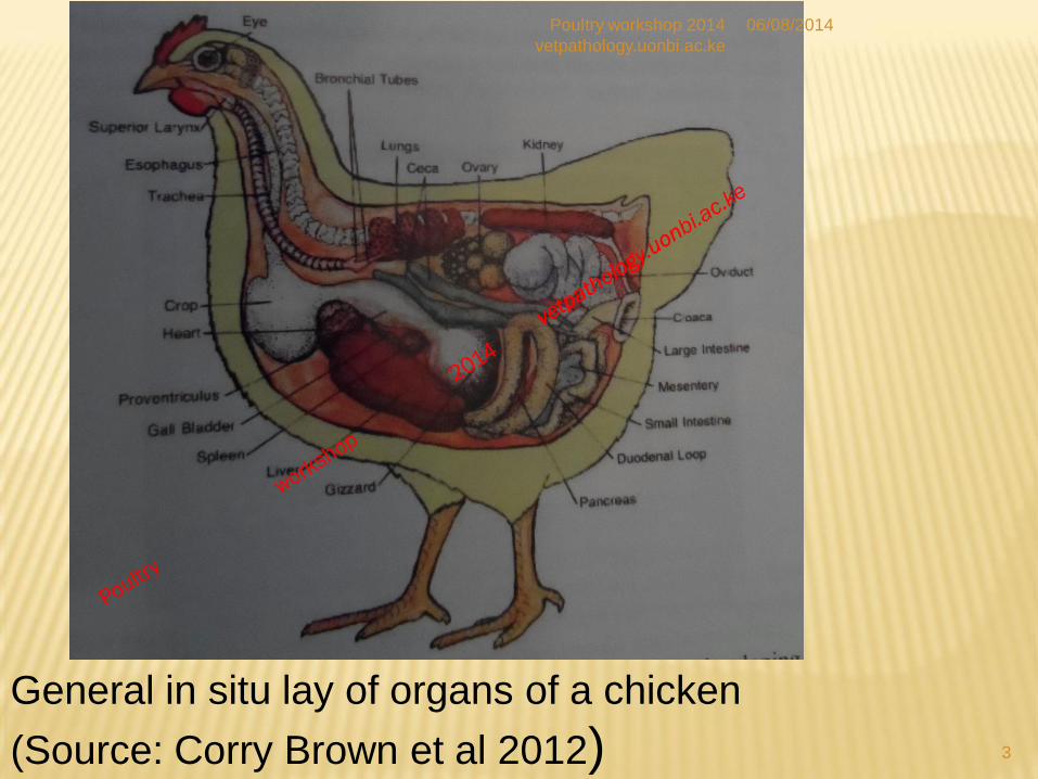
General in situ lay of organs of a chicken
(Source: Corry Brown et al 2012)
06/08/2014Poultry workshop 2014
vetpathology.uonbi.ac.ke

1. THEMES IN POST- MORTEM DIAGNOSTICS
1. Clinical presentation diversity and flock history
2. Disease pathogenesis, pathogen virulence, and inflammation
3. Multiple infections, diversity of disease manifestations - atypical presentations
4. Differentials
5. Epidemiology and flock health management
6. Forensic implications
4
06/08/2014Poultry workshop 2014
vetpathology.uonbi.ac.ke
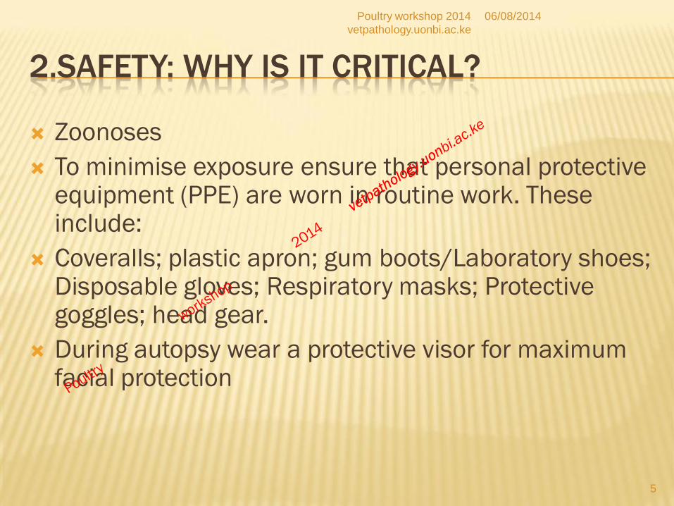
2.SAFETY: WHY IS IT CRITICAL?
Zoonoses
To minimise exposure ensure that personal protective equipment (PPE) are worn in routine work. These include:
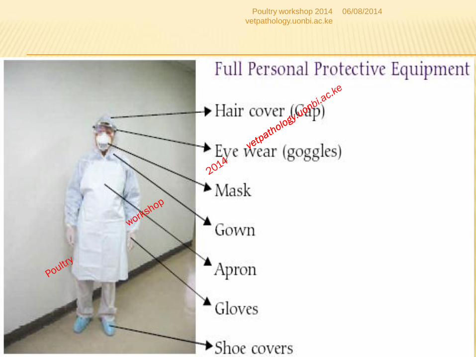
Coveralls; plastic apron; gum boots/Laboratory shoes; Disposable gloves; Respiratory masks; Protective goggles; head gear.
During autopsy wear a protective visor for maximum facial protection
5
06/08/2014Poultry workshop 2014
vetpathology.uonbi.ac.ke
6
06/08/2014Poultry workshop 2014
vetpathology.uonbi.ac.ke
3. EQUIPMENT FOR POSTMORTEM
Disinfectants
Scissors; knifes; forceps; scalpel blades
Tray (40x 30 cm)
Personal protective equipment (PPE):
Surgical gloves of proper size
Rubber gloves- heavy duty
Mask(N95)
Gown-full body cover
Rubber boots
Plastic apron
Head dress and goggles7
06/08/2014Poultry workshop 2014
vetpathology.uonbi.ac.ke
EQUIPMENT
Sampling containers:
with 10 % formalin for histological samples
Sterile polyester swabs with plastic shaft
Viral transport medium (VTM) for virology
samples in cryovials.
Swabs and carrying medium for bacteriology
Cool box and ice packs
Sample packaging for diagnostic specimen
transport8
06/08/2014Poultry workshop 2014
vetpathology.uonbi.ac.ke

EXAMINATION PROCEDURE: STEPS
1. Obtain flock history
2. Examine the bird externally
3. Open the body; observe organs in situ
4. Remove the organs for detailed examination and
sampling
5. Examine and sample the organs
6. Consider observations for d/diagnosis and write the
report
9
06/08/2014Poultry workshop 2014
vetpathology.uonbi.ac.ke
3. GENERAL EXAMINATION PROCEDURE
General body examination: Plumage; body symmetry; palpate for crepitation, ascites; abdominal tumors.
General body condition- weight: normal, low or high
Skin, legs, feet etc.- Indicate effect of management and environment ; fracture and trauma (self or externally inflicted)
Inflammation of feet may indicate: poor litter, wet bedding, chronic Pasteurella multocida infection of the head, fowl pox, ectoparasites
Cloaca examination may give a clue to : enteritis, salphingitis, uraemia, criminal abuse;
10
06/08/2014Poultry workshop 2014
vetpathology.uonbi.ac.ke

GENERAL EXAMINATION
Examine for ectoparasites: around the head, on
the body; under the wings and thighs
Examine mucous membranes: conjuctiva, oral
cavity and cloaca (DD: anaemia; inflammation,
hemorrhages, purulent discharges;
avitaminosis A);;
11
06/08/2014Poultry workshop 2014
vetpathology.uonbi.ac.ke

DISSECTION PROCEDURE 1
Carcass is placed on its right side with head pointed towards the examiner and dampened with disinfectant
Infra-orbital sinus is opened and examined for inflammation: Mycoplasma infections, Haemophilus paragallinarum, Newcastle disease, Avian influenza, Infectious laringotracheitis, Infectious bursal disease.
12
06/08/2014Poultry workshop 2014
vetpathology.uonbi.ac.ke

DISSECTION PROCEDURE 2
The beak is opened and the oral cavity is opened ( the blunts part of the scissors within the cavity)
Incision is extended down the neck to open the oesophagus and the crop
Oral cavity, pharynx, oesophagus and crop are examined for inflammation – fowl pox, ILT, degenerative changes e.g. avitaminosis A
13
06/08/2014Poultry workshop 2014
vetpathology.uonbi.ac.ke
DISSECTION PROCEDURE 3
Larynx and trachea are subsequently opened
and examined for inflammation or other
pathology: NDV, ILT, syngamus trachea , wet
form of fowl pox, etc.
Thymus is inspected and incised ( well
developed in young birds (broilers) atrophied in
old birds (laying flocks)
14
06/08/2014Poultry workshop 2014
vetpathology.uonbi.ac.ke
DISSECTION PROCEDURE 4
The carcass is placed on its back with the legs towards the examiner
The skin is incised transversely behind the xyphoid process and the incision is extended towards both knees. The skin is removed over the pectoral muscles by pulling in a cranial direction
Parasternal bursa is inspected for: inflammation associated with recumbency (immobile birds); inflammation due to mycoplasma and reovirus infections
15
06/08/2014Poultry workshop 2014
vetpathology.uonbi.ac.ke

DISSECTION PROCEDURE 5
Pectoral muscles are incised and
examined for lesions e.g. Tumours due to
marek’s disease, Haemorrhages in
septicaemic diseases, deep pectoral
necrosis, and IBD variant manifestations
Legs and hips are bent outwards until each
femoral head is dislocated from the
acetabulum 16
06/08/2014Poultry workshop 2014
vetpathology.uonbi.ac.ke
DISSECTION PROCEDURE 6
Transverse incision is made behind the
xyphoid process to open into the thoracic
cavity
Incisions are made on both sides of the
thorax up to the brachial region or
shoulders to open the thoracic inlet
17
06/08/2014Poultry workshop 2014
vetpathology.uonbi.ac.ke
DISSECTION PROCEDURES 7
Sternum is slightly lifted to examine the abdominal
and thoracic cavities insitu for evidence of
mycoplasmal, bacteriological or virological infections
e.g. swollen organs, haemorrhages, exudates etc. If
lesions are present take swabs or tissue samples
aseptically for microbiological examination
Sternum with pectoral muscles is cut and rotated
upwards and cranially to expose the abdominal and
thoracic cavities18
06/08/2014Poultry workshop 2014
vetpathology.uonbi.ac.ke

DISSECTION PROCEDURE 8
Examine all air sacs in situ for evidence of
inflammation( cloudiness; thickening;
hemorrhagic reaction, oedema)
Remove the heart with pericardial sac and
incise and examine it for lesions (incisions of
the heart are made along the blood circulation
19
06/08/2014Poultry workshop 2014
vetpathology.uonbi.ac.ke

DISSECTION PROCEDURE 9
The liver and gall bladder are removed and
examined
The spleen is removed and examined
Presence of an enlarged liver (with rounded
edges) and an enlarged spleen are typical for
septicaemia
20
06/08/2014Poultry workshop 2014
vetpathology.uonbi.ac.ke
DISSECTION PROCEDURE 10
make transverse incision cranial to the proventriculus and remove the whole intestinal tract in a caudal direction. Make a transverse incision 1-2 cm cranial to the cloaca and remove the entire intestines
Examine the serosal surface of the intestines and then open the intestines in a caudal direction starting from the proventriculus. Simultaneously examine the pancreas.
21
06/08/2014Poultry workshop 2014
vetpathology.uonbi.ac.ke
DISSECTION PROCEDURE 11
Examine the entire intestines for:
Haemorrhages
Oedema
Contents (ingesta, parasites, foreign bodies)
Ulcers, neoplasms
Thickening or thinning of the intestinal wall
22
06/08/2014Poultry workshop 2014
vetpathology.uonbi.ac.ke
DISSECTION PROCEDURE 12
Examine the intestinal mucosa for
inflammation:
Necrotic enteritis
Salmonellosis (typhilitis)
Newcastle disease; avian Influenza; coccidiosis
Haemorrhages
Ulcerations
Thickening; Parasite infestations
23
06/08/2014Poultry workshop 2014
vetpathology.uonbi.ac.ke
DISSECTION PROCEDURES 14
In females: cut the reproductive tract free of its
ligaments in a caudal direction and
subsequently open the infundibulum, magnum,
isthmus, uterus and vagina ( these are
inspected both from the serosal and mucosal
surfaces) – E coli often causes purulent
inflammation at these sites.
24
06/08/2014Poultry workshop 2014
vetpathology.uonbi.ac.ke
DISSECTION PROCEDURE 15
In young birds the Bursa of Fabricius is opened
through its cloacal opening and examined for
swelling, oedema, haemorrhages.
Gumboro disease often causes swelling, oedema or
haemorrhages in the Bursa of Fabricius similar to
HPAI
Tumours such as Leucosis( not Mareks) often occur
in the Bursa of Fabricius25
06/08/2014Poultry workshop 2014
vetpathology.uonbi.ac.ke
DISSECTION PROCEDURE 16
Examine the kidneys for lesions such as
atrophy, nephropathies, inflammation, tumours.
Kidneys are often pale in Clostridia infections
as a result of toxaemia
Examine for congestion and haemorrhages in
HPAI
Swelling – visceral gout
26
06/08/2014Poultry workshop 2014
vetpathology.uonbi.ac.ke

DISSECTION PROCEDURE 17
Examine the thoracic air sacs for cloudiness,
thickening
Remove the lungs and examine for size, colour,
consistency, oedema tumors
Examine the heart sac for adhesions, lesions,
fluid contents
Examine the heart for lesions (hemorrhages,
myocardial dystrophy,neoplasms)27
06/08/2014Poultry workshop 2014
vetpathology.uonbi.ac.ke

DISSECTION PROCEDURE 18
Examine the brachial plexus for Mareks
disease
Examine the sciatic nerve bilaterally for Mareks
disease by exposing it through a blunt
dissection to separate the gracilis muscle
Check for swelling and loss of striations
28
06/08/2014Poultry workshop 2014
vetpathology.uonbi.ac.ke
29
DISSECTION PROCEDURE 19
Palpate all the joints and open them in case of swellings
and asymmetry
Open the knee and hock joints and inspect the tendons
and tendon sheaths for haemorrhage, ulcers, exudates.
Joint disease can be caused by:
Staphylococcus aureus, Reoviruses, Mycoplasma
synoviae, E. coli, Pasteurella multocida and Salmonella
gallinarum / pullorum
06/08/2014Poultry workshop 2014
vetpathology.uonbi.ac.ke
DISSECTION PROCEDURE 20
Examine young birds for ossification of the ribs
and their spinal and costo-chondral junctions.
Swellings may occur in avitaminosis D;
neoplasia.
Make parallel incisions on the tibial/tarsal
bones to examine for TD (Thiamine deficiency)
lesions
30
06/08/2014Poultry workshop 2014
vetpathology.uonbi.ac.ke
DISSECTION PROCEDURE 21
Remove the brain by gently lifting the cranial
bones and by detaching the dura mater
Inflammatory foci in the brain occur in avian
epidemic tremor,
Encephalomalacia occurs in Vitamin E
deficiency (crazy chick disease)
Tumours can spread to the brain
31
06/08/2014Poultry workshop 2014
vetpathology.uonbi.ac.ke
DIFFERENTIAL DIAGNOSIS AND CASE MANAGEMENT
Lesions observed are summarised and their pathogenesis reviewed
A differential diagnosis is made based on clinical history, symptoms, lesions and other epidemiological factors narrated by the farmer = what are at least three most probable diseases
A tentative diagnosis is made and samples taken
Confirmatory diagnosis is made from results of sample analysis in the laboratory.
Several birds may be examined for an effective flock diagnosis
32
06/08/2014Poultry workshop 2014
vetpathology.uonbi.ac.ke
SAMPLES
Swabs: oropharyngeal / tracheal, cloacal, ocular
Organs: Trachea, lungs, duodenum, caecal tonsil,
brain, liver, spleen and any organ with lesions from
fresh carcasses
Blood for serum: sick and recovered birds
For each sample, n = at least 20 per affected flock
At least 5 whole fresh carcases should be submitted
for autopsy wrapped in double layer polythene bags
and accompanied by full case history as seen by the
referring clinician
33
06/08/2014Poultry workshop 2014
vetpathology.uonbi.ac.ke

SPECIMEN COLLECTION AND SHIPMENT
A) Oropharyngeal swabs:
Swab the oral pharynx and choanae rotating the swab several times
Transfer the swab aseptically into a cryovial containing 1ml viral transport medium
Label the sample appropriately.
Cryovials are wrapped in absorbent material and placed in 50ml plastic tubes which are decontaminated on the outer surface with 70% alcohol
All samples are placed in a third sealable biohazard container and placed in a cool box packed with ice for shipment .
Appropriate labelling and markings are made on the packaging for the specimen shipping to the laboratory
34
06/08/2014Poultry workshop 2014
vetpathology.uonbi.ac.ke

SPECIMEN COLLECTION AND SHIPMENT
35
06/08/2014Poultry workshop 2014
vetpathology.uonbi.ac.ke
SPECIMEN COLLECTION AND SHIPMENT
B)Cloacal swab:
Insert the swab into the cloaca through the vent and
twist gently round the surface of the cloaca to pick
some fecal material and withdraw genntly.
C) Conjuctival swab
Gently rotate the swab in the conjuctiva sac twice
Ship the swab as described for oral pharyngeal swab
D) Tears
Collected for viral nucleic acid and mucosal antibody
testing36
06/08/2014Poultry workshop 2014
vetpathology.uonbi.ac.ke
SPECIMEN COLLECTION AND SHIPMENT
E) Blood sample for serum
Use gauge 23-25 needle, and 2-5 ml disposable syringe
Pluck the feathers from the wing web and swab the wing web
Collect 2ml blood from the wing web and transfer to a 5 ml sterilin tube or a sterile bijoux bottle without an anticoagulant (Patience and experience are vital)
Decontaminate syringe and needle in a disinfectant and dispose the needle in a sharps container
Decontaminate the surface of the sample bottle and ship in the same manner as for the swabs with full history from the farmer
37
06/08/2014Poultry workshop 2014
vetpathology.uonbi.ac.ke

38
06/08/2014Poultry workshop 2014
vetpathology.uonbi.ac.ke
39
06/08/2014Poultry workshop 2014
vetpathology.uonbi.ac.ke
SPECIMEN COLLECTION AND SHIPMENT
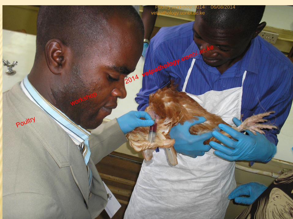
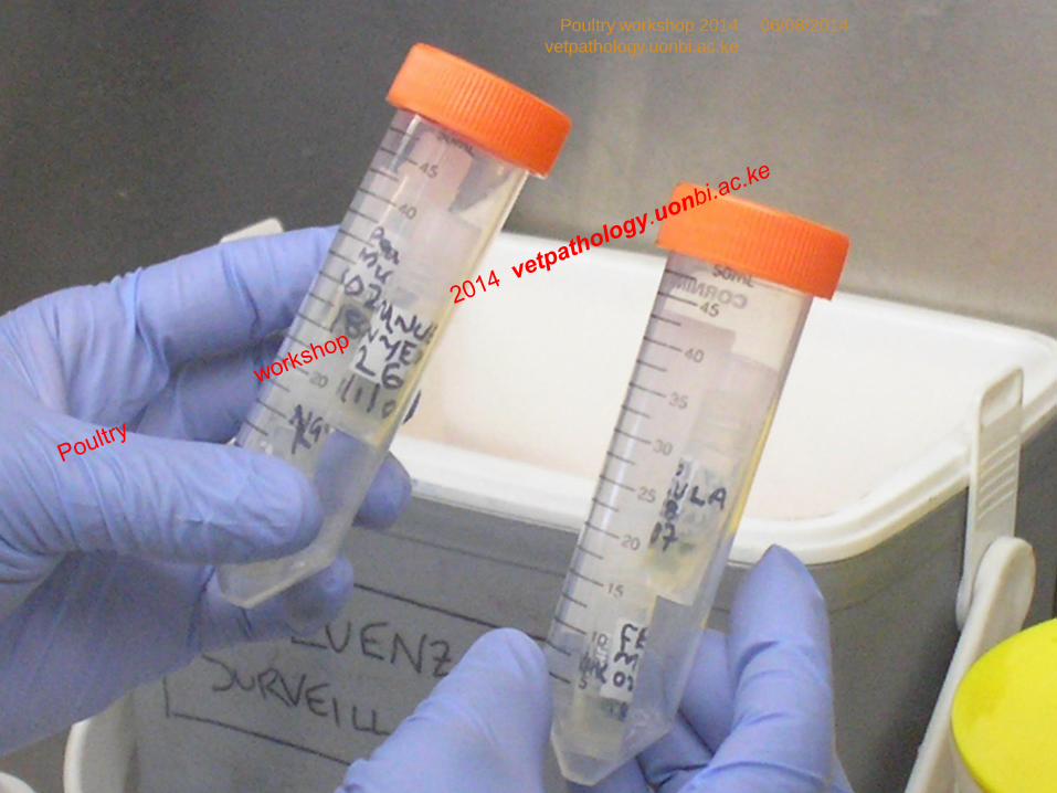
F) Organ sampling
As soon as the carcass is opened and before any opening of the intestinal, aseptically obtain 1-2 cm of organ samples and transfer each organ into a separate sterile tube containing transport medium for viral isolation, bacterial culture and special buffer for nucleic acid detection by PCR technique.
Obtain a sample and put into neutral buffered formalin solution for histopathology
Label the samples appropriately40
06/08/2014Poultry workshop 2014
vetpathology.uonbi.ac.ke

41
TRANSPORTING SPECIMENS FROM FIELD TO
LAB
06/08/2014Poultry workshop 2014
vetpathology.uonbi.ac.ke
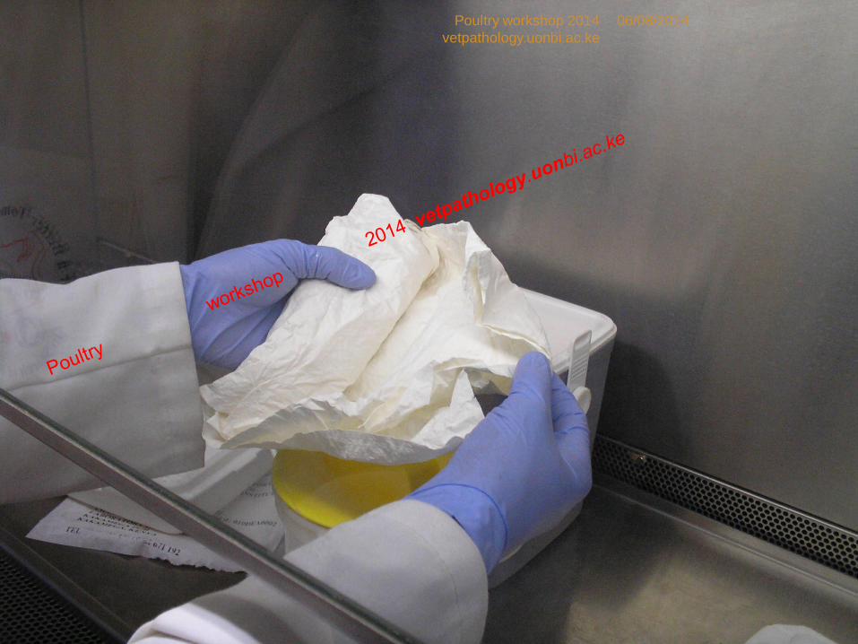
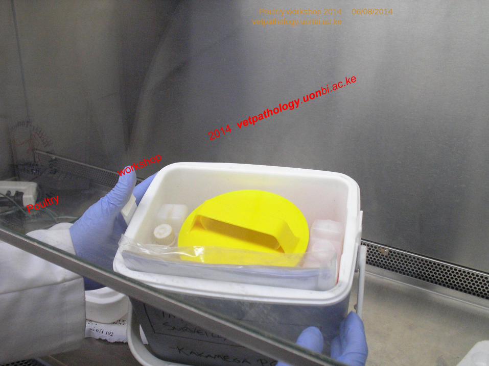
EMERGENCY SAMPLE SHIPMENT
Improvise the tripple parking principle:
The samples, in their collection vials are packed in double
layer plastic bags with absorbent tissue wrappers. The
bags with samples are assembled in a carton which is
then sealed and wrapped in a polythene bag
Put the sealed carton in a clean outer carton and Label
this third container carton appropriately with the name of
the sender; consignee and what materials are contained
in the carton.
Transfer the package for shipment to civilian section of
the lab and attach a filled sample shipment form with all
details entered in a sample submission form.
42
06/08/2014Poultry workshop 2014
vetpathology.uonbi.ac.ke
43
06/08/2014Poultry workshop 2014
vetpathology.uonbi.ac.ke
44
06/08/2014Poultry workshop 2014
vetpathology.uonbi.ac.ke
45
06/08/2014Poultry workshop 2014
vetpathology.uonbi.ac.ke
46
06/08/2014Poultry workshop 2014
vetpathology.uonbi.ac.ke

47
06/08/2014Poultry workshop 2014
vetpathology.uonbi.ac.ke
SAMPLE TRANSPORT
All samples must be properly labelled and
accompanied with a sample submission record
containing full historical description including the
suspected diease for which laboratory confirmation is
being sought
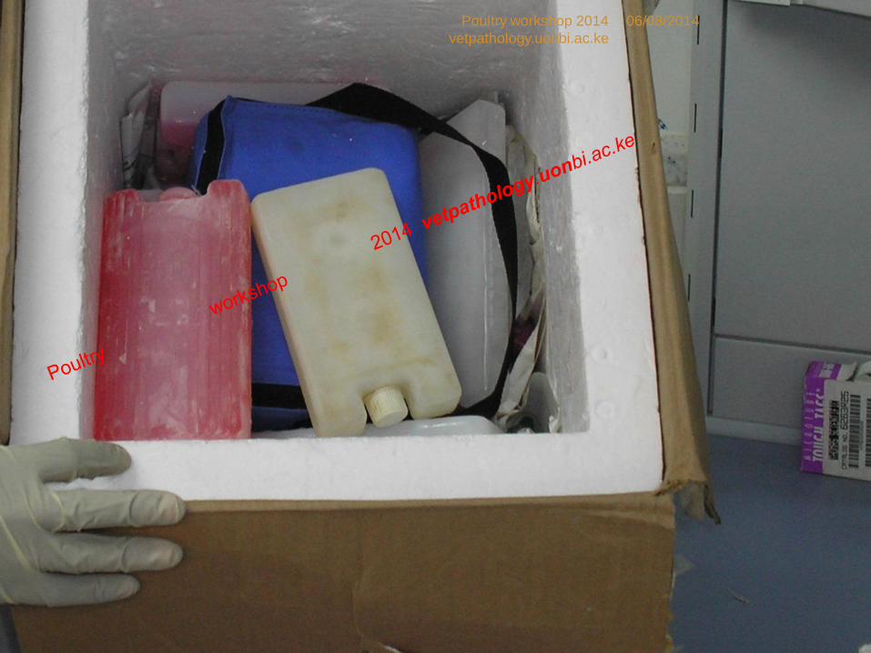
Pack the samples in ice to ensure that the shipment
temperature are between 2-8oC
Faeces and organs : in sterile leak proof boxes
Cloacal swabs: transport in sterile cryovials with viral
transport medium, separate from the fecal samples
48
06/08/2014Poultry workshop 2014
vetpathology.uonbi.ac.ke
DISPOSAL OF INFECTED MATERIAL
Disinfect the carcass and organs while still on the pm tray
Secure the carcass in two water tight biosafety bags
All carcasses are wrapped and transported for incineration in a tightly closed litter bucket or tank internally lined with a watertight polythene disposal bag
Carcasses and all disposable items are incinerated to ashes
Decontaminate all buckets, tanks or other containers and carriers and any other items used to move the carcasses at the incineration facility
49
06/08/2014Poultry workshop 2014
vetpathology.uonbi.ac.ke
DECONTAMINATION OF EQUIPMENTS AND
SURFACES
Disinfect all equipments and surfaces used,
wash thoroughly in running tap water and
disinfect a second time
Send the postmortem kit for autoclaving
Fumigate the postmortem room appropriately
50
06/08/2014Poultry workshop 2014
vetpathology.uonbi.ac.ke
CONCLUSIONS
Themes help clarify observations made
Careful observations and recording help make
accurate differential diagnosis
Samples: well selected and preserved help
confirm diagnosis
Personal safety is essential for personal safety
PPE be available
Diagnostic post-mortem key to clinical disease
management in avian medicine51
06/08/2014Poultry workshop 2014
vetpathology.uonbi.ac.ke
52
THANK
YOU
06/08/2014Poultry workshop 2014
vetpathology.uonbi.ac.ke

53
THANK
YOU
06/08/2014Poultry workshop 2014
vetpathology.uonbi.ac.ke