post op cardiac care review
TRANSCRIPT

7/30/2019 Post Op Cardiac Care Review
http://slidepdf.com/reader/full/post-op-cardiac-care-review 1/11
Section Editor, Jonathan E. Sevransky, MD, MHSConcise Definitive Review
Management of the postoperative pediatric cardiac surgical patient
Ronald A. Bronicki, MD; Anthony C. Chang, MD, MBA
The field of pediatric cardiac in-tensive care continues toevolve, which is in large partthe result of collaborative ef-
forts from anesthesia, surgery, cardiol-ogy, critical care, and other subspecial-ties, including neonatology, neurology,and endocrinology. Examples include anincreasing number of surgeries in verylow birth weight infants; the extension of technology such as cerebral oximetryfrom the operating room into the inten-sive care setting; and innovations in me-chanical circulatory devices. Industry-
sponsored studies and initiatives from theNational Institutes of Health such as thePediatric Heart Network have contrib-uted to the evolution of pediatric cardiacintensive care. These collective efforts aredemonstrated in the Prophylactic Intra-
venous Use of Milrinone After CardiacOperation in Pediatrics (PRIMACORP)study and more recently the single ven-tricle reconstruction trial and thyroidsupplementation study (to be discussed).
The increase in complexity of dis-ease, innovations in technology, and
evolving therapeutic strategies as wellas national quality initiatives, individu-ally and collectively, place a tremen-dous demand on the team, necessitat-ing a focused, concerted effort by allmembers to challenge current practices
while practicing state-of-the-art care.In this review, we discuss the salientaspects and latest advances in the pri-mary challenge after surgery, which isto maintain adequate cardiopulmonaryfunction and to ensure adequate tissueoxygenation.
The Inflammatory Response to
Cardiopulmonary Bypass
Exposure of the blood componentsto the nonendothelialized circuit, pul-monary and myocardial reperfusion in-
jury, and the formation of heparin–protamine complexes stimulate therelease of systemic and local proinflam-matory mediators and activate the co-agulation/fibrinolytic pathways (1). Vir-tually every inflammatory pathway and
parenchymal cell contributes to the in-flammatory cascade. The magnitude of the inflammatory response to cardio-pulmonary bypass (CPB) has been asso-ciated with preoperative conditionssuch as shock and heart failure (2);intraoperative factors such as the dura-tion of bypass and cardioplegic arrest(3); and genetic variances have beenidentified that affect the inflammatoryresponse to several stimuli, includingCPB (4, 5).
Several studies have found a relation-ship between the inflammatory responseto bypass and the development of multi-organ dysfunction and postoperativemorbidity (3, 6). Prospective, randomizedstudies in children found the administra-tion of glucocorticoids before bypass sig-nificantly reduced the proinflammatoryand augmented the compensatory anti-inflammatory responses to bypass, re-duced the extent of myocardial injury,and improved the postoperative course(7–10). Two prospective studies failed todemonstrate benefit from the use of glu-
cocorticoids however they limited enroll-ment to older children (11, 12). A major-ity of pediatric cardiac centers useglucocorticoids to ameliorate the inflam-matory response to bypass (13).
Assessment of Hemodynamics
and Tissue Oxygenation
An accurate and timely determinationof cardiac output (CO), systemic O2 de-livery (DO2), and tissue oxygenation isessential to optimizing outcomes. Stud-
ies have demonstrated that it is not un-common for there to be discordance be-tween measurements of these parametersand estimations based on the physicalexamination and interpretation of con-
ventional hemodynamic parameters suchas heart rate, blood pressure, and rightatrial pressure (14, 15). Compensatory in-creases in systemic vascular resistancemaintain arterial blood pressure as COfalls and central venous/right atrial pres-sures may not correlate well with right
From the Children’s Hospital of Orange County(RAB, ACC), Orange, CA; and the David Geffen Schoolof Medicine at the University of California Los Angeles(RAB), Los Angeles, CA.
The authors have not disclosed any potential con-flicts of interest.
For information regarding this article, E-mail:[email protected]
Copyright © 2011 by the Society of Critical CareMedicine and Lippincott Williams & Wilkins
DOI: 10.1097/CCM.0b013e31821b82a6
Objective: To review the salient aspects and latest advances in
the management of the postoperative pediatric cardiac patient.
Data Source: A Medline-based literature source.
Conclusion: The practice of pediatric cardiac intensive care
has evolved considerably over the last several years. These efforts
are the result of a collaborative effort from all subspecialties
involved in the care of pediatric patients with congenital heart
disease. Discoveries and innovations that are representative of
this effort include the extension of cerebral oximetry from the
operating room into the critical care setting; mechanical circula-
tory devices designed for pediatric patients; and surgery in very
low birth weight neonates. Advances such as these impact post-
operative management and make the field of pediatric cardiac
intensive care an exciting, demanding, and evolving discipline,
necessitating the ongoing commitment of various disciplines to
pursue a greater understanding of disease processes and how to
best go about treating them. (Crit Care Med 2011; 39:1974–1984)
K EY WORDS: pediatric cardiac surgery; postoperative manage-
ment; cardiopulmonary bypass; hemodynamic monitoring; ven-
tricular dysfunction; cardiopulmonary interaction; vasoactive sup-
port
1974 Crit Care Med 2011 Vol. 39, No. 8

7/30/2019 Post Op Cardiac Care Review
http://slidepdf.com/reader/full/post-op-cardiac-care-review 2/11
ventricular filling or left atrial pressures(16, 17). The primary determinants of
ventricular filling are the ventricular di-astolic transmural pressure (ventricular di-astolic pressure – surrounding pressure)
and ventricular compliance (Fig. 1). Withpericardial disease, increases in intratho-racic pressure, and impaired diastolic func-tion, the optimal filling pressure increases.
Echocardiography provides an assess-ment of ventricular filling and function andquantification of valvular regurgitation.Doppler measurements assess obstructionsand pulmonary arterial pressures. Trendingserial lactate levels has prognostic value(18, 19). A low serum HCO3
– level and basedeficit are reflective of tissue hypoxia to the
extent that serum lactate levels are elevated,producing an anion gap; otherwise, these ab-normalities are consistent with a hyperchlo-remic metabolic acidosis (20–23).
The routine placement of central ve-nous catheters readily enables the clini-cian to use venous oximetry to assessoxygen transport balance (Table 1). Thecritical O2 extraction ratio is defined bythe onset of shock and ranges from 50%to 60% (Table 2) (24). The critical DO2
changes in parallel with changes in oxy-gen consumption (VO2); however, the
critical O2 extraction ratio remains con-stant, which is an important consider-ation in the postoperative period whenoxygen demand changes over time and
with therapy (25). When mixed venousoxygen saturations are unavailable, alter-native sampling sites include the rightatrium, superior vena cava, jugular vein,and inferior vena cava–right atrial junc-tion (Table 2) (26–28). As CO falls, bloodflow is redistributed to maintain vital or-gan perfusion. As a result, inferior venacava saturations fall first, which is ac-
companied by a less severe drop in mixed venous oxygen saturations. As CO fallsfurther, superior vena cava and jugularsaturations begin to decrease (28, 29).
Venous oximetry has been shown to im-prove outcomes in pediatric patients atrisk for developing shock, including se-
vere sepsis and the hypoplastic left heartsyndrome (30, 31).
Cerebral near-infrared spectroscopynoninvasively assesses cerebral oxygen-ation. Cerebral oximetry relies on the rel-
ative transparency of biological tissue tonear-infrared spectroscopy light whereoxy- and deoxy hemoglobin have distinctabsorption spectra (32). The oximetermonitors the nonpulsatile signal reflect-ing the microcirculation where 75–85%of the blood volume is venous. Thus, theO2 saturation is used as an indicator of O2
extraction for the area of the brain im-
mediately beneath the probe (frontal cor-tex). Because of technical constraints, thetechnology is limited to relative quanti-tation and is thus useful for trackingchanges for a given patient. Studies havedemonstrated a good correlation betweencerebral O2 saturations and jugular bulband superior vena cava saturations (33,34). A study by Li and colleagues (35)evaluated the relationship between cere-bral oxygenation and hemodynamic andoximetric parameters in neonates follow-ing the Norwood procedure. They foundthat cerebral oxygen saturations closelyand negatively correlated with the O2 ex-traction ratio. Cerebral oximetry is alsoused as a surrogate for central and mixed
venous oxygen saturations; however, thecorrelation between these readings ismarginal (36, 37). The reasons for thisinclude the technical constraints de-scribed; the fact that in a low CO state,blood flow is redistributed to maintainperfusion of vital organs; and cerebralblood flow is sensitive to changes in ar-terial PCO2.
Maintenance of Tissue
Oxygenation
The primary goal after surgery is tomaintain adequate tissue oxygenation,
which is accomplished by optimizing ox- ygen transport balance (i.e., minimizingoxygen demand and consumption andmaintaining adequate oxygen delivery(Table 3) (38). Underlying respiratory andcirculatory dysfunction, the inflamma-
Figure 1. The relationship of ventricular fillingpressure ( EDP ), ventricular compliance, and in-trathoracic pressure ( ITP ) to ventricular filling.The EDP is 15 for each variable. A, The ITP isϩ10 (positive pressure ventilation). B, The ITP isϪ5 (spontaneous breathing). C, Ventricular com-pliance is reduced. Ventricle A vs. B, Ventricular
compliance is the same; however, because ven-tricle B has a greater Tmp, it fills to a greaterextent. Ventricle B vs. C, The Tmp is the same;however, because ventricle B is more compliant,it fills to a greater extent. Tmp, ventricular trans-mural pressure; Tmp, intracavitary Ϫ extracavi-tary pressure.
Table 1. Oxygen transport balance
Fick equation: VO2 ϭ CO ϫ CaO2 Ϫ CvO2
By ignoring the amount of O2 dissolved in blood,the Fick equation may be simplified to:SaO2 Ϫ SmvO2 ϭ VO2 /DO2 ϭ O2 transport
balance
VO2, O2 consumption (mL/min); CO, cardiac
output (L/min); Ca Ϫ CmvO2, arterial Ϫ mixed
venous O2 content difference (mL/dL); SaO2, ar-
terial O2 saturation; SmvO2, mixed venous O2
saturation; DO2, oxygen delivery (CO2 ϫ CaO2;
mL/min).
Table 2. Oxygen extraction ratio (O2ER)
O2ER ϭ SaO2Ϫ SmvO2 /SaO2
SaO2, arterial O2 saturation; SmvO2, mixed venous O2 saturation;Oxygen extraction ratios based on mixed venous oximetry
25% Normal30% to 40% Increased40 to 50% Impending shockϾ50% to 60% Shock, elevated lactate levels
Normal O2 extraction ratios for central venous oximetryRight atrium 25%Jugular vein 35%Superior vena cava 30%Inferior vena cava 20%
Table 3. Factors responsible for increased
metabolic requirements
Catecholamines, endogenous and exogenousSystemic inflammatory responseFeverConsciousness, pain and anxietySpontaneous breathingEnteral nutrition
1975Crit Care Med 2011 Vol. 39, No. 8
7/30/2019 Post Op Cardiac Care Review
http://slidepdf.com/reader/full/post-op-cardiac-care-review 3/11
tory response to CPB, myocardial and
pulmonary reperfusion injury, and sur-
gery all place the patient at risk for de-
veloping shock (39, 40).
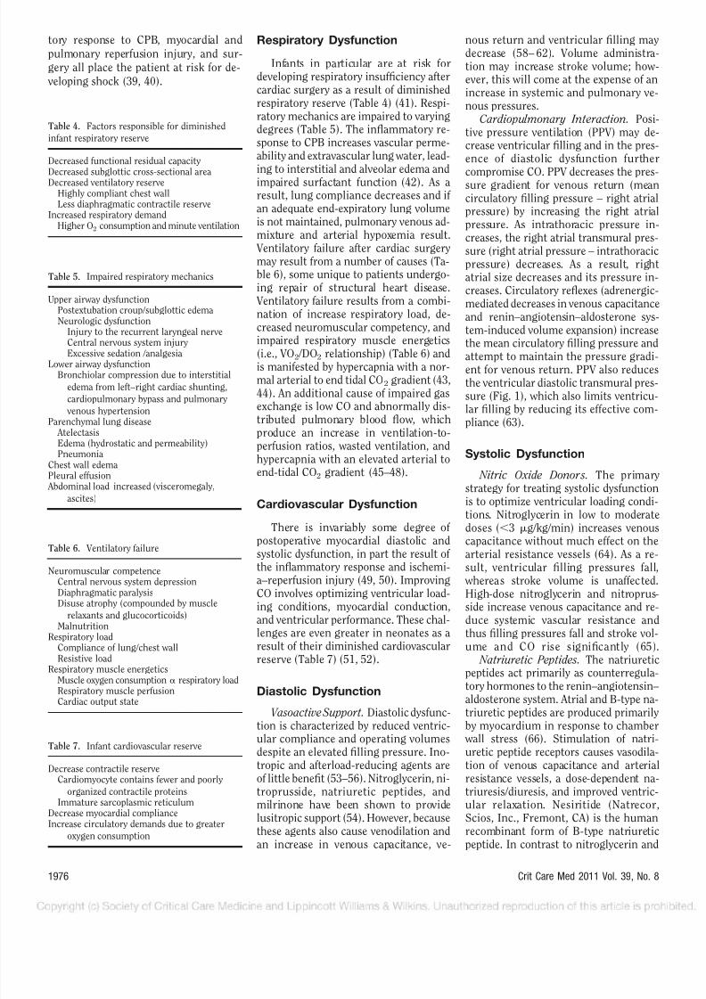
Respiratory Dysfunction
Infants in particular are at risk fordeveloping respiratory insufficiency aftercardiac surgery as a result of diminishedrespiratory reserve (Table 4) (41). Respi-ratory mechanics are impaired to varyingdegrees (Table 5). The inflammatory re-sponse to CPB increases vascular perme-ability and extravascular lung water, lead-ing to interstitial and alveolar edema andimpaired surfactant function (42). As aresult, lung compliance decreases and if an adequate end-expiratory lung volumeis not maintained, pulmonary venous ad-mixture and arterial hypoxemia result.
Ventilatory failure after cardiac surgerymay result from a number of causes (Ta-ble 6), some unique to patients undergo-ing repair of structural heart disease.
Ventilatory failure results from a combi-nation of increase respiratory load, de-creased neuromuscular competency, andimpaired respiratory muscle energetics
(i.e., VO2 /DO2 relationship) (Table 6) andis manifested by hypercapnia with a nor-mal arterial to end tidal CO2 gradient (43,44). An additional cause of impaired gasexchange is low CO and abnormally dis-tributed pulmonary blood flow, whichproduce an increase in ventilation-to-perfusion ratios, wasted ventilation, andhypercapnia with an elevated arterial toend-tidal CO2 gradient (45–48).
Cardiovascular Dysfunction
There is invariably some degree of postoperative myocardial diastolic andsystolic dysfunction, in part the result of the inflammatory response and ischemi-a–reperfusion injury (49, 50). ImprovingCO involves optimizing ventricular load-ing conditions, myocardial conduction,and ventricular performance. These chal-lenges are even greater in neonates as aresult of their diminished cardiovascularreserve (Table 7) (51, 52).
Diastolic Dysfunction
Vasoactive Support. Diastolic dysfunc-tion is characterized by reduced ventric-ular compliance and operating volumesdespite an elevated filling pressure. Ino-tropic and afterload-reducing agents areof little benefit (53–56). Nitroglycerin, ni-troprusside, natriuretic peptides, andmilrinone have been shown to providelusitropic support (54). However, becausethese agents also cause venodilation andan increase in venous capacitance, ve-
nous return and ventricular filling maydecrease (58– 62). Volume administra-tion may increase stroke volume; how-ever, this will come at the expense of anincrease in systemic and pulmonary ve-nous pressures.
Cardiopulmonary Interaction. Posi-tive pressure ventilation (PPV) may de-crease ventricular filling and in the pres-ence of diastolic dysfunction further
compromise CO. PPV decreases the pres-sure gradient for venous return (meancirculatory filling pressure – right atrialpressure) by increasing the right atrialpressure. As intrathoracic pressure in-creases, the right atrial transmural pres-sure (right atrial pressure – intrathoracicpressure) decreases. As a result, rightatrial size decreases and its pressure in-creases. Circulatory reflexes (adrenergic-mediated decreases in venous capacitanceand renin–angiotensin–aldosterone sys-tem-induced volume expansion) increasethe mean circulatory filling pressure andattempt to maintain the pressure gradi-ent for venous return. PPV also reducesthe ventricular diastolic transmural pres-sure (Fig. 1), which also limits ventricu-lar filling by reducing its effective com-pliance (63).
Systolic Dysfunction
Nitric Oxide Donors. The primarystrategy for treating systolic dysfunctionis to optimize ventricular loading condi-tions. Nitroglycerin in low to moderate
doses (Ͻ3 g/kg/min) increases venouscapacitance without much effect on thearterial resistance vessels (64). As a re-sult, ventricular filling pressures fall,
whereas stroke volume is unaffected.High-dose nitroglycerin and nitroprus-side increase venous capacitance and re-duce systemic vascular resistance andthus filling pressures fall and stroke vol-ume and CO rise significantly (65).
Natriuretic Peptides. The natriureticpeptides act primarily as counterregula-tory hormones to the renin–angiotensin–
aldosterone system. Atrial and B-type na-triuretic peptides are produced primarilyby myocardium in response to chamber
wall stress (66). Stimulation of natri-uretic peptide receptors causes vasodila-tion of venous capacitance and arterialresistance vessels, a dose-dependent na-triuresis/diuresis, and improved ventric-ular relaxation. Nesiritide (Natrecor,Scios, Inc., Fremont, CA) is the humanrecombinant form of B-type natriureticpeptide. In contrast to nitroglycerin and
Table 4. Factors responsible for diminished
infant respiratory reserve
Decreased functional residual capacity
Decreased subglottic cross-sectional areaDecreased ventilatory reserveHighly compliant chest wallLess diaphragmatic contractile reserve
Increased respiratory demandHigher O2 consumption and minute ventilation
Table 5. Impaired respiratory mechanics
Upper airway dysfunctionPostextubation croup/subglottic edemaNeurologic dysfunction
Injury to the recurrent laryngeal nerveCentral nervous system injury
Excessive sedation /analgesiaLower airway dysfunction
Bronchiolar compression due to interstitial
edema from left–right cardiac shunting,
cardiopulmonary bypass and pulmonary
venous hypertensionParenchymal lung disease
AtelectasisEdema (hydrostatic and permeability)Pneumonia
Chest wall edemaPleural effusion
Abdominal load increased (visceromegaly,
ascites)
Table 6. Ventilatory failure
Neuromuscular competenceCentral nervous system depressionDiaphragmatic paralysisDisuse atrophy (compounded by muscle
relaxants and glucocorticoids)Malnutrition
Respiratory loadCompliance of lung/chest wallResistive load
Respiratory muscle energeticsMuscle oxygen consumption ␣ respiratory loadRespiratory muscle perfusion
Cardiac output state
Table 7. Infant cardiovascular reserve
Decrease contractile reserveCardiomyocyte contains fewer and poorly
organized contractile proteinsImmature sarcoplasmic reticulum
Decrease myocardial complianceIncrease circulatory demands due to greater
oxygen consumption
1976 Crit Care Med 2011 Vol. 39, No. 8

7/30/2019 Post Op Cardiac Care Review
http://slidepdf.com/reader/full/post-op-cardiac-care-review 4/11
nitroprusside, tachyphylaxis does developto the hemodynamic effects of nesiritide(67) and its use is not associated withreflex stimulation of systemic and cardiacadrenergic activity (68).
Studies in adults have demonstratedits efficacy in treating congestive heartfailure, whereas studies in children havebeen limited (69). A study by Jefferies etal (70) evaluated the safety and efficacy of
nesiritide in pediatric decompensatedheart failure by prospectively monitoring55 separate infusions in 32 patients. Theyfound results similar to those found inadults: a significant increase in diuresis;reduction in ventricular filling pressuresand increase in CO; and a marked im-provement inthe mean New York Heart Associationfunctional class. No hypotension or ar-rhythmias were noted during 478 cu-mulative days of therapy and serum cre-atinine levels trended downward aftertherapy.
Catecholamines. The use o f c at -echolamines is limited because they arechronotropic, arrhythmogenic, and in-crease global and myocardial VO2 (68).Dopamine and dobutamine provide mod-est inotropic support. In contrast to do-butamine, dopamine decreases venouscapacitance and as a result ventricularfilling pressures do not decrease (72, 73).
At higher doses (Ͼ10 g/kg/min), sys-temic vascular resistance begins to in-crease as a result of -agonist activity.Dobutamine decreases systemic vascular
resistance, which is thought to be theresult of 2 agonist activity. Epinephrineprovides unparalleled inotropic supportand in low doses (Ͻ0.05–0.10 g/kg/ min) reduces systemic vascular resis-tance (74, 75).
Phosphodiesterase Inhibitors. Milri-none, and its predecessor amrinone, actthrough selective inhibition of phospho-diesterase III. The advantages of milri-none over catecholamines are that it isnot chronotropic and arrhythmogenic, itdoes not increase myocardial VO2, and it
is unaffected by adrenergic receptor de-sensitization. Milrinone provides mod-est inotropic support, vasodilates pul-monary and systemic vessels, and exertsa lusitropic effect (76). As a result, ven-tricular filling pressures decrease whilestroke volume and CO increase (77). A study by Hoffman and colleagues (78)evaluated the efficacy and safety of pro-phylactic milrinone in pediatric pa-tients undergoing cardiac surgery. Pa-tients (n ϭ 238) were randomized to
low-dose (25 g/kg/min) or high-dose(75 g/kg/min) milrinone or placebo.High-dose milrinone significantly re-
duced the risk of developing low COsyndrome compared with placebo, a64% relative risk reduction, and there
was no significant difference in the in-cidence of hypotension or arrhythmiacompared with placebo.
Levosimendan. Levosimendan repre-
sents the first of a new class of agents,the calcium sensitizers, which are inuse in Europe (79). These agents pro-
vide modest inotropic support by en-hancing the sensitivity of the myofila-
m e nt s t o t he p r ev a il i ng c y to s ol i ccalcium concentration. Because lessenergy is consumed in the cycling of calcium, myocardial VO2 remains un-changed. By stimulating adenosine
5-triphosphate-dependent Kϩ channelsin vascular smooth muscle cells, levo-simendan decreases systemic and pul-
monary vascular resistance. In contrastto other vasoactive agents, levosimen-dan has a long duration of action as aresult of the generation of active me-tabolites with an elimination half-life of 3–4 days.
Namachivayam and colleagues evalu-ated levosimendan in 15 children withsevere ventricular dysfunction who werecatecholamine-dependent (80). A combi-nation of a loading dose and continuous
infusions (24–48 hrs) allowed for a sub-stantial reduction in catecholamine infu-
sions with discontinuation in a majorityof patients. A prospective randomizedtrial was performed by Momeni and col-
leagues (81) of children (n ϭ 36) un-dergoing cardiac surgery in which pa-tients received either milrinone orlevosimendan with initiation of CPB forup to 48 hrs. There was no difference
between groups in terms of serum lac-tate levels (primary aim) or oxygen ex-traction; however, the rate pressure in-dex was significantly greater in thosereceiving milrinone.
Cardiopulmonary Interaction. In sys-tolic heart failure, PPV improves CO. Asdiscussed, PPV may compromise ventric-ular filling (82). However, so long as thedilated ventricle resides on the flat por-
tion of its pressure volume curve, a de-crease in venous return will not affectstroke volume. PPV reduces systemic
ventricular afterload and by unloadingthe respiratory pump respiratory muscle
VO2 decreases, allowing for a redistribu-tion of a limited CO (83, 84).
Other Strategies for Improving
Circulatory Function
Arginine Vasopressin. Arginine vaso-pressin (AVP) plays a vital role in thedefense of arterial blood pressure, be-cause physiological levels are required fornormal vascular tone of venous capaci-tance and arterial resistance vessels. Therationale behind the use of exogenous
AVP in critical illness is that neurohy-pophyseal stores of AVP may become ex-hausted, leading to vasodilatory shock(85). Prospective, randomized studies inadults have yielded equivocal results (86,87). There have been a few reports on theuse of AVP in children (88, 89). A retro-spective analysis was performed byRosenzweig et al of their experience with
AVP in 11 children after cardiac surgery. All had refractory hypotension and all buttwo had normal or mildly depressed left
ventricular function. During the firsthour of AVP, there was a significant im-provement in blood pressure and the ino-trope score.
Glucocorticoids. A similar rationaleexists for the use of glucocorticoids (90).Cortisol is essential for maintaining vas-cular tone and may affect myocardialfunction (91). Particularly in prematureand stressed term neonates, a relative ad-renal insufficiency may develop as a re-sult of the effects of inflammation on animmature hypothalamus–pituitary axis(92). Although the definition of a relativeadrenal insufficiency remains unclear,
most would agree that a normal or near-normal cortisol level in a critically illpatient is indicative of an inadequate hy-pothalamus–pituitary axis response (93).Prospective randomized studies of criti-cally ill premature infants with refractoryhypotension demonstrated improved he-modynamics in those treated with gluco-corticoids (94, 95). Studies in children
with refractory hypotension after cardiacsurgery have been limited to several ret-rospective studies and one small prospec-tive randomized study. All studies dem-
onstrated a significant improvement inarterial blood pressure over the first sev-eral hours after the initiation of therapythat coincided with a weaning of vasoac-tive support (96–99).
Thyroid Hormone. After CPB, serumthyroid hormone levels are depressed(100). Because of the known effects of thyroid hormone on ventricular function,several studies have evaluated the role of thyroid replacement therapy in childrenafter cardiac surgery (101–104). A rela-
1977Crit Care Med 2011 Vol. 39, No. 8

7/30/2019 Post Op Cardiac Care Review
http://slidepdf.com/reader/full/post-op-cardiac-care-review 5/11
tively large prospective randomized study(nϭ 193) was performed by Portman andcolleagues of T3 supplementation in chil-dren undergoing cardiac surgery. Analy-sis using age stratification found thosepatients Ͻ5 months of age who receivedT3 experienced a significantly shortenedtime to extubation (primary end point),improved cardiac function, and requiredless vasoactive support.
A study by Mackie and colleagues ran-domized 42 infants undergoing the Nor- wood procedure or repair of interruptedaortic arch to T3 supplementation or pla-cebo. There was no difference in the pri-mary end points of clinical outcome scoreand CO between groups; however, thosereceiving therapy did have a significantlyhigher systolic blood pressure. A study byChowdhury and colleagues demonstratedan improved postoperative course includ-ing reduced need for vasoactive supportin children (n ϭ 28) randomized to T3 asdid the study by Bettendorf and col-leagues that demonstrated an improvedpostoperative course and systolic func-tion in those children (n ϭ 40) random-ized to treatment.
Postoperative Management:
Lesion-Specific
Single Ventricle Physiology. Patients with single ventricle physiology are atrisk for developing shock, the result of systolic dysfunction compounded by theinefficiencies of a parallel circulation.
The aortic–pulmonary artery shunt al-lows for continuous flow throughout thecardiac cycle, leading to excessive pulmo-nary perfusion (Qp), which occurs at theexpense of systemic perfusion (Qs), dia-stolic hypotension, and potentially inad-equate coronary perfusion. The distribu-tion of total CO (Qp ϩ Qs) is determinedby the systemic to pulmonary vascularresistance ratio, which is much greaterthan one. Pulmonary overcirculation in-creases as this ratio increases; as Qs
wanes, neurohormonal systems are acti-
vated, which further increases the sys-temic to pulmonary vascular resistanceratio, and a vicious cycle ensues culmi-nating in shock.
Management begins with an accurateassessment of Qs. Studies have demon-strated the limitations of inferring Qs andsystemic DO2 from arterial oxygen satu-rations and studies have found improvedoutcomes using venous oximetry fromthe superior vena cava (31, 105). Thefocus on postoperative management is to
not increase pulmonary vascular resis-tance but rather to lower systemic vascu-lar resistance, which improves stroke vol-ume, Qs, and Qp, producing a markedincrease in systemic DO2 (106–108). Mil-rinone is an ideal agent for this strategy.Low-dose epinephrine (Ͻ0.05– 0.10 g/ kg/min) provides greater inotropic sup-port while reducing systemic vascular re-sistance (109). Dopamine provides
modest inotropic support but does notreduce systemic vascular resistance andhas been shown to worsen oxygen trans-port balance by increasing VO2 withoutan appreciable effect on systemic DO2
(110, 111). An alternative strategy for the initial
palliation of the hypoplastic left heartsyndrome is the use of a right ventricle topulmonary artery conduit. Becauseshunting occurs only during systole, theQp/Qs ratio tends to be lower and thediastolic blood pressure higher compared
with the standard approach (112, 113).The single ventricle reconstruction trialprospectively randomized (n ϭ 549) in-fants with the hypoplastic left heart syn-drome or a related single, morphologicright ventricular anomaly and a plannedNorwood procedure to one of these twostrategies (114). The rate of transplanta-tion-free survival at 12 months was sig-nificantly better in those infants receiv-ing the right ventricular to pulmonaryartery conduit (74 vs. 64%, p ϭ .01);however, after 12 months, there was nodifference in transplantation-free survival
between groups ( p ϭ .06). The rate of composite “serious adverse events”(death, acute shunt failure, cardiac ar-rest, extracorporeal membrane oxygen-ation, or necrotizing enterocolitis) dur-ing the Norwood hospitalization wassignificantly lower in the right ventricleto pulmonary artery conduit group (36
vs. 48%, p ϭ .02). Bidire ctiona l Glenn. The primary
challenge after the bidirectional Glenn ishypoxemia. This may be the result of pul-monary venous admixture, systemic ve-
nous collaterals, pulmonary arterio- venous malformations, or inadequate Qp(115–116). One strategy to improve Qpand oxygenation is permissive hypercap-nia, which uncouples cerebral blood flowfrom cerebral metabolism allowing for anincrease in superior vena cava flow andQp (118). A study by Hoskote et al (119)demonstrated that progressive increasesin PaCO2 led to increases in Qp and oxy-genation without increasing pulmonary
vascular resistance. These changes were
accompanied by a decrease in systemic vascular resistance and increase in Qsand a marked increase in DO2. In theabsence of systemic hypotension, manip-ulation of systemic blood pressure willnot affect cerebral blood flow and oxygen-ation because cerebral pressure autoreg-ulation is intact (120).
Fontan Procedure. The primary chal-lenge after the Fontan procedure is a low
CO state, which results from inadequate ventricular volume. Diastolic dysfunctionis present to varying degrees. In contrastto the bidirectional Glenn, all systemic
venous return must overcome the resis-tance of the pulmonary circulation with-out a subpulmonic pumping chamber,
which further compromises ventricularfilling (60, 61, 121). Inotropic supportprovides marginal support as a result of limited venous return and ventricularfilling (54). Negative pressure ventilationimproves ventricular filling and CO byincreasing venous return and the ventric-ular diastolic transmural pressure and isin part the rationale for an early extuba-tion after surgery (Fig. 1) (122, 123). Vol-ume administration increases the up-stream driving pressure for venousreturn, whereas venodilators increase ve-nous capacitance compromising venousreturn (59). Even modest increases inpulmonary vascular resistance may notbe tolerated and normalizing the func-tional residual capacity, pH, and alveolarPO2 may improve Qp (124); some pa-tients may respond to inhaled nitric oxide
(125).Hypoxemia may also complicate the
postoperative course and may be the re-sult of systemic venous collaterals (126).Transudative and chylous pleural andpericardial effusions are not uncommonafter the Fontan procedure and may re-sult from traumatic injury to a lymphatictributary or elevated systemic or pulmo-nary venous pressure impeding lym-phatic drainage (127, 128). Pleural fluidaccumulation may also be the result of systemic–pulmonary artery collaterals
(129). Any space-occupying thoracic le-sion will increase intrathoracic pressureand compromise the gradient for venousreturn; a pericardial effusion will furtherreduce the ventricular diastolic transmu-ral pressure; either lesion will furthercompromise an already under filled
ventricle.Tetralogy of Fallot. After repair of te-
tralogy of Fallot, a low CO state maydevelop as a result of right ventriculardiastolic failure (130); this most likely
1978 Crit Care Med 2011 Vol. 39, No. 8

7/30/2019 Post Op Cardiac Care Review
http://slidepdf.com/reader/full/post-op-cardiac-care-review 6/11
results from exposure of the hypertro-phied right ventricle to CPB and reperfu-sion injury. Because systolic function isintact, inotropic agents are of little ben-efit (55, 56). Low CO output results frominadequate right ventricular filling,
which is compounded by a reduction inthe effective compliance of the left ven-tricle. During diastole, the hypertensiveright ventricle alters the normal transep-tal pressure gradient, causing the ven-tricular septum to bow into the left ven-tricle (130, 131). As discussed, a fall inintrathoracic pressure as occurs with theuse of a cuirass or after extubation hasbeen shown to improve hemodynamicsand tissue oxygenation (132, 133). Invari-ably some degree of right bundle branch
block is present after surgery and cardiacresynchronization therapy has beenshown to improve hemodynamics insome patients (134–136).
Mechanical Circulatory Support. Me-chanical circulatory support devices maybe used as a bridge to recovery, like infulminant myocarditis and the postcar-diotomy syndrome, or as a bridge totransplantation. The mainstay of short-term pediatric mechanical circulatorysupport has been and remains extracor-poreal membrane oxygenation and the
centrifugal ventricular assist device(VAD). By and large, long-term mechan-ical circulatory support in pediatrics hasrelied on extracorporeal membrane oxy-genation with a 47% to 57% success rateas a bridge to transplantation (134, 135).Over the last several years, several long-term VADs have been designed for orapplied to the pediatric patient, leading toimproved outcomes. A study of the Pedi-atric Heart Transplant Study databasefound a survival to transplantation rate of
77% using VADs from 1993–2003 with aneven higher success rate from 2000–2003of 86% (139). Despite improved overalloutcomes, the success rate is signifi-cantly lower in patients with congenitalheart disease and in the smaller, youngerpatients.
The Berlin Heart Excor (Berlin GmbH,Berlin, Germany) is the first commer-cially available VAD designed for the pe-diatric population (Fig. 2). It has special-ized cannulae and a miniaturizedpneumatically driven pump that pro-duces pulsatile flow. The device is suit-able for patients with a body weight of Ͼ2.5 kg and may be implanted as a bilat-eral VAD, left VAD, or right VAD. TheBerlin Excor has been in use in Europe
since 1992 and recently was approved bythe Food and Drug Administration undera limited investigational device exemp-tion. A prospective multicentered trial isunderway in the United States as part of an effort to obtain Food and Drug Admin-istration approval. The MEDOS HIA VAD(MEDOS Medizintechnik GmbH, Stoll-berg, Germany) is a paracorporeal pneu-matically driven pump that that is in usein Europe and been used in children assmall as 3 kg.
Other paracorporeal VADs designed
for adults have been used in children witha body surface area of Ͼ0.7 m2. TheHeartMate II by Thoratec (ThoratecCorp., Pleasanton, CA) is a relatively newintracorporeal device that relies on rotarypump technology to provide continuousflow and has been used in patients downto a body surface area of 1.3 m2 (140).The MicroMed Debakey VAD Child (Mi-croMed Technology, Houston, TX) is animplantable device with an actuated axialflow pump that provides nonpulsatile
flow for patients with a body surface areaof 0.7–1.5 m2. The pediatric experiencehas been limited.
Pulmo nary Hypertension. Patients with cardiac disease are prone to devel-oping pulmonary hypertension, whichmay be exacerbated by the adverse effectsof CPB and reperfusion injury on pulmo-nary vascular, respiratory, and right ven-tricular function (141, 142). Initial ther-apies include normalization of blood pH;correction of alveolar hypoxia with titra-tion of end-expiratory pressure to achievea normal functional residual capacity;and right ventricular inotropy. Pharmaco-logic strategies to reduce pulmonary vascu-lar resistance are included in Figure 3.Nonselective vasodilators may precipi-
tate systemic hypotension if systemic vas cul ar res ist anc e falls to a muc hgreater extent than pulmonary vascularresistance and may impair oxygenationb y r el ea si ng h yp ox ic p u lm on a ry
vasoco nstriction.Inhaled nitric oxide (iNO) is highly
effective at reducing pulmonary vascularresistance and its effects are limited tothe pulmonary circulation (144). Toorapid a discontinuation of iNO may leadto downregulation of endogenous iNOand rebound pulmonary hypertension,
which may be ameliorated with the use of oral sildenafil (145, 146). The use of iNOin left-sided obstructive lesions such asmitral stenosis and “obstructed” totalanomalous pulmonary venous returnleads to an increase in pulmonary venouspressure and may precipitate pulmonaryedema without improving CO (147, 148).In patients with pulmonary hypertensionsecondary to left ventricular systolicheart failure, the reduction of right ven-tricular afterload with iNO exacerbates
Figure 2. Berlin Heart pumps of different sizes (range 10–60 mL).
1979Crit Care Med 2011 Vol. 39, No. 8

7/30/2019 Post Op Cardiac Care Review
http://slidepdf.com/reader/full/post-op-cardiac-care-review 7/11
left ventricular congestive heart failure if the left ventricle is not unloaded (149). A study by Argenziano and colleagues (150)demonstrated the utility of iNO in adultpatients with severe left ventricular fail-ure whose left ventricle was unloaded
with a left VAD but whose flow rate waslimited by elevated pulmonary vascularresistance. The use of iNO in this settingallowed for a significant increase in left
VAD flow. Arrhythmia. After cardiac surgery,
temporary epicardial pacing wires are fre-quently attached to the right atria andright ventricle or single ventricle en-abling atrial, atrioventricular sequential,and ventricular pacing. Cardiac resyn-chronization therapy after surgery is arelatively new strategy to improve cardiacfunction. Cardiac resynchronizationtherapy involves nonconventional pacingstrategies targeting prolonged atrioven-tricular and intraventricular conduction,
which may improve ventricular fillingand lessen the degree of discoordinate
ventricular contraction (151, 152).Several studies have evaluated cardiac
resynchronization therapy after pediatriccardiac surgery. A study by Zimmermanand colleagues (153) demonstrated sig-nificant improvements in hemodynamicsand CO by using multisite ventricularpacing after pediatric cardiac surgery in29 patients with single and biventricularanatomy and prolonged QRS duration. Ina follow-up study from the same center,26 single ventricle patients regardless of
electrocardiographic criteria demon-strated improved hemodynamics and CO
with multisite ventricular pacing (154). A study by Janousek and colleagues (135,136) demonstrated improved blood pres-sure using atrial synchronous right ventri-cle and biventricular pacing for primarilyatrioventricular and intraventricular con-duction delay after pediatric cardiac sur-gery. A study by Jeewa et al (155) evaluatedcardiac resynchronization therapy in chil-dren undergoing biventricular repair of
congenital heart disease. Patients wereatrioventricular paced using right ventricleor biventricular pacing. There was no he-modynamic improvement using eitherstrategy. A similar study conducted byPham et al (156) demonstrated improvedCO during biventricular pacing but notduring conventional atrioventricularpacing. The QRS duration for eachstudy was not prolonged. Althoughthere are variations between these stud-ies, including the underlying cardiaclesion, baseline conduction, and pacing
strategies, the longer the baseline QRSduration, the more likely cardiac resyn-chronization therapy will be hemody-namically beneficial and a normal QRSduration does predict lack of benefit.
Summary
The practice of pediatric cardiac inten-sive care has evolved considerably overthe last several years. Exciting advancesare being made in other areas of periop-
erative care, which directly or indirectlyimpact postoperative management andoutcomes. Industry-sponsored studiesand the advent of the Pediatric HeartNetwork of the National Institutes of Health will contribute to the growth of the field by conducting randomized con-trolled studies. Discoveries and innova-tions in imaging, genomics, and mechan-ical circulatory devices are occurring;refinements in CPB are taking place,
which target myocardial reperfusion in-
jury, the inflammatory response to by-pass, and neurologic sequelae. Some cen-ters are bridging the catheterizationlaboratory and the operating room by ex-ploring hybrid techniques. Fetal inter-
ventions and surgery in very low birth weight neonates are providing additionalstrategies for dealing with the most chal-lenging lesions, whereas an ever-increas-ing population of adults with congenitalheart disease is emerging, necessitatingthe development of a new subspecialty.The field of pediatric cardiac intensive
care is an exciting, demanding, and evolv-ing discipline, necessitating the ongoingcommitment of various disciplines topursue a greater understanding of diseaseprocesses and how to best go about treat-ing them.
REFERENCES
1. Seghaye M-C: The clinical implications of
the systemic inflammatory reaction related
Figure 3. Physiologic approach to the treatment of pulmonary hypertension. Ca 2ϩ, calcium; cAMP , cyclic adenosine monophosphate; cGMP , cyclicguanosine monophosphate; ET-1, endothelin-1; iNO, inhaled nitric oxide; NO, nitric oxide; PDE5 , type V cGMP-specific phosphodiesterase; PGI2,prostacyclin; SMC, smooth muscle cell; TxA2, thromboxane A 2. Reprinted with permission from Abman SH (143).
1980 Crit Care Med 2011 Vol. 39, No. 8
7/30/2019 Post Op Cardiac Care Review
http://slidepdf.com/reader/full/post-op-cardiac-care-review 8/11
to cardiac operations in children. Cardiol
Young 2003; 13:228–239
2. Mou SS, Havdek SB, Lequier L, et al: Myo-
cardial inflammatory activation in children
with congenital heart disease. Crit Care
Med 2002; 30:827–832
3. Kirklin JK, Westaby S, Blackstone EH, et al:
Complement and the damaging effects of
cardiopulmonary bypass. J Thorac Cardio-
vasc Surg 1983; 86:845–857
4. van Deventer SJH: Cytokine and cytokine
receptor polymorphisms in infectious dis-ease. Intensive Care Med 2000; 26:
S98–S102
5. Gaudino M, Di Castelnuovo A, Zamparelli R,
et al: Genetic control of postoperative sys-
temic inflammatory reaction and pulmo-
nary and renal complications after coronary
artery surgery. J Thorac Cardiovasc Surg
2003; 126:1107–1112
6. Seghaye MC, Duchateau J, Grabitz RG, et al:
Complement activation during cardiopul-
monary bypass in infants and children. Re-
lation to postoperative multiple system or-
gan failure. J Thorac Cardiovasc Surg 1993;
106:978–987
7. Bronicki RA, Backer CL, Baden HP, et al:Dexamethasone reduces the inflammatory
response to cardiopulmonary bypass in chil-
dren. Ann Thorac Surg 2000; 69:1490–1495
8. Schroeder VA, Pearly JM, Schwartz, et al:
Combined steroid treatment for congenital
heart surgery improves oxygen delivery and
reduces postbypass inflammatory mediator
expression. Circulation 2 00 3; 1 07 :
2823–2828
9. Checchia PA, Backer CL, Bronicki RA, et al:
Dexamethasone reduces postoperative tro-
ponin levels in children undergoing cardio-
pulmonary bypass. Crit Care Med 2003; 31:
174210. Malagon I, Hogenbirk K, van Pelt J, et al:
Effect of dexamethasone on postoperative
cardiac troponin T production in pediatric
cardiac surgery. Intensive Care Med 2005;
31:1420–1426
11. Lindberg L, Forsell C, Jogi P, et al: Effects of
dexamethasone on clinical course, C-reac-
tive protein, S100B protein and von Wille-
brand factor antigen after paediatric cardiac
surgery. Br J Anaesthesia 2003; 90:728–732
12. Varan B, Tokel K, Mercan S, et al: Systemic
inflammatory response related to cardiopul-
monary bypass and its modification by
methylprednisolone: High dose versus low
dose. Pediatr Cardiol 2002; 23:437–44113. Checchia PA, Bronicki RA, Costello JM, et
al: Steroid use before pediatric cardiac op-
erations using cardiopulmonary bypass: An
international survey of 36 centers. Pediatr
Crit Care Med 2005; 6:441–444
14. Connors AF Jr, McCaffree DR, Gray BA:
Evaluation of right-heart catheterization in
the critically ill patient without acute myo-
cardial infarction. N Engl J Med 1983; 308:
263–267
15. Tibby SM, Hatherill M, Marsh MJ, et al:
Clinicians’ abilities to estimate cardiac in-
dex in ventilated children and infants. Arch
Dis Child 1997; 77:516–518
16. Reuse C, Vincent J-L, Pinsky MR: Measure-
ments of right ventricular volumes during
fluid challenge. Chest 1990; 98:1450–1454
17. Toussaint GPM, Burgess JH, Hampson LG:
Central venous pressure and pulmonary
wedge pressure in critical surgical illness.
Arch Surg 1974; 109:265–269
18. Hatherill M, Sajjanhar T, Tibby SM, et al:
Serum lactate as a predictor of morality
after paediatric cardiac surgery. Arch DisChild 1997; 77:235–238
19. Munoz R, Laussen PC, Palacio G, et al:
Changes in whole blood lactate levels dur-
ing cardiopulmonary bypass for surgery for
congenital cardiac disease: An early indica-
tor of morbidity and mortality. J Thorac
Cardiovasc Surg 2000; 119:155–162
20. Fencl V, Leith DE: Stewart’s quantitative
acid-base chemistry: Applications in biology
and medicine. Respir Physiol 1993; 91:1–16
21. Ring T, Frische S, Nielsen S: Clinical re-
view: Renal tubular acidosis—A physico-
chemical approach. Crit Care 2005;
9:573–580
22. Kellum J: Fluid resuscitation and hyper-chloremic acidosis in experimental sepsis:
Improved short-term survival and acid-base
balance with hextend compared with saline.
Crit Care Med 2002; 30:300–305
23. Hatherill M, Salie S, Waggie Z, et al: Hyper-
chloraemic metabolic acidosis following
open cardiac surgery. Arch Dis Child 2005;
90:1288 –1292
24. Vincent JL: Determination of oxygen deliv-
ery and consumption versus cardiac index
and oxygen extraction ratio. Crit Care Clin
1996; 12:995–1006
25. Li J, Zhang G, McCrindle BW, et al: Profiles
of hemodynamics and oxygen transport de-rived by using continuous measured oxygen
consumption after the Norwood procedure.
J Thorac Car diovasc Surg 2007; 133:
441–448
26. Perez AC, Eulmesekian PG, Minces PG, et
al: Adequate agreement between venous ox-
ygen saturation in right atrium and pulmo-
nary artery in critically ill children. Pediatr
Crit Care Med 2009; 10:76–79
27. Scheinman MM, Brown MA, Rapaport E:
Critical assessment of use of central venous
oxygen saturation as mirror of mixed ve-
nous oxygen in severely ill cardiac patients.
Circulation 1969; XL:165–172
28. Lee J, Wright F, Barber R, et al: Central venous oxygen saturation in shock: A study
in man. Anesthesiology 1972; 36:472–478
29. Scheinman MM, Brown MA, Rapaport E:
Critical assessment of use of central venous
oxygen saturation as mirror of mixed ve-
nous oxygen in severely ill cardiac patients.
Circulation 1969; XL:165–172
30. de Oliveira CF, de Oliveira DSF, Gottschald
AFC, et al: ACCM/PALS haemodynamic sup-
port guideline for paediatric septic shock:
An outcomes comparison with and without
monitoring central venous oxygen satura-
tion. Intensive Care Med 2008; 34:
1065–1075
31. Tweddell JS, Hoffman GM, Mussatto KA, et
al: Improved survival of patients undergo-
ing palliation of hypoplastic left heart syn-
drome: Lessons learned from 115 consecu-
tive patients. Circulation 2002; 106(Suppl
I):I-82–I-89
32. Watzman HM, Kurth CD, Montenegro LM,
et al: Arterial and venous contributions to
near-infrared cerebral oximetry. Anesthesi-
ology 2000; 93:947–95333. Kirshbom PM, Forbess JM, Kogon BE, et al:
Cerebral near infrared spectroscopy is a re-
liable marker of systemic perfusion in
awake single ventricle children. Pediatr
Cardiol 2007; 28:42–45
34. Nagdyman N, Fleck T, Schubert S, et al:
Comparison between cerebral tissue oxy-
genation index measured by near-infrared
spectroscopy and venous jugular bulb satu-
ration in children. Intensive Care Med
2005; 31:846–850
35. Li J, Zhang G, Holtby H, et al: The influence
of systemic hemodynamics and oxygen
transport on cerebral oxygen saturation in
neonates after the Norwood procedure. J Thor Cardiovasc Surg 2008; 135:83–90
36. McQuillen PS, Nishimoto MS, Bottrell CL,
et al: Regional and central venous oxygen
saturation monitoring following pediatric
cardiac surgery: Concordance and associa-
tion with clinical variables. Pediatr Crit
Care Med 2007; 8:154–160
37. Bhutta AT, Ford JW, Parker JG, et al: Non-
invasive cerebral oximeter as a surrogate for
mixed venous saturation in children. Pedi-
atr Cardiol 2007; 28:34–41
38. Bronicki RA: Shock states. In: Pediatric
Hospital Medicine. Perkin R, Anas N, Swift J
(Eds). Philadelphia, Lippincott Williams & Wilkins, 2007, pp. 192–207
39. Graham TP Jr: Ventricular performance in
congenital heart disease. Circulation 1991;
84:2259–2274
40. Shernan SK: Perioperative myocardial isch-
emia reperfusion injury. Anesth Clin N Am
2003; 21:465–485
41. Nichols DG: Respiratory muscle perfor-
mance in infants and children. J Pediatrics
1991; 118:493–502
42. McGowan FX Jr, Ikegami M, del Nido PJ, et
al: Cardiopulmonary bypass significantly re-
duces surfactant activity in children. J Tho-
rac Cardiovasc Surg 1993; 106:968–977
43. Supinski G, Ketai DL, Altose M: Reversibil-ity of diaphragm fatigue by mechanical hy-
perfusion. Am Rev Respir Dis 1988; 138:
604–609
44. Supinski G, DiMarco A, Dibner-Dunlap M:
Alterations in diaphragm strength and fati-
gability in congestive heart failure. J Appl
Physiol 1994; 76:2707–2713
45. Burrows FA: Physiologic dead space, venous
admixture, and the arterial to end-tidal car-
bon dioxide difference in infants and chil-
dren undergoing cardiac surgery. Anesthe-
siology 1989; 70:219–225
1981Crit Care Med 2011 Vol. 39, No. 8

7/30/2019 Post Op Cardiac Care Review
http://slidepdf.com/reader/full/post-op-cardiac-care-review 9/11
46. Fletcher R: Relationship between alveolar
deadspace and arterial oxygenation in chil-
dren with congenital cardiac disease. Br J
Anaesth 1989; 62:168–176
47. Tamir A, Melloul M, Berant M, et al: Lung
perfusion scans in patients with congenital
heart defects. J Am Coll Cardiol 1992; 19:
383–388
48. Matsushita T, Matsuda H, Ogawa M, et al:
Asse ssme nt of the in trap ulmon ary
ventilation–perfusion distribution after the
Fontan procedure for complex cardiacanomalies: Relation to pulmonary hemody-
namics. J Am Coll Cardiol 1990; 15:
842–848
49. Blatchford DW, Barragry TP, Lillehei TJ, et
al: Effects of cardioplegic arrest on left ven-
tricular systolic and diastolic function of
the intact neonatal heart. J Thorac Cardio-
vasc Surg 1994; 107:527–535
50. Chaturvedi RR, Herron T, Simmons R, et al:
Passive stiffness of myocardium from con
genital heart disease and implications for
diastole. Circulation 2010; 121:979–988
51. Legato MJ: Cellular mechanisms of normal
growth in the mammalian heart: I. Quali-
tative and quantitative features of ventricu-
lar architecture in the dog from birth to five
months of age. Circ Res 1979; 44:250–262
52. Marijianowski MH, Van Der Loos CM,
Mohrschladt M, et al: The neonatal heart
has a relatively high content of total colla-
gen and type I collagen, a condition that
may explain the less compliant state. J Am
Coll Cardiol 1994; 23:1204
53. Zile MR, Brutsaert DL: New concepts in
diastolic dysfunction, diastolic heart failure:
Part II. Causal mechanisms and treatment.
Circulation 2002; 105:1503–1508
54. Senzaki H, Masutani S, Kobayashi J, et al:
Ventricular afterload and ventricular workin Fontan circulation. Comparison with
normal two-ventricle circulation and sin-
gle-ventricle circulation with Blalock-
Taussig shunts. Circulation 2002; 105:
2885–2892
55. Jaccard C, Berner M, Rouge JC, et al: He-
modynamic effect of isoprenaline and do-
butamine immediately after correction of
tetralogy of Fallot. J Thorac Cardiovasc
Surg 1984; 87:862–869
56. Berner M, Rouge JC, Friedli B: The hemo-
dynamic effect of phentolamine and dobut-
amine after open-heart operations in chil-
dren: Influence of the underlying heartdefect. Ann Thorac Surg 1983; 35:643–650
57. Matter CM, Mandinov L, Kaufmann PA, et
al: Effect of NO donors on LV diastolic func-
tion in patients with sever pressure-
overload hypertrophy. Circulation 1999; 99:
2396–2401
58. Thomson HL, Morris-Thurgood J, Atherton
J, et al: Reflex responses of venous capaci-
tance vessels in patients with hypertrophic
cardiomyopathy. Clin Sci 1998; 94:339–346
59. Krishnan US, Taneja I, Gewitz M, et al:
Peripheral vascular adaptation and ortho-
static tolerance in Fontan physiology. Cir-
culation 2009; 120:775–1783
60. Mace L, Dervanian P, Bourriez A, et al:
Changes in venous return parameters asso-
ciated with univentricular Fontan circula-
tion. Am J Physiol Heart Circ Physiol 2000;
279:H2335–H2343
61. Myers CD, Ballman K, Riegle LE, et al:
Mechanisms of systemic adaptation to uni-
ventricular Fontan conversion. J Thorac
Cardiovasc Surg 2010; 140:850–856
62. Braunwald E, Oldham HN, Ross J Jr, et al:The circulatory response of patients with
idiopathic hypertrophic subaortic stenosis
to nitroglycerin and to the Valsalva maneu-
ver. Circulation 1964; XXIX:422–431
63. Bronicki RA, Anas NG: Cardiopulmonary in-
teraction. Pediatr Crit Care Med 2009; 10:
313–322
64. Hill NS, Antman EM, Green LH, et al: In-
travenous nitroglycerin. A review of phar-
macology, indications, therapeutic effects
and complications. Chest 1981; 79:69–76
65. Ilbawi MN, Idriss FS, DeLeon SY, et al:
Hemodynamic effects of intravenous nitro-
glycerin in pediatric patients after heart
surgery. Circulation 1985; 72(Suppl II):
II-101–II-107
66. Levin ER, Gardner DG, Samson WK: Natri-
uretic peptides. N Engl J Med 1998; 339:
321–328
67. Gori T, Parker JD: Nitrate tolerance. Circu-
lation 2002; 106:2510–2513
68. Brunner-La Rocca HP, Kaye D, Woods R, et
al: Effect of intravenous brain natriuretic
peptide on regional sympathetic activity in
patient with chronic heart failure as com-
pared with healthy control subjects. J Am
Coll Cardiol 2001; 37:1221–1227
69. Colucci WS, Elkayam U, Horton D, et al:
Intravenous nesiritide, a natriuretic pep-tide, in the treatment of decompensated
congestive heart failure. N Engl J Med 2000;
343:246–253
70. Jefferies JL, Denfield SW, Price JF, et al: A
prospective evaluation of nesiritide in the
treatment of pediatric heart failure. Ped
Cardiol 2006; 27:402–407
71. Atgie C, D’Allaire F, Bukowiecki LJ, et al:
Role of B1- and B3-adrenoceptors in the
regulation of lipolysis and thermogenesis in
rat brown adipocytes. Am J Physiol 1997;
273:C1136–C1142
72. Loeb HS, Bredakis J, Gunnar RM: Superi-
ority of dobutamine over dopamine for aug-mentation of cardiac output in patients
with chronic low output cardiac failure.
Circulation 1977; 55:375–381
73. Pang C: Autonomic control of the venous
system in health and disease. Effects of
drugs. Pharmacol Ther 2001; 90:179–230
74. Allwood MJ, Cobbold AF, Ginsburg J: Pe-
ripheral vascular effects of noradrenaline,
isopropyl noradrenaline and dopamine. Br
Med Bull 1963; 19:132–136
75. Greenway CV: Mechanisms and quantitative
assessment of drug effects on cardiac out-
put with a new model of the circulation.
Pharm Rev 1982; 33:213–251
76. Yano M, Kohno M, Ohkusa T, et al: Effect of
milrinone on left ventricular relaxation and
Ca2ϩ uptake function of cardiac sarcoplas-
mic reticulum. Am J Physiol Heart Circ
Physiol 2000; 279:H1898–H1905
77. Chang AC, Atz AM, Wernovsky G, et al:
Milrinone: Systemic and pulmonary hemo-
dynamic effects in neonates after cardiac
surgery. Crit Care Med 1995; 23:1907–1914
78. Hoffman TM, Wernovsky G, Atz AM, et al:Efficacy and safety of milrinone in prevent-
ing low cardiac output syndrome infants
and children after corrective surgery for
congenital heart disease. Circulation 2003;
107:996–1002
79. Nieminen MS, Akkila J, Hasenfuss G, et al:
Hemodynamic and neurohumoral effects of
continuous infusion of levosimendan in pa-
tients with congestive heart failure. J Am
Coll Cardiol 2000; 36:1903–1912
80. Namachivayam P, Crossland DS, Butt WW,
et al: Early experience with levosimendan in
children with ventricular dysfunction. Pedi-
atr Crit Care Med 2006; 7:445–448
81. Momeni M, Rubay J, Matta A, et al: Levosi-
mendan in congenital cardiac surgery: A
randomized, double-blind clinical trial.
J Cardiothorac Vasc Anesth 2010 Sep 7
[Epub ahead of print]
82. Bark H, LeRoith D, Myska M, et al: Eleva-
tions in plasma ADH levels during PEEP
ventilatio n in the dog: Mechanisms in-
volved. Am J Physiol 1980; 239:E474–E480
83. Peters J, Kindred MK, Robotham JL: Tran-
sient analysis of cardiopulmonary interac-
tions. II. Systolic events. J Appl Physiol
1988; 64:1518–1526
84. Viires N, Aubier SM, Rassidakis A, et al:
Regional blood flow distribution in dog dur-ing induced hypotension and low cardiac
output. J Clin Invest 1983; 72:935–947
85. Landry DW, Oliver JA: The pathogenesis of
vasodilatory shock. N Engl J Med 2001; 345:
588–595
86. Russell JA, Walley KR, Singer J, et al: Vaso-
pressin versus norepinephrine infusion in
patients with septic shock. N Engl J Med
2008; 358:877–887
87. Dunser MW, Mayr AJ, Ulmer H, et al: Argi-
nine vasopressin in advanced vasodilatory
shock. A prospective, randomized, con-
trolled study. Circulation 2003; 107:
2313–231988. Rosenzweig EB, Stare TJ, Chen JM, et al:
Intravenous arginine–vasopressin in chil-
dren with vasodilatory shock after cardiac
surgery. Circulation 1999; 100(Suppl II):II-
182–II-186
89. Choong K, Kissoon N: Vasopressin in pedi-
atric shock and cardiac arrest. Pediatr Crit
Care Med 2008; 9:372–379
90. Lamberts SWJ, Bruining HA, De Jong FH:
Corticosteroid therapy in severe illness.
N Engl J Med 1997; 337:1285–1292
91. Wehling M: Specific, nongenomic actions of
1982 Crit Care Med 2011 Vol. 39, No. 8

7/30/2019 Post Op Cardiac Care Review
http://slidepdf.com/reader/full/post-op-cardiac-care-review 10/11
steroid hormones. Annu Rev Physiol 1997;
59:365–393
92. Sasidharan P: Role of corticosteroids in
neonatal blood pressure homeostasis. Clin
Perinatol 1998; 25:723–740
93. Bronicki RA: Is cardiac surgery sufficient to
create insufficiency? Pediatr Crit Care Med
2010; 11:150–151
94. Ng PC, Lee CH, Bnur FL, et al: A double-
blind, randomized, controlled study of a
‘stress dose’ of hydrocortisone for rescue
treatment of refractory hypotension in pre-term infants. Pediatrics 2006; 117:367–375
95. Kopelman AE, Moise AA, Holbert D, et al: A
single very early dexamethasone dose im-
proves respiratory and cardiovascular adap-
tation in preterm infants. J Pediatr 1999;
135:345–350
96. Ando M, Park I-S, Wada N, et al: Steroid
supplementation: A legitimate pharmaco-
therapy after neonatal open heart surgery.
Ann Thorac Surg 2005; 80:1672–1678
97. Shore S, Nelson DP, Pearl JM, et al: Useful-
ness of corticosteroid therapy in decreasing
epinephrine requirements in critically ill
infants with congenital heart disease. Am J
Cardiol 2001; 88:591–59498. Millar KJ, Thiagarajan RR, Laussen PC:
Glucocorticoid therapy for hypotension in
the cardiac intensive care unit. Pediatr Car-
diol 2007; 28:176–182
99. Suominen PK, Dickerson HA, Moffett BS, et
al: Hemodynamic effects of rescue protocol
hydrocortisone in neonates with low car-
diac output syndrome after cardiac surgery.
Pediatr Crit Care Med 2005; 6:655–659
100. Mainwaring RD, Healy RM, Meier FA, et al:
Reduction in levels of triiodothyronine fol-
lowing the first stage of the Norwood recon-
struction for hypoplastic left heart syn-
drome Cardiol Young 2001; 11:295–300101. Bettendorf M, Schmidt KG, Grulich-Henn
J, et al: Tri-iodothyronine treatment in chil-
dren after cardiac surgery: A double-blind,
randomised, placebo-controlled study. Lan-
cet 2000; 356:P529 –P534
102. Chowdhury D, Ojamaa K, Parnell VA, et al:
A prospective randomized clinical study of
thyroid hormone treatment after operations
for complex congenital heart disease. J Tho-
rac Cardiovasc Surg 2001; 122:1023–1025
103. Mackie AS, Booth KL, Newburger JW, et al:
A ra ndo miz ed, do ubl e-b lind, place bo -
controlled pilot trial of triiodothyronine in
neonatal heart surgery. J Thorac Cardio-
vasc Surg 2005; 130:810–816104. Portman MA, Slee A, Olson AK, et al: Trii-
odothyronine supplementation in infants
and children undergoing cardiopulmonary
bypass (TRICC). A multicenter placebo-
controlled randomized trial: Age analysis.
Circulation 2010; 122(Suppl 1):S224–S233
105. Taeed R, Schwartz SM, Pearl JM, et al: Un-
recognized pulmonary venous desaturation
early after Norwood palliation confounds
Qp:Qs assessment and compromises oxygen
delivery. Circulation 2001; 103:2699–2704
106. Hoffman GM, Tweddell JS, Ghanayem NS,
et al: Alteration of the critical arteriovenous
oxygen saturation relationship by sustained
afterload reduction after the Norwood pro-
cedure. J Thorac Cardiovasc Surgery 2004;
127:738–745
107. De Oliveira NC, Ashburn DA, Khalid F, et al:
Prevention of early sudden circulatory col-
lapse after the Norwood operation. Circula-
tion 2004; 110(Suppl II):II-133–II-138
108. Stieh J, Fischer G, Scheewe J, et al: Impact
of preoperative treatment strategies on the
early perioperative outcome in neonates with hypoplastic left heart syndrome. J Tho-
rac Cardiovasc Surg 2006; 131:1122–1129
109. Reddy VM, Liddicoat JR, McElhinney DB, et
al: Hemodynamic effects of epinephrine, bi-
carbonate and calcium in the early postna-
tal period in a lamb model of singleventricle
physiology created in utero. J Am Coll Car-
diol 1996; 28:1877–1883
110. Li J, Zhang G, Holtby H, et al: Adverse
effects of dopamine on systemic hemody-
namic status and oxygen transport in neo-
nates after the Norwood procedure. J Am
Coll Cardiol 2006; 48:1859–1864
111. Riordan CJ, Randsbaek F, Storey JH, et al:
Inotropes in the hypoplastic left heart syn-drome: Effects in an animal model. Ann
Thorac Surg 1996; 62:83–90
112. Maher KO, Pizarro C, Gidding SS, et al:
Hemodynamic profile after the Norwood
procedure with right ventricle to pulmo-
nary artery conduit. Circulation 2003; 108:
782–784
113. Mair R, Tulzer G, Sames E, et al: Right
ventricular to pulmonary artery conduit in-
stead of modified Blalock-Taussig shunt im-
proves postoperative hemodynamics in
newborns after the Norwood operation.
J Thorac Car diovasc Surg 2003; 126:
1378–1384114. Ohye RG, Sleeper LA, Mahony L, et al: Com-
parison of shunt types in the Norwood pro-
cedure for single-ventricle lesions. N Engl
J Med 2010; 362:1980–1992
115. McElhinney DB, Reddy VM, Hanley FL, et
al: Systemic venous collateral channels
causing desaturation after bidirectional
cavopulmonary anastomosis: Evaluation
and management. J Am Coll Cardiol 1997;
30:817–824
116. Gatzoulis MA, Shinebourne EA, Redington
AN, et al: Increasing cyanosis early after
cavopulmonary connection caused by ab-
normal systemic venous channels. Br
Heart J 1995; 73:182–186117. Chang R-KR, Alejos JC, Atkinson D, et al:
Bubble contrast echocardiography in de-
tecting pulmonary arteriovenous shunting
in children with univentricular heart after
cavopulmonary anastomosis. J Am Coll
Cardiol 1999; 33:2052–2058
118. Bradley SM, Simsic JM, Mulvihill DM: Hy-
poventilation improves oxygenation after
bidirectional superior cavopulmonary con-
nection. J Thorac Cardiovasc Surg 2003;
126:1033–1039
119. Hoskote A, Li J, Hickey C, et al: The effects
of carbon dioxide on oxygenation and sys-
temic, cerebral, and pulmonary vascular he-
modynamics after the bidirectional superior
cavopulmonary anastomosis. J Am Coll
Cardiol 2004; 44:1501–1509
120. Simsic JM, Bradley SM, Mulvihill DM: So-
dium nitroprusside infusion after bidirec-
tional superior cavopulmonary connection:
Preserved cerebral blood flow velocity and
systemic oxygenation. J Thorac Cardiovasc
Surg 2003; 126:186–190
121. Garofalo CA, Cabreriza SE, Quinn A, et al: Ventricular diastolic stiffness predicts peri-
operative morbidity and duration of pleural
effusions after the Fontan operation. Circu-
lation 2006; 114(Suppl I):I-56–I-61
122. Shekerdemian LS, Bush A, Shore DF, et al:
Cardiopulmonary interactions after Fontan
operations. Augmentation of cardiac output
using negative pressure ventilation Circu-
lation 1997; 96:3934–3942
123. Cooper DS, Costello JM, Bronicki RA, et al:
Current challenges in cardiac intensive
care: Optimal strategies for mechanical
ventilation and timing of extubation. Car-
diol Young 18 suppl 2:226–233, 2008
124. Williams DB, Kiernan PD, Metke MP, et al:Hemodynamic response to positive end-
expiratory pressure following right atrium-
pulmonary artery bypass (Fontan proce-
dure). J Thorac Cardiovasc Surg 1984; 87:
856–861
125. Goldman AP, Delius RE, Deanfield JE, et al:
Pharmacological control of pulmonary
blood flow with inhaled nitric oxide after
the fenestrated Fontan operation. Circula-
tion 1996; 94(Suppl II):II-44–II-48
126. Buheitel G, Hofbeck M, Tenbrink U, et al:
Possible sources of right-to-left shunting
in patients following a total cavopulmo-
nary connection. Cardiol Young 1998;8:358–363
127. Mellins RB, Levine R, Fishman AP: Effect of
systemic and pulmonary venous hyperten-
sion on pleural and pericardial fluid accu-
mulation. J Appl Physiol 1970; 29:564–569
128. Szabo G, Magyar Z: Effect of increased sys-
temic venous pressure on lymph pressure
and flow. A m J P hysiol 1967; 212:
1469–1474
129. Kantner KR, Vincent RN, Raviele AA: Im-
portance of acquired systemic-to-pulmo-
nary collaterals in the Fontan operation.
Ann Thorac Surg 1999; 68:969–675
130. Cullen S, Shore D, Redington A: Character-
ization of right ventricular diastolic perfor-mance after complete repair of tetralogy of
Fallot. Circulation 1995; 91:1782–1789
131. Bronicki RA, Baden HP: Pathophysiology of
right ventricular failure in pulmonary hy-
pertension. Pediatr Crit Care Med 2010; 11:
S15–S22
132. Shekerdemian L, Bush A, Shore DF, et al:
Cardiorespiratory responses to negative
pressure ventilation after tetralogy of Fallot
repair: A hemodynamic tool for patients
with a low-output state. J Am Coll Cardiol
1999; 33:549–555
1983Crit Care Med 2011 Vol. 39, No. 8

7/30/2019 Post Op Cardiac Care Review
http://slidepdf.com/reader/full/post-op-cardiac-care-review 11/11
133. Bronicki RA, Herrera M, Mink R, et al: He-
modynamics and cerebral oxygenation fol-
lowing repair of tetralogy of Fallot: The
effects of converting form positive pressure
ventilation to spontaneous breathing. Cong
Heart Dis 2010; 5:416–421
134. Mikesell CE, Bronicki RA, Domico M, et al:
Right ventricular synchronization therapy
following repair of tetralogy of Fallot. Pedi-
atr Crit Care Med 2010; 11(Suppl):S102
135. Janousek J, Vojtovic P, Hucin B, et al: Re-
synchronization pacing is a useful adjunctto the management of acute heart failure
after surgery for congenital heart defects.
Am J Cardiol 2001; 88:145–152
136. Janousek J, Vojtovic P, Chaloupecky V, et al:
Hemodynamically optimized temporary
cardiac pacing after surgery for congenital
heart defects. PACE 2000; 23:1250–1259
137. Del Nido PJ, Armitage JM, Ficker FJ, et al:
Extracorporeal membrane oxygenation sup-
port as a bridge to pediatric heart transplan-
tation. Circulation 1994; 90:II-66–II-69
138. Gajarski RJ, Mosca RS, Ohye RG, et al: Use
of extracorporeal life support as a bridge to
pediatric cardiac transplantation. J Heart
Lung Transplant 2003; 22:28–34139. Blume ED, Naftel DC, Bastardi HJ, et al:
Outcomes of children bridged to heart
transplantation with ventricular assist de-
vices. A multi-institutional study. Circula-
tion 2006; 113:2313–2319
140. Owens WR, Bryant R III, Dreyer WJ, et al:
Initial clinical experience with the Heart-
Mate II ventricular assist system in a pedi-
atric institution. Artif Organs 2010; 34:
600–614
141. Eppinger MJ, Ward PA, Bolling SF, et al:
Regulatory effects of interleukin-10 on lung
ischemia–reperfusion injury. J Thorac Car-
diovasc Surg 1996; 112:1301–1306
142. Serraf A, Sellak H, Herve P, et al: Vascular
endothelium viability and function after to-
tal cardiopulmonary bypass in neonatal pig-
lets. Am J Respir Crit Care Med 1999; 159:
544–551
143. Abman SH: Pulmonary hypertension in
children: A historical overview. Pediatr Crit
Care Med 2010; 11(Suppl 1):S4 –S9144. Wessel Dl, Adatia I, Giglia TM, et al: Use of
inhaled nitric oxide and acetylcholine in the
evaluation of pulmonary hypertension and
endothelial function after cardiopulmonary
bypass. Circulation 1993; 88:2128–2138
145. Atz A, Adatia I, Wessel DL: Rebound pulmo-
nary hypertension after inhalation of nitric
oxide. Ann Thorac Surg 1996; 62:1759–1764
146. Atz AM, Wessel DL: Sildenafil ameliorates
effects of inhaled nitric oxide withdrawal.
Anesthesiology 1999; 91:307–310
147. Phoon CKL, Silverman NH: Conditions
with right ventricular pressure and volume
overload, and a small left ventricle: ‘Hyp-
oplastic’ left ventricle or simply a squashed ventricle? J Am Coll Cardiol 1997; 30:
1547–1553
148. Atz AM, Adatia I, Jonas RA, et al: Inhaled
nitric oxide in children with pulmonary hy-
pertension and congenital mitral stenosis.
Am J Cardiol 1996; 77:316–319
149. Seminigran MJ, Cockrill AB, Kacmarek R,
et al: Hemodynamic effects of inhaled nitric
oxide in heart failure. J Am Coll Cardiol
1994; 24:982–988
150. Argenziano M, Choudhri AF, Moazami N, et
al: Randomized, double-blind trial of in-
haled nitric oxide in LVAD recipients with
pulmonary hypertension. Ann Thorac Surg
1998; 65:340–345
151. Leclercq C, Kass DA: Retiming the failing
heart: Principles and current clinical status
of cardiac resynchronization. J Am Coll
Cardiol 2002; 39:194–201
152. Nishimura RA, Hayes DL, Holmes DR, et al:
Mechanism of hemodynamic improvement
by dual-chamber pacing for severe left ven-tricular dysfunction: An acute Doppler and
catheterization hemodynamic study. J Am
Coll Cardiol 1995; 25:281–288
153. Zimmerman FJ, Starr JP, Koenig PR, et al:
Acute hemodynamic benefit of multisite
ventricular pacing after congenital heart
surgery. Ann Thorac Surg 2003; 75:
1775–1780
154. Bacha EA, Zimmerman FJ, Mor-Avi F, et al:
Ventricular resynchronization by multisite
pacing improves myocardial performance in
the postoperative single-ventricle patient.
Ann Thorac Surg 2004; 78:1678–1683
155. Jeewa A, Pitfield AF, Potts JE, et al: Does
biventricular pacing improve hemodynam-ics in children undergoing routine congen-
ital heart surgery? Pediatr Cardiol 2010;
31:181–187
156. Pham PP, Balaji S, Shen I, et al: Impact of
conventional versus biventricular pacing
on hemodynamics and tissue Doppler im-
aging indexes of resynchronization post-
operatively in children with congenital
heart disease. J Am Coll Cardiol 2005;
46:2284–2289
1984 Crit Care Med 2011 Vol. 39, No. 8