post-therapeutic i-131 whole body scan in patients with … · post-therapeutic i-131 whole body...
TRANSCRIPT

9
Post-Therapeutic I-131 Whole Body Scan in Patients with Differentiated Thyroid Cancer
Ho-Chun Song and Ari Chong Chonnam National University Medical School,
Republic of Korea
1. Introduction
Therapy with radioiodine (I-131) has been used for patients with well-differentiated thyroid
cancer such as papillary and follicular thyroid carcinoma to ablate normal thyroid tissue or
to treat metastatic lesions. The improvement of survival rates and decrease of recurrence
rates after I-131 therapy has been documented by retrospective studies (Mazzaferri &
Jhiang, 1994; Samaan et al., 1992). Even those patients categorized as low risk also had
significantly lower recurrence and death rates after they received I-131 (Dietlein et al., 2005).
Diagnostic I-123 or I-131 whole body scan (DxWBS) can be performed to detect persistent
disease before radioiodine therapy. The post-therapeutic I-131 whole body scan (RxWBS)
can be done after administration of therapeutic dose of I-131.
1.1 Rationale of RxWBS
The purpose of RxWBS is for detection and localization or exclusion of functioning thyroid
remnants, persistent or recurrent local disease or distant metastasis in patients receiving I-
131 therapy (Luster et al., 2008). There are several reports regarding the higher detection
ability of RxWBS than DxWBS. Therefore, the disease stage also can be changed after
RxWBS. Fatourechi et al. reported that 13% of 117 patients of thyroid papillary cancer
demonstrated abnormal foci on RxWBS, which were not seen on DxWBS (111 MBq (3 mCi),
I-131) (Fatourechi et al., 2000). They also reported that RxWBS changed management
strategy in 9% of 81 patients. Souza Rosario et al. reported that RxWBS on first ablation
changed the disease stage in 8.3% of the patients and therapeutic approach in another 15%
among total 106 patients (Souza Rosario et al., 2004). They also reported that RxWBS
provided clinically relevant information for 26% of patients with 1 previous ablation. In
their report, even when excluding cases whose lesions were known before scanning, the
therapeutic approach was influenced by RxWBS in 15.6% of the patients.
There are also several reports regarding comparison of I-123 DxWBS and RxWBS. In the
retrospective study by Donahue et al., they reported that RxWBS could find more lesions in
18% of 108 patients and clinical upstaging occurred in 10% of patients compared with
DxWBS (I-123) (Donahue et al., 2008). In a study by Alzahrani et al., they showed 209 pairs
of 238 pairs of RxWBS and DxWBS (I-123) were concordant (87.8% concordance rate)
(Alzahrani et al., 2001). They also revealed that there were 29 discordant pairs, 13 RxWBS
(5.5%) demonstrated additional foci of uptake at sites that were already positive on DxWBS
www.intechopen.com

12 Chapters on Nuclear Medicine
232
Fig. 1. Detection of metastatic lymph nodes on RxWBS, not on DxWBS. A 53-year-old woman with papillary thyroid cancer who underwent total thyroidectomy with left lateral neck dissection. A. I-123 DxWBS reveals a focal uptake in the anterior neck, suggesting remnant thyroid tissues in the thyroid bed. B. On RxWBS obtained 2 days after administration of 6.66 GBq (180 mCi) I-131, several focal hot uptake are noted in her left lateral neck and right highest mediastinal area. Neck ultrasonography revealed (image not shown) lesions suspicious of metastatic lymph nodes.
(I-123). Distant metastasis of thyroid cancer is not extremely rare. There have been studies reported the distant metastasis was found in 7-23% (Casara et al., 1991; Cooper et al., 2009; Mazzaferri, 1986; Samaan et al., 1985; Schlumberger et al., 1986). Iwano et al. reported that the concordance rates between DxWBS (I-123) and RxWBS were high for thyroid bed and bone metastases (89% and 86%, respectively), while they were low for lymph node and lung metastases on RxWBS (61% and 39%, respectively) (Iwano et al., 2009). There are several reasons of performing RxWBS. a. Metastasis is not extremely rare in thyroid cancer. b. Presence of distant metastasis can change treatment strategies. c. RxWBS has better detection performances of detecting remnant or metastatic lesions
than DxWBS. d. Performing scan after therapy does not require additional radiation exposure, like other
radiologic evaluation.
2. Protocols of RxWBS
2.1 Imaging protocol 2.1.1 Gamma camera or SPECT
The efficacy of a system for imaging I-131 is dependent on the thickness of the crystal of camera and the collimator (Mazzaferri, 1986). I-131 whole body scan was usually done
www.intechopen.com
Post-Therapeutic I-131 Whole Body Scan in Patients with Differentiated Thyroid Cancer
233
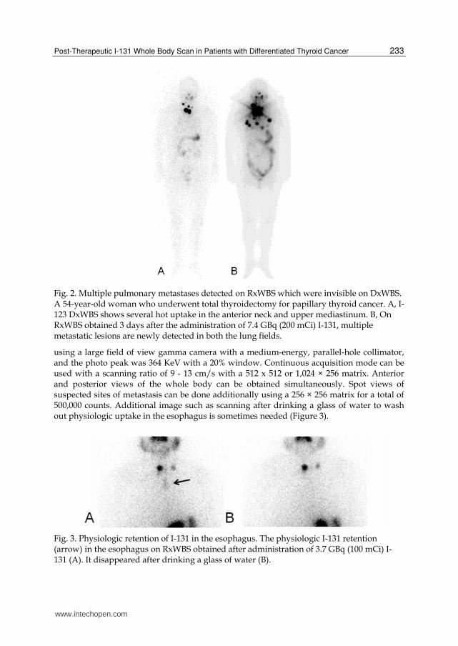
Fig. 2. Multiple pulmonary metastases detected on RxWBS which were invisible on DxWBS. A 54-year-old woman who underwent total thyroidectomy for papillary thyroid cancer. A, I-123 DxWBS shows several hot uptake in the anterior neck and upper mediastinum. B, On RxWBS obtained 3 days after the administration of 7.4 GBq (200 mCi) I-131, multiple metastatic lesions are newly detected in both the lung fields.
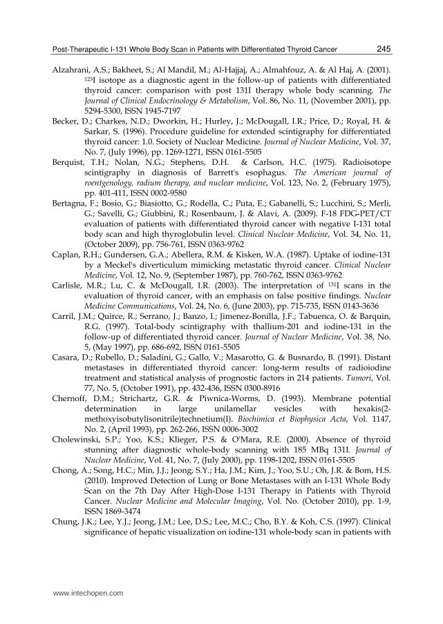
using a large field of view gamma camera with a medium-energy, parallel-hole collimator, and the photo peak was 364 KeV with a 20% window. Continuous acquisition mode can be used with a scanning ratio of 9 - 13 cm/s with a 512 x 512 or 1,024 × 256 matrix. Anterior and posterior views of the whole body can be obtained simultaneously. Spot views of suspected sites of metastasis can be done additionally using a 256 × 256 matrix for a total of 500,000 counts. Additional image such as scanning after drinking a glass of water to wash out physiologic uptake in the esophagus is sometimes needed (Figure 3).
Fig. 3. Physiologic retention of I-131 in the esophagus. The physiologic I-131 retention (arrow) in the esophagus on RxWBS obtained after administration of 3.7 GBq (100 mCi) I-131 (A). It disappeared after drinking a glass of water (B).
www.intechopen.com

12 Chapters on Nuclear Medicine
234
2.1.2 SPECT/CT Whole body imaging with SPECT/CT requires long scan time. Therefore, SPECT/CT is usually performed for specific site after whole body scan. The field of view (FOV) of SPECT/CT is usually determined by nuclear medicine physicians based on the planar image findings. So far, many companies have their models of SPECT/CT. This is the one of the usual protocol of SPECT/CT (Infinia Hawkeye 4) of our institution. First, emission SPECT images are acquired with counts from the 10% energy window at 364 KeV, with a matrix size of 128 x 128. A total image of 64 frames is acquired over 360˚ with an acquisition time of 30 s/frame, angular step of 6˚, and zooming factor of 1. After SPECT acquisition, a CT scan is acquired with a low-dose, helical CT scanner. The CT parameters are 140 KeV and 5 mAs, and no intravenous iodinated contrast is administered. The CT data are used for attenuation correction. The Images are reconstructed with a conventional iterative algorithm, ordered subset expectation maximization (OSEM). A workstation providing multiplanar reformatted images are used for image display and analysis.
2.2 Imaging timing of RxWBS Radioiodine is excreted slowly in hypothyroid patients, especially if they have been on a low iodine diet (Dietlein et al., 2005). Therefore, decreasing background activity is important for visualizing small metastases or remnant thyroid tissue. However, current guidelines for I-131 therapy differ in their recommendations for the optimal time of the RxWBS ranging from 48 – 72 hours (Becker et al., 1996; Luster et al., 2008) to 2 – 10 days (Cooper et al., 2009). Also, there is a lot of discrepancy in the published literature regarding the optimal timing of RxWBS (Cholewinski et al., 2000; Chong et al., 2010; Durante et al., 2006; Hindie et al., 2003; Hung et al., 2009; Khan et al., 1994; Nemec et al., 1979). So far, several studies were done for evaluating efficacy or accuracy of RxWBS which was done at different timing after high-dose I-131 therapy. Khan et al. conducted scans at 2 days and 7 days post-therapy after 3.7–5.6 GBq of I-131 administration (Khan et al., 1994). They reported a higher sensitivity for detection of iodine-avid tissue on RxWBS 7 days post-therapy than earlier RxWBS. Hung et al. analyzed RxWBS at three different time points (first scan performed 3–4 days, second scan 5–6 days, and third scan 10–11 days after I-131 therapy) (Hung et al., 2009). They retrospectively analyzed 239 patients’ scans. Twenty-eight percent of lymph node metastases, 17% of lung metastases, and 16% of bone metastases were missed on the late images on 10-11th day. On the other hand, only 5% of the remnants were missed. The ratio of early washout was different between remnants and metastatic lesions. Chong et al. conducted RxWBS on the third and seventh days after I-131 therapy in 60 cases from 52 patients with lung or bone metastases of thyroid cancer (Chong et al., 2010). They showed that 22% of lung metastases and 33% of bone metastases that were not shown on the third day scan were detected on the seventh day scan (Figure 4 and 5). Lee et al. conducted RxWBS on the third and tenth day after I-131 therapy in 81 patients (Lee et al., 2011). They reported that the I-131 avid lesions on the early scan were more easily detected by visual analysis and had higher uptake ratios than those on the delayed scan. The optimal timing for RxWBS is still needed to be clarified.
2.3 Medication and diet after administration of therapeutic dose of I-131 Published guideline for I-131 therapy recommend that low-iodine diet, when possible, <50 μg/day, starting 1-2 weeks prior to radioiodine administration is recommended (Cooper et al., 2009; Luster et al., 2008). However, the duration of this low-iodine diet varies. Usually, regular diet can be started after the treatment. Information about low-iodine diet can be obtained at Thyroid Cancer Survivors Association website, http://www.thyca.org/rai.htm#diet.
www.intechopen.com

Post-Therapeutic I-131 Whole Body Scan in Patients with Differentiated Thyroid Cancer
235
Fig. 4. Diffuse pulmonary metastases. The metastatic uptake is not seen on I-123 DxWBS (A) and early RxWBS (on the third day after therapy) (B). It only appears on the RxWBS which was performed on the seventh day after the administration of 7.4 GBq (200 mCi ) I-131 (C).
Fig. 5. Multiple osseous metastases. The metastatic lesion in the upper thoracic vertebra (arrow) is only visible on the RxWBS obtained seventh day after administration of 7.4 GBq (200 mCi) I-131 (B). These lesion is not visible on RxWBS obtained third day after therapy (A) or Tc-99m HDP WBS (C).
www.intechopen.com

12 Chapters on Nuclear Medicine
236
Thyroid hormone replacement may be resumed on the second or third day after therapy (Cooper et al., 2009; Luster et al., 2008).
3. Findings of RxWBS
The following data are needed to be reported: the name of patient, ID, age, the date of I-131 therapy, I-131 dose, date of RxWBS. When it is available, the serum level of thyroglobulin, thyroid stimulating hormone and anti-thyroglobulin antibody are to be reported. The sites of significant I-131 uptake are needed to be mentioned.
3.1 False positive RxWBS
False positive RxWBS occurs for nonthyroidal I-131 concentration, including external contamination by the saliva, nasal secretions and sweat containing I-131, internal contamination through nasopharyngeal secretion, as well as physiologic uptake in nonthyroidal tissue such as the choroid plexus, salivary glands, gastric mucosa, and urinary tract. Carliscle et al. summarized false positive findings (Carlisle et al., 2003). I-131 uptake can be seen in the nose, salivary glands, mouth, thyroid bed, lactating breast, liver, gall bladder, stomach, esophagus and sweat (Figure 6). Physiologic uptake of I-131 in the salivary glands, nasal mucosa, gastric mucosa, colon, mammary glands and choroid plexus is due to the NIS presence in these tissues (Carlisle et al., 2003; Riedel et al., 2001). Thymic uptake is rare, there is a report that the incidence of thymic uptake was 1-1.2% of cases (Davidson & McDougall, 2000) (Figure 7).
Fig. 6. Diffuse perspiration. (A) On the RxWBS obtained on the third day after I-131 therapy with 3.7 GBq (100 mCi), mild perspiration is noted in both the axillae. (B) On the RxWBS obtained on the seventh day after I-131 therapy, diffuse perspiration is present in both the axillae, chest, upper arms and hands.
www.intechopen.com

Post-Therapeutic I-131 Whole Body Scan in Patients with Differentiated Thyroid Cancer
237
Fig. 7. Thymic uptake. A 12-year-old girl underwent therapy with 3.7 GBq (100 mCi) I-131.
RxWBS were obtaind on the third day (A) and seventh day (B) after therapy. Hot uptakes
are seen in the right lower neck and upper chest. I-131 SPECT/CT (C) and F-18 FDG
PET/CT images (D) confirmed I-131 uptake in the upper chest to be physiologic uptake in
the thymus.
Diffuse hepatic uptake of I-131 is rarely found due to occult hepatic metastases but more
commonly due to the hepatic de-ionization and conjugation of I-131 which was not
incorporated into the thyroid hormone (Carlisle et al., 2003). Chung et al. investigated
hepatic uptake on DxWBS or RxWBS. They analyzed scans of 399 patients. They reported
that hepatic uptake is more often when higher dose of I-131 was administered. They also
reported that the more uptakes appeared in the residual thyroid, the more it appeared in the
liver. However, they found that 15 patients showed diffuse hepatic uptake without uptake
by the remnant thyroid or metastatic lesion. They followed these patients and metastatic
lesions were found in 7 of 15 patients. So, they insisted that diffuse liver uptake indicated
functioning thyroid remnant or metastasis (Chung et al., 1997) (Figure 8).
There are some reports I-131 false positive uptake in other pathologic condition unrelated to
thyroid cancer: tracheostomy site, bronchiectasis, pulmonary inflammatory disease, pleural
effusion, salivary gland tumor, some other carcinoma such as adenocarcinoma, squamous
carcinoma, Barrett’s esophagus, Meckel’s diverticulum (Ain & Shih, 1994; Berquist et al.,
1975; Caplan et al., 1987; Carlisle et al., 2003; Fernandez-Ulloa et al., 1976; Hoschl et al., 1988;
Misaki et al., 1994; Mitchell et al., 2000; Muratet & Giraud, 1996), and so on. (Figure 9)
www.intechopen.com

12 Chapters on Nuclear Medicine
238
Fig. 8. Diffuse hepatic uptake. A 59-year-old female underwent I-131 therapy with 370 MBq (100 mCi) after total thyroidectomy. On the RxWBS obtained on the third day after therapy, mild focal uptakes in the anterior neck and right lower anterior neck are shown (A). On the RxWBS obtained on the seventh day after therapy, diffuse hepatic uptake appears in addition to the cervical uptakes (B).
Fig. 9. I-131 accumulation in the benign pulmonary disease. A 51-year-old female underwent RxWBS on the third day (A) and the seventh day (B) after the administration of 6.66 GBq I-131. Besides the hot uptake in the neck, slightly increased, focal, linear uptake (arrows on A and B) is shown. On SPECT/CT (C), it is on the medial segment of the right middle lobe. Chest CT (D) shows peribronchial cicatrical consolidation and adhesive atelectasis containing mild traction bronchiectatic change in this lesion.
www.intechopen.com

Post-Therapeutic I-131 Whole Body Scan in Patients with Differentiated Thyroid Cancer
239
3.2 Diagnostic value of RxWBS
The visualizing functioning metastasis as well as remnant thyroid tissue is related to the dose of I-131. Waxman et al. reported that more lesions were detected when the activity administered was increased from 74 to 370 MBq (2 to 10 mCi) and when even higher yields at 1,110 to 3,700 MBq (30 to 100 mCi) (Waxman et al., 1981). Spies et al. reported that with higher therapeutic dose, RxWBS demonstrated additional findings or more accurate localization compared with a diagnostic dose of 185 MBq (5 mCi) in 46% of cases (Spies et al., 1989). The diagnostic accuracy of RxWBS is also related with the time interval from the date of administration of I-131 and that of scanning. Khan et al. and Chong et al. reported that earlier scanning might miss the lesions and the effectiveness of the delayed scan on the seventh day from the administration of I-131 (Khan S et al., 1994). Khan et al. performed RxWBS on the second day and seventh day post-therapy (Khan et al., 1994). They reported that the seventh day scan is more sensitive than third day scan. Chong et al. reported that 22% of lung metastasis and 33% of bone metastases that were not shown on the third day scan, though they were detected on the seventh day scan (Chong et al., 2010) (Figure 10).
Fig. 10. Vertebral metastasis mimicking remnant thyroid tissue. A 61-year-old female underwent RxWBS after administration of 7.4 GBq (200 mCi) I-131. RxWBS shows multiple uptakes in the abdomen and right pelvic area suggesting distant metastases. Focal uptake in the anterior neck was supposed to be usual remnant thyroid tissue. However, on additional SPECT/CT (B, C), the uptake is detected in the cervical vertebra (C5), not in the thyroidectomy site. SPECT/CT also reveals I-131 uptake on thelumbar vertebra (L3, D) and right iliac bone (E).
www.intechopen.com

12 Chapters on Nuclear Medicine
240
Hung et al. conducted different protocol, RxWBS on the third to fourth day, fifth to sixth
day and tenth to eleventh day post-therapy (Hung et al., 2009). They reported that there is a
trend of decreasing visualization of I-131 uptake in sequential images and that 17% of lung
metastasis and 16% of bone metastases were missed on the tenth to eleventh day scans. Lee
et al. conducted RxWBS on the third and tenth day post-therapy (Lee et al., 2011). They also
reported significant reduction in visual analysis scores and uptake ratios of I-131 avid
lesions on the delayed RxWBS.
In addition, diagnostic accuracy of RxWBS is related to the past-history of previous I-131
therapy. Oh et al. reported the sensitivity of RxWBS in detecting distant metastasis after first
I-131 therapy is 75% (Oh et al., 2011). In contrast, in patients with history of multiple
radioiodine therapy, sensitivity of RxWBS in detecting distant metastasis is only 35%. The
sensitivity of RxWBS and SPECT/CT is reported to be similar. However, the specificity of
RxWBS is lower than SPECT/CT (Table 1). The sensitivity of FDG PET/CT is lower than
RxWBS in detecting distant metastasis and the specificity of FDG PET/CT is much higher
than RxWBS or SPECT/CT.
Sens(%) Spec(%) DA(%) PPV(%) NPV(%)
All patients (n=140)
WBS 65 55 59 42 76
SPECT/CT 65 95 85 86 85
PET/CT 61 98 86 93 84
Single therapy (n=101)
WBS 90 56 63 35 6
SPECT/CT 90 96 95 86 97
PET/CT 48 100 89 100 88
Multiple therapies (n=39)
WBS 44 50 46 30 33
SPECT/CT 44 86 59 85 46
PET/CT 72 86 77 90 63
Table 1. Diagnostic performance of WBS, SPECT/CT and PET/CT in detecting distant metastasis (patient-based analysis) (adapted and modified from Oh et al., 2011). Sens, Sensitivity; Spec, Specificity; DA, diagnostic accuracy; PPV, positive predictive value; NPV, negative predictive value
Previous radioiodine scanning might be the reason of decreased sensitivity (Rawson et al.,
1951). Thyroid stunning has been reported as the temporary impairment of thyroid tissue
after a 111 MBq (3 mCi) or greater diagnostic I-131 dose that decreases the final absorbed
dose in ablative therapy. However, Rosario et al. reported that diagnostic scanning using a
185 MBq (5 mCi) of I-131 dose does not interfere with uptake of the ablative dose or with
treatment efficacy when ablation is performed within 72 hours (Rosario et al., 2005). Dam et
al. reported that even though, stunning might occur but there was no significant difference
in treatment success rates (Dam et al., 2004).
www.intechopen.com

Post-Therapeutic I-131 Whole Body Scan in Patients with Differentiated Thyroid Cancer
241
4. Other imaging modalities
4.1 F-18 Fluorodeoxyglucose PET/CT
Fluorodeoxyglucose (FDG) is a glucose analogue. This is taken into the cell and phosphorylated by the same mechanism as glucose. In thyroid cancer, “Flip-flop phenomenone” is reported (Feine et al., 1996; Khan N. et al., 2003). It means the alternating uptake pattern of I-131 and FDG by the differentiated papillary or follicular thyroid cancer, which thought to be related to the differentiation of tumor (Figure 11 and 12). Bertagna et al. reported that F-18 FDG PET/CT positive results correlated with the serum thyroglobulin level in patients with negative I-131 whole body scan and high serum thyroglobulin level. They also reported that F-18 FDG PET/CT showed highest accuracy when the patient’s thyroglobulin level was higher than 21 ng/mL (Bertagna et al., 2009). In addition to that, they also revealed that the levothyroxine therapy regimen does not influence F-18 FDG PET/CT results. Yoshio et al. evaluated 55 cases with differentiated thyroid cancer with F-18 FDG PET/CT and reported that FDG-avid lesions are resistant to radioactive iodine therapy with or without I-131 uptake (Yoshio et al., 2011).
Fig. 11. Flip-flop phenomenone. A 44-year-old male underwent total thyroidectomy due to thyroid papillary carcinoma. I-123 DxWBS (A) shows several hot uptakes in the anterior neck, whereas F-18 FDG PET/CT (B) is negative.
4.2 Tc-99m methoxyisobutylisonitrile (Tc-99m MIBI) Tc-99m MIBI, a lipophilic cationic molecules, was originally developed as a myocardial perfusion imaging agent. Today Tc-99m MIBI is also used for the study of many neoplastic diseases. The mechanism of Tc-99m MIBI accumulation in tumor has been reported to depend on the size of a tumor, the blood flow and the richness of mitochondria in the tumor
www.intechopen.com

12 Chapters on Nuclear Medicine
242
Fig. 12. Iodine negative and F-18 FDG PET positive case. A 65-year-old female underwent I-123 DxWBS (A) and F-18 FDG PET/CT (B,C) for elevated serum thyroglobulin level (3 to 17 ng/ml) after left modified radical lymph node dissection two years ago. I-123 DxWBS (A) does not show abnormal uptake. On F-18 FDG PET/CT (B), focal uptakes were shown in the left lateral neck (arrow). On axial view of fusion image (C), two hypermetabolic lesions were seen in the left cervical level II. Biopsy revealed recurred papillary carcinoma.
cells (Moretti et al., 2005; Piwnica-Worms et al., 1994; Saggiorato et al., 2009). MIBI irreversibly passes into the mitochondria using the electrical gradient generated by a high negative inner transmembrane mitochondrial potential of malignant cells (Chernoff et al., 1993; Moretti et al., 2005; Piwnica-Worms et al., 1994). Also, there is a report that TSH simulates both F-18 FDG PET and Tc-99m MIBI uptake in poorly differentiated papillary thyroid cancer in vitro experiment (Kim et al., 2009). Even though Tc-99m MIBI is inferior to I-131 scintigraphy in detecting I-131 avid lesions (Al Saleh et al., 2007), it can be applied for patients with elevated serum human thyroglobulin levels but negative I-131 whole body scan (Wu et al., 2003). However, there are several reports that F-18 FDG PET is more sensitive than Tc-99m MIBI SPECT in detecting metastatic cervical lymph node in differentiated thyroid cancer patients with elevated serum thyroglobulin level but negative I-131 whole body scan (Iwata et al., 2004; Wu et al., 2003) (Figure 13).
4.3 Tc-99m tetrofosmin
Tetrofosmin is a lipophilic phosphine useful for myocardial perfusion imaging. Significant uptake of Tc-99m tetrofosmin in the thyroid, breast and lung tumors is reported. Tc-99m tetrofosmin is useful in detection of thyroid metastatic disease, particularly when the tumor is not iodine avid. In addition, it does not require withdrawal of thyroid hormone suppression therapy (Sharma et al., 2007). Nishiyama et al. reported that detectability of thyroid cancer metastases using Tc-99m tetrofosmin was 79.4% and that Tc-99m tetrofosmin is more sensitive than I-131 for detection of differentiated thyroid cancer metastasis, particulary for regional lymph node (Nishiyama et al., 2000). Unal et al. reported different results that the sensitivities of Tl-201, Tc-99m tetrofosmin and I-131 in diagnosing distant metastases were comparable (0.85, 0.85, and 0.75, respectively) (Unal et al., 1998).
www.intechopen.com

Post-Therapeutic I-131 Whole Body Scan in Patients with Differentiated Thyroid Cancer
243
Fig. 13. I-131, Tc-99m MIBI and F-18 FDG images. A 62-year-old woman with metastatic papillary thyroid cancer to the left proximal humerus, clavicle and sternum underwent I-131 therapy with 7.4 GBq (200 mCi). Her serum thyroglobulin level was 260 ng/mL. I-131 RxWBS (A) shows focal hot uptakes in the left clavicle and huemrus. On Tc-99m MIBI WBS, irregular hot uptakes are detected in the sternal area and faint uptake in the left clavicle (B). F-18 FDG PET/CT (C-E) shows multiple hypermetabolic lesions in the sternum, both hilar lymph nodes and lungs.
4.4 Thallium-201 Thallium-201 (Tl-201) is a monovalemt cationic radioisotope listed in same group with gallium in the periodic table. The membrane permeability for Tl-201 is almost equal to that for potassium (Elgazzar et al., 1993). It is suggested that intracellular accumulation of thallous ions is due to the transmembrane electropotential gradient (Mullins & Moore, 1960). The primary role of Tl-201 in nuclear medicine is for imaging of myocardial perfusion and viability (Pauwels et al., 1998). Also, Tl-201 chloride has affinity for various malignant tumors, although it is not specific for malignant tumor (Senga et al., 1982). Uptake of Tl-201 in tumor seems to be determined by blood flow, grade of malignancy, viability of tumor cells and tumor necrosis (Pauwels et al., 1998). There are several reports regarding Tl-201 and thyroid cancer. Shiga et al. compared FDG PET with I-131 and Tl-201 scintigraphy and reported that FDG uptake was concordant with I-131 in 38% and with Tl-201 uptake in 94% (Shiga et al., 2001). Senga et al. reported that when tumor showed a positive scan with Tl-
www.intechopen.com

12 Chapters on Nuclear Medicine
244
201 chloride but negative results using Ga-67 citrate, it was differentiated thyroid carcinoma or poorly differentiated adenoma (Senga et al., 1982). Carril et al. analyzed Tl-201 and I-131 in ablated thyroid cancer patients. They reported that the sensitivity and specificity were 94% and 96% for Tl-201 and 29% and 100% for I-131 (Carril et al., 1997). Nakada et al. reported that Tl-201 uptake correlated well with the proliferating cell nuclear antigen index, which represents proliferative activity (Nakada et al., 1999).
4.5 I-124 PET/CT
I-124 is a positron emitter with a half-life of 4.18 days, produced by a cyclotron. It can be used to directly image thyroid cancer using PET scanner. Van Nostrand et al. compared I-124 PET with I-131 DxWBS in detecting residual thyroid tissue and/or metastatic lesion. They reported that I-124 PET identified as many as 50% more foci of radioiodine uptake suggestive of additional residual thyroid tissue and/or metastasis (Van Nostrand et al., 2010). They suggest that I-124 PET produced superior results because the PET scanner provides images with reduced background noise and enhanced spatial and contrast resolution compared with I-131 DxWBS. The longer half-life of I-124 is useful for dose monitoring over an extended time period (Surti et al., 2009). It can be also used for lesion-specific dosimetry. I-131 mean absorbed dose distributions can be calculated from serial I-124 PET image (Erdi et al., 1999; Larson & Robbins, 2002).
5. Conclusion
The I-123 DxWBS or I-131 DxWBS is sensitive and specific for treatable remnant or metastatic lesion. About 25% of RxWBS detect lesions missed by DxWBS. For this reason, following administration of a therapeutic I-131 dose, RxWBS should always be performed in patients with well differentiated thyroid cancer. The timing of RxWBS is currently still controversial. Based on several studies, the timing of post-therapeutic I-131 WBS is within 5th to 10th day, especially more opportune around 7th day after therapy. I-131 WBS with SPECT/CT may be the highly tailored approach for assessing distant metastatic lesions in patients who received a radioiodine therapy if SPECT/CT is available. RxWBS is especially likely to provide the useful information when DxWBSs are negative and the serum thyroglobulin levels are elevated. Other imaging modalities including F-18 FDG PET/CT is useful in detecting remnant thyroid tissues or metastatic thyroid cancer in patient with the elevated serum thyroglobulin level and negative iodine images. I-124 can be also useful in detection of the lesion and evaluation of lesional and whole-body dosimetry in patients with well-differentiated thyroid cancer.
6. References
Ain, K.B. & Shih, W.J. (1994). False-positive I-131 uptake at a tracheostomy site. Discernment with Tl-201 imaging. Clinical Nuclear Medicine, Vol. 19, No. 7, (July 1994), pp. 619-621, ISSN 0363-9762
Al Saleh, K.; Safwat, R.; Al-Shammeri, I.; Abdul Naseer, M.; Hooda, H. & Al-Mohannadi, S. (2007). Comparison of whole body scintigraphy with Tc-99m-methoxyisobutylisonitrile and iodine-131 NA in patients with differentiated thyroid cancer. The Gulf Journal of Oncology, Vol. 1, No. 1, (January 2007), pp. 29-33, ISSN 2078-2101
www.intechopen.com
Post-Therapeutic I-131 Whole Body Scan in Patients with Differentiated Thyroid Cancer
245
Alzahrani, A.S.; Bakheet, S.; Al Mandil, M.; Al-Hajjaj, A.; Almahfouz, A. & Al Haj, A. (2001). 123I isotope as a diagnostic agent in the follow-up of patients with differentiated thyroid cancer: comparison with post 131I therapy whole body scanning. The Journal of Clinical Endocrinology & Metabolism, Vol. 86, No. 11, (November 2001), pp. 5294-5300, ISSN 1945-7197
Becker, D.; Charkes, N.D.; Dworkin, H.; Hurley, J.; McDougall, I.R.; Price, D.; Royal, H. & Sarkar, S. (1996). Procedure guideline for extended scintigraphy for differentiated thyroid cancer: 1.0. Society of Nuclear Medicine. Journal of Nuclear Medicine, Vol. 37, No. 7, (July 1996), pp. 1269-1271, ISSN 0161-5505
Berquist, T.H.; Nolan, N.G.; Stephens, D.H. & Carlson, H.C. (1975). Radioisotope scintigraphy in diagnosis of Barrett's esophagus. The American journal of roentgenology, radium therapy, and nuclear medicine, Vol. 123, No. 2, (February 1975), pp. 401-411, ISSN 0002-9580
Bertagna, F.; Bosio, G.; Biasiotto, G.; Rodella, C.; Puta, E.; Gabanelli, S.; Lucchini, S.; Merli, G.; Savelli, G.; Giubbini, R.; Rosenbaum, J. & Alavi, A. (2009). F-18 FDG-PET/CT evaluation of patients with differentiated thyroid cancer with negative I-131 total body scan and high thyroglobulin level. Clinical Nuclear Medicine, Vol. 34, No. 11, (October 2009), pp. 756-761, ISSN 0363-9762
Caplan, R.H.; Gundersen, G.A.; Abellera, R.M. & Kisken, W.A. (1987). Uptake of iodine-131 by a Meckel's diverticulum mimicking metastatic thyroid cancer. Clinical Nuclear Medicine, Vol. 12, No. 9, (September 1987), pp. 760-762, ISSN 0363-9762
Carlisle, M.R.; Lu, C. & McDougall, I.R. (2003). The interpretation of 131I scans in the evaluation of thyroid cancer, with an emphasis on false positive findings. Nuclear Medicine Communications, Vol. 24, No. 6, (June 2003), pp. 715-735, ISSN 0143-3636
Carril, J.M.; Quirce, R.; Serrano, J.; Banzo, I.; Jimenez-Bonilla, J.F.; Tabuenca, O. & Barquin, R.G. (1997). Total-body scintigraphy with thallium-201 and iodine-131 in the follow-up of differentiated thyroid cancer. Journal of Nuclear Medicine, Vol. 38, No. 5, (May 1997), pp. 686-692, ISSN 0161-5505
Casara, D.; Rubello, D.; Saladini, G.; Gallo, V.; Masarotto, G. & Busnardo, B. (1991). Distant metastases in differentiated thyroid cancer: long-term results of radioiodine treatment and statistical analysis of prognostic factors in 214 patients. Tumori, Vol. 77, No. 5, (October 1991), pp. 432-436, ISSN 0300-8916
Chernoff, D.M.; Strichartz, G.R. & Piwnica-Worms, D. (1993). Membrane potential determination in large unilamellar vesicles with hexakis(2-methoxyisobutylisonitrile)technetium(I). Biochimica et Biophysica Acta, Vol. 1147, No. 2, (April 1993), pp. 262-266, ISSN 0006-3002
Cholewinski, S.P.; Yoo, K.S.; Klieger, P.S. & O'Mara, R.E. (2000). Absence of thyroid stunning after diagnostic whole-body scanning with 185 MBq 131I. Journal of Nuclear Medicine, Vol. 41, No. 7, (July 2000), pp. 1198-1202, ISSN 0161-5505
Chong, A.; Song, H.C.; Min, J.J.; Jeong, S.Y.; Ha, J.M.; Kim, J.; Yoo, S.U.; Oh, J.R. & Bom, H.S. (2010). Improved Detection of Lung or Bone Metastases with an I-131 Whole Body Scan on the 7th Day After High-Dose I-131 Therapy in Patients with Thyroid Cancer. Nuclear Medicine and Molecular Imaging, Vol. No. (October 2010), pp. 1-9, ISSN 1869-3474
Chung, J.K.; Lee, Y.J.; Jeong, J.M.; Lee, D.S.; Lee, M.C.; Cho, B.Y. & Koh, C.S. (1997). Clinical significance of hepatic visualization on iodine-131 whole-body scan in patients with
www.intechopen.com

12 Chapters on Nuclear Medicine
246
thyroid carcinoma. Journal of Nuclear Medicine, Vol. 38, No. 8, (August 1997), pp. 1191-1195, ISSN 0161-5505
Cooper, D.S.; Doherty, G.M.; Haugen, B.R.; Kloos, R.T.; Lee, S.L.; Mandel, S.J.; Mazzaferri, E.L.; McIver, B.; Pacini, F.; Schlumberger, M.; Sherman, S.I.; Steward, D.L. & Tuttle, R.M. (2009). Revised American Thyroid Association management guidelines for patients with thyroid nodules and differentiated thyroid cancer. Thyroid, Vol. 19, No. 11, (November 2009), pp. 1167-1214, ISSN 1557-9077
Dam, H.Q.; Kim, S.M.; Lin, H.C. & Intenzo, C.M. (2004). 131I therapeutic efficacy is not influenced by stunning after diagnostic whole-body scanning. Radiology, Vol. 232, No. 2, (August 2004), pp. 527-533, ISSN 0033-8419
Davidson, J. & McDougall, I.R. (2000). How frequently is the thymus seen on whole-body iodine-131 diagnostic and post-treatment scans? European Journal of Nuclear Medicine, Vol. 27, No. 4, (May 2000), pp. 425-430, ISSN 0340-6997
Dietlein, M.; Moka, D. & Schicha, H. (2005). Radioiodine therapy for thyroid cancer, In: Thyroid cancer, H. J. Biersack & F. Grunwald, pp. 93, Springer, ISBN 3540413901, New York
Donahue, K.P.; Shah, N.P.; Lee, S.L. & Oates, M.E. (2008). Initial staging of differentiated thyroid carcinoma: continued utility of posttherapy 131I whole-body scintigraphy. Radiology, Vol. 246, No. 3, (March 2008), pp. 887-894, ISSN 0033-8419
Durante, C.; Haddy, N.; Baudin, E.; Leboulleux, S.; Hartl, D.; Travagli, J.P.; Caillou, B.; Ricard, M.; Lumbroso, J.D.; De Vathaire, F. & Schlumberger, M. (2006). Long-term outcome of 444 patients with distant metastases from papillary and follicular thyroid carcinoma: benefits and limits of radioiodine therapy. The Journal of Clinical Endocrinology & Metabolism, Vol. 91, No. 8, (August 2006), pp. 2892-2899, ISSN 0021-972X
Elgazzar, A.H.; Fernandez-Ulloa, M. & Silberstein, E.B. (1993). 201Tl as a tumour-localizing agent: current status and future considerations. Nucl Med Commun, Vol. 14, No. 2, (February 1993), pp. 96-103, ISSN 0143-3636 (Print) 0143-3636 (Linking)
Erdi, Y.E.; Macapinlac, H.; Larson, S.M.; Erdi, A.K.; Yeung, H.; Furhang, E.E. & Humm, J.L. (1999). Radiation Dose Assessment for I-131 Therapy of Thyroid Cancer Using I-124 PET Imaging. Clin Positron Imaging, Vol. 2, No. 1, (October 2003), pp. 41-46, ISSN 1095-0397
Fatourechi, V.; Hay, I.D.; Mullan, B.P.; Wiseman, G.A.; Eghbali-Fatourechi, G.Z.; Thorson, L.M. & Gorman, C.A. (2000). Are posttherapy radioiodine scans informative and do they influence subsequent therapy of patients with differentiated thyroid cancer? Thyroid, Vol. 10, No. 7, (August 2000), pp. 573-577, ISSN 1050-7256
Feine, U.; Lietzenmayer, R.; Hanke, J.P.; Held, J.; Wohrle, H. & Muller-Schauenburg, W. (1996). Fluorine-18-FDG and iodine-131-iodide uptake in thyroid cancer. Journal of Nuclear Medicine, Vol. 37, No. 9, (September 1996), pp. 1468-1472, ISSN 0161-5505
Fernandez-Ulloa, M.; Maxon, H.R.; Mehta, S. & Sholiton, L.J. (1976). Iodine-131 uptake by primary lung adenocarcinoma. Misinterpretation of 131I scan. JAMA, Vol. 236, No. 7, (August 1976), pp. 857-858, ISSN 0098-7484
Hindie, E.; Melliere, D.; Lange, F.; Hallaj, I.; de Labriolle-Vaylet, C.; Jeanguillaume, C.; Lange, J.; Perlemuter, L. & Askienazy, S. (2003). Functioning pulmonary metastases of thyroid cancer: does radioiodine influence the prognosis? European
www.intechopen.com

Post-Therapeutic I-131 Whole Body Scan in Patients with Differentiated Thyroid Cancer
247
Journal of Nuclear Medicine and Molecular Imaging, Vol. 30, No. 7, (March 2003), pp. 974-981, ISSN 1619-7070
Hoschl, R.; Choy, D.H. & Gandevia, B. (1988). Iodine-131 uptake in inflammatory lung disease: a potential pitfall in treatment of thyroid carcinoma. Journal of Nuclear Medicine, Vol. 29, No. 5, (May 1988), pp. 701-706, ISSN 0161-5505
Hung, B.T.; Huang, S.H.; Huang, Y.E. & Wang, P.W. (2009). Appropriate time for post-therapeutic I-131 whole body scan. Clinical Nuclear Medicine, Vol. 34, No. 6, (June 2009), pp. 339-342, ISSN 1536-0229
Iwano, S.; Kato, K.; Nihashi, T.; Ito, S.; Tachi, Y. & Naganawa, S. (2009). Comparisons of I-123 diagnostic and I-131 post-treatment scans for detecting residual thyroid tissue and metastases of differentiated thyroid cancer. Annals of Nuclear Medicine, Vol. 23, No. 9, (September 2009), pp. 777-782, ISSN 1864-6433 (Electronic)
Iwata, M.; Kasagi, K.; Misaki, T.; Matsumoto, K.; Iida, Y.; Ishimori, T.; Nakamoto, Y.; Higashi, T.; Saga, T. & Konishi, J. (2004). Comparison of whole-body 18F-FDG PET, 99mTc-MIBI SPET, and post-therapeutic 131I-Na scintigraphy in the detection of metastatic thyroid cancer. European Journal of Nuclear Medicine and Molecular Imaging, Vol. 31, No. 4, (April 2004), pp. 491-498, ISSN 1619-7070
Khan, N.; Oriuchi, N.; Higuchi, T.; Zhang, H. & Endo, K. (2003). PET in the follow-up of differentiated thyroid cancer. British Journal of Radiology, Vol. 76, No. 910, (October 2003), pp. 690-695, ISSN 0007-1285
Khan, S.; Waxman, A.; Nagaraj, N. & G, B. Optimization of post ablative I-131 scintigraphy: comparison of 2 day vs. 7 day post therapy study in patients with differentiated thyroid cancer (DTC), Proceedings of 41st Annual Meeting of the Society of Nuclear Medicine., Cedars-sinai Medical center, Los Angeles, CA, 1994
Kim, C.H.; Yoo Ie, R.; Chung, Y.A.; Park, Y.H.; Kim, S.H.; Sohn, H.S. & Chung, S.K. (2009). Influence of thyroid-stimulating hormone on 18F-fluorodeoxyglucose and 99mTc-methoxyisobutylisonitrile uptake in human poorly differentiated thyroid cancer cells in vitro. Annals of Nuclear Medicine, Vol. 23, No. 2, (February 2009), pp. 131-136, ISSN 0914-7187
Larson, S.M. & Robbins, R. (2002). Positron emission tomography in thyroid cancer management. Seminars in Roentgenology, Vol. 37, No. 2, (July 2002), pp. 169-174, ISSN 0037-198X
Lee, J.W.; Lee, S.M.; Koh, G.P. & Lee, D.H. (2011). The comparison of (131)I whole-body scans on the third and tenth day after (131)I therapy in patients with well-differentiated thyroid cancer: preliminary report. Annals of Nuclear Medicine, Vol. No. (April 2011), pp. ISSN 1864-6433
Luster, M.; Clarke, S.E.; Dietlein, M.; Lassmann, M.; Lind, P.; Oyen, W.J.; Tennvall, J. & Bombardieri, E. (2008). Guidelines for radioiodine therapy of differentiated thyroid cancer. European Journal of Nuclear Medicine and Molecular Imaging, Vol. 35, No. 10, (August 2008), pp. 1941-1959, ISSN 1619-7070
Mazzaferri, E. L. (1986). In: Treatment of carcinoma of follicular epithelium, pp. 1342-1343, Lippincott, ISBN 0397508026 Philadelphia
Mazzaferri, E.L. & Jhiang, S.M. (1994). Long-term impact of initial surgical and medical therapy on papillary and follicular thyroid cancer. American Journal of Medicine, Vol. 97, No. 5, (November 1994), pp. 418-428, ISSN 0002-9343
www.intechopen.com

12 Chapters on Nuclear Medicine
248
Misaki, T.; Takeuchi, R.; Miyamoto, S.; Kasagi, K.; Matsui, Y. & Konishi, J. (1994). Radioiodine uptake by squamous-cell carcinoma of the lung. Journal of Nuclear Medicine, Vol. 35, No. 3, (March 1994), pp. 474-475, ISSN 0161-5505
Mitchell, G.; Pratt, B.E.; Vini, L.; McCready, V.R. & Harmer, C.L. (2000). False positive 131I whole body scans in thyroid cancer. The British Journal or Radiology, Vol. 73, No. 870, (July 2000), pp. 627-635, ISSN 0007-1285
Moretti, J.L.; Hauet, N.; Caglar, M.; Rebillard, O. & Burak, Z. (2005). To use MIBI or not to use MIBI? That is the question when assessing tumour cells. European Journal of Nuclear Medicine and Molecular Imaging, Vol. 32, No. 7, (July 2005), pp. 836-842, ISSN 1619-7070
Mullins, L.J. & Moore, R.D. (1960). The movement of thallium ions in muscle. J Gen Physiol, Vol. 43, No. 4, (March 1960), pp. 759-773, ISSN 0022-1295 (Print) 0022-1295 (Linking)
Muratet, J.P. & Giraud, P. (1996). Thymus accumulation of I-131 after therapeutic dose for thyroid carcinoma. Clinical Nuclear Medicine, Vol. 21, No. 9, (September 1996), pp. 736-737, ISSN 0363-9762
Nakada, K.; Katoh, C.; Morita, K.; Kanegae, K.; Tsukamoto, E.; Shiga, T.; Mochizuki, T. & Tamaki, N. (1999). Relationship among 201T1 uptake, nuclear DNA content and clinical behavior in metastatic thyroid carcinoma. Journal of Nuclear Medicine, Vol. 40, No. 6, (August 1999), pp. 963-967, ISSN 0161-5505
Nemec, J.; Rohling, S.; Zamrazil, V. & Pohunkova, D. (1979). Comparison of the distribution of diagnostic and thyroablative I-131 in the evaluation of differentiated thyroid cancers. Journal of Nuclear Medicine, Vol. 20, No. 2, (February 1979), pp. 92-97, ISSN 0161-5505
Nishiyama, Y.; Yamamoto, Y.; Ono, Y.; Takahashi, K.; Nakano, S.; Satoh, K.; Ohkawa, M. & Tanabe, M. (2000). Comparison of 99mTc-tetrofosmin with 201Tl and 131I in the detection of differentiated thyroid cancer metastases. Nuclear Medince Communications, Vol. 21, No. 10, (October 2000), pp. 917-923, ISSN 0143-3636
Oh, J.R.; Byun, B.H.; Hong, S.P.; Chong, A.; Kim, J.; Yoo, S.W.; Kang, S.R.; Kim, D.Y.; Song, H.C.; Bom, H.S. & Min, J.J. (2011). Comparison of (131)I whole-body imaging, (131)I SPECT/CT, and (18)F-FDG PET/CT in the detection of metastatic thyroid cancer. European Journal of Nuclear Medicine and Molecular Imaging, Vol. 38, No. 8, (August 2011), pp. 1459-1468, ISSN 1619-7089
Pauwels, E.K.; McCready, V.R.; Stoot, J.H. & van Deurzen, D.F. (1998). The mechanism of accumulation of tumour-localising radiopharmaceuticals. European Journal of Nuclear Medicine, Vol. 25, No. 3, (March 1998), pp. 277-305, ISSN 0340-6997
Piwnica-Worms, D.P.; Kronauge, J.F.; LeFurgey, A.; Backus, M.; Hockett, D.; Ingram, P.; Lieberman, M.; Holman, B.L.; Jones, A.G. & Davison, A. (1994). Mitochondrial localization and characterization of 99mTc-SESTAMIBI in heart cells by electron probe X-ray microanalysis and 99Tc-NMR spectroscopy. Magnetic Resonance Imaging, Vol. 12, No. 4, (January 1994), pp. 641-652, ISSN 0730-725X
Rawson, R.W.; Rall, J.E. & Peacock, W. (1951). Limitations and indications in the treatment of cancer of the thyroid with radioactive iodine. The Journal of Clinical Endocrinology & Metabolism, Vol. 11, No. 10, (October 1951), pp. 1128-1142, ISSN 0021-972X
Riedel, C.; Dohan, O.; De la Vieja, A.; Ginter, C.S. & Carrasco, N. (2001). Journey of the iodide transporter NIS: from its molecular identification to its clinical role in cancer.
www.intechopen.com

Post-Therapeutic I-131 Whole Body Scan in Patients with Differentiated Thyroid Cancer
249
Trends in Biochemical Sciences, Vol. 26, No. 8, (August 2001), pp. 490-496, ISSN 0968-0004
Rosario, P.W.; Barroso, A.L.; Rezende, L.L.; Padrao, E.L.; Maia, F.F.; Fagundes, T.A. & Purisch, S. (2005). 5 mCi pretreatment scanning does not cause stunning when the ablative dose is administered within 72 hours. Arquivos Brasileiros de Endocrinologia & Metabologia, Vol. 49, No. 3, (March 2006), pp. 420-424, ISSN 0004-2730
Saggiorato, E.; Angusti, T.; Rosas, R.; Martinese, M.; Finessi, M.; Arecco, F.; Trevisiol, E.; Bergero, N.; Puligheddu, B.; Volante, M.; Podio, V.; Papotti, M. & Orlandi, F. (2009). 99mTc-MIBI Imaging in the presurgical characterization of thyroid follicular neoplasms: relationship to multidrug resistance protein expression. Journal of Nuclear Medicine, Vol. 50, No. 11, (November 2009), pp. 1785-1793, ISSN 1535-5667
Samaan, N.A.; Schultz, P.N.; Haynie, T.P. & Ordonez, N.G. (1985). Pulmonary metastasis of differentiated thyroid carcinoma: treatment results in 101 patients. The Journal of Clinical Endocrinology & Metabolism, Vol. 60, No. 2, (February 1985), pp. 376-380, ISSN 0021-972X
Samaan, N.A.; Schultz, P.N.; Hickey, R.C.; Goepfert, H.; Haynie, T.P.; Johnston, D.A. & Ordonez, N.G. (1992). The results of various modalities of treatment of well differentiated thyroid carcinomas: a retrospective review of 1599 patients. The Journal of Clinical Endocrinology & Metabolism, Vol. 75, No. 3, (September 1992), pp. 714-720, ISSN 0021-972X
Schlumberger, M.; Tubiana, M.; De Vathaire, F.; Hill, C.; Gardet, P.; Travagli, J.P.; Fragu, P.; Lumbroso, J.; Caillou, B. & Parmentier, C. (1986). Long-term results of treatment of 283 patients with lung and bone metastases from differentiated thyroid carcinoma. The Journal of Clinical Endocrinology & Metabolism, Vol. 63, No. 4, (October 1986), pp. 960-967, ISSN 0021-972X
Senga, O.; Miyakawa, M.; Shirota, H.; Makiuchi, M.; Yano, K.; Miyazawa, M. & Takizawa, M. (1982). Comparison of Tl-201 chloride and Ga-67 citrate scintigraphy in the diagnosis of thyroid tumor: concise communication. Journal of Nuclear Medicine, Vol. 23, No. 3, (March 1982), pp. 225-228, ISSN 0161-5505
Sharma, R.; Chakravarty, K.L.; Tripathi, M.; Kaushik, A.; Bharti, P.; Sahoo, M.; Chopra, M.K.; Rawat, H.; Misra, A.; Mondal, A. & Kashyap, R. (2007). Role of 99mTc-Tetrofosmin delayed scintigraphy and color Doppler sonography in characterization of solitary thyroid nodules. Nuclear Medicine Communcations, Vol. 28, No. 11, (September 2007), pp. 847-851, ISSN 0143-3636
Shiga, T.; Tsukamoto, E.; Nakada, K.; Morita, K.; Kato, T.; Mabuchi, M.; Yoshinaga, K.; Katoh, C.; Kuge, Y. & Tamaki, N. (2001). Comparison of (18)F-FDG, (131)I-Na, and (201)Tl in diagnosis of recurrent or metastatic thyroid carcinoma. Journal of Nuclear Medicine, Vol. 42, No. 3, (March 2001), pp. 414-419, ISSN 0161-5505
Souza Rosario, P.W.; Barroso, A.L.; Rezende, L.L.; Padrao, E.L.; Fagundes, T.A.; Penna, G.C. & Purisch, S. (2004). Post I-131 therapy scanning in patients with thyroid carcinoma metastases: an unnecessary cost or a relevant contribution? Clinical Nuclear Medicine, Vol. 29, No. 12, (Novemeber 2004), pp. 795-798, ISSN 0363-9762
Spies, W.G.; Wojtowicz, C.H.; Spies, S.M.; Shah, A.Y. & Zimmer, A.M. (1989). Value of post-therapy whole-body I-131 imaging in the evaluation of patients with thyroid carcinoma having undergone high-dose I-131 therapy. Clinical Nuclear Medicine, Vol. 14, No. 11, (November 1989), pp. 793-800, ISSN 0363-9762
www.intechopen.com

12 Chapters on Nuclear Medicine
250
Surti, S.; Scheuermann, R. & Karp, J.S. (2009). Correction technique for cascade gammas in I-124 imaging on a fully-3D, Time-of-Flight PET Scanner. IEEE Trans Nucl Sci, Vol. 56, No. 3, (August 2009), pp. 653-660, ISSN 0018-9499
Thyroid cancer survivors' Association, Inc. (05.23.2011). Radioactive iodine (RAI), In: Thyroid Cancer Survivors Association website, 06.22.2011, Available from: <http://www.thyca.org/rai.htm#diet>
Unal, S.; Menda, Y.; Adalet, I.; Boztepe, H.; Ozbey, N.; Alagol, F. & Cantez, S. (1998). Thallium-201, technetium-99m-tetrofosmin and iodine-131 in detecting differentiated thyroid carcinoma metastases. Journal of Nuclear Medicine, Vol. 39, No. 11, (November 1998), pp. 1897-1902, ISSN 0161-5505
Van Nostrand, D.; Moreau, S.; Bandaru, V.V.; Atkins, F.; Chennupati, S.; Mete, M.; Burman, K. & Wartofsky, L. (2010). (124)I positron emission tomography versus (131)I planar imaging in the identification of residual thyroid tissue and/or metastasis in patients who have well-differentiated thyroid cancer. Thyroid, Vol. 20, No. 8, (August 2010), pp. 879-883, ISSN 1050-7256
Waxman, A.; Ramanna, L.; Chapman, N.; Chapman, D.; Brachman, M.; Tanasescu, D.; Berman, D.; Catz, B. & Braunstein, G. (1981). The significance of 1-131 scan dose in patients with thyroid cancer: determination of ablation: concise communication. Journal of Nuclear Medicine, Vol. 22, No. 10, (October 1981), pp. 861-865, ISSN 0161-5505
Wu, H.S.; Huang, W.S.; Liu, Y.C.; Yen, R.F.; Shen, Y.Y. & Kao, C.H. (2003). Comparison of FDG-PET and technetium-99m MIBI SPECT to detect metastatic cervical lymph nodes in well-differentiated thyroid carcinoma with elevated serum HTG but negative I-131 whole body scan. Anticancer Research, Vol. 23, No. 5b, (December 2003), pp. 4235-4238, ISSN 0250-7005
Yoshio, K.; Sato, S.; Okumura, Y.; Katsui, K.; Takemoto, M.; Suzuki, E.; Katayama, N.; Kaji, M. & Kanazawa, S. (2011). The local efficacy of I-131 for F-18 FDG PET positive lesions in patients with recurrent or metastatic thyroid carcinomas. Clincal Nuclear Medicine, Vol. 36, No. 2, (February 2011), pp. 113-117, ISSN 0363-9762
www.intechopen.com

12 Chapters on Nuclear MedicineEdited by Dr. Ali Gholamrezanezhad
ISBN 978-953-307-802-1Hard cover, 304 pagesPublisher InTechPublished online 22, December, 2011Published in print edition December, 2011
InTech EuropeUniversity Campus STeP Ri Slavka Krautzeka 83/A 51000 Rijeka, Croatia Phone: +385 (51) 770 447 Fax: +385 (51) 686 166www.intechopen.com
InTech ChinaUnit 405, Office Block, Hotel Equatorial Shanghai No.65, Yan An Road (West), Shanghai, 200040, China
Phone: +86-21-62489820 Fax: +86-21-62489821
The development of nuclear medicine as a medical specialty has resulted in the large-scale application of itseffective imaging methods in everyday practice as a primary method of diagnosis. The introduction of positron-emitting tracers (PET) has represented another fundamental leap forward in the ability of nuclear medicine toexert a profound impact on patient management, while the ability to produce radioisotopes of differentelements initiated a variety of tracer studies in biology and medicine, facilitating enhanced interactions ofnuclear medicine specialists and specialists in other disciplines. At present, nuclear medicine is an essentialpart of diagnosis of many diseases, particularly in cardiologic, nephrologic and oncologic applications and it iswell-established in its therapeutic approaches, notably in the treatment of thyroid cancers. Data from officialsources of different countries confirm that more than 10-15 percent of expenditures on clinical imaging studiesare spent on nuclear medicine procedures.
How to referenceIn order to correctly reference this scholarly work, feel free to copy and paste the following:
Ho-Chun Song and Ari Chong (2011). Post-Therapeutic I-131 Whole Body Scan in Patients with DifferentiatedThyroid Cancer, 12 Chapters on Nuclear Medicine, Dr. Ali Gholamrezanezhad (Ed.), ISBN: 978-953-307-802-1, InTech, Available from: http://www.intechopen.com/books/12-chapters-on-nuclear-medicine/post-therapeutic-i-131-whole-body-scan-in-patients-with-differentiated-thyroid-cancer
© 2011 The Author(s). Licensee IntechOpen. This is an open access articledistributed under the terms of the Creative Commons Attribution 3.0License, which permits unrestricted use, distribution, and reproduction inany medium, provided the original work is properly cited.