poster abstract booklet -...
TRANSCRIPT
Poster Abstract Booklet
Poster Session 1 - Saturday, 31th March 2007 Poster 1 – 77 / MT I – 9, N – 41, R – 18, I – 9 Poster Session 2 - Sunday, 1st April 2007 Poster 78 – 152 / MT II/CB – 33, D – 7, M – 37 See also appendix at the end for additional contributions
Venue: Lecture Hall BuildJustus Liebig Un
ing, Institute for Anatomy and Cell Biology iversity, Aulweg 121, 35385 Giessen, Germany
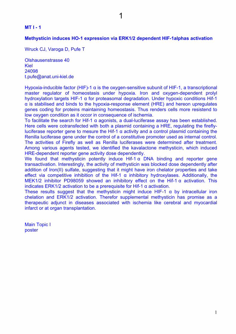
1MT I - 1 Methysticin induces HO-1 expression via ERK1/2 dependent HIF-1alphas activation Wruck CJ, Varoga D, Pufe T Olshausenstrasse 40 Kiel 24098 [email protected] Hypoxia-inducible factor (HIF)-1 α is the oxygen-sensitive subunit of HIF-1, a transcriptional master regulator of homeostasis under hypoxia. Iron and oxygen-dependent prolyl hydroxylation targets HIF-1 α for proteasomal degradation. Under hypoxic conditions Hif-1 α is stabilised and binds to the hypoxia-response element (HRE) and hereon upregulates genes coding for proteins maintaining homeostasis. Thus renders cells more resistend to low oxygen condition as it occor in consequence of ischemia. To facilitate the search for Hif-1 α agonists, a dual-luciferase assay has been established. Here cells were cotransfected with both a plasmid containing a HRE, regulating the firefly-luciferase reporter gene to mesure the Hif-1 α activity and a control plasmid containing the Renilla luciferase gene under the control of a constitutive promoter used as internal control. The activities of Firefly as well as Renilla luciferases were determined after treatment. Among various agents tested, we identified the kavalactone methysticin, which induced HRE-dependent reporter gene activity dose dependently. We found that methysticin potently induce Hif-1 α DNA binding and reporter gene transactivation. Interestingly, the activity of methysticin was blocked dose dependently after addition of Iron(II) sulfate, suggesting that it might have iron chelator properties and take effect via competitive inhibition of the Hif-1 α inhibitory hydroxylases. Additionally, the MEK1/2 inhibitor PD98059 showed an inhibitory effect on the Hif-1 α activation. This indicates ERK1/2 activation to be a prerequisite for Hif-1 α activation. These results suggest that the methysticin might induce HIF-1 α by intracellular iron chelation and ERK1/2 activation. Therefor supplemental methysticin has promise as a therapeutic adjunct in diseases associated with ischemia like cerebral and myocardial infarct or at organ transplantation. Main Topic I poster
1
2MT I - 2 Cellular defects in Ataxia-telangiectasia Dunner EM, Newman JV and Watters DJ. School of Biomolecular and Physical Sciences and The Eskitis Institute for Cell and Molecular Therapies, Griffith University, Nathan, Queensland, 4111, Australia. Ataxia-telangiectasia (A-T) is a progressive neurodegenerative disease characterised by oxidative stress and cancer predisposition. Extensive research has characterised the DNA damage response role for nuclear ATM (ataxia-telangiectasia-mutated), the protein defective in A-T, however very little is known about the role of cytoplasmic ATM. In this study, novel mechanisms contributing to disease progression in A-T were investigated. We examined the endosomal recycling of membrane receptors for epidermal growth factor (EGF) and transferrin in control and A-T fibroblasts, as well as the cellular distribution of cholesterol and lipid droplets in control and A-T fibroblasts, and in astrocytes from wild-type and A-T mice. Transferrin and EGF remained localised to the perinuclear region in A-T fibroblasts, indicating defective recycling in A-T. Immunofluorescence studies showed partial colocalisation of transferrin with recycling endosome markers. Other cellular anomalies observed included increased intracellular cholesterol. Using the unesterified cholesterol-binding fluorescence antibiotic, filipin, a bright punctate intracellular staining pattern in cytoplasmic vesicles was revealed in the perinuclear region of A-T fibroblasts while minimal internal staining was seen in controls. Nile red labelling for lipid droplets indicated increased lipid droplets in A-T fibroblasts compared to controls. This was also observed in A-T astrocytes relative to wild-type. Treatment of control fibroblasts with hydrogen peroxide led to punctate cytoplasmic filipin staining and increased lipid droplets in the treated fibroblasts similar to that seen in A-T fibroblasts, suggesting that the increased lipid droplets and abnormal cholesterol distribution may be due to oxidative stress. We have also observed defective sphingolipid trafficking and mitochondrial abnormalities in A-T cells. All of these features could contribute to neurodegeneration in A-T. Main Topic I poster
2
3MT I – 3 Einfluss von Vitamin C auf die UV-induzierte Bildung von NO in humanen Endothelzellen (HMEC-1) S.Rodemeister, D.Schilling, J.Wurster und D.Nohr Garbenstr. 30 Stuttgart 70593 [email protected] UV-Strahlung (v.a. UV-A) führt in diversen Zelltypen über die Aktivierung der Transkriptionsfaktoren AP-1 und NF-kB zur Synthese der induzierbaren NO-Synthase (iNOS), die in Anwesenheit von molekularem Sauerstoff und weiterer Cofaktoren die Aminosäure L-Arginin in L-Citrullin umwandelt. Hierbei entsteht das freie Radikal NO, welches unter physiologischen Bedingungen als Vasodilatator wirkt. In zu hohen Konzentrationen und vor allem bei gleichzeitiger Anwesenheit von reaktiven Sauerstoffspezies (ROS) wirkt NO schädlich. Es reagiert mit den ROS zu sogenannten reaktiven Stickstoffverbindungen (RNOS), die schließlich zur Schädigung von Proteinen, Membranlipiden sowie von DNA führen und somit die zellulären Funktionen auf mehreren Ebenen beeinträchtigen. Vitamin C kann aufgrund seiner antioxidativen Wirkung die Bildung von ROS konzentrationsabhängig einschränken. Dies führt sowohl zur geringeren Bildung von RNOS als auch zu einer Verringerung der NO-Produktion, welche durch ROS über die Induktion der iNOS-Synthese erhöht wird. Eine weitere Reduzierung der NO-Konzentration wird über eine direkte Interaktion zwischen Vitamin C und NO erreicht. Am Modell humaner Endothelzellen (HMEC-1) haben wir untersucht, ob die präventive Gabe von Vitamin C zu einer Verringerung der NO-Bildung nach durch UV-Bestrahlung induziertem oxidativem Stress führt. Hierzu wurden die Zellen mit Vitamin C in zwei verschiedenen Konzentrationen supplementiert, bevor sie mit einer Intensität von 25 J/cm2 UV-A-Strahlung stimuliert wurden. Anschließend wurde der NO-Gehalt in den Zellen mittels DAF-Fluoreszenz-imaging gemessen. Dabei konnten wir zeigen, dass der NO-Gehalt nach UV-Bestrahlung erwartungsgemäß ansteigt, dieser Anstieg jedoch nach präventiver Gabe von Vitamin C wesentlich niedriger ist als in vor der Bestrahlung nicht supplementierten Zellen. Ebenfalls führt die alleinige Gabe von Vitamin C ohne anschließende Bestrahlung zu einer Absenkung des NO-Niveaus in der Zelle. Eine ausreichende Versorgung mit Vitamin C kann somit Endothelzellen vor oxidativem und nitrosativem Stress und seinen Folgeerscheinungen schützen. Main Topic I poster
3
4Poster 4 Naphthalene-induced Clara cell injury is greatly reduced in murine lungs pretreated with Keratinocyte Growth Factor (KGF) Yildirim AÖ#, Veith M#, Van Winkle LS*, Müller B#, Plopper CG*, Fehrenbach H# #Clinical Research Group Chronic Airway Diseases, Phillips-Univ. Marburg, Germany *Veterinary Medicine, APC, UC Davis, CA, United States Naphthalene (NA) is widely used as a feedstock in chemical industry and is a component of cigarette smoke. NA causes Clara cell damage due to conversion into 1R,2S-oxide catalyzed by cytochrome P450 monooxygenases (CYP), particularly isoform 2F2. Because KGF was shown to protect lung epithelial cells against various types of injury, we investigated whether pretreatment of lungs with human recombinant (rHu) KGF protects against NA-induced Clara cell damage in vivo, and sought to identify the underlying molecular mechanisms. Male C57BL/6 mice were i.p. injected with NA (100, 200, or 300 mg/kg b.w.) or corn oil (250 microL) 24 hrs after instillation of rHuKGF (10 mg/kg b.w.) or PBS (80 microL). Distal airways were isolated by microdissection 12 hrs later. Microdissected airways, embedded into paraffin, were stained for Clara cell specific protein CC10 to quantify Clara cell numbers using a physical disector approach. Frozen airways were used for mRNA isolation and real time RT-PCR. Additionally, mRNA expression was analysed in isolated airway epithelial cells enriched in Clara cells (approx. 80% purity), which were incubated with or without rHuKGF. Injection of NA resulted in dose-dependent Clara cell injury. Distal airways of mice pretreated with rHuKGF prior to NA injection (200 mg/kg) exhibited significantly higher Clara cell number (1.9fold) and volume (4.3fold) per basal membrane area compared with PBS pretreated mice. RT-PCR showed increased mRNA expression of NRF2 and PCNA in rHuKGF pretreated mice. Normalized to CC10 expression, CYP2F2 mRNA was reduced by about 50% compared to control mice. Airway epithelial cells enriched in Clara cells incubated with rHuKGF exhibited a similar reduction in CYP2F2 mRNA (relative to GAPDH). Our results demonstrate that pr-treatment with rHuKGF protects the airway epithelium against NA-induced injury. We suggest that rHuKGF exerts its beneficial effect through an increase in NRF2 and a decrease in CYP2F2 expression. Experimental morphology [email protected]
4
5MT I – 5 Ascorbic acid blocks the UV-induced upregulation of iNOS in human endothelial cells (HMEC-1) Dirk Schilling, Julia Wurster, Sandra Rodemeister, Mareike Schultze and Donatus Nohr Garbenstr. 30 Stuttgart 70599 [email protected] Introduction: Reactive oxygen species (ROS) play an important role in UVA-induced cell damage and the activation of inducible nitric oxide synthase (iNOS) in several cell types including endothelial cells. Both activation factors NFkB and AP-1 are also involved in this iNOS-activating pathway. Antioxidants like ascorbic acid (AA) are most important components to support the intracellular balance between oxidants and antioxidants thus avoiding oxidative stress and its damaging consequences. Aim: The aim of our study was to investigate the influence of UV-induced oxidative stress and of antioxidative ascorbic acid on the expression of iNOS mRNA and protein in human endothelial cells (HMEC-1). Materials and Methods: HMEC-1 were incubated with AA in concentrations of 50 and 100 µM for 24 hours to determine intracellular uptake. At the optimal AA concentration after 12 h cells were UV-irradiated with 25 J/cm². Cells were harvested and iNOS mRNA were determined by real time PCR. For immunocytochemistry cells were grown on coverslips. Results and Discussion: After UV irradiation we found an increase of iNOS mRNA by 50 % in comparison to non-irradiated cells. The preventive supplementation with AA resulted not only in a prevention of the increase but also to levels (67%) belone that of the UV-irradiated cells. These results were “confirmed” by equal effects on the protein level, i.e. an increase of iNOS after UV-irradiation which was decreased by AA. Also the final production of NO showed comparable results (shown elsewhere). Our results indicate a protective potential of AA on the induction and modulation of iNOS in irradiated cells not only by the known direct interaction with NO but maybe also at other levels of the iNOS-NO signalling pathway. Main Topic I poster
5
6MT I – 6 Neuronal death in cerebellar cultures from PEX13-knockout mice is mediated by oxidative stress Barbara Ahlemeyer1, Tam Nguyen2, Catharina Müller2, Denis Crane2, Eveline Baumgart-Vogt1 1Aulweg 123, 2Forest Court N75 1Gießen, 2Brisbane 1 35385, 2 QLD 4111 [email protected] Patients with defects of the peroxin protein Pex13p, a peroxisomal membrane protein involved in the import of matrix proteins, suffer from Zellweger syndrome (ZS), the most severe form of a peroxisomal biogenesis disorder. Increased neuronal death and defects in neuronal migration as well as delay in neuronal differentiation has been described in brain samples of these patients. Recently, several animal knockout mouse models with deletions of distinct peroxin genes, exhibiting the same phenotype as the corresponding patients, have been generated from our groups to study the molecular pathogenesis of brain defects. In the present study, we used newborn PEX13Dexon2 knockout mice for the preparation of primary neuronal cultures from the cerebellum. Six days after preparation, cultures constituted about 90% cerebellar granule neurons and less than 10% astrocytes. As expected for peroxisomal biogenesis disorders, catalase was mislocalized to the cytoplasm in cultured neurons and astrocytes from homozygous PEX13(-/-) animals. In contrast, catalase was present exclusively in peroxisomes of the cells from heterozygous and wild-type animals. Determination of cell death revealed 75% apoptotic neurons in cultures from PEX13(-/-) animals (= 3.5-fold increase in comparison to controls). Interestingly, we also found 50% apoptotic neurons in cultures from PEX13(+/-) animals. Next, we measured the intracellular level of reactive oxygen species (ROS) using the oxidant-sensitive dye dihydroethidine to evaluate whether ROS contribute to the observed neuronal death. The mean basal cellular ROS levels in cultures from PEX13(+/-) and PEX13(-/-) mice were increased 1.5 and 2-fold compared to controls. Our results suggest that oxidative stress plays an important role in the development of neuronal death in peroxisomal biogenesis disorders. Main Topic I poster
6
7MT I – 7 Imbalance of antioxidative enzymes in PEX11ß deficient mouse lungs. Srikanth Karnati and Eveline Baumgart-Vogt Aulweg 123 Giessen 35385 [email protected] The lung is an organ which is exposed to high levels of atmospheric oxygen. Owing to its large surface area and extensive blood supply, this organ is susceptible to oxidative injury by reactive oxygen species (ROS) and free radicals. Alterations in lung antioxidant balance can lead to a variety of airway diseases such as asthma, chronic obstructive pulmonary dysplasia (COPD) and idiopathic pulmonary fibrosis (IPF). Peroxisomes are organelles with extensive metabolism of reactive oxygen species. However, to date, nothing is known about the functions of peroxisomes and the role of PEX11ß in mouse lungs, a peroxin involved in peroxisome proliferation. Even though peroxisomes are present in PEX11ß-deficient mice, these animals display several pathologic features shared by knockout mouse models of Zellweger syndrome in which peroxisomes are absent. In this study, we characterised the regulation of antioxidant enzymes in PEX11ß-deficient mouse lungs. Our results indicate that, changes of antioxidant enzyme expressions occur in the lungs of these knockout animals. A marked increase of mitochondrial manganese-superoxide dismutase (SOD2) and significant down regulations of extracellular superoxide dismutase (EC-SOD), glutathione S- transferase 1 (GST-1), glutathione peroxidase (GPx), peroxiredoxin V (Prx V) and hemoxygenase 1 (hmox1) was noted in PEX11ß-deficient mouse lungs, suggesting an accumulation of reactive oxygen species in these knockout animals. Due to the strong alterations in ROS metabolizing enzymes, we conclude that peroxisomes play an important role in the control and regulation of ROS metabolism in the lung. Further studies have to prove which relevance peroxisomal ROS metabolism has in the protection against lung injury and chronic-inflammatory lung diseases. Main Topic I poster
7
8MT I – 8 Exercise-induced alterations in skeletal muscle of heterozygous MnSOD-deficient mice Lena Willkomm, Klara Brixius, Nadine Lange, Lisa Richters, Norbert Treiber, Angelika Kümin, Sabine Werner, Karin Scharffetter-Kochanek, Wilhelm Bloch Carl-Diem-Weg 6 Köln 50933 [email protected] Physical exercise is going along with an increased generation of oxygen-derived free radicals. The present study investigates the influence of endurance-exercise on skeletal muscle in male transgenic mice with a heterozygous deficiency of the manganese-dependent superoxide dismutase (MnSOD) in comparison to wildtype animals (WT). WT- and MnSOD-mice performed an exercise training of 60 min/d at 5 d/week for a duration of 8 weeks at a velocity of 15 m/min and an inclination of 5°. In addition, analysis for skeletal muscle alterations were performed in age matched sedentary WT and MnSOD. The exercise-performance did not alter the diameter of the Vastus lateralis muscle in WT, but muscle cell diameter was significantly increased in MnSOD after the training. In WT but not in MnSOD a shift in the staining intensity of the succinate dehydrogenase was observed towards a higher succinate dehydrogenase activity. After running exercise, 8-isoprostan-staining, a marker for the generation of reactive oxygen species, was significantly increased in WT but not in MnSOD. No alterations were observed for nitrotyrosin, a marker for reactive nitrogen species. Conclusions: A chronical decrease in MnSOD-activity seems to be compensated by other antioxidative defense mechanisms. This compensation results in a loss of the exercise-induced ROS generation and alterations in the skeletal adaptation. Main Topic I poster
8
9MT I – 9 BENEFICIAL EFFECT OF LIPOIC ACID ON THE SUPPRESSED WOUND HEALING INDUCED BY N-3 DIETARY FATTY ACIDS IN RATS Andres Arend and Marina Aunapuu University of Tartu, Department of Anatomy, Ravila 19 Biomedicum Tartu, [email protected] The present study was performed to test the influence of alpha-lipoic acid (LA), a natural anti-oxidant, on the inhibition of connective tissue proliferation and on the increased level of lipid peroxidation in the rat liver wound induced by dietary n–3 polyunsaturated fatty acids (n–3 PUFAs). Rats were fed with a commercial pellet diet (control group) or with diets enriched with 10% of sunflower oil (n–6 PUFAs group) or 10% of fish oil (n–3 PUFAs group) for 8 weeks followed by addition of LA to the same diets for 10 days. Then a liver thermic wound was induced and the administration of LA was continued for 6 days. The proliferation of the connective tissue and the content of prostaglandins (PGs) E2 and F2alpha·were measured in the liver wounds. The level of lipid peroxidation in the liver wound was assessed via the level of thiobarbituric-acid-reactive substances (TBARS) and lipid peroxidizability via the level of iron-induced TBARS. LA prevented the suppression of connective tissue proliferation in the healing wound induced by n–3 PUFAs, avoided the increase in peroxidation of lipids, reduced peroxidizability of lipids and modulated the decrease in PGE2 and PGF2alpha content. The protective effect of LA against the adverse effects of an n–3 PUFA diet may be mediated by different mechanisms including the ability to scavenge different reactive oxygen species and increase de novo synthesis of cellular anti-oxidant glutathione. It seems that the elevation of the level of reduced glutathione (GSH) in liver might have an impact on the protective effect of LA. It can be concluded that the protective effect of LA on the suppression of wound healing induced by n–3 PUFAs includes the prevention of increased lipid peroxidation and the maintenance of a suitable spectrum and/or level of 2-series PGs. Main Topic I poster
9
10N1 The neuroanatomists\' dirty little secret Krueger-M, Mahlo-J, Rappert-A, Bechmann-I Theodor-Stern-Kai 7 Frankfurt/Main 60590 [email protected] In 1900, summarizing his experiments with toxins and Ehrlich\'s earlier observations with intravital dyes, the Berlin physician Lewandowski concluded that \"brain capillaries must hold back certain molecules\". The term \"Bluthirnschranke\" (blood-brain barrier, BBB) describes this phenomenon with persuasive beauty, but its extension of meaning into the context of leukocyte recruitment is misleading. Endothelial expression of BBB-typical \"belts \" of tight junctions requires their direct interaction with astrocytes provided in the capillary segment of the vascular tree, but not in postcapillary venules, where infiltration takes place (Bechmann et al., Trends in Immunology 2007). We injected classical markers of BBB integrity and found that the brain parenchyma indeed was not labeled. However, dyes accumulated in the vascular wall and perivascular macrophages of pre- and postcapillary vessels and the choroid plexus. Thus, Ehrlich\'s observation that the brain remains \"white as snow\" after tracer injected refers to the neuropil proper, while the site of entry of leukocytes in neuroinflammation is permissive for BBB-markers under normal conditions. Neuroimmunology poster
10
11N2 Influence of Interferon-gamma receptor knockout on microglial and astroglial reactions Michael Bette*, Tilman Krieger*, Bernhard Dietzschold# and Eberhard Weihe* * Institute of Anatomy and Cell Biology, Department of Molecular Neuroscience, Philipps-University Marburg, Germany # Center of Neurovirology, Deptartmen of Microbiology and Immunology, Thomas Jefferson University, Philadelphia, PA USA [email protected] Intranasal infection of normal adult 129/SvEv mice with the attenuated rabies virus (RV) CVS-F3 resulted in the development of a transient RV disease characterized by loss of body weight and appetite depression, which peaked at 13 days post infection (p.i.). To investigate the involvement of the interferon-gamma receptor (IFNgR) in the appearance on astroglial and microglial reactions in the RV-infected brain and its influence on the elimination of RV from the CNS, we studied the course of glial activation and virus clearance in knockout (k.o.) mice lacking the IFNgR. Immunhistochemical analysis of C1q as marker for microglia activation, and GFAP, as marker for astrocyte reactions revealed a significant delay of both glial responses in the RV-infected IFNgR k.o. mouse as compared to wild type mice. Additionally, there was not RV clearance in the IFNgR k.o. mouse until day 21 p.i. which represents a date at which wild type mice had completely cleared RV from the brain. These observations support the hypothesis, that one of the anti-viral effector mechanism of IFNg; mediated through the IFNgR, is be due to the activation of microglial and astroglial cells. Furthermore, local glial responses seem to be essential for rapid clearance of RV antigen from neurons. Neuroimmunology poster
11
12N3 Induction of antimicrobial peptide rCRAMP by bacterial components in glial cells Lars-Ove Brandenburg1, Nikoleta Nikolaeva1, Deike Varoga1, Stephen Leib2, Henrik Wilms3, Rainer Podschun4, Thomas Pufe1, Jobst Sievers1 and Ralph Lucius1
1 Department of Anatomy, University of Kiel, Germany 2 Institute for Infectious Diseases, University of Bern, Schwitzerland 3 Clinic of Neurology, University of Kiel, Germany 4 Institute of Infection Medicine, University of Kiel, Germany [email protected] Antimicrobial peptides are a part of the innate immune system at epithelial surface, and may also have important functions in the brain. However, little is known about the expression of antimicrobial peptides in the CNS and whether neural cells can secrete these peptides. We have used cell cultures, real-time PCR, immunohistochemistry, ELISA and an animal model to get more information about the role of antimicrobial peptides in the CNS. In detail, we have investigated the expression of the antimicrobial peptide rCRAMP (homologe of the human LL-37) in rat glial cells (astrocytes and microglia) after incubation with bacterial components. Furthermore, we used cerebrospinal fluid (CSF) and serum from patients with bacterial meningitis to detect LL-37 and other antimicrobial peptides. Finally, we investigated the occurrence of rCRAMP in an animal model of bacterial meningitis. We here demonstrate (i) not only the expression but also secretion of biological active rCRAMP in glial cells, and (ii) the occurrence of antimicrobial peptides in the cerebrospinal fluid of meningitis patients. Moreover, we could show an involvement of rCRAMP in the rat meningitis model pointing to a role of rCRAMP in the pathogenesis of this disease. Our results suggest that rCRAMP respectively LL-37 is an important part of the innate immunity in the brain against bacterial CNS pathogens. Neuroanatomy/Neurobiology poster
12

13N4 In vivo cell tracking via NMR spectroscopy allows online observation of therapeutic approaches in a rat glioma model S. Arnhold1, U. Himmelreich2, U. Hoehn2, T. Berhorn1, K. Addicks1, K. Nohroudi1
1 Department of Anatomy I, University of Cologne, Germany, 2 Max-Planck-Institute of Neurological Research, Cologne, Germany, Josef-Stelzmannstr. 9, 50931 Köln [email protected] Glioblastoma multiforme are highly malignant brain tumors, which are resistant to surgery, chemotherapy and radiation. Due to the insufficiency of conventional therapies, new therapeutic strategies need to be established. In recent years several attempts using viral vectors and/or stem cells have been made to develop more effective therapies. One major problem of all these approaches was the uncontrolled distribution of the injected particles and disappearance of cells respectively. In order to avoid the necessity of large numbers of animals NMR spectroscopy is a promising, non-invasive technique that allows the detection and observation of tumor masses in vivo. However, so far the present labelling methods did not allow the follow up of injected cells due to degradation and low contrast, requiring more than 1000 labelled cells on a spot to detect them. Using highly stable iron beads, we developed a method to label mesenchymal stem cells (MSC), making them detectable by NMR imaging. With the effective concentration of beads as few as 10 cells on a spot can be detected and using this concentration in vitro neither an inhibition of proliferation nor of the migratory potential is observed. Furthermore, MSCs labelled with iron beads and a fluorescent marker can be detected as migrating cells in rat C6 glioma model. In conclusion, this new method is a promising tool for researchers developing cell based therapies by providing the possibility to monitor the distribution of cells in individual animals. Neuroanatomy/Neurobiology poster
13
14N5 Suppression of C6 glioma cell proliferation by bone marrow derived stromal cells (BMMCs) K. Nohroudi, E. Schnell, K. Addicks, S. Arnhold Department of Anatomy I, University of Cologne, Germany [email protected] Conventional treatment of malignant brain tumors like glioblastoma multiforme is insufficient to eradicate the tumor cells and leaves the patients with a poor prognosis. Therefore it is necessary to develop new therapeutic strategies, e.g. viral or cell based therapies. In recent years several more or less effective approaches in this direction have been made, using viral vectors and neural or embryonic stem cells or the combination of both. However, the major problem of these experiments, to overcome the elimination of transplanted viruses or cells by the hosts immune system is still not solved and furthermore, the usage of neuronal or embryonic stem cells is tainted with ethical concern. In contrast, bone marrow stromal cells (BMMCs) can be obtained as autologous cells from the patient itself, overcoming both pitfalls. We isolated and cultured rat BMMCs in vitro over more than 20 passages, showing that the cells can be expanded to large numbers in short time. Co-culturing of C6 glioma cells with BMMCs lead to a striking inhibition of proliferation of the glioma cells and after stereotactic injection BMMCs showed a directed migration to and infiltration of tumor masses in vivo. Together with our previous findings that BMMCs own the capacity to differentiate into neural phenotypes, BMMCs seem to be a promising approach in developing a cell based therapy for glioblastoma. Neuroanatomy/Neurobiology poster
14
15N6 Dexamethasone reduces gap junctional intercellular communication in F98 and U87 glioma cell lines Daniel Hinkerohe*, Dörte Wolfkühler, Aiden Haghikia, Uwe Schlegel*, Rolf Dermietzel, Carola Meier, Pedro M. Faustmann *Department of Neurology, Knappschafts-Hospital, Ruhr-University Bochum, Germany Department of Neuroanatomy and Molecular Brain Research, Ruhr- University Bochum Background: Glucocorticoids are the most efficient therapeutics in cerebal edema associated with brain tumors but can also reduce cytotoxicity and growth inhibition induced by chemotherapeutics. Like astrocytes in the CNS, different astroglioma cells form a well coupled syncytium via gap junctional communication (GJC). Astroglial gap junctional communication serves for a number of physiological properties related to CNS homeostasis. The major gap junction protein in astrocytic and astroglioma cells is connexin43 (Cx43). Purpose: To test the effects of dexamethasone on astroglial intercellular coupling and astroglial membrane resting potential (MRP) in a F98 rat glial cell line and a human U87 glial cell line and to prevent in a second experiment the effects of dexamethasone by addition of mifepristone, a glucocorticoid receptor-specific antagonist. Material and Methods: We evaluated the influence of dexamethasone (0.1, 1, 10µmol) and mifepristone (1, 10 µmol) on GJC and MRP in U87 and F98 glial cell lines. MRP was measured by patch clamp technique (whole cell), GJC was tested by Lucifer Yellow dye-application and presence of Cx43 was analysed by Western blotting and immuno-cytochemistry. Results: Dexamethasone causes a dose dependent significant reduction of functional coupling and Cx43 expression in both glial cell lines accompanied by an astroglial depolarisation. Furthermore, preincubation with mifepristone could prevent dexamethasone induced astroglial uncoupling in a dose dependent manner. Discussion: Our results indicate that dexamethasone reduces astroglial functional coupling and astroglial Cx43 expression through activation of glucocorticoid receptors. This reduced GJC could be one possible mechanism by which glucocorticoids attenuate the efficacy of several chemotherapeutic drugs used in the therapy of human malignant gliomas. Neuroanatomy/Neurobiology poster
15
16N7 The retinoblastoma – aspects of anatomy and pathology Anca Indrei, Gabriela Dumitrescu, Danisia Haba, Carmen Lăcrămioara Zamfir, D. Costin Universitatii Str, 16 Iasi ROMANIA 700115 [email protected] Retinoblastoma is the most common primary intraocular malignancy of children. It is now clear that the cell of origin of retinoblastoma is neuronal. In approximately 40% of cases, retinoblastoma occurs in individuals who inherit a germ line mutation of one RB allele. The pathology of retinoblastoma of both ereditary and sporadic types is identical. Tumors may contain both undifferentiated and differentiated elements. The former appear as collections of small, round cells with hyperchromatic nuclei. In well-differentiated tumors there are Flexner-Wintersteiner rosettes and fleurettes reflecting photoreceptor differentiation. Focal zones of dystrophic calcification are characteristic of retinoblastoma. Our study was realised on 30 eyes from children aged between 6 month and 3 years, with diagnosis of retinoblastoma, sended to the Laboratory of Pathology of the Iaşi „Sfanta Treime” Hospital, for the pathological examination. Because the prognosis is adversely affected by extraocular extension and invasion along the optic nerve and by choroidal invasion, the purpose of our work is to study the kind of growth of this tumor and the intra and extraocular invasion. Key words: retinoblastoma, Flexner-Wintersteiner rosettes, optic and choroidal invasion. Neuroanatomy/Neurobiology poster
16
17N8 Amniotic fluid derived cells express stem cell markers and show neuronal differentiation characteristics C. Post
1, K. Nohroudi
1, F.-J. Klinz
1, M. Hoopmann
2, K. Addicks
1, S. Arnhold
1
Department of Anatomy I1, Clinic for Obstetrics and Gynaecology
2, University of Cologne
[email protected] In the search for cell populations suitable for cell replacement strategies, a variety of stem cell populations are being discussed. Recently, stem cells from the amnion or the amniotic fluid have shifted into the focus of interest, as it has been shown, that because of their plasticity, these cells have the potency to differentiate into a variety of cell types. However, at least three different cell types have been described to occur in the amniotic fluid. In order to obtain a homogenous cell population from the amniotic fluid and to keep these cells in culture for some time, we have carried out a selction step by means of the magnetic associated cell sorting (MACS) using an antibody against the surface epitope CD90. With this procedure a homogenous cell population of epithelial-like cells with typical characteristics for epithelial cells could be obtained. Analysing stem cell characteristics, with RT-PCR transcripts for Oct-4, Nanog and Sox-2 could be detected. Furthermore the majority of cells is immunopositive for the stem cell marker SSEA-1 (CD15), while in primary cells only a small subpopulation of cells express this marker. In regard of a therapeutical application of these cells in diseases of the central nervous system, the neural differentiation capacity of amnion fluid derived cells was stimulated by cultivating cells in the presence astrocyte conditioned media and analysed using RT-PCR, ELISA as well as immunocytochemistry. After stereotactic transplantation of vector transduced cells from the amniotic fluid into the striatum of adult rats, neuronal and glial morphologies could be observed. Neuroanatomy/Neurobiology poster
17
18N9 Targeting of defined neuronal populations by retrograde recombinase transport Kathrin Dethleffsen, Michael Meyer Pettenkoferstr. 12 Munich 80336 [email protected] Site-specific recombinases (SSR) are powerful tools for conditional mutagenesis in the mouse. Spatial and temporal specificity of these mutations reflects temporal and spatial availability of the active SSR. A major limitation for their use is the lack of gene regulatory elements that could control their availability in defined cell populations of complex tissues. Such elements may either be difficult to identify or they may not exist at all. In this project we attempt to alleviate this situation by exploring an alternative method applicable in the nervous system, one of the most complex tissues in the body. Many subpopulations of neurons have characteristic innervation targets, i.e. they are distinguished from neighbouring neurons by their projections. We suggest to deliver Cre recombinase to specific neuronal subpopulations by retrograde transport from these targets. This approach would be particularly useful for analysis of the actions of neurotrophins and there receptors on corticospinal neurons (Giehl et al., J. Neuroscience 21, 3492; Harrington et al., Proc. Natl. Acad. Sci. USA 101, 6226). We are currently investigating in vitro and in vivo properties of appropriately engineered variants of Cre-recombinase. Neuroanatomy/Neurobiology poster
18
19N10 Distribution of the Golgi-Associated Protein PIST in Rat Brain and Co-Localization with Somatostatin Receptor Subtype 5 Annie Chen1, Hans-Jürgen Kreienkamp2, and Thomas Stroh1 1Montreal Neurological Institute, 3801 University St., Montreal, Quebec, H3A 2B4, Canada;
2Institut für Humangenetik, Universitätsklinikum Eppendorf, Martinistraße 52, 20246
Hamburg, Germany. [email protected] The protein interacting specifically with Tc10 (PIST) is a Golgi-associated protein involved in trafficking and targeting of membrane proteins. Recently, it was shown to play a key role in regulating the targeting of AMPA-receptor subunits to synapses. Moreover, we demonstrated that PIST interacts via its PDZ domain with somatostatin receptor subtype 5 (sst5) and retains the bulk of sst5 in the Golgi apparatus. To date, little is known of its regional and cellular distribution in the brain. In the present study, we used a specific antibody to investigate the distribution of PIST in rat brain by immunohistochemistry. PIST was detected in neurons but not glial cells. In the telencephalon, PIST-like immunoreactivity (PIST-l IR) was abundant in olfactory areas, the basal forebrain, and the neocortex. In the latter, layer V pyramidal cells were intensely stained. The CA2 field of the hippocampus contained densely packed PIST-positive neurons, whereas labelled cells were sparse in CA1, CA3, and the dentate gyrus. Anterodorsal, paraventricular, ventrolateral and laterodorsal thalamic nuclei contained PIST-positive neuronal cell bodies, whereas intralaminar nuclei were almost unlabelled. In the hypothalamus, neurons of the supraoptic nucleus were intensely immunostained while the lateral hypothalamus, tuber cinereum, para- and periventricular nuclei were moderately labelled. PIST-l IR was widespread in the brainstem. Substantia nigra pars compacta, central gray, interpeduncular and pontine nuclei stood out by high numbers of intensely immunolabelled cells. In the cerebellar cortex, PIST-l IR was restricted to Purkinje cells and interneurons. Using an sst5-specific antibody we were able to show that PIST co-localizes extensively with this somatostatin receptor subtype in basal forebrain nuclei involved in the regulation of cortical activation. These findings suggest that its interaction with sst5 may play a role in vivo in the mediation of the effects of somatostatin on cortical activation and sleep. Neuroanatomy/Neurobiology poster
19
20N11 A graphical data base of neuropeptide systems in non-mammalian brains I.Neubert, M. Kolsch, and S. Blaehser Aulweg 123 Giessen 35385 [email protected]. uni-giessen.de A graphical data base of neuropeptide systems in non-mammalian brains I. Neubert, M. Kolsch and S. Blaehser Institut für Anatomie und Zellbiologie, Justus-Liebig Universität, Aulweg 123, D-35385 Gießen Our data base, founded on immunoreactive systems (i. e., perikaryal clusters and their projection areas), enables a phylogenetically oriented analysis of neuropeptide systems such as arginine-vasotocin, mesotocin, corticoliberin (CRF), vasointestinal polypeptide, gonadoliberin (GnRH), somatostatin, met-enkephaline, neuropeptide Y, neurotensine, and substance P, all incorporated into graphics of serially cut brain sections of Myxine glutinosa, Lampetra planeri, Clarias batrachus, Rana temporaria, Calotes versicolor, and Gallus gallus domesticus. The possibility to project several (or all) immunoreactive systems simultaniously onto one sectional plane enables to recognize, e. g., the spatial characteristic of each system, overlapping areas, or functionally indicative compartimentation of projection areas. Comparison between neuropeptide systems in these species demonstrates the evolution of the systems in relation to brain development, their morphological constancy as well as their spatial relationship to functional circuitries. The data base will be accompanied by text, tables, and references in English, German, and French. Neuroanatomy/Neurobiology poster
20
21N12 Calcitonin gene-related peptide is a marker for early pre-symptomatic motoneuron pathology in a mouse model of amyotrophic lateral sclerosis Cornelia Ringer, Eberhard Weihe, Burkhard Schütz Robert-Koch-Strasse 8 Marburg 35037 [email protected] Introduction: Superoxide dismutase-1 (SOD1) transgenic mice are a model to study motoneuron degeneration and pathology of the human disease amyotrophic lateral sclerosis (ALS). While loss of spinal motoneurons and deficits in motor tasks appear at around postnatal day (P) 90 of life in these mice, structural changes at neuromuscular junctions have already been detected at P50. No signs of early pre-symptomatic pathology at the site of motoneuron cell bodies, however, can bee seen. Recently, we showed that calcitonin gene-related peptide (CGRP) immunoreactivity (IR) not only served as a marker for spinal motoneurons, but also labelled aberrant-shaped dendrites and axons (neurites) in the spinal cord ventral horn in P90 SOD1 mice (Schütz et al., J. Neurosci., 2005). In the present report we investigated the onset and progression of CGRP-related neuropathology in this mouse model of ALS to test whether CGRP could serve an early marker of spinal motoneuron pathology. Methods: Spinal cord tissue from SOD1 and control mice was analysed from P20 to P130 in 10 day intervals by immunohistochemistry for CGRP, choline acetyltransferase (ChAT), the vesicular acetylcholine transporter (VAChT), and other neuronal markers (MAP2, NF200). Results: In control mice of all ages investigated, CGRP-IR labelled the majority of lumbar motoneuron cell bodies and few thin neurites. Starting at P40, SOD1 mice showed additional CGRP-IR in ring-shaped structures, approx. 2µm in diameter. These structures most likely represented dendrites of motoneurons, because they were MAP2-positive, but lacked NF200 and VAChT. From P50 on these structures were also ChAT-positive. The numbers and diameters of ring-shaped CGRP-IR neurites increased with age, with a peak between P90 and P110, and then disappeared almost completely until P130. Conclusions: In the SOD1 transgenic ALS mouse model, CGRP-IR is a novel marker for early motoneuron pathology at the spinal level which uncovers motoneuron degeneration around P40. Neuroanatomy/Neurobiology poster
21
22N13 The neuropeptides PACAP and NPY mediate antagonistic lipolytic effects in a time-dependent manner Martin Gericke, Marcin Nowicki, Joanna Kosacka, Katharina Spanel-Borowski Institute of Anatomy, University of Leipzig [email protected] Neuropeptides play an important role in energy homeostasis of the body. In the central nervous system, they are involved in the regulation of food-intake. They may also directly act on fat tissue via neuropeptid-receptors, because pituitary adenylate cyclase-activating polypeptide (PACAP) and neuropeptide Y (NPY) cause lipolysis and lipogenesis, respectively. In this study, 3T3-L1 adipocyte cultures were stimulated with PACAP and NPY (10-6 M each) at different time points, and lipolysis determined by measurement of glycerol concentration in the supernatant. The 4-h-incubation of 3T3-L1 cells with PACAP and NPY resulted in lipolysis increase by 40% and 35%, respectively. After 24 h both neuropeptides caused a lipolysis reduction for about 30% compared to untreated controls. Stimulatory and inhibitory effects were statistically significant. Hence, PACAP and NPY display time-dependent-antagonistic lipolytic effects in 3T3-L1 adipocytes. We then validated the lipogenetic effect in a long-term co-culture model of 3T3-L1 adipocytes with sensory dorsal root ganglia neurons, which likely produce PACAP and NPY. According to oil red staining the co-cultured 3T3-L1 cells contained significantly more lipid droplets than the 3T3-L1 adipocytes alone. This finding indicates that under transmitter release lipogenesis occurs in 3T3-L1 adipocytes. Future blocking experiments will define PACAP and NPY to be the responsible transmitter. We suppose that an initial cAMP increase by PACAP and NPY causes a short-term lipolytic effect. Intracellular cascades of long-term regulation are initiated in parallel that finally mediate enhanced lipogenesis. Neuroanatomy/Neurobiology poster
22
23N14 Receptors for substance P and C GRP are present in human liver: possible involvement in neuro-immunomodulation Dominik Abt*, Gisa Tiegs*, Thomas Papadopoulos**, Winfried Neuhuber, Institut für Anatomie, *Institut für Exp. und Klin. Pharmakologie und Toxikologie, **Institut für Pathologie, Universität Erlangen-Nürnberg, Krankenhausstraße 9, Fahrstraße 17 bzw. Krankenhausstraße 8-10, resp., 91054 Erlangen, Germany Krankenhausstraße 9 Erlangen D-91054 [email protected] Recent experiments in mice demonstrated a significant pro-inflammatory role of peptidergic primary afferent neurons in hepatitis (Tiegs et al., 1999; Bang et al., 2003, 2004). The models used included T cell- (ConA, GalN/SEB), macrophage- (GalN/LPS), cytokine- (GalN/TNFa) and Fas ligand- (CD95) mediated hepatocyte injury. Involvement of both, substance P (SP) and calcitonin gene-related peptide (CGRP), released from primary afferents was revealed by neonatal capsaicin treatment and neurokinin-1 receptor (NK-1R) antagonists which significantly prevented liver damage (Bang et al., 2003, 2004). The aim of the present study was to identify key components of the proposed primary afferent immunomodulatory mechanism in human liver. Immunohistochemistry for SP, CGRP, NK-1R, CRLR and markers for various immune cells as well as RT-PCR for NK-1R, CRLR, RAMP-1 and RCP were used on tumor-free liver tissue obtained at surgery for metastatic cancer. Nerve fibers immunoreactive for SP and CGRP were found both in periportal fields and lobules. Immunostaining for both NK-1R and CRLR was detected in hepatocytes, either singly or clustered in groups of about 20 cells. mRNA for NK-1R and all three components of the CGRP receptor (CRLR, RAMP-1 and RCP) could be detected by RT-PCR. Co-staining of NK-1R and CRLR, respectively, with specific markers for immunocytes revealed presence of these receptors on macrophages (CD68), granulocytes (CD31), Kupffer cells (CD14), dendritic cells (CD83) and Ito cells (GFAP, a-sma) but not on lymphocytes (CD20, CD4, CD8). These data suggest that immunomodulation by SP and CGRP released from primary afferents may work also in human liver. Thus, application of the respective receptor antagonists could represent a novel therapeutic option in immune-mediated hepatitis. (Supported by DFG NE 534/1-2/2-2) Neuroimmunology poster
23
24N15 Probabilistic cytoarchitectonic mapping of the human frontal operculum Martina Haeck
1; Katrin Amunts
2,3,4; Simon B. Eickhoff
1,2,3; Lars Hoemke
2; Axel
Schleicher 1; Karl Zilles
1,2,3;
1 C. and O. Vogt Institute for Brain Research, Heinrich Heine University Duesseldorf, Germany 2 Institute of Medicine, Research Center Juelich, Germany 3 Brain Imaging Center West (BICW) Juelich, Germany 4 Department of Psychiatry und Psychotherapy, RWTH Aachen University, Germany [email protected] Previous studies of our own group revealed four new areas (Op1-Op4) in the parietal operculum1. In continuation of this study, we here mapped the frontal operculum in histological sections of ten post-mortem brains, and generated three-dimensional probability maps of these areas. Material and Methods Postmortem brains were serially sectioned at 20 µm and stained for cell bodies2. Borders between cortical areas were defined using a multivariate statistical approach which detects differences in the laminar pattern of the volume fraction of cell bodies3. The histological sections with the delineated areas were 3D-reconstructed, and spatially normalised to the MNI single subject template4. Probability maps were generated, which reflect the location, size and variability of the areas in stereotaxic space. Results and Discussion Three dysgranular areas (Op5-Op7) were identified. Op5 was located rostral to Op4 in the anterior subcentral gyrus. Op6 was found in the inferior precentral gyrus, Op7 in the frontal operculum reaching BA44. Op5 was dominated by darkly stained bands of LII, LIV, LVI, as well as large pyramidal cells in deep LIII. Op6 was defined by an accentuation of LIV and LVI. Op7 was nearly isodens, and its LV was subdivided into 2 sublayers. Probability maps of the areas revealed inter-subject and inter-hemispheric differences with the left areas being located more posterior than the right ones. The localization of the areas between BA44 and the somatosensory cortex suggests that they are involved in speech production and/or visceromotoric processes. Such hypotheses can now be tested by combining the probabilistic maps with data from functional imaging studies. Neuroanatomy/Neurobiology poster
24
25N16 Glutamate-induced regulation of NF-�B-related proteins in the axon initial segment P. Golinskia, H.-G. Königc, C. Politia, J.H.M. Prehnc, D. Kögelb, T. Dellera, C. Schultza aInstitute for Clinical Neuroanatomy, J.W. Goethe-University, Theodor-Stern-Kai 7, 60590 Frankfurt, Germany bExperimental Neurosurgery, Center for Neurology and Neurosurgery, J.W. Goethe-University, 60590 Frankfurt, Germany cDepartment of Physiology, RCSI Neuroscience Research Centre, Royal College of Surgeons in Ireland, Dublin 2, Ireland The transcription factor NF-kappaB (NF-�B) is inactivated in the cytosol by the inhibitory protein I�Bα. Constitutive activation of NF-�B in neurons occurs via phosphorylation of I�Bα at Serine 32/36. This phosphorylation is mediated by the activated Ikappa kinase (IKK). Recently, we noted a coincident accumulation of activated IKK and phosphorylated I�B� (pI�B�) in the axon initial segment (AIS) of neurons. In the present study we examined whether this AIS-specific accumulation of NF-�B-related proteins is regulated by the excitatory neurotransmitter L-glutamate. The effect of L-glutamate on activated IKK and pI�B� was studied in primary hippocampal neurons using bath-application of L-glutamate (100 µM / 1h). The intensity of immunofluorescent labeling for activated IKK and pI�B� was measured in the AIS and compared with untreated control cultures. A coincident and highly significant reduction of both activated IKK and pI�B� was revealed after glutamate treatment. This glutamate-induced effect was largely prevented by the specific NMDA-receptor antagonist MK-801, indicating that IKK activation and I�B� phosphorylation in the AIS is regulated by NMDA receptors. Furthermore, we studied whether activation of extrasynaptic NMDA receptors contributes to this glutamate-induced effect. To this end, cultures were treated with Ifenprodil (5 µM), a specific inhibitor of extrasynaptic NMDA-receptors. Ifenprodil partially prevented the glutamate-induced downregulation of activated IKK and pI�B�. These results suggest that NF-�B related proteins in the AIS are regulated by activation of extrasynaptic NMDA receptors. Neuroanatomy/Neurobiology vortrag
25
26N17 Distribution and localisation of the protocadherin Fat1 in postsynaptic densities in hippocampal neurons of human and rat brain and patients suffering from schizophrenia. Phillip Grant, Klaus Kuchelmeister, Eveline Baumgart-Vogt, Monika Wimmer Aulweg 123 Giessen, Germany 35385 [email protected] Recent studies on the pathogenesis of schizophrenia suggest the involvement of the glutamatergic neurotransmitter system. Glutamate is an excitatory transmitter abundantly expressed in pyramidal neurons amongst others. It is also involved in the formation of long-term-potentiation (LTP) through binding with N-methyl-D-aspartate (NMDA)-receptors. LTP primarily occurs in the hippocampal formation and is believed to be linked to positive schizophrenic symptoms under circumstances of pathologically reduced glutamate transmission. The giant protein Fat1 is a member of the cadherin superfamily and shown to directly interact with Homer signalling scaffolding proteins, which in turn regulate and maintain signal transduction, receptor trafficking and transmitter levels in glutamatergic systems. Both Homer proteins and Fat1, as well as their interaction, are therefore believed to play a potential role in the pathogenesis of psychotic disorders like schizophrenia. In this study we examined the cellular and subcellular localisation of Fat1 in hippocampal sections taken from human and rat. The rat tissue was used to localise Fat1 in the postsynaptic densities of Gray I-synapses in hippocampal pyramidal neurons through immunogold-staining in transmission electron microscopy. We also checked for distribution and expression of Fat1 in hippocampal sections taken from healthy and schizophrenic human patients. Using peroxidase-based immunohistochemistry as well as immunofluorescence on formalin-fixed, paraffin-embedded sections we were able to detect Fat1 in the human hippocampal formation and compare expression levels between control and schizophrenic patients. Neuroanatomy/Neurobiology poster
26
27N18 Morphology of ageing of CA1 of hippocampus – abnormal synaptic pattern Brichová H. Albertov 4 Prague 2 120 00 [email protected] The process of ageing was studied in two groups of rat males of Wistar strain: I. P360 - 500, II. P900 -1080. Groups P30 and P90 were used as controls. Histological, electron-microscopical (EM), immunocytochemical methods were performed. EM, anti-GFAP, anti-GFAP+GSA, GSA and anti-Ox42 demonstrated a highly increased number of astrocytes, microglia cells and pericytes in the tissue of hippocampus at P900 - 1080. Hypertrophic and swollen astrocytes separating neurons of CA1 layer and forming thick envelopes of the synapses were observed only at males of group II. Activated microglia accumulated together with hypertrophic and swollen astrocytes in the vicinity of capillaries formed a barrier between the capillary and nervous tissue. It has been concluded that the ultrastructural abnormalities of the wall of cerebral capillaries were causally related to decrease of cerebral blood flow and created a condition that favored neurodegenerative mechanisms. In the neuronal population between impaired hypoxic elements with a high content of inclusions in the cytoplasm and ultimately degenerated neurons the ”normal” neurons were present. Differences were observed in the ultrastructure of axons. In some of the animals of group II a highly increased number of the densely accumulated microtubules and synaptic vesicles formed large fields in the axoplasm of the synaptic compartments. The amount of synapses significantly decreased. Neurons, synaptic contacts of which had been destroyed, degenerated. (EM, NF1, b-III-tubulin, calcium binding proteins). It is supposed that abundance of microtubules, found in the axonal synaptic region, was developed due to the effort of the cell to improve the mediator transport in the age / hypoxia changed tissue; however, the transport of synaptic mediator might have been effectively blocked by the tight accumulation of microtubules in these compartments. Supported by MSM111100001 Neuroanatomy/Neurobiology poster
27
28N19 Localization of estrogen receptors α in hippocampal neurons of male rabbits. Krakowska Izabela, Jaworska-Adamu Jadwiga, Boratyski Zbigniew Akademicka 12 Lublin 20-950 [email protected] Recent studies prove the presence of estrogen receptors in some areas of the central nervous system of both male and female population of different animal species. Due to these receptors, estrogens have in both sexes a significant influence on nerve and glial cells. The aim of this study was to determine the estrogen receptors α (ERα) in the hippocampus of male adult rabbits. The study was carried out on 8 animals. Immunocytochemical reaction on ERα using 6F11 antibody (Novocastra) was determined. The presence of ERα was confirmed in the hippocampal granular neurons in gyrus dentatus, in CA4, CA3, CA2 and CA1 subfields of pyramidal neurons and subiculum of male adult rabbits. The expression of these receptors was the similar in all CA fields. Localization of ERα in hippocampal nerve cells takes place largely outside the neurons. However, in the subiculum and hilus gyrus dentatus immunostaining in the nucleus of nerve cells was observed. Immunoreactivity was seen similarly in apical dendrites of pyramidal neurons. Estrogens in adult mammal male may be produced in neurons and astrocytes because these cells carry an aromatase which changes androgens to estrogenic steroid hormones. Our results show that cytoplasmic ERα play an important role in hippocampal neurons of adult male rabbits. Neuroanatomy/Neurobiology poster
28
29N20 17β – estradiol impact on expression of α estrogen receptors in hippocampal astrocytes of ovariectomized rabbits Jaworska-Adamu Jadwiga, Krakowska Izabela, Krakowski Leszek Akademicka 12 Lublin 20-950 [email protected] The impact of estrogens on α estrogen receptors (ERα) expression in hippocampus structures in rabbit has not been well studied yet; therefore the study regarding the problem has been undertaken. The study comprised sexually mature female rabbits that underwent ovariectomy. The animals were divided into two experimental groups. Group I included the ovariectomized rabbits and the group II – the ovariectomized animals treated with 17β – estradiol. The immunocytochemical reaction was conducted with the application of two antibodies against estrogen receptors α . In ovariectomized rabbits which did not receive 17β – estradiol and in group II after the application of 17β – estradiol a similar results were obtained, expression of ERα was found in hippocampus astrocytes and neurons. In astrocytes, these receptors are localized in the cell body and initial processes and rarely in cell nuclei. The results suggest that astrocytes are the target cells for estrogens, changing their function and modulating hippocampal neurons activity. Neuroanatomy/Neurobiology poster
29
30N21 EXPRESSION OF STEROID HORMON RECEPTORS IN ADULT TISSUE Autor:Bezdickova Marcela, Molikova Radka, Bebarova Linda, David Ondrej Hnevotinska 3 Olomouc, Czech Republic 77515 [email protected] Steroid hormones regulate cellular processes by binding to intracellular receptors. Binding of these hormones to their receptors in the formation of hormone-receptor complexes which will eventually bind directely to chromosomal DNA and activate transcription of specific gene. Presence of the SR within the hypothalamus and hippocampus of the human brain were proved. We are therefore interested in the areas of the neo-cortex mainly. The imunnohistochemical localization of SR (ER, AR, PR) was used. Gender and developmental aspects are taken into consideration. From the various places of the different neo-cortex areas (praecentral, postcentral, frontal gyruses) we have confirmed mainly cytoplasmic but very weak nuclear positivity expression of SR too (AR positive expression within preacentral gyrus in male, age of 53). Nuclear SR staining was observed within the steroid-dependent tissue we tested within each case as a control in relation the gender (prostate, ovary). In conclusion, the expression of SR within the brain cortex shows interesting revelation, which could be first step to understand new relation and action of the steroid hormone or neurotransmitters. Consequent experiments have to be done and relation between the SR expression remains to be revealed. Our research is at the beginning, the first preliminary study has been done, but the result should be proved. Anatomy and Pathology Depts. Faculty of Medicine and Dentistry, Palacky University are involved. Supported by Faculty of Medicine guideline number LF – B3-1/2006-PN and 91110071-39. Neuroanatomy/Neurobiology vortrag
30
31N22 Treatment with ethinylestradiol in fish during development leads to differential changes in the expression of IGF-I, IGF-II, GH and ER-α mRNA in the pituitary Natallia Shved, Eliane Häusermann, Giorgi Berishvili, Jean-François Baroiller, Manfred Reinecke, Elisabeth Eppler * Division of Neuroendocrinology, Institute of Anatomy, University of Zürich, Winterthurerstrasse 190, CH-8057 Zürich, Switzerland; † CIRAD-EMVT UPR20, Campus International de Baillarguet, F-34398 Montpellier, France The potential impact of estrogenic compounds has attracted growing interest in current fish research. This is mainly due to recent indications that environmental (xeno)estrogens disturb relevant physiological systems in many species. However, there is only preliminary evidence that estrogens may interfere with the growth hormone (GH)/insulin-like growth factor (IGF) system that is essentially involved in differentiation, growth and reproduction of fish. In the present study, we aimed to investigate the potential impact of estrogen treatment on the GH/IGF system at the level of pituitary. A balanced population (n=300) of Nile tilapia was fed during the sensitive period, i.e. 10-40 days post fertilisation (DPF), with high-dose 17α-ethinylestradiol (EE2) which is a common approach to induce functional feminisation of fish in commercial aquaculture. Gene expression of IGF-I, IGF-II, GH and estrogen receptor (ER)-α was measured using real-time RT-PCR. In male pituitary, IGF-I gene expression was significantly up-regulated at 75 DPF and down-regulated at 90 DPF, but no significant changes were observed in females. In contrast, IGF-II gene expression was affected in both sex. It was transiently upregulated at 75 DPF and suppressed at 90 DPF. In males, no significant changes in GH mRNA were observed at 50-165 DPF but in females GH gene expression was significantly suppressed at 75 and 90 DPF. The EE2 effects on the GH/IGF system were accompanied by transient down-regulation (75 and 90 DPF) and later elevation (165 DPF) of the ER-α in males and elevated levels in females indicating that the observed effects may be mediated via the ER-α. In situ hybridisation of tilapia pituitary revealed IGF-I and IGF-II gene expression in numerous endocrine cell subpopulations which also expressed ER-α. Thus, feeding with EE2 during fish development has caused distinct changes in the gene expression of IGF-I, IGF-II, GH and ER-α mRNA in the pituitary.
31
32N23 Targeted deletions of melatonin receptors affect pCREB levels in lactotroph and pars intermedia cells of mice Pjotr Sheynzon, Horst-Werner Korf Theodor-Stern-Kai 7 Frankfurt am Main 60590 [email protected] The pineal hormon melatonin acts on target cells through two subtypes of membrane-bound, G-protein-coupled receptors: the MT1 (Mel1a) and MT2 (Mel1b) melatonin receptors. Through receptors in the pars tuberalis (PT) of the pituitary, melatonin regulates prolactin secretion from the pars distalis of the hypophysis. Here we analyzed the activity state of lactotroph cells in the pars distalis of melatonin-proficient C3H and melatonin-deficient C57BL mice at four different time points of a light/dark cycle. These analyses were performed by immunocytochemical demonstration of Ser133-phosphorylated pCREB in identified lactotroph cells and have revealed a subpopulation of lactotroph cells whose pCREB levels differed between the two mouse strains. To identify the melatonin receptor type responsible for this difference we analyzed the levels of Ser133-phosphorylated pCREB in immunocytochemically identified lactotroph cells of wild-type mice (MelAABB) and of mice bearing targeted deletions of Mel1a receptor (MelaaBB), the Mel1b receptor (MelAAbb) or of both receptor types (Melaabb) at five different time points of a light/dark cycle. In wild-type and MelAAbb mice the percentage of lactotroph cells with nuclear pCREB immunoreactions varied significantly over a 24-h period, whereas in MelaaBB and Melaabb mice no significant differences were found between the five time points analyzed. Interestingly, pCREB levels were also influenced by melatonin in the pars intermedia. Here pCREB levels did not show rhythmic variations in wild type or Melaabb animals but wild type mice had a higher number of pCREB immunoreactive cells than Melaabb. In conclusion, melatonin appears to be involved in the control of the activity state of lactotroph and pars intermedia cells of mice, primarily by acting upon Mel1a melatonin receptors. The interface through which melatonin influences the pars intermedia cells remains to be determined. Neuroanatomy/Neurobiology poster
32
33N24 Melatonin and the clock gene complex regulate rhythmic gene expression in the mouse pars tuberalis Claudia Unfried1,2, Guido Burbach3, Horst-Werner Korf2 and Charlotte von Gall1,2 1Emmy Noether-Nachwuchsgruppe, 2Institut für Anatomie II, 3Institut für Anatomie I, Dr. Senckenbergische Anatomie, Johann Wolfgang Goethe-Universität, Frankfurt am Main, Germany Theodor-Stern-Kai7 Frankfurt am Main, Germany 60590 [email protected] The rhythm of the pineal hormone melatonin is driven by a master circadian clock and provides information about phase and length of the photoperiod to the body. Melatonin acting on a peripheral oscillator in the hypophyseal pars tuberalis (PT) via the MT1 receptor influences prolactin secretion in the pars distalis by a yet unknown paracrine factor. Furthermore, in this tissue the rhythmic activation of MT1 receptors drives the rhythmic expression of clock genes which encode for transcriptional regulators of tissue specific gene expression. In order to study the expression of genes in the PT that are directly or indirectly regulated by the MT1 receptor, we analyzed total RNA from MT1 receptor deficient mice (MT1-/-) and the corresponding wildtype (WT) at midday (CT06, low endogenous melatonin levels) and midnight (CT18, high endogenous melatonin levels) by Affymetrix cDNA microarrays. Our previous studies showed that the negative regulatory complex of the clock gene products mPER and mCRY (NRC) is only present at CT06 in WT mice. Therefore, this analysis also allows the identification of genes that are regulated by the NRC. We found a variety of genes that are regulated by daytime but independently of melatonin, and some genes that seem to be regulated by the MT1 receptor or the NRC. Among the genes that were presumably regulated by the MT1 receptor, we found mCry1, Timeless, Neurod1, Npas4 and the Tsh receptor. Among the genes that are presumably regulated by the NRC we found Fshb, Lhb and again the Tsh receptor. After validation of the results by in situ hybridization and quantitative PCR, the regulation of these genes and the functional relevance of the corresponding gene products will be further analyzed by transcription assays and in vitro experiments. Neuroanatomy/Neurobiology poster
33
34N25 Effect of sleep deprivation on the expression of clock genes in the suprachiasmatic nucleus of golden hamsters Ha Vy Do, Qian Zhang and Frank Nürnberger Theodor-Stern-Kai 7 Frankfurt am Main 60590 [email protected] The suprachiasmatic nucleus (SCN) is the main mammalian oscillator, which is responsible for the circadian rhythmus of biochemical and physiological processes. The timing of sleep and wakefulness in mammals is not only governed by a sleep homeostatic process, but also by the circadian clock of the SCN. The present study focuses on the effect of sleep deprivation on the expression of the clock genes CLOCK, BMAL1, PER1 and CRY1 in the suprachiasmatic nucleus of golden hamsters. For sleep deprivation, animals were kept awake by gentle manual handling for 3 hours prior to their sacrifice at ZT03.00. The changes of clock genes were examined on cryosections of the brain of sleep deprived hamster by use of immunocytochemistry. The CLOCK protein was strongly depressed after 3 hours of sleep deprivation. A similar result was obtained for BMAL1, there is a significant reduction of BMAL1-protein after 3 hours of sleep deprivation. However, the immunoreactivity to PER1 and CRY1 were generally weak, and no significant differences were observed for the immunoreactivity to PER1 and CRY1 between sleep deprived and control hamsters. These results indicate that the sleep deprivation affects the essential components of the circadian clock, e.g. CLOCK and BMAL1 and, in reverse, the SCN may influence the timing of sleep. The unchanged protein level of PER1 and CRY1 in parallel to decreased CLOCK and BMAL1 levels may indicate regulation mechanisms of the clock genes in the SCN not yet deciphered. Neuroanatomy/Neurobiology poster
34
35N26 Prevalence and distribution of spiny Purkinje cell axons in ankyrinG-deficient mice J. Sie, C. Politi, D. Del Turco, T. Deller, C. Schultz Theodor-Stern-Kai 7 Frankfurt am Main D-60590 [email protected] The axon initial segment (AIS) contains a membrane-associated diffusion barrier implicated in maintaining neuronal polarity. We examined this putative function of the AIS by investigating mice with a cerebellum-specific loss of ankyrinG (AnkG), a crucial component of the diffusion barrier. Surprisingly, a fraction of Purkinje cell (PC) axons in the cerebellum of AnkG -/- mice acquired a dendritic phenotype characterized by cytoplasmic protrusions closely resembling dendritic spines. To further examine this phenomenon, we have studied the prevalence and distribution pattern of spiny PC axons in AnkG -/- mice. Serial sagittal sections (50 µm) were cut from the cerebellum of adult AnkG -/- mice (n=3) and of age-matched controls (n=3). The morphology of PC axons was visualized using fluorescent labeling for the calcium-binding protein calbindin. Spiny axons were entirely absent in control animals. A total of 1756 axons were studied in AnkG -/- mice. The overall percentage of axons carrying spines was 11.9% (n=209). Interestingly, spiny axons tended to cluster in circumscribed cerebellar regions. Thus, the highest percentage of spiny axons was found in lobule X accounting for 45% of all axons (56 out of 124). This was followed by lobule IX, where 17% of all axons (33 out of 196) showed a spiny phenotype. In contrast, other cerebellar lobules were largely spared, including lobules VII and VIII, where only 1.5% of axons exhibited aberrant spines. Taken together, our observations demonstrate that disruption of the AIS-specific diffusion barrier in PC axons of AnkG -/- mice leads to formation of axonal spines in a region-specific manner. Neuroanatomy/Neurobiology poster
35
36N27 Neurons of the porcine facial nucleus – reaction to axotomy Wasowicz K, Cacka J, Podlasz P, Bukowski R, Zacki M Oczapowskiego 13 Olsztyn, Poland 10-957 [email protected] Neurons of the facial nucleus of rodents react to axotomy with upregulation of the expression of vasoactive intestinal polypeptide (VIP). VIP is a peptide with strong neuroprotective function. However, porcine sympathetic and sensory neurons never react to axotomy with VIP up-regulation, whereas the peptide up-regulated in axotomized porcine neurons is galanin (GAL). We decided to check whether axotomy of a branch of the facial nerve will induce in porcine facial neurons the expression of VIP and PACAP. In addition, we decided to check expression of some other neuropeptides known to be up-regulated in axotomized neurons: GAL, SP and CGRP. In 6 juvenile female pigs (body weight ca. 20 kg) the dorsal buccal nerve, a branch of the facial nerve, was localized and fluorescent tracer Fast Blue was injected into the nerve trunk. In 4 animals the trunk was transected distally from the injection site. 2 animals were sacrificed 2 weeks and 2 animals were sacrificed 4 weeks after axotomy. Two remaining animals were used as controls. In frozen sections of the fixed brain medulla neurons containing tracer were identified and in selected slides immunohistochemical stainings for VIP, GAL, PACAP, SP and CGRP were performed. Neither after 2 weeks, nor after 4 weeks the expression of VIP, PACAP, GAL, SP and CGRP was found in the tracer-containing neurons of the facial nucleus. It may be concluded from these findings that the porcine neurons of the facial nucleus react to axotomy in a manner different from that found in laboratory animals. This may indicate mechanisms of neuronal degeneration and regeneration different from those in laboratory animals. Neuroanatomy/Neurobiology Poster
36
37N28 A comparison of the distribution and morphology of ChAT-, VAChT-immunoreactive and AChE-positive neurons in the thoracolumbar and sacral spinal cord of the pig J. Calka*, M. Zalecki, K. Wasowicz and M. Lakomy Oczapowskiego 13 Olsztyn 10-719 [email protected] Present knowledge concerning the organization of cholinergic structures of the spinal cord has been derived primarily from studies on small laboratory animals, while there is complete lack of information concerning its structure in the pig. In the present study we employed choline acetyltransferase (ChAT) and vesicular acetylcholine transporter (VAChT) immunocytochemistry and acetylcholinesterase (AChE) histochemistry to identify the cholinergic neuronal population in the thoracolumbar and sacral spinal cord of the pig. The distribution of ChAT-, VAChT- and AChE-positive cells was found to be similar. Distinct groups of cholinergic neurons were observed in gray matter of the ventral horn, the intermediolateral nucleus, the intermediomedial nucleus as well as individual stained cells were found in the area around the central canal and in the base of the dorsal horn. Double staining confirmed complete colocalization of ChAT with AChE in the ventral horn and intermediolateral nucleus. Although in the intermediomedial nucleus only 64 % of the AChE-positive neurons expressed ChAT-immunoreactivity, indicating unique, region restricted, diversity of ChAT and AChE staining. Our results revealed details concerning spatial distribution and morphological features of the cholinergic neurons in the thoracolumbar and sacral spinal cord of the pig. We also found that the pattern of distribution of cholinergic neurons in the porcine spinal cord shows great similarity to organization of the cholinergic system in other mammalian species studied. Neuroanatomy/Neurobiology poster
37
38N29 Can a subthreshold input to spinal dorsal horn neurones induce an increased cFos-expression? U. Hoheisel, T. Taguchi, S. Mense Im Neuenheimer Feld 307 Heidelberg 69120 [email protected] Electrophysiological recordings of dorsal horn neurones in the rat showed that the main target region of suprathreshold afferent activity originating in the gastrocnemius-soleus (GS) muscle was the medial dorsal horn of the lumbar spinal segments L4, L5, and L6. In these segments, electrical stimulation of the muscle nerve excited about 15 % of the neurones, i.e. these neurones exhibited action potentials following the stimulation. In spinal segment L3, the proportion of excited neurones was very low (2.7 %), and the electrical stimulation never elicited responses in the dorsal horn contralateral to stimulation. In contrast, the activation of muscle afferent fibres by intramuscular injection of 5% formalin into the rat GS muscle induced a strong expression of the cFos protein in dorsal horn neurones with wide rostrocaudal distribution. Compared to control conditions (0.9% saline), 5 % formalin i.m. caused a significant increase in the number of cFos-immunoreactive dorsal horn neurones in all lumbar spinal segments (L1 to L6) even in those spinal regions where no action potentials could be recorded in the electrophysiological experiments. The significant increase in the number of cFos-immunoreactive neurones (P < 0.001) was found ipsilateral and contralateral to the stimulation site. A comparison of both sets of data suggests that not only excited dorsal horn neurones – i.e. neurones that produce action potentials following stimulation - but also neurones that receive a subthreshold input (excitatory postsynaptic potentials), only, can show an increased expression of the cFos protein. Neuroanatomy/Neurobiology poster
38
39N30 Vesicular glutamate transporters define subsets of excitatory neurons in sensory spinal pathways Schafer MK-H, Erickson JD, Weihe E Robert-Koch-Str. 8 Marburg and New Orleans, USA 35032 [email protected] Glutamate, the major excitatory neurotransmitter in the mammalian brain, is packaged by a selective transport system into synaptic vesicles prior to its exocytotic release from neurons. Three vesicular transport proteins (VGLUT1, VGLUT2, VGLUT3) have been identified as members of the SLC17 transporter family. Previous work of our group and others has shown that the complementary and pathway-specific expression patterns of VGLUTs, though partially overlapping, define specific subsets of glutamatergic neurons with unique intrinsic activities, trafficking patterns or regulatory properties of vesicular glutamate transport. Here, we analyzed the differential expression patterns of VGLUTs in primary afferent and spinal sensory pathways of rodents and their coexpression with markers of nociceptive and other sensory systems at the mRNA and protein level using double-labeling techniques. Furthermore, we tested the influence of experimentally induced inflammatory and neuropathic pain. VGLUT2 was the most abundantly expressed isoform in spinal cord. VGLUT2 occurred in almost all excitatory interneurons and nociceptive projection neurons of the superficial and deep dorsal horn. Under chronic pain conditions, dynorphin and enkephalin opiods were specifically induced in VGLUT2 dorsal horn neurons. VGLUT1 and VGLUT3 mRNA expression was restricted to distinct minor subpopulations of spinal neurons. While VGLUT2-operated glutamatergic synapses covered all spinal cord areas including the substantia gelatinosa, VGLUT1 synapses were concentrated in mechanosensory target areas. In dorsal root ganglion, a partial coexpression of VGLUT1 and VGLUT2 mRNA was observed, but VGLUT1 protein was exclusively located in neurofilament 200 positive large diameter afferents including those that expressed the noxious heat responsive TRP channel TRPV2. In contrast, VGLUT2 predominated in small diameter afferents and was coexpressed with nociceptive markers such as substance P, TRPV1 and/or isolectin B4. We propose that pharmacological targeting of VGLUT isoforms selectively modulates quantal size of glutamatergic neurotransmission in sensory pathways providing novel opportunities to treat chronic pain.and sensory neuropathies. Neuroanatomy/Neurobiology poster
39
40N31 The Siglec-4 inhibitor BENZ induces neurite outgrowth and survival of rat dorsal root ganglia cell cultures (DRG) Marcin Nowicki, Joanna Kosacka, Jürgen Borlak* and Katharina Spanel-Borowski Institute of Anatomy, University of Leipzig, Liebigstr. 13, 04103 Leipzig, Germany; *Fraunhofer Institute of Toxicology and Experimental Medicine, Nikolai-Fuchs-Str. 1, 30625 Hannover, Germany. [email protected] The myelin associated glycoprotein (MAG) is a member of the Ig superfamily of cell adhesion molecules and is expressed in myelinating glial cells. This protein inhibits neurite outgrowth during nerve repair. As of today the signaling pathway of MAG remains uncertain. We studied the selective MAG inhibitor BENZ, which binds to the N-acetylneuraminic acid (Neu5Ac) portion of the N-terminal Ig-like domain of MAG. Treatment of cultures of DRG with 100nM BENZ induced outgrowth of neurofilament (NF) 200–positive neurites and significantly improved survival of neurons. Nonetheless, MAG protein expression was strongly repressed. Furthermore, BENZ treatment of DRG cultures increased glial fibrillary acidic protein immunoreactive cells as determined by fluorescence and confocal laser microscopy and by Western immunoblotting. By gene chip analysis, we identified the small GTPase RhoA to be repressed. In strong contrast, Rho GTP activating proteins 5 and 24 were induced, as was protein kinase A, all of which inactivate Rho A. Additionally, we observed repression of Rho-associated protein kinase 2 (Rock2) and of p21-activated kinase (PAK4), which are inhibitors of neurite outgrowth, but cofilin 1, a promoter of axonal growth, was induced. Gene and protein expression of these key regulators agreed well excepted for PAK4. We also observed regulation of several genes in BENZ treated DRG cultures with established or putative roles in neurite regeneration. Therefore, MAG inhibition by BENZ inactivates the RhoA-ROCK-cofilin pathway to promote neurite outgrowth. Our findings will be validated by BENZ-treatment in an animal model for nerve repair. Neuroanatomy/Neurobiology poster
40
41N32 PREGANGLIONIC INPUT TO THE SUPERIOR CERVICAL GANGLION IN THE DOMESTIC PIG Joanna Wojtkiewicz, Slawomir Gonkowski, Agnieszka Bossowska, Cezary Skobowiat, Zbigniew Liminowicz, Mariusz Majewski Oczapowskiego 13 Olsztyn 10-917 [email protected] The superior cervical ganglion (SCG) is a sole centre of the sympathetic innervation of all the head and neck organs. In animal species studied so far, sympathetic preganglionic neurons (SPN) supplying the SCG were found in cervico-thoracal neuromeres, being confined to following nuclei: 1) the nucleus intermediolateralis pars principalis (IMLpp), 2) the nucleus intermediolateralis pars funicularis (IMLpf), 3) the nucleus intercalatus spinalis (IC), 4) the nucleus intercalatus spinalis pars paraependymalis (ICpe). As there is lack of data concerning both the distribution and chemical organization patterns of these neurons in the pig, in the present study we decided to examine the distribution and chemical coding of nerve fibers apposed to retrogradely labelled SPN supplying porcine SCG. After unilateral injection of the retrograde tracer (Fast Blue; FB) into left SCG, labeled neurons were found solely on the ipsilateral side. SPN projecting to the porcine SCG were found in neuromeres C8 to Th6, with the vast majority (approximately 98%) of them located in segments Th1-Th3. SPN retrogradely labelled from the SCG were predominantly distributed in the IMLpp and IMLpf (approximately 97% of all neurons) and most of them (approximately 80%) were simultaneously nitric oxide synthase (NOS) and/or choline acetyltransferase-immunoreactive (ChAT-IR). However, in addition to cells exhibiting solely NOS- or ChAT-IR, some of the FB+ SPN were NOS- and ChAT-immunonegative. Retrogradely labelled SPN were surrounded by very dense network of opioidergic (LENK-, DYN A- or αNEO-IR), GABA-, PACAP- and VAChT-IR nerve terminals of probably supraspinal origin, while SP-, SOM-, CALB-, CRT- or TH-IR nerve processes were moderately dense. CCK-, VIP-, PHI-, NPY-, 5HT-, GAL- or CGRP-IR fibres were scarcely distributed around SCG projecting SPN. Thus, the present study provides a detailed description of neuroarchitecture of porcine preganglionic neurons controlling the SCG, giving the basis for further studies concerning the SPN plasticity under experimental/pathological conditions. Neuroanatomy/Neurobiology poster
41
42N33 Neurodegenerative alterations of neurons and satellite cells in the human superior cervical ganglion following ischemic stroke G. Liutkiene, R. Stropus, A. Dabuzinskiene, M. Pilmane* A. Mickeviciaus Street 9; *Dzirciema 16 Kaunas, Lithuania; *Riga, Latvia Kaunas 44307; *Riga, LV 1007 [email protected] The sympathetic nervous system participates in the modulation of cerebrovascular autoregulation. The effect of sympathetic nervous system is realized through neurons of the superior cervical ganglion (SCG) whose sympathetic nerve fibers innervate cerebral arteries. Human sympathetic ganglia alterations related to the injury to peripheral tissue have not been enough analyzed. The aim of the present study was to evaluate the influence of ischemic stroke on the morphology of SCG, and to investigate signs of neurodegenerative alteration, including apoptosis of neuronal and glial cells in the human superior cervical ganglion following ischemic stroke. We investigated human superior cervical ganglia in eight patients who died from ischemic stroke as well as in seven control subjects who died of diseases not related to heart and/or brain disorders using TUNEL method and biotin-streptavidin immunohistochemistry for detecting apoptotic cells and myelin protein and neurofilament in sympathetic neurons and nerve fibres, respectively. The present investigation showed that: (1) signs of neurodegenerative alteration (dark-stained and deformed neurons with vacuoles, lymphocytic infiltrates, gliocyte proliferation) were markedly expressed in stroke affected ganglia; (2) apoptotic neuronal and glial cell death was observed in the human SCG in old age and after stroke; (3) heterogenic distribution of apoptotic neurons and glial cells as well as individual variations in both checked groups were identified; (4) higher apoptotic index of sympathetic neurons (89%) in the stroke group than in the control group was found; (5) lower percentage of neurofilament positive neurons (45%) was detected in both checked groups; (6) numerous myelinated fibres in the stroke-affected ganglia and only occasional myelinated fibres in the control group were observed. In summary, findings of this study revealed axotomy-like changes in the human superior cervical ganglia following ischemic stroke, and these alterations are possibly related to distal axonal damage. Neuroanatomy/Neurobiology poster
42
43N34 Expression of ciliary neurotrophic factor (CNTF) in cultured olfactory ensheathing cells (OEC) Bömmel H, Steinke A, Asan E Koellikerstr. 6 Würzburg 97070 [email protected] OEC, specialized glial cells of the olfactory nerve, promote axonal regeneration in vivo and in vitro. Little is known about the mechanisms responsible for this capacity. CNTF is implicated in neuroprotection and axonal regeneration in the lesioned central nervous system and is constitutively highly expressed in rodent OEC. As a basis for investigations of the role of CNTF for regeneration-promoting capacities of OEC, we characterized OEC cultures with respect to their CNTF production under different experimental conditions. OEC isolated from olfactory bulbs of postnatal rats and mice were cultured according to established methods. RT-PCR demonstrated CNTF mRNA expression, lack of signal in OEC from CNTF-deficient mice proved detection specificity. Morphological studies and immunostaining of OEC markers indicated increasing homogeneity and purity of rat OEC cultures with culture duration (e.g. 83% s100β-immunoreactive OEC after first passage p1, 88% after p2 and later). The relative frequency of CNTF-immunoreactive(ir) OEC decreased from 35% at p1 to 20% at p2 and <10% at p4. First experiments on wildtype mouse OEC cultures gave similar results. Neither omission of forskolin, a mitotic agent, from the growth medium, nor culturing of OEC on laminin influenced relative frequency of CNTF-ir OEC. Overnight culturing of olfactory receptor neurons (ORN) on late passage (>p2) cultures significantly increased the relative proportions of CNTF-ir OEC. Particularly intense CNTF-immunoreactivity was found in OEC in contact with ORN cell bodies and/or processes. Our findings show that OEC cease to synthesize CNTF in cell culture. Coculture experiments indicate that CNTF-production in OEC is reinduced by ORN, possibly via direct contact. Thus, the system can be used to study possible CNTF effects by comparing axon regeneration-promoting capacities of wildtype and CNTF deficient OEC, and to study aspects of interactions between OEC and different neuron types. Neuroanatomy/Neurobiology poster
43
44N35 Peripheral nerve regeneration after end-to-side nerve coaptation – functional and morphological analysis Haastert K1, Samii M2, Lipokatic´E1, Feigl G2, Grothe C1 1Hannover Medical School, Dep. of Neuroanatomy and Center for Systems Neuroscience (ZSN) Hannover and 2International Neuroscience Institute (INI) Hannover, 30625 Hannover, Germany [email protected] The gold standard for reconstruction of peripheral nerve transection is end-to-end coaptation of the nerve stumps. However, substantial tissue loss and long gaps between the severed ends of the injured peripheral nerve do not allow tension-free anastomosis and nerve grafting is needed. End-to-side nerve sutures have been used in clinical practise with the idea to induce collateral sprouting from the healthy donor and to reduce donor nerve morbidity. The current study was performed to analyse in more detail if sprouting takes place, how many neurons show collateral sprouting and what functional outcome could be expected. Adult rat peroneal or tibial nerves were transected and either end-to-end coapted or the distal stump was sutured to an epineurial window of the tibial nerve or fibular nerve, respectively. Six weeks after surgery the nerve was dissected and electrically stimulated to evaluate functional motor recovery by means of EMG recordings from the peroneus muscle or the gastrocnemius muscle respectively. Retrograde tracing of neurons projecting in both nerves was performed by placing of crystals of DiI (tibial nerve) or FluoroGold (peroneal nerve) at the transected stump 10 days prior to sacrifice of the animals. Double fluorescence of spinal motoneurons (L4-L6) and or dorsal root ganglion neurons (L4-L6) will elucidate the number of sprouting neurons. Morphometrical analysis of nerve cross sections of epon embedded nerve tissue proximal and distal to the coaptation site will demonstrate the quality and quantity of myelinated axons inside the donor and the regenerated nerve. Preliminary results show that there is no difference in motor function after end-to-side as compared to end-to-end coaptation. Suggesting that end-to-side nerve repair indeed provides a reliable and less dramatic alternative to autologous nerve transplants. Morphological criteria and results of retrograde neuron tracing are under current investigation. Neuroanatomy/Neurobiology poster
44
45N36 The Grüneberg Ganglion in the tree shrew Tupaia belangeri Claudia Sieblist, Hans-Jürg Kuhn and Cordula R. Malz Kreuzbergring 36 Göttingen 37075 [email protected] The Grüneberg Ganglion (GG), a cluster of neuronal pericarya is located bilaterally in the most anterior and dorsal part of the nasal septum. Recently, the cells have been found to express olfactory marker protein (OMP) and a distinct vomeronasal receptor as well as prenatally the odorant receptor mOR256-17 (Fleischer et al. 2006). Axon projections to the main olfactory bulb have been identified, suggesting an involvement in chemosensory processes. To further investigate the first hints of an age-dependent significance of the GG, we studied histological sections of tree shrews, Tupaia belangeri from prenatal day 26 to adult using various immunohistochemical and lectin histochemical methods. Prenatally, ganglion-shaped OMP-positive cell groups were observed in this antero-dorsal part of the septum while postnatally to adult the arrangement changed to a more filiform layer underneath the epithelium. In adult Tupaia belangeri, the number of ganglion-like structures was considerably reduced in the dorsal mucosa, only a few grouped cells or a few sporadic single cells then remaining. These data support the idea of a correlation between age and ganglion function, suggesting greater importance in younger stages of development. With the lectin Ulex europaeus agglutinin I (UEA I), a binding pattern similar to that in olfactory cells was found, thus indicating a possible olfactory function. Moreover, single staining tests of the GG with LHRH antibody, characteristic for nervus terminalis ganglion cells, were negative. Nevertheless, function und relevance still remain elusive, since the cells of the GG are not constant intraepithelial elements like olfactory and vomeronasal receptor cells, but are situated in the submucosa apart from the latter. Neuroanatomy/Neurobiology poster
45
46N37 Intracardiac nerves and ganglia in diabetic Goto Kakizaki rats Batulevicius D.1, Frese T.2, Peschke E.2, Batuleviciene V.3, Pauza D.H.1
1Institute of Anatomy, Kaunas University of Medicine, Kaunas, Lithuania, 2Institute of Anatomy and Cell Biology, Martin Luther University Halle-Wittenberg, Halle, Germany, 3 Medicine and Social Sciences Study Centre, Kaunas College, Kaunas, Lithuania [email protected] Diabetes impacts the ultrastructure, cytochemistry and function of nerves and neurons in various divisions of the nervous system. Recently, a reduced number of the central and autonomic neurons has been reported in type 2 diabetic patients and animal models of the type 2 diabetes. We performed this study to investigate the structure of the cardiac autonomic nervous system in Goto Kakizaki (GK) rats, a model of the type 2 diabetes. Twelve GK rats (276 ± 17 days of age, 443 ± 5 g in weight; mean ± SEM) and 13 metabolic healthy Wistar rats (262 ± 5 days of age, 512 ± 10 g in weight) as controls were used for this study. Blood glucose was determined using test stripes, plasma insulin by RIA. The total hearts were prepared and stained for acetylcholinesterase to visualise the intrinsic nerves and ganglia. The intracardiac neural structures were observed with a stereomicroscope. The area of the intracardiac ganglia and the density of the left atrial epicardial nerves were measured using image analysing software. The GK rats exhibited significantly increased blood glucose levels compared to the control ones (11.0 ± 0.6 vs. 5.9 ± 0.1 mmol/l, P < 0.001), but the concentration of plasma insulin did not differ significantly between GK- and Wistar rats (83.9 ± 9.3 vs. 67.4 ± 10.8 pmol/l, P = 0.27). The total area of the intracardiac ganglia in control rats was 2.23 ± 0.10 mm2. It was significantly decreased to 1.38 ± 0.06 mm2 (P < 0.001) in GK rats. The disposition of the cardiac ganglia and nerves on the cardiac surface was not altered. The calculated density of the epicardiac nerves did not differ significantly between controls and GK rats. Results of the present study imply a 38% decrease of the total area of the intracardiac ganglia in GK- compared to Wistar rats. We conclude that the marked decrease of the area of the intracardiac ganglia in GK rats reflects a loss of the intrinsic neurons due to diabetic metabolic disorders. It seems likely that type 2 diabetes impacts anatomy of the cardiac autonomic nervous system in a functional relevant extent. Neuroanatomy/Neurobiology poster
46
47N38 Proliferative enteropathy (PE)-induced changes in the number of ZnT3–like immunoreactive (ZnT3-LI) colonic neurons in the pig Gonkowski Slawomir, Bossowska Agnieszka, Wojtkiewicz Joanna, Skobowiat Cezary, Majewski Mariusz Oczapowskiego 13 Olsztyn 10-917 [email protected] ZnT3, a member of the SLC30 family of solute carriers, has recently been reported to be present in the central and peripheral nervous system of rodents and pigs. As this may implicated an important role of Zn2+ ions in the process of synaptic transmission, the present study was aimed at disclosing whether the expression of this transporter molecule in porcine colonic neurons may be affected by proliferative enteropathy (PE), allowing a better understanding of the mechanisms involved in enteric neurons plasticity. Pieces of distal colon were collected from three control female pigs (C group) and from three animals with clinically diagnosed Lawsonia intracellularis infection (PE group) after transcardially perfusion with 4% buffered, freshly prepared paraformaldehyde. Ten-μm-thick cryostat sections were then prepared and subjected to routine double-labelling immunofluorescence using mouse monoclonal anti-PGP9.5 and rabbit polyclonal anti-ZnT3 antisera. In the control group, ZnT3-LI neurons constituted 25.6% of all perikarya observed in the myenteric plexus (MP), 15% in the outer submucous plexus (OSP) and 34.8% in the inner submucous plexus (ISP). In contrast, a distinct increase in the number of ZnT3-LI neurons has been observed in each of the three plexus studied in PE group, reaching 32.4%, 40.4% and 42.4% of all neurons in the MP, OSP and ISP, respectively. The present results clearly demonstrate that proliferative enteropathy, induced by Lawsonia intracellularis is able to evoke dramatic increase in the number of ZnT3-LI colonic neurons in the pig and further suggest that ZnT3 may participate in mechanisms of neural control of colonic activities under physiological and pathological conditions. However, the exact role of this transporter molecule, as well as of Zn2+ ions in the neural control of distal bowel function remains to be elucidated in detail. Neuroanatomy/Neurobiology poster
47
48N39 Resiniferatoxin-induced changes in the chemical coding of sensory neurons supplying the porcine urinary bladder. Agnieszka Bossowska, Piotr Radziszewski1, Sławomir Gonkowski, Cezary Skobowiat, Joanna Wojtkiewicz, Mariusz Majewski Oczapowskiego 13, Lindley’a 4, Olsztyn, Warszawa 10-917 [email protected] Resiniferatoxin (RTX) is nowadays used as one of neurourological drugs targetting the vanilloid receptors of afferent nerves supplying the bladder wall. However, there is a paucity of data concerning changes in the chemical coding of DRG neurons induced by instillation of this substance into the urinary bladder. Therefore, the present study was aimed at revealing the chemical coding of DRG neurons supplying porcine urinary bladder (used here as an animal model of the human organ), after intravesical RTX treatment. Urinary bladder wall was injected with retrograde tracer Fast Blue (FB) in twelve juvenile female pigs and an intravesical instillation of RTX (500 nmol per animal) was performed three weeks later in six of them. After a week, DRGs of interest were collected from all animals and processed for single-immunofluorescence labelling on 10-µm-thick cryostat sections. In control ganglia, FB+ neurons were substance P- (SP), calcitonin gene-related peptide- (CGRP), pituitary adenylate cyclase-activating peptide- (PACAP), galanin- (GAL), nitric oxide synthase- (NOS) and somatostatin-immunoreactive (SOM-IR), constituting 40%, 35%, 26%, 9%, 6% and 6% of all FB+ DRG cells, respectively. Approximately 40% of FB+ cells were immunonegative to all studied substances. Moreover, there were differences in the number of neurons containing particular neurotransmitters between DRGs studied: while the vast majority of CGRP-, SOM-, NOS- and/or GAL-IR FB+ neurons was located in lumbar DRG, the greater part of FB+, PACAP-IR neurons was found in DRG S3 and S4. After RTX treatment, a significant decrease in the number of FB+ neurons containing CGRP, SOM and NOS was observed (9%, 1, 5% and 0%, respectively), while the number of SP-, PACAP- and GAL-IR bladder afferent cells slightly increased (to 44%, 31% and 12%, respectively). Thus, the urinary bladder instillation with RTX is able to profoundly change the neurochemical architecture of afferent limb of peripheral micturition reflexes. Neuroanatomy/Neurobiology poster
48
49N40 IMMUNOHISTOCHEMICAL PROPERTIES OF ANTERIOR PELVIC GANGLION (APG) NEURONS PROJECTING TO THE PORCINE TESTIS Sienkiewicz W. University of Warmia and Mazury, Faculty of Veterinary Medicine, Department of Functional Morphology, Division of Animal Anatomy, 10-719 Olsztyn, Oczapowskiego str 13, Poland. e-mail: [email protected]
Until now, studies disclosing immunohistochemical properties of pelvic ganglia neurons supplying male gonads were performed only on laboratory animals, so this is the first paper dealing with this topic in the farm animal.
Three sexually mature boars were used. The animals were anaesthetized and injected with FB into the right testis. Three weeks later the animals were re-anaesthetized and transcardially perfused with 10 liters of 4% buffered paraformaldehyde. Collected ganglia were washed in PB and stored in 18% sucrose solution. The cryostat sections were stained using antisera against TH or DβH, VACHT, NPY, VIP and Gal. Preparations were studied using fluorescent microscope.
FB positive (FB+) neurons were found in right APG-s. APG-s contained 15.4% of all FB+ neurons. Immunohistochemical staining revealed that 60% of FB+ neurons contained immunoreactivity to DβH, whereas 12% of FB+ cells were VACHT-positive. Within the ganglia very dense network of VACHT-IR nerve fibers was observed. Among FB+/DBH+ neurons, 55% contained NPY and less than 1% stained for Gal. All FB+/VACHT+ neurons were also VIP+. 46% of FB+ somata contained immunoreactivity to NPY, whereas VIP was found in 19% of FB+ neurons.
In conclusion, three subpopulations of the porcine testis–projecting neurons can be distinguished (from the largest to the smallest one) including adrenergic, NANC and cholinergic. Supported by grant: P 06K 003 27. Neuroanatomy/Neurobiology poster
49
50N41 Exogenously applied melatonin is able to change the chemical coding of intraovarian nerve fibers in Mustela lutreola Autor:C. Skobowiat, A. Sroka, A. Bossowska, J. Wojtkiewicz, S. Gonkowski, K. Baran, M. Majewski Oczapowskiego 13, Olsztyn, Gdańsk 10-917 [email protected] As there is lack in available data concerning not only the distribution and chemical coding patterns of intraovarian nerve fibres in Mustela lutreola, but also a hypothetical influence of exogenously applied melatonin (a hormone, which may accelerate maturation and improve quality of Mustela lutreola fur), on ovarian nerve fibers, we decided to study, using antibodies against DβH, NPY, VIP and CGRP, whether or not melatonin is able to interfere with the chemistry of intraovarian nerve fibres in Mustela lutreola. To reach the goal of the study, double-immunofluorescence technique was used to demonstrate the distribution of sympathetic and non-sympathetic intraovarian nerve fibres in both control (n=4) and melatonine-treated (n=4; Melakryl, 6 mg, Inpolimed AO, Russia) animals. While we were not able to demonstrate any nerve fibers immunoreactive (IR) to VIP or CGRP in control ovaries, numerous DβH- and/or NPY-IR nerve fibers were found both in the cortex and medulla. Approximately 80% of the DβH-IR teminals exhibited simultaneously NPY-immunoreactivity. DβH-/NPY-IR nerve fibers surrounded mainly vessels and, to a lesser extend, ovarian follicles, especially maturating ones. In contrast, we found CGRP- and/or VIP-IR terminals in ovaries of melatonin-treated animals, especially around small vessels of the corpus luteum (CL) and cortex, while medullar vessels were devoid of these terminals. Although no differences in the distribution pattern of DβH/NPY-IR intraovarian nerves was found between control and melatonin-treated animals, we have found that approximately 75% of observed VIP-IR axons ran in close vicinity to DβH-IR terminals. Furthermore, in melatonin-treated animals we were able to demonstrate numerous CGRP-IR varicose terminals around vessels of the CL, cortex and, to a lesser extend, medulla. Thus, the exogenously applied melatonin is able to change the chemical coding of intraovarian nerves in Mustela lutreola, however, the physiological relevance of this phenomenon remain to be elucidated. Neuroanatomy/Neurobiology poster
50
51R1 C-kit receptor positive (c-kit+) cells in the bovine corpus luteum originate from the thecal cell layer S. Löffler, K. Sass, E. Brylla, A. Ricken, M. Sakurai and K. Spanel-Borowski Liebigstraße 13 Leipzig 04103 [email protected] The c-kit receptor CD117 plays a key role in growth and maturation of oocytes and follicles. Little is known about c-kit occurrence in the corpus luteum (CL). We first detected the mRNA and protein message in homogenates of the cyclic CL. We then localized c-kit+ cells in paraffin sections in different CL stages. At the CL stage of development, a c-kit+ ribbon-like band was observed in capsule/septum regions representing the former theca. The late stage of secretion was differentiated from the early stage by a distinctly lower amount of c-kit+ cells in the CL septum. Additionally, a c-kit+ network of small luteal cells was seen in the outer zone of the CL parenchyma; the network was independent on the factor VIIIr+ capillary bed as detected by double immunofluorescence localization. In the pregnant CL mRNA and protein levels of c-kit expression appeared comparable from early to late pregnancy. Immunolocalization revealed fibroblast-like c-kit+ cells scattered around small septa as well as clusters of large-sized c-kit+ luteal cells. We conclude that small c-kit+ cells are derived from the former theca, that they reside as immature cells within the septum from where they appear to migrate into the outer zone of the parenchyma in support of CL renewal in case of pregnancy. Reproductve Biology
51
52R2 The corpus luteum: a source of progenitors for endothelial and granulosa cells? Käßmeyer, Plendl Koserstr. 20 Berlin 14195 [email protected] Introduction: The corpus luteum is an excellent model of blood vessel development in the adult organism. Endothelial cultures of the bovine corpus luteum established in preliminary studies and clearly positive for several endothelial markers showed different angiogenic potency (high to non angiogenic). Our studies showed that vascular development of high angiogenic cultures may be initiated by stem cell. However, up to now there is no explanation for the angiogenic heterogeneity particularly the presence of non angiogenic cells in the cultures. Therefore the aim of the present study was to characterize the non angiogenic cells of the luteal cultures on a morphological and molecular level. Material and Methods: Cells were analysed via transmission electron microscopy. Immunolocalization was performed for VEGF-R2, CD 31, CD 34, CD 117 and CD 51/61. Labelled cells were counted. Results: Specific cells of the non angiogenic cultures showed ultrastructural features of both endothelial as well as granulosa cells. Immunolabelling demonstrated the presence of VEGF-R2 (40% of cells labelled), CD31 (65% labelled), CD34 (negative) and CD117 (67 % labelled). Approximately 30% of the cells were not labelled at all. CD51/61 was clearly visible at the cell borders of all cells. Summary: It is known that VEGF-R2, CD31, CD34 and CD117 are specific markers of the endothelial differentiation cascade. Granulosa cells and their progenitors also express VEGF-R2 and CD31 but not CD117. Therefore we hypothesize that the 30% unlabeled cells in the cultures may represent cells of the granulosa differentiation cascade. The presence of cells in the cultures positive for integrin CD51/61, which is relevant in both granulosa differentiation and endothelial lumenization, strongly supports our hypothesis on the existence of bipotent progenitors. Reproductive Biology poster
52
53R3 The angiogenesis inhibitor 16kDa prolactin reduces the number of corpora lutea (CL) in mice Anke Heinrich1, Albert Ricken1, Ngoc-Quynh-Nhu Nguyen2, Anne Cornet2, Ingrid Struman2, Joseph Martial2, Katharina Spanel-Borowski1 1Department of Anatomy, University of Leipzig, Leipzig, Germany; 2Laboratoire de Biologie Moleculaire et de Genie Genetique, Universite de Liege, Belgium. [email protected] New blood vessels physiologically arise during growth of follicles and the development of CL in the cyclic ovary. Tumor growth may be prevented by inhibitors of tumor vessel sprouting such as the 16kDa prolactin fragment. The effect of the anti-angiogenic 16kDa on the ovary is unknown. Here we investigated the ovaries of 7-week-old C57BL/6J syngenic mice systemically challenged with B16-F10 mouse melanoma cells and treated two days later with recombinant adenoviruses that either expressed the 16kDa prolactin (Ad5-16K PRL) or not (Ad5-null control). 15 days later the mice were sacrificed for tumor assessment. We studied the ovaries after having grouped the mice by the estrous cycle stages according to their vaginal histology. Ovaries were serially cut at 7 µm, and every third section was haematoxylin-eosin-stained. No difference were observed between the two adenovirus vector-treated groups on the preantral follicle numbers and diameters. Yet the number of antral follicles appeared to be lower in number for the Ad5-16K PRL at the proestrous stage, this observation being not however statistically significant. Interestingly, when counting first, second and third generations of CL, a statistical significant lower number of CL was observed in the Ad5-16K PRL-treated mice. vs Ad5-Null-treated mice. The microvascular bed of the Ad5-16K PRL group showed a regular morphology as displayed by laminin staining for the basal lamina and by and Griffonia simplicifolia agglutinin 1 staining for endothelial cells in CL of estrous and metestrous. Our retrospective data indicate that, in 16kDa-PRL-treated mice, a transient decline in the number of ovulations occurs. A prospective study with superovulated immature female mice is wanted to determine the ovulation rate. More generally, an anti-tumor therapy with angiogenesis inhibitors may interfere with ovarian functions. Reproductive Biology poster
53
54R4 Relaxin receptor (LGR7) expression in the human first and second trimester decidua Krusche CA, Kroll T, von Rango U Wendlingweg 2 Aachen 52074 [email protected] It is discussed that relaxin plays a role in the paracrine/autocrine modulation of human implantation as well as in decidual and placental differentiation. However, although recently relaxin receptor expression was demonstrated in human endometrium, term placenta and fetal membranes, relaxin receptor expression in early human decidua remains largely unkown. Consequently, we studied relaxin receptor mRNA and protein expression in human first and second trimester decidua. Relaxin receptor mRNA expression was assessed in 38 decidual samples as well as in epithelial cells, fibroblasts and leukocytes isolated from 1st trimester decidua (n=3). Relaxin receptor protein expression was assessed by immunohistochemistry. Double immunohistochemical labelling with CD45 and cytokeratin was performed to confine protein expression to immune cells as well as to glandular epithelial cells and extravillous trophoblast, respectively. Relaxin receptor mRNA is expressed constitutively in the human decidua during the studied time period. Furthermore, relaxin receptor mRNA expression was detected in decidual epithelial and stromal cells as well as leukocytes. By immunohistochemistry, relaxin receptor protein expression was found in glandular epithelial cells, stromal and decidual cells. Immunodouble labelling with CD45 showed that a small proportion of immune cells express the relaxin receptor protein. In addition, doublelabelling with cytokeratin identified few relaxin receptor expressing extravillous trophoblast cells in first and second trimester decidua. Our data show - to the best of our knowledge for the first time - that in the early human decidua different cell types express the relaxin receptor and are potential effector cells of locally or systemically derived relaxin. These data suggest that paracrine and/or autocrine relaxin effects are possibly involved in the regulation of human placental and decidual differentiation and development. Reproductive Biology poster
54
55R5 The human granulosa cell line KGN as a model to investigate the role of the AhR pathway for granulosa cell function K Horling, C Fröhlich, S Tonack, A Navarrete Santos, B Fischer Department of Anatomy and Cell Biology, Martin Luther University Faculty of Medicine,
06097 Halle (Saale), Germany
[email protected] The arylhydrocarbon receptor (AhR) pathway participates in various cellular and toxicological processes. The activation of the cytosolic receptor by exogenous ligands such as dioxin (TCDD) leads to transcription of target genes, especially the cytochrome P450 family(Cyp1a1, Cyp1a2). Potent endogenous ligands are not known. A physiological role of the AhR for ovarian folliculogenesis and endocrine function has been deduced from an abnormal folliculogenesis in AhRKO mice. We employed the human granulosa cell line KGN to analyse the influence of the AhR pathway on granulosa cell function. As in primary human granulosa cells, FSH receptor (FSHR) and estrogen receptor (ER) alpha and members of the the AhR pathway (AhR, arylhydrocarbon receptor nuclear translocator (ARNT), arylhydrocarbon receptor repressor (AhRR) and Cyp1a1) could be demonstrated in KGN cells on the mRNA level and AhR and Cyp1a1 on the protein level. In untreated cells the AhR was localized in the cytoplasm. Addition of 4-androstene-3,17-dione stimulated estradiol production. TCDD exposure caused an increased Cyp1a1 expression and decreased expression of AhR and FSHR in a time dependent manner. In conclusion we demonstrate that the AhR pathway is functional in KGN cells. This cell line has typical characteristics of granulosa cells and is a suitable model to investigate the role of the AhR pathway in human granulosa cells. Reproductive Biology poster
55
56R6 Regulatory effect of insulin and glucose on the gluconeogenetic enzyme phosphoenolpyruvate carboxykinase (PEPCK) in rabbit blastocysts Nicole Ramin, Anne Navarrete Santos and Bernd Fischer Department of Anatomy and Cell Biology, Martin Luther University Faculty of Medicine,
06097 Halle (Saale), Germany
[email protected] The increasing role of glucose as the major energy substrate during blastocyst formation leads to the assumption of a crucial regulatory role for insulin in preimplantation embryo metabolism. Insulin is known to control hepatic gluconeogenesis. In order to better understand the metabolic role for insulin and insulin like growth factor 1 (IGF1) in embryos, we analysed the activation of the gluconeogenetic enzyme phosphoenolpyruvate carboxykinase (PEPCK) in rabbit blastocysts. Blastocysts were cultured in vitro for 1 hour with insulin (170nM) or IGF1 (1.3nM) supplemented media. PEPCK RNA amount was quantified in whole blastocysts and isolated embryoblast (Em) and trophoblast (Tr) cells by real time PCR. PEPCK RNA was decreeased by 40% after one hour stimulation only by insulin and not by IGF1. After insulin treatment a clear decrease of PEPCK mRNA was found in Tr cells but not in Em cells. The downregulation of PEPCK was mediated by the phosphatidylinositol 3- kinase (PI3-K) pathway because the decrease in PEPCK transcription was reversible by PI3-K inhibition with Ly 294002. The glucose concentration in the culture media was found to be another crucial factor of influence for PEPCK expression. Blastocysts cultured in media containing 1mM glucose showed a twofold higher PEPCK amount than blastocysts cultured with 10mM glucose. Additional stimulation with insulin in both media resulted in a downregulation of PEPCK expression. In conclusion, insulin and glucose but not IGF1 control PEPCK transcription in rabbit blastocysts and gluconeogenesis seems to be restricted to trophoblast cells. supported by German Research Foundation (DFG Fi 306/13-1) Reproductive Biology poster
56
57R7 Deregulated expression of Gap Junction Proteins in human preeclamptic placentae
Jan Scheidler1, Caroline C. Dunk2, Markus Schmidt3, Elke Winterhager1 and Alexandra Gellhaus1 1Departments of Anatomy and 3Gynecology, University Hospital Essen, Essen, Germany; 2Department of Obstetrics and Gynecology, Mount Sinai Hospital, Toronto, Canada. Preeclampsia affecting 5-10% of pregnancies is one of the leading causes of maternal and fetal mortality. One hypothesis is that it is caused by shallow invasion of the extravillous trophoblast (EVT) leading to uteroplacental insufficiency and hypoxia. Gap Junction proteins, connexins (Cx), play a fundamental role in placental development. Cx40, characteristic for the proliferating EVT, plays a critical role in the switch from the proliferative to the invasive trophoblast, whereas Cx43 is involved in the villous trophoblast fusion process. Here we investigated the regulation of Cx40 and Cx43 in the human placenta during normal pregnancy compared to preeclamptic and HELLP placentae. During normal pregnancy both connexins demonstrated elevated expression levels in villous placental development pointing to increased placental proliferation and differentiation. Cx40 was localized in proliferating EVT, fetal vessels and interstitial EVT cells, Cx43 was detected in the villous trophoblast, mesenchyme, fetal vessels and interstitial EVT. Though Cx43 mRNA increased during placental development it is not any more detected in villous trophoblast cells from the 10th week of gestation onwards. The increase is probably due to an increase of placental endothelial and mesenchymal cells during pregnancy. Furthermore, we identified a new connexin expressed in the human placenta, Cx29 which could be detected in the villous mesenchyme, macrophages, fetal vessels and interstitial EVT but not in the villous trophoblast. Like the other connexins, we revealed an increased Cx29 expression in the placenta during pregnancy. Interestingly, we found elevated transcript levels of Cx43 and Cx40 in early-onset but not in late-onset preeclampsia and also an upregulation of Cx40 in HELLP placentae. Our data show that the expression of Cx43 and Cx40 seems to be deregulated upon hypoxia in early-onset preeclampsia and HELLP (Cx40) and that both connexins could be involved in the pathogenesis of this disease through their effect on placental development. Reproductive Biology poster
57
58R8 Regulation of CCN proteins CYR61 (CCN1) and NOV (CCN3) in human trophoblast
cells under hypoxia
Nadine Goebel1, Markus Schmidt2, Caroline C. Dunk3, Stilla Frede4, Elke Winterhager1 and Alexandra Gellhaus1 1Departments of Anatomy, 2Gynecology and 4Physiology, University Hospital Essen, Essen, Germany; 3Department of Obstetrics and Gynecology, Mount Sinai Hospital, Toronto, Canada Preeclampsia complicates 5-10% of pregnancies and is one of the leading causes for maternal and fetal mortality. Up to now the pathophysiology of preeclampsia is poorly understood. The prevalent theory of this disease is a shallow invasion of the extravillous trophoblast into the decidua and maternal vessels, which in turn leads to hypoxia and uteroplacental insufficiency. Since angiogenesis and/or migration of the trophoblast are affected in this disorder we have focused on molecules that are discussed as key players in these processes, CYR61 (CCN1) and NOV (CCN3). We could show that both matricellular molecules are strongly expressed in placental vessels and in the invasive trophoblast. Interestingly, we found a significant downregulation of CYR61 and NOV in the villous compartment, responsible for feto-maternal exchange, of early-onset preeclamptic placentae compared to matched normal controls. Since in preeclampsia it is known that angiogenic factors are regulated by oxygen we investigated the expression of CYR61 and NOV in the invasive trophoblast cell line Jeg3 under hypoxic conditions using qRT-PCR and western blotting. The cells were cultured for up to 24 h in either standard O2 conditions (20% O2) or hypoxia (1% O2). Our results showed a significant increase (3-4fold) of both CYR61 and NOV transcripts upon hypoxia. NOV protein is already upregulated upon hypoxia after 1h whereas the CYR61 protein was not increased before 4-8h. In conclusion, our results showed that CYR61 and NOV are hypoxia-inducible genes in the invasive trophoblast cell line Jeg3. Therefore, we assume that an imbalance in the production of both CCN molecules at the maternal-fetal interface, resulting from a low oxygen supply, could be the reason for the shallow invasion and immature remodelling of the uterine vessels observed in the pregnancy disorder preeclampsia. This results will be compared to the in vivo situation using placental villous explant cultures. Reproductive Biology poster
58
59R9 Decidual leukocyte subpopulations in cases of pathological increased trophoblast invasion Selma Schenkel1, Joachim Alfer2, Axel Wellmann3, Gisela Knöpfle4, Ulrike von Rango1 1Department of Anatomy and Reproductive Biology, RWTH University of Aachen, Wendlingweg 2, 52074 Aachen, Germany 2 Department of Pathology Düren, Germany 3Department of Pathology, RWTH University of Aachen, Germany 4Department of Pathology, Rheinische Friedrich-Wilhelm University Bonn, Germany [email protected] In successful pregnancies invasion of extravillous trophoblast (EVT) is tolerized by the maternal immune system. However, invasion normally is limited by decidua to exclude the destruction of the uterine wall. In ectopic tubal pregnancies, which are characterized by EVT over-invasion, altered leukocyte patterns were found in comparison to the eutopic pregnancy. Whereas this suggests that leukocytes influence the increased invasive capacity of EVT in the tubal wall, there is no information concerning the situation in eutopic pregnancies characterized by increased EVT invasion. Consequently, we analyzed leukocyte populations in pathological eutopic pregnancies (placenta accreta, increta and percreta; n=10; pregnancy week 6- 16) compared to cases of induced abortions of pregnancies without complications (n=13; pregnancy week 6- 16). By double immunohistochemical staining procedures we co-localized different leukocyte populations (stained for their markers CD3, CD8, CD20, CD45, CD56 or CD68) to the invading trophoblast cells (stained for cytokeratin). Leukocytes were counted within an area of 0.085 mm2 using a high-power magnification (x400) in decidua parietalis, weakly invaded area and strongly invaded area. Normal pregnancies demonstrate a clear invasion front of EVT. In contrast, cases characterized by over-invasion lack a defined invasion front whereas showing irregular deep spread into the myometrium. Normal as well as over-invaded decidua showed characteristic leukocyte patterns concerning the tissue areas. In cases of over-invasion significantly less CD45+ leukocytes were found, which may be due to the also detected decreased number of CD8+ T-cells. Our data suggest a regulating influence of these immune competent cells on trophoblast invasion in eutopic cases of EVT over-invasion. Analysis of additional subpopulations as dendritic cells or regulatory T-cells may give further insight into the mechanism of limitation of trophoblast invasion. Reproductive Biology poster
59
60R10 NOV (CCN3) protein alters the proliferative capacity of malignant trophoblast cells
Janne Schmitz1, Norbert Schuetze2, Elke Winterhager1 and Alexandra Gellhaus1 1Department of Anatomy, University Hospital Essen, Essen, Germany; 2Orthopedic Center for Musculoskeletal Research, Molecular Orthopedics, University of Wuerzburg, Wuerzburg, Germany. NOV (CCN3) belongs to a family of matricellular growth regulatory proteins which can be considered as integrators of signal transduction processes since they bind to various cell surface receptors. Recently we revealed that NOV is upregulated in Jeg3 trophoblast cells by induction of connexin43 (Cx43). Interestingly, NOV could be detected in the nucleus / cytoplasm and is shifted to the membrane by binding to Cx43 which lead to a drastic reduction in proliferation. Jeg3 cells transfected with NOV showed that intracellular NOV has an antiproliferative effect combined with a shift in NOV localization from the nucleus to the cell membrane. Here we investigated the effect of secreted NOV on the proliferative capacity of parental Jeg3 and NOV transfected cells by cultivation with recombinant NOV. The NOV treated cultures of Jeg3 cells showed a significant increase in proliferation already after 24 h compared to controls. Interestingly, both proliferation rates of NOV treated and untreated cells converged after 48 h. However, after 72 h we detected again a strong increase in proliferation pointing to a pulse generator function of NOV. Preincubation of recombinant NOV with a NOV neutralizing antibody inhibited this effect thus confirming the results. Moreover, the external treatment with NOV in Jeg3 cultures led to a shift of NOV to the cell membrane and an upregulation of cellular NOV protein like it has already been observed in NOV transfected cells. When cultivating NOV transfected cells additionally with external NOV, however, neither an increase in proliferation nor an obvious change in NOV localization is demonstrated. In conclusion, our results point to a regulatory function of NOV dependent on its localization. External application of NOV seems to enhance proliferation possibly by binding to membrane receptors such as integrins. In contrast, NOV if expressed intracellularly leads to a downregulation by binding to Cx43. Reproductive Biology poster
60
61R11 The effects of steroids and Trichostatin A on CXCR4 mRNA expression in T47D and MCF7 breast cancer cells Rampelbergh HM, von Rango U, Eisner S, Krusche CA Wendlingweg 2 Aachen 52074 [email protected] Very recently it became apparent that the chemokine receptor CXCR4 is responsible for metastasis of cancer cells originating from various organs, including breast cancer. Furthermore, it was shown that histone deacetylase (HDAC) inhibitors, like Trichostatin A (TSA), reduce CXCR4 expression of leukemia cells. However, there is only rare information on hormonal regulation and the effects of TSA on CXCR4 expression in steroid hormone receptor positive breast cancer cells. Consequently, we studied CXCR4 mRNA expression in T47D and MCF7 breast cancer cells cultured for four days a) with vehicle (ethanol), b) with E (10-8M) or c) with E (10-8M) + MPA (10-6M). Furthermore, these three groups were studied after cotreatment with either 0,2 or 0,5 µM/L TSA. CXCR4 mRNA expression was analysed by real-time RT-PCR using the LightCycler Instrument (Roche). In MCF7 cells, the addition of steroids (E alone or E+MPA) decreased CXCR4 mRNA expression. In contrast, in T47D cells highest CXCR4 mRNA expression was found in five out of seven experiments in cells treated with E alone, whereas T47D cells cultured without steroids or with E+MPA showed an significantly lower CXCR4 mRNA expression level. TSA treatment only exerted an effect on MCF7 cells cultured with E. After administration in a dosis of 0,5 µM/L CXCR4 mRNA expression was increased. These data show that steroid hormones are involved in the regulation of CXCR4 mRNA expression in breast cancer cells. However, the way of regulation may vary between different breast cancer cells lines. Furthermore, TSA did not reduce CXCR4 mRNA expression as it was shown in leukaemia cells. This has to be taken into account during therapeutic approaches using steroid hormones, their competitive antagonists or HDAC inhibitors. Reproductive Biology poster
61
62R12 The human androgen-dependent prostate cancer models LNCaP and PC-346C: growth regulatory and mechanistic studies with glucocorticoids, Vitamin D3 (EB1089) and green tea ingredients in comparison to (anti-)androgens in vitro and in vivo. Michna H1, Jeffrey R2, Tenniswood M2, Welsh JE2, Nishino T1 Connollystraße 32 Munich 80809 [email protected] The most commonly used model system for androgen-dependent early stage localized prostate cancer (PC) is the cell line LNCaP and there is evidence that the action of pregnenolone, dexamethasone, desoxycorticosterone, EB1089, (-)epigallocatechin-3-gallate (EGCG) and quercetin in LNCaP cells is androgen-dependent. It is a good model of early stage localized prostate cancer in that it is non-metastatic and shows appropriate androgen responsiveness in vitro. Aside from the anti-androgen Casodex, it does not respond well to anti-androgens due to a mutated androgen-receptor (AR). A less studied model system of early stage localized human prostate cancer is the cell line PC-346C. This cell line shares the other advantages of LNCaP cells but has a wildtype AR and therefore responds to all anti-androgens. Anti-androgens like Casodex® or flutamide, used as standard hormone therapy for early prostate cancer (EPC) induce prostate tumor regression by initiating cell death and apoptosis. In vitro, anti-androgens induce apoptosis in most androgen-dependent epithelial cells, however a small population fail to die, become resistant to anti-androgen therapy. This phenomenon is reflected in the clinic, where resistance to anti-androgens eventually develops in nearly all patients. While glucocorticoids (5nM) stimulate proliferation of PC cells in vitro, EB1089 (3-100nM) showed almost no effect on proliferation both in vitro and in vivo. In the last ten years a number of well designed epidemiological studies have shown that consumption of green tea ingredients may be associated with a lower risk of prostate cancer. EGCG and quercetin (10-100µM) induced massive apoptosis in LNCaP cells. The combination of anti-androgens and green tea ingredients displayed highly significant cell death in cell culture and reduction in tumor mass in LNCaP and PC-346C xenografts. Nevertheless, no studies examined the effects of these components on PC progression and possible adverse effects of these components on standard hormone therapies, particularly the anti-androgen therapy. Reproductive Biology poster
62
63R13 Mechanistic and morphological studies on the combinatory effect of a Vitamin D3 analog and an androgen receptor antagonist in prostate cancer cells in vitro and in vivo. Nishino, Jeffrey, Tenniswood, Welsh, Michna Connollystraße 32 Munich 80809 [email protected] The most common mono-therapy for prostate cancer is the hormone therapy using androgen receptor antagonists. Risk increases with age, a high fat and high calcium diet. Other risk factors include low exposure to sunlight and vitamin D deficiency. Therefore, we completed a test trial of early stage prostate cancer treated with vitamin D and derivatives in combination with androgen receptor antagonist Casodex in vitro as well as in in vivo experiments. In cell culture, we used LNCaP and PC-346C cells. The crystal violet assays (CV) of LNCaP cells showed an increase in absorbance in the control group over a 12 day period. Absorbance in the cells treated with 1,25(OH)2D3 at concentrations of between 3nM and 100nM all showed the same increase as the control group and although the treated groups appeared to not increase to the same extent as the control group, no differences between mean absorbance in any treatment and the control group for that time point were significant. CV´s of PC-346C cells showed cells treated with 50nM 1,25(OH)2D3 to not have a mean absorbance significantly different from the control group. PC-346C cells treated with 25μM Casodex showed a decline in mean absorbance over 6 days and was significantly lower than control cells after 48 hours post treatment. The cells treated with 50nM 1,25(OH)2D3 and 25μM Casodex combined did not show mean absorbance to be significantly different than cells treated with Casodex alone. The mass of tumors treated with EB1089 was not significantly less than in control mice and there was no significant difference in tumor mass between mice treated with Casodex combined with EB1089 compared to mice solely on Casodex. Reproductve Biology poster
63
64R14 TYROSINE PHOSPHORYLATION IN CAPACITATING HUMAN SPERM BOUND TO Sevil Cayli1, Leyla Sati2, Denny Sakkas3, Ramazan Demir2 and Gabor Huszar3. 1Justus Liebig University, Institute of Anatomy and Cell Biology, Giessen, Germany; 2Akdeniz University, Department of Histology and Embryology, Antalya, Turkey; 3Yale University School of Medicine, Department of Obstetrics and Gynecology, New Haven, CT,USA. [email protected] During spermiogenesis there is a sperm plasma membrane remodeling that facilitates the formation of zona pellucida and hyaluronic acid (HA) binding sites. Upon binding to zona pellucida, human sperm undergo capacitation-related changes. Sperm with arrested maturation can fail to bind to the zona pellucida and HA. Also, the sperm selection attributes of HA and zona pellucida is similar. Previous studies have shown that protein tyrosine phosphorylation (TP) changes in spermatozoa are related to events associated with capacitation and zona-binding. In the present study, we have studied TP patterns in spermatozoa isolated from media or bound to HA. Immunofluorescence was performed with antisera for TP. We prepared sperm smears, HA- and zona-bound sperm from each semen sample suspended in HTF medium (Irvine Scientific) and using HA-coated slides (MidAtlantic Diagnostics). Data analyses were carried out with SigmaStat (Jandel, CA). We determined the TP pattern in different regions of sperm. TP occurred in the equatorial segment, acrosomal area, neck, and principal piece of sperm. The extent of TP significantly increased at 0 and 4 hours. We observed major TP changes, related to elapsed time and HA-binding, only in the neck region and principal piece of sperm. The % sperm with neck and principal piece phosphorylation in HTF at 0 time and 4 hours were: 14.4±5.3 and 41.5±8.8 (p=0.02, N=3777 sperm) compared to 21.9%±4.9% and 51.5±5.5% (p=0.01, N=3555 sperm) in the HA-bound sperm fraction, respectively. The capacitation-related pattern of TP in different regions of the sperm increased in a time-related fashion. The extent of TP in the sperm neck and principal piece, a pattern that is characteristic for marker of sperm activation, increases with time and contacts with HA. Furthermore, it is of interest that the sperm TP pattern Reproductive Biology poster
64
65R15 In vitro-Fusion of bull sperm to epididymosomes Anja Richter; Beate Wilhelm; Gerhard Aumüller; Gunther Wennemuth Department of Anatomy and Cell Biology, Philips-University Marburg, Germany [email protected] Mammalian spermatozoa receive their fertilizing ability during the epididymal transit. This maturation includes the exchange of several components like lipids and proteins between spermatozoa and vesicles originating from the epididymal epithelium (epididymosomes). Epididymosomes are membranous vesicles pinched off by the epididymal principle cells and are subsequently present in epididymal fluid of rat, hamster and bull. Epididymosomes are potentially involved in the transfer of proteins secreted by epididymal proteins to spermatozoa. In this study the fusion of bovine epididymosomes to spermatozoa was analyzed by determining octadylrhodamine (R18) selfquenching. Spermatozoa and epididymosomes were isolated from epididymis fluid using different centrifugation steps. The homogeneity of epididymosomes was analyzed by electron microscopy. Purified epidymosomes were loaded with the lipophilic fluorescent dye octadylrhodamine (R18). Non integrated R18 was removed by Sephadex G-50 chromatography. The fusion assay was performed in a cuvette containing 0,32 mol/l sucrose + 20 mmol/l MES or 2 mmol/l HEPES and unloaded spermatozoa. The fusion process was initiated by adding loaded epididymosomes and the emission at 580 nm was recorded using a fluorometer. The in vitro fusion was carried out at several conditions (pH, sperm/epididymosomes ratio). The experiments were performed at pH 5 to guarantee maximum fusion (23%). The fusion was absent at pH above 7. The results show that the amount of fusion depends on the sperm to epididymosomes protein ratio and varied from 6% to 23% during a recording time of 600s. The results indicate that the process of pH-depend fusion of epididymosomes with epididymal spermatozoa can modify the protein contents of sperm plasma membranes. Reproductive Biology poster
65
66R16 EXPRESSION OF TESTIS-SPECIFIC HISTON (H1T) GENE IN SPERMATOGENESIS OF THE STALLION MCO Cavalcanti ¹, J Geyer ², Failing K, M Bergmann ¹. Frankfurterstr.98 Giessen 35392 [email protected] The histones can be divided into five classes, identified H1, H2A, H2B, H3 and H4. The H1 protein family consists of seven subtypes, termed H1.1-H1.5, H1°, and H1t. H1t is expressed in spermatogonia and primary spermatocytes and is believed to facilitate the histone to protamine exchanges during spermatogenesis. The precise process of histone-to-protamine displacement during spermiogenesis has not been studied. In the stallion, peripubertal infertility was shown to be associated with a prolonged H1 expression, but the existence and gene expression of H1t has not yet been demonstrated. A fragment of equine H1t gene was cloned (GenBank Accession No.AJ865320) for the first time and used for H1t mRNA expression analyses by RT- PCR, Real Time PCR and in situ hybridization. H1t gene expression at mRNA level was detected in fetal prespermatogonia, prepubertal spermatogonia and in primary spermatocytes up to mid pachytene. H1t protein was detected by Western Blot at 29 kDa using a polyclonal antibody specifically detecting the equine H1t. H1t protein was only detected in primary spermatocytes. Analysis of pre (6 m – 1, 5 J), peri (2-3 J) and postpubertal (> 4 J) testes through RT-PCR and Real Time RT-PCR confirmed the presence of H1t mRNA transcripts as expected. The protein expression was first detected in one year old prepubertal testes together with the occurrence of primary spermatocytes. Real time RT-PCR revealed an increase of H1t mRNA expression with a wide range of individual variety between 2 and 4 year old animals indicating a stable expression in animals more than 4 years old. These data could explain the well-known peripubertal infertility in the stallion. Reproductive Biology poster
66
67R17 Expression and cellular localization of estrogenrezeptor alpha (ER-α) mRNA in the testis of different mammals Lekhkota O, Bergmann M Institute of Veterinary -Anatomy, -Histology, and -Embryology Justus-Liebig University of Giessen In the testis of the different mammals are different amounts of estrogene synthesize. The precise cellular localisation of the estrogen receptor subtypes within the testis was so far mainly based on protein expression studies using different antibodies in several species shows contradictory results. To better define the cellular localisation of estrogen receptor alpha in reproductive functions, we have proceeded to the messenger RNA (mRNA) localisation in human, mouse, dog and horse testis. We evaluated the ER-α mRNA expression using RT-PCR in testis homogenates and after UV-laser-assisted cell microdissection combined with in situ hybridization. In the all species ER-α mRNA were found in testis homogenates. In the testes of human, mouse and horse ER-α expression was demonstrated in spermatogonia type A and type B and in primary spermatocytes up o the mid-pachytene stage. In the testes of dog ER-α was expressed in both types of spermatogonia and in primary spermatocytes. Interstitial Leydig cells revealed neither ERα mRNA. The expression of ER-α suggests that estrogen directly affect germ cells during testicular development and spermatogenesis. There are no species specific differences in the cellular localization of ER-α mRNA level in the examined species. Reproductive Biology poster
67
68R18 Costimulatory Molecules, Chemokine Receptors and Proinflammatory Cytokines in Dendritic Cell Population in Normal and Chronically Inflamed Rat Testis. M. Fijak1, L. Lustig2, W. von Wulffen3, R. Iosub1, VA. Guazzone2, E. Schneider1, A. Meinhardt1, C. Rival2 Aulweg 123 Giessen 35392 [email protected] Dendritic cells (DC) are potent antigen presenting cells and presentation of self antigens by DC is likely to play an important role in the initiation of autoimmunity and its progression. Our recent characterization of testicular autoantigens in a model of chronic testicular inflammation, i.e. experimental autoimmune orchitis (EAO) prompted us to investigate the presence the DC in normal and EAO rat testis. DC in the testes were quantified by immunohistochemistry using the DC specific antibodies Ox-62 and CD11c followed by stereological analysis. The number of DC in EAO testes (ca. 7x10⁵/testis) increased significantly compared to adjuvant and untreated control rats (ca. 1x10⁵/testis). The activation state of the DC is crucial in determining the outcome of antigenic challenge viz the development of either T cell immunity or tolerance. To better understand the role of DC in testicular inflammation, we performed a detailed analysis of different maturation markers such as costimulatory molecules, chemokine receptors and cytokines. We analyzed the expression of CD80, CD86 and MHC class II molecules on DC by flow cytometry in testicular single cell suspensions. Moreover, we have isolated testicular DC from adjuvant control and EAO adult rats by magnetic beads separation followed by FACS sorting and determined the expression of mRNAs for chemokine receptors (CCR2 and CCR7), IL10 and IL12. Our preliminary results showed no significant differences in the expression of CD80, CD86 and MHC II between the investigated groups, but a significantly upregulated expression of CCR7 and a strong decrease of IL12 mRNA in the EAO group. The CCR2 mRNA level in EAO animals was not significantly changed comparing to adjuvant controls. These data suggest that the DC in EAO testis have already processed (auto)antigens and are in a status to migrate to the local nymph nodes for T cell activation. They are in a ready migratory state, but functionally immature. Our data provide the first firm evidence for the existence of DC in the testis and in conjunction with the previous characterization of autoantigens a new tool for the investigation of the underlying causes of male factor immunological infertility. Reproductive Biology poster
68

69I1 Identification and molecular characterization of binding partners for the pro- inflammatory cytokine MIF Sevil CAYLI, Ana-Maria DOBRE, Suada FRÖHLICH, Henning Urlaub, Andreas MEINHARDT, Jörg KLUG Aulweg 123 Giessen 35385 [email protected] Macrophage migration inhibitory factor (MIF) is a pleiotropic cytokine that plays a critical role in several inflammatory conditions and cancer. MIF’s constitutive expression profile, enzymatic functions and its non-receptor-based uptake in addition to CD74/CD44-mediated signaling strongly indicate intracellular functions for this cytokine. Therefore, we aimed at identifying MIF interacting proteins (MIPs) by utilizing a tagged MIF fusion protein that is expressed in NIH 3T3 cells. At its C-terminal end were fused a calmodulin binding peptide, a cleavage site for the TEV protease and a peptide that is biotinylated in vivo by the bacterial birA biotin ligase that is co-expressed in the same stable NIH 3T3 clone. Biotinylated MIF and associated MIPs were purified in a single step by binding to streptavidin agarose beads, cleavage of the biotin tag with TEV protease and separation of eluted proteins by 1D and 2D SDS-PAGE. A stable NIH 3T3 clone that expresses birA ligase only was used as control. Mass spectrometry identified fetuin-A, valosin-containing protein (VCP) and phosphoglycerate kinase 1 (Pgk-1) as candidate MIPs besides peroxiredoxin-1 and RP S19, which are known MIF interacting partners. Interactions between MIF and fetuin-A, VCP and Pgk-1 were validated by co-immunoprecipitation experiments. Using pull-down experiments, His-tagged VCP directly interacted with recombinant MIF in vitro. Studies are underway to determine if the binding of fetuin-A, VCP or Pgk-1 can modify MIF functions. Immune Biology poster
69
70I2 Expression and regulation of β-defensins at the ocular surface Fabian Garreis1, Thomas Schlorf1, Deike Varoga2, Friedrich P. Paulsen1
1Institut für Anatomie und Zellbiologie, Martin-Luther-Universität Halle-Wittenberg, Germany, 2Anatomisches Institut, Christian-Albrechts-Universität Kiel, Germany β-defensins are a family of small, cationic peptides that play an importent role in the innate immune system by directly killing a broad spectrum of microorganisms. They exhibite a multiplicity of biological activities like induction of chemotaxis, migration, proliferation and activation of cells and cytokines. The aim of our study was to investigate the inducibility of β-defensins in ocular surface tissues by different bacterial supernatants of clinical interest by means of cell culture and animal experiments. Three cell lines, a sebocyte (SCL), a corneal (HCE) and a conjunctival epithelial cell line (HCjE) were treated with different concentrations of supernatants of heat-inactivated Eschericha coli (EC), Staphylococcus aureus (SA), and Pseudomonas aeroginosa (PA). Real-time-PCR and ELISA experiments were performed to evaluate the effect on the inducibility of human ß-defensins 2 (hBD-2) and 3 (hBD-3). The inducibility of mouse β-defensins (mBD)-2, -3 and -4 was tested in a mouse ocular surface scratch model with and without treatment of PA and afterwards analsis by immunohistochemistry. Real-time PCR and ELISA revealed that supernatants of SA but not of any other supernatant tested stimulated the cells to significantly increase their gene expression and protein production of hBD-3 but not of hBD-2. Data obtained in the mouse model revealed that there is only an induction of mBDs -3 and -4 but not mBD2 in corneal as well as conjunctival epithelial cells if the ocular surface epithelium was scratched and get in direct contact with supernantant of PA. PA alone (without scratch) or scraching alone (without PA) did not significantly induced mBDs3 and 4. Our results indicate specific regulation of the innate immune system against specific bacterial components. Induction of ß-defensins against PA seems only necessary in case of a damaged ocular surface. Otherwise, the content of antimicrobial substances in tear fluid seems to protect against PA infection. Immune Biology Poster
70
71I3 Promotion of corneal ulcer healing after local application of bone marrow cells and CD117+ stem cells Sel S1, Schilling M1, Friebel K1, Vetter E1, Ehrich D2, Simm A3, Nass N3, Nakhai H4, Kalinski T5, Hoppe C1, Duncker GIW1, Paulsen F6 Grosse Steinstr. 52 Halle (Saale) 06097 [email protected] Bone marrow cells have the ability to differentiate into different cell types and produce a variety of growth factors and cytokines. In this study, we verify the hypothesis whether topically applied bone marrow cells and CD117+ stem cells play a role in healing of corneal ulcers. Bone marrow cells from 30 syngenic Balb/c mice were isolated. Half of the volume of the bone marrow cells were transferred into cell culture medium and the other half of the volume were used to isolate CD117+ cells by means of the MACS method. The quality of CD117+ stem cells were verified by FACS analysis. Corneal ulcers were created on mice eyes by application of alkali-soaked (0.5M NaOH) filter paper disk. We assigned the mice to 3 treatment groups: Group 1 (control): cell culture medium (33 eyes). Group 2: cell culture medium plus bone marrow cells (30 eyes). Group 3: cell culture medium plus CD117+ cells (27 eyes). All treatments were applied as eye drops 3 times per day. The corneal ulcers were visualized by fluorescein eye drops and the healing process was photo-documented for 7 days. The defect area was measured with the software package Sigma Pro 5.0 and the statistical analysis was performed using SPSS for Windows 12.0. Kaplan-Meier analysis revealed that the topical application of bone marrow cells (log rank test p<0.0001) and CD117+ stem cells (log rank test p<0.0001) as ophthalmic eye drops accelerates the healing of corneal ulcers in comparison to the control group. There was no statistically significant difference between the application of bone marrow cells or CD117+ stem cells (p=0.91). Topically application of bone marrow cells and CD117+ stem cells promote the healing of corneal ulcers. This could be another more effective option for the treatment of patients with therapeutically resistant corneal ulcers. Immune Biology poster
71
72I4 Detection and function of psoriasin at the ocular surface Maria Gottschalt1, Fabian Garreis1, Jürgen Harder2, Regine Gläser2, Dieter Worlitzsch3, Lars Bräuer1, Friedrich P. Paulsen1 1 Department of Anatomy and cell Biology Martin-Luther-University Halle-Wittenberg 2 Department of Dermatology Christian-Albrecht-University Kiel 3 Department of Hygiene Martin-Luther-University Halle-Wittenberg Große Steinstr. 52 Halle/Saale 06097 [email protected] The 11-kDa Zn²+ -binding peptide, psoriasin, has been identified as a principal E-coli -bactericidal factor. Psoriasin is produced by the epidermis, around hair follicles and by sebaceous glands of the scalp, face, axilla and palmar surfaces of hands and feet. We questioned whether psoriasin has also a protective role at the ocular surface. Different tissues of the lacrimal appartus (lacrimal gland, eyelid, cornea, conjunctiva, meibomian gland, and epithelium of the nasolacrimal ducts) were investigated by means of RT-PCR, Western blot and immunohistochemistry for their ability to express and produce psoriasin. Inducebility of psoriasin was analysed in three epithelial cell lines (sebocyte [HSC]-, conjunctiva-[HCjE] and cornea[HCE]) after treatment with supernatants of Eschericha coli (EC), Staphylococcus aureus (SA), Pseudomonas aeroginosa (PA) and Burkholderia XXX (BX) as well as different concentrations of IL-1ß, vascular endothelial growth factor (VEGF), TNFα, and trefoil factor family peptide 3 (TFF3) and analysed by ELISA. Additionally we examined lacrimal fluid of different persons for its proriasin concentration by ELISA. RT-PCR and Western blot revealed expression and presence in cornea, conjunctiva and nasolacrimal ducts but not in lacrimal gland. Psoriasin could be immunolocalized in the epithelum of conjunctiva, around hair follicles as well as in the secretions of meibomian glands but was absent in lacrimal gland. No induction of psoriasin was observed after stimulation with supernatants of different bacteria, TNFα or TFF3 whereas IL-1ß and VEGF strongly induced psoriasin. Highest amounts of psoriasin were detected in lacrimal fluid (~170 ng/ml) and meibomian glands. The results suggest psoriasin as an important part of the innate immune system of the ocular surface. It is mainly produced by Meibomian glands and induced by inflammatiory stimuli. Moreover, the results suggest that psoriasin may also play a role in tear film stability. Immune Biology poster
72
73I5 Trefoil factor family peptide 3 (TFF3) in osteoarthritic cartilage Sophie Rösler,1 Tobias Hase,1 Horst Claassen,1 Dagmar Riemann,2 Saadettin Sel,3 Deike Varoga,4 Dieter Worlitzsch,5 Brigitte Müller-Hilke,6 Mary B. Goldring,7 David Wohlrab,8 Friedrich P. Paulsen1
[email protected] TFF3 has been implicated in epithelial-cell restitution. We questioned whether TFF3 also has a function in articular cartilage restitution. Healthy human articular cartilage and osteoarthritic (OA) cartilage were compared with regard to their ability to express and produce TFF3 by means of RT-PCR, Western-Blot and immunohistochemistry. Moreover, TFF3 expression was determined by RT-PCR in cultured chondrocytes after administration of TNFalpha;, IL-1beta;, LPS, peptidoglykane (PGN), and supernatants of Pseudomonas aeruginosa (PA) and Staphylococcus aureus (SA). Knee joints of STR/Ort mice (genetically predisposed to develop OA like lesions) of different age and healthy Balb-c mice after administration of supernatants of SA into the knee joint space (induction of pyogenic arthritis) were analysed immunohistochemically with antibodies against Tff3. The influence of recombinant TFF3 (rTFF3) on apoptosis was investigated with caspase-3/7 assays in cultured chondrocytes after treatment with IL-1beta; and TNFalpha;. ELISAs were used to study the effect of rTFF3 on induction of OA specific matrixmetalloproteinases (MMPs) and their inhibitors (TIMPs). We found mRNA expression and production of human TFF3 only in OA cartilage whereas healthy cartilage did not express TFF3. In a C28/I2 chondrocyte cell line TFF3 expression was increased after administration of IL-1beta;, TNFalpha;, LPS, PGN, PA and SA supernatants, whereas in cultured human primary chondrocytes TFF3 expression was only visible after administration of LPS, PGN, PA and SA supernatants. STR/Ort mice demonstrated positive TFF3 immunostaining of some but not all chondrocytes, especially in early stages of OA. Balb-c mice only showed positive TFF3 staining after administration with SA supernatant. Caspase-3/7 activity, which indicates apoptosis, was increased after treatment of cultured chondrocytes with rTFF3. Treatment of cultured chondrocytes and cartilage discs resulted in an increase of cartilage-degrading MMPs and a down-regulation of TIMPs. These findings suggest that TFF3 is an additional factor in the pathogenesis of OA. Immune Biology poster
73
74I6 Distribution of Receptor-Activity-Modifying Protein 1 (RAMP1) in the non-inflamed and inflamed ileum of wild-type and c-Kit-deficient mice K. Lantermann1, L. Van Nassauw1, J. Van Op den bosch1, E. Van Marck2, J.-P. Timmermans1 Groenenborgerlaan 171 Antwerp BE2020 [email protected] During the acute phase of intestinal schistosomiasis, the murine ileum is characterised by massive recruitment of mucosal mast cells (MMCs) in close association with extrinsic calcitonin gene-related peptide (CGRP)-immunoreactive (ir) afferent nerve fibres. To reveal the presence of CGRP-receptors, we aimed to localise the RAMP1-subunit of the CGRP1-receptor by immunofluorescent stainings on cryosections and whole-mount preparations of non-inflamed and 8 weeks Schistosoma mansoni-infected ileum of wild-type and c-Kit-deficient mice. RAMP1 was detected on extrinsic CGRP-ir visceral afferent nerve fibers and fibers running adjacent to these, both in control and inflamed wild-type mice. In the latter, the number of RAMP1-ir fibers in the lamina propria appeared to be increased and a prominent network of RAMP1-ir nerve fibers surrounding the crypts of Lieberkühn was noted, however, recruited MMCs did not show RAMP1-ir. No obvious difference in CGRP- and RAMP1-distribution was observed in non-infected wild-type and mast cell-deficient KitWsh/Wsh-mice. RT-PCR confirmed the expression of RAMP1 in the non-inflamed and inflamed wild-type and KitWsh/Wsh-ileum. Furthermore, despite knocking out the c-Kit-gene, in this inflammatory model a significant recruitment of MMCs could be demonstrated in the KitWsh/Wsh-ileum during inflammation, in line with observations in murine Trichinella spiralis-infected KitW/Wv-intestine (Alizadeh and Wakelin 1984). In conclusion, the presence and increased expression of RAMP1 within the enteric nervous system under normal and inflammatory conditions is indicative of a substantial role of CGRP1-receptor-mediated nerve pathways in (patho-)physiological circumstances in the murine ileum. Furthermore, these and earlier (De Jonge et al. 2004) data support the hypothesis of Mousli and coworkers (1993) that CGRP-induced mediator-release by MMCs is not a receptor-induced response, but involves a peptidergic pathway mediated by Gi-proteins (Ferry et al. 2002) as demonstrated in our earlier in vitro study showing that exogenously applied CGRP leads to mast cell secretion (De Jonge et al. 2004). Supported by FWO-grant G.0377.04. Immune Biology poster
74
75I7 5HT increases ciliary particle transport speed in the mouse trachea independently from acetylcholine Benjamin R. Krain, Wolfgang Kummer, Peter König Abteilung für Anaesthesie, Intensivmedizin, Schmerztherapie, Uniklinikum Giessen und Marburg – Standort Giessen, 35385 Giessen, Germany; Institut für Anatomie und Zellbiologie, Justus-Liebig-Universität Giessen, 35385 Giessen, Germany; Institut für Anatomie, Universität zu Lübeck, 23538 Lübeck, Germany [email protected] 5HT (serotonin) is released in the airways from mast cells and neuroendocrine cells and is proposed to induce the release of acetylcholine from airway epithelial cells. Since acetylcholine increases mucociliary clearance via muscarinic receptors, we asked whether 5HT is indeed able to increase cilia driven particle transport speed (PTS) in the mouse trachea and if this process really depends on acetylcholine. To address this question, we measured the effect of 5HT on PTS in an explanted mouse trachea via automated particle tracking. Stimulation with 5HT started to increase PTS at a concentration of 10-6 M and doubled PTS at 10-4 M as compared to base line. The increase of PTS that was induced by 10-4 M 5HT was comparable to the effect of 10-4 M ATP. Ondansetron (10-5) which blocks 5HT3 receptors, and Ketanserin (10-5 M), a blocker of 5HT1D, and 5HT2A-C receptors, blocked the 5HT effect but also reduced the ATP-induced increase in PTS. Methysergide, which blocks 5HT1, 5HT2, and 5HT7 receptors, reduced the 5HT response at 10-6 M and totally blocked the effect of 5HT at 10-4 M without affecting the ATP response. Inhibition of all muscarinic receptors by atropine (10-6 M) did not affect the 5HT-induced increase in PTS but completely inhibited the muscarine effect on PTS. These data show that 5HT increased PTS in the mouse trachea. This effect can be specifically blocked by methysergide without interfering with ATP-signaling. In contrast to the hypothesis, the 5HT effect was independent from acetylcholine. Thus, 5HT that is released by mast cells or neuroendocrine cells is a likely candidate to increase the transport rate of particles in the airways. This hypothesis will be tested in further experiments. Immune Biology poster
75
76I8
Microtopographic peculiarities of some immune system organs in Vistar rats both under stress and antistress action of the delta sleep-inducing peptide A.A.Bakhmet, M.R.Sapin, W.Kuhnel Moscow Medical “Setchenov” Academy, Mokhovaja, 11, bld.3., 103904,Moscow, Russia and Department of Anatomy of Lübeck Medical University, Ratzeburger Allee, 160,D-23538, Lübeck, Germany
The microtopography of a spleen, inguinal lymph nodes and lymphoid (Payer’s) patches of 104 Vistar male rats of both experimental and control groups with various type of individual stability to stress was investigated. The rats were killed by decapitation one hour after emotional stress. The material was prepared and stained by standard histological and immunohistochemical methods. Changes of cell type composition and cytostructure of different functional zones of the spleen, inguinal lymph nodes and lymphoid (Payer’s) patches were estimated. It was found that the delta sleep-inducing peptide injection inhibited the emotional stress influence on the increasing of macrophage proliferative and destructive processes in the functional active zones of these organs in both predisposed and stable to stres experimental animals. After the injection of the delta sleep-inducing peptide there was found the the increase of the relative quantity of blasts up to 5.5% (in the control group - 3.6 %), large lymphocytes up to 6.3 % (in the control group - 4.6 %), medium-sized lymphocytes up to 38% (in the control group - 28 %) and small lymphocytes - 46.2 % (in the control group - 28 %) in the germinal centers of the inguinal lymph nodes of experimental rats predisposed to stress in comparison with the control groups of animals. 1 hour after the influence of emotional stress without preliminary injection of delta sleep-inducing peptide the content of cells mentioned above, in the in the germinal centers of the inguinal lymph nodes of experimental animals decreased in comparison with the data of control groups animals. Immune biology poster
76
77I9 The histology of the hemal nodes of the Egyptian water buffalo (Bos bubalus) M. Zidan and R. Pabst Carl-Neuberg-Str. 1 Hannover 30625 [email protected] The hemal nodes are independent lymphatic organs found in different mammals and also in some birds. They play a role in the defense against blood-born infections. The structure of the hemal node in different species has been described, but as yet not that of the Egyptian Buffalo. Therefore, the present study aimed to do this. Specimens were obtained in the slaughter house from 10 clinically healthy animals (five animals: 2-3months and five animals: 2-8 years of age). Six hemal nodes were obtained from mesenteric and perirectal regions of each animal. Fixed samples were prepared for examination under the light microscope and transmission electron microscope. The hemal nodes were spherical or bean shaped, dark red to brown in color, with one hilus. Through this hilus arteries and nerves entered, and veins and lymphatics left the node. The hemal node had a thin capsule of connective tissue. The parenchyma was composed of irregular lymphoid cords rich in erythrocytes and macrophages. Other blood cells, platelets and plasma cells were also observed in the cords. These cords were separated by blood sinusoids. The blood sinuses were formed from endothelial cells resting on the basement membrane and collagen fibers. The blood supply entered the hemal node as the hilar artery and divided into smaller branches which inserted in blood sinusoids. These sinusoids were drained by supcapsular sinuses which collected the blood and supplied the hilar vein. Neither lymph follicles, nor high endothelial venules, nor afferent lymphatics were found. Few lymph vessels were seen in the parenchyma. No relationship between structure and age was observed. The buffalo hemal nodes may play a role in filtration of the blood and can be classified as hemal nodes. Immune Biology poster
77
78MT II/CB1 Aberrant DNA methylation of imprintig contol region of IGF2/H19 gene in prostate cancer compared with benign prostate hyperplasia Agnieszka Paradowska, Irina Fenic, Lutz Konrad, Wolfgang Weidner, Klaus Steger
Department of Urology and Pediatric Urology, Justus-Liebig-University of Giessen, Germany Department of Urology, Phillips University of Marburg, Germany [email protected] Insulin-like growth factor 2 (IGF2) and H19 are a pair of imprinted genes at 11p15 chromosome. The expression of IGF from the paternal allele and H19 from the maternal allele depends on the methylation on their common imprinting control region (ICR). In a variety of tumors, the somatic imprinting pattern of IGF and H19 may be lost due to epigenetic modifications. In this project, we studied the DNA methylation of the promoter and the differentially methylated region (DMR) of IGF2/H19 in non malignant prostate tissue and prostate carcinoma. Methylation specific PCR followed by bisulfite sequencing was performed from genomic DNA obtained from frozen tissue collected after radical prostatectomy. Fourteen tumor sections with histologically estimated high Gleason score (7) and sixteen noncancerous tissue were analyzed. Additionally, RT-PCR from mRNA was amplified with AMACR primers as epithelial tumor marker and housekeeping gene HALAS or HPRT to confirm histological diagnosis. Sequencing results were compared with the unmethylated genomic sequence and analyzed using BIQ Analyzer software tool for DNA methylation. Methylation state of 17 CpG islands within 227 bp of H19 fragment could be characterized from each DNA sample. All BPH samples showed more than 80% methylation of CpGs. One unmethylated CpG island was indicated in ICR of two BPH samples. In contrast, we found 41% of CpGs methylation in 9 out of 14 prostate cancer specimens. We were able to observed differences in methylation state between cancer and BPH especially in ICR. Our data demonstrated that methylation analysis of ICR of IGF2/H19 provides important insight into the early steps of carcinogenesis and, therefore, may contribute to improve diagnosis of prostate carcinoma. Cell Biology poster
78
79MT II/CB2 NUCLEOSOME AND CHROMATIN FIBER Mazzotti G., Teti G., Falconi M. Dip. Scienze Anatomiche Umane e Fisiopatologia dell’Apparato Locomotore, Università di Bologna, Italia. The importance of chromatin organization and its remodeling during the regulation of nuclear events such as transcription, replication and repair of DNA, has been well recognized. Chromatin structure plays a critical role in many aspects of gene regulation and a full understanding of chromatin remodeling events and their functional significance requires knowledge of the 3D arrangements of components and the mechanisms and dynamics of their assembly and disassembly. In our previous works we studied chromosomal 3D architecture and chromatin organization in interphase cells and mitosis and we identified the localization of DNA in replication units. The purpose of this work is to study chromatin structure and architecture with high resolution morphological techniques in combination with immunolabeling methods, showing the three-dimensional organization of chromatin, highlighting the nucleosome fiber and identifying the localization of DNA in it. HL60 cells were fixed, cryosectioned and then processed for high resolution scanning electron microscope (FEISEM). Some samples were metal coated to better demonstrate the presence of nucleosomes in the 10 nm fiber. For the detection of DNA in the nucleosome fiber, HL60 cells were incubated with bromodeoxiuridine as a marker of DNA synthesis, then fixed, cryosectioned and processed for FEISEM analysis. Interphase chromatin appeared as a three-dimensional network with fibers of different diameter overlapping each others. 10nm fibers were mainly represented and they were organized in loops of about 80-100nm. High resolution images of samples coated with platinum or carbon showed the 10nm fiber as a linear repeating array of small particles with the same size of nucleosome. By BrdU immunostaining it was possible to demonstrate the presence of gold particles on 10nm fibers, suggesting a possible correspondence with nucleosome fiber. Our data suggest a model of chromatin arrangement in which 10nm fibers represent the main order of nuclear organization. The high resolution morphological analysis by FEISEM in combination with immunogold labeling methods allow the detection could represent a different approach to better understand fundamental nuclear functions connected with gene regulation. Cell Biology poster
79
80MT II/CB3 Nuclear bodies: distinct nuclear substructures are regulated by an intracellular growth factor Alexander-Francisco Bruns, Jeroen van Bergeijk , Julia Jungnickel, Claudia Grothe, and Peter Claus Carl-Neuberg-Str. 1 Hannover 30625 [email protected] Nuclear bodies are distinct subnuclear structures. The survival of motoneuron (SMN) protein, which is either mutated or deleted in patients with the neurodegenerative disease spinal muscular atrophy (SMA), is a protein marker for one class of nuclear bodies denoted nuclear gems. SMN has also been found in Cajal bodies, which colocalize with nuclear gems in many cell lines. Interestingly, patients with SMA display a reduced number of nuclear gems. Little is known about the regulation of nuclear body formation and stabilization. We have previously shown that a nuclear isoform of the fibroblast growth factor-2 (FGF-2) binds directly to SMN. In this study, we analyzed the consequences of FGF-2 binding to SMN with regard to nuclear body formation. On the molecular level, we showed that FGF-2 competed with Gemin2, a component of the SMN complex that is necessary for nuclear gem stabilization, for binding to SMN. Gemin2 down-regulation by siRNA caused destabilization of SMN-positive nuclear bodies. On the cellular level, FGF-2 decreased the number of SMN-positive nuclear bodies. The same effect could be observed in motoneurons in FGF-2 transgenic mice. These results demonstrate a role for a nuclear growth factor in processes that regulate nuclear body number. Main Topic II Poster
80
81MT II/CB4 Peroxisomal heterogeneity in distinct cell types of the testis. Stage dependent alterations of the peroxisomal compartment in seminiferous tubules. Anca Nenicu (1), Georg H. Lüers (2), Martin Bergmann (3), Eveline Baumgart-Vogt (1) Aulweg 123 Giessen 35385 [email protected] Human disorders, resulting from impaired peroxisome function, show a range of testicular pathologies including arrest of spermatogenesis and testicular atrophy. However, only sparse information is available on the functional role of peroxisomes in distinct cell types of the testis. Peroxisomes in general are organelles involved in the metabolism of lipids and reactive oxygen species. In the present study, we characterized the peroxisomal compartment in testicular somatic and germ cells by 1) IF-stainings on Paraffin sections of human and mouse testis, 2) GFP-fluorescence in peroxisomes in cryosections from transgenic “GFP-SKL”-mice, 3) comparative analysis of peroxisome distribution correlated to distinct stages of spermatogenesis, 4) prolonged EM-DAB-cytochemistry for catalase, 5) comparative analyses of expression levels of peroxisomal proteins from isolated murine Leydig-, peritubular myoid- and Sertoli cells in primary culture, 5) comparative RT-PCR. Our results obtained with several antibodies to peroxisomal marker proteins are indicative for the presence of this organelle in all cell types in the testis, including late spermatids and residual bodies. Peroxisomes are altered in shape and abundance in late spermatids and are clustered together. These clusters are still present in residual bodies. As shown with all peroxisomal markers used, peroxisomes are not present in mature and well developed spermatozoa. Stainings with an anti-LAMP2 antibody revealed labeling of autophagic vacuoles only in Sertoli cells, which would suggest the degradation of peroxisomal clusters by phagocytosis of residual bodies from Sertoli cells. Peroxisomal enzyme composition is extremely heterogeneous in distinct somatic cell types of the testis, with a selective enrichment of lipid transporters and Acox2&3 in Sertoli cells. RNA transcripts for enzymes in ether-lipid synthesis are almost equally distributed. Our results are suggestive for important and cell-type specific functions of peroxisomes in the testis and point to a special role of Sertoli cell peroxisomes in testicular lipid metabolism and peroxisome degradation. Main Topic II
81
82MT II/CB5 Pex14p, the ideal marker of peroxisomes for comparative morphometry in paraffin-embedded tissue samples. Ingra Weßel, Phillip Grant, Eveline Baumgart-Vogt Aulweg 123 Giessen, Germany 35385 [email protected] Catalase is an enzyme commonly found in eukaryotes. It is endogenous to peroxisomes, functioning in the decomposition of hydrogen peroxide to water and oxygen. Catalase is commonly used in morphological studies as marker enzyme for peroxisomes. Having, however, one of the highest turnover rates in all enzymes, positive peroxisomal localisation by using antibodies against catalase turns out to be imprecise and heavily dependent on the metabolism of the respective cell type. A similar statement can be made about another common peroxisomal marker, namely PMP70, a 70-kDa peroxisomal membrane protein. This ABC-transporter is a major component of the peroxisomal membrane and is involved in transport of lipid derivatives through the peroxisomal membrane. PMP70 expression is also highly dependent on cell metabolism, wherefore it’s morphological and morphometric applicability is similarly limited. In this study we examined a wide variety of antibodies against peroxisomal matrix and membrane proteins in combination with peroxidase-based immunohistocemistry and immunofluorescence on Paraffin-embedded sections of a large set of tissues taken from mouse, rat and human to check for optimal ubiquitous and homogeneous labelling of peroxisomes for morphometric studies. Our results suggest Pex14p as the most accurate peroxisomal marker, labelling peroxisomes in all tissues and cell types in a comparable manner. Pex14p (peroxin 14 protein) is a peroxisomal biogenesis protein involved in the docking of both PTS1- and PTS2-linked peroxisomal matrix enzymes, present within every intact peroxisomal membrane and expressed in similarly high levels in distinct cell types and organs. We will present several examples of the striking differences in peroxisomal visualisation between catalase- and Pex14p-staining from various tissues, such as salivary glands, kidneys, ovaries and others. Our results show significant numbers of catalase-negative peroxisomes marked through our Pex14p-antibody in parallel sections of most tissues. Main Topic II poster
82
83MT II/CB6 The PEX19-knockout mouse - a new model for Zellweger Syndrome and to study peroxisomal membrane biogenesis Anja Beck (1), James C. Morrell (2), Stephen J. Gould (2), Eveline Baumgart-Vogt (1) (1) Aulweg 123, (2) 725 North Wolfe S (1) Giessen, (2) Baltimore (1) 35385 , (2) MD 21205 [email protected] Peroxisomes are organelles found in all eukaryotic cells, involved in ROS and lipid metabolism. Peroxisomal membrane and matrix proteins are synthesized on free ribosomes in the cytoplasm and are imported post-translationally into the pre-existing organelles. Even though plenty of knowledge is available on peroxisomal matrix protein import, the biogenesis of the peroxisomal membrane is still a matter of debate. Three peroxines, Pex3p, Pex16p and Pex19p, are suggested to be involved in the early steps of peroxisomal membrane assembly. Pex19p is predominantly a cytoplasmic protein but is also anchored via binding to Pex3p at the outer peroxisomal membrane. Pex19p has a bifunctional role 1) as a chaperone by binding to most peroxisomal membrane proteins and 2) as an import receptor at the peroxisomal membrane. Fibroblasts of patients with PEX19, PEX16 and PEX3 deficiency lack any detectable peroxisomal membranes and a number of peroxisomal membrane proteins are consequently unstable or mislocalized. In this study, we have generated the first knockout mouse model by deletion of Exons 2-6 of the PEX19 gene to study peroxisomal membrane biogenesis. After homologous recombination in E14 ES cells two correctly targeted clones (3B9 and 3E5) were injected into BL6 blastocysts. Together eight male chimeras (three derived from 3B9 and five derived from 3E5) were obtained and backcrossed into C57BL/6J wildtype mice. Heterozygous animals were intercrossed to generate PEX19 knockout mice. PEX19 pups show an identical phenotype as corresponding patients with Zellweger syndrome, the severest peroxisomal biogenesis disorder. As shown by indirect immunofluorescence on paraffin sections of newborn PEX19-/- animals, Pex14p, a Pex19p-import-dependent peroxisomal membrane protein, is mislocalized into mitochondria and all peroxisomal matrix proteins remain in the cytoplasm. Isolated cell lines of different tissue origin of PEX19-/- mice will allow to study the molecular mechanisms of peroxisomal membrane biogenesis in the near future. Main Topic II
83
84MT II/CB7 PEX11beta deficiency impairs bone ossification Guofeng Qian (1), Martin Obert (2), Barbara Ahlemeyer (1), Horst Traupe (2), Eveline Baumgart-Vogt (1)
(1) Institute for Anatomy and Cell Biology II, Justus Liebig University, 35392 Giessen, Germany (2) Department of Neuroradiology, University Hospital Giessen/Marburg, 35392 Giessen, Germany [email protected] The Pex11beta-protein is an integral component of the peroxisomal membrane and appears to play an important role in peroxisome division. PEX11beta-deficient mice exhibit numerous pathologic features of Zellweger syndrome, including hypotonia, development delay, and neuronal migration defects. In patients with Zellweger syndrome also ossification defects have been described. However, the functions of peroxisomes in skeletal tissues and the requirements of Pex11beta-protein for bone ossification are still unknown. In the present study, we compared bone ossification in P0.5 control and PEX11beta-deficient mice with 1) Alcian blue/Alizarin red skeleton staining, 2) volumetric computer tomography (VCT) analyses, 3) PAS-staining of sagittal sections of paraffin-embedded newborn mice. In addition, primary osteoblasts were isolated from the calvariae of E19 control and PEX11beta-deficient mice, and mRNA expressions of genes of peroxisomal proteins, bone markers and corresponding transcription factors were analyzed by RT-PCR. Skeleton staining of PEX11beta-deficient mice revealed defects in ossification of distal bone elements of the limbs as well as parts of the skull and the sternum. VCT-analyses revealed a substantially lower total bone volume in PEX11beta-deficient mice in comparison to controls. Whole-body bone mineral density was also lower in PEX11beta-deficient mice than in control littermates. Heterozygous mice showed no significant differences in comparison to controls. The enchondral ossification retardation in PEX11beta-deficient mice was corroborated by PAS-staining of paraffin sections of complete animals. IF- stainings in primary osteoblasts revealed a decrease in abundance of peroxisomes, and an increase in tubular elongation as well as cluster-formation of these organelles. RT-PCR analysis revealed the absence of PEX11beta-mRNA in knockout osteoblasts. Expression levels of peroxisomal genes were not altered in KO-osteoblasts. However, significant differences in the mRNA-expression levels of bone marker proteins and corresponding transcription factors were noted. Taken together, our data suggest an important role of PEX11beta for ossification. Main Topic II poster
84
85MT II/CB8 Bacterial induction of HBD-3 in osteoblasts is dependent on Toll-like receptor-2 Varoga DJ, Wruck, CJ, Tillmann, BN, Pufe T Olshausenstrasse 40 Kiel 24098 [email protected] Introduction: Gram-positive bacterial bone infections are an important cause of morbidity especially in immunocompromised patients. Antimicrobial peptides (AP) are effectors of the innate immune system by directly killing microorganisms in the first hours after microbial infection. The aim of the present investigations was to study the expression and regulation of gram-positive specialized human β-defensin-3 (HBD-3) in osteoblasts and bone. Methods: Samples of healthy and osteomyelitic human bone were assessed for the expression of HBD-3. Using primary and immortalized osteoblasts (SAOS-2 cells), expression and regulation of HBD-3 was studied after exposure to Staphylococcus aureus and/or corticosteroids by PCR, immunohistochemistry and ELISA. To determine the role of Toll-like-receptor-2 and -4 (TLR-2/-4), siRNA was used to downregulate TLRs. An osteomyelitis mouse model was performed to investigate the expression of murine β-defensins by immunohistochemistry and RT-PCR. Results: Cultured osteoblasts and human bone are able to produce HBD-3 under standard conditions, but a fast and clear induction was observed only in cultured osteoblasts after bacterial exposure but not in chronic infected bone samples. SiRNA-technology revealed the necessity of TLR-2 and -4 in the induction of HBD-3 in osteoblasts. Blocking protein synthesis with cycloheximide, indicated that the rapid induction of HBD-3 is not due to a translational de-novo synthesis and is not affected by glucocorticoids. The murine osteomyelitis model demonstrated the in vivo upregulation of MBD-4 in bone. Conclusions: This report shows the bacterial induction of HBD-3 via TLR-2 and -4 in osteoblasts and suggests a central role of antimicrobial peptides in the early phase of bacterial bone infection. The rapid and effective induction of HBD-3 in infected osteoblasts seems more likely a result of a quick secretion of preformed HBD-3 by osteoblasts rather than a result of enhanced gene expression. Furthermore, the increased incidence of gram-positive bacterial bone infection in patients with regular intake of glucocorticoids is not caused by a deranged HBD-3 production in osteoblasts. Cell Biology
85
86MT II/CB9 Evaluation of androgen, estrogen, insulin, and progesterone receptor expression in chondrocyte cell lines C-28/I2 and T/C-28a2 by RT-PCR and Western blot analysis Claassen H1, Brandt J2, Ebersbach R1, Goldring M3, Reuse K1, Thate A1, Paulsen F1
1Department of Anatomy and Cell Biology, Martin-Luther-University Halle-Wittenberg, Grosse Steinstrasse 52, D-06097 Halle (Saale), Germany, 2Department of Orthopaedics, Martin-Luther-University Halle-Wittenberg, Magdeburger Straße 22, D-06097 Halle (Saale), Germany, 3Beth Israel Deaconess Medical Center, Harvard Medical School, Boston, MA, USA Grosse Steinstrasse 52 Halle (Saale) D-06097 [email protected] A decreased level of sexual hormones is hypothesized to be involved in the pathogenesis of osteoarthritis in both genders. Due to hormonal changes in midlife and the so-called metabolic syndrome, insulin may play a role in modifying articular cartilage metabolism. Recently, we showed that 17b-estradiol suppresses the anabolic effects of insulin in cultured articular chondrocytes. To further determine the role of sex hormones in articular cartilage we studied the expression pattern of estrogen receptors a/b (ERa/b), progesterone receptor (PR), androgen receptor (AR) and insulin receptor (IR) in immortalized human chondrocyte cell lines C-28/I2 and T/C-28a2 by RT-PCR and Western blot analysis. Chondrocytes were incubated without or with different doses of 17b-estradiol, progesterone, dihydrotestosterone or insulin during a serum free culture period. RT-PCR analysis revealed the predicted products of ERa, ERb, AR and IR, but not PR mRNAs in both cell lines. Using Western blot analysis we detected ERa, ERb, AR and IR in both cell lines. Different antibodies raised against PR revealed two bands of 70 kDa and 55 kDa. Increasing concentrations of 17b-estradiol diminished the 95 kDa band of IR. Although the mRNA expression of ERa, ERb, AR and IR was confirmed at the protein level as ERa, ERb, AR and IR, the PR isoforms found in cell lines were distinct from the conventional isoforms of human PR of 116 kDa (PR-B) and 94 kDa (PR-A) found in other tissues. Interestingly, the two cell lines expressed the so-called membrane-bound PR of 55 kDa, which plays a role in spermatogenic cells of human testis. Our finding that 17b-estradiol has an influence on the expression of IR suggests that ER signaling may have negative effects on cartilage anabolism during hormonal imbalance. Further work will address the influence of ER on insulin-dependent signals, such as Akt, Grb2 and ERK 1/2. Cell Biology poster
86
87MT II/CB10 Detection of androgen and progesterone receptors in human articular chondrocytes analysed by RT-PCR and Western Blot Ebersbach R1, Brandt J2, Paulsen F1, Reuse K1, Rosemeier A2, Thate A1, Claassen H1
1Department of Anatomy and Cell Biology, Martin-Luther-University Halle-Wittenberg, Große Steinstraße 52, D-06097 Halle (Saale), Germany, 2Department of Orthopaedics, Martin-Luther-University Halle-Wittenberg, Magdeburger Straße 22, D-06097 Halle (Saale), Germany [email protected] Osteoarthritis (OA) is characterized by loss of articular cartilage. While women seem to be protected against OA until menopause, men suffer from this disease beginning at the age of 30. Very early experiments in mice showed a detrimental effect of testosterone on articular cartilage. By contrast, recent clinical studies showed a positive correlation between testosterone and volume of tibial cartilage. Unlike to that, women with OA showed increased testosterone levels. To evaluate a possible role of testosterone and progesterone in articular cartilage metabolism, we tried to detect androgen (AR) and progesterone (PR) receptors in human primary articular chondrocytes. Human primary articular chondrocytes were cultured in monolayers at 5% O2 in medium containing serum for one week, followed by application of 10-6M-10-9M dihydrotestosterone (DHT) or progesterone (PG) during a serum-free culture period of two days. We studied the expression pattern of AR and PR by RT-PCR and Western blot analysis. RT-PCR revealed the expected products of AR and PR mRNAs in male chondrocytes. Using Western blot analysis we found AR at 110 kDa, PR at 55 kDa and 70 kDa in female chondrocytes. We detected AR and PR on mRNA and protein level. Interestingly all cells expressed the so-called membrane-bound PR of 55 kDa, which was found in spermatogenic cells of human testis. The conventional isoforms of human PR of 116 kDa and 94 kDa could not be detected. Further experiments will show if DHT and PG can influence articular cartilage metabolism and extracellular matrix composition. Cell Biology poster
87
88MT II/CB11 Estrogen and insulin receptors in human primary articular chondrocytes analysed by Western blot Reuse K1, Brandt J2, Ebersbach R1, Paulsen F1, Rosemeier A2, Thate A1, Claassen H1
1Department of Anatomy and Cell Biology, Martin-Luther-University Halle-Wittenberg, Grosse Steinstrasse 52, D-06097 Halle (Saale), Germany, 2Department of Orthopaedics, Martin-Luther-University Halle-Wittenberg, Magdeburger Strasse 22, D-06097 Halle (Saale), Germany [email protected] The incidence of osteoarthritis (OA)increases with age in both men and women, but compared to men, women have a higher prevalence of OA later in age, especially in postmenopause. Articular cartilage of diabetic patients is softer possessing changed biomechanical properties. Therefore we suppose that patients with diabetes suffer from OA more often. Recent studies have shown that 17b-estradiol suppresses anabolic effects of insulin in female bovine articular chondrocytes. Here we tried to evaluate the expression of estrogen (ERa, ERb) and insulin (IR) receptors in human primary articular cartilage cells. Articular chondrocytes were obtained from knee joint surgery in patients aged between 50 and 75 years. Cells were seeded in a density of 400,000 cells/25cm2 and cultured in monolayers in medium containing serum at 5% O2. During a serum-free culture period of two days cells were incubated with different concentrations of 17b-estradiol and insulin. ERa, ERb and IR were determined by Western blot analysis. Using Western blot analysis we found ERa at 66kDa in a 73-year-old woman and ERb at 57kDa in a 72-year-old woman. Furthermore, IR was detected at 95kDa in a 70-year-old and a 72-year-old women. Since ERa, ERb and IR were detected on protein level, we suppose that estrogens and insulin are involved in human articular cartilage metabolism, too. Further experiments will show whether these receptors are also found on mRNA level and whether extracellular matrix composition of articular cartilage can be influenced by 17β-estradiol and insulin. Cell Biology poster
88
89MT II/CB12 Changes in gene expression in human trabecular meshwork cells after treatment with BMP-7 and/or TGF-beta2 Rudolf Fuchshofer, Sebastian Heersink,1 Keri E. Ramsey,2 Dietrich A. Stephan,2 Paul Russell,3 Ernst R. Tamm Institute of Human Anatomy and Embryology, University of Regensburg, Germany 1Georgetown University, Washington, DC 2Neurogenomics Division, Translational Genomics Research Institute, Phoenix, Arizona 3School of Veterinary Medicine, University of Wisconsin, Madison Purpose: Transforming growth factor (TGF)-β2 is found in higher amounts in the aqueous humor of patients with glaucoma. In vitro, TGF-β2 causes an accumulation of extracellular matrix in the human trabecular meshwork (HTM) and an increase in TM outflow resistance. In a previous study, we showed that bone morphogenetic protein (BMP)-7 signaling antagonizes the effects of TGF-β2 on HTM cells. The purpose of the present study was to determine in vitro genomic expression changes in HTM cells after treatment with TGF-β2, BMP-7 and combined TGF-β/BMP7. Methods: RNA was isolated from HTM cells after 72 h treatment with TGF-β2, BMP-7, or combined TGF-β/BMP7. Changes in gene expression were determined by hybridization of gene microarrays, and confirmed by real-time RT-PCR and western blotting. Results: Several distinct changes in the expression of signaling molecules were observed. The expression of Smad-7, an inhibitory Smad of the TGF-β pathway was upregulated by TGF-β2 treatment. The upregulation was considerably increased following treatment with BMP-7 alone and the combination of TGF-β/BMP7. The expression of gremlin, a member of the DAN family of BMP antagonists, showed a significant downregulation after BMP-7 and the combined TGF-β/BMP7 treatment, but was not changed after treatment with TGF-β2 alone. BMP-7 and the combined TGF-β/BMP7 treatment caused a substantial downregulation of CDC42bpb, a downstream effector of CDC42 in cytoskeletal reorganization, whereas CDC42bpb was increased after TGF-β2 treatment. TGF-β2 showed an increase in VEGF expression, which was slightly downregulated by the combined TGF-β/BMP7 treatment. BMP-7 had no effect on VEGF expression. Conclusions: Treatment of HTM with TGF-β2, BMP-7, or TGF-β/BMP7 causes distinct changes in HTM gene expression. The identification and functional analysis of differentially expressed genes will facilitate to understand the biological function and interaction of both growth factors in the HTM and their roles in glaucoma. Supported by DFG grant Ta 115/15-1 Cell Biology poster
89
90MT II/CB13 Connective tissue growth factor affects the homeostasis of the extracellular matrix in trabecular meshwork cells Benjamin Junglas, Ernst Tamm, Rudolf Fuchshofer Institute of Human Anatomy and Embryology, University of Regensburg, Germany Purpose: A pathological increase in intraocular pressure (IOP) is the major risk factor for glaucoma. The increase in IOP is due to an increase in outflow resistance to aqueous humor, in the trabecular meshwork, which is associated with an increase in extracellular matrix deposition. In glaucoma the amount of transforming growth factor (TGF-β2) is increased in aqueous humor. Connective tissue growth factor (CTGF) is the main mediator of the profibrotic action of TGF-β2. In the present study we investigated the effect of CTGF on the expression of ECM-proteins, integrins and matrix metalloproteinases (MMP) in trabecular meshwork cells (TMC). Methods: Human embryonic kidney cells 293 (HEK293) were tranfected with an eukaryotic expression plasmid (pDNA3.1-CTGF-myc-His). The secreted CTGF was purified by Fast Performance Liquid Chromatography (FLPC). TMCs were incubated for 24h with CTGF-concentrations from 2.5ng/ml to 100ng/ml. The expression of ECM-proteins and integrins was investigated by real-time PCR, western blot analyses and fluorescent labelling. The activity of MMPs was investigated by gelatine zymogram analyses. Results: CTGF-treated TMCs show an increased expression of fibronectin (2.5 fold), integrin β1 (3.0 fold), β3 (2 fold) and αV (4 fold), receptors known to bind to fibronectin and CTGF. At the same time the synthesis of MMP-2 and MMP-9 was increased after CTGF treatment. Discussion: CTGF has the ability to alter the ECM in the TMC in vitro, including an increased synthesis of ECM proteins and increased expression of MMPs. Further the composition of integrins is changed after treatment with CTGF. Thus, CTGF may have an influence on the aqueous humor outflow facility through the trabecular meshwork. Supported by DFG grant Ta 115/15-1 Cell Biology poster
90
91MT II/CB14 Cyclic strain is involved in the downregulation of VEGF in tendons Wruck CJ, Varoga DJ, Tillmann BN, Pufe T Olshausenstrasse 40 Kiel 24098 [email protected] Neovascularization is involved in beneficial and detrimental processes of tendon pathology. We investigated the influence of cyclic stretching on the activation of hypoxia-inducible factor-1 α (HIF-1 α). HIF-1 α is a key regulator of the expression of vascular endothelial growth factor (VEGF) by binding the hypoxia response element (HRE) within the promoter of VEGF gene. To measure Hif-1 α activity in fibroblasts under cyclic stretching a dual-luciferase assay has been established. Here fibroblasts were co-transfected with both a plasmid containing a HRE, regulating the firefly-luciferase reporter gene to measure the HIF 1 α activity and a control plasmid containing the Renilla luciferase gene under the control of a constitutive promoter used as internal control. The activities of Firefly as well as Renilla luciferases were determined after treatment. To generate cyclic stretching transfected fibroblasts were plated onto type I collagen-coated flexible-bottom wells. Subsequently the cells were subjected to cyclic strain at 0,5 Hz (1 s of deformation alternating with 1 s of relaxation) for 24 h, using a computer-controlled vacuum strain apparatus (Flexercell Strain Unit, FlexCell International) with 6% strain. Control samples were maintained under the same conditions without cyclic stretch. In control fibroblasts a HRE activity was measured suggesting a constitutive activity of HIF-1 α. Whereas cells undergo cyclic stretching downregulates HIF-1 α activity significantly under control levels as measured by reduced HRE activation. These results demonstrate that mechanical factors are involved in the regulation of HIF-1 α actvity in tendon tissue. The downregulation of HIF-1 α may be necessary for the development of hypovascular tissues like tendons. Cell Biology poster
91
92MT II/CB15 Dioxin interferes specifically with glucose transport in myoblast and hepatoma cells Friederike Meinhard, Bernd Fischer, Anne Navarrete Santos and Sarah Tonack Grosse Steinstrasse 52 Halle (Saale) 06097 [email protected] 2,3,7,8 tetrachlorodibenzo-p-dioxin (TCDD) belongs to a family of lipophilic halogenated aromatic hydrocarbons and is one of the most toxic environmental contaminants. A well-described effect of TCDD intoxication is loss of bodyweight (wasting syndrome). One proposed mechanism is the disturbance of cellular glucose uptake by TCDD. We have shown previously in embryonic stem cells that TCDD lowers the protein amounts of facilitative glucose transporter isoforms 1 and 3 (GLUT 1, 3) and interferes with the cellular distribution of GLUT 1. Here we report that the TCDD effects are not restricted to GLUT 1 and embryonic cells. We choose somatic cells expressing a specific GLUT isoform pattern. Mouse myoblast cells (line C2C12) and rat hepatoma cells (line MH1C1) were treated with 10nM TCDD for 2 days (d). Samples were obtained at 2d, 5d, 7d and 10d after culture and analysed for GLUT 1, 2, 4 mRNA and protein. Glucose uptake and localization of GLUT 1, 4 were analysed by incorporation of 3-O-methyl-D-[1-3H] glucose and immunohistochemistry, respectively. The RNA amounts of all investigated GLUT isoforms were not changed by TCDD treatment. Also, GLUT1 protein was not affected in both cell lines. However, in MH1C1 GLUT2 and in C2C12 GLUT4 protein were significantly decreased by TCDD. Localisation of GLUT1 and GLUT4 changed from membrane to cytoplasmic in C2C12. The reduced GLUT expression correlated clearly with a significant decrease in glucose uptake in MH1C1. We conclude that a general mechanism of TCDD metabolic toxicity is the reduction of cellular glucose uptake. The main TCDD target seems to be the processing of the facilitative glucose transporters. It is noteworthy that TCDD suppresses particularly the cell-specific predominant GLUT isoforms. Cell Biology poster
92
93MT II/CB16 PTPIP 51 in human skeletal muscle cells is closely related with differentiation processes Justus Barop, Dietmar Schreiner, Claudia Tag, Monika Wimmer Aulweg 123 Giessen 35392 [email protected] Protein tyrosine phosphatase interacting protein 51 (PTPIP 51), a putative substrate of the closely related cytosolic Tcell protein tyrosine phosphatase TcPTP) and PTP1B, was found expressed in various tissues and organs of human, rat, mouse, pig and guniea pig. PTPIP51 was also found in skeletal muscle tissue. Here, a strict correlation between PTPIP51 expression and fast twitch oxidative, glycolytic fibres (type IIa) could be established. To investigate this relationship with the type of the fibre, preliminary experiments with rat muscles being submitted to type fibre transformation from fast to slow were investigated. The process of transformation resulted in an increase of PTPIP51 positive fibres. Analyzing the time course of PTPIP51 increase semiquantitatively showed the highest augmentation in protein to take place in the early phase of stimulation when most of the fibres were in the process of redifferentiation. Basing on these results, we used five different in-vitro myoblast cultures of human rectus femoris muscle to investigate the effect of proliferation versus differentiation in muscle cells cultured in vitro. Myoblasts were cultivated in medium inducing proliferation. At defined time intervals the myoblasts were switched to a medium inducing differentiation into multinuclear myotubes. At various time intervalls the proliferating as well as the differentating cultures were analyzed for their expression pattern of PTPIP51. To control the observed effects, differentiating cultures were resubmitted to proliferating conditions, which resulted in a reduction in PTPIP51 expression to levels found in the first phase of proliferation. In conclusion, we postulate a strong correlation of PTPIP51 expression and differentiation processes in human muscle cells. Cell Biology poster
93

94MT II/CB17 PTPIP51 is associated with processes of differentiation and apoptosis Stenzinger A, Gassauer M, Tag C, Wimmer M Aulweg 123 Giessen 35385 [email protected] Originally identified by its ability to interact with the protein tyrosine phosphatases PTP1B and TCPTP (Porsche 2001), the novel protein tyrosine phosphatase interacting protein 51 (PTPIP51: SwissProt Accession code Q96SD6, EMBL accession No. AK001441) is furthermore characterized by its association to highly differentiating tissues, such as keratinocytes of the epidermis (Stenzinger et al 2005). Keratinocytes of the basal layer, showed no detectable amount of PTPIP51, whereas keratinocytes of the suprabasal epidermal layers were PTPIP51-positive. Further investigations of PTPIP51 expression in cultured cells led to the conclusion that PTPIP51 may not only be associated with but also driving apoptosis and differentiation of cells (Lv et al 2006, Stenzinger et al 2007), which at a first glance seem to be distinct and in some tissues possibly antagonistic processes. However, tissues with a high cell turnover, such as epidermis require a carefully balanced interplay between proliferation, differentiation and apoptosis. It has been shown for epidermal keratinocytes in particular that signal cascades mandatory for apoptosis are crucial for the terminal differentiation of these cells, even suggesting an interlocking of these two processes. To further address this issue, HaCaT cells were submitted to apoptosis-inducing concentrations of nocodazole or chelerythrine. Subsequently, cells were immunostained for PTPIP51 protein and the subset of apoptotic cells was identified and labeled with an in-situ TUNEL assay. Results were corroborated by double staining of PARP p85 fragment, indicating caspase cleavage, and PTPIP51. Whereas only 7% of control cells were apoptotic, treatment with nocodazole led to an increase of 40% apoptotic cells. All apoptotic cells showed a strong reaction with the PTPIP51 antibody. This was observed for different concentrations and incubation periods. Yet, another 30% of cells, which were found to undergo differentiation, were also positive for PTPIP51. These findings were corroborated by the use of zVAD-FMK, a specific caspase inhibitor. Cell Biology poster
94
95MT II/CB18 Expression of the novel protein PTPIP51 in human keratinocyte carcinomas and their surrounding stroma Koch P, Stenzinger A, Viard M, Mayser P, Wimmer M Department of Anatomy and Cell Biology, Justus-Liebig-University Giessen, Aulweg 123, 35392 Giessen, Germany ps,[email protected] The novel protein PTPIP51 (SwissProt accession code Q96SD6) is associated with processes of cellular differentiation and apoptosis in various mammalian tissues, especially in human follicular and interfollicular epidermis. Since PTPIP51 protein is expressed in all suprabasal layers of normal epidermis, it was of interest to study the distribution of PTPIP51 protein and mRNA in human keratinocyte carcinomas which are characterized by the loss or disturbance of these two processes pivotally required for normal tissue homeostasis. Therefore, paraffin-embedded sections of 20 human basal cell carcinomas (BCC) and squamous cell carcinomas (SCC) respectively were analyzed by means of immuocytochemistry and in situ hybridization. Preliminary results revealed only a distinct subset of clustered malignant cells being positive for PTPIP51 protein. A strong reaction was especially observed for BCC and to a somewhat lesser degree for SCC. However, it was noticed that PTPIP51 was also expressed in peritumoral mesenchymal cells as well as cells comprising inflammatory peritumoral infiltrates of both tumor entities, such as granulocytes. Furthermore, endothelial cells of capillaries and vessels located in the surrounding stroma were PTPIP51-positive. The latter observation is of interest since especially BCC is well known to interact with its mesenchymal stroma thereby establishing a specific milieu, which seems to be mandatory for its growth in the upper corium. This study aims to elucidate the role of PTPIP51 in malignant cells of keratinocyte carcinomas as well as to track down a possible link to those stromal cells that express PTPIP51. Cell Biology poster
95
96MT II/CB19 Vitamin- and cytokine-mediated expression of the novel protein PTPIP51 in epidermal cells Stenzinger A, Schreiner D, Tag C, Wimmer M JUSTUS-LIEBIG-UNIVERSITY OF GIESSEN, INSTITUTE OF ANATOMY AND CELL BIOLOGY, D-35385 GIESSEN, GERMANY [email protected] The novel protein tyrosine phosphatase interacting protein 51 (PTPIP51), which has been found to interact with protein tyrosine phosphatases of the PTP1B/TcPTP subfamily is restricted in its expression to the suprabasal layers of epidermis. To study the underlying regulatory mechanism, a human keratinocyte cell line (HaCaT) was used as a model system. These keratinocytes were submitted to different hormonal agents inducing either proliferation or differentiation, Results were obtained by immunocytochemistry and subsequent statistical analysis. Additionally, immunoblotting was performed to detect the possible occurrence of distinct molecular weight forms. HaCaT cells were subjected to treatment with factors that are crucial among others for the regulation of proliferation and differentiation of keratinocytes in human epidermis: epidermal growth factor (EGF), retinoic acid (RA) and 1,25-dihydroxyvitamin D3 (1,25 (OH)2D3). Epidermal growth factor receptor (EGFR) expressed in HaCat cells was inhibited by PD153035. Without these treatments, PTPIP51 protein could only be detected in about a third of HaCaT cells. Whereas cells treated with increasing concentrations of 1,25 (OH)2D3, thereby mediating differentiation processes, showed a stepwise numerical increase of PTPIP51-positive cells, treatment with RA did not influence the number of PTPIP51-positive cells except for application of supraphysiological concentrations. In accordance with those results, low concentrations of proliferation-inducing EGF led to a signifcant decrease, whereas –possibly due to a downregualtion of EGFR- increasing EGF concentrations led to a stepwise increase of PTPIP51-positive cells, finally reaching control levels. The intracellular distribution of PTPIP51 resembled the localization of its known interacting partner TcPTP. In summary, these data indicate a possible association of PTPIP51 expression with differentiation as well as with apoptosis of immortal keratinocytes. Taken previous studies into account, one could hypothesize that PTPIP51 plays a role in the planned cell death pathway regulating the homeostasis of mammalian epidermis. Cell Biology poster
96
97MT II/CB20 Specific tissue- and organ-distribution of P30 - a spectrin-like protein Schulte-Kreutz T, Tag C, Stenzinger A, Wimmer M Institute of Anatomy and Cell Biology, Justus-Liebig-University Giessen, 35385 Giessen [email protected] P30, a recently identified protein with a molecular weight of 58.4 kDa has been first isolated from human brain tissue. This protein, which contains multiple spectrin repeats and an amino-terminal actin binding site is also known as Syne-1, Nesprin, myne, Enaptin, MSP-300, and Ank-1. P30 is a detergent-resistant cytoskeletal strcuture that has the ability to bind to both, the nuclear envelope and the Golgi. It also functions as a structural link for tER sites and the Golgi. Furthermore, P30 was found to be tyrosine-phosphorylated by src-kinases and also interacts with the TcellPTP. Although P30 is biochemically quite nicely characterized, not much is known so far about its tissue-specific distribution. Therefore, the expression of P30 was investigated by immunocytochemical staining for its occurrence in all adult organs of rat, mouse and guinea pig as well as in cell cultures. The expression of P30 during developmental process was studied by means of mouse embryos. The sarkolemma and nuclear membrane of all different fiber types of skeletal muscle were P30-positive. Fiber type transformation by electrical stimulation led to an increase in staining-intensity. In contrast, embryonic tissue showed a markedly lower expression of P30. Myoepithelial cells of lactating mammary gland were also P30-positive. In all investigated organs, endothelial cells of venous as well as arterial vessels expressed P30; furthermore arterial and venous myocytes were also positive for P30 protein. Respiratory epithelium as well as ciliated cells of the ependym showed a positive reaction in the apical cell pole, where the kinetosomes are localized. Acrosome-phase spermatides of seminiferous epithelium expressed P30 exclusively in the cap. P30 is also present in ganglia cells of the central as well as peripheral nervous system. Epithelia, such as epidermis and cornea were also P30-positive. The importance of this study is further underlined by the fact that nesprin mutations may contribute to a broad range of human syndromes, including laminopathies. Cell Biology
97
98MT II/CB21 Human Ceruminous Gland: Ultrastructure and Histochemical Analysis of Antimicrobial and Cytoskeletal Components Mechthild Stoeckelhuber1, Christoph Matthias2, Michaela Andratschke2, Beate M. Stoeckelhuber3, Claudia Koehler1, Sabine Herzmann1, Astrid Sulz1, Ulrich Welsch1 1 Anatomical Institute, Ludwig-Maximilians-University Munich, Munich 2 Department of Otorhinolaryngology, Ludwig-Maximilians-University Munich, Munich 3 Department of Radiology, University of Luebeck, Luebeck The ceruminous glands in the skin of the human external auditory canal are modified apocrine glands which together with sebaceous glands produce the cerumen, the ear wax. Cerumen plays an important role in the protection of the ear canal against physical damage and microbial invasion. We studied the morphology of the glandular cells by light and electronmicroscopy. Antimicrobial and cytoskeletal components of the ceruminous glands were investigated by immunohistochemical methods. Numerous antimicrobial proteins and peptides are present in the ceruminous glandular cells: ß-defensin-1, ß-defensin-2, cathelicidin, lysozyme, lactoferrin, MUC1, secretory component of IgA. These data indicate a crucial role in the innate host defense against diverse pathogens. The apocrine secretion mechanism is a special mode of secretion by which the apical part of the cell cytoplasm surrounded by a membrane is pinched off. We could show that the presence of actin-filaments, CK19 and CK7 seems to play a role in the pinching-off mechanism. Finally, we showed the secretion of lipid vesicles from the ceruminous gland. We could extend the number of detected antimicrobial peptides and proteins in human ceruminous glandular cells that protect the surface of the external auditory meatus. In addition, we detected proteins involved in the apocrine secretion mode of the ceruminous gland. Cell Biology poster
98
99MT II/CB22 Impaired Actin Dynamics in CD2AP-Deficient Podocytes E. Doroshenko1, N. Endlich1, T. Welsch2, A.S. Shaw3, K. Endlich1 1 Dept. of Anatomy and Cell Biology, Ernst Moritz Arndt University, Greifswald, Germany 3 Dept. of General, Visceral and Transplantation Surgery, University of Heidelberg, Germany 2 Pathology and Immunology, Washington University School of Medicine, St. Louis, MO, USA CD2AP (CD2-associated protein) knock-out mice die of renal failure at the age of 6-7 weeks. The renal phenotype can be rescued by podocyte-specific expression of CD2AP, confirming the critical function of CD2AP in podocytes. Since CD2AP localizes to specific F-actin structures in podocytes and since CD2AP interacts with F-actin, cortactin, and capping protein, CD2AP may play an important role in the regulation of the actin cytoskeleton in podocytes. We therefore examined the actin cytoskeleton in conditionally immortalized podocytes derived form CD2AP knock-out mice and in wild-type (WT) podocyte cell lines. CD2AP deficient (CD2AP-/-) podocytes predominantly possess a polygonal shape with more stress fibers, larger focal adhesions and less lamellipodia as compared to WT podocytes. Upon treatment with cytochalasin, CD2AP-/- podocytes show an incomplete disruption of the actin cytoskeleton. In response to stimulation with epidermal growth factor (EGF), the formation and motility of ring-like actin structures (RiLiS) are markedly reduced in CD2AP-/- podocytes. The morphology of RiLiS is altered in CD2AP-/- podocytes as well. The phenotype of the actin cytoskeleton and of actin dynamics is rescued by transfection of CD2AP-/- podocytes with a GFP-CD2AP construct. In contrast to the N-terminal half of CD2AP, the C-terminal half of CD2AP, containing cortactin, capping protein and F-actin binding sites, was sufficient to confer localization into RiLiS, and to rescue RiLiS formation in CD2AP-/- podocytes. Our data demonstrate that CD2AP plays a non-redundant role in actin dynamics in podocytes, possibly representing a critical function of CD2AP in podocytes in vivo. Cell Biology Poster
99
100MT II/CB23 Cell biology of acute vasopressin-stimulated activation of the kidney Na,K,2Cl cotransporter of thick ascending limb Mutig K, Paliege A, Böhlick A, Welker P, and Bachmann S Philippstraße 12 Berlin 10115 [email protected] Na,K,2Cl cotransporter (NKCC2) of the thick ascending limb (TAL) is controlled by vasopressin (AVP) via vasopressin receptor V2R. In mice, the short term activation of NKCC2 includes its phosphorylation and luminal membrane insertion. Our first aim was the evaluation of acute V2R-mediated NKCC2-phosphorylation in AVP deficient Brattleboro rats (DI). The second aim was to assess the potential involvement of protein biosynthesis and/or changes in protein stability in short term regulation of NKCC2. Antibodies against phosphorylated NKCC2 (pT-NKCC2) and antibodies unrelated with NKCC2 phosphorylation state were produced and characterized. The short term effects of the V2R-agonist, (dDAVP; 30min to 1h), were investigated in DI rats, control Long-Evans (LE) rats, and C57/Bl6 mice using immunohistochemistry and western blot. In steady state, immunohistochemical signal for pT-NKCC2 was apparently absent in the renal medulla of DI rats, whereas in the cortex, the signal was reduced compared to LE rats. Macula densa cells showed selectively stronger expression of pT-NKCC2 than adjacent TAL-cells. Detection of NKCC2 independently of its phosphorylation state revealed decreased but significant expression of the NKCC2 in kidney medulla and cortex of DI rats, as compared to LE rats. dDAVP treatment in DI rats enhanced NKCC2 immunoreactivity in the medulla (+36% in the inner and +9% in the outer stripe) as revealed by semiquantitative confocal analysis. In mice, dDAVP treatment caused an increase in NKCC2 content as shown by western blot from whole kidney homogenates (+48%) and plasma membrane fractions (+25%). These increases in mice were further confirmed by immunohistochemistry. In conclusion, AVP-deficient DI rats display short term effects of AVP, involving V2R-mediated NKCC2 phosphorylation. The zonal localization of this effect corresponds to the known renal distribution of V2R expression. Increase in NKCC2 content via short term translational activation and/or altered protein stability complements the activation of the transporter by AVP. Cell Biology poster
100
101MT II/CB24 Amphiphysin is associated with apical canaliculi in rat kidney proximal tubules Stephanie Groos and Ernst J. Ungewickell Carl-Neuberg-Str. 1 Hannover 30625 [email protected] BAR-domain containing proteins are suspected to sense or even induce plasma membrane bending. This property has been shown upon overexpression of such proteins in cultured cells. In addition it is known that in situ an isoform of the BAR-domain containing protein Amphiphysin (Amphiphysin 2 or Bin 1) is a component of T-Tubules, the stable infoldings of the sarcolemm in striated muscle. Moreover, evidence is growing about a close relationship of BAR-domain containing proteins and vesicle formation in clathrin-mediated endocytosis. In the kidney considerable amounts of material previously filtrated in the glomerulus is reabsorbed in the proximal tubule. Thus this segment of the nephron is characterized by unusually deep tubular invaginations of the apical plasma membrane termed (amongst others) apical canaliculi reflecting sites of extensive clathrin mediated endocytosis. However, as yet it is not known whether amphiphysin is present in the kidney and if it is involved in membrane bending and/or clathrin mediated endocytosis in this organ. To address this issue we investigated rat kidney samples for the presence and distribution of Amphiphysin by means of immunoblotting, immunofluorescence and electron microscopy. Western blot analysis detected Bin 1 (Amphiphysin 2) in rat kidney. Confocal microscopy after immunofluorescent labelling of rat kidney with a monoclonal antibody directed against Bin 1 showed its presence in proximal tubules. It was localized with a linear pattern at the apical edge of the cells. Double labelling experiments showed that it is deposited slightly above the apical localization of megalin, a member of the LDL-receptor family as well as of proteins associated with clathrin-mediated endocytosis, i.e. clathrin, adaptor protein 2 (AP2), clathrin assembly lymphoid myeloid leukemia protein (CALM), Epsin and Eps15. Immunoelectron microscopy showed that the latter are associated with the lower part of the apical canaliculi. From these findings we deduce a role for Amphiphysin 2 in the generation and/or maintenance of the clathrin coated membrane invaginations in rat kidney proximal tubules and thereby in the renal reabsorption of macromolecules. Main Topic II poster
101
102MT II/CB25 Downregulation of caveolin-1 influences the P2XR expression in lung alveolar epithelial cells Barth Kathrin, Weinhold Karina, Kasper Michael Fetscherstr. 74 Dresden 01307 [email protected] P2X receptors are cationic-selective ion channels gated by extracellular ATP. The P2X7 receptor has recently been described as a marker for lung alveolar epithelial type I cells. After demonstrating robust levels of P2X7 protein expression in the lung epithelial cell line E10, we have studied the localization as well as the association of P2X7 with lipid rafts and consider the hypothesis that lipid rafts might contribute to a specific subcellular targeting and functional activation of P2X7 receptor. A marked reduction in P2X7 immunoreactivity was observed in lung sections prepared from caveolin-1 knockout mice, indicating that caveolin-1 expression was required for full expression of P2X7 protein. Indeed, suppression of caveolin-1 protein expression in E10 cells using siRNA resulted in a large reduction in P2X7 protein expression. Taken together these data indicate a possible interaction between P2X7 and caveolin-1 in lipid rafts. Main Topic II poster
102
103MT II/CB26 Intermedin: expression and localisation in the asthmatic mouse lung R. Haberberger, Y. de Graaf, *SYT Hsu, IL Gibbins GPO Box 2100 Adelaide 5001 [email protected] The peptide intermedin/adrenomedullin 2 (IMD,/ADM2) builds together with the calcitonin gene related peptide (CGRP) and adrenomedullin (ADM) one subgroup of the calcitonin/calcitonin-gene-related-peptide (CGRP) family. Application of IMD/AMD2 led to a decrease in blood pressure and has cardioprotective effects. Other members of the peptide family, CGRP and ADM respectively, have profound effects on the lung function whereas the localisation and effects of IMD in the lung are unknown. Therefore we investigated the presence of IMD in the mouse lung under normal and chronic asthmatic conditions using the model of ovalbumin (OVA)-sensitized and 8 days -challenged C57Bl6 mice. We compared the expression levels and the localisation of IMD between asthmatic and non-asthmatic mice. The IMD-mRNA could be detected in asthmatic and non-asthmatic lungs, and quantitative RT-PCR showed an 2-3 fold increase in the IMD expression in the chronic asthmatic lung whereas the level of related peptide ADM was unchanged. Immunoreactivity (IR) for IMD was present in CD31-positive endothelial cells of pulmonary arteries and in basal and ciliated bronchiolar epithelial cells whereas the vascular and airway smooth muscle showed virtually no IR. Strong IMD-IR was also present in cells with morphological characteristics of neutrophils, in CD68-positive macrophages and in yet-to-identify cells of the lung parenchyma. Preabsorption of the antiserum abolished the immunolabelling. The localisation and intensity of the immunolabelling did virtually not change under asthmatic conditions but the number of neutrophils and CD68-positive cells with IMD-IR increased in asthmatic lungs. The results show the presence of IMD in non-neuronal epithelial and immune competent cells and suggest an involvement of the novel peptide in the inflammatory process of asthma. Cell Biology poster
103
104MT II/CB27 Allergic lung inflammation induces the remodelling of the pulmonary vasculature in sphingosine kinase 1 deficient mice R.V. Haberberger, #S. Runciman, *R. Proia, I.L. Gibbins GPO Box 2100 Anatomy & Histology, Adelaide 5001 [email protected] The bioactive sphingolipid sphingosine 1-phosphate (S1P) is a good candidate for the modulation of lung function under inflammatory conditions like chronic asthma. The S1P concentration is increased the BAL in asthmatic patients and S1P has been shown to constrict airways and pulmonary arteries. S1P acts as an extracellular messenger via five different S1P-receptors but is also suggested to function as an intracellular messenger. It is synthesized by two sphingosine kinase isoforms (SK1 and SK2), and degraded by sphingosine phosphatases (Spp1, Spp2) and sphingosine lyase (SPL). The precursor sphingosine is generated from ceramide by ceramidases (Cer). SK1 mRNA is highly expressed in the lung whereas SK2 is highest in liver. In the current study we used SK1-KO mice and the model of ovalbumin-induced chronic asthma to investigate the effects of SK1-deficiency on airways and pulmonary vasculature. Quantitative RT-PCR of lung cDNA showed that the mRNA-expression levels for the S1P synthesizing and degrading enzymes did not change under chronic asthma, whereas the mRNA-levels for the markers of atopic inflammation, IL-4 and eotaxin, were increased. The asthmatic SK1-KO animals showed a remarkable remodelling of pulmonary arteries with reduction of luminal size and deposition of longitudinally oriented a-smooth muscle-actin positive smooth muscle cells and fibrosis of the intima. The remodelled arteries also exhibited immunoreactivity for the muscarinic M2 acetylcholine receptor subtype at the adventitia-media border although the M2-mRNA-expression in the lung was generally reduced. The results point to an important role of the SK1-S1P system in the regulation of pulmonary vascular tone under inflammatory conditions. Cell Biology poster
104
105MT II/CB28 Translocation and phosphorylation of eNOS in rat palatal mucosa epithelium overlaying gl. palatinae H. Korkmaz1, S. Arnhold1, Y. Korkmaz2, W. H. -M. Raab2, W. Bloch3, K. Addicks1 1Department I of Anatomy, University of Cologne, Cologne; 2Department of Operative and Preventive Dentistry, Heinrich-Heine-University, Düsseldorf; 3Department of Molecular and Cellular Sport Medicine, German Sport University, Cologne [email protected] To determine the effects of mucous saliva on translocation and phosphorylation of eNOS in palatal mucosa epithelium, we investigated the localization of antibodies recognizing the total eNOS, translocated eNOS and phosphorylation sites of eNOS at Ser-1177, Thr-495 and at Ser-116 in decalcified sections (40 µm) of palatal epithelium overlaying in absence or presence of gl. palatinae. The signal intensities between both epithelia were compared by densitometry. The staining of total eNOS (Transduction) in both epithelia indicates, that eNOS is equally expressed in palatal epithelium and epithelium overlaying gl. palatinae. The higher staining intensities of the eNOS antibodies from Biomol and Santa Cruz in epithelial cells overlaying palatine glands reveals a translocation of eNOS. The homogenous staining of Ser-1177 in both epithelia indicates that the activity of eNOS is increased constitutively by p-eNOS at Ser-1177 independent of mucous saliva effects. In comparison to the weak staining intensities of Ser-116 and Thr-495 in the palatal epithelium, the higher staining intensities of Ser-116 and Thr-495 in the epithelium overlaying palatine glands indicate the mucous saliva effects on the p-eNOS at Ser-116 and at Thr-495 in epithelial cells and is compatible with a decrease of the eNOS activity. It was concluded that under pathological conditions (in the absence of adequate tetrahydrobiopterin levels) p-eNOS at Ser116 and at Thr495 is associated with enhanced production of O2- in epithelial cells overlaying gl. palatinae of the palatal mucosa. Cell Biology poster
105
106MT II/CB29 cAMP stabilizes barrier function in VASP-deficient myocardial microvascular endothelium in vitro N. Schlegel, S. Burger, D. Drenckhahn, J. Waschke University of Wuerzburg, Institute of Anatomy and Cell biology, Koellikerstraße 6, D-97070 Würzburg, [email protected] Previously it has been demonstrated that increased levels of cAMP lead to stabilization of endothelial barrier functions. In this respect, it has been suggested that vasodilatator-stimulated phosphoprotein (VASP) plays an essential role as an actin-based regulator. The present study aimed to investigate the role of VASP in regulation of the endothelial barrier using VASP wt and VASP (-/-)microvascular myocardial endothelial cells. Treatment with forskolin (5 µM) and rolipram (10 µM) for 60 min to increase intracellular cAMP did not affect monolayer integrity and VE-cadherin distribution in VASP wt and VASP (-/-) cells. Moreover, increased cAMP reduced stress fiber formation in both cell-lines. VASP was diffusely distributed in the cytoplasm in VASP wt cells after forskolin/rolipram treatment and did not localize to cell junctions which is in contrast to previous findings. Nevertheless, a shift of the VASP specific band from 46 kDa to 50 kDa was detected by western blotting indicating that VASP was phosphorylated at position Ser157 under these conditions. To study the effect of VASP and increased cAMP on VE-cadherin-mediated adhesion, laser tweezer experiments using VE-coated microbeads were performed. We found that both wt and (-/-) had similar levels of bead binding which was not affected by increased levels of cAMP (102% in VASP wt and 100% in VASP (-/-) compared to controls). However, in both cell lines FITC-dextran flux across the endothelial monolayer was significantly reduced (p<0.05) but not different from each other in VASP wt (47% ± 17%) and VASP (-/-) (36% ± 6%) after incubation with forskolin/rolipram. Taken together, our data demonstrate that increased levels of cAMP in VASP-deficient microvascular endothelial cells were effective to stabilize endothelial barrier functions. This indicates that VASP may not be critically involved in cAMP-mediated regulation of the endothelial barrier. Cell Biology poster
106
107MT II/CB30 The role of LINE-1 elements in vascular morphogenesis Ferya Banaz-Yaşar
1, Gyde Stefen
2, Jessica Hauschild
2, Ergin Kilic
3, Bärbel Gobs-
Hevelke1, Gerald Schumann
4, Süleyman Ergün
1
1
Institute for Anatomy, University Hospital Essen, Essen, Germany, 2
Institute of Anatomy I,
3Institute of Pathology, University Hospital Hamburg-Eppendorf, Hamburg, Germany,
4
Paul-Ehrlich-Institut, Langen, Germany Line-1 retrotransposons (L1) play a significant role in shaping the mammalian genome. We could detect the expression of L1-encoded proteins ORF-1 and ORF2 in human male gonads and vascular endothelial cells. To analyse the role of L1 in vascular morphogenesis we performed immunhistochemical staining on paraffin sections from normal and tumor tissues of human testis, prostate and urinary bladder. Furthermore, we performed proliferation and migration assays with primary human dermal micro vascular edothelial cells (HDMEC) and porcine aortic endothelial cells (PAE). The PAE cells were stably transfected with L1 and used for retratransposition assay. While endothelial cells of normal tissues of testis, prostate and bladder exhibited a clear immunostaining for ORF-2p, immature tumor vessels of these organs were completely negativ for L1 ORF-2p. In comparison to WT PAE cells the proliferation and migration of PAE clones with retrotransposition events were significantly downregulated. Likewise the immunostaining for the proliferation marker Ki67 revealed also a reduced number of cells with positive nuclear staining in L1 PAE clones versus WT PAE cells. Double-immunostaining of L1 ORF-2p and Ki67 revealed that particularly those cells show a strong Ki67 staining in which L1 ORF-2p is not detectable or present in a lower amount. Finally, in vitro endothelial tube formation in response to VEGF or bFGF was also significantly reduced in L1 PAE clones with retrotransposition events in comparison to WT PAEs. We also performed similar overexpression studies with HDMEC. Our data show that the expression of the L1-encoded proteins ORF-1p and ORF-2p is decreased in tumor blood vessels. The increased presence of these proteins in endothelial cells and/or L1-mediated retrotranspositions events result in decreased migration, proliferation and tube formation in vitro. This data suggest that L1-encoded proteins and/or L1-mediated reinsertion events in ednothelial cells might be involved in maturation of blood vessels and tumor vacularisation. Cell Biology poster
107
108MT II/CB31 Contribution of anatomists to replace animal testing: Mahtab Bahramsoltani and Johanna Plendl Koserstr. 20 Berlin 14195 [email protected] Vascular research, particularly the investigation of angiogenesis and anti-angiogenesis, is a hot topic of modern anatomy. Many studies are done in animal models. In order to replace animal testing one focus of our work is to establish an in vitro model recognized officially as a valid method by the responsible institutions at international level. As a first step we have developed a method for quantitation of angiogenesis in vitro based on microvascular endothelial cells isolated from the bovine ovary (Bahramsoltani and Plendl, ALTEX 2005; Bahramsoltani and Plendl, APMIS, in press). In opposite to in vivo systems this method includes the staging of angiogenesis in strictly defined steps and thus allows quantitation of all phases of angiogenesis up to the development of capillary-like structures. Lab-intern validation of the method showed that routine and reproducible accomplishment should be possible for different investigators with a maintainable effort of time and costs. Human endothelial cell cultures derived from the myocardium, lung and skin were used for standardisation of this method. We investigated whether the steps of the angiogenic cascade in vitro found for the bovine can be assigned to these cultures. The results show that the defined stages in vitro for the bovine are adaptable to the human cultures and therefore also in these cultures quantitation of angiogenesis can be carried out by staging the angiogenic cascade. The next target in the procedure of official recognition is the external validation in three independent laboratories. After this has been done successfully the method may be presented to ECVAM (European Centre for the Validation of Alternative Method), an institution which will finally review and assess it as an alternative method. Cell Biology poster
108
109D1 PTP interacting protein 51 in embryonic mouse tissue Märker D, Tag C, Wimmer M Aulweg 123 35392 Giessen Germany [email protected] Protein tyrosine phosphatase interacting protein 51(PTPIP51: SwissProt Accession code Q96SD6) was identified by a yeast two hybrid approach as a potential substrate of PTP1B and TCPTP. By immunocytochemical screening PTPIP51 was identified in different tissues, such as skeletal muscle, nerve tissue, germinal tissue and epithelia in every investigated mammalian species. Within epithelia, the protein was associated with differentiation processes as well as apoptotic processes. Studies of cultured myocytes confirmed the association of PTPIP 51 to differentiation processes. The aim of this study was to investigate the question, whether PTPIP51 is a necessary prerequisite for differentiation processes taking place in organogenesis. By immunoblotting as well as in situ hybridization the presence and expression of PTPIP 51 in the developing embryo was confirmed. Immunocytochemical localization studies of the protein were done with sections from embryos aged 12 days pc up to newborn mouse. Strong reactions of the protein could be observed in the developing central nervous system, plexus choroidei and eye. The same holds true for organs like the respiratory tract including lung, different parts of the intestine, the urogential system, epidermis and the skeletal muscles. Possible associations to apoptotic processes, a common mechanism during embryogenesis, were tested either by doublestainings with caspase 3 or by TUNEL assays. The prevalence of PTPIP 51 in tissues originating from ectoderm, entoderm as well as mesoderm and in different organs imply an important role of the protein in mouse embryogenesis. Developmental Biology
109
110D2 Elevated TGF- ß signalling in the eyes of transgenic mice inhibits development of the choroidal vasculature Andreas Ohlmann, Michael Scholz1, Ernst R. Tamm Universitätsstr. 31 Regensburg 93053 [email protected] Purpose: The aim of the study was to investigate the influence of TGF-β1 signalling on the development of the choroidal vasculature. Methods: Transgenic mice with a strong expression of active TGF-β1 under control of the lens specific βB1-crystallin promoter (Flügel-Koch et al., Dev. Dyn. 2002) were bred in a FVB/N or mixed FVB/N x CD1 background. Retinal and choroidal phenotypes of heterozygous βB1-TGFβ1 mice from postnatal day (P) 0 to 14 were investigated by light and electron microscopy and by immunostaining for GFAP, α- smooth muscle actin, Thy-1, p-Smad2/3 and activated caspase-3. In addition, TUNEL labelling and staining with biotinylated Griffonia simplicifolia lectin was performed. Results: Mice with a transgenic overexpression of TGF-β1 do not develop a layer of choriocapillaris underlying the retinal pigment epithelium. Also, no capillaries develop in the inner layers of the retina. In addition, βB1-TGFβ1 mice show a significant increase of apoptotic retinal neurons between P7 and P9 compared to wild-type littermates. A second peak of apoptotic cell death is observed around P14, when apoptosis is mainly found in the outer nuclear layer. Comparable findings were seen when FVB/N mice were compared with those with mixed FVB/N x CD31 background. Conclusions: In transgenic in vivo conditions, TGF-β1 signalling inhibits growth of retinal and choroidal capillaries and leads to an increase in apoptotic cell death of retinal neurons. Supported by DFG grant Ta 115/15-1 Developmental Biology poster
110

111D3 ATOH8, a bHLH transcription factor, may involve in a novel myopathy Dai F, Morosan-Puopolo G, Tröster F, Lu Y, Zhao W, Yusuf Y, Gamel AJ, Brand-Saberi B Institute of Anatomy and Cell Biology, Department of Molecular Biology, Freiburg University, Albertstrasse 17, 79104 Freiburg, Germany [email protected] The genome encodes basic helix-loop-helix transcription (bHLH) factors conserved in evolution that are required for skeletal muscle development. We have analyzed gene expression profiles in a patient who suffered from a severe lethal myopathy with an unfused myoblast phenotype. Using cDNA microarray we could previously show that many genes have significantly altered expression levels. Among them, the expression of a bHLH transcription factor gene, atonal homolog 8 (ATOH8), was significantly reduced (ratio <-4). Using quantitative real-time RT-PCR the down-regulation of the expression of this patient’s ATOH8 was confirmed. The bioinformatics analysis shows that bHLH domains of ATOH8s are highly conserved among different species. As there is no report describing the expression pattern of ATOH8 during embryogenesis, we cloned a fragment of cATOH8 and performed in situ hybridization in chicken embryos of different stages with the probe. The results show that ATOH8 in chick is specifically expressed in the somites (demomyotome/myotome) and muscle cells. Silencing of cATOH8 with RNA interference constructs significantly inhibits MyoD expression, and upregulates Pax3 expression in the chick embryos. Our preliminary results suggest that beside some known bHLH transcription factors, like MyoD, Myf5 and Myogenin, ATOH8 that had not been reported in the context of myogenesis before, represents another bHLH transcription factor involved in muscle development. Developmental Biology poster
111
112D4 Role of Thymosin beta 15 in muscle development Rudloff S, Dathe V, Yusuf F, Dai F, Pröls F, Rehimi R, Kleff V, Hofmann DK, Brand-Saberi B Institute of Anatomy and Cell Biology, Department Molecular Embryology, University of Freiburg, Albertstrasse 17, 79104 Freiburg, Germany Institut für Anatomie und Zellbiologie, Große Steinstr.52 06097 Halle, Germany Entwicklungsphysiologie der Tiere, Lehrstuhl Spezielle Zoologie, Ruhr-Universität Bochum, Univerisitätsstr. 150, Gebäude ND 7/28 D-44780 Bochum [email protected] Cytoskeletal dynamics underlying cell motility, substrate adhesion, cell shape and chemotaxis are crucial for embryogenesis, as well as cancer progression. Without the large body of ABPs (actin-binding proteins) the elaborate flow of actin subunits between monomeric and force generating filamentous form would be impossible. The highly conserved family of beta thymosins belongs to the group of small actin sequestering proteins regulating the availability of G-actin. Thymosin beta15 was originally described from metastatic prostate cancer cell lines. Besides its relevance as an important marker for tumor prognosis, little is known about its physiological function. We cloned the first avian homologue, which exhibits some extraordinary alterations in its amino acid sequence. Using the chick model system we studied Thymosin beta involvement in embryogenesis. Thymosin beta15 expression starts at early stages of development. Most obvious is the expression domain in developing muscles of limbs and back. The expression pattern is highly dynamic and distinct from that of the related thymosin beta4 gene. For targeted overexpression studies we electroporated constructs carrying EGFP as a marker and full-length thymosin beta15 into early somites. We could induce infected cells to trespass segment borders leading to changes in normal muscle cell patterns, including fusion of myotomes. Furthermore, the upregulation of Thymosin beta 15 comes along with changes in sclerotomal and myogenic gene expression. Remarkably, the knock down of thymosin beta15 with bi-cistronic RNAi constructs seems to be accompanied with a clear reduction in the number of developing myofibers. Developmental Biology poster
112
113D5 Chemokine, Stromal-derived factor-1 (SDF-1), expression during early chick embryo development Rehimi R, Khalida N, Yusuf F, Dai F, Morosan-Puopolo G, Brand-Saberi B Institute of Anatomy and Cell Biology, Department of Molecular Biology, Freiburg University, Albertstrasse 17, 79104 Freiburg, Germany Cell migration plays a fundamental role in a wide variety of biological processes including development, tissues repair, and disease. These processes depend on directed cell migration along and through cell layers. Chemokines are small secretory proteins that exert their effects by activating a family of G-protein coupled receptors and have been shown to play numerous fundamental roles in the control of physiological and pathological processes during development and in adult tissues respectively. Stromal-derived factor-1 (SDF-1/CXCL12) a ligand of the chemokine receptor, CXCR4, is involved in providing cells with directional cues as well as in controlling their proliferation and differentiation. Recently, it has been proved that SDF-1 and its receptor (CXCR4) play an important part in the development of the CNS. For example, SDF-1 or CXCR4 receptor knockout mice show an abnormal development of the internal granule layer of the cerebellum. Here we studied the expression pattern of SDF-1 in the developing of chick embryo. We could detect a specific expression of SDF-1 in the ectoderm, the sclerotome, the intersomitic spaces and the developing limbs. The expression domain of SDF-1 in the limb reflects its role in somitic precursor migration into the limbs. Developmental Biology poster
113
114D6 Bcl-2 family in hepatolienal period of hematopoiesis Lužná P., Kylarová D., Kašpárková E., Erdösová B., Wagner F., Lichnovský V. Institute of Histology and Embryology, Palacký University, Olomouc, Czech Republic [email protected] Bcl-2 family members are the important regulators of programmed cell death during hematopoiesis and they are probably involved in cell cycle and cell differentiation control as well. Our investigation was focused on the presence of four Bcl-2 family members (Bax, Bcl-2, Bcl-Xl and BimL) in developing blood elements in liver sinusoids of human and mouse embryos. Dynamics of hematopoietic cells proliferation in different stages of mouse and human IUD and changes of number of CD34 positive precursors in liver were also being observed. Liver tissues of 6 murine embryos, aged from 10th to 16th days of IUD and 20 human embryos and fetuses between 7 - 30 weeks of IUD were used. All the tissues under study were taken from histologically normal embryos. Labelling of proliferating cells (PCNA) and Bcl-2 proteins was proved by means of the indirect three step immunohistochemical method. Following antibodies were applied: MAb Anti-bcl-2 Oncoprotein (BioGenex), human/mouse MAb AntiBcl-XL (R&D Systems), MAb Bax (Immunotech), and Rat Anti BimL MAb (human/mouse)(Chemicon), human CD34 (Australia Serological Co.), Mouse CD34 (Hy Cult Biotechnolog b.v. Cell science) and PCNA83 (MOÚ). In the early phase of liver hematopoiesis, we observed very low number of positive cells for all studied Bcl-2 family members both in mouse and man. In the middle phase characterized by culmination of liver hematopoietic tissue growth, pronounced increase of both pro-apoptotic members (Bax and BimL) has been registered. The most prominent change was observed during decreasing proliferative activity in liver sinusoids in the later fetal period when numbers of Bcl-2 and Bcl-XL positive cells (antiapoptotic members) were significantly higher than in previous stages. The work was supported by MSM 6198959205 Developmental Biology poster
114
115D7 Congenital absence of right tympanic bone Antohe Ion, Antohe Dan Stefan, Puisoru Mihaela, Moraru Marius 16, Universitatii Street Iasi 700115 [email protected] Our study aims to present a case of right tympanic bone and external auditory meatus absence and to discuss the morphogenetic hypotheses of this malformation. Material and methods. Our case was discovered fortuitously on a skull coming from an 18th century ossuary. The skull base was observed on surgical microscope Zeiss, the images were recorded on a Sony digital camera and then examined CT 2D at University Hospital of Neurosurgery. Finally, we attempted by means of dental drill, to approach the tympanic cavity and the facial canal. Results and discussions. In the middle part of right skull external base we found the absence of the tympanic bone allowing to observe all external auditory fossa, processus tegmentalis of the petrous part and the fissure of Glasser. The narrowed stylomastoid foramen suggested the involvement of right facial nerve. The CT examination revealed the hypoplasia of the auditory tube and tympanic cavity and remnants of the malleus head and stapes synostotic to the lateral aspect of the ethmoidal labyrinth. These anatomical aspects allowed us to suppose a precocious malformation afflicting the rostral part of pharyngeal apparatus. Developmental Biology poster
115
116M1 Tympanic bone revisited Antohe Dan Stefan, Fatu Constantin, Varlam Horatiu, Antohe Ion 16, Universitatii Street Iasi 700115 [email protected] Aim. The tympanic bone is often neglected or forgotten. Our study proposes to reevaluate the classical data concerning its anatomy and to establish a model of functional organization. Material and methods. The anatomical material studied consisted of 50 dry skulls and 100 CT examinations of adult patients without otological diseases. Tympanic bone was carved along petrotympanic, petrosquamous and petrosphenoidal sutures by means of a dental drill in order to observe its medial inferior and superior surfaces. On living subjects were performed 1 mm thin CT slices in coronal and orbitomeatal planes. Images were recorded on a digital camera and computer processed. Results and discussions. Our study confirmed that the tympanic bone closes inferolaterally the external auditory meatus, tympanic cavity and auditory tube, and allowed us to perform an original regionalization of its medial surface. We also described new anatomical details as processus caroticus, processus tubalis, processus sphenoidalis and jugular and carotic scutum. Gross Anatomy/Clinical Anatomy poster
116
117M2 Syngnathia in Historic Skulls: A Systematic Methodological Investigation of a Rare Pathologic Condition Involving the Facial Skeleton D. Schamall1, D. Holzinger2,8, M. Teschler-Nicola1, C. Czerny3, H. Imhof3, F. Brandstätter4, B. Patzak5, St. Tangl6, 7, M.L. Pretterklieber8 1 Department of Anthropology, Natural History Museum Vienna, Austria 2 University Hospital of Cranio-Maxillofacial and Oral Surgery, Medical University of Vienna 3 Division of Musculo-Skeletal and Head and Neck Radiology, Department of Radiology, Medical University of Vienna, Austria 4 Department of Mineralogy and Petrography, Natural History Museum Vienna, Austria 5 Federal Pathologic-Anatomical Museum Vienna, Austria 6 Department of Oral Surgery, Dental School of the Medical University of Vienna, Austria 7 Ludwig Boltzmann Institute for Experimental and Clinical Traumatology, Vienna, Austria 8 Center of Anatomy and Cell Biology, Medical University of Vienna, Austria Burgring 7 Wien 1010 Wien [email protected] Craniofacial malformations due to coalition of the jaws (syngnathia) are infrequently found pathologies. Such alterations are either caused by connective tissue alone (synechiae) or manifest themselves in bony junctions (synostoses) and represent congenital or acquired pathologic conditions. We present two new cases with complete unilateral bony adhesion. The fragments of one individual originate from the awar period (7th-9th century) and are part of the Natural History Museum Vienna. They represent the oldest sample being affected by this condition in our country. The other individual lived in the 19th century. His skull is housed in the Federal Pathologic-Anatomical Museum (FPAM), Vienna, Austria. Following conventional radiography, 3D-reconstructions of the deformations were created from series of high-resolution computed tomography images. Subsequently, samples of the tissue connecting the jaws were prepared for conventional light- (LM) and scanning electron microscopy (SEM) investigation in backscattered electron (BSE) mode following standardized procedures. We show that these deformations resulting in complete or incomplete blocking of the jaws may either be caused by trauma and / or chronic inflammation. Gross Anatomy/Clinical Anatomy poster
117
118M3 Anatomical and clinical considerations on the maxillary sinus floor V Nimigean, A Mihai, VR Nimigean, N Maru, MC Rusu 8, Bd.Eroilor Sanitari Bucharest RO-76241 [email protected] The present study was performed in order to establish the mean distances between the maxillary sinus floor and the roots of the lateral maxillary teeth, respectively the mean height of the available bone for oral implantology at edentulous persons in the corresponding area. For the study 25 dry skulls were morphologically and morphometrically investigated and correlations were performed by use of 40 CT scans of the targeted area. Also 12 human adult cadavers were bilaterally dissected in order to bring topographical evidence at that level. After the statistical analysis it may be concluded that the maxillary sinus floor represents the danger zone for the oral implantology at the level of the lateral maxillary area. Gross Anatomy/Clinical Anatomy poster
118
119M4 Anatomical and morphometrical study of the pterygoid canal Antohe Ion, Antohe Dan Stefan, Chistol Raluca-Ozana 16, Universitatii Street Iasi 700115 [email protected] Aim. The aim of our study was to realize an anatomical macro- and mesoscopical study of the pterygoid canal openings and course and their morphometrical variability on dry skulls and on CT images of non-neurological patients. Material and methods. The anatomical material studied consisted of 100 dry skulls, 100 CT examinations of cephalic extremity of non-neurological patients and the CT images of 10 intact dry skulls. In order to point out and measure the pterygoid canal, we have performed frontal sections of skull base between foramen lacerum and pterygopalatin fossa as well as the minute dissection of the canal in the spongy tissue of pterygosphenoidal junction. Morphometry was performed on both dissected canal and CT images. Results and discussions. Our study allowed us to describe apertura posterior canalis pterygoidei as well as the estuary shape of its apertura anterior. The course was divided into crus anterior and crus posterior and the value of the in-between angle was determined. The same details were displayed on CT images and the comparative morphometry was performed demonstrating qualitative and quantitative concordance of mesoscopical data on dry skulls and CT images on living patients. Gross Anatomy/Clinical A
119
120M5 Morphology and morphometry of the jugular foramen Scutaru Mihai Dinu, Varlam Horatiu, Antohe Dan Stefan, Antohe Ion “Gr. T. Popa” University of Medicine and Pharmacy 16, Universitatii Street Iasi 700115 Romania E-mail: [email protected]
The jugular foramen is generally described as an irregular space between the lateral part of the occipital bone and the petrous part of the temporal bone. From a clinical point of view, the pathology of the jugular foramen is represented by tumors. Thanks to the recent technological advances in radiological imaging and microsurgery it is now possible to diagnose more tumors and to remove them using different techniques. However, surgical access to the jugular foramen is still difficult. Numerous studies tried to describe and evaluate the jugular foramen and its content. The aim of our study was to approach the jugular foramen both from morphological and morphometrical points of view. The anatomical material was represented by 25 dry skulls. We analyzed the jugular foramen from the interior of he cranial cavity as well as its aspect on norma basalis. The morphometry of the jugular foramen consisted of the evaluation of several parameters using well-known anatomical details as landmarks (anteroposterior, lateral and oblique diameters and high of its different compartments) using the program KS-300. We used t-test paired two samples for means to compare different parameters of right and left jugular foramina. Morphologically, we were able to describe the architecture of the jugular foramen and the morphometrical data allowed us to establish useful relations between parameters for its surgical approach. Gross Anatomy/ Clinical Anatomy poster
120
121M6 An morpholgical and imagistic preimplantary survey of the lateral mandibular area V Nimigean, I Sirbu, VR Nimigean, N Maru, MC Rusu 8, Bd.Eroilor Sanitari Bucharest RO-76241 [email protected] The study was performed in order to establish the anatomical preimplantary obstacles at the level of the lateral region of the mandible: the mandibular canal, the compact plates and the trabeculated bone density. For this study were used 30 dry mandibles that were serially cut and CT scans obtained from 25 human adults with lateral mandibular edentations. Also correlative dissections on 10 human adult cadavers were performed. The obtained results were statistically processed for further use in the surgical oral implantology. The present study lead to the conclusion that the mandibular canal and its contents represents the main anatomical obstacle for the oral implantologists. Gross Anatomy/Clinical Anatomy poster
121
122M7 Morphology of thyroid cartilage: is there a link between thyroid angle and the type of hyoid bone? Kovac Tanja (1), Leksan Igor (1), Nikolic Vasilije (1), Marcikic Mladen (2), Radic Radivoje (1), Jo Osvatic Ana (1) Huttlerova 4. Osijek, Croatia 31000 [email protected] Thyroid cartilage is the largest laryngeal cartilage. The superior edge (superior corni) is attached to the hyoid bone by the thyroid membrane, and inferior corni to cricoid cartilage. Although it is well known that there is difference in thyroid angle in men and women, the corelation between thyroid angle and the shape of hyoid bone has not yet been researched. In this research 50 fixed thyroid cartilages and hyoid bones of both sexes (32 male and 18 female) were analyzed. Hyoid bones are classified into three types according to the angle between greater horns: U-type, V-type and assymetrical type. We compared thyroid angle with the angle between greater horns of hyoid bone. The results were compared according to sex and type of thyroid superior corni. Gross Anatomy/Clinical Anatomy poster
122
123M8 REGIONAL SPECIFIC FEATURES OF FACIAL SUPERFICIAL MUSCULOAPONEUROTIC SYSTEM Marius Hinganu, Francu Laurian Lucian, Ramona Hinganu bd.INDEPENDENTEI IASI 700115 [email protected] A clear anatomical definition of SMAS doesn’t exist even if the importance of SMAS in facial rejuvenation has been proved long time ago. From this reason, the purpose of this work is to study 10 cadavers using the microdisection of the pieces from different regions of face with all the layers from skin to bone. The existence of SMAS was demonstrated but this area has regional specific features, specific to parotid, zygomatic, infraorbitary, upper lip and nasolabial groove region. SMAS appears like continuous conjunctive layer which connects facial muscles to derm which contains variable quantities of adipose tissue. Particularly topographic situation was demonstrated for the branches of facial nerve: in posterolateral areas have deep position between SMAS and deep fascia or intraglandulary, in anterior areas are situated between superficial and deep layer of facial muscles. The existence of these regional differences involves different plastic surgery techniques depending on SMAS structure, regional stratigraphy, and quantity of fibrous and adipose tissue. Gross Anatomy/Clinical Anatomy poster
123
124M9 The structural changes of the human gum in adult persons *Sapin M.R., **Erofeyeva L.M., *Bakhmet A.A. *Moscow Medical "Sechenov" Academy, Mokhovaja, 11. bed. 3, 103904 ** Moscow, Russia, and Moscow State Medical Stomatological University, Delegatskaja, 20, 103473, Moscow, Russia Gross Anatomy/Clinical Anatomy poster
124
125M10 Anatomo-clinical considerations on the hypoglossal nerve MC Rusu, V Nimigean, N Maru 8, Bd.Eroilor Sanitari Bucharest RO-76241 [email protected] Evidence-based anatomical studies were bilaterally performed on 20 human adult cadavers of both sexes in order to recognize and topographically describe the relevant anatomical parameters of the hypoglossal nerve course and relations. The intracranial and extracranial segments of the hypoglossal nerve were dissected. In the extracranial course the hypoglossal nerve topography was established in the parapharyngeal space, carotid triangle, submandibular region and at the level of the tongue. The most vulnerable segments of the nerve are those in the carotid triangle and submandibular region and surgical procedures can damage either the hypoglossal nerve either adjacent blood vessels. The descriptive anatomy is discussed with relevant clinical and surgical implications. Gross Anatomy/Clinical Anatomy poster
125
126M11 Normal CT-Appearance of the Soft-Tissues within the Face and Neck: A Study Comparing High-Resolution CT-Images and 3D-Reconstructions with Anatomic Cross-Sections and Stratigraphic Dissections D. Holzinger1,2, A. Frank2, C. Czerny3, S. Plischke3, H. Imhof3, M.L. Pretterklieber2 1 University Hospital of Cranio-Maxillofacial and Oral Surgery, Medical University of Vienna 2 Center of Anatomy and Cell Biology, Medical University of Vienna, Austria 3 Division of Musculo-Skeletal and Head and Neck Radiology, Department of Radiology, Medical University of Vienna, Austria Waehringer Guertel 18-20 Wien 1090 [email protected] CT-scans of the orofacial region as well as of the adjacent parts of the neck are frequently used in the differential diagnosis of pathologic condidions. Modern CT-scanners offer a highly improved spatial resolution and thus a more detailed visualization of the soft tissues. Therefore radiologists have to be familiar with the anatomic structures forming the floor of the mouth and the triangles of the neck. The purpose of our study is to point out which structures can be delineated on native CT-scans. We have used two formol-fixed anatomic head specimens taken from the collection of the Center of Anatomy and Cell Biology of the Medical University of Vienna. High-resolution native axial CT-scans were taken on the 64-row CT-scanner (Philips Brilliance 64) of the Division of Musculo-Skeletal and Head and Neck Radiology of the Medical University of Vienna. From this data set, serial CT-scans in the coronal and sagittal plane as well as 3D-reconstructions have been generated. Finally, gross anatomic cross-sections corresponding to the CT-slices have been obtained from the specimens. For exact definition of the anatomic structures visible on the 3D-reconstructions, additional specimens underwent stratigraphic anatomic dissection. For documentation, digital photographs have been taken from of all the anatomic specimens. Our results show that even on native scans, the recently developed CT-scanners give a very detailed insight into the anatomy of the face and the neck. Gross Anatomy/Clinical Anatomy poster
126
127M12 Phenotypic variability and pathology of fetuses with holoprosencephaly in the Meckel Anatomical Collections at the University of Halle, Germany Göbbel L.(1), Schultka R.(1), Klunker R.(2), Stock K.(3), Olsson L.(4), Tönnies H.(5)
1) Department of Anatomy and Cell Biology, Martin-Luther University Halle-Wittenberg, Halle/Saale, Germany 2) Medical Clinic I, St. Elisabeth and St. Barbara Hospital, Halle/Saale, Germany
3) University Clinic and Policlinic for Diagnostic Radiology, Martin-Luther University Halle-Wittenberg, Halle/Saale, Germany 4) Institut für Spezielle Zoologie und Evolutionsbiologie mit Phyletischem Museum, Friedrich-Schiller-Universität, Jena, Germany 5) Department of Human Genetics, Charité, Medical University Berlin, Augustenburger Platz 1, Berlin, Germany In 1826, when Johann Friedrich Meckel the Younger, the founder of developmental pathology, described cyclopia (“Über die Verschmelzungsbildung”), he had studied this condition for over 15 years and had assembled a collection of 28 preparations - eleven piglets, seven lambs, five calves, a kitten, and four humans – which display this disorder in varying degrees of severity. Cyclopia is an extreme facial malformation and is almost always associated with the alobar form of holoprosencephaly (HPE), a major brain anomaly. There are variable types of HPE, ranging from severe alobar, semilobar and lobar type, to microforms with microcephaly, corpus callosum-, septum pelucidum-agenesis/dysgenesis and arhinencephaly. Facial dysmorphies associated with HPE include cyclopia, ethmocephaly, cebocephaly, premaxillary agenesis, ocular hypotelorism or a single maxillary central incisor. The etiology is extremely heterogeneous and involves environmental and genetic factors. Here, we present essential material upon which Meckel and his medical students based their descriptions. Moreover, our re-evaluation of the preparations led to the discovery of a wide range of HPE forms. The face dysmorphic fetuses in the Meckel Collections can be classified into synaphtolmia, ethmocephaly, cebocephaly, and premaxillary agenesis. Modern diagnostic techniques, such as MRI and CT scanning and comparative genetic hybridization (CGH) are currently being used to investigate these fetuses. In the present paper, we discuss the human fetuses with HPE under consideration of the embryopathology of the facial malformations. Gross Anatomy/Clinical Anatomy Poster
127
128M13 Toughness of the sternum Selthofer Robert (1), Nikolic Vasilije (1), Mrcela Tomislav (2), Radic Radivoje (1), Igor Lekšan (1) Huttlerova 4. Osijek, Croatia 31000 [email protected] Introduction. Median sternotomy is the most frequently used approach in cardiac surgery. Sternal dehiscence is a rare, but very serious complication followed by high mortality rate, up to 40%. The properties of sternum are a very important factor in developing sternal dehiscence. We studied sternum strength and toughness by determining regional sternum toughness. Materials and methods. Research was conducted on 96 cadaveric sternums. The samples were obtained from persons of both sexes, mainly from elderly people. Sternums were cut into six defined pieces, each manubrium and body of the sternum was divided into a median and two lateral pieces. This procedure was done in order to create the regional map of the sternum strength. Toughness of these samples was determined by Charpy pendulum. Results. There were great differences in sternum toughness, independent on the sex. Sternum toughness varied in great range from 12 to 40 J/cm2. The map of regional toughness was created. In both female and male samples the manubrium had greater toughness than the body of the sternum. Lateral parts of the manubrium and the body of the sternum were stronger than median parts. Conclusion. Sternum strength determined by toughness is an important factor in estimating the risk of sternal dehiscence after median sternotomy. New findings of regional differences in sternum toughness can help in discovering optimal techniques of sternum cerclage after median sternotomy. Gross Anatomy/Clinical Anatomy poster
128
129M14
An Anatomical Contribution to explain Arthritis and „Blockade“ of Thoracic Zygapophyseal Joints Schulte, Tobias L.1; Struwe, Patrick2; Herrmann, Edwin2; Bullmann, Viola1; Filler, Timm J.2 1 Klinik und Poliklinik für Allgemeine Orthopädie, Universitätsklinikum Münster 2 Institut für Anatomie, Universitätsklinikum Münster
The importance of the intra-articular structures of intervertebral joints for pathogenesis of pain in the back is discussed contradictorily in the scientific literature and, moreover, has been described inadequately for the thoracic spine so far. The aim of the presented study is to investigate the morphology of human thoracic facets joints with regard to meniscal components and pathological changes of the cartilage. From 12 formaldehyde-fixed human thoracic spines (from C7 to L1) 268 meniscal folds were identified and isolated. Incidence, orientation, and morphology had been classified and quantified macroscopically and exemplarily by histology from the different identified classes. As a clinical parameter correlation with the quality of the cartilage was performed. 3 types of folds originating from the capsule could be differentiated according to their macroscopic morphology. A thin, densely-fibrous type of folds, entering the cartilaginous joint cavity prevails. All folds have been vascularised at their bases, thus producing in some cases an obviously recurring haemorrhage in the respective joint. A second type of folds consists of adipose tissue covered with synovial epithelium. In nearly half of the cases this type shows a continuous fatty joining to the epidural space passing the ligamenta flava. In 183 joints one or more (up to four) folds could be demonstrated. The amount of folds did not depend on gender or side. However, significant relations existed between severity code of cartilage damage with number of folds as well as between the affected segment with the amount of folds. Furthermore, significant correlation was found between arthritis with the mode of rip attachment (Costae verae, spuriae, et fluitantes). In this study intraarticular meniscal folds could be classified within thoracic intervertebral joints and associated to pathological changes of cartilage for the first time. Morphology and frequency suggest that these articular components are an anatomical correlative for thoracic pain of back concerning the theory of blockade and arthritis of the zygapophyseal joints. Moreover, haemorrhage of the joints accruing from these folds could cause arthritis. On the other hand, the reduction of mobility caused by the different types of the rips seems to have a gradual protective effect against arthritis. Gross Anatomy/Clinical Anatomy poster
129
130M15 ROD-THROUGH-PLATE FIXATION OF SHEEPS DIAPHYSEAL FRACTURES. A PILOT STUDY Marina Aunapuu1, Andres Arend1, Peeter Roosaar1, Helle Evi Simovart1, Piret Männik1, Ragnar-Toomas Kibur1, Denis Uksov1, Kalev Nõupuu1, Tõnu Järveots2, Vladimir Andrianov2
1 – Department of Anatomy, University of Tartu, Ravila 19 Biomedicum Tartu, 2 – Estonian University of Life Sciences, Fr. Kreutzwaldi 62 Tartu, Estonia [email protected] Nowadays, in orthopaedic surgery for the treatment of fractures of tubular bones of small animals both conservative and operative methods are used. This is due to the diversity of bone fractures, whereas none of the methods can have maximum efficiency for all types of fractures of tubular bones. The main factors that ensure speedier healing of a bone fracture are precise reposition, stable fixation of the fracture and as early mobilisation of the limb as possible. The combined fixator was first used by A. Seppo in 1979 in humans. In animals, it is extremely important that the construction used for fixing the broken fragments has maximum strength and is compact and stable, because we cannot expect animals to stay immobile during the process of recovery. The rod-through-plate fixator is made of stainless steel and consists of a support plate, two curved lamellae and two cortical bone screws. The method increases the fixator’s shoulder on account of the intra- and extramedullar osteosynthesis of the bone, which in turn reduces pressure in the area of the fracture and guarantees the required strength. Thus, the main aim of the method is to maximally reduce the traumatization of soft tissues during the operation and ensure strong fixation of bone fragments. Experimental bone fractures were induced in the central third of the diaphysis of the sheep’s femur and the regeneration of the bone tissue was studied using radiographical and morphological methods at week 10. The working hypothesis is that attaching the rod-through-plate fixator on the fracture of a long tubular bone will contribute to the development of a positive proportion of connective tissue/ cartilage tissue/ osteoid tissue in the process of reparative regeneration. Preliminary results with active periostal bone tissue formation and decreased amount of cartilage tissue (8-10%) in callus tend to support the hypothesis. Experimental Morphology poster
130
131M16 The anterior surface of the distal radius shows many anatomical variations – Does this have a relevance for surgical operations? *N.P. Tesch, , **H.G. Clement, **K. Tanzer, **W. Pichler, **W. Grechenig, *L.Hausleitner *Institut für Anatomie, Karl- Franzens- Universität Graz, Harrachgasse 21, A 8010 Graz **Universitätsklinik für Unfallchirurgie, Auenbruggerplatz 7a, A 8036 Graz Surgical operations at the distal radius have become daily routine. Usually anatomically preformed plates are used for operations at the palmar side of the radius. The aim of our study was to show if the structure of the distal radius allowed surgical operations without complications regardless of possible variations. We examined 100 isolated radii, which had not been macerate previous to the study, as well as the radii of 14 skeletons. None of the isolated bones or the skeletons showed pathological changes at the macroscopic level. We noticed that the sliding surfaces for the tendons of the abductor pollicis longus and the extensor pollicis brevis muscles had a strong influence on the shaping of the palmar surface of the distal radius. 56 % of the cases showed a distinctive projection at the palmar surface, which influenced the radius of curvature in such a way that it was not possible to use anatomically preformed plates despite the fact that 4 different models had been tried. In another 34 % of cases the norm plate could only be made to fit with considerable effort, although the radius did not show any conspicuous abnormities. These consideration call for two demands: on the one hand the variations in the anatomy of the distal radius must be taken into consideration during the planning phase of an operation and on the other hand the radius of curvature of the plates should be more variable to meet the exigency of the anatomical situation. Gross Anatomy/Clinical Anatomy poster
131
132M17 Morphological and morphometrical study of the lateral malleolus Varlam Horatiu, Antohe Ion, Chistol Raluca-Ozana, Antohe Dan Stefan “Gr. T. Popa” University of Medicine and Pharmacy 16, Universitatii Street Iasi 700115 Romania E-mail: [email protected] The aim of our study was to realize a detailed description of the lateral malleolus and to complete the knowledge about its morphology with data obtained from analysis of the measurements of its surfaces. Material and methods. The study was carried out on 50 fibulae without having any information of their origin (age or sex). Fibulae were divided in two groups, right and left. For each group we have studied the morphology of the distal fibula. We established and defined the points anatomically significant (landmarks). Direct measurements were performed with a manual vernier caliper. Images were recorded by means of a digital photo camera. Classical morphometry techniques allowed us to evaluate the length of different parameters. Graphical analysis by means of thin-plate spline functions and Procruste analysis were also performed. Results and discussion. Classical morphometry techniques allowed us to determine the different length, surfaces and angles of the lateral malleolus that are missing from the literature. The originality of our study was represented by the graphical and the statistical multivariate analysis that allowed us to quantify the individual variations of the specimens. Thin-plate spline analysis pointed out the deformations of the contours related to an average shape. Conclusions. The average shape demonstrated could be an important criterion for anatomical definitions of the surfaces of lateral malleolus useful for surgical techniques and reconstructions of talocrural joint. Gross Anatomy/Clinical Anatomy poster
132
133M18 Underfoot pressure distribution in patient with unilateral ankylosis of talonavicular joint – case report. Lorkowski J, Trybus M, Brongel L., Häadki W. Kopernika 21 Cracow, Poland 31-501 [email protected] Isolated anatomical changes in lower extremities limited only to talocalcaneonavicular joints in rheumatoid arthritis are rare and so the results of pedobarographic examination on this pathology have not been published in literature. The aim of this study was to estimate the underfoot pressure distribution using pedobarographic examination in a 56 year old woman with III/IV degree of rheumatoid arthritis with isolated degenerative changes of the talocalcaneonavicular joint with severe degenerative changes and partial ankylosis of right talonavicular joint connected with elongation of the plantar talonavicular ligament. Physical, radiological, ultrasographic and pedobarographic examination were used in the patient’s analysis. Static and postural pedobarographic examination (apparatus PEL 38) was performed after complete withdraw of pain due to pharmacotherapy and physiotherapy. The underfoot pressure was determined at foot regions distinguished on the basis of the classification of Blomgren. The control group consisted of 20 healthy women of the same age. On the static and postural pedobarographic examination on the bipedal standing under the T region there was found increased maximal pressure on the right sides. Value of pressure differences was greater on postural pedobarographic examination. Pedobarographic examination did not reveal changes on underfoot pressure distribution on other regions in comparison with the control group. Concluding, results of this study suggest changes of undefoot pressure distribution in case of unilateral lack of one element of lower extremity articular chain. Gross Anatomy/Clinical Anatomy poster
133
134M19 Morphology of the anterior inferior tibiotalar ligament, a new structure with clinical relevance Keller, K1; Jerosch, J2; Filler, TJ1 Universitätsklinikum Münster Münster 48129 [email protected] Background and Hypothesis This descriptive anatomical study was designed to validate the existence of an additional regular ligament (lig.) at the upper ankle joint and to consider its possible clinical significance. The intraoperative observable fibre streaks follow a course that could make them responsible for a lateral impingement of the talus dome. Material and Methods We examined the anatomical relationships of the anterior ligg. of the ankle (tibiofibulare, inferior tibiofibulare, talofibulare, and accessory tibiotalare) on 33 specimens in Thiels fixation. Their length and angles to each other were measured. A classification was evolved on these bases. Histological analyses were also carried out at samples surgically acquired. Results A fibre streak running from the tibia to the talus was seen in 27 preparations. It had an average length of 26mm (±3mm) at 70° plantar flexion and an average angle of 40° to the anterior inferior tibiofibulare lig. These bundles could be categorized in two classes and four types. A significant laterality was noticed relating to the course of the fibre streak and to the existence of a tibiotalar ligament. There was also a significant side-difference in length of the anterior tibiofibulare lig. and in the angle between the anterior accessory tibiotalar and anterior inferior tibiofibulare ligg. whereas no lateralities were seen concerning the other ligaments. Histological examination revealed elastic fibres and hyaline cartilage at different stages of proliferation, in addition to tense collagen connective tissue. Free nerve endings could be detected in both perivascular and connective tissues. Discussion and Conclusions Because this structure was detected in a large fraction of the samples, it can be considered constant. Its morphology, innervation, and unmistakable identifiability are characteristically for a ligament. Its histology alludes to tensile and pressure load. Depending on the degree of development, this structure can explain pathologies such as anterolateral ankle impingement. Gross Anatomy/Clinical Anatomy poster
134
135M20 In vitro viability test of preserved tendons for grafting K. Sass, C. Corterier*, S. Löffler, E. Brylla, M. Steen*, K. Spanel-Borowski Liebigstraße 13 Leipzig 04103 [email protected] Surgical reconstruction of tendons requires viable tendon grafts. For skin transplantation the graft viability has been validated by assessment of cellular metabolic activity like oxygen consumption, MTT-assay, etc. We employed the quantitative MTT-assay in which a tetrazolium salt is reduced by mitochondrial dehydrogenases in living cells to colored formazan. We compared the viability of four tendon sample groups treated as follows: freezing in liquid nitrogen using two cryoprotecting agents, storage at 4°C and fresh samples. Tendon discs of reproducible size were punched and incubated in the MTT-solution. We calculated the viability of the sample discs as a ratio between the optical density produced in the MTT-test and its dry weight (viability index, O.D./mg). The comparison showed that the viability of fresh and conserved tendon samples did not significantly differ regardless of the cryoprotecting agent used. Further, we successfully cultivated tenocytes from cryopreserved tendon samples and confirmed their viability and their potential to proliferate. Thus, cryoconserved tendons might qualify for autografts. In future, surgical reconstruction of preserved tendons in an animal model has to validate their suitability for transplantation. Gross Anatomy/Clinical Anatomy poster
135
136M21 Comparison of cross-linked and non cross-linked human amniotic membrane for corneal reconstruction M. Zeigermann 1,2, M. Amm 1, F. Paulsen 2, G. I. W. Duncker 1 1 Ophthalmological Clinic, Martin-Luther-University Halle-Wittenberg, Halle, Germany 2 Department of Anatomy and Cell Biology, Martin-Luther-University Halle-Wittenberg, Halle, Germany Große Steinstr. 52 Halle 06097 [email protected] Based on its capacity to support corneal wound healing human amniotic membrane is more and more used to treat different corneal diseases. Aim of the present study was to analyse whether differences exist in the expression of several cytokines, enzymes and adhesion molecules in cross-linked and non cross-linked (native) human amniotic membrane. So called cross-linking of amniotic membrane is performed with glutaraldehyde to make the membrane more stable and to ease handling during surgery. Formalin-fixed amniotic membranes (native, non cross-linked) and amniotic membranes that were previously cross-linked by dipping them in a 0,1% glutaraldehyde solution for 5min and afterwards fixation in paraformaldeyde were embedded in paraffin. Section were analysed immunohistochemically for the presence of the following factors: nerve growth factor (NGF), proNGF, fibroblast growth factor (FGF) 2, FGF7, epidermal growth factor (EGF), trefoil factor family peptide (TFF) 3, laminin, fibronectin, alpha-1-antitrypsin and inter-alpha-trypsin-inhibitor. Immunereactivity of proNGF, FGF2, (laminin and alpha-1-antitrypsin) was reduced in cross-linked membranes compared to non-cross-linked membranes. No differences were observed for all other factors analysed. Although it seems that cross-linking of amniotic membrane with glutaraldehyde reduces slightly the capacity of amniotic membrane to express and produce some factors that may be helpful in ocular surface regeneration. The advantage of cross-linking amniotic membrane with glutaraldehyde is in the longer durability after amniotic membrane transplantation. Nevertheless, the changes should be taken into consideration when using cross-linked human amniotic membrane for reconstruction of corneal surfaces. Gross Anatomy/Clinical Anatomy poster
136
137M22 Ultrastastructural and immunohistochemical analysis of idiopathic epiretinal membranes of the cellophane and non-cellophane type. Michaela Kritzenberger1, Jost Hillenkamp2, Karin Kobuch2, Parykshit Saikia2, Carsten Framme2, Horst Helbig2, Ernst R. Tamm1
Institute of Human Anatomy and Embryology1 Department of Ophthalmology2 University of Regensburg, Universitätstr. 31, 93053 Regensburg [email protected] Idiopathic epiretinal membranes (IEM) are characterised by the formation of non-vascularized epimacular membranous tissue without any known underlying retinal disease. In vitroretinal surgery, considerable differences in the degree of difficulty of the removal of IEMs between patients occur. Surgically, IEMs can be classified as either “easy to peel” with a cellophane-like appearance or “difficult to peel” of the non-cellophane type. In this study we investigated structural and molecular differences between both different types of IEMs with TEM and with immunohistochemistry using antibodies against collagens I, II, III, IV, VI, fibronectin, laminin and smooth muscle α-actin. Both, cellophane and non-cellophane type IEMs consisted of (1) an inner cellular layer of myofibroblast-like cells, (2) an intermediate layer, which contained dense extracellular fibrillar material and (3) the inner limiting membrane. Major differences between the two types of membranes were found in the intermediate layer of fibrillar extracellular material: In cellophane type IEMs, the major fibril type was 8-12 nm in diameter without obvious periodicity. In contrast, in non-cellophane type IEMs, most of the fibrils were considerably thicker with a diameter of 40-50 nm and the typical 68 nm periodicity of collagen fibrils. Occasionally, aggregates of long-spacing collagen with a periodicity of approximately 80 nm were observed. The presence of various collagen types (I, II, III, IV, VI) and of laminin and fibronectin was demonstrated with immunohistochemistry in both types of IEMs. Still, in cellophane type IEMs, fibronectin was considerably more abundant than in non-cellophane type IEMs. In contrast, collagen VI was stronger expressed in non-cellophane type IEMs than in cellophane type IEMs. The different structure and molecular composition of an intermediate layer of fibrillar extracellular matrix very likely accounts for the different clinical appearance and the different mechanical properties of cellophane and non-cellophane like IEMs. Supported by DFG grant TA115/15-1 Gross Anatomy/Clinical Anatomy poster
137
138M23 The ophthalmic artery origin from the terminal branches of basic artery of the brain in chinchilla (Chinchilla laniger) – case report. J. Kuchinka, E. Nowak A. Szczurkowski. T. Kuder Department of Comparative Anatomy, Institute of Biology, Swiętokrzyski University, 15 Swiętokrzyska St., 25-406 Kielce, Poland [email protected]
Our investigations were performed on twelve adult individuals of chinchilla (Chinchilla laniger, Molina) of either sex. The animals were injected using stained acryl latex through left heart ventricle. Heads of animals were fixed in formalin and after the calcification in 5% HNO3 injected arteries of the base of the brain were exposed.
During routine dissection of the base arteries of the brain the atypical position of
ophthalmic artery was observed. In one case, at the left side, the ophthalmic artery originated from the terminal branches of the basilar artery of the brain. This artery leaved basilar artery just before division on anterior cerebral artery and middle cerebral artery. Described ophthalmic artery forms the thick trunk at the inferior and lateral surface of the brain at the level optic chiasm near the left optic nerve. Neuroanatomy/Neurobiology poster
138
139M24 Surgical Relevance of the origins of the inferior mesenteric artery branches M. Zamfir, C. Zamfir , D. Paduraru, C. Stan, E. Cojocaru Universitatii Str, , nr,. 16 Iasi 700115 [email protected] The knowledge of the descendent and sigmoid colon vascularisation seems to be very important for the resection surgery of cancerous rectum.The resistence and the viability of the down-drawed loop depend on the surgical method of optimal ligatures and section of inferior mesenteric collateral branches.Because it is still a problem of a real pattern of distribution of the inferior mesenteric artery branches,our research is based on macro- and microscopic dissection of 57 formolised bodies,together with arterial injections and selective arteriographies,made in vivo and postmortem.We found six patterns of distribution;at least three of them being of special surgical relevance.2 patterns present a distinct origin of the sigmoid arteries from the inferior mesenteric arterial trunk,creating problems of choosing the place of ligature in colorectal surgery.Another pattern reveals the problem of a critical point of Sudeck and the arch of Mondor.The authors also insist on the notion of\"dominating collateral trunk\",in order to make the most suitable ligature,to descend the colon towards perineum.Key words:inferior mesenteric artery,pattern,surgery,trunk. Gross Anatomy/ Clinical Anatomy Poster
139
140M25 THE PAUCIVASCULAR ARTERIAL AREAS OF THE ESOPHAGEAL MUSCULARIS Goian Lucian, Frincu Doina Lucia, Hinganu Marius, Veliceasa Bogdan Arterial supply of the esophagus, particularly intraparietal distribution of arterial vessels, is a subject which was not treated very often but has given rise to some controversies. The esophagus receives arteries from different sources at different levels. The differences come when discussing about intraparietal distribution of arteries and about the existence of paucivascular anatomic areas in cervical region, at the tracheal bifurcation and at the esophageal hiatus of diaphragm. In our study, we want to quantify the vascular distribution pattern in esophageal muscular layer, by quantitative computerized analysis, to sustain or not the existence of paucivascular microanatomic arterial areas. We made cadaver studies of three esophageal areas: the junction between cervical and thoracic esophagus, thoracic esophagus at the level of tracheal bifurcation and at esophageal hiatus of diaphragm. The fragments have been processed by paraffin technique. The quantitative study has been done with a digital interactive program which utilized stereology of percentual volumes of esophageal components (muscles cells, vessels and connective tissue). We compared each section with a similar section from 2 cm above and found differences in arterial pattern only at the level of tracheal bifurcation and esophageal hiatus of diaphragm. The esophagus receives an excellent blood supply through longitudinally oriented intramural arteries that permit anastomoses at any level. Our computerized morphometry objectively sustain the existence of paucivascular arterial areas of tunica muscularis, at the same level with macroscopic low arterial supply, but the submucosal network can compensate this anatomic detail. The paucivascular arterial areas, seems to have decisive surgical importance in esophageal successful anastomoses, being an important tool in prevention of anastomoses fistula in reconstructive esophageal surgery. Gross Anatomy/Clinical Anatomy poster
140
141M26 The anterior descending septal artery – morphological and topographical considerations MC Rusu*, CI Petrescu**, M.Vlad*, MC Niculescu** 8, Bd.Eroilor Sanitarii Bucharest RO-76241 [email protected] The aim of the study was to investigate the anterior descending septal artery (ADSA). For the study 23 human adult normal hearts were used. Intramyocardial dissections of ADSA were performed. Each specimen presented with an individual morphology of the septal arteries emerged from the anterior interventricular artery so a general morphological pattern is difficult to be defined. Two main patterns of the arterial suppliers of the anterior two-thirds of the interventricular septum can be defined, one determined in the presence of the ADSA and other evidenced in the absence of that artery. When present, ADSA is responsible for supplying the middle third of the interventricular septum. When a well-configured ADSA was missing, the anterior septal arteries, emerged from the anterior interventricular artery, supplied both the anterior and the middle thirds of the interventricular septum. In all specimens ADSA adopted a similar course: it first descended posterior and inferior, in the right myocardial layer of the interventricular septum, towards the mural attachment of the muscle of Luschka. Then it continued embedded in the supraventricular crest and continued under the septal attachment of the moderator band. A branching pattern of ADSA altered by individual variations could be observed; the artery gives off 3 groups of branches: anterior septal branches, for the anterior third of the interventricular septum, middle septal branches, for the middle third of the interventricular septum and left septal branches, for the left layer of the interventricular septum. From these, the middle septal branches were specifically distributed to the right septal papillary muscles, the supraventricular crest and the moderator band. The anterior descending septal artery must be considered as the main supplier of the conducting system at the level of the interventricular septum and its morphology must be taken into account by clinicians, radiologists and surgeons. Gross Anatomy/Clinical Anatomy poster
141
142M27 Evaluation of the branching of the portal hepatic vein at hilum level according to the morphologic aspect of the liver Matusz P, Niculescu V, Pusztai AM, Hordovan E. 2, Piata Eftimie Murgu Timisoara 300041 [email protected] The intraparenchymal branching of the portal hepatic vein (PHV) was, along time, the morphological base for the segmentation of the hepatic parenchyma. The modal way of branching is represented by the bifurcation of the trunk of the PHV into the right branch (RBr) and the left branch (LBr). In turn, RBr gives birth to the anterior branch (ABr) and to the posterior branch (PBr). As well, the left branch gives birth to the medial and lateral branches. We analyzed the branching modality of the PHV on a study material of 150 hepatic corrosion casts. We also correlated the several types of branching of the PHV with the morphologic type of liver (ventro-petal and dorso-petal). The corrosion casts were made by injecting with plastic (AGO II paste and TECHNOVIT 7143) the vasculo-ductal systems. The hepatic parenchyma was corroded with hydrochloric acid. We found 4 morphological types of branching of the PHV: Type I (modal type – 96%); Type II (2%) with the trifurcation of the PHV into the LBr, Abr and PBr; Type III (0.67%) with the bifurcation of the PHV into the ABr ands PBr, the LBr originating from the ABr; Type IV (1.33%) where the PHV bifurcates into the PBr and LBr, the ABr originating from the first portion of the LBr. According to the development of the left hepatic lobe parenchyma, 74% casts were of ventro-petal type (with a reduced development of the left hepatic lobe) and 26% casts were of dorso-petal type, with a more important development of the for mentioned. Most of the casts having variations of branching of the PHV (3.33% out of 4%) were found in the dorso-petal type of liver, where the left hepatic lobe has a larger volume. This morphologic correlation should be known by surgeons performing hepatic surgery. Supported by CEEX 175/2006. Gross Anatomy/Clinical Anatomy poster
142
143M28 Autonomic innervation of the human pulmonary veins: the targets for cardiac interventionalists R. Vaitkevicius1, 2, I. Saburkina1, R. Zaliunas3, N. Pauziene1, DH. Pauza1 A. Mickeviciaus Street 9 Kaunas, Lithuania LT 44307 [email protected] Cardiac interventionalists perform an extensive autonomic denervation of the human pulmonary veins (PVs) using a radiofrequency ablation in order to treat atrial fibrillations originated within the PVs. The aim of the study was to determine how intrinsic nerves supply the human PVs. Twenty-one human hearts containing the full set of PVs were investigated applying a histochemical methods for acetylcholinesterase to stain intrinsic neural structures with their subsequent stereomicroscopic examination. Findings of the study demonstrate that epicardiac nerves varying in diameter from heart to heart extend to walls of both left PVs from the left and the middle dorsal epicardiac subplexuses. The epicardiac nerves reached the right superior PV from the dorsal right atrial ganglionated subplexus, whereas a neural network of the right inferior PV was composed by extensions from both the dorsal right atrial and the middle dorsal subplexuses. Epicardiac ganglia, from which nerves extended onto the PVs, concentrated mainly at the roots of PVs. The quantitative analysis of these ganglia showed that: (1) the left and the right superior PVs contained 49 ± 4 and 35 ± 3 ganglia, whereas 30 ± 2 and 70 ± 2 ganglia supplied respectively the left and the right inferior PVs; (2) the size of ganglia was similar at all four veins and it was 0.032 ± 0.002 mm2; (3) the overall area of ganglia distributed at every PV did not differ specifically and it was 6.47 ± 0.43 mm2. Solitary ganglia were also scattered distally from the roots of PVs, but their area was significantly lesser. The present findings demonstrate that the most rich in ganglia sites from which intrinsic nerves supply the human PVs are located at the posterior sides of the both inferior and the left superior PVs and, therefore, these sites should be the primary targets for cardiac surgeons. Gross Anatomy/Clinical Anatomy poster
143
144M29 Prenatal growth and remodeling of neural epicardiac ganglia in human fetuses I. Saburkina, R. Vaitkevicius, V. Geguzis, D.H. Pauza A.Mickeviciaus 9 Kaunas, Lithuania LT-44307 [email protected] In neonates and infants, the intrinsic neural pathways are considered to be important for radiofrequency ablation that is indicated in cases of incessant supraventricular and atrioventricular nodal re-entry tachycardia. The anatomy of epicardiac neural plexus in the human fetus, unfortunately, has not been in a scope of special investigation so far. The aim of the present study was to determine the anatomy of intrinsic cardiac ganglia in respect of the epicardiac nerve plexus in the human fetuses at different gestation stages. Twenty-one fetal hearts were investigated applying a technique of histochemistry for acetylcholinesterase to visualize the epicardiac neural ganglionated plexus with its subsequent examinations on total hearts. Epicardiac neural plexus of fetuses at 15-40 weeks of gestation with numerous (929 ± 62) ganglia might be clearly differentiated into seven ganglionated subplexuses, topography and structural organization of which were typical for adult human heart. Most ganglia were more or less oval in shape, whereas some ganglia had the irregular appearance due to their extensions at the interganglionic nerves. Mean area of fetal ganglia was 0.03 ± 0.008 mm2 and the largest ganglia were mainly concentrated on the dorsal surface of both atria. A direct positive correlation was revealed between the fetal age and the ganglion size (mm2) as well as between the fetal age and the number of interganglionic nerves (p < 0.05). In conclusion, this study demonstrates that the distribution of the epicardiac ganglia in the human fetuses at 15-40 weeks of gestation is not age dependent, but appearance of epicardiac ganglia confirms their prenatal growth and remodeling. Gross Anatomy/Clinical Anatomy poster
144
145M30 The human glomera coccygea – a group of paraganglia? Alexander Merkel, Hans-Jürgen Bratzke und Horst-Werner Korf, and Helmut Wicht Theodor-Stern-Kai 7 Frankfurt 60590 [email protected] If there were a class of organs termed „organa humana neglecta“, the glomera coccygea would certainly qualify as members of this group. They are of about the size and shape of small grains and consist of coiled ateriovenous anastomoses embedded in tough connective tissue. About 5-10 of them are found in the vicinity of the caudal end of the arteria sacralis mediana and its side branches, on the ventral surface and at the tip of the os coccygis. They were first described by Luschka in 1860, the last major anatomical investigation was carried out by Staubesand in 1953, half a century ago. The more recent literature consists of scattered clinical reports on rare cases of hypertrophic and tumorous alterations of these glomera. We decided to take a closer microscopical look at the glomera, which were obtained from standard anatomical preparations and from forensic dissections, fixed by formaldehyde. The H.-E.-stained paraffin sections confirmed Staubesand’s observations. The glomerular vessels posses a gappy intima and the media has a glandular appearance resembling that of chromaffine tissue, including large cells with pale nuclei, which resembled neurons. Sections stained by immunohistochemistry revealed tyrosine-hydroxylase-like immunoreactive (ir) and dopamine-ß-hydroxylase-ir cells within the media which thus may function as a catecholaminergic endocrine gland. Thus, the glomera coccygea may actually not belong into the class of “organa neglecta” but into that of the “paraganglia” like, e.d., the glomera aortica. Gross Anatomy/Clinical Anatomy poster
145
146M31 ANATOMICAL BASIS OF CONSTITUTIONAL FACTOR IN WOMEN WITH GENITAL PROLAPSE Delia Farcaß;, Doina Lucia Frîncu, Eduard Crauciuc, Ramona Hinganu bd. Independentei Iasi 700115 [email protected] The weakness of genital female suspension and holding systems causes pelvic-perineal prolapse (PPP), which is a global health concern affecting adult women of all ages, but with unknown etiology. The collagen state is very important in endopelvic connective tissue weakness that explains the genesis of PPP. This is liable to an important dissarangement reported to age, hormones, trauma and pregnancy. The aim of this study is to illustrate the importance of constitutional factor in PPP etiology , investigating the joint mobility. For this we studied a group of 60 patients with PPP and a group of 10 witness patients without PPP. Inside this last group, we evaluated the joint mobility using the Carter-Wilkinson criteria, which are beeing used on large scale for classifying the joint hypermobility in general population. Microscopic studies made on intraoperatory pieces from patients with PPP, the connective peri- and intralevatorian tissue was also investigated. The results of this study reveals that the existence of joint hypermobility in women with PPP is in relationship with fragmentary and dissociation of connective perilevatorian tissue same time with disorders of the intralevatorian connective tissue. As a result of these studies the general conclusions are that the joint mobility is often associated with genital prolapse and these relationship can indicate an anomaly of the connective tissue related to a predisposition towards genital prolapse. Gross Anatomy/Clinical Anatomy poster
146
147M32 ASSESSMENTS REGARDING THE LUTEINISING OVARIAN STROMAL CELLS Ramona Hinganu, Frincu Doina Lucia, Crauciuc Eduard, BD.INDEPENDENTEI IASI 700115 [email protected] The stromal cells, most of them fusiform, with low quantity of cytoplasm are separated by a plentifully reticular network, variable quantity of collagen more in superficial cortex. Luteinising stromal cells are usually represented at older women and they are not related with hormonal dysfunctions but they also can be represented at younger women, in pregnancy or stromal hipertecosis. The increase of androgenic secretion is associated with endometrial carcinogenesis. The purpose of this study was to study this last type of stroma cells represented in the ovaries of women during genital activity or in menopause, trying to establish relations with the endometrial tumors. We studied histological cross sections colored with usually or special methods from 30 ovaries proceeded from women who had surgery for non-ovaries diseases and 10 of them with endometrial cancer. Representative sections were studied quantitative with an interactive digital video program to measure the density of luteinising stromal cells in mm2 of stroma in the two areas of the ovaries. Luteinising stromal cells, isolated or in small groups at certain distance from ovarian follicles have an increase density in medullar area. The number of luteinising cells is significantly increased during pregnancy, possibly due to the increased level of circulate gonadotropin in this period of time. In stromal luteinising cells are represented diffuse or nodular but uniformly represented in both ovarian areas. In postmenopausal we have this in 76,66 % of the studied ovaries, most frequently bilateral, especially in medullar area, the biggest density of luteinising stromal cells belonging to women with endometrial adenocarcinoma. The focuses of stromal cells are associated with dense network of expanded capillaries and lymphocytes conglomeration. The significantly raised number of stromal cells in the ovaries of women with adenocarcinoma coincides with raised steroidogenesis, which points their role in carcinogenesis, angiogenesis and cancer evolution. Gross Anatomy/Clinical Anatomy poster
147
148M33 Morphology of a sling-like structure during laparoscopic total extraperitoneal (TEP) inguinal hernia repair F Mainik1, A. Koschek2, A Brehmer3, WL Neuhuber3, A Kuthe1, F Schrödl3 1Dept. of Surgery, Clementinenhaus Hannover, Germany 2Dept. of Pathology, Klinikum Coburg, Germany 3Dept. of Anatomy I, FAU Erlangen-Nürnberg, Germany [email protected] PURPOSE: During laparoscopic total extraperitoneal (TEP) inguinal hernia repair a persistent sling-like structure coursing dorsal of the spermatic cord is detectable. For a proper mesh placement it is necessary to transect this sling-like structure. Since the morphological features of this sling are unknown, a histological description was aim of the study. MATERIAL AND METHODS: Tissue samples of the sling-like structure were obtained during TEP hernia repair in accordance with the Declaration of Helsinki. Tissue was prepared for alternating serial sections followed by standard hematoxilin-eosin (HE) stain, by combined elastica-van-Gieson stain, and by Masson-Goldner stain, respectively. For documentation, light microscopy was used. RESULTS: Standard HE staining revealed that the sling-like structure mainly consists of collagenous fibres, containing blood vessels with up to 100µm in diameter. Elastica-van-Gieson stain revealed, that numerous elastic fibres were present in the collagen fibre network. Occasionally, nerve fibres of up to 30µm in diameter were found. Some of the capillaries detected might represent lymphatic vessels. CONCLUSION: The sling-like structure described here most likely represents a condensation of fascial structures within the abdominal wall of yet unknown origin. The functional meaning of this condensation is unclear. Detected blood vessels supply parts of the abdominal wall and might derive from the inferior epigastric artery. If the detected nerve fibers represent sensory or vasomotor nerves has to be clarified in upcoming studies. Nevertheless, the sling-like structure presented here is important for all endoscopic techniques in inguinal hernia surgery since it hinders a proper mesh placement and therefore might provoke a recurrent hernia, if not transected. Gross Anatomy/Clinical Anatomy poster
148
149M34 Evaluation of an NO donor effect on rat kidney,after zinc exposure Carmen Zamfir,M.Zamfir,Elena Cojocaru Universitatii str.nr16 Iasi 700115 [email protected] If zinc is allready known as a key regulator in vital homeostatic processes in the body,the NO donors are still controversed,even they can determine nitric tolerance,it seems that they are able to develop same particular properties,depending on target organ we explore.Because the possibility of discovering of new drugs capable to release NO without adverse effects,their use can be a real alternative in many therapeutic directions.Materials and methods:We have used 4 distinct lots of rats,each of 10 rats,with a weight of 165-175g,treated after the schedule:lot1-witnesslot;lot 2 was treated with ZnSO4 0,1mg/kgbodyweight/day;lot 3 was treated with an synthetic NO donor(cod name NX)4,5mg/kgbodyweight/day;lot4 received both ZnSO4 and NX,in similar doses as lot 2 and 3;t the substances were intraperitoneally administered,once a day,for 2 weeks.At the end of the experiment,the animals were euthanasized,the kidneys were prelevated and specifically treated for microscopic exam.Results and discussions:The tissular fragments prelevated from the first lot were normal.The fragments from lot2 revealed enlarged vascular lumens,with variable degree of wall alterations.There are inflammatory reactions in intertubullary areas,but there are no zinc deposits.The exam of lot3 revealed a moderate vasodilatation, especially for large blood vessels.Lot 4 presented no inflammatory signs,but a moderate vasodilatation still persisted.Conclusions:Our synthetic NO derivative,NX,was able to reduce the inflammatory reactions produced by the excessfull zinc on renal parenchyma,being a real candidate for an antiinflammatory therapy.Key words:NO donor,ZnSO4,antiinflammatory,renal parenchyma Experimental Morphology poster
149
150M35 Untersuchung der morphologischen Veränderungen des Lungengewebes mittels 3D-Optischer Kohärenztomographie während der Perfusionsfixierung L. Knels, M. Wendel, S. Meissner, A. Krueger, E. Koch, T. Lambeck, AR Heller, T. Koch Fetscherstraße 74 Dresden 01307 [email protected] Zur Beurteilung der Lungenschädigung bei verschiedenen pulmonalen Erkrankungen spielen histomorphologische Untersuchungen eine wichtige Rolle. Zentrale Voraussetzungen hierfür ist die nachgewiesene Artefaktfreiheit der Lungengewebsfixierung. In der vorliegenden Studie untersuchten wir an der isolierten perfundierten Kaninchenlunge 2 verschiedene Verfahren der Perfusionsfixierung und verfolgten dabei mittels der Optischen Kohärenztomographie (OCT) die morphologischen Veränderungen im Verlauf des Fixierungsprozesses. In einer vorangegangenen Pilotstudie konnten wir zeigen, dass die OCT bis zu 500 µm in das subpleurale Lungengewebe eindringt und im Vergleich zu rasterlektronenmikroskopischen Aufnahmen die alveoläre Geometrie realistisch abbildet (1). Isolierte perfundierte Kaninchenlungen wurden unter kontinuierlichem CPAP von 10 mbar nach 2 verschiedenen Protokollen perfusionsfixiert. Protokoll 1 (P1): Perfusion mit 1,5% Glutaraldehyd + 1,5% Paraformaldehyd in HEPES-Puffer und Protokoll 2 (P2): Perfusion mit 1,5% Glutaraldehyd + 1,5% Paraformaldehyd in HEPES-Puffer gefolgt von einer Ethanolfixierung. Während des Fixierungsprozesses wurde dieselbe Alveolarstruktur in fünfminütigen Abständen mittels 3D-Optischer Kohärenztomographie (OCT) berührungsfrei erfasst. Wir beobachteten bei P1 eine unveränderte alveoläre Geometrie wärend der Fixierung. Spirometrie und Volumetrie nach Beendigung der Perfusionsfixierung und Dekonnektion der Trachea zeigten, dass es nicht zu einer Schrumpfung des Lungengewebes kam. Die zusätzliche Fixierung mit Ethanol bei P2 führte unter aufsteigenden Ethanolkonzentrationen zu einer transienten Flutung der Alveolen, die nach absteigender Ethanolreihe und Reperfusion mit der Fixierungslösung reversibel war. Nach Beendigung der Fixierung und Dekonnektion der Trachea kam es zu einem vollständigen Kollaps der Lunge. Wir zeigen hiermit erstmals, dass es mittels OCT möglich ist, Veränderungen des Lungengewebes während des Fixierungsprozesses direkt zu beobachten. Unsere Ergebnisse belegen, dass eine Perfusionsfixierung mit Glutaraldehyd und Paraformaldehyd nicht zu Schrumpfungsartefakten führt und somit für die Fragestellung des Projektes geeignet ist. (1) Popp et al. JBO 2006;11(1):14 Experimental Morphology poster
150
151M36 A method for staining thin plastinated body slices Steinke H Liebigstr. 13 Leipzig 04103 [email protected] Thin body slices sawn from frozen blocks are not differentiated by staining until now, when they are plastinated. The reason is that slices/tissues are impregnated by polymerized epoxy resin impeding color access. To circumvent this obstacle, we scratched the E12/E6 polymer and ground the slice from one side. This allowed the successful staining with Giemsa, (a mixture of azur A and B, eosin, methylenblue) and with resorcin- fuchsin. For the procedure, rehydration of the slices was not necessary. Ground slices were stained according to the well-known histological methods between 20-24 h. We used a Giemsa stock solution, which was diluted in Tris-buffer, pH 7.5, to stain muscles (green) and connective tissue (blue). Air-dried slices were recoated with a thin layer of epoxy resin E12/E6. After polymerization we obtained slices, which were robust during handling. We also tried to remove the polymerised both E12/E6 and E12/E1 resin by a saturated sodium methylate solution in absolute methanol. Resin removal was not possible for E12/E1 slices, but for E12/E6. Sodium methylate deplastination allowed Giemsa staining. Our methods were applicable to thin plastinated body slices prepared years ago thus allowing in depth analysis of thin body plastinate’s topography. Methods/Teaching poster
151

152M37 Learning by teaching: peer instruction in veterinary anatomy and histology courses Johanna Plendl, Hana Hünigen, Mahtab Bahramsoltani and pre-clinical students of veterinary medicine (2002-2007) Koserstr. 20 Berlin 14195 [email protected] Anatomy and histology courses at our institution comprise a one-hour lecture followed by a two-hour lab twice (anatomy) or once (histology) a week. During anatomy labs students dissect animal bodies topographically, partly guided by camera and beamer. In the histology lab slides are presented via beamer and explained by a lecturer. Listening, reading and seeing are passive learning methods while speaking and doing are active ways of learning. One of the best tests of knowledge retention is the ability to practice what one has learned. For this reason and to optimize quality of our teaching, we introduced peer instruction into our histology labs since 2002 and into anatomy labs since 2006. Peer instruction is the process by which a student with a teacher\'s guidance helps one or more students at the same grade level learn a skill or concept. In each lab a group of students has the option to act as peer instructors, i.e., they have to take responsibility to prepare themselves optimally for this lab. During the lab an interactive teaching team is built up in which the lecturers support – and in case necessary, correct and enhance - instructing by peers. Additionally the peers get electronic pictures (e-pics) and slides (e-slides) which they label. If necessary the labels are corrected by the teachers and students, and the students are provided with the labelled pics and slides on a closed platform (Blackboard). With the exception of very few students (less than 2%) participation and engagement was outstanding. Results of this new teaching project showed that students not only improve their understanding of lab content, but also develop communication skills, team-playing and confidence. Students unisonously state that this method increases their skills in anatomy and histology enormously resulting in highly effective knowledge retention and dismantling barriers between them and lecturers. Methods/Teaching Poster
152
153
Appendix MT II / CB 32
Protein import into peroxisomes via the PTS2-pathway Grunau S, Girzalsky W, and Erdmann R Ruhr-Universität Bochum, D-44780 Bochum, Institut für Physiologische Chemie, Germany Tel.: +49 234/32-24943, Fax: +49 234/32-14266 The matrix protein import into peroxisomes occurs in a posttranslational manner and uses either one of the two well-characterized peroxisomal targeting signals, PTS1 or PTS2 that are recognized and bound by specific receptor proteins Pex5p and Pex7p. Recognition of PTS-proteins by their import receptors is supposed to take place in the cytosol, which however, has not been confirmed. Whereas Pex5p is capable to perform its role in PTS1 protein targeting on its own, the PTS2-receptor Pex7p needs the auxiliary proteins Pex18p and Pex21p. After its formation, the receptor-cargo complex docks to distinct proteins at the peroxisomal membrane, probably Pex13p and Pex14p. These both proteins and Pex17p were thought to form the receptor docking complex being responsible for the initial binding of the receptor-cargo complex at the peroxisomal membrane. In the present work, we focussed on the PTS2-dependent protein import into peroxisomes with Fox3p (thiolase) as target protein. In order to isolate, concentrate and characterize Fox3p-complexes, we constructed strains expressing a Fox3p-fusion protein containing two IgG binding domains derived from Staphylococcus aureus proteinA (ProtA). This fusion-protein turned out to be fully biological active. Fox3p-TEV-ProtA was used to isolate protein complexes from either a soluble cell fraction or from Digitonin-solubilized membranes. Affinity chromatography with immobilized human IgG revealed that soluble Fox3p was associated with the PTS2-receptor Pex7p and the auxiliary protein Pex18p. The membrane-bound Fox3p-complex also contains components of the docking complex. Furthermore, we analyzed the size of the protein-complexes by blue native gelelectrophorese and gelfiltration. From these experiments, we estimated an approximate size of 200 and 300 kD for the soluble and membrane associated Fox3p-complex, respectively. Our data demonstrates that the receptor-cargo recognition in PTS2-dependent protein import takes place in the cytosol and that it does not depend on components of the docking complex. Email: [email protected]
153
154 Appendix MT II / CB 33 Recycling of the peroxisomal import receptor Pex5p is dependent on its ubiquitination Platta HW, Grunau S, El Magraoui F, Korneli A, Schlee D, Girzalsky W, and Erdmann R
Ruhr Universität Bochum, D-44780 Bochum, Institut für Physiologische Chemie, Germany The peroxisomal import receptor Pex5p binds its cargo proteins in the cytosol and targets them to the peroxisomal membrane where the receptor releases the cargo proteins into the peroxisomes and shuttles back to the cytosol. Dislocation of the yeast PTS1 receptor Pex5p from the peroxisomal membrane to the cytosol after cargo release is performed by the peroxisomal AAA-proteins Pex1p and Pex6p. At the peroxisomal membrane, Pex5p is modified by mono- and polyubiquitination but the functional role of this modification was not known. We demonstrate that Pex5p is polyubiquitinated when the export event is blocked. This type of modification is catalyzed by Ubc4p and serves for the subsequent degradation by the 26S proteasome. In contrast, monoubiquitination is also present under wild-type condition most likely as a physiological modification of Pex5p. By both, in vivo and in vitro ubiquitination experiments, we clearly demonstrate that Pex4p facilitates Pex5p monoubiquitination. These observations are of particular importance as the molecular target of Pex4p/Ubc10p, which is the only ubiquitin-conjugating enzyme known to be involved in the biogenesis of an organelle, has been a mystery for several years. The functional role of ubiquitination was tested by in vitro export assays. Prevention of Pex5p polyubiquitination had no effect on the release of the receptor in vitro, whereas only a minor portion of Pex5p could be exported under Pex4p-deficient condition when monoubiquitination was not present. However, when mono- and polyubiquitination of Pex5p were blocked, release of the receptor from the membrane was completely inhibited. From this, we conclude that ubiquitination of Pex5p is a prerequisite for its dislocation from the peroxisomal membrane by the AAA-peroxins. As ubiquitination is essential for the recycling of the PTS1-receptor, we have expanded the energy-requirement of the peroxisomal import pathway by a second ATP-dependent step, namely receptor-monoubiquitination. Email: [email protected]
154
155Poster Nr. 4 Naphthalene-induced Clara cell injury is greatly reduced in murine lungs pretreated with Keratinocyte Growth Factor (KGF) Yildirim AÖ#, Veith M#, Van Winkle LS*, Müller B#, Plopper CG*, Fehrenbach H# #Clinical Research Group Chronic Airway Diseases, Phillips-Univ. Marburg, Germany *Veterinary Medicine, APC, UC Davis, CA, United States Naphthalene (NA) is widely used as a feedstock in chemical industry and is a component of cigarette smoke. NA causes Clara cell damage due to conversion into 1R,2S-oxide catalyzed by cytochrome P450 monooxygenases (CYP), particularly isoform 2F2. Because KGF was shown to protect lung epithelial cells against various types of injury, we investigated whether pretreatment of lungs with human recombinant (rHu) KGF protects against NA-induced Clara cell damage in vivo, and sought to identify the underlying molecular mechanisms. Male C57BL/6 mice were i.p. injected with NA (100, 200, or 300 mg/kg b.w.) or corn oil (250 microL) 24 hrs after instillation of rHuKGF (10 mg/kg b.w.) or PBS (80 microL). Distal airways were isolated by microdissection 12 hrs later. Microdissected airways, embedded into paraffin, were stained for Clara cell specific protein CC10 to quantify Clara cell numbers using a physical disector approach. Frozen airways were used for mRNA isolation and real time RT-PCR. Additionally, mRNA expression was analysed in isolated airway epithelial cells enriched in Clara cells (approx. 80% purity), which were incubated with or without rHuKGF. Injection of NA resulted in dose-dependent Clara cell injury. Distal airways of mice pretreated with rHuKGF prior to NA injection (200 mg/kg) exhibited significantly higher Clara cell number (1.9fold) and volume (4.3fold) per basal membrane area compared with PBS pretreated mice. RT-PCR showed increased mRNA expression of NRF2 and PCNA in rHuKGF pretreated mice. Normalized to CC10 expression, CYP2F2 mRNA was reduced by about 50% compared to control mice. Airway epithelial cells enriched in Clara cells incubated with rHuKGF exhibited a similar reduction in CYP2F2 mRNA (relative to GAPDH). Our results demonstrate that pr-treatment with rHuKGF protects the airway epithelium against NA-induced injury. We suggest that rHuKGF exerts its beneficial effect through an increase in NRF2 and a decrease in CYP2F2 expression. Experimental morphology [email protected]
155
156Poster 12 A Focal cerebral ischemia induces up-regulation of Beclin 1 and autophagy Althaus J1, Blondeau N2 and Rami A1 1Institute of Molecular and Cellular Anatomy, Faculty of Medicine, Theodor-Stern-Kai 7, 60590 Frankfurt/Main, Germany and 2CNRS. –IPMC, 660 route des Lucioles Sophia-Antipolis 06560 Valbonne, France Autophagy is a highly regulated cellular mechanism for the bulk degradation of cytoplasmic contents which seems to be implicated in a variety of physiological and pathological conditions relevant to neurological diseases. We examined whether autophagy is involved in mechanisms of cell death after focal cerebral ischemia. Protein level and distribution of Beclin 1 (Bcl2 interacting protein) and microtubule-associated protein 1 light chain 3 (LC3) were investigated, both of which were previously found to promote autophagy. We found a dramatic elevation in Beclin 1 levels in the penumbra of rats challenged by cerebral ischemia. Interestly, Beclin 1 upregulation starts at early postischemic stages (6 h) and lasts for at least 48 h. A subpopulation of cells with high Beclin 1-levels is also expressing the active form of caspase-3. In addition, not all cells with high levels of Beclin1 display dense staining of LC3. Some neuronal cells that overexpress Beclin 1 exhibited DNA damage and others not, which indicates that not all the Beclin 1-upregulating cells are predestined to die. Moreover, many blood vessels in the penumbra showed punctuate dots of Beclin 1 expression in the endothelial cells at 24 and 48 hours postischemia. The upregulation of Beclin 1 and related changes of LC3 in the ischemic penumbra may represent an enhanced autophagy either as a mechanism to rescue injured cells or a process leading to cell demise. Supported by Paul-Cilli-Weill-Stiftung Neuroanatomy/Neurobiology
156
157Poster 16 A Transitory up-regulation of the heterogenous nuclear ribonucleoprotein C1-C2 and X-linked inhibitor of apoptosis protein (XIAP) in staurosporine treated HT22 cells
Spahn A and Rami A
Institute of Molecular and Cellular Anatomy, Faculty of Medicine, Theodor-Stern-Kai 7,
60590 Frankfurt/Main, Germany
The inhibitors of apoptosis proteins (IAPs) are to date the only identified endogenous regulators of the caspases. X-linked inhibitor of apoptosis protein (XIAP) is a most powerful and ubiquitous inhibitor of apoptosis. The translation of XIAP mRNA seems to be controlled by a potent internal ribosome entry site (IRES) element. XIAP IRES seems to be activated under conditions of cellular stress. We were interested to know if such regulatory mechanisms exist in neuronal structures by using the model of staurosporine-induced apoptosis in hippocampal HT22 cells. We examined the protein level and distribution of caspase-3, XIAP and hnRNP C1-C2 by immunhistochemistry and immunoblotting at different times after stimulation with staurosporine (STP). We found that STP activated caspase 3 and altered XIAP- and hnRNPC1-C2-expression. XIAP expression exhibited two paradigms of regulation in HT22 cells treated with STP: there is a moderate up-regulation of XIAP which is accompanied by a subsequent cleavage. Moreover, we found that hnRNP C1-C2 protein which interacts with XIAP-translation was also up-regulated in STP-treated HT22 cells. Here, we demonstrated that increased cellular levels of hnRNPC1-C2 may modulate XIAP expression, probably by interacting with the XIAP-IRES. We suggest that IRES-mediated translation by hnRNP C1-C2 could be selectively regulated to achieve a balance between the pro- and antiapoptotic signals, safeguarding the cellular homeostasis. IAPs are potent inhibitor of apoptosis by virtue of blocking caspase activation. It is tempting to speculate that the up-regulation of hnRNPC1-C2 is targeted to foster the synthesis of IAPs as a protective pathway by which neurons try to counteract degeneration under stress conditions.
"A. Rami" <[email protected]>
Neuroanatomy
157
158Poster 77 A The Organization of the Intra-Placental Myocytes in a Self Contractile System - Immuno-Histochemical Essay Corina Daniela Frandes1, Paduraru D2 , Motoc A 3 Damian Gratian 1 1. Universitatea de Vest "Vasile Goldis" Arad, Romania 2. UMFT "Grigore T Popa" Iasi, Romania 3. UMFT "Victor Babes" Timisoara Romania Abstract The purpose of the present research is the actual detection of muscle actin in the placental structures. The subjects for our experiment were placental fragments from 51 births with newborns that presented malformations of any kind and 1/3 normal births with normal newborns, delivered on term. The IHC experiment was performed with anti-human muscle actin provided by DAKO, using the DAKO LSAB2 System method. Immunochemical experiments in the present research have succeeded to identify muscle cells in the walls of blood vessels to be found in villous system of the placenta. They were observed inside cellular structures located peripheral on blood vessels and also at a distance from those. Immunochemical reaction for muscle actin indicated not only a contractile function of placental blood vessels, but also the existence of a contractile system outside the vessels. There were no differences between the subjects of experiment and the subjects used as witness. If the identified myocytes strangle the blood vessels until they are suffocated, certain vascular placenta segments will be excluded, as well as the contact surface between the uterus and the placenta will be affected. All these alterations will lead to several hypoxic repercussions on the concept product whose development will be eventually altered. On the other hand, the contraction of the myocytic contractile structures organized in a compact ring-shaped system right at the periphery of the placenta, after the action of some inducing factors, will determine premature delivery of the placenta followed by premature birth. If there is simultaneity in the association of the peri-vascular cuff-mycoytes with those of the periphery-ring we can refer to it as a placenta's self-contractile system independent to the uterus contractions. We consider that the existence of this system is worthy for more investigations, as well as for a better systematization of these myocytes. Keywords: placenta, antiactin antibody, myocytes, self contractile system. Immunobiology [email protected]
158
159Poster 84 A
Protein import into peroxisomes via the PTS2-pathway Grunau S, Girzalsky W, and Erdmann R Ruhr-Universität Bochum, D-44780 Bochum, Institut für Physiologische Chemie, Germany Tel.: +49 234/32-24943, Fax: +49 234/32-14266 The matrix protein import into peroxisomes occurs in a posttranslational manner and uses either one of the two well-characterized peroxisomal targeting signals, PTS1 or PTS2 that are recognized and bound by specific receptor proteins Pex5p and Pex7p. Recognition of PTS-proteins by their import receptors is supposed to take place in the cytosol, which however, has not been confirmed. Whereas Pex5p is capable to perform its role in PTS1 protein targeting on its own, the PTS2-receptor Pex7p needs the auxiliary proteins Pex18p and Pex21p. After its formation, the receptor-cargo complex docks to distinct proteins at the peroxisomal membrane, probably Pex13p and Pex14p. These both proteins and Pex17p were thought to form the receptor docking complex being responsible for the initial binding of the receptor-cargo complex at the peroxisomal membrane. In the present work, we focussed on the PTS2-dependent protein import into peroxisomes with Fox3p (thiolase) as target protein. In order to isolate, concentrate and characterize Fox3p-complexes, we constructed strains expressing a Fox3p-fusion protein containing two IgG binding domains derived from Staphylococcus aureus proteinA (ProtA). This fusion-protein turned out to be fully biological active. Fox3p-TEV-ProtA was used to isolate protein complexes from either a soluble cell fraction or from Digitonin-solubilized membranes. Affinity chromatography with immobilized human IgG revealed that soluble Fox3p was associated with the PTS2-receptor Pex7p and the auxiliary protein Pex18p. The membrane-bound Fox3p-complex also contains components of the docking complex. Furthermore, we analyzed the size of the protein-complexes by blue native gelelectrophorese and gelfiltration. From these experiments, we estimated an approximate size of 200 and 300 kD for the soluble and membrane associated Fox3p-complex, respectively. Our data demonstrates that the receptor-cargo recognition in PTS2-dependent protein import takes place in the cytosol and that it does not depend on components of the docking complex. Email: [email protected]
159
160Poster 84 B Recycling of the peroxisomal import receptor Pex5p is dependent on its ubiquitination Platta HW, Grunau S, El Magraoui F, Korneli A, Schlee D, Girzalsky W, and Erdmann R
Ruhr Universität Bochum, D-44780 Bochum, Institut für Physiologische Chemie, Germany The peroxisomal import receptor Pex5p binds its cargo proteins in the cytosol and targets them to the peroxisomal membrane where the receptor releases the cargo proteins into the peroxisomes and shuttles back to the cytosol. Dislocation of the yeast PTS1 receptor Pex5p from the peroxisomal membrane to the cytosol after cargo release is performed by the peroxisomal AAA-proteins Pex1p and Pex6p. At the peroxisomal membrane, Pex5p is modified by mono- and polyubiquitination but the functional role of this modification was not known. We demonstrate that Pex5p is polyubiquitinated when the export event is blocked. This type of modification is catalyzed by Ubc4p and serves for the subsequent degradation by the 26S proteasome. In contrast, monoubiquitination is also present under wild-type condition most likely as a physiological modification of Pex5p. By both, in vivo and in vitro ubiquitination experiments, we clearly demonstrate that Pex4p facilitates Pex5p monoubiquitination. These observations are of particular importance as the molecular target of Pex4p/Ubc10p, which is the only ubiquitin-conjugating enzyme known to be involved in the biogenesis of an organelle, has been a mystery for several years. The functional role of ubiquitination was tested by in vitro export assays. Prevention of Pex5p polyubiquitination had no effect on the release of the receptor in vitro, whereas only a minor portion of Pex5p could be exported under Pex4p-deficient condition when monoubiquitination was not present. However, when mono- and polyubiquitination of Pex5p were blocked, release of the receptor from the membrane was completely inhibited. From this, we conclude that ubiquitination of Pex5p is a prerequisite for its dislocation from the peroxisomal membrane by the AAA-peroxins. As ubiquitination is essential for the recycling of the PTS1-receptor, we have expanded the energy-requirement of the peroxisomal import pathway by a second ATP-dependent step, namely receptor-monoubiquitination. Email: [email protected]
160
161Poster 129 A Anatomical study on the origin of coronary arteries Constantin Fătu, Dan Ştefan Antohe, Paula Drosescu, Ion Constantin Fătu, Horaţiu Varlam, Mihaela Puişoru Department of Anatomy and Clinical Anatomy, University of Medicine and Pharmacy « Gr. T. Popa” Iaşi, Romania Coronary arteries are responsible for the vascularization of the myocardium and the conducting system of the heart. This study aims to analyse the origin of the coronary arteries, the shape and size of the coronary ostium. A number of 51 formalin-fixated human hearts were studied, 29 from male and 22 from female cadavers, aged between 29 and 67 years. Measurements were performed both on right and left coronary arteries. The results showed, for the right coronary artery, the origin below the sinutubular junction in 57% cases (35.4% in male and 21.6% in female cases), at the junction in 23.5% cases (13.7% in male and 9.8% in female hearts) and above the sinotubular junction in 19.5% cases (11.7% male and 7.8% female). The origin of the left coronary artery was found in 51% below the junction (31.3% in male and 19.6% in female cadavers), at the junction in 17.6 % cases (11.7% in male and 5.9% in female arteries) and in 31.4% above the junction (21.5% in male and 9.8% in female cases). The coronary ostia were circular in 68.6% cases (39.2% in male and 29.4 % in female cases), ellipsoid in 19.6% cases (13.7% in male and 5.9% in female cases) and semilunar in 11.8% cases (7.8% in male and 3.9% in female specimens).The average diameter was found 3.5 mm on the right and 4.2 mm on the left, 1-2 mm smaller in females than in males. In conclusion, the origin of the coronary arteries is below the sinotubular junction in most of cases, in no relation to gender. The dominant shape is the circular one and the inner diameter is about 3.5-4.2 mm, 1-2 mm smaller in female, larger on the left side compared to the right side. Gross Anatomy constantin fatu <[email protected]>
161
162Poster 129 B Vascularization of the atroventricular node Mihaela Puişoru, Dan Ştefan Antohe, Paula Drosescu, Ion Constantin Fătu, Horaţiu Varlam, Constantin Fătu Department of Anatomy and Clinical Anatomy, University of Medicine and Pharmacy « Gr. T. Popa” Iaşi, Romania Atriventricular node is supplied by branches of the right coronary artery and the posterior descending artery. A number of 49 human formalin-fixed hearts, 31 from male and 18 from female cadavers were studied. We have found two arterial branches from the posterior descending artery that supply the atrioventricular node: the main artery has 0.8-1.3 mm in calibre and the accessory artery is 0.6-1 mm in calibre. In conclusion, in all our cases, the atriovntricular node has two arterial sources, a main artery and an accessory one. Both of them are branching from the posterior descending artery. The calibre is variable, both for the main and for the accessory arteries. Gross Anatomy
162
163Poster 142 A THE VASCULAR BASIS OF THE DESCEND OF THE ILEAL POUCH IN THE ILEO-ANAL ANASTOMOSIS D. Pãduraru, M. Zamfir, Corina Frandeº1 , Motoc A2. C.I. Stan, O.I. Motaº, S. Luncã U.M.F. "Gr. T. Popa" Iaºi, 1 West "Vasile Goldiº" University of Arad, Romania 2 U.M.F. "Victor Babes" Timisoara Total coloproctectomy followed by ileo-anal anastomosis (IAA) with pouch had become the elected surgical technique in family colic polyposis and some forms of ulcerative recto-colitis. IAA results are depending on how low is the pouch in the pelvis, descend that allows some lax anastomosys, keeping the vascularization of the last ileal bowel. It is necessary to determine if the tip of the pouch will reach the anal canal. The anatomical landmark is the pubic symphysis. The anastomosis is always possible with no tension if the distance between the tip of the pouch and the lower margin of the pubic symphysis is at least 6 cm. For this purpose we have studied the vasculature of the last ileal bowel with its possible variations. We have dissected 40 bodies and performed selective angiography of the superior mesenteric artery. It seems that the section of the ileo-colic artery is to be preferred leading to a better descend. The loss in elasticity for these arteries suggests that the age over 60 is a criterion that excludes the surgical approach, even though there are no precise anatomic studies in this direction. Keywords: vascular sections, ileal pouch, ileo-anal anastomosis. Macroscopy and Clinical Anatomy [email protected]
163
164Poster 153 Plastinated slices of the zonula fibers and the corpus vitreum after shrinkage reduction Steinke H, K. Spanel-Borowski (1) and T. Saito (2): (1) Insitute of Anatomy, Liebigstr. 13, Nürnberger Str. 57, D-04103 Leipzig, Germany, (2) Department of Anatomy, Nippon Medical School, 6-8-33 Kagawa 253-0082 Chigasaki, Tokyo, Japan Liebigstr. 13 Leipzig 04103 [email protected] Plastination means to exchange the specimen's water against polymerizing resin by an intermedium, e.g. acetone. Increase in water content of the specimen parallels increase in shrinkage problems during the plastination procedure. The method to reduce shrinkage in the brain (90% water) is insufficient for delicate components of the inner eyeball, which consists of 99% water. We used eyeballs of the cow to improve shrinkage reduction. The zonula apparatus and the corpus vitreum were dissected under 0.9% NaCl solution. Shock-freezing was conducted at -85°C in 85% acetone and 15 % water followed by very slow dehydration with ascending acetone concentrations at -25°C. The dehydrated specimens were incubated with a mixture of Epoxid for Plastination (E12/E6) and xylene (1:7 at -25°C). Then we warmed the impregnated probes to 50°C to make acetone evaporate, whereas xylene and resin remained. This step enabled the mixture of xylene and resin to intrude into the very tender structures without vacuum. The mixture became gelatinous after one week, because the polymerized Epoxy was softened by xylene, which we extracted under low vacuum over days. The polymerised probes now looked like cotton wool. It meant that solely polymerized Epoxy stabilizes the probe. We finally plastinated the cotton-wool like specimen by use of pure acetone as intermedium with E12/E6. By our step-wise embedding procedure we obtained plastinated blocks to be finally cut into slices. They may give new insights into the close-to-native-behaviour of the zonula fibers and the corpus vitreum after having developed an appropriate staining procedure. Poster Makroskopie and Clinical Anatomy
164