poster abstracts v1 -...
TRANSCRIPT
Barcelona GPCR Spring Conference 2014 28 to 30 April 2014
POSTER ABSTRACTS
1. Sensing membrane protein activation and modulation in a high-throughput format FRET assay
P. Scholler1,2, E. Doumazane1, L. Fabre1, G. Donsimoni2, F. Charrier-Savournin2, J. M. Zwier2, E. Trinquet2, P. Rondard1, J.-P. Pin1 1Institut de Génomique Fonctionnelle, Department of Molecular Pharmacology, CNRS UMR5203 – INSERM U661 – Universités Montpellier 1 & 2, Montpellier, France 2Cisbio Bioassays, Codolet, France
Membrane receptors are key players in cell communication, and represent great potential targets for the development of novel therapeutic drugs. Although FRET-based biosensors have been early seen as promising high throughput screening (HTS) tools to identify new modulators of cell surface proteins, they suffer a number of limitations that prevent their use in HTS. Here, we describe a toolbox that can be used to selectively label cell surface proteins with various FRET compatible fluorophore pairs, and demonstrate its potential for the development of HTS compatible FRET-based sensors that reflects conformational changes in the protein complex upon ligand activation. The approach is based on i) the fusion of various tags with different size in the extracellular part of the membrane protein, and ii) their labeling with pairs of fluorophores compatible with time-resolved FRET with various R0. The approach was first validated with examples from three different GPCR classes, and further applied to tyrosine kinase receptors. Optimization of the FRET signal measurement and sample preparation allowed an increase in the signal to noise ratio, and lead to the qualification of such biosensors with a z' higher than 0.5 under HTS conditions, with the possibility to screen for full or partial agonists, antagonists, and positive and negative allosteric modulators.
2. Conformational restriction of G-proteins Coupled Receptors (GPCRs) upon complexation to G-proteins: a putative activation mode of GPCRs?
Louet Maximea, Karakas Esraa, Perret Alexandrea, Perahia Davidb, Martinez Jeana and Floquet Nicolasa* aInstitut des Biomolecules Max Mousseron (IBMM, CNRS UMR5247), Faculte de Pharmacie, 15 avenue Charles Flahault, BP 14491, 34093 Montpellier Cedex 05, France bLaboratoire de Biologie et Pharmacologie Appliquee (LBPA), CNRS UMR8113, Ecole Normale Superieure de Cachan, 61 Avenue du President Wilson, 94235 Cachan Cedex, France *[email protected]
GPCRs undergo large conformational changes during their activation. Starting from existing X-ray structures in the PDB, we used Normal Modes Analyses (NMA) to study the collective motions of the agonist-bound β2-adrenergic receptor both in its isolated "uncoupled" and G-protein "coupled" conformations. We interestingly observed that the receptor was able to adopt only one major motion in the protein:protein complex. This motion corresponded to an anti-symmetric rotation of both its extra- and intra-cellular parts, with a key role of previously identified highly conserved proline residues. Because this motion was also retrieved when performing NMA on 7 other GPCRs which structures were available, it is strongly suspected to possess a significant biological role, possibly being the "activation mode" of a GPCR when coupled to G-proteins.
Barcelona GPCR Spring Conference 2014 28 to 30 April 2014
We discuss how this motion can lead to different motions of the G-protein on the other side of the membrane. Future work will include energy calculations to decipher how agonist binding in thededicated crevice could favor such a motion.
3. New insights in class B receptor activation. Is extrapolation from class A possible?
Arnau Cordomí1, Minos Matsuokas1, Chantal Escrieut2, Sadek Ismail2, Katerina Spyridaki3, Maria Papadokostaki3, Daniel Fourmy2, George Liapakis3 and Leonardo Pardo1 1Laboratori de Medicina Computacional, Universitat Autònoma de Barcelona, Catalonia 2Université de Toulouse 3, EA 4552, Toulouse, France; 3Department of Pharmacology Faculty of Medicine University of Crete Voutes Heraklion, 71003 Crete, Greece
Although there is significant knowledge on the molecular mechanisms by which class A receptors are activated, little is known regarding class B GPCRs. The recent release of the crystal structures of the transmembrane domain of the corticotropin-releasing factor receptor 1 (CRF1R)1 and glucagon receptor,2 class B GPCRs, represented an important step towards understanding their structure and function. The incorporation of these class B structures to the pool of class A structures, already available, permits the unambiguous sequence alignment between these two families. This allows, for the first time, to assess whether the residues that participate in class A activation and G protein binding are also relevant in class B GPCRs. In this communication we show two examples on which we have combined molecular modeling and experimental techniques (site-directed mutagenesis, radioligand binding and biological assays) to assess the residues involved in class B receptor activation, relying on the current knowledge of class A receptor activation. Firstly, we addressed a structural analysis of the third membrane-spanning domain of the CRF1R and examined its role in receptor activation and allosteric antagonism. Secondly, we focused on residues responsible for G protein binding in the gastric inhibitory polypeptide receptor (GIPR) based on the crystal structure of the β2-adrenergic receptor in complex with Gs.3 These two studies show that despite the low degree of sequence similarity between classes A and B they share conserved elements in their activation mechanism.
1. Hollenstein, K. et al. Structure of class B GPCR corticotropin-releasing factor receptor 1. Nature 2013;499:438-443.
2. Siu, F. Y. et al. Structure of the human glucagon class B G-protein-coupled receptor. Nature 2013;499:444-449.
3. Rasmussen, S. G. et al. Crystal structure of the beta2 adrenergic receptor-Gs protein complex. Nature 2011;477:549-555.
4. The relation between sequence and structure conservation in membrane proteins
Mireia Olivella1, Angel González2, Leonardo Pardo2, Xavier Deupi2# 1Grup de Bioinformàtica I Estadística Mèdica, Universitat de Vic, Catalonia. 2Laboratori de Medicina Computacional. Unitat de Bioestadística, Facultat de Medicina, Universitat Autònoma de Barcelona, Bellaterra, Catalonia #Current Address: Condensed Matter Theory Group and Laboratory of Biomolecular Research, Paul Scherrer Institut, Villigen PSI, Switzerland
Integral polytopic membrane proteins contain only two types of folds in their transmembrane domains: α-helix bundles and β-barrels. The increasing number of available crystal structures of
Barcelona GPCR Spring Conference 2014 28 to 30 April 2014
these proteins permits an initial estimation of how sequence variability affects the structure conservation in their transmembrane domains. We, thus, aim to determine the pairwise sequence identity necessary to maintain the transmembrane molecular architectures compatible with the hydrophobic nature of the lipid bilayer. Root mean square deviation (rmsd) and sequence identity were calculated from the structural alignments of pairs of homologous polytopic membrane proteins sharing the same fold. Analysis of these data reveals that transmembrane segment pairs with sequence identity in the so-called “twilight zone” (20-35%) display high structural similarity (rmsd < 1.5 Å). Moreover, a large group of β-barrel pairs with low sequence identity (< 20%) still maintain a close structural similarity (rmsd < 2.5 Å). Thus, we conclude that fold preservation in transmembrane regions requires less sequence conservation than for globular proteins. These findings have direct implications in homology modeling of evolutionary related membrane proteins.
5. Protein function prediction by using binary relevance multilabeling method
Georgina Mirceva
6. Linking Class A and Class C GPCRs activation mechanisms
Isaias Lans, James Dalton and Jesús Giraldo. Institut de Neurociències and Unitat de Bioestadística, Universitat Autònoma de Barcelona
G protein-coupled receptors (GPCRs) are transmembrane proteins responsible for the transduction of signals from the extracellular to the cytoplasmic side of a cell [1]. GPCRs respond to many hormones and neurotransmitters of living organisms and, because of this, their malfunctioning is associated with a large number of pathologies, which makes them one of the most important target classes of current pharmaceutical research [2-4]. GPCRs can be grouped into five main families or classes [rhodopsin (class A), secretin (class B), glutamate (class C), adhesion and frizzled/taste2] according to phylogenetic analyses [5]. We have studied the activation mechanism of class A GPCRs, from structural and molecular dynamic analyses, to examine the specific role of particular residues in the activation process. Similar studies are being done with metabotropic glutamate subtypes 4 and 5 class C receptors for which no crystal structure was available at the time. Comparative analysis may provide a link between class A and class C activation mechanisms, which can be of help for a general understanding of GPCR function and for the rational design of new drugs. 1. Rosenbaum DM, Rasmussen SG & Kobilka BK. The structure and function of G-protein-coupled
receptors. Nature 2009 459: 56-363. 2. Arnis S, Fahmy K, Hofmann KP & Sakmar TP. A conserved carboxylic acid group mediates light-
dependent proton uptake and signaling by rhodopsin. J Biol Chem 1994 269:23879-23881. 3. Jacobson KA & Gao ZG Adenosine receptors as therapeutic targets. Nat Rev Drug Discov 2006 5:247-
264. 4. Overington JP, Al-Lazikani B & Hopkins AL. How many drug targets are there? Nat Rev Drug Discov
2006 5: 993-996. 5. Fredriksson R1, Lagerström MC, Lundin LG, Schiöth HB. The G-protein-coupled receptors in the human
genome form five main families. Phylogenetic analysis, paralogon groups, and fingerprints. Mol Pharmacol. 2003 63(6):1256-72.
Barcelona GPCR Spring Conference 2014 28 to 30 April 2014
7. FRET- and FRAP-based evidence for ligand-independent preassociation of 5-HT7 receptors and Gs – Mapping of possible interaction domains
Andrea H. Ulsund, Ornella Manfra, Finn Olav Levy and Kjetil W. Andressen
Department of Pharmacology, Institute of Clinical Medicine, University of Oslo and Oslo University Hospital, P.O.Box 1057 Blindern, 0316 Oslo, Norway; K.G. Jebsen Cardiac Research Centre and Center for Heart Failure Research, Faculty of Medicine, University of Oslo, Oslo, Norway Background How G-protein-coupled receptors, G proteins and effectors interact is important for their biological function and their function as pharmacological targets. We have previously compared the signaling and binding properties of the Gs-coupled 5-HT4 and 5-HT7 serotonin receptors and found that 5-HT4 functions by classical collision coupling, whereas the pharmacological properties of 5-HT7
indicate preassociation with Gs in the absence of ligand. In this study, we directly examine the interaction between 5-HT7 receptors and Gs by FRET (Fluorescence Resonance Energy Transfer) and FRAP (Fluorescence Recovery After Photobleaching) techniques, and try to determine the intracellular domains of the receptor responsible for the preassociation.
Methods FRET and FRAP experiments were performed in HEK293 cells transfected with β1-adrenergic, 5-HT4 or 5-HT7 receptors and G protein subunits labeled with YFP or CFP. To identify the intracellular structural determinants of the receptor responsible for the preassociation, we performed similar experiments on a series of chimeric receptors, in which the intracellular segments of the 5-HT7 receptor were systematically exchanged with corresponding segments from the 5-HT4 receptor.
Results Agonist activation of β1-adrenergic or 5-HT4 receptors increased the FRET signal, consistent with the expected interaction by collision coupling. In contrast, for the 5-HT7 receptor, FRET experiments with either labeled Gα or Gγ G protein subunits indicated an initial conformational change within and a subsequent dissociation of a preassociated complex of 5-HT7 receptor and Gs. Consistent with this, FRAP experiments with antibody-immobilized receptors demonstrated that 5-HT7, in contrast to 5-HT4 receptors, prevented or delayed Gs diffusion in the cell membrane, confirming preassociation of 5-HT7 receptors with Gs. Both FRET and FRAP experiments with the chimeric constructs identified the C-tail and, to some extent the intracellular loop 3 as responsible for the preassociation, as these segments of the 5-HT4 receptor converted 5-HT7 into a collision-coupled receptor that associated with G protein upon agonist activation. Conclusion Taken together these data indicate that the 5-HT7 receptor preassociates with Gs, and that this preassociation is dependent on the C-tail and intracellular loop 3 of the receptor.
8. Residue His264 in Extracellular Loop 3 of Adenosine A2A Receptor is Critical for the Kinetics of Ligand Binding
Javier Rodrígueza, Rocío A. de la Fuentea, José Breaa, David Rodríguezb, María Isabel Lozaa, Hugo Gutiérrez-de-Teránc, Marián Castroa aBiofarma Research Group. Department of Pharmacology. Center for Research in Molecular Medicine and Chronic Diseases (CIMUS). University of Santiago de Compostela, Spain bDepartment of Biochemistry and Biophysics, Stockholm University, Sweden. cDepartment of Cell and Molecular Biology, Uppsala University, Sweden
Barcelona GPCR Spring Conference 2014 28 to 30 April 2014
Adenosine A2A receptors (A2AAR) are drug targets in asthma, pain, Parkinson or Huntington. The crystal structures of A2AAR show a polar interaction between extracellular loops (EL) 2 and 3 through the family conserved residue Glu5.30 in EL2 and the His7.29 in EL3, one of the residues distinguishing A2A from the related A2BAR (where 7.29 is occupied by Asn). Molecular dynamics simulations of both receptors suggested that the interaction between Glu5.30 and His7.29 in A2AAR is key to maintain low mobility of the extracellular loops1, which act as a lid closing the binding site and stabilizing the ZM241385/A2AAR complex. We hypothesize that the His7.29Asn replacement is responsible for a higher diffusion rate of ligands, which might explain the lower affinity for most ARs ligands on A2BAR. Considering that residence time of drugs on their targets [tR, the inverse of the dissociation rate constant (koff)] may be a critical parameter to predict in vivo efficacy2, we aimed to determine the effect of the mutation His264Asn on ligand binding kinetics of [3H]-ZM241385 at A2AAR. Kinetic binding dissociation experiments in membranes of HEK293 cells stably expressing the receptors provided values of koff = 0.01417 ± 0.00242 min-1 and 0.1516 ± 0.02610 min-1 (mean ± SEM, n = 4) at wild type and His264Asn mutant A2AAR, respectively (tR of [3H]-ZM241385 = 70.57 min and 6.59 min, respectively), being koff value at A2AAR similar to that previously reported (0.01 min-1)3. These experimental results confirm a role of EL3 mobility in ligand binding kinetics.
1. Rodríguez D, Piñeiro A, Gutiérrez-de-Terán H. Molecular dynamics simulations reveal insights into key structural elements of adenosine receptors. Biochemistry. 2011;50:4194-4208.
2. Copeland R. The dynamics of drug-target interactions: drug-target residence time and its impact on efficacy and safety. Expert Opin Drug Discov. 2010;5:305-310.
3. Guo D, Mulder-Krieger T, P IJzerman A and H Heitman L. Functional efficacy of adenosine A2A receptor agonists is positively correlated to their receptor residence time. Br J Pharmacol. 2012;166: 1846-1859.
9. Design of a Muscarinic Receptor with Inverted Electrophysiological Phenotype
Juan Carlos Mobarec1, Andreas Rinne2, Moritz Bünemann3 and Peter Kolb1 1Department of Pharmaceutical Chemistry, Philipps-University Marburg, Marbacher Weg 6-10, 35032 Marburg, Germany 2Department of Physiology, Ruhr-University Bochum, Universitätsstrasse 150, 44780 Bochum, Germany 3Department of Pharmacology and Clinical Pharmacy, Philipps-University Marburg, Karl-von-Frisch-Str. 1, 35043 Marburg, Germany Muscarinic G protein-coupled receptors are important components of electrically excitable tissue and organs. It has been reported that they are sensitive to the membrane potential. However, the mechanism of this sensitivity remains a mystery. It has been argued that the G protein class, the ligand type, or the receptor itself are factors which control the voltage sensitivity.
We have used computational tools to design a muscarinic receptor mutant which has a reversed electrophysiological phenotype. Demonstrating that voltage sensitivity does not depend on the G protein class, but on the particular ligand and receptor pair. Our computational predictions were validated using fluorescence energy transfer (FRET) microscopy and patch clamp.
Barcelona GPCR Spring Conference 2014 28 to 30 April 2014
10. Molecular determinants of Arrestin Recruitment to the Human Y4 Receptor
Lizzy Wanka, Stefanie Babilon, Kerstin Burkert, Vsevolod V. Gurevich and Annette G. Beck-Sickinger. Leipzig University, Institute of Biochemistry
The neuropeptide Y receptor system gained interest because of its important role in different physiological functions and its involvement in diseases such as obesity and cancer. [1] This multiligand/multireceptor system consists of four Y-receptor subtypes that have been cloned from human genome: hY1, hY2, hY4 and hY5. The hY4-receptor, which is predominantly expressed in the gastro-intestinal tract and conveys anorexigenic effects, is an attractive target for the development of new drugs to treat obesity. [1] To this end, it is essential to understand the molecular mechanism of receptor signaling, including arrestin recruitment. For the investigation of the distinct amino acids that are important for arrestin recruitment, different hY4-receptor mutants were generated. The influence of a hypothetical internalization motif within the C-tail of the hY4-receptor was studied first and distinct positions were varied. The mutants that either contain single amino acid replacements or partial truncation within this motif were investigated in an inositol triphosphate accumulation assay and by bioluminescence resonance energy transfer to investigate G protein activation and arrestin recruitment, respectively. It was shown that the deletion of the possible C-terminal internalization motif and the substitution of glutamic residues by alanine within this motif led to the inhibition of internalization and of arrestin recruitment. The replacement of serine or threonine residues by alanines within this motif resulted in a decrease of internalization as well as of arrestin recruitment. Thus, we identified for the first time individual residues that contribute to arrestin recruitment and subsequent internalization of the human Y4-receptor.
[1] Babilon S, Mörl K, Beck-Sickinger AG. Towards improved receptor targeting: anterograde transport, internalization and postendocytic trafficking of neuropeptide Y receptors. Biol Chem. 2013: 394(8):921-936
11. Electrostatic contribution to the coupling specificity of GPCRs to G proteins
B. Taddese1, H. Castel2, P. Rodien1 and M. Chabbert1 1UMR CNRS 6214 – INSERM 1083, University of Angers 2INSERM U982, University of Rouen
GPCRs transduce signal from extracellular stimuli to intercellular processes via heterotrimeric G proteins. However, the selectivity of the interaction between GPCRs and G proteins is not clear. Some closely related GPCRs with same ligands are known to pair with different G proteins, whereas GPCRs with low sequence similarity and different ligands can bind the same G proteins to elicit signal transduction. Due to the recently available structure of a receptor-G protein complex, it is now possible to study these interactions on a structural basis, in an attempt to rationalise the coupling specificity of the GPCR-G protein interaction.
Here, using the crystal structure of the active β2 adrenergic receptor–Gs protein complex (PDB code: 3SN6) as template, we built atomistic models for diverse active class A GPCRs and Gα proteins from different subfamilies (Gs, Gi/o, Gq/11 and G12/13) and we compared the compatibility/complementarity of the electrostatic properties of the interfaces. Indeed, the promiscuity of GPCR-G protein interactions is highlighted by the alike electrostatic potential surfaces of GPCRs and G proteins from different subfamilies, with only subtle differences. The exceptions to this pattern are observed for G12 and G13 proteins, whose putative interface of interaction with GPCRs has an unusual positive potential. Here, we propose that this property is
Barcelona GPCR Spring Conference 2014 28 to 30 April 2014
helpful in controlling specificity of G12 and G13 binding to GPCRs. It might also suggest different binding modes or the involvement of accessory proteins to aid GPCR-G-protein binding.
12. Fluorescent biosensors for monitoring of different second messenger molecules downstream of GPCR signaling
Olga Mazina, Sergei Kopanchuk, Ago Rinken. Institute of Chemistry, University of Tartu, Estonia
GPCR activation mediates signal transduction via different pathways inside the cell. Our aim is to follow these pathways in live cells.
Our approach is to monitor receptor activation by measuring the changes in cellular second messenger levels: cyclic AMP and Ca2+ ions. Levels of both these second messenger species are elevated as a result of an active enzyme downstream of ligand binding to the receptor. The first reaction we monitor using a genetically encoded cAMP FRET biosensor (TEpacVV) [1] and for the second genetically encoded calcium indicator (GCaMP5) [2]. For these measurements we use fluorescence intensity and/or lifetime microscopy (FLIM). Moreover, a way to monitor GPCR downstream signaling independent of the particular G protein is to look at the desensitization of the activated receptor. To look at the close proximity of the cell membrane, where β-Arrestins bind to phosphorylated receptors, we use total internal reflection (TIRF) microscopy. We can specifically trace the membrane region and monitor β-Arrestin-Clover fluorescence as a measure of receptor desensitization.
Application of BacMam system for expression of these fluorescent tools has enabled a convenient approach to study GPCR signaling inside the cell downstream the binding event [3]. In B16F10 murine melanoma cells we have studied the endogenously expressed melanocortin-1 receptor (MC1R). Using cAMP biosensor we characterized a set of MC1R full and partial agonists and demonstrated how bivalent ions modulate ligand potencies. Currently studies on dopamine receptor subtypes are in progress.
1. Klarenbeek, Jeffrey B., et al. "A mTurquoise-based cAMP sensor for both FLIM and ratiometric read-out has improved dynamic range." PloS one 6.4 (2011): e19170.
2. Akerboom, Jasper, et al. "Optimization of a GCaMP calcium indicator for neural activity imaging." The Journal of Neuroscience 32.40 (2012): 13819-13840.
3. Mazina, Olga, et al. "BacMam System for FRET-Based cAMP Sensor Expression in Studies of Melanocortin MC1 Receptor Activation." Journal of Biomolecular Screening 17.8 (2012): 1096-1101.
13. Oxytocin Receptor “Biased Agonists” Modulate β-arrestins Recruitment
Marta Busnelli1,2, Marianna Leonzino 1, Lara Anelli 1, Erika Donà1, Bianca Silva1, Maurice Manning3 and Bice Chini1
1CNR, Inst. of Neuroscience, Milan, Italy 2Dept. BIOMETRA, Univ. of Milan, Milan, Italy 3Dept. of Biochemistry and Cancer Biology, Univ. of Toledo, Toledo, OH, USA
The oxytocin receptor (OTR), activated by oxytocin, couples to several G-proteins (Gq, Gi1, Gi2, Gi3, Go) to activate intracellular signalling pathways and recruits β-arrestins (β-arrestin1 and β-arrestin2) which promote receptor internalization (1)
We investigated the recruitment of β-arrestin1 and β-arrestin2 by four OT analogs: atosiban and DNalOVT that specifically activate Gi proteins and carbetocin and dLVT which activate both Gq and Go or Gi proteins respectively.
Barcelona GPCR Spring Conference 2014 28 to 30 April 2014
In a BRET based assay, no recruitment of either β-arrestin1 nor β-arrestin2 was observed with atosibanand DNalOVT (1). Carbetocin also did not appear to recruit β-arrestins, while dLVT was found to selectively recruit β-arrestin2.
To further confirm these observations, we generated fluorescent derivatives of dLVT and atosiban in order to perform in vivo confocal microscopy experiments. In accordance with BRET observations, fluorescent atosiban was unable to recruit β-arrestins and didn’t induce OTR internalization. On the contrary, fluorescent dLVT led to a rapid receptor endocytosis accompanied by a fast β-arrestin2 redistribution from the cytoplasm to the plasma membrane.
These data confirm the idea that different receptor conformations are stabilized by different ligands to influence G-protein activation, β-arrestins recruitment and receptor internalization.
The ligands identified are instrumental in delineating the roles of different β-arrestins in OTR intracellular signalling and trafficking.
1. Busnelli M, Saulière A, Manning M, Bouvier M, Galés C, Chini B. Functional selective oxytocin-derived agonists discriminate between individual G protein family subtypes. J Biol Chem. 2012; 287(6):3617-29
14. Applying label free technology on the GLP-1 receptor
Mie Fabricius Wöldike, Maria Waldhoer, Sanne Moeller Knudsen, Lene Martini
During the last few years, the label free technology has gained increased attention in order to understand receptor signalling in greater detail. Using this technology, signalling events can be investigated without any prior knowledge of specific pathways or kinetic measures. Furthermore, label free technology is non-invasively and therefore, both recombinant cells and cells endogenously expressing a receptor of interest can be investigated. Altogether, the label free technology can be used to better understand current in vitro tools and to build better future in vivo predictive assays. However, a big challenge in the label free technology remains to be deconvolution of the integrated cell response. Application of specific pathway inhibitors can be a powerful tool to facilitate the understanding of traces obtained in the label free assays.
Here, we used HEK293 cells stably expressing the hGLP-1R and tested two different Gs inhibitors (cholera toxin and SQ22536), a Gq inhibitor (QIC) and a Gi inhibitor (pertussis toxin). Using a plethora of different receptor ligands, the functionality of the inhibitors were first tested in conventional assays for specific second messengers, and subsequently applied in the label-free dynamic mass redistribution Epic assay.
15. A molecular dynamics study of functional selectivity at serotonin receptors
Maria Martí-Solano
16. Modeling An Allosteric Functional Mechanism In Metabotropic Glutamate Receptor 5
James Dalton & Jesús Giraldo. Institut de Neurociències and Unitat de Bioestadística, Universitat Autònoma de Barcelona
Metabotropic glutamate receptors (mGluRs) are involved in several neurological diseases and are high-profile GPCR drug targets, with the controlled allosteric modulation of mGluR5 in particular a potentially valuable treatment for schizophrenia, depression or chronic pain. We have modeled mGluR5 with several positive and negative allosteric modulators (PAMs and NAMs) in both active and inactive receptor states, from multiple Class A GPCR templates, using a specialized protocol
Barcelona GPCR Spring Conference 2014 28 to 30 April 2014
that includes homology to increase docking accuracy, and receptor relaxation to generate an induced-fit with the allosteric modulator1 . Results implicate two residues significantly involved in NAM and PAM function: W785, which facilitates receptor inactivation, and S809, which facilitates both receptor activation and inactivation. Specifically, docking supports the existence of an H-bond between S809 and allosteric modulator, in agreement with experimental information, with the precise orientation of the H-bond influencing the shape of the allosteric site and potentially overall receptor state. Although PAMs and NAMs bind in the same allosteric pocket and share similar binding modes, they have different effects on receptor conformation and, as a result, a possible allosteric activation mechanism has been identified, which may be useful in the rational design of new allosteric modulators for mGluR5. 1Dalton, J.A.; Jackson, R.M. Homology-modelling protein-ligand interactions: allowing for ligand-induced conformational change. J. Mol. Biol. 2010, 399, 645-661.
17. The chemoattractant cluster – allosteric modulation by downstream effectors and monovalent cations
Lisa Jödicke, Ulrike Hermsdorf, Julia Preu, Hartmut Michel. Max Planck Institute of Biophysics, Department of Molecular Membrane Biology, Frankfurt am Main, Germany
The complex nature of G protein coupled receptor (GPCR) signalling involving ligand binding and the coupling of downstream effectors as a result of receptor flexibility, is mediated by allosteric regulation.
In the classical view of GPCR signal transduction, the binding of a ligand activates the receptor and allows the coupling of G proteins and recruitment of arrestins. By now it has become evident that pre-coupling of different Gα subunits can influence the conformation of the binding pocket and thereby allosterically regulate ligand affinities. Further allosteric regulators of ligand binding include sodium ions, which differentially influence ligand affinities by so far poorly understood mechanisms.
Here, we present the differential regulation of chemoattractant cluster GPCRs by pre-coupling of Gα subunits and binding of sodium ions. The chemoattractant cluster mainly groups the angiotensin, kinin, anaphylatoxin, apelin and f-MLF receptors with respect to conserved residues in their ligand binding cavities [1]. Selected chemoattractant GPCRs were co-expressed with different Gα subunits in Sf9 insect cells to assess the influence of G protein pre-coupling on ligand binding properties. Especially for the angiotensin II type 1a receptor, differential regulation of the affinity state of the receptor is detected dependent on the presence of Gα subunits and the concentration of monovalent cations. Coexpression of chemoattractant cluster GPCRs results in the formation of heterodimeric complexes with cooperative binding properties. Together, these findings underline the complexity of GPCR regulation and signalling bias.
[1] J.-S. Surgand, J. Rodrigo, E. Kellenberger, and D. Rognan, “A chemogenomic analysis of the transmembrane binding cavity of human G-protein-coupled receptors.,” Proteins, vol. 62, no. 2, pp. 509–538, Feb. 2006.
Barcelona GPCR Spring Conference 2014 28 to 30 April 2014
18. Simulations of Biased Agonists in the β2 Adrenergic Receptor with Accelerated Molecular Dynamics
Irina G. Tikhonova1*, Balaji Selvam1, Anthony Ivetac2, Jeff Wereszczynski3, J. Andrew McCammon 2, 4, 5 ,6
1Molecular Therapeutics, School of Pharmacy, Medical Biology Centre, Queen’s University Belfast, BT9 7BL, Northern Ireland, United Kingdom, [email protected] 2Department of Chemistry & Biochemistry, University of California at San Diego, La Jolla, CA 92093-0365, USA 3Department of Physics, Illinois Institute of Technology, Chicago IL 60616-3893, USA 4Center for Theoretical Biological Physics, University of California San Diego, La Jolla, California 92093-0365, United States 5Howard Hughes Medical Institute, University of California San Diego, La Jolla, California 92093-0365, United States 6Department of Pharmacology, University of California San Diego, La Jolla, California 92093-0365, United States
The biased agonism of the G protein-coupled receptors (GPCRs), where in addition to a traditional G protein-signalling pathway a GPCR promotes intracellular signals though β-arrestin, is a novel paradigm in pharmacology. Biochemical and biophysical studies have suggested that a GPCR forms a distinct ensemble of conformations signalling through the G protein and β-arrestin. Here we report on the dynamics of the β2 adrenergic receptor bound to the β-arrestin and G protein biased agonists and the empty receptor to further characterize the receptor conformational changes caused by biased agonists. We use conventional and accelerated molecular dynamics (aMD) simulations to explore the conformational transitions of the GPCR from the active state to the inactive state. We found that aMD simulations enable monitoring the transition within the nanosecond timescale while capturing the known microscopic characteristics of the inactive states, such as the ionic lock, the inward position of F6.44, and water clusters. Distinct conformational states are shown to be stabilized by each biased agonist. In particular, in simulations of the receptor with the β-arrestin biased agonist, N-cyclopentylbutanepherine we observe a different pattern of motions in helix 7 when compared to simulations with the G protein biased agonist, Salbutamol that involves perturbations of the network of interactions within the NPxxY motif. Understanding the network of interactions induced by biased ligands and the subsequent receptor conformational shifts will lead to development of more efficient drugs.
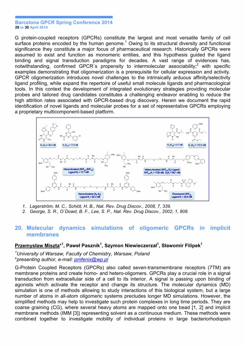
19. Multicomponent-Assisted GPCR Modulation: Evolutionary Approaches For Ligand Discovery and Molecular Probes Assembly
Jhonny Azuaje1, Abdelaziz El Maatougui1 , Abel Crespo1, Carlos Carbajales1, Vicente Yaziji1, Paula López1, Alba Casas1, Alejandro Fuentes1, Manuel González1, Alberto Coelho1, Alba Iglesias2, José Brea2, M. Isabel Loza2, Jana Selent3, María Martí3, Manuel Pastor3, Hugo Gutiérrez de Terán4 and Eddy Sotelo1,2*
1Center for Research in Biological Chemistry and Molecular Materials (CIQUS) and 2Center for Research in Molecular Medicine and Chronic Diseases (CIMUS). University of Santiago de Compostela, Santiago de Compostela 15782, Spain; 3Computer-Assisted Drug Design Lab, Research Programme on Biomedical Informatics (GRIB), PRBB, Barcelona 08003, Spain. 4Department of Cell and Molecular Biology, Uppsala University, Uppsala SE-75124.
Barcelona GPCR Spring Conference 2014 28 to 30 April 2014
G protein-coupled receptors (GPCRs) constitute the largest and most versatile family of cell surface proteins encoded by the human genome.1 Owing to its structural diversity and functional significance they constitute a major focus of pharmaceutical research. Historically GPCRs were assumed to exist and function as monomeric entities, and this hypothesis guided the ligand binding and signal transduction paradigms for decades. A vast range of evidences has, notwithstanding, confirmed GPCR´s propensity to intermolecular associability;2 with specific examples demonstrating that oligomerization is a prerequisite for cellular expression and activity. GPCR oligomerization introduces novel challenges to the intrinsically arduous affinity/selectivity ligand profiling, while expand the repertoire of useful small molecule ligands and pharmacological tools. In this context the development of integrated evolutionary strategies providing molecular probes and tailored drug candidates constitutes a challenging endeavor enabling to reduce the high attrition rates associated with GPCR-based drug discovery. Herein we document the rapid identification of novel ligands and molecular probes for a set of representative GPCRs employing a proprietary multicomponent-based platform.
1. Lagerström, M. C., Schiöt, H. B., Nat. Rev. Drug Discov., 2008, 7, 339. 2. George, S. R., O´Dowd, B. F., Lee, S. P., Nat. Rev. Drug Discov., 2002, 1, 808.
20. Molecular dynamics simulations of oligomeric GPCRs in implicit membranes
Przemysław Miszta*1, Paweł Pasznik1, Szymon Niewieczerzał1, Sławomir Filipek1
1University of Warsaw, Faculty of Chemistry, Warsaw, Poland *presenting author, e-mail: [email protected]
G-Protein Coupled Receptors (GPCRs) also called seven-transmembrane receptors (7TM) are membrane proteins and create homo- and hetero-oligomers. GPCRs play a crucial role in a signal transduction from extracellular side of a cell to its interior. A signal is passing upon binding of agonists which activate the receptor and change its structure. The molecular dynamics (MD) simulation is one of methods allowing to study interactions of this biological system, but a large number of atoms in all-atom oligomeric systems precludes longer MD simulations. However, the simplified methods may help to investigate such protein complexes in long time periods. They are coarse graining (CG), where several heavy atoms are mapped onto one bead [1, 2] and implicit membrane methods (IMM [3]) representing solvent as a continuous medium. These methods were combined together to investigate mobility of individual proteins in large bacteriorhodopsin
MULTICOMPONENT SYNTHESIS
Antagonists
Allosteric Modulators
Agonists
Fluorescent Ligands
Hetero-bivalent Ligands
Homo-Bivalent Ligands
Bitopic Ligands
Biased Ligands
N
H
O
N
Me
O
O
MeO
OO
N
H
O
N
Me
O
O
OMe
I
I
NH2
Me
NH2Me
N
N NH
N
O
OMe
MeOO
Me
NH
O
OO
O NH
N
O Me
O
O
NH
N
N
OMe
OMe
N
OCH3
ON N
MeOO
NH
O
NH
Me
N
OMe
O
O
I
MeO
OO
Me
NH2
N
H
O
O
MeO
I
NH2Me
NS
H
O O
N
Hetero-bivalent (5HT2A-D2) Ligand 5HT2A KI = 1720 nM, D2KI = 80.1 nM
Fluorescent 5HT2A Ligand KI = 25.0 nM
Homo-bivalent (5HT2A-5HT2A) Ligand KI = 17.7 nM
Homo-bivalent (A3-A3) Ligand KI = 62.3 nM
N
N
O
O
H
Me
O
N O
OO
H2N
O O
OMe
N
KI (A2B) = 4.7 nM KI (A3) = 172.5 nM
N
NN
CN
Me
H
Cl
O
HN
NH
O
CH3O
O
I OO
KI (A1) = 56.3 nM KI (A2A) = 17.5 nM

Barcelona GPCR Spring Conference 2014 28 to 30 April 2014
oligomers as well as to test stability of different GPCR dimers. In a current version of software an extensive testing of parameters was performed and tested on known oligomeric structures.
1. Levitt, M.: A simplified representation of protein conformations for rapid simulation of protein folding. J. Mol. Biol. 104(1), 59–107 (1976)
2. Levitt, M., Warshel, A.: Computer simulation of protein folding. Nature 253(5494), 694–698 (1975) 3. Lazaridis, T.: Effective energy function for proteins in lipid membranes. Proteins 52(2), 176–192 (2003)
21. Role of Histamine H3-glutamate mGlu5 receptor heteromers in neuronal activity modulation
Estefanía Moreno*, Hanne M. Hoffmann, Gemma Navarro, Josefa Mallol, Antoni Cortés, Vicent Casadó, Carme Lluís, Jordi Ortiz, Michel Vignes*, Enric Canela*, Peter J. McCormick*. University of Barcelona
Group 1 metabotropic glutamate receptors (mGlu1R and mGlu5R) are required for both persistent forms of memory and persistent synaptic plasticity. These characteristic suggest they are potential targets for memory and plasticity-related disorders. We hypothesized that one way to target mGluRs might be via receptor heteromers. Heteromers are complexes between different receptors with biochemically distinct properties from the single receptors. Thus, we investigated potential mGlu5R partners in the hippocampus. Here by co-immunoprecipitation, Bioluminescent Resonant Energy Transfer and Proximity Ligation Assays, we report a novel receptor heteromer between mGlu5R and the histamine H3 receptors (H3R) in transfected cells and in rat hippocampus. A sharp inhibition of signaling by the agonist of either receptor in the presence of the agonist or the antagonist of the other receptor was seen. These results indicate a negative cross-talk in signaling when heteromers are activated with both agonists and also a cross-antagonism between receptors that might be attributed to an allosteric interaction between receptors in the H3R-mGlu5R heteromers. Heteromer formation thus leads to the H3R-mediated modulation of mGlu5R signaling.
The cross-antagonism and the negative cross-talk at the level of signaling in hippocampal slices were also seen at the level of extracellular field potential recordings and on pyramidal neuron Ca+2 mobilization and excitation. Thus, targeting H3R-mGlu5R heteromers via H3R ligands might be an efficient and potent way to modulate mGlu5R-mediated neuronal signaling and excitability as well as neuronal plasticity. The results demonstrate that H3R-mGlu5R heteromers are new targets to treat neurocognitive diseases where reduced mGlu5R signaling is desired.
22. Interaction of CRIP1a protein with CB1 repector: A molecular modeling study
Agnieszka Kaczor1, 2, Antti Poso2
1Medical University of Lublin, Faculty of Pharmacy with Division of Medical Analytics, Department of Chemical Technology of Pharmaceutical Substances with Computer Modeling Lab, 4a Chodźki St., 20-093 Lublin, Poland 2University of Eastern Finland, School of Pharmacy, Department of Pharmaceutical Chemistry, Yliopistonranta 1, P.O. Box 1627, FI-70211 Kuopio, Finland; E-mail: [email protected]
The activity of the cannabinoid CB1 receptor is regulated by multiple proteins that interact with the receptor. These include a novel protein, the cannabinoid receptor interacting protein 1a, CRIP1a, which has been demonstrated to suppress the constitutive activity of the CB1R. Moreover, it has been shown that the effect of the CRIP1a on the CB1 receptor is mediated through the interaction
Barcelona GPCR Spring Conference 2014 28 to 30 April 2014
with the C-terminus of the receptor. The aim of this work was to build homology models of CB1 receptor and CRIP1a protein, to use protein-protein docking to construct a complex of CB1 receptor and CRIP1a protein and to check in molecular dynamics simulation how CRIP1a protein affects CB1 receptor functioning.
Homology modeling with Modeller9v3 was used to build models of CB1 receptor in inactive and active conformation. The model in inactive conformation was built using adenosine A2A receptor (PDB ID: 4EIY, sequence identity 24.40%) and kappa opioid receptor as a template (PDB ID: 4DJH, sequence identity 28.70%). The model in the active conformation was built using as a template β2 adrenergic receptor in complex with Gs protein (PDB ID: 3SN6, sequence identity was significantly lower). The active model of CB1 receptor was built in complex with Gi3, Go and Gs proteins. The models were inserted into POPC bilayer, hydrated and neutralized with ions. Ions were then added to the concentration 0.15 M of NaCl. Molecular docking of CB1 ligands was performed with Glide and Surflex which is a part of Sybyl-X suite of programs. The best scored ligand-receptor complexes were subjected to molecular dynamic simulations with Gromacs. The complexes embedded in membrane were first minimized and then subjected to 1 ns MD in NVT ensemble, followed by 100 ns MD in NPT ensemble. The model of CRIP1a protein was built using crystal structure of UVRA2 as a template (PDB ID: 2VF8). Sequence identity was 21%. To build the initial complexes between the CB1 and the CRIP1a, different protein-protein docking tools (e.g. PatchDock, ClusPro and ZDOCK) were tried. The initial complexes were refined with ROSETTA.
It was found that nine C-terminal residues of CB1 receptor are involved in the interaction with CRIP1a protein. In order to check the effect of CRIP1a protein on the CB1 receptor functioning, MD simulations were performed as described above. It was discovered that CRIP1a destabilizes CB1 receptor active conformation which can lead to reduction of receptor basal activity and which is in agreement with experimental data. Moreover, it was determined that CRIP1a blocks CB1 receptor interactions with Gi and Go proteins.
Acknowledgements
The research was performed under Marie Curie IEF fellowship for Agnieszka A. Kaczor. Calculations were performed under computational grant by Interdisciplinary Center for Mathematical and Computational Modeling (ICM), Warsaw, Poland, grant number G30-18 and using resources of CSC, Finland.
23. Different role of NCS-1 and Calneuron in the adenosine A2A-dopamine D2 receptor heteromer signaling
D. Aguinaga, E. Moreno, M. Mikhaylova, E. Angelats, A. Cortés, J. Mallol, V. Casadó, C. Lluís, P.J. McCormick, M. Kreutz, E.I. Canela and G.Navarro
It is known that adenosine A2A (A2AR) and dopamine D2 (D2R) receptors form heteromers in which A2AR activation inhibits D2R signaling, but it is not known if this negative cross-talk is regulated by the physiological status. Here we demonstrated that the neuronal calcium binding proteins NCS1 and calneuron interact with A2AR-D2R heteromers in transfected cells and in primary cultures of rat striatal neurons. The alternative binding of NCS1 or calneuron to the A2AR-D2R heteromers transduces a local change in Ca2+ concentrations to the receptor heteromer function. In fact, the A2AR-mediated negative cross-talk of D2R in cAMP signaling is prevented by the binding of NCS1 at low Ca+2 concentration or by the binding of calneuron-Ca+2 complex at high Ca+2 levels. Thus, in striatal neurons, independently of Ca+2, D2R agonists can reduce the cAMP levels in the presence or the absence of A2AR agonists. Moreover, the binding of calneuron-Ca+2 complex, but not the NCS1, to the heteromer induced a potent decrease of ERK 1/2 signaling when the heteromer is co-activated. Thus, when Ca+2 levels increase, calneuron favors the A2AR agonist-induced
Barcelona GPCR Spring Conference 2014 28 to 30 April 2014
blockade of D2R MAPK signaling. Our results indicate that in striatal neurons A2AR in the A2AR-D2R-calcium binding protein complexes act as a biased signaling modulator of the D2R function.
24. The molecular interface of the heteromer between D1 and D3 dopamine receptors
Vicent Casadó1, Estefanía Moreno1, Xavier Guitart2, Gemma Navarro1, Ning-Sheng Cai2, Marta Sánchez2, Verònica Casadó, Daniel Farré1, Josefa Mallol1, Carme Lluís1, Peter J. McCormick1, Antoni Cortés1, Enric I. Canela1, Sergi Ferré2
1Molecular Neurobiology Group, Department of Biochemistry and Molecular Biology, Faculty of Biology, University of Barcelona, Barcelona, Spain 2Integrative Neurobiology Section, National Institute on Drug Abuse, Intramural Research Program, National Institutes of Health, Baltimore, MD, USA
Receptor heteromers are becoming the focus of extensive research in the field of G-protein-coupled receptors (GPCRs) (1,2). Recent studies have shown evidence for the existence and co-localization of dopamine D1 (D1R) and D3 receptors (D3R) in specific neuronal populations in the striatum. We have already shown the existence of a physical interaction between these receptors and a positive cross-talk as a biochemical property of the D1R-D3R heteromer (3). The existence of these heteromers also suggests that their cross-talk may be important in regulating some aspects of rewarding mechanisms as well as of emotional and cognitive processes in the appearance and development of motor dysfunctions. Here, we address the study of the molecular interactions possibly involved in the heteromerization of D1R and D3R, particularly the role of the different GPCR transmembrane TM domains. We have used synthetic peptides containing the amino acid sequences of several D1R TM domains to determine biochemical properties of the D1R-D3R heteromer (cross-talk and cross-antagonism). We show the disruption of D1R-D3R positive cross-talk and cross-antagonism by D1 TM5 and TM6 peptides. The same disruption strategy allowed us to identify another biochemical property of the heteromer: a selective reduction in the potency of raclopride at antagonizing agonist-induced MAPK activation and β2-arrestin recruitment. The information obtained from this experimental work may help clarifying the role of the overexpression of D3R described in several neuropsychiatric disorders, and may also help in the design of new molecular entities able to selectively interact with the D3R in its homomeric or heteromeric forms.
1. Ferré S, Baler R, Bouvier M, Caron MG, Devi LA, Durroux T, Fuxe K, George SR, Javitch JA, Lohse MJ. Building a new conceptual framework for receptor heteromers. Nat Chem Biol. 2009;5:131–134.
2. Ferré S, Casadó V, Devi LA, Fillizola M, Jokers R, Lohse MJ, Milligan G, Pin JP, Guitart X. G Protein-Coupled Receptor oligomerization revisited: Functional and pharmacological perspectives. Pharmacol Rev. 2014;66:413-34.
3. Marcellino D, Ferré S, Casadó V, Cortés A, Le Foll B, Mazzola C, Drago F, Saur O, Stark H, Soriano A, Barnes C, Goldberg SR, Lluis C, Fuxe K, Franco R. Identification of dopamine D1-D3 receptor heteromers. Indications for a role of synergistic D1-D3 receptor interactions in the striatum. J Biol Chem. 2008; 283:26016-25.
Barcelona GPCR Spring Conference 2014 28 to 30 April 2014
25. Targeting dopamine D1-D3 receptor heteromers for L-DOPA-induced dyskinesias in the 6-OHDA rat model
Daniel Farré1, Estefanía Moreno1, Júlia Canet1, Josefa Mallol1, Antoni Cortés1, Carme Lluís1, José-Luis Lanciego2, José-Luis Labandiera3, Enric I. Canela1, Ana Muñoz3, Rafael Franco1, Vicent Casadó1
1Molecular Neurobiology Group, Department of Biochemistry and Molecular Biology, Faculty of Biology, University of Barcelona, Barcelona, Spain
2Centro de Investigación Médica Aplicada, University of Navarr, Spain 3Department of Morphological Sciences, University of Santiago de Compostela, Spain
Parkinson’s disease (PD) is a progressive neurodegenerative disorder caused by the degeneration of the pigmented neurons of the substantia nigra pars compacta that provide dopamine input to the striatum. Chronic dopamine replacement therapy leads to involuntary aimless movements known as L-DOPA-induced dyskinesia (LID), due to an increased of dopamine D1 receptors (D1R) signaling promoted by an up-regulation of D3R. In this study, we investigate by radioligand binding techniques changes in D1R and D3R protein expression in the striatum of a unilateral PD rat model. We determine the receptor levels in the right (lesioned) side and in the left (non-lesioned) side of the striatum in control, 6-OHDA-lesioned and in non-dyskinetic and dyskinetic L-DOPA-treated rats. A high increase in D3R is observed in both striatum sides only in dyskinetic rats. We also investigate the presence of cross-talk between D1R and D3R as fingerprint of the D1R-D3R heteromer by competition binding experiments. Lesioned side of dyskinetic rat striatum is the only case showing positive cross-talk of D3R on D1R. In all cases, left side shows higher D1R affinity than right side and positive cross-talk is not longer observed. By in situ proximity ligation assay, we detect the higher number of positive cells containing D1-D3
receptor heteromers in striatum of dyskinetic animals. All these results show that the increase of D3R expression in dyskinetic rats induces heteromerization in both striatum sides. In this case, D3R activation increases D1R affinity in right side and catch up the high affinity detected in left side. This fact can be responsible of the dyskinetic pattern in rats.
1. Feyder, M., A. Bonito-Oliva, and G. Fisone, L-DOPA-Induced Dyskinesia and Abnormal Signaling in Striatal Medium Spiny Neurons: Focus on Dopamine D1 Receptor-Mediated Transmission. Front Behav Neurosci, 2011. 5: p. 71.
2. Ghiglieri, V., et al., Corticostriatal Plastic Changes in Experimental L-DOPA-Induced Dyskinesia. Parkinsons Dis, 2012. 2012: p. 358176.
3. Jenner, P., Molecular mechanisms of L-DOPA-induced dyskinesia. Nat Rev Neurosci, 2008. 9(9): p. 665-77.
4. Kelly, P.H. and K.E. Moore, Mesolimbic dopamine neurons: effects of 6-hydroxydopamine-induced destruction and receptor blockade on drug-induced rotation of rats. Psychopharmacology (Berl), 1977. 55(1): p. 35-41.
5. Lebel, M., et al., Striatal inhibition of PKA prevents levodopa-induced behavioural and molecular changes in the hemiparkinsonian rat. Neurobiol Dis, 2010. 38(1): p. 59-67.
6. Marcellino, D., et al., Identification of dopamine D1-D3 receptor heteromers. Indications for a role of synergistic D1-D3 receptor interactions in the striatum. J Biol Chem, 2008. 283(38): p. 26016-25.
7. Miklyaeva, E.I., D.J. Martens, and I.Q. Whishaw, Impairments and compensatory adjustments in spontaneous movement after unilateral dopamine depletion in rats. Brain Res, 1995. 681(1-2): p. 23-40.
8. Muir, G.D. and I.Q. Whishaw, Ground reaction forces in locomoting hemi-parkinsonian rats: a definitive test for impairments and compensations. Exp Brain Res, 1999. 126(3): p. 307-14.
9. Santini, E., et al., Critical involvement of cAMP/DARPP-32 and extracellular signal-regulated protein kinase signaling in L-DOPA-induced dyskinesia. J Neurosci, 2007. 27(26): p. 6995-7005.
Barcelona GPCR Spring Conference 2014 28 to 30 April 2014
26. Biophysical behaviour of model membranes of 1,2-dimyristoyl-sn-glysero-3-posphocholine
Daniela Vojta
27. DAFT CXCR4: in silico study of protein dimerization
Kristyna Pluhackova, Stefan Gahbauer, Tsjerk Wassenaar and Rainer A. Böckmann. Computational Biology, Department of Biology, Friedrich-Alexander Universität Erlangen-Nürnberg
G-protein coupled receptors are family of receptors and signal transductors that are the target of about 40% of the drugs on the market currently. Th chemokine receptor type 4 (CXCR4) protein is believed to be one of several chemokine receptors that HIV can utilize to infect T-cells in the human body. Toth and colleagues have shown in 2004 that the CXCR4 dimerization is involved in the gp120 and SDF-1alpha-induced signaling events [1].
In order to predict interaction interfaces of transmembrane peptides and proteins and the process of their dimerization we developed an in silico Docking Assay For Transmembrane proteins (DAFT) which uses large sets of coarse-grained molecular dynamics simulations.
The DAFT method was applied to study the dimerization of the CXCR4 protein whose crystal structure was published recently [2]. The dimerization interfaces of this protein are influenced by the presence of the CVX15 cofactor as well as by the specific lipid environment. After the dimerization, representative dimer structures were selected and converted back to the atomistic resolution by the tool backward [3] in order to analyze all possible interactions between the two proteins at the atomistic level.
[1] Toth et al, J Pharmacol Exp Ther 2004 [2] Wu et al, Science 2013 [3] Wassenaar and Pluhackova et al, JCTC 2013
28. Studying the complex membrane environment of GPCR using the MEMBPLUGIN
Juan Manuel Ramírez Anguita
29. Interaction between the β2-adrenergic and the insulin receptor: correlation between the BRET and bioinformatics results
Maja Mandić1, Luka Drinovec2, Sanja Glisic3, Nevena Veljkovic3, Jane Nøhr4, Milka Vrecl1
1Institute of Anatomy, Histology & Embryology, Veterinary Faculty, University of Ljubljana, Slovenia 2Aerosol d. o. o. Ljubljana, Slovenia 3Center for Multidisciplinary Research, Institute of Nuclear Sciences VINCA, Belgrade, Serbia. 4Department of Incretin & Islet Biology, Novo Nordisk A/S, Måløv, Denmark
Functional interplay between different classes of receptors has emerged as a notable factor of cellular response in health and disease. Glucose metabolism is under cooperative regulation of both insulin receptor (IR) and β2-adrenergic receptor (β2-AR) representing the receptor tyrosine kinases (RTKs) and the seven transmembrane receptors (7TMRs), respectively. To test possible direct interaction between the β2-AR and IR we first employed BRET2 saturation and competition
Barcelona GPCR Spring Conference 2014 28 to 30 April 2014
assays. Saturation assay data suggested a constitutive β2-AR and IR homo- as well as heteromerization. Calculated AD50 value as a measure of the relative affinity for homo- and heteromers formation was comparable for both homomers but differed between heteromers, which could not be explained by a simple dimer model. In heterologous competition assays a transient increase in BRET2 signal with a later hyperbolical decrease was observed further suggesting higher order heteromer formation. To complement BRET2 data we next applied informational spectrum method (ISM), a virtual spectroscopy method for investigation of protein-protein interactions. Computational peptide scanning of β2-AR and IR identified domains encompassing residues at the end of 7th TM domain and C-terminal tail of β2-AR and the cytoplasmic part of IR beta chain as prospective interaction domains. ISM analysis of β2-AR and IR also revealed that affinity for formation of β2-AR: β2-AR: IR trimer is higher compared to the affinity for β2-AR: IR: IR trimer formation. In summary, our data suggest direct interaction and higher order oligomer formation between the β2-AR and IR as well higher propensity of β2-AR for heteromerization.
30. Targeting dopamine D1-histamine H3 receptor heteromers reverts cognitive and motor deficits in a mouse model of Huntington’s disease
Mar Rodríguez-Ruiz1,2*, David Moreno-Delgado1,2*, Mar Puigdellívol3*, Estefanía Moreno1,2, Joaquín Botta1,2, Paola Gasperini1,2, Arnau Cordomí5, Gemma Navarro1,2, Josefa Mallol1,2, Antoni Cortés1,2, Sergi Ferré5, Manuel Guzmán1,6, Leonardo Pardo4, Jordi Alberch1,3, Vicent Casadó1,2, Enric I. Canela1,2, Carme Lluís1,2, Sílvia Ginés1,3*, Peter J. McCormick1,2,7* 1Centro de Investigación Biomédica en Red sobre Enfermedades Neurodegenerativas 2.Department of Biochemistry and Molecular Biology, Faculty of Biology, University of Barcelona, Spain 3Departament of Cellular Biology, Immunology and Neurosciences, Faculty of Medicine, University of Barcelona, Spain 4Computational Medicine Laboratory, Faculty of Medicine, Autonomous University of Barcelona, Bellaterra, Spain 5National Institute on Drug Abuse, National Institutes of Health, Department of Health and Human Services, Baltimore, MD 21224 Maryland, USA 6Dept. Biochemistry and Molecular Biology I, School of Biology, Complutense University of Madrid, Spain 7School of Pharmacy, University of East Anglia, Norwich Research Park, Norwich, NR4 7TJ. *These authors contributed equally to this work
In early stages of Huntington’s disease (HD) there is an excess of dopamine production and an over-activation of dopamine D1 receptors (D1R) that can produce not only an imbalance in dopaminergic neurotransmission but also can directly lead to signaling cascades that induce cell death. Here we propose a new and provocative strategy to reduce the D1R over-activation effects in HD by targeting the recently described receptor complexes of D1R and the histamine receptors H3 (H3R). We show the expression of D1R-H3R heteromers in a HD model of striatal neuronal progenitor cells and in different brain areas of mouse models of HD in the early but not in the late stages of the illness as well as in human control subjects and in grade 2 HD patients but not in grade 3 or 4 HD patients. Upon co-activation of D1R-H3R heteromers, H3R ligands act as a “molecular brake” for D1R signaling. D1R-induced cell death in cells and in brain slices and the signaling cascades responsible for this death are reduced by H3R ligands targeting D1R-H3R heteromers. Treatment of presymptomatic mouse models of HD with the H3R antagonist thioperamide can restore both cognitive and motor deficits of these animals and inhibits the loss of heteromer expression observed in non-treated mice in the late stages of the illness. Our results demonstrate that D1R-H3R heteromers play a pivotal role in controlling dopaminergic
Barcelona GPCR Spring Conference 2014 28 to 30 April 2014
neurotransmission and indicate that D1R-H3R heteromers can be target for treating HD in the pre-symptomatic stages of the illness.
1. Garrett MC, Soares-da-Silva P. Increased cerebrospinal fluid dopamine and 3,4- dihydroxy-phenylacetic acid levels in Huntington’s disease: evidence for an overactive dopaminergic brain transmission. J Neurochem. 1992;58(1):101-6.
2. Paoletti P, Vila I, Rifé M, Lizcano JM, Alberch J, Ginés S. Dopaminergic and glutamatergic signaling crosstalk in Huntington’s disease neurodegeneration: the role of p25/cyclin-dependent kinase 5. J Neurosci Off J Soc Neurosci. 2008;28(40):10090-101.
3. Ross CA, Tabrizi SJ. Huntington’s disease: from molecular pathogenesis to clinical treatment. Lancet Neurol. 2011;10(1):83-98.
31. Studying the activity of metabotropic glutamate receptor 4 allosteric modulators
Fanny Malaire1, Xavier Rovira1, Jordi Rodrigo de Losada2, Patricia González3, Amadeu Llebaria3, Jesus Giraldo4, Jean-Philippe Pin1, Cyril Goudet1 1CNRS,UMR 5203, Institut de Génomique fonctionnelle, Montpellier, France and INSERM, U661, Montpellier, France and Université Montpellier,1,2, Montpellier, France. 2Université Paris-Sud, CNRS, BioCIS-UMR 8076, Laboratoire de Chimie Thérapeutique, Faculté de Pharmacie, 92296 Châtenay-Malabry, France. 3Research Unit on Bioactive Molecules, Departament of Biomedicinal Chemistry, Institute of Advanced Chemistry of Catalonia CSIC, 08034 Barcelona, Spain. 4Laboratory of Systems Pharmacology and Bioinformatics, Institut de Neurociencies and Unitat de Bioestadistica, Universitat Autònoma de Barcelona, 08193 Bellaterra, Spain.
Glutamate is the main neurotransmitter of the mammalian central nervous system. When released in the synaptic cleft, glutamate activates two types of receptors: ionotropic glutamate receptors which are ligand-gated ion channels and metabotropic glutamate receptors (mGlu) which are G-protein coupled receptors mediating the neuromodulatory action of glutamate. The last-mentioned receptors are involved in the physiopathology of various CNS disorders, thus raising the therapeutic interest of targeting mGlu to modulate and restore impaired neurotransmission associated with these diseases. Among them, mGlu4 appears as a promising therapeutic target for chronic pain and Parkinson disease. However, the current pharmacology of mGlu4 is somewhat unsatisfactory. Orthosteric ligands are limited due to their lack of selectivity among the different mGlu subtypes and allosteric ligands often have intrinsic agonist activity, low potency and poor solubility. Therefore, new knowledge is necessary to understand the receptor functioning and to find molecules with improved pharmacological profiles.
In the present work the allosteric binding site of mGlu4 is explored in order to gain mechanistic insight into the modulatory function of ligands acting at this part of the protein. Both, theoretical and experimental procedures are used to obtain information about important determinants for the interaction of ligands in this site. The results of this study will help to better understand the functioning of this receptor and the design of new selective compounds that will improve the pharmacological toolbox available to regulate mGlu4 activity.
Barcelona GPCR Spring Conference 2014 28 to 30 April 2014
32. Synthesis and studies of putative GPCR ligands
Gunars Duburs, Einars Loza, Egils Bisenieks, Olga Bobileva, Brigita Vigante, Janis Klovins, Ilona Mandrika, Ramona Petrovska, Iveta Luntena.
Latvian Institute of Organic Synthesis, Aizkraukles Str. 21. Riga, LV-1006, Latvia; Biomedical Research and Study Centre, Ratsupites Str. 1, Riga, LV-1067, Latvia
Synthesis and functional receptor studies of acyclic and cyclic �-carbamoyl-vinylcarbonyl compounds were performed.
Novel aryl derivatives of 2-amidocyclohex-1-ene carboxylic acid 1 were synthesized. Instead of the saturated ethane linker in the amide part of the molecules they contain rigidity elements: E-double bond, triple bond and trans- or cis- substituted cyclopropane ring; their potencies on the activation of HCA1, HCA2 and HCA3 receptors by cAMP assay were evaluated. Incorporation of rigidity elements in appropriate molecules 1 allowed modulating the potency and selectivity of the activation of HCA2 receptor.
OH
O
NH
XO Y Ar
NH
O
OH
OAr
O
O
R1
NH
ON
+
n
1 2 3
R1, R2 = H; Me
n = 1-3
A small series of 5-substituted 6-oxo-1,4,5,6-tetrahydropyridine-3-carboxylic acids 2, where 2-amido substituent was a component part of cyclohexene cycle, was prepared. Compounds 2 exhibited minor activation of HCA2 receptor by cAMP assay.
A series 3 of potential ligands for HCA1 receptors involving the anthranil amide moiety linked to heterocyclic cations were synthesized.
33. Expert curated information on GPCRs in the IUPHAR/BPS Guide to PHARMACOLOGY
Joanna L. Sharman1, Adam J. Pawson1, Helen E. Benson1, Elena Faccenda1, Christopher Southan1, Anthony J. Harmar1, Michael Spedding2 and NC-IUPHAR. 1IUPHAR Database and Guide to PHARMACOLOGY Web Portal Group, Centre for Integrative Physiology, School of Biomedical Sciences, University of Edinburgh, Hugh Robson Building, Edinburgh, EH8 9XD, UK. 2Spedding Research Solutions SARL, 6 Rue Ampere, Le Vésinet 78110, France
G protein-coupled receptors (GPCRs) are the largest family of human drug targets representing ~19% of the targets of current drugs and many more in clinical trials [1]. We have developed the IUPHAR/BPS Guide to PHARMACOLOGY portal (the new home of the IUPHAR Database) to provide free access to curated information on important pharmacological targets and the substances that act on them [2]. The database includes data on 394 GPCRs including 130 “orphans” without confirmed endogenous ligands. This includes the recent addition of 33 adhesion class GPCRs. Development of the database is overseen by the International Union of Basic and Clinical Pharmacology (IUPHAR) Committee on Receptor Nomenclature and Drug Classification (NC-IUPHAR). Expert subcommittees for each GPCR family provide recommendations on receptor nomenclature and peer-reviewed summaries of the literature covering properties such as function, pharmacology, signalling mechanism, important variants, available assay systems and
Barcelona GPCR Spring Conference 2014 28 to 30 April 2014
mouse knockout phenotypes. Ligands are annotated with their chemical structures, or sequences and post-translational modifications for peptides. Links are provided to other online databases including GPCRDB, UniProt, Ensembl and PubChem. Current work includes identifying the mechanism of action for approved drugs treating human diseases, mapping them to their primary targets and curating supporting data in the literature (e.g. Ki, Kd, IC50). In some cases we also annotate data-supported polypharmacology where interactions of comparable in vitro potencies against multiple targets have been published. The database is available online at http://www.guidetopharmacology.org.
1. Rask-Andersen M, Masuram S, Schiöth HB. (2014) The druggable genome: evaluation of drug targets in clinical trials suggests major shifts in molecular class and indication. Annu Rev Pharmacol Toxicol., 54: 9-26.
2. Pawson AJ, Sharman JL, Benson HE et al. (2014) The IUPHAR/BPS Guide to PHARMACOLOGY: an expert-driven knowledgebase of drug targets and their ligands. Nucl. Acids Res., 42 (Database Issue): D1098-106.
34. Application of SPILLO-PBSS to GPCRs: predicting selectivity and off-target interactions of potential new drugs.
Alessandro Di Domizio, Ingrid Pogu, Giulio Vistoli, Alessandro Pedretti. Department of Pharmaceutical Sciences Faculty of Pharmacy, University of Milan, Italy
G protein-coupled receptors (GPCRs) constitute a superfamily of proteins which play a pivotal role as pharmacological targets for a number of pathologies. However, due to the high level of sequence similarity, the risk of low selectivity usually represents a serious problem when designing new GPCR-targeted drugs, especially within the same GPCR class. On these grounds, the possibility to predict selectivity or, in other words, the off-target interactions of a therapeutic agent could be of significant support in Drug Discovery and Development processes. The present study is focused on the GPCR members belonging to the class A (Rhodopsin-like) the structures of which were modelled by homology techniques (503 models), exploiting the increasing availability of GPCR resolved structures. With the aim to predict the selectivity and the off-target interactions, these homology models were exploited by using the new software SPILLO-Potential Binding Sites Searcher (SPILLO-PBSS1), which searches similar binding sites starting from well-defined reference structures. In detail, the program was utilized for a set of purposely generated complexes for which it was able to identify similar binding sites among the screened GPCR. Very often these potential sites correspond to experimentally known off-targets of the considered drugs and in some cases the performed simulations were able to evidence also unknown potential off-targets which may provide useful suggestions for a better understanding of the polypharmacological effects and clinically-observed side effects. 1. Di Domizio A, Vitriolo A, Vistoli G, Pedretti A. SPILLO-PBSS: detecting hidden binding sites within
protein 3D-structures through a flexible structure-based approach. J. Chem. Inf. Model. 2014 [submitted]
Barcelona GPCR Spring Conference 2014 28 to 30 April 2014
35. Preclinical evidence on the “anti-alcohol” properties of the positive allosteric modulators of the GABAB receptor
Giancarlo Colombo1, Paola Maccioni1, Mauro A.M. Carai1, Claudia Mugnaini2, Federico Corelli2 1Neuroscience Institute, National Research Council of Italy, Section of Cagliari, Monserrato (CA), Italy 2Department of Biotechnologies, Chemistry and Pharmacy, University of Siena, Siena (SI), Italy
Multiple preclinical and clinical lines of evidence have demonstrated that pharmacological activation of the orthosteric GABAB receptor suppressed several alcohol-related behaviors; as a result of these studies, the prototypic GABAB receptor agonist, baclofen, is currently undergoing evaluation as possible pharmacotherapy for alcohol use disorders (AUDs). More recently, it has been discovered that activation of the positive allosteric modulatory binding site may represent a more favourable way to pharmacologically manipulate the GABAB receptor. Treatment with the presently available, in vivo effective positive allosteric modulators (PAMs) of the GABAB receptor reproduced indeed the “anti-alcohol” effects of baclofen, displaying however fewer side-effects (e.g., sedation) and a remarkably higher therapeutic index. Specifically, acute or repeated treatment with all GABAB PAMs tested to date (namely, CGP7930, GS39783, BHF177, rac-BHFF, ADX71441, and COR659) have invariably resulted in marked and selective reductions in excessive alcohol drinking, binge-like alcohol drinking, operant alcohol self-administration, and alcohol seeking behavior in mice and rats (including those belonging to lines selectively bred for high alcohol preference and consumption). Because of the predictive validity of the experimental procedures used in these studies, data collected to date suggest that GABAB PAMs may represent a novel class of agents with therapeutic potential for AUDs.
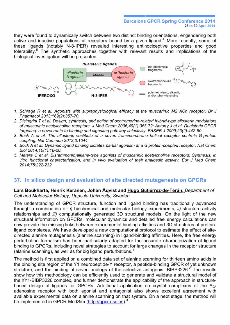
36. Novel pharmacological tools which activate mAChRs: a question of “dualsterism”
Carlo Materaa, Clelia Dallanocea, Marta Quadria, Ulrike Holzgrabeb, Elisabetta Barocellic , Klaus Mohrd, Marco De Amicia aDepartment of Pharmaceutical Sciences - University of Milan – Milan, Italy bInstitute of Pharmacy - University of Würzburg – Würzburg, Germany cDepartment of Pharmacy - University of Parma – Parma, Italy dInstitute of Pharmacy - University of Bonn – Bonn, Germany
Muscarinic acetylcholine receptors (mAChRs) represent an excellent model system to study orthosteric and allosteric interactions. The high sequence homology shown by orthosteric sites of mAChRs has hampered the development of subtype selective agonists. On the other hand, allosteric recognition sites are less conserved among the various mAChR subtypes.
We synthesized a series of hybrid ligands designed to simultaneously interact with both orthosteric and allosteric sites (“dualsteric” compounds) by fusing orthosteric activators with M2-selective allosteric fragments (W84 and Naphmethonium). In particular, among the oxotremorine-like orthosteric agents, iperoxo emerged as a potent agonist with supraphysiological efficacy but devoid of subtype selectivity.1 To explore the whole chemical space of the binding region, we modified the structure of the three component parts (orthosteric and allosteric moieties and spacer) of dualsteric ligands.2
These ligands permitted to prove for the first time that GPCR’s allosteric vestibule is able to control the extent of receptor movement to govern a hierarchical order of G-protein coupling.3 In addition,
theyactithestolebiol
1. SP
2. Dot
3. Bc
4. BB
5. Mv2
37.
LarCel
Thethrorelastrunowligadirepertbind(ala
Thethe strushothe basadeavabe i
y were fouive and inse ligands
erability.5 Togical inve
Schrage R Pharmacol 2Disingrini T of muscarinargeting: a
Bock A et acoupling. NaBock A et alBiol 2014;10Matera C evitro functio2014;75:222
. In silic
rs Boukhall and Mole
e understaough a comationships uctural infow provide tand compleected alaniturbation fding to GPanine scan
e method isbinding sit
ucture, andow how this
hY1-BIBPsed designenosine reailable expeimplement
und to dynaactive pops (notablyThe synthestigation w
et al. Agon2013;169(2)et al. Desigic acetylchonovel routeal. The alloat Communl. Dynamic 0(1):18-20. t al. Bis(am
onal charac2-232.
co design
arta, Henriecular Biolo
anding of mbination and iii) co
ormation othe missingexes. We hne mutageformalism
PCRs, incluning), as w
s first applte region od the binds methodo
P3226 comn of ligandeceptor wierimental dted in GPC
amically swpulations oy N-8-IPEetic approwill be pres
nists with s2):357-70. gn, synthesoline recepte to binding osteric vestn 2012;3:104ligand bindi
mmonio)alkacterization,
n and eva
k Keränenogy, Uppsa
GPCR strof: i) biochomputationn GPCRs,g links betwhave develenesis (alahas been
uding novewell as for b
lied on a cof the Y1 ning of sev
ology can bplex, and fds for GPth both adata on ala
CR-ModSim
witch betweof receptorR) revealeoaches togsented.
supraphysio
is, and actiotors. J Med and signalitibule of a 44. ing dictates
ane-type agand in vivo
aluation
n, Johan Åala Univers
ructure, funhemical annally gene, moleculaween expeoped a ne
anine scanparticularll strategiesbig ligand
combined deuropeptid
ven analogbe efficientfurther dem
PCRs. Addagonist ananine scan
m (http://gp
B
een two dirs bound bed interesgether wit
ological effi
on of oxotred Chem 200ing pathwayseven tran
s partial ago
gonists of mo evaluatio
of site d
Åqvist andsity, Swede
nction andnd molecurated 3D
ar dynamicerimental bew computanning) in ligly adapteds to accouperturbatio
data set ofde-Y recepgs of the stly used tomonstrate ditional apnd antagonnning on thpcr.usc.es)
Barcelona
stinct bindby a givensting antinth relevant
icacy at the
emorine-rel06;49(1):366y selectivitynsmembran
onism at a G
muscarinic an of their a
directed m
d Hugo Guen
d ligand bular biologystructural
cs and detabinding affinational progand-bindind for the ant for largeons.1
f alanine scptor, a peptselective ao generate the applica
pplication onist also hat system.3
a GPCR Sp
ing orientan ligand.4 nociceptivet results a
e muscarini
ated hybrid6-72; Antonyy. FASEB J e helical re
G protein-co
acetylcholinanalgesic a
mutagen
utiérrez-de
inding hasy experimemodels. O
ailed free nities and
otocol to esng affinitie
accurate che changes
canning fotide-bindin
antagonist and valida
ability of thon crystal shows ex
m. On a nex
pring Con28
ations, engMore rece
e propertieand implic
ic M2 ACh
d-type allostny J et al. D2009;23(2)eceptor con
oupled rece
ne receptorsactivity. Eur
nesis on
e-Terán, D
s traditionents, ii) strOn the ligenergy ca3D structustimate the
es. Here, thharacterizain the rece
or thirteen ag GPCR oBIBP3226ate a struche approac complexe
xcellent agxt stage, th
ference 208 to 30 April 2
gendering bently, somes and gcations of
h receptor.
teric modulaualsteric GP:442-50. ntrols G-pro
eptor. Nat C
rs: Synthesir J Med C
GPCRs
Department
ally advanructure-actht of the
alculations ures of prote effect of she free eneation of ligeptor struc
amino acidof yet unkn6.2 The resctural modech in structes of the greement he method
014 2014
both e of
good the
Br J
ators PCR
otein
Chem
is, in Chem
t of
nced tivity new can
tein-site-ergy
gand cture
ds in own sults el of ture-
A2A with
d will
Barcelona GPCR Spring Conference 2014 28 to 30 April 2014
1. Boukharta L, Gutiérrez-de-Terán H, Åqvist J. Computational prediction of alanine scanning and ligand binding energetics in G-protein coupled receptors. Submitted
2. a) Sautel M, Rudolf K, Wittneben H, Herzog H, Martinez R, Munoz M, Eberlein W, Engel W, Walker P, Beck-Sickinger AG (1996) Neuropeptide Y and the nonpeptide antagonist BIBP 3226 share an overlapping binding site at the human Y1 receptor. Mol Pharmacol 50: 285-292. b) Aiglstorfer I, Hendrich I, Moser C, Bernhardt G, Dove S, et al. (2000) Structure-activity relationships of neuropeptide Y Y1 receptor antagonists related to BIBP 3226. Bioorg Med Chem Lett 10: 1597-1600.
3) Gutiérrez-de-Terán H, Bello X, Rodríguez D (2013) Characterization of the dynamic events of GPCRs by automated computational simulations. Biochem. Soc. Trans. 41: 205-212.
38. Lignad binding mode analysis using new 5-HT1A receptor models developed by ALiBERO methodology
Dawid Warszycki1, Manuel Rueda2, Kurt Kristiansen3, Ingebrigt Sylte3, Ruben Abagyan2, Andrzej Bojarski1
1Department of Medicinal Chemistry, Institute of Pharmacology, Polish Academy of Sciences, Kraków, Poland 2Skaggs School of Pharmacy & Pharmaceutical Sciences, University of California, San Diego, U.S. 3Medicinal Pharmacology and Toxicology, Department of Medicinal Biology, Faculty of Health Sciences, UiT The Arctic University of Norway, Tromsø, Norway
ALiBERO (Automated Ligand-guided Backbone Ensemble Receptor Optimization) [1] is a new computational tool which expands the pocket selection from single to multiple, allowing for automatic iteration of the sampling-selection procedure. ALiBERO heuristic search consists of two main steps: (i) generation of multiple receptor conformations and selection of the best individual pockets according to the flexible-docking static-receptor small-scale docking performance; (ii) out of the best performing models the next generation of receptor conformations are constructed. This iterative process is repeated until the threshold for the fitness function is reached.
AliBERO algorithm was applied for a well-recognized 5-HT1A receptor and several factors (template selection, influence of training set composition etc.), influencing models performance, were investigated. Finally, 26 different approaches were validated on test set generated with DUD formalism and assessed by area under the receiver operating characteristic curve (ROC) [2] and Normalized Square root AUC (NSQ_AUC) metric [3].
Ligand binding mode of structurally diverse compounds with high affinity for 5-HT1A receptor (arylpiperazines, indoles, tetralines, etc.) was analyzed with use of SIFt methodology [4], and then compared to data extracted from literature.
Acknowledgments
The study was partially supported by the grant PLATFORMEX (Pol-Nor/198887/73/2013) financed within the Polish-Norwegian Research Programme.
References
1. Rueda M. et al., 2012, J. Chem. Inf. Model., 52, 2705-14. 2. Teramoto R. et al., 2007, J. Chem. Inf. Model., 47, 526-534. 3. Katritch V. et al., 2011, Neuropharm., 60, 108-115. 4. Mordalski, S. et al., 2011 Bioorg. Med. Chem. Lett., 21, 6816-6819
Barcelona GPCR Spring Conference 2014 28 to 30 April 2014
39. GPCR ligand selectivity through structure-based methods
David Rodríguez, Anirudh Ranganathan, and Jens Carlsson
Science for Life Laboratory, Department of Biochemistry and Biophysics and Center for Biomembrane Research; Stockholm University, SE-106 91 Stockholm, Sweden.
G Protein-Coupled Receptors (GPCRs) are grouped in receptor families that have evolved to recognize the same ligand. This enables the natural agonist to finely regulate signalling through differential binding affinities, downstream effectors, and tissue distributions of the receptor subtypes involved. However, the structural similarities among the binding sites of these also lead to difficulties in finding new chemical scaffolds that can selectively bind to specific receptors, a requirement in most drug discovery efforts.1
The 5-HT1B receptor is a target for migraine and mood disorders, but binding to the 5-HT2B receptor can lead to cardiovascular side effects. The recent determination of crystallographic structures for the human 5-HT1B and 5-HT2B receptors bound to ergotamine gave the opportunity to understand the molecular determinants of ligand binding to these receptors.2 Moreover, they were proposed to be predicted by computational methods before their release, in the context of the GPCR Dock 2013 assessment.3 Our submitted complexes were among the most accurate, achieving the best and fifth-best predictions for 5-HT1B and 5-HT2B receptors, and presenting the best descriptions of the binding sites. However, despite the accuracy of our predictions, they did not capture the most important structural features responsible for ligand selectivity. Retrospective analyses of a series of drugs against migraine revealed that the crystallographic structures could predict 5-HT1B selective ligands, but not the homology models. The potential of using crystallographic structures4 and homology models5,6 in drug design for GPCR ligands will be discussed.
1. Giacomini KM, Krauss RM, Roden DM, Eichelbaum M, Hayden MR, Nakamura, Y. When good drugs go bad. Nature. 2007; 446:975-977.
2. Wang C, Jiang Y, Ma J, Wu H, Wacker D, Katritch V, Han GW, Liu W, Huang XP, Vardy E, McCorvy JD, Gao X, Zhou XE, Melcher K, Zhang C, Bai F, Yang H, Yang L, Jiang H, Roth BL, Cherezov V, Stevens RC, Xu HE. Structural basis for molecular recognition at serotonin receptors. Science. 2013;340:610-614.
3. http://gpcr.scripps.edu/GPCRDock2013/ 4. Kruse AC, Weiss DR, Rossi M, Hu J, Hu K, Eitel K, Gmeiner P, Wess J, Kobilka BK, Shoichet BK.
Muscarinic Receptors as Model Targets and Antitargets for Structure-Based Ligand Discovery. Mol. Pharmacol. 2013;84:528-540.
5. Katritch V, Kufareva I, Abagyan R. Structure based prediction of subtype-selectivity for adenosine receptor antagonists. Neuropharmacology. 2011;60:108-115.
6. Kolb P, Phan K, Gao ZG, Marko AC, Sali A, Jacobson KA. Limits of ligand selectivity from docking to models: in silico screening for A(1) adenosine receptor antagonists. PLoS One. 2012;7:e49910.
40. Cloning and expression optimization of TGR5 in HEK293 cells for ligand screening
Elisabetta De Filippoa, Juan Carlos Mobarecb, Peter Kolbb, Anke C. Schiedela
aPharmaceutical Chemistry I, Pharmacenter Bonn, University of Bonn, An der Immenburg 4, D-53121 Bonn, Germany bDepartment of Pharmaceutical Chemistry, Philipps-University of Marburg, MarbacherWeg 6, 35032 Marburg, Germany
Bile acids are essential for the solubilization and absorption of dietary lipids and are the major products of cholesterol catabolism. So far, several nuclear receptors for bile acids have been
Barcelona GPCR Spring Conference 2014 28 to 30 April 2014
identified. However, a novel G protein-coupled receptor, TGR5, has recently been identified as first cell-surface receptor responsive to bile acids. Previous studies showed that bile acids are low-affinity agonists for this receptor, enabling TGR5-expressing cells to mediate G protein binding, MAPK activation and intracellular cAMP production as well as receptor internalization. The physiological role of this receptor is not well understood yet. Therefore, the design of specific TGR5 receptor ligands is important to gain deeper insight into TGR5 function.
For this purpose, we first cloned the TGR5 receptor into different vectors with and without tags for detection and then we generated transiently and stably transfected HEK293 cell lines. A high expression level of TGR5 receptors is required to generate a cell-based assay for ligand screening. We evaluated the expression level of TGR5 using immunofluorescence and Western blot analyses. A functional assay measuring the intracellular cAMP accumulation in TGR5-expressing cell lines was optimized using a known ligand for the receptor, lithocholic acid. This assay will be used for further compound testing in order to find ligands of the TGR5 receptor.
41. Mining Nature's Diversity for Selective GPCR Ligands
Markus Muttenthalera,b, Aline Dantas de Araujoc, Asa Anderssonc, Irina Vetterc, Marta Busnellid, Christian Bergmayere, Sebastien Dutertrec, Mehdi Moblic, Stuart M. Brierleyf, Philip E. Dawsonb, Fernando Albericioa, Chini Biced, Christian W. Grubere, Richard J. Lewisc, Paul F. Alewoodc aThe Institute for Research in Biomedicine, Barcelona, Spain bThe Scripps Research Institute, La Jolla, CA, USA cThe University of Queensland, Brisbane, QLD, Australia dCNR - Institute of Neuroscience, Milan, Italy eThe University of Vienna, Vienna, Austria fThe University of Adelaide, Adelaide, Australia
We still lack a complete toolbox of selective agonists and antagonists for the four known Oxytocin (OT) and arginine-vasopressin (AVP) GPCRs. Considering the ubiquitous presence of these receptors in the periphery and CNS in addition to their role in a multitude of important physiologically processes, it is central to have a plethora of selective ligands for concluding studies. Ideally, these ligands should not only be selective, but should also preserve their selectivity in different species allowing for translational studies important for drug development. While there has been great medicinal chemistry conducted over the last decades on shaping such ligands, we decided to take a different approach, taking advantage of the fact that the OT/AVP signaling system has been well preserved in the kingdom of life for millions of years, and started exploring these natural sources for novel ligands.
The venom of the cone snail, a traditional plant extract and the genome of social ants turned out to be such natural sources, all yielding excellent lead compounds for the design of receptor selective ligands. Here we would like to discuss the rationale behind our approaches, the techniques involved and how structure-activity relationship studies of these lead peptides influenced our ligand design, eventually leading to the identification of an agonist to antagonist switch for AVP, the development of a selective OT receptor agonist that retained its selectivity profile in mice and humans, the first oxytocic plant cyclotide, and the development of stable ligands towards the treatment of chronic abdominal pain.
Barcelona GPCR Spring Conference 2014 28 to 30 April 2014
42. An application of ligand interaction profiles as a novel approach in virtual screening of GPCR ligands
Jagna Witek, Sabina Smusz, Krzysztof Rataj, Stefan Mordalski, Andrzej J. Bojarski
Department of Medicinal Chemistry, Institute of Pharmacology Polish Academy of Sciences, 12 Smętna Street, 31-343 Kraków, Poland
Cheminformatic methods, such as Virtual Screening (VS), constitute vital part of modern drug design. VS enables effective database mining, being particularly useful tool in search for ligands of desired activity. The scope of this study was to find an efficient methodology to automatically discriminate between active and inactive compounds towards the target, thus facilitating VS procedure.
In this research we present a workflow combining docking, application of interaction profiles for ligands1 created on the basis of structural interaction fingerprints2 (SIFt) and machine learning algorithms. Due to the fact, that the method is strongly dependent on the target structure, it was examined against demanding conditions, that is homology models of G-protein coupled receptors.
Models of beta1 and beta2 adrenergic receptors were constructed on a number of class A GPCRs crystal structures. They underwent validation by docking of active and decoy compounds, and subsequent enrichment calculation. For the main experiment, three best homology models per each template, and three crystal structures of beta1 and beta2 receptors from PDB repository were chosen. Sets of active and inactive compounds towards each target were retrieved from ChEMBL database. Moreover, the set of inactives was enriched by random selection of compounds from ZINC database, and generation of decoys. All compounds were docked to their corresponding targets. Finally, interaction profiles created for all docked compounds constituted an input to machine learning algorithms. Performance of interaction profiles was compared with benchmark approach, which utilizes molecular fingerprints to discriminate between active and inactive compounds.
Acknowledgements
The study partially is supported by a grant PRELUDIUM 2013/09/N/NZ2/01917 financed by the National Science Centre. This study is partially supported by project “Diamentowy Grant” DI 2011 0046 41 financed by Polish Ministry of Science and Higher Education.
References
1. Witek, J.; Smusz, S.; Rataj, K.; Mordalski, S.; Bojarski, A.J.: ‘An application of machine learning methods to structural interaction fingerprints – a case study of kinase inhibitors’, Bioorg Med Chem Lett, 2014, 24 (2), 580–585.
2. Deng Z, Chuaqui C: Structural Interaction Fingerprint (SIFt): A Novel Method for Analyzing Three-Dimensional Protein-Ligand Binding Interactions. J Med Chem 2004, 47:337-344.
43. From Bitterness to GPCR Promiscuity and Back
Antonella Di Pizio and Masha Y. Niv. Institute for Biochemistry, Food Science and Nutrition, The Robert H. Smith Faculty of Agriculture, Food and Environment, and Fritz Haber Center for Molecular Dynamics, The Hebrew University of Jerusalem, Israel
The human bitter receptor (hTAS2R) family is made up of 25 members, representing about 4% of the GPCRs in humans. Better understanding of molecular recognition of bitter compounds may provide improved tools for rational design of functional foods and a rational way for designing drugs with acceptable taste and higher compliance rates. Moreover, hTAS2Rs were recently shown to be expressed extraorally, thus their ligands emerge as novel candidates for treatment of
Barcelona GPCR Spring Conference 2014 28 to 30 April 2014
metabolic and respiratory disorders. Due to the relevance of these targets, our studies are focused on investigating the structural basis of ligand recognition by hTAS2Rs in order to design target-selective hTAS2R modulators.
Bitter receptors are interesting targets in terms of molecular recognition: a few receptors are activated by thousands of structurally diverse compounds, with some of the receptors more broadly-tuned than others.1 Using chemoinformatics and modeling approaches, simple and predictive model of the diversity of possible GPCR antagonists was recently developed based on the sequence and the structure of the receptor.2 Currently we apply a similar method to study relations between GPCR properties and the diversity of the agonists sets. Preliminary results highlight intrinsic differences between selectivity towards agonists and towards antagonists and suggest intriguing differences between Family A GPCRs (for which more antagonists than agonists are known) and bitter taste receptors (for which agonists abound but antagonists are hard to find). Insights into these differences could promote understanding of the activation processes and faciliate rational design of bitter taste antagonists.
1 Wiener, A., Shudler, M., Levit, A. & Niv, M. Y. BitterDB: a database of bitter compounds. Nucleic acids research 40, D413-419 (2012).
2 Levit, A., Beuming, T., Krilov, G., Sherman, W. & Niv, M. Y. Predicting GPCR Promiscuity Using Binding Site Features. Journal of Chemical Information and Modeling 54, 184-194, doi:10.1021/ci400552z (2013).
44. Machine Learning analysis of class C GPCRs from their sequences
Martha-Ivón Cárdenas1,2, Caroline König2, Raúl Cruz-Barbosa3, René Alquézar2, Alfredo Vellido2 and Jesús Giraldo1 1Institut de Neurociències, Unitat de Bioestadística, Universitat Autònoma de Barcelona 08193, Bellaterra (Barcelona) – Spain 2Departament de Llenguatges i Sistemes Informàtics, Universitat Politècnica de Catalunya 08034, Barcelona – Spain 3Universidad Tecnológica de la Mixteca, 69000, Huajuapan, Oaxaca, México
Class C is one of the five main families in which GPCRs are divided [1]. Distinct structural features of class C GPCRs, such as a large extracellular domain where agonists bind and dimeric configuration, together with their involvement in many neurological disorders, makes them particularly attractive for pharmacological research [2]. According to GPCRDB [3], class C is subdivided into seven subtypes: Metabotropic glutamate (mG), Calcium sensing (CS), GABA-B (GB), Vomeronasal (VN), Pheromone (Ph), Odorant (Od) and Taste (Ta). The absence of crystal structures for full-length class C receptors prompts the use of their primary amino acid sequences for their analysis using homology detection methods. In our research, we have implemented diverse machine learning (ML) approaches to build robust models that should help experts to obtain more insight into class C subtype specific features that might be related to their functionalities. We have defined supervised [9,10] classification methods to build reliable models for the classification of class C GPCRs into its seven subtypes, together with unsupervised [4-7] and semi-supervised [8] ML methods for exploratory visualization and clustering. These methods are either based directly on the amino acid sequences or on their physicochemical transformations. We aim to continue this investigation with more specialized ML approaches for subtype-specific motif search, so as to identify overlapping structures in GPCRs that could be further analyzed using hierarchical techniques. The results provided by the ML models can be easily compared with the information at the evolutionary level using phylogenetic trees [7], as well as be analyzed in detail using data from existing literature
Barcelona GPCR Spring Conference 2014 28 to 30 April 2014
1. Fredriksson R1, Lagerström MC, Lundin LG, Schiöth HB. The G-protein-coupled receptors in the human genome form five main families. Phylogenetic analysis, paralogon groups, and fingerprints. Mol Pharmacol. 2003; 63(6):1256-72.
2. Kniazeff J, Prézeau L, Rondard P, Pin JP, Goudet C. Dimers and beyond: The functiona puzzles of class C GPCRs. Pharmacol Ther. 2011; 130(1):9-25
3. http://www.gpcr.org/7tm/ 4. Olier,I., Vellido, A., and Giraldo, J. “Kernel Generative Topographic Mapping”.In M. Verleysen, editor,
proceedings of the 18th European Symposium on Artificial Neural Networks (ESANN 2010), pp. 481-486, (2010)
5. Vellido, A., Cárdenas, M.I., Olier, I., Rovira, X., and Giraldo, J. “A probabilistic approach to the visual exploration of G Protein-Coupled Receptor sequences”. In M. Verleysen, editor, proceedings of the 19th European Symposium on Artificial Neural Networks (ESANN 2011), pp.233-238, (2011)
6. Cárdenas, M.I., Vellido, A., Olier, I., Rovira, X. and Giraldo, J. (2012) Kernel Generative Topographic Mapping of Protein Sequences. Book Chapter. Medical Applications of Intelligent Data Analysis: Research Advancements, published by IGI Global, ISBN 978-1-4666-1803-9.
7. Cárdenas, M.I., Vellido, A., Olier, I., Rovira, X. and Giraldo, J. (2012) Complementing kernel-based visualization of protein sequences with their phylogenetic tree. Proceedings of the 8th International Meeting on Computational IntelligenceMethods for Bioinformatics and Biostatistics, CIBB. Lecture Notes in Bioinformatics (LNCS/LNBI), Springer-Verlag.
8. Cruz-Barbosa, R., Vellido, A. and Giraldo, J. (2013) Advances in Semi-Supervised Alignment-Free Classification of G Protein-Coupled Receptors. In Proc. of the International Work-Conference on Bioinformatics and Biomedical Engineering (IWBBIO), 759-766.
9. König, C., Cruz-Barbosa, R., Alquezar, R. and Vellido, A., (2013) SVM-based classification of class C GPCRs from alignment-free physicochemical transformations of their sequences. ICIAP 2013 Workshops, Lecture Notes in Computer Science 8158, pp. 336-343, Springer, 2013.
10. König, C., Alquezar, R., Vellido, A. and Giraldo, J. (2014) Finding class C GPCR subtypediscriminating n-grams through feature selection, In Proceedings of the 8th International Conference on Practical Applications of Computational Biology & Bioinformatics (PACBB), Salamanca, Spain
45. Phylogenetic classification and analysis of class A GPCRs binding site
Santiago Ríos, Laboratory of Computational Medicine. Universitat Autònoma de Barcelona
G-protein coupled receptors are one the biggest integral membrane protein family (estimated 2-3% of the human proteome)1. They role of transducing extremely diverse external signals from the extracellular environment to the intracellular environment makes this protein family the pharmacological target of around 30% of market drugs to-date2.
Since more than 800 human GPCRs are known and because of the variety of cellular process they are involved in, many classification schemes have been attempted in order to cluster GPCRs based on their properties. In this work we made a classification of human nonolfactory class A GPCRs based on their ligand binding sites. Using structural data of GPCRs co-crystallized with a variety of ligands, we defined a selection of 35 residues representing all possible amino acids potentially involved in ligand binding. This selection of amino acids was used to classify class A GCPRs using phylogenetic reconstruction methods. From a total of 284 receptors taken in consideration, we were able to classify 243 of them in 18 main groups. The rest of the receptors belong to independent branches and correspond to peptide or orphan receptors.
1. Fredriksson, R. & Schiöth, H. B. The Repertoire of G-Protein–Coupled Receptors in Fully Sequenced Genomes. Mol. Pharmacol. 67, 1414–1425 (2005).
2. Imming, P., Sinning, C. & Meyer, A. Drugs, their targets and the nature and number of drug targets. Nat. Rev. Drug Discov. 5, 821–834 (2006).
Barcelona GPCR Spring Conference 2014 28 to 30 April 2014
46. Evolutionary Computation Approach to Parameter Estimation in Pharmacology
David Roche1, Debora Gil2 and Jesús Giraldo1
1Institut de Neurociències and Unitat de Bioestadística, Universitat Autònoma de Barcelona 2Computer Vision Center, Department of Computer Science, Universitat Autònoma de Barcelona
Multiple minima topology is a typical case for parameter estimation in mechanistic pharmacological models [1]. Given that the local gradient descent method strongly depends on the initial seed, a global optimization approach is needed to treat with this kind of functions. Evolutionary Algorithms (EAs) are a class of stochastic global optimization methods that simulate the process of natural evolution [2-4]. Unlike gradient descent methods that evolve a single initial value each time, EAs maintain a population of possible solutions that evolve according to bio-inspired simulated rules. This population-based strategy implements a global approach to multiple minima optimization problems. A hybrid design combining an EA first stage method to explore the possible seeds and a posterior gradient descent technique is an effective approach to solve the parameter estimation problem. We propose an EA-gradient descent hybrid method with a suitable stop criterion for the EA algorithm to avoid useless computational effort as a computational tool to deal with the parameter estimation problem in pharmacology.
1. Roche D, Gil D, Giraldo J. Mathematical modeling of g protein-coupled receptor function: what can we
learn from empirical and mechanistic models? Adv Exp Med Biol. 2014;796:159-81 2. Introduction to Evolutionary Computing. A.E.Eiben and J.E.Smith. Natural Computing Series. Springer-
Verlag, Berlin (2003). 3. Evolutionary Algorithms in Theory and Practice. Thomas Bäck. Oxford University Press (1996). 4. Ravikumar, V. and Panigrahi, B. K. (2010) Comparative Study of Evolutionary Computing Methods for
Parameter Estimation of Quality Signals. Int. J. App. Evol. Comp. 1, 28-59
47. Metadynamics Meets Pharmacophore Modelling
Vojtěch Spiwok, Barbora Šmolíková. Institute of Chemical Technology, Prague
Energies stabilizing protein-ligand complexes are determined not only by the interaction itself, but also by energy changes associated with conformational transitions in the structure of protein or ligand. Conformational changes in proteins have been addressed by numerous studies, however, importance of conformational changes in ligands is usually ignored. In order to decipher the effect of ligand flexibility on the overall affinity it is necessary to use accurate methods to explore its conformational space and to predict underlying free energies. Here we report a preliminary application of method combining metadynamics method [1] with an alignment-free “fuzzy” 3D pharmacophore (sophisticated quantification of interaction distribution – SQUID) [2]. 3D pharmacophore is used to describe the conformation of a molecule, whereas metadynamics is used to improve sampling.
The project was supported by the COST action GLISTEN (CM1207, LD14133). Access to computing and storage facilities MetaCentrum (LM2010005) and CERIT-SC (CZ. 1.05/3.2.00/08.0144) is greatly appreciated.
1. Laio A, Parrinello M. Escaping free-energy minima. Proc. Natl Acad Sci USA. 2002;99(20):12562–12566.
2. Renner S, Schneider G. Fuzzy pharmacophore models from molecular alignments for correlation-vector-based virtual screening. J Med Chem. 2004;47(19):4653-4664.
Barcelona GPCR Spring Conference 2014 28 to 30 April 2014
48. New insights into the stereo chemical requirements of the bradykinin B2 receptor antagonists binding
Cecylia S. Lupala and Juan J. Perez. Dept of Chemical Engineering Universitat Politecnica de Catalunya
Bradykinin (BK) is a nonapeptide involved in several pathophysiological conditions including among others septic and haemorrhagic shock, anaphylaxis, arthritis, rhinitis, asthma, inflammatory bowel disease. Accordingly, BK antagonists have long been sought after for therapeutic intervention. Action of BK is mediated through two different G-protein coupled receptors known as B1 and B2. Although there are several B2 antagonists reported their pharmacological profile is not yet optimal, thus new molecules need to be described. In the present work we have constructed an atomistic model of the B2 receptor and docked the available non-peptide antagonists in order to get a deeper insight into the structure-activity relationships of this receptor [1]. The model was constructed by homology modelling using the chemokine CXC4 receptor as template. The model was further refined using molecular dynamics with the protein embedded in a POPC bilayer. Simulations were run for 600 ns. From the simulations we were able to obtain an average structure that was used for docking studies using the Glide program. Antagonists selected for the docking studies include fasitibant, anatibant, WIN64338, bradyzide, FR167344, FR173657 and CHEMBL442294. The results of the docking study show the role of specific receptor residues in ligand binding. These results permit the definition of a pharmacophore that describes the sterochemical requirements of antagonist binding, and can be used for the developing of new compounds. 1. Filizola M, Llorens O, Carteni-Farina M, Perez JJ. New insights into the conformational requirements of
B2 bradykinin antagonism. Bioorg. Med. Chem. Lett., 1998; 6:1491-1500.
49. From ethnopharmacology to drug design
Johannes Koehbach1, Markus Muttenthaler2, Michael Freissmuth1 and Christian W. Gruber1
1Center for Physiology and Pharmacology, Medical University of Vienna, Schwarzspanierstrasse 17, 1090 Vienna, Austria 2Institute for Research in Biomedicine, Barcelona Science Park, 08028 Barcelona, Spain
The diversity in nature has long been and still is one of the biggest resources of pharmaceutical lead compounds and many natural products often exhibit biological activity against unrelated biological targets, thus providing us with starting points for drug discovery.[1,2] We used genome-mining and peptidomics to determine the molecular structure of naturally-occurring oxytocin-like peptides and we investigated their pharmacological profile on human oxytocin and vasopressin receptors.[2] For example, cyclotides are plant peptides comprising a circular backbone and three conserved disulfide bonds that confer them with exceptional stability. They were originally discovered in Oldenlandia affinis based on their use in traditional African medicine to accelerate labor. Their unique structural topology, high stability and tolerance to sequence variation make them promising templates for the development of peptide-based pharmaceuticals. The cyclotide kalata B7 was found to induce strong contractility on human uterine smooth muscle cells. Radioligand displacement and second messenger-based reporter assays confirmed the oxytocin and vasopressin V1a-receptors, members of the G protein-coupled receptor (GPCR) family, as molecular targets for this cyclotide. Furthermore we show that cyclotides can serve as templates for the design of selective GPCR ligands by generating an oxytocin-like peptide with nanomolar affinity. This nonapeptide elicited dose-dependent contractions on human myometrium. These observations provide a proof-of-concept for cyclotide-based ligand development.[4] At a more
Barcelona GPCR Spring Conference 2014 28 to 30 April 2014
general level, mining of naturally-occurring peptides is a promising tool for the identification and the design of novel GPCR ligands.
[1] Gruber et al. (2010) Curr Pharm Des 16:3071-88 [2] Gruber et al. (2012) Future Med Chem 4:1791-98 [3]Gruber & Muttenthaler (2012) PLoS One 7(3):e32559 [4]Koehbach et al. (2013) Proc Natl Acad Sci USA 110:21183-88
50. 2D fingerprints – a new way of chemical structure description
Krzysztof Rataja, Wojciech Czarneckib, Sabina Smusza, Andrzej J. Bojarskia
a Institute of Pharmacology, Polish Academy of Sciences, Smętna 12, Krakow, Poland
b Faculty of Mathematics and Computer Science, Jagiellonian University, Łojasiewicza 6, Kraków, Poland
Substructural and topological fingerprints (FPs) are the most popular tools for analysis of chemical compounds’ structure. The bit-string form of encryption allows to perform numerous calculations, such as clustering, similarity search, or activity prediction, exploiting soft computing methods like machine learning. Substructural FPs depict the composition of a ligand, based on a predefined set of chemical groups.1 There are few available systems, the most known being MACCS,2 SubFP and Klekota-RothFP.3 Though well designed and commonly used, the information contained within those representations is insufficient to properly classify ligands.
To address this issue we used substructure keys from MACCS and SubFP to construct 2-dimensional substructure fingerprints, which carry additional information of interconnectivity of substructures present within the molecule. The FPs are represented by a square matrix, where rows and columns mark the connections between respective substructures. A simple linearization of such matrix can be used for ligand activity classification via machine learning methods. The initial results prove, that even though more computationally demanding, 2D-FPs outperform the original substructural fingerprints.
1. Barnard, J. M. & Downs, G. M. Chemical Fragment Generation and Clustering Software. J. Chem. Inf. Model. 37, 141–142 (1997).
2. Ewing, T., Baber, J. C. & Feher, M. Novel 2D fingerprints for ligand-based virtual screening. J. Chem. Inf. Model. 46, 2423–2431
3. Klekota, J. & Roth, F. P. Chemical substructures that enrich for biological activity. Bioinformatics 24, 2518–25 (2008).
51. 2D-SIFt – a matrix describing detailed interactions between ligand and receptor
Stefan Mordalski, Andrzej J. Bojarski. Institute of Pharmacology Polish Academy of Sciences, Smetna 12, 31-343 Krakow, Poland
Structural Interaction Fingerprints (SIFts), as defined in [1], encode a ligand-receptor complex in form of a bit string with detailed description of the receptor interactions but ignoring the pharmacophore features of ligand. In this work we show a modification of original SIFt methodology, encapsulating interactions between the features of ligand and receptor in form of 6x9xN matrix (6 standard pharmacophore features, 9 types of interactions with amino acid [2], N – number of residues in described receptor). Matrix fields can take values greater than 1, there can be more than one separate pharmacophore feature of one type within ligand interacting with one residue (for instance three phenyl groups surrounding a phenylalanine).
Barcelona GPCR Spring Conference 2014 28 to 30 April 2014
Analogously to the previously demonstrated methodology, such matrices can be averaged to create profiles showing the most important interactions, thus being a hybrid between structure-based pharmacophore model and classical interaction fingerprint.
Acknowledgements
The study was partially supported by the grant PLATFORMEX (Pol-Nor/198887/73/2013) financed within the Polish-Norwegian Research Programme.
References
1. Deng Z, Chuaqui C, Singh J. Structural interaction fingerprint (SIFt): a novel method for analyzing three-dimensional protein-ligand binding interactions. J Med Chem. 2004;47(2):337-44.
2. Mordalski S, Kosciolek T, Kristiansen K, Sylte I, Bojarski AJ, Protein binding site analysis by means of Structural Interaction Fingerprint patterns, Bioorg Med Chem Lett 2011 Nov 15;21(22):6816-9.
52. Mining the Chemical Universe Database GDB-17 for New Adrenergic Ligands
Ricardo Visini, Lars Ruddigkeit, Mahendra Awale and Jean-Louis Reymond*. University of Berne
The chemical space, which contains all possible drug-like organic molecules, is extremely vast, but only very sparsely populated by known molecules. This leads to the "empty space" phenomenon, i.e. each molecule is relatively isolated and good analogs are hard to find when considering small sets of only a few million molecules, for example databases of known molecules such as PubChem, ChEMBL or ZINC. Here we searched for analogs of modulators of the adrenergenic receptor (a GPCR), a drug target for various health problems such as high blood-pressure and cardiac dysrhythmia, by mining the Chemical Universe Database GDB-17, which contains 166.4 billion molecules up to 17 atoms and populates the chemical space in unprecedented depth (Ruddikeit et al., J. Chem. Inf. Model. 2012, 52, 2864-2875). Using three successive layers of ligand similarity search with increasing sophistication, we discovered thousands of yet unknown analogs of various adrenergenic ligands with high pharmacophore and shape similarity yet broad scaffold diversity. These ligands can now be considered for further modelling evaluation such as docking to select compounds for chemical synthesis and experimental evaluation.
1. Ruddikeit et al., J. Chem. Inf. Model. 2012, 52, 2864-2875.
53. Uncertainty of the in vitro experiments in the predictive models construction
Sabina Smusz1, Wojciech Czarnecki2, Dawid Warszycki1, Andrzej J. Bojarski1 1Department of Medicinal Chemistry, Institute of Pharmacology Polish Academy of Sciences, Smętna 12, Krakow, Poland 2Faculty of Mathematics and Computer Sciences, Łojasiewicza 6, Kraków, Poland
The great majority of molecular modeling tasks require a construction of a model applied for compounds evaluation. Although various types of them actually exist [1] (machine learning models, pharmacophores, docking, etc.), they all use at some stage knowledge about the activity of a given group of compounds.
There is a number of databases providing quantitative information about the biological activity of chemical compounds, such as ChEMBL, PDSP and PubChem. However, due to the inconsistency of the results obtained in in vitro experiments, for some compounds there is more than one Ki
Barcelona GPCR Spring Conference 2014 28 to 30 April 2014
(or equivalent) value provided (e.g. for cocaine, there are 815 different activity records (with differences occurring also within the same assays) stored in ChEMBL database, version 17, Sep 2013).
In the study, we developed a machine learning-based methodology that takes into account the uncertainty of biological experiments using SVM [2] as a classification algorithm. It was verified on data fetched from the ChEMBL database regarding 8 protein targets. This approach was compared with the results obtained for the model developed without the consideration of the uncertainty of the in vitro data what proves its superiority over the standard methods.
Acknowledgments
The study was partially supported by the grant PLATFORMEX (Pol-Nor/198887/73/2013) financed within the Polish Norwegian Research Programme.
References
1. Reddy AS, Pati, SP, Kumar PP, Pradeep HN, Sastry GN. Virtual screening in drug discovery -- a computational perspective. Curr Prot Pept Sci, 2007, 8(4), 329–351.
2. Han LY, Ma XH, Lin HH, Jia J, Zhu F, Xue Y, Li ZR, Cao ZW, Ji ZL, Chen YZ. A support vector machines approach for virtual screening of active compounds of single and multiple mechanisms from large libraries at an improved hit-rate and enrichment factor. J Mol Graph Model, 2008, 26(8), 1276–1286.
54. Fragment-based Similarity Searching: Infinite Color Space with Reduced Graphs
Jakub Gunera and Peter Kolb, Department of Pharmacuetical Chemistry, Philipps-Universität Marburg, Marbacher Weg 8, 35032 Marburg, Germany