poster session - eventsforce
TRANSCRIPT

Poster Session (P1) Liquid-like behaviour transition in soft solid wetting
M Lin1, B Tan1, 3, H An1, 3 and T Tran1, 2 1Nanyang Technological University, Singapore, 2Singapore-MIT Alliance for Research and Technology, Singapore, 3Griffith University, Australia
On an infinitely stiff substrate, the equilibrium shape of a droplet at the three-phase contact line obeys young’s law [1]. However, on a sufficiently soft substrate, the vertical component of the interfacial tension pulls up a sharp wetting ridge from the substrate, whose shape is determined by a balance with the substrate’s elasticity [2,3]. Over the past decades, there have been significant strides in the understanding of wetting ridges on soft solids, with the development of numerous experimental techniques to visualize them optically; theories exhibit strong quantitative agreement with the experiments [2–4]. Due to limits in the spatial resolution of widely-used techniques, which primarily utilize optical visualization, the regime where the droplets are smaller than elastocapillary length ∼γ/E of the substrate has never been experimentally resolved. In our work, we study the wetting ridges of microdroplets ∼100nm that are substantially smaller than previously investigated, beating the diffraction limit encountered by optical techniques by using atomic force microscopy (AFM). Substrates with contrasting softness (elastic modulus) were used to probe the regimes across the elastocapillary length. Our study shows that for microdroplets on a relatively stiff substrate (>γ/E), the wetting ridges are well predicted by elastic theory [3,5]. However, similar microdroplets on a softer substrate (<γ/E), the elasticity theory breaks down. Remarkably, the experimentally resolved wetting ridges are well-fitted to an equation arising from classical fluid statics, in effect treating the soft but solid gel as though it were a liquid [6]. This finding provides a crucial guiding principle in the design of physical mechanisms to actuate or manipulate nanoscopic objects on gels and other soft objects and offers a twist to the previous expectation that wetting ridge behaviour is fully understood.
Figure. Behaviour of the wetting ridge on a soft gel (E=3kPa), γ/E ∼10 μm, with varying droplet footprint radii a. “Large” droplets (a>>10μm) have ridge heights that are predicted by elasticity theory. Yet the theory underestimates the ridge height h
of microdroplets by about two orders of magnitude. Microdroplets are found to be much better fitted to capillarity theory.
[1] P. Roura and J. Fort, Journal of Colloid and Interface Science 272, 420 (2004) [2] S. J. Park, B. M. Weon, J. S. Lee, J. Lee, J. Kim, and J. H. Je, Nature Communications 5, 4369 (2014) [3] R. W. Style, R. Boltyanskiy, Y. Che, J. S. Wettlaufer, L. A. Wilen, and E. R. Dufresne, Phys. Rev. Lett. 110,
066103 (2013) [4] M. C. Lopes and E. Bonaccurso, Soft Matter 8, 7875 (2012) [5] R. W. Style and E. R. Dufresne, Soft Matter 8, 7177 (2012) [6] D. F. James, Journal of Fluid Mechanics 63, 657 (1974)
(P2) Dynamical properties of confined hard-sphere glasses
G Jung and T Franosch
University of Innsbruck, Austria
The structural relaxation of dense liquids displays a drastic slowing down upon compression or cooling, a phenomenon which is commonly identified with the glass transition. Many dynamical properties of the glass transition in bulk liquids have been successfully described by mode-coupling theory (MCT [1]). Even more involved is the description of confined liquids since they already exhibit interesting phenomena, like layering and gradients in the diffusivity, below the glass transition due to the breaking of the translational symmetry. In recent years, a MCT for confined hard-sphere liquids has been derived and its static properties have been compared to results from computer simulations [2,3]. Most importantly, it was discovered that the confinement induces a multiple reentrant glass transition. This could lead to the coexistence of glass and liquid in a wedge geometry [3].
Here we go further and investigate the dynamical properties of confined glass-forming liquids. By applying the standard scaling analysis [4], we find that the multiple decay channels (parallel and perpendicular to the walls) lead to additional, non-trivial contributions to the asymptotic solutions of the MCT. This qualitatively changes the connection between the bifurcation of the nonergodicity parameter and the long-time behavior of the coherent scattering function. Additionally, we present a detailed comparison of the dynamical properties predicted by MCT to results from computer simulations of hard-spheres (using event-driven molecular dynamics simulations). This includes the investigation of the coherent and incoherent scattering functions as well as an analysis of the αand relaxation processes.
[1] Götze, W.; Complex Dynamics of Glass-Forming Liquids, Oxford University Press (2009) [2] Lang, S.; Phys. Rev. Let. 105, 125701 (201) [3] Mandal, S. et al.; Nat. Comm. 5, 4435 (2014) [4] Franosch, T. et al.; Phys. Rev. E 55, 7153 (1997)
(P3) Tuning the mechanical and flow properties of colloidal gels with ultrasound
T Gibaud1, N Dagès1, P Lidon1, G Jung1, LC Ahouré1, M Sztucki2, A Poulesquen3,4,5, N. Hengl6, F Pignon6 and S Manneville7, 8
1Université Claude Bernard Lyon, France, 2ESRF - The European Synchrotron, France,3 CEA, France, 4DEN, France, 5Université de Montpellier, France, 6Université Grenoble Alpes, France, 7Université Claude Bernared Lyon, France and 8MultiScale Material Science for Energy and Environment, USA
Colloidal gels, where nanoscale particles aggregate into an elastic yet fragile network, are at the heart of materials that combine specific optical, electrical and mechanical properties. Tailoring the viscoelastic features of colloidal gels in real-time thanks to an external stimulus currently appears as a major challenge in the design of “smart” soft materials. Here we show that ultrasound allows one to achieve this goal in well-controlled conditions. By using a combination of rheological (Fig. a) and structural characterization (Fig. b), we evidence and quantify a strong softening (Fig. c) in colloidal gels submitted to ultrasonic vibrations (with submicron amplitude and frequency 20--45∼kHz). This softening is attributed to the fragmentation of the gel network into large clusters that may or may not fully re-aggregate once ultrasound is turned off depending on the acoustic intensity. Ultrasound is further shown to dramatically decrease the gel yield stress and accelerate shear-induced fluidization, thus opening the way to a full control of elastic and flow properties by ultrasonic vibrations [1].

Fig. 1. (a) Image of the rheometer setup combined with ultrasound. Scale bar: 1 cm. (b) Image of the
SAXS setup combined with ultrasound used at ESRF to study micro-structure change in the carbon black gel. Scale bar: 4 cm. (c) Elastic modulus of carbon black gel in function of time. When ultrasound
is turned on, G’ immediately drops and recovered its initial value when ultrasound is switched off. There is a strong softening.
[1] arXiv:1905.07282
(P4) Protein binary gelation via depletion interaction
J Li
University of Bristol, UK
Proteins offer a large potential to create new materials. With this in mind, we have developed a novel platform for the development of a new class of functional biomaterials, protein binary gels, where domains of each component are distinguishable and the potential properties of the proteins are preserved within the structure. Here we improve the said platform by using depletion interaction to yield the functional gels. Fluorescent proteins are employed to monitor the preservation of the protein structure and the polymer PEG is used as the depletant agent.
(P5) Roughness-dependent tribology effects on discontinuous shear thickening
C Hsu, S Ramakrishna, M Zanini, N Spencer and L Isa
ETH Zürich, Switzerland
In recent years, a growing consensus has developed around the role played by frictional interactions in the discontinuous shear thickening (DST) of dense suspensions [1]. In particular, only very few studies have explored the role played by the surface roughness of the flowing particles and direct measurements of the tribological properties of inter-particle contacts remain largely unexplored. We aim to shed light on the role of roughness on DST by investigating the correlation between macroscopic rheological properties and microscopic tribology of model rough colloids.
In this work, we have examined the role played by surface roughness in DST by producing a library of all- silica raspberry-like colloids with tunable nanoscale surface roughness [2]. We then examined their compressive rheology via sedimentation experiments to measure their maximum packing fraction [3], followed by shear rheology experiments to measure the critical shear rate for the onset of shear thickening. We have finally produced planar surfaces with a surface morphology mimicking that of the rough colloids and we have measured their friction using lateral force microscopy with rough colloidal probes.
Interestingly, we observe a dependence of the sediment’s volume fraction on surface roughness.
The same behavior is also found by measuring the critical shear rate for the onset of shear thickening, confirming the correlation between the two macroscopic rheological quantities [3]. Moreover, introducing surface roughness changes the qualitative shear-thickening behavior of the suspensions, leading to the disappearance of continuous thickening and to the occurrence of dilatant DST (N1 > 0) for much lower volume fractions and rates. Here, we propose a novel microscopic mechanism to explain these observations based on nanoasperity interlocking. The nanotribology characterization reveals that the rough surfaces with the highest friction correspond to the particles with the lowest maximum packing fraction, which are also those that shear thicken first. The surface furthermore displays stick-slip frictional behavior, indicating that interlocking of the probe with the surface asperities plays a crucial role. Consequently, rougher colloids experience higher effective friction and DST is caused by asperity interlocking. This leads to the conclusion that adding few smooth particles displaying lower friction that disrupt interlocking can have a dramatic effect on the suspension’s rheology [4].
Increasing surface roughness enables a great reduction of the critical volume fraction for DST. As a consequence, while retaining very strong thickening, lower viscosities are found in the unthickened region of the flow curve, which could be of interest for fluids used in vibration or impact-absorption applications. Our results therefore show that surface design of the particles is a powerful tool to engineer the DST response of dense suspensions [4].
[1] J. F. Morris, Phys Rev Fluids 3, (2018) [2] M. Zanini, et al., Colloid Surface A 532, 116-124, (2017) [3] N. Fernandez , et al., Phys Rev Lett 111, 108301, (2013) [4] C. P. Hsu, et al., Proc Natl Acad Sci U S A 115, 5117-5122 (2018)
(P6) A DNA origami-based chiral plasmonic sensing device
Y Huang, M Nguyen, V Nguyen and A Kuzyk
Aalto University, Finland
Accurate and reliable biosensing is crucial for environmental monitoring, food safety, and diagnostics. Various materials and biochemical entities hold potentials for biorecognition and/or transduction in sensors. The difficulty often lies in the integration of them for realizing efficient and reliable biosensing, which is especially challenging at nanoscale. We engineered a nano-sensing device that combines the beneficial properties of DNA origami technique, chiral plasmonics, and aptamers. This combination enables selective and sensitive detection of targets.
The nanosensor was composed of aptamer-based locks for biorecognition, a reconfigurable DNA origami for transduction, and gold nanorods (AuNRs) for optical detection. The state of the aptamer-based lock changes upon target binding. The DNA origami structure transduces the state of the lock (closed/open) into two spatial configurations of the AuNRs dimer (chiral/relaxed), which exhibit distinct circular dichroism (CD) responses. In this work [1], the adenosine molecule was used as a model analyte, and corresponding aptamer sequence was inserted into the DNA origami through Watson-Crick base pair. The AuNRs-origami construct was assembled through thiol-functionalized oligonucleotides. Transmission electron microscopy (TEM) and CD spectroscopy were used to characterize the nanosensor [2].
Two types of locks were designed for optimizing the system. The double stranded (ds)-lock consisted of an aptamer and a complementary strand and undergoes closed to open transition upon analyte binding. The split aptamer (sp)-lock consisted of partial aptamer strands and closes upon target binding. The AuNRs were properly assembled onto the DNA origami and the construct with ds-lock or sp-lock generated CD signal decrease or increase, respectively, in addition to adenosine while remained the same in the presence of control molecules. After combing the two locks, the nanosensor demonstrated significant CD responses

corresponding to the concentration of adenosine in the range from 30 μM to 10 mM (Figure 1). In addition, the nanosensor enables optical detection in environments with strong optical extinction, which would simplify preparation procedures of biologically relevant samples.
Figure 1: The schematics and characterizations of the nanosensor
We developed a highly programmable plasmonic nano-sensing device, which exhibited selectivity and sensitivity in a wide detection range. By manipulating the properties of the lock and the characteristics of the AuNRs, the nanosensor can be developed for various applications. We also expect that the presented sensing scheme can be adapted to a wide range of analytes and tailored to specific needs.
[1] Y. Huang, M. Nguyen, A. Natarajan, V. Nguyen, and A. Kuzyk, ACS Appl. Mater. Interfaces. 2018 10 (51), 44221-44225
[2] Y. Huang, M. Nguyen, A. Kuzyk, J. Vis. Exp. 2019 (145), e59280
(P7) Phase behavior of poly(vinyl alcohol-co-vinyl acetate) copolymer films with additives
E Patyukova and M Wilson
University of Durham, UK
Polyvinyl alcohol (PVA) is a widely used biodegradable and water soluble, partially crystalline polymer. In particular PVA films with low molecular weight additives are used for packaging of liquid detergents which we all use at home.
PVA cannot be synthesized directly by polymerization of ethenol but instead it is obtained by postpolymerization modification of polyvinyl acetate (PVAc), and in most applications in fact poly(vinyl alcohol-co-vinyl acetate) (PVA/c) copolymers are used. It is well known that depending on the synthetic route resulting copolymers are characterized by different degree of blockiness (both blocky and alternating copolymers can be produced) and as a result physical properties of PVA/c films vary considerably [1,2].
This project is designed to study the relation between the statistics of copolymer sequence and phase behavior of these copolymer materials on its own and when mixed with low molecular weight substances preferentially interacting with one of the copolymer components.
Theoretically phase behavior of statistical polymers has been studied extensively, however the existing works whether consider purely random monomer sequences [3] or those described by first-order Markov model for addition the next segment during sequential copolymerization [4]. Clearly none of these models can describe statistics of PVA/c copolymers. Our aim is using an extensive information on sequence distribution in PVA/c

available in the literature to study theoretically macrophase and microphase separation in PVA/c films.
[1] R. K. Tubbs, J. Polym. Sci., Part A -1, 4, 623 (1966) [2] Yu. I. Denisova et al, Polym. Sci. Ser. B, 54, 375 (2012) [3] E. I. Shakhnovich and A. M. Gutin, J. Phys (France), 50, 1843 (1989) [4] G. H. Fredrickson, et al, Macromolecules, 25, 6341 (1992)
(P8) Supported polymeric membranes
R Goodband, M Staykova, and C Bain
Durham University, UK
Amphiphilic polymers, like lipids are able to self-assemble to form bilayer structures, which are fluid and able to reconstitute membrane proteins. Polymeric membranes are increasingly replacing lipid membranes in biotechnological applications due to their greater mechanical stability and resistance to air exposure. However not much is known on the formation and properties of supported polymeric membranes. To address that, we use a combination of AFM, fluorescence microscopy and mechanical tests to study membrane patches, formed by fusing giant polymeric vesicles to glass and silicone substrates. We present some unique observations on the fluidity and morphology of supported polymeric membranes and how they respond to mechanical stretch.
(P9) Colloidal SU-8 polymer rods for three-dimensional confocal imaging and optical tweezing
C Fernández-Rico, T Yanagishima, A Curran, D Aarts and R Dullens
University of Oxford, UK
Colloidal rod-like particles have been used as a powerful model system to study liquid crystalline (LC) behaviour. In recent years, confocal microscopy has become an increasingly popular technique to obtain three-dimensional (3D) real space information on the structure and dynamics of concentrated colloidal dispersions. This technique has also been applied to study dense suspensions of rods including PMMA ellipsoids [1], silica rods [2] and fd-virus particles [1]. However, the small dimensions of fd-viruses, the large mass density of the silica rods and the limited yield and shape of the PMMA ellipsoids, make 3D confocal experiments with tuneable effects of gravity at the single particle level cumbersome.
Here, we describe the bulk synthesis of fluorescent colloidal SU-8 polymer rods with tuneable length and diameter that are stable in both aqueous and apolar solvent mixtures [4]. The colloidal SU-8 rods are prepared by shearing an emulsion of SU-8 polymer droplets [5] and then exposing the resulting non Brownian rods to sonication, which breaks them into colloidal rods with typical lengths of 3.5 - 10 μm and diameters of 0.4 - 1 μm. By varying the composition of the solvent mixture, both the difference in refractive index and mass density between the particles and the solvent can be independently controlled. This enables the use of colloidal SU-8 rods in 3D confocal microscopy and optical trapping experiments, and even in experiments combining both techniques. We demonstrate this by imaging the liquid crystalline phases and the isotropic-nematic interface formed by the colloidal SU-8 rods. In addition, we show the optical trapping of single SU-8 rods in water and the simultaneous optical manipulation and confocal imaging of multiple rods in the isotropic phase.
[1] A. Mohraz and M.J. Solomon, Langmuir 21, 5298 (2005) [2] O. J. Dammone, I. Zacharoudiou, R. P. A. Dullens, J. M. Yeomans, M. P. Lettinga, and D. G. A.
L. Aarts, Physical Review Letters 109, 1 (2012)

[3] H. E. Bakker, S. Dussi, B. L. Droste, T. H. Besseling, C. L. Kennedy, E. I. Wiegant, B. Liu, A.
Imhof, M. Dijkstra, and A. Van Blaaderen, Soft Matter 12, 9238 (2016) [4] C. Fernández-Rico, T. Yanagashima, A. Curran, D.G.A.L. Aarts, and R.P.A.Dullens, Adv. Mater,
(2019), in press [5] R.G. Alargova, K.H. Bhatt, V.N. Paunov and O.D. Velev, Adv. Mater. 16, 1653 (2004)
(P10) Investigating the solution chemistry and ageing process of triple cation perovskite solar cell precursors
M O'Kane and D Lidzey
University of Sheffield, UK
Perovskite solar cells (PSCs) have attracted significant interest in the solar research community over the last 10 years, achieving impressive photovoltaic (PV) performance of up to 24.2% power conversion efficiency (PCE). [1] These materials are usually made from and stored as ‘inks’, which can be completely solution processed, with the potential for significantly reduced manufacturing costs compared to the market-leader, silicon. However, many questions remain regarding the perovskite solution chemistry and its subsequent effects on film formation. In particular, acquiring data on how these solutions age is paramount for maximising the ‘shelf life’ of inks, to transition these materials from research to commercial production.
It has been suggested that an intermediate complex phase forms in the PSC precursor solution, and it might be a colloidal suspension rather than a traditional solution. [2] Previous studies have used dynamic light scattering (DLS) to analyse the size of particles in the precursors. These results have shown a bimodal distribution of particles at roughly 1nm and some particles at 100-1000nm size.[3]–[5] In general, a reduction in this peak at larger particle size correlates with improved film quality.
Here we present initial results investigating fundamental changes in a variety of perovskite precursor inks and the performance of devices fabricated from these inks is studied over time. We have initially used DLS to investigate colloid size during the ink ageing process. This data show a change in colloid sizes within precursor solutions over time that could contribute to reduced device performance. We have also conducted preliminary research with small angle neutron scattering (SANS) to investigate this further. Our initial results indicate that both solvent and perovskite composition can affect the solution and will assist in the rational design of stable perovskite inks.
[1] National Renewable Energy Laboratory, “Best research-cell efficiencies,” p. 2020, 2019 [2] M. Jung, S.-G. Ji, G. Kim, and S. Il Seok, “Perovskite precursor solution chemistry: from
fundamentals to photovoltaic applications,” Chem. Soc. Rev., 2019 [3] N. K. Noel et al., “Unveiling the Influence of pH on the Crystallization of Hybrid Perovskites,
Delivering Low Voltage Loss Photovoltaics,” Joule, vol. 1, no. 2, pp. 328–343, 2017 [4] P. Boonmongkolras, D. Kim, E. M. Alhabshi, I. Gereige, and B. Shin, “Understanding effects of
precursor solution aging in triple cation lead perovskite,” RSC Adv., vol. 8, no. 38, pp. 21551– 21557, 2018
[5] K. Yan et al., “Hybrid Halide Perovskite Solar Cell Precursors: Colloidal Chemistry and Coordination Engineering behind Device Processing for High Efficiency,” J. Am. Chem. Soc., vol. 137, no. 13, pp. 4460–4468, 2015
(P11) Chiral stresses in nematic cell monolayers
L Hoffmann1, K Schakenraad1, R Merks1,2 and Luca Giomi1 1 Leiden University, The Netherlands, 2 Centrum Wiskunde & Informatica, The Netherlands
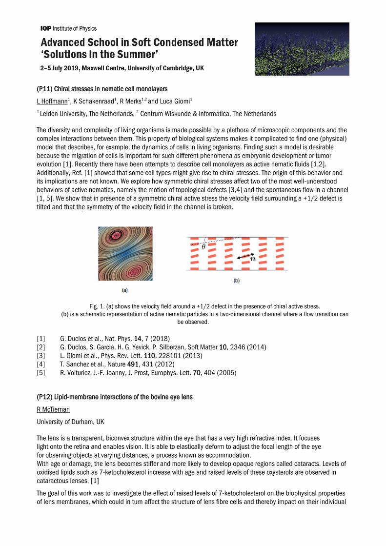
The diversity and complexity of living organisms is made possible by a plethora of microscopic components and the complex interactions between them. This property of biological systems makes it complicated to find one (physical) model that describes, for example, the dynamics of cells in living organisms. Finding such a model is desirable because the migration of cells is important for such different phenomena as embryonic development or tumor evolution [1]. Recently there have been attempts to describe cell monolayers as active nematic fluids [1,2]. Additionally, Ref. [1] showed that some cell types might give rise to chiral stresses. The origin of this behavior and its implications are not known. We explore how symmetric chiral stresses affect two of the most well-understood behaviors of active nematics, namely the motion of topological defects [3,4] and the spontaneous flow in a channel [1, 5]. We show that in presence of a symmetric chiral active stress the velocity field surrounding a +1/2 defect is tilted and that the symmetry of the velocity field in the channel is broken.
Fig. 1. (a) shows the velocity field around a +1/2 defect in the presence of chiral active stress. (b) is a schematic representation of active nematic particles in a two-dimensional channel where a flow transition can
be observed.
[1] G. Duclos et al., Nat. Phys. 14, 7 (2018) [2] G. Duclos, S. Garcia, H. G. Yevick, P. Silberzan, Soft Matter 10, 2346 (2014) [3] L. Giomi et al., Phys. Rev. Lett. 110, 228101 (2013) [4] T. Sanchez et al., Nature 491, 431 (2012) [5] R. Voituriez, J.-F. Joanny, J. Prost, Europhys. Lett. 70, 404 (2005)
(P12) Lipid-membrane interactions of the bovine eye lens
R McTiernan
University of Durham, UK
The lens is a transparent, biconvex structure within the eye that has a very high refractive index. It focuses light onto the retina and enables vision. It is able to elastically deform to adjust the focal length of the eye for observing objects at varying distances, a process known as accommodation. With age or damage, the lens becomes stiffer and more likely to develop opaque regions called cataracts. Levels of oxidised lipids such as 7-ketocholesterol increase with age and raised levels of these oxysterols are observed in cataractous lenses. [1]
The goal of this work was to investigate the effect of raised levels of 7-ketocholesterol on the biophysical properties of lens membranes, which could in turn affect the structure of lens fibre cells and thereby impact on their individual
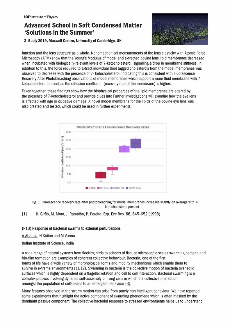
function and the lens structure as a whole. Nanomechanical measurements of the lens elasticity with Atomic Force Microscopy (AFM) show that the Young’s Modulus of model and extracted bovine lens lipid membranes decreased when incubated with biologically-relevant levels of 7-ketocholesterol, signalling a drop in membrane stiffness. In addition to this, the force required to extract individual thiol tagged cholesterols from the model membranes was observed to decrease with the presence of 7- ketocholesterol, indicating this is consistent with Fluorescence Recovery After Photobleaching observations of model membranes which support a more fluid membrane with 7- ketocholesterol present as the diffusion coefficient (recovery rate of the membrane) is higher.
Taken together, these findings show how the biophysical properties of the lipid membranes are altered by the presence of 7-ketocholesterol and provide clues into Further investigations will examine how the eye lens is affected with age or oxidative damage. A novel model membrane for the lipids of the bovine eye lens was also created and tested, which could be used in further experiments.
Fig. 1. Fluorescence recovery rate after photobleaching for model membranes increases slightly on average with 7-ketocholesterol present
[1] H. Girão, M. Mota, J. Ramalho, P. Pereira, Exp. Eye Res. 66, 645–652 (1998)
(P13) Response of bacterial swarms to external perturbations
A Abdulla, H Kotian and M Varma
Indian Institute of Science, India
A wide range of natural systems from flocking birds to schools of fish, at microscopic scales swarming bacteria and bio-film formation are examples of coherent collective behaviour. Bacteria, one of the first forms of life have a wide variety of morphological forms and motility mechanisms which enable them to survive in extreme environments [1], [2]. Swarming in bacteria is the collective motion of bacteria over solid surfaces which is highly dependent on a flagellar rotation and cell to cell interaction. Bacterial swarming is a complex process involving dynamic self-assembly of living cells in which the collective interaction amongst the population of cells leads to an emergent behaviour [3].
Many features observed in the swarm motion can arise from purely non intelligent behaviour. We have reported some experiments that highlight the active component of swarming phenomena which is often masked by the dominant passive component. The collective bacterial response to stressed environments helps us to understand

the active intelligent decision-making component of the bacterial system. Bacterial swarm was exposed to antibiotics and external force fields. These experiments elucidate the active component of bacterial swarm motion. Mutant strains of the bacteria lacking a particular trait were used in the swarming experiments to understand the collective motion better.
A mathematical model that can explain the behaviour of bacterial swarm motion in response to external perturbations have been developed based on the surfactant driven fluid flow model proposed by Sarah Trinschek et al. [4]. We aim to develop a comprehensive model that can encompass all the subtleties of this collective motion.
[1] C. R. Woese and G. E. Fox, Proc. Natl. Acad. Sci. USA, 74, 5088 (1977) [2] R. Glud et al., Nature Geoscience, 6, 284 (2013) [3] M. F. Copeland and D. B. Weibel, Soft Matter, 5, 1093 (2009) [4] S. Trinschek, K. John and U. Thiele, Soft Matter, 14, 4464 (2018)
(P14) Diffusion and active transport in porous media
P Zuk1,2 and S Datta2
1 Institute of Fundamental Technological Research Polish Academy of Sciences, Poland, 2Princeton University, USA
Diffusive and active transport in porous media are phenomena of great importance for living organisms on microscopic and mesoscopic length scales. In the micro scale the nearest neighborhood of the each cell in living organisms can be seen as porous medium through which molecules like nutrients or extracellular signaling proteins are transported. The mesoscopic scale example is soil, that contains abundant variety of bacteria. Here not only the diffusion but also active transport determines fate of whole populations of organisms. I would like to present a simulation framework built on the Generalized Rotne-PragerYamakawa approximation of hydrodynamic interactions that addresses diffusion and active transport in porous media and some obtained preliminary results.
(P15) Microscopic theory of dielectric and mechanical response in disordered materials
B Cui1 and A Zaccone1,2, 1University of Cambridge, UK, 2University of Milan, ItalyA framework called nonaffine lattice dynamics was recently developed [1, 2], which can quantitatively describe the macroscopic response (mechanical, dielectric) of disordered materials. This framework is able to predict the mechanical stability, elastic moduli and yielding failure in terms of the local bonding environment [3]. The mechanical response is an extremely sensitive marker of the internal bonding and mechanical integrity of amorphous materials. This principle could be exploited to develop an efficient non-destructive testing protocol for soft materials (a typical example being the polymer adhesives used by aircraft companies in their components). To make this approach applicable, we implement a systematic description of both the shear modulus and the dielectric function, with the vibrational density of states due to lattice vibrations being an input.
After the elastic constant is measured on a sample as a function of frequency, the data can be fitted to our theoretical model as in the figure below, where the imaginary part of the shear modulus for supercooled metallic glass right at the glass transition temperature Tg, when the material is marginally rigid, is compared. The shape of the curve, for a given material, can be directly related to the mechanical relaxation as a function of frequency, which provides unambiguous information about the mechanical integrity of the material.

Fig. 1. Comparison between our theory and the experimental data [4]
[1] A. Zaccone and E. Scossa-Romano, Phys. Rev. B 83, 184205 (2011) [2] A. Zaccone, J.R. Blundell and E.M. Terentjev, Phys. Rev. B 84, 174119 (2011) [3] (a) A. Zaccone and E.M. Terentjev, Phys. Rev. Lett. 110, 178002 (2013); (b) A. Zaccone, P. Schall and
E.M. Terentjev, Phys. Rev. B 90, 140203(R) (2014) [4] B. Cui, Z. Evenson, B. Fan, M. Li, W.-H. Wang and A. Zaccone, Phys. Rev. B 98, 144201 (2018)
(P16) Complex formulations drying on complex substrates
C Perez and Job H. J. Thijssen
University of Edinburgh, UK
Drying of droplets containing colloidal particles is critical to many industrial practices, such as inkjet printing, microelectronics and pesticide spraying on plant leaves. For the latter, the deposit pattern can affect the uptake through the leaf. In the literature, one can find many examples of strategies developed to suppress the coffee-ring effect. [1] However, little is known about how the surface texture affects the drying process and hence the dried deposit. Additionally, from a fundamental point of view, many more interesting questions remain unanswered, such as: how does the microstructure relate to the substrate properties; how the wetting properties vary with the deposit coverage; and what is the most adequate microstructure for a certain application. [2] Different roughness length-scales have been explored experimentally, ranging from the size of the colloidal particles (μm), to the order of magnitude of the drop size (mm). An in-house Python code has been developed for the image analysis of the dried deposits, defining a parameter that relates the roughness of the substrate to the dried pattern. Furthermore, the chosen substrates have been selected as they exhibit similar properties to those found in waxy leaves. This, along with exploring particle and droplet sizes comparable to those used for pesticide spraying, will help create a model system for crop care applications.
ACKNOWLEDGEMENTS: The authors thank the EPSRC Centre for Doctoral Training in Soft Matter and Functional Interfaces, grant number EP/L015536/1 and Dr. Marie-Capucine Pope from CRODA CropCare Market Sector for her support and enlightening discussions.
[1] D. Mampallil and H. B. Eral, Adv.Colloid and Interfac. 252, 38-54 (2018) [2] D. Quéré, Annu. Rev. Mater. Res. 38, 71-99 (2008)

(P17) Wavefront propagation speeds in a bacteriophage-bacteria system
R Claydon and A Brown
University of Edinburgh, UK
Wave fronts are realised in many biological situations, and indeed a large number of physical systems. An understanding of such front propagation has applications to predicting the invasion rates of viruses into a healthy population, or the escape speed of a healthy species from an infected area. There are many important applications of the use of bacteriophages (phages), viruses which attack bacteria, to curb the populations of pathogenic bacteria.
Given the serious threat of antibiotic resistance to the healthcare system in the future, phage therapies provide a potential solution. Understanding the spatial dynamics of the virus and bacteria will be vital information in the effort to control infections. To this end, a system of T4 bacteriophages invading a population of Escherichia coli in a 1D channel were modelled using a system of coupled partial differential equations. In contrast to previous studies, which have either omitted spatial dynamics or considered stationary bacteria with other coupled fields (i.e. nutrient or phage concentration) to diffuse, the dynamics in this system assumes stationary phages and motile bacteria. Essentially, the bacteria swim and are not prone to significant biofilm formation. Numerical investigation predicted wave fronts converging to constant spreading speeds in all cases considered. Intriguingly, the viral infection can invade the population of Escherichia coli in the absence of diffusion purely by commandeering the hosts.
Furthermore, there appears to have been very few examples in the literature of fronts converging to constant propagation speeds into states which are growing exponentially. It was unexpected that the bacteria concentration exhibited near self-similar solutions whereas the phage concentration did not. With applications to food waste reduction and wound dressing, the bacteria were then modelled as propagating in a constant background of bacteriophages. The dynamics were again found to converge to solutions with wave-fronts tending to an asymptotic speed. Surprisingly, it was found that wave fronts in this scenario were significantly slower than the wavefront speed of the phage invading an exponentially growing population of bacteria. The numerical simulations are supplemented by analytic predictions of the asymptotic front speed. The ability for bacteria to evolve phage resistance is an important consideration in making more realistic predictions and was incorporated into the model. In addition to this, 2D simulations were also performed to investigate spreading on surfaces.

(P18) Rheologically fingerprinted bacterial biofilms: the multiplicity of yielding states
S Charlton, S Jana, J Chen and T Curtis
Newcastle University, UK
Biofilms are a communal form of bacterial growth, which attach to and colonise surfaces. The defining feature of biofilm is the production of self-secreted extracellular polymeric substances (EPS), which encapsulate bacteria and confer protection from biological and physicochemical challenges. EPS is composed of polysaccharides, eDNA, lipids and proteins. The EPS/bacterial composite is a nonequilibrium system where spatial architecture is controlled by a combination of phenotypic responses and physicochemical interactions within the EPS. The diversity in genotype and environmental conditions results in architectural and rheological variability in different species of biofilms. Our work utilises nonlinear rheological techniques (LAOS) to quantify the yielding dynamics of biofilms and provide a standardised way of cataloguing biofilm structure-function relationships. We adopt the sequence of physical process’s (SPP) to fingerprint biofilms and demonstrate how these fingerprints can be interpreted. Firstly we reveal how structure function relationships in P. fluorescens biofilms can be modified using chemical treatment. We show that addition of divalent cations (CaCl2 and FeCl2) to P. fluorescens biofilm results in a transition in behaviour from a repulsive to attractive gel like state. Secondly we show how genetic mutation of capsular polysaccharide (CPS) production in Pantoea sp.controls packing and causes a transition from liquid to glassy rheology. Finally we fingerprint 4 different strains of bacteria: S. aureus (cocci, gram positive), N. polysaccharea (cocci, gram negative),P. fluorescens (rod, gram negative), and C. denitrificans (rod, gram positive) and reveal 4 different yielding characteristic mechanisms. We pose that a systematic approach to rheological fingerprinting of biofilm isolates would be beneficial towards the discovery of novel proteins and polysaccharides.
(P19) Correlated diffusion of colloidal particles in two-dimensional random confinement
M Bell-Davies1, M Mordan1, A Sonn-Segev1, R Dullens1 and Y Roichmann2 1University of Oxford, UK, 2Tel Aviv University, Israel
Correlated diffusion of tracer particles embedded in a quasi two-dimensional model porous medium made of a random matrix of large colloidal particles sandwiched between two glass plates is investigated. We use the two-point mean squared displacement (2P-MSD), a measure of the coupled motion of particle pairs. A range of matrix porosities were achieved by pressing suspensions of small and large colloids between two glass slides such that the large particles are stuck between the slides, forming the matrix, while the small particles are mobile. We find that at all matrix packing fractions the 2P-MSD has the same form as that of particles confined solely between two plates – namely it decays with the inter-particle distance r as 1/r2 . However, the magnitude of the hydrodynamic coupling decreases with increasing matrix packing fraction. In addition, there is anomalous time dependence at high matrix packing fractions, with sub-diffusive behaviour even at short lag times, which is related to a decrease in the mean pore size.
(P20) Interface properties of phase separated colloid-polymer mixtures
B Dohni, H Razak, L Verhoeff, Dirk G Aarts and R Dullens
University of Oxford, UK
Liquid-liquid interfaces in phase separated colloidal suspensions have an ultralow interfacial tension. We study aqueous colloid-polymer (PMMA-Xanthan) mixtures, where the presence of non-absorbing polymers induces an attraction between the colloidal particles via the depletion interaction. This leads to phase separation in a colloid-

poor (polymer-rich) “gas” phase and a colloid-rich (polymer-poor) “liquid” phase. In this system, thermal capillary waves at the interface can be directly visualised and studied with confocal scanning laser microscopy. We analyse the capillary fluctuations of the interface between the two phases, which gives access to interfacial properties such as the surface tension, capillary length and time. In addition, we use optical tweezers to actively manipulate the interface between the liquid and the gas phase. This allows the study of the deformation and relaxation of the interface.
(P21) Grain growth in impurity-doped two-dimensional hard sphere colloidal crystals
J Hutchinson1, F Lavergne2, D Aarts1 and R Dullens1
1University of Oxford, UK, 2University of Fribourg, Switzerland
Understanding how the microscopic structure of metals and alloys can be controlled is key for the fine tuning of material properties for specialised uses [1]. Through the use of colloidal crystals we study how dopants can control grain size and grain boundary properties. This is important for manipulating how stress microscopically propagates through the material and therefore macroscopic properties such as strength and stiffness [2].
Grains are domains of crystalline particles with similarly oriented neighbours. Growth occurs when particles realign to join the crystallite, as a means of minimising interfacial energy. Grain growth can occur in two regimes. The first is during crystallisation, where grains grow from nucleation sites and develop until reaching an impediment, such as a differently orientated crystallite. Secondly, when a polycrystalline state is reached, crystallites are directly adjacent and coarsening is induced by minimisation of grain boundary length. The addition of impurities to a monodisperse system can impede both regimes of growth, and as such, careful doping can be applied to alter the mechanical properties of materials.
To better understand how doping in alloys can be effectively utilised, we examine grain growth in impurity doped colloidal crystals. We achieve this by creating a polycrystalline monolayer of small hard spheres, with the addition of a larger dopant colloidal hard sphere. We use optical microscopy study the system's evolution from formation over thousands of Brownian times, and evaluate the effect of different concentrations of impurities. By resolving individual particle locations, we then analyse how dynamics and specific mechanisms behind the structural evolution of the system are affected by doping.
[1] G. De Faveri. Innovation in Materials Conference Proceedings: Materials tailor-made to solve the world's biggest problems. Royal Society of Engineering, 2014
[2] W. Sylwestrowicz and E. O. Hall. The deformation and ageing of mild steel. Proc. Phys. Soc. B, 64(6):495-502, 1951