potential effects of heavy metals bioaccumulation on
TRANSCRIPT
CATRINA (2020), 21(1): 75-82
© 2020 BY THE EGYPTIAN SOCIETY FOR ENVIRONMENTAL SCIENCES
______________________________________
* Corresponding author e-mail: [email protected]
Potential Effects of Heavy Metals Bioaccumulation on Oxidative Stress Enzymes of
Mediterranean clam Ruditapes decussatus
Gamal Abd El-Fattah Gabr1,2
, Mostafa Fathi Masood3,4
, Eman Hashem Radwan5,
Khalid Hashem Radwan2 and Aml Zaki Ghoenim
5
1 Department of Pharmacology and Toxicology, College of Pharmacy, Prince Sattam Bin Abdulaziz University, Al-Kharj, KSA
2 Agricultural Genetic Engineering Research Institute (AGERI), Agric. Res. Center, Giza, Egypt 3 Department of Zoology, Faculty of Science, Al Azhar University, Assiut, Egypt
4 Department of Biology, Faculty of Science, Jazan University, Kingdom of Saudi Arabia 5 Department of Zoology, Faculty of Science, Damanhour University, Egypt
ABSTRACT The bivalves have capability to accumulate the toxicant substances as heavy metals in their body tissues,
therefore, they might be used as a good bio-indicators of water contamination. The present work aimed to
measure the concentration of Cd, Cu, Pb, Mn, and Zn in the soft tissues of Ruditapes decussatus which
collected between December 2018 and February 2019 from two sites of Mediterranean Sea. One from
Alexandrian Port (Site I) and the other from Port Said (Site II), Egypt, as well as to estimate the potential
physiological change of the clam affected by these pollutants. Samples from Site I gives comparatively
higher water salinity and metals concentration in soft tissues. The statistical analysis shows significant
increase in the level of malondialdehyde (MDA) while superoxide dismutase (SOD) and glutathione
peroxidase were found decreased in the R. decussatus soft tissue collected from Site I. The correlation
coefficient of physicochemical parameters, heavy metals and oxidative stress biomarkers in Site I shows that
glutathione peroxidase and superoxide dismutase have positive correlation with acetylcholinesterase
(r=0.912) and (r=0.929), respectively. SOD, on the other hand, was having negative correlation with MDA
(r= -0.886). The reported values in this study are considered as basic data in monitor of the anthropogenic
activities in future along the coast, as well as it is starter point in assessment of pollution that maybe effect on
the aquatic organisms in the Mediterranean marine environment.
Keywords: Bivalves, Environment, Heavy metals, Oxidative stress, Protein.
INTRODUCTION
The human activities lead to increase of the
environmental pollutants that can penetrate and
accumulate in the aquatic organisms, thus effecting
their reproduction and health (Andreia et al., 2019).
These pollutants including, the different forms of
released chemicals from industrial works, xenobiotic
and pesticides which might be increase the oxidative
stress through the reactive oxygen species (ROS)
yielding (Zhang et al., 2016; Wu et al., 2017). The
overproduction of ROS caused by toxic heavy metals is
considered the main problem as they might lead to
changing in the activities of antioxidants enzymes or
induce genotoxic effects in the aquatic flora (Nordberg
and Arner, 2001). The important roles of the oxidative
stress inside the aquatic organism bodies are they have
a potential biomarker of the environmental pollutions
(Verdelhos et al., 2015; Murtala et al., 2012). In order
to understand the heavy metals toxic effects at the
molecular and cellular levels in the aquatic organisms,
the multiple antioxidant enzymes such as superoxide
dismutase, glutathione peroxidase and catalase should
be determining their activities (Bakhtawar et al., 2016),
in addition to, the genotoxicity, DNA damage and
immunotoxicity also required to be identified (Almeida
et al., 2007; Marisa et al., 2016). Similarly, the heavy
metals can be accumulated inside the nervous system
leading to necrosis and degeneration effects on the
marine aquatic species (Gao et al., 2012; Mannan et
al., 2018).
Aquatic biota has been utilized as bio-indicators of
environmental pollution due to their capability of
pollutants accumulation when exposed to high
concentrations of these contaminants such as heavy
metals, the different organs of aquatic organisms may
accumulate some amounts of these heavy metals
(Campillo et al., 2013). The aquatic filter feeders such
as clams, oysters and mussels due to their tolerance
against pollutants are expected to use as bio-indicators
and marine biomonitoring (Sayka and Vladimir, 2019).
Due to the release of large quantity of untreated sewage
and wastewater every year into Alexandria beachfront
region through nearby sewage framework (El Khodary
et al., 2018), the biological organisms such as R.
decussatus which available in the area under study can
be used as bio-indicator for detection the polluted area
with high concentration levels of different heavy
metals and other toxicants (Abd El Ghany, 2017;
Hazrat et al., 2019). The main target of the current
study is to investigate the concentrations of Cd, Cu, Pb,
Mn, and Zn in R. decussatus tissues and assess its
effects on the biomarkers and antioxidants enzymes.
MATERIALS AND METHOD
Study areas and sample collection
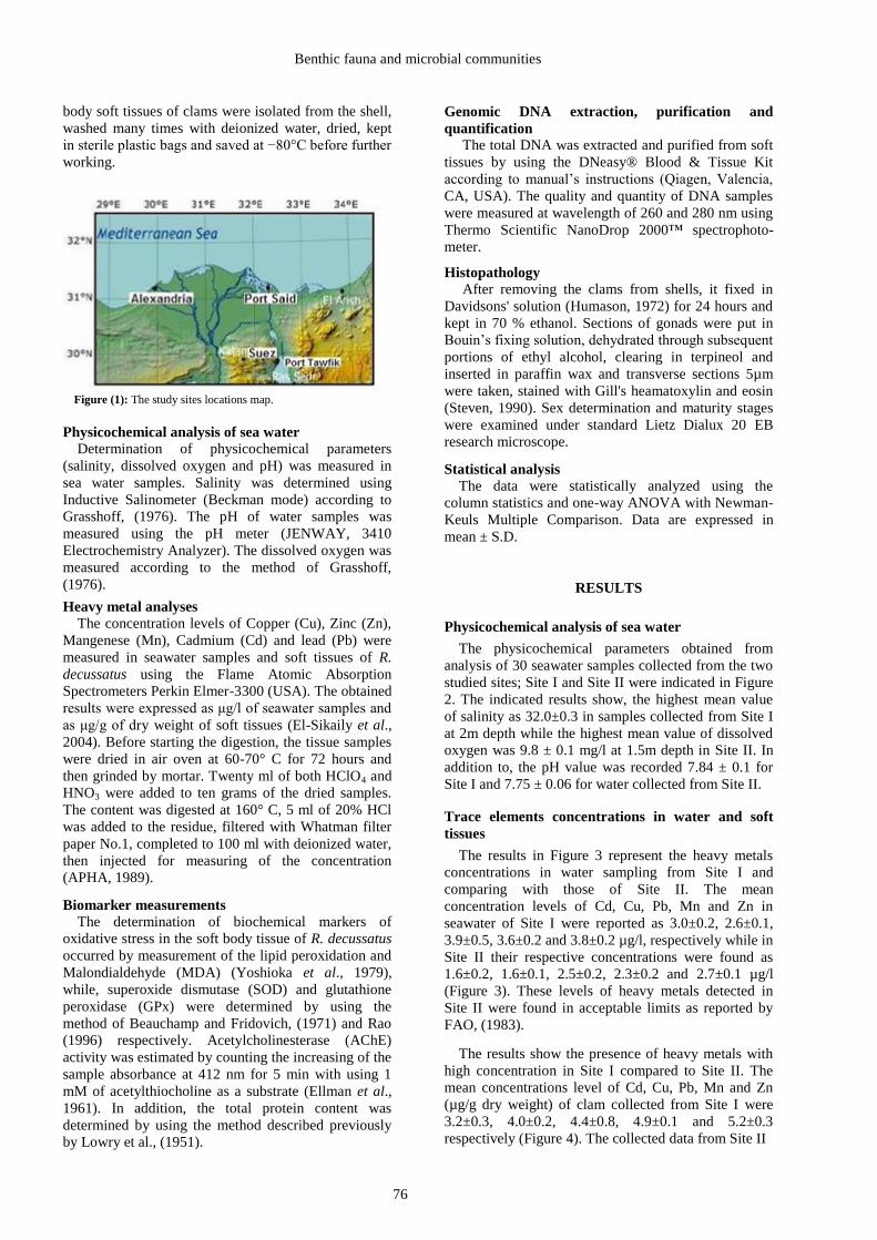
The present investigation was carried out at two
sites in the Mediterranean Sea, Egypt. The first site is
Alexandria Port (Site I) and the second one is Port Said
(Site II). A total of 100 adult R. decussatus clam and 30
sea water samples were collected from each site
between December 2018 and February 2019 from
definite depth consistent to the clam habitation from
each location, while, dead or damaged R. decussatus
clam samples were excluded and only those with
identical shell size (3.5-3.8mm) were used. Thereafter,
it transported immediately in ice to the laboratory. The
Benthic fauna and microbial communities
body soft tissues of clams were isolated from the shell,
washed many times with deionized water, dried, kept
in sterile plastic bags and saved at −80°C before further
working.
Figure (1): The study sites locations map.
Physicochemical analysis of sea water
Determination of physicochemical parameters
(salinity, dissolved oxygen and pH) was measured in
sea water samples. Salinity was determined using
Inductive Salinometer (Beckman mode) according to
Grasshoff, (1976). The pH of water samples was
measured using the pH meter (JENWAY, 3410
Electrochemistry Analyzer). The dissolved oxygen was
measured according to the method of Grasshoff,
(1976).
Heavy metal analyses
The concentration levels of Copper (Cu), Zinc (Zn),
Mangenese (Mn), Cadmium (Cd) and lead (Pb) were
measured in seawater samples and soft tissues of R.
decussatus using the Flame Atomic Absorption
Spectrometers Perkin Elmer-3300 (USA). The obtained
results were expressed as μg/l of seawater samples and
as μg/g of dry weight of soft tissues (El-Sikaily et al.,
2004). Before starting the digestion, the tissue samples
were dried in air oven at 60-70° C for 72 hours and
then grinded by mortar. Twenty ml of both HClO4 and
HNO3 were added to ten grams of the dried samples.
The content was digested at 160° C, 5 ml of 20% HCl
was added to the residue, filtered with Whatman filter
paper No.1, completed to 100 ml with deionized water,
then injected for measuring of the concentration
(APHA, 1989).
Biomarker measurements
The determination of biochemical markers of
oxidative stress in the soft body tissue of R. decussatus
occurred by measurement of the lipid peroxidation and
Malondialdehyde (MDA) (Yoshioka et al., 1979),
while, superoxide dismutase (SOD) and glutathione
peroxidase (GPx) were determined by using the
method of Beauchamp and Fridovich, (1971) and Rao
(1996) respectively. Acetylcholinesterase (AChE)
activity was estimated by counting the increasing of the
sample absorbance at 412 nm for 5 min with using 1
mM of acetylthiocholine as a substrate (Ellman et al.,
1961). In addition, the total protein content was determined by using the method described previously
by Lowry et al., (1951).
Genomic DNA extraction, purification and
quantification
The total DNA was extracted and purified from soft
tissues by using the DNeasy® Blood & Tissue Kit
according to manual’s instructions (Qiagen, Valencia,
CA, USA). The quality and quantity of DNA samples
were measured at wavelength of 260 and 280 nm using
Thermo Scientific NanoDrop 2000™ spectrophoto-
meter.
Histopathology
After removing the clams from shells, it fixed in
Davidsons' solution (Humason, 1972) for 24 hours and
kept in 70 % ethanol. Sections of gonads were put in
Bouin’s fixing solution, dehydrated through subsequent
portions of ethyl alcohol, clearing in terpineol and
inserted in paraffin wax and transverse sections 5µm
were taken, stained with Gill's heamatoxylin and eosin
(Steven, 1990). Sex determination and maturity stages
were examined under standard Lietz Dialux 20 EB
research microscope.
Statistical analysis
The data were statistically analyzed using the
column statistics and one-way ANOVA with Newman-
Keuls Multiple Comparison. Data are expressed in
mean ± S.D.
RESULTS
Physicochemical analysis of sea water
The physicochemical parameters obtained from
analysis of 30 seawater samples collected from the two
studied sites; Site I and Site II were indicated in Figure
2. The indicated results show, the highest mean value
of salinity as 32.0±0.3 in samples collected from Site I
at 2m depth while the highest mean value of dissolved
oxygen was 9.8 ± 0.1 mg/l at 1.5m depth in Site II. In
addition to, the pH value was recorded 7.84 ± 0.1 for
Site I and 7.75 ± 0.06 for water collected from Site II.
Trace elements concentrations in water and soft
tissues
The results in Figure 3 represent the heavy metals
concentrations in water sampling from Site I and
comparing with those of Site II. The mean
concentration levels of Cd, Cu, Pb, Mn and Zn in
seawater of Site I were reported as 3.0±0.2, 2.6±0.1,
3.9±0.5, 3.6±0.2 and 3.8±0.2 µg/l, respectively while in
Site II their respective concentrations were found as
1.6±0.2, 1.6±0.1, 2.5±0.2, 2.3±0.2 and 2.7±0.1 µg/l
(Figure 3). These levels of heavy metals detected in
Site II were found in acceptable limits as reported by
FAO, (1983).
The results show the presence of heavy metals with
high concentration in Site I compared to Site II. The
mean concentrations level of Cd, Cu, Pb, Mn and Zn
(µg/g dry weight) of clam collected from Site I were
3.2±0.3, 4.0±0.2, 4.4±0.8, 4.9±0.1 and 5.2±0.3
respectively (Figure 4). The collected data from Site II
76

Gabr et al.
Figure (2): The concentration level of physicochemical characteristics of water samples collected from the two sites.
Data expressed in mean ±SE. **, statistically significant at p ≤0.05; ***, statistically significant at p ≤ 0.001.
Figure (3): Heavy metal concentration (μg/ml) in sea water of the
two studied sites, Alexandria and Port Said harbors. Data are represented in mean ±SD. *, statistically significant at p ≤0.05;
***, statistically significant at p ≤ 0.001.
indicated the heavy metals in clam tissue were 1.6±0.5,
2.6±0.4, 2.6±0.5, 2.6±0.3 and 3.8±0.8 respectively. The
present result reported that the clam collected from Site
I had more heavy metals accumulation in clam tissues
than that collected from Site II.
Biochemical biomarkers responses
In the current study, the levels of MDA in tissues
collected from Site I and Site II reported as 12.9±0.3
U/g and 9.7±0.2 U/g, respectively and this indicates the
clams in the Site I were under much stress of pollution
than those of Site II. The mean activity values of GPx
in Site I was 10.6±0.3 U/g and that of Site II was
11.6±0.3 U/g. The mean activity values of SOD in Site
I was 1.8±0.1 U/g whereas, that of the other location
was 1.1±0.0 U/g. The total protein in Site I was 5.5±0.6
mg/g while that of Site II was 6.5±0.2 mg/g. Moreover,
the AchE in Site I reported as 446.2±1.9 U/l where that
of Site II reported as 468.0±1.9 U/l (Table 1).
The RNA /DNA in the Site I was reported as 4.5±0.2
and that of Site II was as 2.9 ± 0.1. The data show significant difference between the sites under
investigation at p<0.001.
Figure (4): Mean and standard deviation (Mean± SD) of heavy
metals in water (μg/l) in the two sites. Data was expressed by using mean ± S.E. Statistically significant at ***p
≤ 0.001, nsp ≥ 0.05.
Histopathology
The parasites were infected the digestive glands
causing hypertrophy and necrosis of the host cells
(Figure 5 A & B). It infected also, the stomach
epithelial cells of the clam as cleared in Figure (5 D &
F). The infected cells are lysed the lumen of the
digestive tubules and a wide spread evidence of
necrosis of the digestive tissues with destruction of
large regions of the digestive tissues (Figure 5 B). In
the healthy clam tissue, the nuclei were basically
located and the individual cells were easily
distinguishable (Figure 5 C & E).
77

Benthic fauna and microbial communities
Table (1): Antioxidant activity and damage biomarkers in Clam Ruditapes. Glutathione peroxidase (GPx) and
superoxide dismutase (SOD) activities, Total protein content, Acetylcholinesterase (AchE), RNA/DNA and
lipid peroxidation (malondialdhyde, MDA level) of Ruditapes decussatus collected from the both sites.
Biochemical Parameters†
Studied Sites GPx (U/g) SOD (U/g)
Total protein (mg/g)
AchE (U/l) MDA (U/g)
Alexandria Harbor (Site I) 10.6±0.3 1.8±0.1 5.5±0.6 446.2±1.9 12.9±0.3
Port Said harbor (Site II) 11.6±0.3 1.1±0.0 6.5±0.2 468.0±1.9 9.7±0.2
P* 0.001* <0.001* 0.008* <0.001* <0.001*
t-test 5.315* 11.501* 3.532* 18.167* 20.032*
† Data expressed in mean ± SE. *statistically significant at p ≤ 0.05.
Figure (5): Photomicrograph of transverse section through the digestive gland (A, B and C) and testes (D, E and F) of
Ruditapes decussatus, (A), normal uninfected digestive gland (NC); (B), mild infected digestive gland with cercaria larvae (arrows); C, heavy infections by cercaria larva causing cell autolysis (arrow); D, normal tissue of testes of the clam; E and
F, mild to sever infection that showing the presence of trematode sporocyst with germ cells of different types of sporocyst
containing cercariae (blue arrows) and sever infection causes cell autolysis (yellow arrows).
78

Gabr et al.
DISCUSSION
The heavy metals maybe accumulated in the aquatic
environments comes from different sources such as
urban overflow, stations of sewage treatments,
industrial chemical wastes, mining industries, ship
movement, local waste junkyards and agricultural
pesticides overflow (Alemdaroglu et al., 2003). Earlier
studies demonstrated that, the increasing of pollution
sources maybe increase the heavy metal accumulation
by aquatic organisms and fishery products
(Chandrasekhar et al., 2004; Zhang et al., 2007).
The current results indicated, the increasing level
concentrations of Pb, Cd, Zn, Mn and Cu in seawater
and soft tissues of Site I were higher than Site II and
this may be due to increasing in the contamination
resources, which mainly from the industrial effluents,
domestic, sewage effluents and wastewater, that effect
directly or indirectly on the physiological functions of
aquatic organisms and human being health (Arafa and
Ali, 2008). Early studies reported that, the heavy
metals are entering into water effluents through
industrial, agricultural and municipal wastes and
affects the rate of reproductive system of aquatic
organisms leading to a gradual extinction of their
generations; also, disturb their physiological functions
(Shenai et al., 2017; García-Medina et al., 2017).
The toxic of heavy metals effect at both of cellular
and molecular levels were identified through
determination the antioxidant biomarkers in the living
organisms (Bejaoui et al., 2018). The obtained results
show a significant increase in MDA concentration in
clam soft tissues collected from Site I compared to that
of Site II. The heavy metals may lead to increasing of
the ROS production and causing the oxidative damage
through inhibition of the antioxidant enzymes activity
(Dayem et al., 2017).
One study reported that, lead positively correlated
with the MDA levels and the increasing damage of the
cell membrane and subsequently enhances the
oxidative stress (Chalkiadaki et al., 2015). In addition,
the cell damage exhibited positive correlation with the
antioxidant enzymes and toxic substances leakage and
it frequently accompanied by increasing of the cell
membrane permeability (Fernandez et al., 2010). The
obtained results show high significant decreasing in the
total protein and GPx activity of clam collected from
Site I compared to those of Site II and this result was in
agreement with Parate and Kulkarnim, (2003) who
found, at protein content decreasing, this might be due
to the proteins degraded into free amino acids which
converted to α keto acid and participate the tri-
carboxylic acid cycle for ATP production.
Furthermore, the decreasing in the total proteins maybe
reflect also the decreasing in the oxidative enzymes
activity. The exposure to heavy metals was previously
reported to decrease the activity of GPx (Maria and
Bebianno, 2011) and reduce the capacity to scavenge
H2O2 (Saidani et al., 2019). The contamination of
water sources by genotoxic compounds is a worldwide
problem (Sahayanathan et al., 2018). The probability
of using changes in DNA integrity as markers of
exposure effects of Geno-toxicants materials has been
previously estimated in different aquatic organisms
using cytogenetic analysis (Letelier et al., 2005).
The poisonous metals are equipped for aggravating
the regular oxidation lessening balance in the cells
through different components originating from their
own particular complex redox responses with
endogenous oxidants and consequences for cell cancer
prevention agent frameworks (Wiem et al., 2019). The
subsequent oxidative stress causes DNA harm which
may add to metals danger (Tlili et al., 2010).
The results of the current research indicate the
samples of Site I represented 5% of the samples were
parasitized whereas, Site II samples represented only
3% of the samples were parasitized. Environmental
stress such as parasites and heavy metal pollutants may
have negative effects on the physiological properties of
the host organisms, leading to high response to diseases
causing agents (Radwan et al., 2018).
The data of correlations coefficient in Site I
concluded that, the dissolved oxygen showed highly
negative correlation with Zn, as r=-0.881 and presence
of a positive correlation between Zn and Pb in sea
water samples expressed as r=0.891. While, Zn was
also positively correlated with Pb in clam tissue and
expressed as r =0.975. In addition to, Pb is negatively
correlated with AchE as r =0.950 and Zn has positive
correlation with RNA/DNA as r =0.905. Finally, Cd is
negatively correlated with AchE as r =0962. While, the
data of correlations coefficient in Site II show there
was high positive correlations among S% and DO as r
=0.910. In addition, there was a high negative
correlation between Cu and SOD as r=-0.965. Cu has
positive correlation with MDA as r= 0.934, whereas,
Cd is positively correlated with Cd in the tissue of the
clam as r=0.962. Mn is negatively correlated with Zn in
the tissues of the clam as r =0.953. GPx has positive
correlation with AchE activity as r =0.912 and SOD is
also positively correlated with AchE as r=0.929.
Finally, SOD is negatively correlated with MDA as r=-
0.886.
CONCLUSION
The current study was evaluated the toxic effects of
the heavy metals on R. decussatus. It reduces and
destroys the oxidative stress enzymes of R. decussatus
soft tissues. Additionally, there are different positive
correlation between the oxidative enzymes activity and
the inhibition of AChE activity in the soft tissues.
Finally, the current results are considered as the
baseline data in the biomonitoring of the pollutants
along the Egyptian coast and we need more research
studies for complete the assessment process of
pollution effectors on the aquatic organisms.
ACKNOWLEDGMENT
The authors would like to express their thanks to
college of pharmacy at Prince Sattam Bin Abdulaziz
79
Benthic fauna and microbial communities
University for providing the necessary facilities to
precede this research work.
REFERENCES
ABD EL GHANY, S., 2017. Heavy metal
bioaccumulation in the edible bivalve Venerupis
decussates collected from Port Said, Egypt.
Wulfenia Journal, 24, 54-55.
ALEMDAROGLU T., E. ONUR AND F. ERKAKAN.
2003. Trace metal levels in surface sediments of
lake Manyas, Turkey and Tributary River.
International Journal of Environmental Studies, 60,
287-298.
ALMEIDA, E. A., A. C. DIAS BAINY, A. P. DE
MELO LOUREIRO, G. R. GLAUCIA REGINA
MARTINEZ, S. MIYAMOTO, J. ONUKI, L. F.
BARBOSA, C. C. MACHADO GARCIA, F. M.
PRADO, G. E. RONSEIN, C. A. SIGOLO, C. B.
BROCHINI, A. G. MARTINS, M. G. DE
MEDEIROS AND P. MASCIO. 2007. “Oxidative
stress in Perna perna and other bivalves as
indicators of environmental stress in the Brazilian
marine environment: Antioxidants, lipid
peroxidation and DNA damage,” Comp. Biochem.
Physiol. Part A: Mol & Integ. Physiol. 146, 588-
600.
ANDREIA, F. M., M. SÉRGIO, J. C. MARQUES, F.
J. MARQUES, M. GONÇALVES AND M. ANA.
2019. Copper sulphate impact on the antioxidant
defense system of the marine bivalves
Cerastoderma edule and Scrobicularia plana.
Scientific Reports, 9:16458.
APHA, 1989. Standard Methods for the Examination
of Water and Wastewater. 17th edition. American
Public Health Association, Washington D.C, 1-268.
ARAFA, M.M. AND A.T. ALI. 2008. Effect of some
heavy metals pollution in Lake Mariout on
(Oreochromis niloticus) fish. Egypt. J. Comp. Path
and Clin. Path., 21(3), 191 – 201.
BAKHTAWAR, R., J. MUHAMMAD, A. FAIZA
AND L. FARIHA. 2016. Toxic Effect of Lead
Chloride on Antioxidant Enzyme in the Liver and
Kidney of Fish. J. Bioresource Manage. 3, 1-8.
BEAUCHAMP, C. AND I. FRIDOVICH. 1971.
Superoxide dismutase: improved assays and an
assay applicable to acrylamide gels. Analytical
Biochemistry, 44, 276–87.
BEJAOUI, S. K., I. TELAHIGUE, I. CHETOUI, C.
RABEH, W. FOUZAI, I. TRABELSI, M. HOUAS-
GHARSALLAH AND N. SOUDANI. 2018.
“Integrated Effect of Metal Accumulation,
Oxidative Stress Responses and DNA Damage in
Venerupis Decussata Gills Collected from Two
Coast Tunisian Lagoons,” J. E. C. E, 2, 44-51.
CAMPILLO, J.A., M. ALBENTOSA, N.J. VALDÉS,
R. MORENO-GONZÁLEZ AND V. M. LEÓN.
2013. Impact assessment of agricultural inputs into
a Mediterranean coastal lagoon (Mar Menor, SE
Spain) on transplanted clams (Ruditapes
decussatus) by biochemical and physiological
responses. Aquat. Toxicol. 142–143, 365–379.
CHALKIADAKI, O., M. DASSENAKIS, N.
LYDAKIS–SIÙANTIRIS AND M. SCOULLOS.
2015. Tissue specific, time and dose dependence
impact of Lead to a commercial marine
Mediterranean organism, Lesvos Island. Greece:
11th Panhellenic Symposium on Oceanography and
Fisheries, Mytilene, 2015.
CHANDRASEKHAR K., N.S. CHARY, C.T.
KAMALA, D.S.S. RAJ AND A.S. RAO. 2004.
Fractionation studies and bioaccumulation of
sediment bound heavy metals Kolleru Lake by
edible fish. Environment International, 29(7), 1001-
1008.
DAYEM, A. A., M. K. HOSSAIN, S. B. LEE, K. KIM,
S. K. SAHA, G. M. YANG, H. Y. CHOI AND S.
G. CHO. 2017. “The Role of Reactive Oxygen
Species (ROS) in the Biological Activities of
Metallic Nanoparticles,” Int. J. Mol. Sci. 18, 120.
EL KHODARY, G.M., E.H. RADWAN, M.M. EL
GHAZALY AND D. EL BAHNASAWY. 2018.
Marine pollution by some heavy metals and
physiological response of Ruditapes decussatus.
Journal of Bioscience and Applied Research, 4,
199-217.
ELLMAN, G.L., K.D. COURTNEY, V. ANDRES,
R.M. FEATHERSTONE. 1961. A new and rapid
colorimetric determination of acetylcholinesterase
activity. Biochem. Pharmacol. 7 (2), 88–95.
EL-SIKAILY, A., A. KHALED AND A. EL-NEMR.
2004. Heavy metals monitoring using bivalves from
Mediterranean Sea and Red Sea. Environmental
Monitoring and Assessment 98, 41–58.
FAO. 1983. Food and Agriculture Organization
Compilation of legal limits for hazardous
substances in fish and fishery products”, Fish Circ,
464, 5-100.
FERNANDEZ, B., J. A. CAMPILLO, C. MARTINEZ-
GÓMEZ AND J. BENEDICTO. 2010.
“Antioxidant responses in gills of mussel (Mytilus
galloprovincialis) as biomarkers of environmental
stress along the Spanish Mediterranean coast”
Aquat. Toxicol, 99, 186–197.
GAO, X., C. HE, H. LIU, H. LI, D. ZHU, S. CAI, Y.
XIA, Y. WANG AND Z. YU. 2012. Intracellular
Cu/ Zn superoxide dismutase (Cu/Zn-SOD) from
hard clam Meretrix meretrix: its cDNA cloning,
mRNA expression and enzyme activity. Mol. Biol.
Rep. 39, 10713–10722.
GARCÍA-MEDINA, S., M. GALAR-MARTÍNEZ, L.
M. GÓMEZ-OLIVÁN, K. RUIZ-LARA, H.
ISLAS-FLORES, E. GASCA-PÉREZA. 2017.
“Relationship between genotoxicity and oxidative
stress induced by mercury on common carp
(Cyprinus carpio) tissues,” Aqua. Toxicol, 192,
207–215.
GRASSHOFF, K. 1976. Methods of sea water analysis.
Verlag. Chemie. Chapter 4; Determination of
oxygen.
HAZRAT, A., K. EZZAT AND I. IKRAM. 2019.
Environmental Chemistry and Ecotoxicology of
Hazardous Heavy Metals: Environmental
Persistence, Toxicity and Bioaccumulation. Journal
80

Gabr et al.
of Chemistry Volume 2019, Article ID 6730305, 14
pages.
HUMASON, G.L. 1972. Animal tissue techniques.
WH Freeman & Company, San Francisco, CA.
LETELIER, M. E., ANA MARÍA L., MARIO, F.,
JULIA, S., RIGOBERTO, M., HERNÁN, S., 2005.
Possible mechanisms underlying copper-induced
damage in biological membranes leading to cellular
toxicity. Chem. Biol. Interactions 151, 71–82.
LOWRY, O.H., N.J. ROSEBROUGH, A. L. FARR, R.
J. RANDALL. 1951. Protein Measurement with the
Folin Phenol Reagent. Journal of Biological
Chemistry, 193, 265–275.
MANNAN, M. A., M. S. HOSSAIN, M. A. A.
SARKER, M. M. HOSSAIN AND L. CHANDRA.
2018. Bioaccumulation of Toxic Heavy Metals in
Fish after Feeding with Synthetic Feed: A Potential
Health Risk in Bangladesh. J Nutr Food Sci. 8, 728.
MARIA, V. L. AND M. J. BEBIANNO. 2011.
Antioxidant and lipid peroxidation responses in
Mytilus galloprovincialis exposed to mixtures of
benzo(a)pyrene and copper. Comp. Biochem.
Physiol. C. 154, 56–63.
MARISA, I., V. MATOZZO, M. MUNARI, A.
BINELLI, M. PAROLINI, A. MARTUCCI, E.
FRANCESCHINIS, N. BRIANESE AND M.G.
MARIN. 2016. In vivo exposure of the marine clam
Ruditapes philippinarum to zinc oxide
nanoparticles: responses in gills, digestive gland
and haemolymph. Environ. Sci. Pollut. Res. 23
(15), 15275–15293.
MURTALA, B. A., P.H.D. ABDUL, A. ADEOLU
AND P.H.D. AKINYEMI. 2012. Bioaccumulation
of Heavy Metals in Fish (Hydrocynus forskahlii,
Hyperopisus bebe occidentalis and Clarias
gariepinus) Organs in Downstream Ogun Coastal
Water, Nigeria. Journal of Agricultural Science. 4,
51-59.
NORDBERG, J. AND E.S.J. ARNER. 2001. Reactive
oxygen species, antioxidants, and the mammalian
thioredoxin system. Free Radic. Biol. Med. 31,
1287–1312.
PARATE S.K. AND K.M. KULKARNI. 2003. Toxic
influence on the total protein content in the mussels
and gills of the freshwater crab, Paratephusa
jacqimontii exposed to cypermethrin. Aquatic
Biology, 18 (1):111-113.
RADWAN, E. H., A. A. HASSAN, G. H. FAHMY, S.
S. EL SHEWEMI AND S. SALAM. 2018. Impact
of environmental pollutants and parasites on the
ultrastructure of the Nile bolti, Oreochromis auruis.
J of Biosciences and applied Research, 4, 58-83.
RAO, G.S. 1996. Glutathionyl hydroquinone: a potent
pro-oxidant and a possible toxic metabolite of
benzene. Toxicology, 106(1–3), 49–54.
SAHAYANATHAN, G. J., G. SHREYOSHI, C.
ARULVASU. 2018. Antiproliferative Effect of
Crude Proteins Extracted from Marine Clam Donax
Variables on Human Cancer Cell Lines. IJPSR, 9,
3180-3188.
SAIDANI, W., B. SELLAMI, A. KHAZRI, A.
MEZNI, M. DELLALI, O. JOUBERT, D.
SHEEHAN AND H. BEYREM. 2019. Metal
accumulation, biochemical and behavioral
responses on the Mediterranean clams Ruditapes
decussatus exposed to two photocatalyst
nanocomposites (TiO2 NPs and AuTiO2NPs).
Aquat Toxicol. 208, 71–79.
SAYKA, J. AND S. VLADIMIR. 2019. Assessment of
trace elements pollution in the sea ports of New
South Wales (NSW), Australia using oysters as
bioindicators. Scientific Reports. 9, 1416.
SHENAI, P., M. U. TIRODKAR AND M. W. A.
GAUNS. 2017. “Antioxidant responses in gills and
digestive gland of oyster Crassostrea madrasensis
(Preston) under lead exposure.,” Ecotoxicology
Environmental Safety, 142, 87-94.
STEVENS, A. 1990. The haematoxylins. In: Bancroft
JD, Stevens A (eds) Theory and Practice of
histological technique` s. Churchill Livingstone,
New York, p 107–118.
TLILI, S., I. MÉTAIS, H. BOUSSETTA AND C.
MOUNEYARC. 2010. Linking changes at sub-
individual and population levels in Donax
trunculus: assessment of marine stress.
Chemosphere. 81, 692–700.
VERDELHOS, T., J. C. MARQUES AND P.
ANASTÁCIO. 2015. The impact of estuarine
salinity changes on the bivalves Scrobicularia plana
and Cerastoderma edule, illustrated by behavioral
and mortality responses on a laboratory assay. Ecol.
Ind. 52, 96–104,
WIEM, S., S. BADREDDINE, K. ABDELHAFIDH,
M. AMINE, D. MOUHAMED, J. OLIVIER, S.
DAVID AND B. HAMOUDA. 2019. Metal
accumulation, biochemical and behavioral
responses on the Mediterranean clams Ruditapes
decussatus exposed to two photocatalyst
nanocomposites (TiO2 NPs and AuTiO2NPs).
Aquatic Toxicology 208, 71–79.
WU, H., Y. ZHANG, X. SHI, J. ZHANG AND E. MA.
2017. Overexpression of Mn-superoxide dismutase
in Oxya chinensis mediates increased malathion
tolerance. Chemosphere 181, 352–359.
YOSHIOKA, T., K. KAWADA AND T. SHIMADA.
1979. Lipid peroxidation in maternal and cord
blood and protective mechanisms against activated
oxygen toxicity in the blood. Am J Obster Gynecol.
135, 372–376.
ZHANG I. AND M.H. WONG. 2007. Environmental
mercury contamination in China: Sources and
impacts. Environmental International, 33, 108-121.
ZHANG, H.C., C.Y. SHI, L.Q. SUN, F. WANG, G.W.
CHEN. 2016. Toxic effects of ionic liquid 1- octyl-
3-methylimidazolium bromide on the antioxidant
defense system of freshwater planarian, Dugesia
japonica. Toxicol. Ind. Health 32, 1675–1683.
81

Benthic fauna and microbial communities
مه الىوع للمعادن الثقلة على إوزمات الإجهاد التأكسذي لمحار البحر الأبض المتوسط الحويالتأثرات المحتملة للتراكم
Ruditapes decussatus
خبش ذخبل عبذ انفخب
2،3 يسعد يصطفى فخح ،
4،5، اب بشى سظا
6، خبنذ بشى سظا
3 ايم صك غى
6
2 ، انخشج، انهكت انعشبت انسعدتضقسى عهى الادت انسو، كهت انصذنت، خبيعت الأيش سطبو ب عبذ انعض
3، يشكض انبحد انضساعت، اندضة، يصشيعذ بحد انذست انساثت انضساعت
4قسى عهى انحا، كهت انعهو، خبيعت الاصش، أسغ، يصش
5قسى انبنخ، كهت انعهو، خبيعت خبصا، انهكت انعشبت انسعدت
6 قسى عهى انحا، كهت انعهو، خبيعت ديس، يصش
الملخص العرب
ك نزنكثقهت ف أسدت أخسبيب، ان هثبث يثم انعبدانببنقذسة عهى حدع اناد انسبيت يثم انحبسانبحشت تشخان بثحاانحخخع
بعط انعبصش انثقهت يثم انضك، اندض، انشصبص، إنى قبط حشكضز انذساست جذفنزنك . اسخخذايب كؤششاث حت خذة نخهد انب
ي يقع ف انبحش 3122فبشاش 3122ب دسبش ف انفخشة بحدعانخ حى R. decussatus حبسف الأسدت انشخة نـ انحبط انكبديو
ببلإظبفت انى حقذش انخغشاث انفسنخت انحخهت لأسدت انحبس خدت ،(، يصش3( بسسعذ )انكب 2ب الإسكذست )انكب الأبط انخسػ
انطقت الأنى. حأثش بخهك انهثبث. اثبخج انخبئح ا يهحت انبء حشكضاث انعبصش انثقهت ف اسدت انحبس كبج أعهى ف انعبث انأخرة ي
اضى اضى انسبش اكسذ دسحضبب اخفعج يسخبث كم ي انبنذذف حشكض كزنك أظحج انخحهلاث الإحصبئت صبدة يعت
، انعبصش انثقهت يعبيم الاسحببغ نهعهبث انفضبئت انكبئتاندهحبث بشكسذض ف اسدت انحبس ف انطقت الأنى. كزنك عذ قبط كم ي
اضى اندهحبث انسبش اكسذ دسحضاضى أظشث انخبئح ا ف انطقت الأنى أعطى كم ي كب كسذة، انؤششاث انحت نعبداث الا
اضى عهى انخان. عهى اندبب الاخش اعطى (r=0.929) (r=0.912)يع اضى الاسخم كن اسخشض يعت بشكسذض علاقت إدببت
سصذ الأشطت انبششت إيكبت بببث أسبست ف حعخبش خبئح ز انذساست نزا .(r= -0.886) انبنذذعلاقت سهبت يع انسبشاكسذ دسحض
ف ، فعلا ع أب قطت بذات نخقى انخهد انزي قذ ؤثش عهى انكبئبث انبئت ف انبئت انبحشت سبحم انبحش الأبط انخسػعهى غل يسخقبهب
ض انبحش الأبط انخسػ.ح
82