potentially virulence-related extracellular proteins of ... · potentially virulence-related...
TRANSCRIPT
Potentially Virulence-Related Extracellular Proteins of
Streptococcus equi
Jonas Lannergård Faculty of Natural Resources and Agricultural Sciences
Department of Microbiology Uppsala
Doctoral thesis Swedish University of Agricultural Sciences
Uppsala 2006
Acta Universitatis Agriculturae Sueciae 2006: 80 Horse photograph courtesy of JavaJane: www.javajane.co.uk Cover illustration design and agar plate photograph by Jonas Lannergård ISSN 1652-6880 ISBN 91-576-7129-X © 2006 Jonas Lannergård, Uppsala Tryck: SLU Service/Repro, Uppsala 2006
Abstract
Lannergård, J. 2006. Potentially Virulence-Related Extracellular Proteins of Streptococcus equi. Doctoral thesis. ISSN 1652-6880. ISBN 91-576-7129-X. Equine strangles, a disease of the upper respiratory tract caused by the bacterium Streptococcus equi subspecies equi, is one of the most commonly diagnosed and serious diseases in horses. However, the molecular basis of S. equi subsp. equi infection is poorly understood and there are no safe and effective vaccines on the market. The main objective of this study was to identify and characterise extracellular proteins used by S. equi subsp. equi to initiate infection and cause disease.
Extracellular proteins, which can be secreted or cell surface-located, play an important role in the initiation of infection and in continued bacterial survival inside the host. Adhesins are a specific class of virulence-related proteins that are used by the bacteria to attach to host tissues. This study focused on a number of cell-surface anchored proteins that specifically adhere to collagen and fibronectin, two major extracellular matrix proteins of vertebrates. The binding characteristics of the fibronectin-binding protein FNEB were compared to two previously studied fibronectin-binding proteins, FNE and SFS. A follow-up investigation showed that FNE and FNEB are part of a family of six similar proteins encoded by S. equi subsp. equi. One of the four novel proteins identified (FNEE) was shown to bind to fibronectin and all four to collagen. In another study, a secreted immunoglobulin-specific protease was characterised. This enzyme could be used by the bacteria to interfere with the immune response of the infected horse. A useful tool in this research was the public S. equi subsp. equi genome database, which can be used to identify homologues to virulence-related proteins of other pathogens.
An applied objective of the present study was to identify potential components for a future vaccine against strangles. One of the collagen-binding proteins characterised (CNE) is currently being used in vaccination trials as a component of a protein subunit vaccine for horses. Keywords: Streptococcus equi, Streptococcus zooepidemicus, strangles, virulence, fibronectin, collagen, extracellular matrix, adhesin, immunoglobulin Author’s address: Jonas Lannergård, Department of Microbiology, Uppsala Genetic Centre, SLU, Box 7025, SE-750 07 Uppsala, Sweden. E-mail: [email protected]
Abbreviations
α2M α2-macroglobulin aa Amino acids BLAST Basic Local Alignment Search Tool cDNA Complementary DNA CNA Collagen-binding protein of S. aureus CNE Collagen-binding protein of S. equi subsp. equi C-terminal Carboxy-terminal EAG α2-macroglobulin-, albumin- and immunoglobulin G-
binding protein of S. equi subsp. equi ECM Extracellular matrix Fab Fragment antigen binding of immunoglobulin Fc Fragment crystallisable of immunoglobulin Fg Fibrinogen Fn Fibronectin FNE (B, C, D, E, F) Fibronectin-binding protein of S. equi subsp. equi F1, F2, F3 Fibronectin modules type 1, 2 and 3 FNZ Fibronectin-binding protein of S. equi subsp.
zooepidemicus IdeE, IdeS, IdeZ Immunoglobulin-degrading enzymes of S. equi subsp.
equi, S. pyogenes and S. equi subsp. zooepidemicus
IgG Immunoglobulin G IS element Insertion sequence element kDa KiloDalton LTA Lipoteichoic acid mRNA Messenger RNA NMR Nuclear magnetic resonance N-terminal Amine-terminal ORF Open reading frame PBS Phosphate buffered saline PCR Polymerase chain reaction PMN Polymorphonuclear leukocyte rRNA Ribosomal RNA RT-PCR Reverse-transcript PCR Scl (C-I) Streptococcal collagen-like protein SDS-PAGE Sodium dodecyl sulphate polyacrylamide gel electrophoresis SeM S. equi subsp. equi M-like protein SFS Secreted fibronectin-binding protein of S. equi SS Signal sequence (signal peptide) Subsp. Subspecies TA Teichoic acid TLR Toll-like receptor
Contents
General background 7 Streptococci and streptococcal infections 7 The three subspecies of Streptococcus equi 9 Strangles 10
Clinical signs and conditions 10 Pathogenesis 11 Transmission and persistent carriage 11 Antibiotic treatment 12
Vaccination against S. equi 12 S. equi subsp. equi evolved through gene acquisition and gene loss 13 Extracellular proteins of streptococci 14
The streptococcal cell wall 14 Export of extracellular proteins 15 Sortase-anchored proteins 15 Common structures and functions of cell wall-anchored proteins 16
Bacterial binding to components of the extracellular matrix 17 Fibronectin 17 Collagens 17 Bacterial proteins with fibronectin-binding repeats 18 Collagen-binding bacterial proteins 20 Fibronectin- and collagen-binding proteins as vaccine components 21
Potential virulence factors of S. equi subsp. equi 22 Capsule 22 M-like proteins 22 Other receptins and adhesins 25 Secreted toxins and enzymes 26
Present investigation 27 CNE, a collagen-binding protein of S. equi subsp. equi (I) 27 Studies of fibronectin-binding proteins of Streptococcus equi (II) 29 IdeE, an IgG-endopeptidase of S. equi subsp. equi (III) 30 FNE belongs to a novel family of fibronectin-binding and collagen-binding proteins of Streptococcus equi subsp. equi (IV) 32 Future perspectives 34 References 36 Acknowledgements 46
Appendix
Papers I-IV The present thesis is based on the following papers, which are referred to in the text by their Roman numerals:
I. Lannergård J., Frykberg L. & Guss B. 2003. CNE, a collagen-binding protein of Streptococcus equi. FEMS Microbiology Letters 222: 69-74.
II. Lannergård J., Flock M., Johansson S., Flock J-I. & Guss B. 2005. Studies of fibronectin-binding proteins of Streptococcus equi. Infection and Immunity 73: 7243-51.
III. Lannergård J. & Guss B. 2006. IdeE, an IgG-endopeptidase of Streptococcus equi ssp. equi. FEMS Microbiology Letters 262: 230-235.
IV. Lannergård J. & Guss B. FNE belongs to a novel family of fibronectin-binding and collagen-binding proteins of Streptococcus equi subsp. equi. (Manuscript)
Papers I, II and III are reproduced with the kind permission of the journals concerned. My contributions to the papers were as follows:
I. Took part in planning the study and analysing the results. Performed the majority of the laboratory work. Wrote the paper together with B. Guss.
II. Planned the study together with B. Guss and J-I. Flock. Performed the majority of the laboratory work, excluding the antibody titre measurements. Main writer of the paper.
III. Major part in planning the study, performing the laboratory work and analysing the results. Wrote the paper.
IV. Major part in planning the study and analysing the results. Performed all laboratory work. Wrote the manuscript.
7
General background
The Earth is inhabited by a massive array of bacterial species, each adapted to their particular ecological niche through the struggle for existence. Many bacterial species colonise higher organisms, and the vast majority of these have adapted to peacefully coexist with their hosts. In many cases this partnership can be described as symbiotic, for example when the bacteria provide essential nutrients and co-factors. Other bacteria have evolved a pathogenic lifestyle, when the colonisation of the host results in disease. Generally speaking, a pathogen multiplies and persists more successfully in nature when it is transmitted between healthy and mobile hosts. For that reason, a successful pathogen cannot afford to cause too much injury during the course of infection. The more severe bacterial diseases observed in nature are often the result of opportunistic infections in compromised individuals, or ‘accidental’ infections of species that are irrelevant for the survival of the bacteria. However, some bacterial species are specifically adapted to colonise privileged sites of their hosts, such as the lymphatic system, which often results in a strong inflammatory response and severe disease symptoms. A bacterial infection usually involves several steps, such as the initial attachment and sometimes entry into the body, local or general dissemination, multiplication, and finally shedding from the body and transmission to new hosts. To prevent infections, vertebrates are equipped with exterior defences, such as the skin as a physical barrier and the pH of the stomach, but also innate and acquired immunity. During the evolutionary arms race between pathogens and their hosts, the bacteria have acquired a wide arsenal of cell surface structures, enzymes and toxins that specifically interfere with this immune response. This thesis is based on studies of the important horse pathogen Streptococcus equi subspecies equi. The main objective was to identify and characterise extracellular proteins used by S. equi subsp. equi to initiate infection and cause disease. Papers I, II and IV present studies of cell surface-anchored proteins that specifically adhere to collagen and fibronectin, two major extracellular matrix proteins of vertebrates. In III a secreted immunoglobulin-specific protease is characterised. As a general background to these studies, the subject of S. equi subsp. equi infection is discussed, followed by a review of streptococcal virulence factors in general and the virulence factors of S. equi subsp. equi in particular. Streptococci and streptococcal infections Bacteria belonging to the genus Streptococcus are Gram-positive and catalase-negative cocci, growing in pairs or chains. They are facultatively anaerobic, homofermentative and with complex nutritional requirements. The temperature optimum varies between streptococcal species, but is usually around 37 ºC. Some species form a hyaluronic acid capsule during the early phase of growth (Hardie, 1986). The cell wall is composed mainly of cross-linked peptidoglycan, with carbohydrates, teichoic acid and proteins attached and exposed to the surface
8
(Fischetti, 2000). Streptococci comprise a large part of the commensal flora of man and animals, where they mainly colonise the mucus membranes of the mouth, respiratory, alimentary and genitourinary tract (Hardie & Whiley, 1995). Most species can act as opportunistic pathogens and some are highly virulent. One of the first characters used to differentiate isolates was the ability of certain clinically important streptococci to cause complete (β-) haemolysis when grown on solid medium containing blood (Hardie & Whiley, 1995). In 1933, Lancefield introduced a method to further differentiate the β-haemolytic streptococci based on carbohydrate ‘group’ antigens (Lancefield, 1933). The main limitation of Lancefield’s system is that no streptococcal species, with the exception of S. agalactiae, possesses a unique group antigen that can be used for identification to species level. In 1937, Sherman made the next major contribution to streptococcal taxonomy by dividing species into four major groups based on different phenotypic properties (Sherman, 1937). Sherman’s groups were the pyogenic group, the viridans (oral) group, the lactic group and the enterococci. Today, only the two first groups remain in the genus Streptococcus, the lactic group and the enterococci having been reclassified as separate genera (Hardie & Whiley, 1995). Since the publication of the second volume of Bergey’s Manual in 1986, increased genotypic and phenotypic information has led to further changes in the genera. Several groups of Gram-positive cocci have split off the Streptococcus genus and new species of streptococci have been discovered, as reviewed by Facklam (2002). With the progress in bacterial genomics, the use of biochemical and serological tests to classify streptococcal isolates has largely been replaced by DNA-based molecular methods. A majority of the most pathogenic streptococci belong to the pyogenic (pus-forming) group (Table 1), which mainly includes the β-haemolytic species with defined group antigens (Hardie & Whiley, 1995; Facklam, 2002). S. pyogenes, commonly referred to as Group A Streptococci (GAS), is the most common cause of streptococcal infection in man. Primary infection often occurs in the form of sore throat or skin lesions, which may become invasive and lead to potentially life-threatening conditions such as bacteraemia, septicaemia or necrotizing fasciitis. S. agalactiae (Goup B Steptococci), another potent pathogen in the pyogenic group, is an important cause of neonatal sepsis and meningitis. The most important human pathogen outside of the pyogenic group, S. pneumoniae, is the major cause of community-acquired pneumonia. (Hardie & Whiley, 1995)
9
Table 1. The pyogenic group of streptococci
Species Lancefield group β-haemolysis OriginS. pyogenes A + HumanS. agalactiae B + Human, bovineS. dysgalactiae subsp. equisimilis A/C/G/L -/+ Human, mammals subsp. dysgalactiae C - BovineS. equi subsp. zooepidemicus C + Mammals, human subsp. ruminatorium C + Sheep, goat subsp. equi C + HorseS. canis G + Dog, humanS. iniae - -/+ Dolphin, fish, humanS. parauberis E/P - BovineS. porcinus E/P/U/V/- + Swine, humanS. uberis E/P/G - Bovine
The three subspecies of Streptococcus equi S. equi subsp. equi (for simplicity here referred to as S. equi) is a group C streptococcus, which typically is highly capsulated, giving large mucoid colonies with clear haemolytic zones when grown on blood agar. In phenotypic tests, S. equi is separated from related group C streptococci by its inability to ferment lactose, sorbitol and trehalose (Grant et al., 1993). Different isolates show very little serologic or antigenic variation (Galan & Timoney, 1988), and are generally considered as clonal. The genome is approximately 2.25 Mbp in length, with a G+C content of 41% (Sanger Institute, 2006). S. equi is an obligate parasite in the upper respiratory tract of Equidae (horses, donkeys and mules) where it causes a severe disease called strangles (Timoney, 1993). Unlike its related subspecies, S. equi is highly host-adapted, a recently reported case of strangles in dog being a rare exception (Ladlow et al., 2006). At least one human infection with S. equi has been reported, where it was isolated from the cerebrospinal fluid of a child with meningitis (Elsayed et al., 2003). No molecular tests were used in the identification though, so it cannot be ruled out that the infection was due to a related species with a fermentation pattern similar to S. equi. S. equi subsp. zooepidemicus (S. zooepidemicus) is, as the name suggests, not strictly adapted to horses and can be found on the mucus membranes of a wide range of other animals, including humans. S. zooepidemicus exist as an opportunistic commensal, causing disease when the host is subjected to stress, for example due to virus infection or tissue injury (Timoney, 2004). In horses the most common complications include pyogenic infections of the joints, lymph nodes, nasal cavities, uterus and the respiratory tract. It has also been associated with bovine mastitis (Sharp et al., 1995). S. zooepidemicus has been associated with serious infections in man, including septicaemia, meningitis, pneumonia, septic arthritis, endocarditis and poststreptococcal acute glomerulonephritis (Nicholson et al., 2000; Kuusi et al., 2006). Infections have mainly been reported in patients over the age of 70 and can often be traced to consumption of dairy products, such as unpastorized cheese, which caused two recent outbreaks in Gran
10
Canaria (Bordes-Benitez et al., 2006) and Finland (Kuusi et al., 2006). The novel subspecies S. equi subsp. ruminatorium was recently isolated and identified from milk samples from sheep and goats with mastitis (Fernandez et al., 2004). Phenotypic and genomic assays confirmed that these new isolates were clearly divergent from subspecies equi and zooepidemicus, and should be given separate subspecies status. On the basis on DNA hybridisation, S. equi has formally been regarded as the archetype of S. zooepidemicus (Farrow & Collins, 1984). Later examinations of multilocus enzyme electrophoresis (Jorm et al., 1994) and the composition of the interspacer sequences for the 16S and 23S rRNA genes (Chanter et al., 1997) have indicated that S. equi actually evolved from an S. zooepidemicus ancestor. These studies and more recent analyses of the bacterial genomes (Artiushin et al., 2006; Mitchell et al., 2006) suggest that S. equi should be reclassified as a biovar of S. zooepidemicus. The ongoing annotation of the genomes of these subspecies, which are both accessible online (http://www.sanger.ac.uk/Projects/Microbes/), will give a more complete insight into their evolutionary relationship. This subject is discussed further in the section S. equi subsp. equi evolved through gene acquisition and gene loss.
Strangles Equine strangles, the disease caused by S. equi, is one of the most commonly diagnosed and serious diseases in horses. The disease and its transmission have been discussed in the medical literature since the late eighteenth century, and the first description of strangles goes back to 1251 (Timoney, 1993). Today the disease is spread worldwide, leading to both severe suffering in the affected animals and large economic losses. The following description is based on reviews written by Timoney (1993), Sweeney et al. (2005) and Waller & Jolly (2006). Clinical signs and conditions Early signs of S. equi infection are a rapid onset of fever, followed by nasal discharge, coughing, loss of appetite and swelling of the lymph nodes of the head and neck. As the disease progresses, the inflamed lymph nodes become hard and painful and may restrict the airway, hence the name of the disease. Abscesses in infected lymph nodes usually rupture within 7 to 14 days after onset of disease, resulting in mucopurulent drainage through the nostrils of the horse. The majority of animals are fully recovered from infection within a 4-6 week period. Severe cases of strangles are more common in younger horses, while older animals with residual immunity to S. equi often develop a clinically milder form of the disease. In severe cases, bacteria disseminate and form abscesses in other parts of the body, such as the lungs, brain or abdominal lymph nodes. This condition, called ‘bastard strangles’, often leads to death or euthanasia. Streptococcus equi infection may also lead to the potentially fatal condition purpura haemorrhagica, an immune system-mediated acute inflammation of peripheral blood vessels characterised by subcutaneous oedema.
11
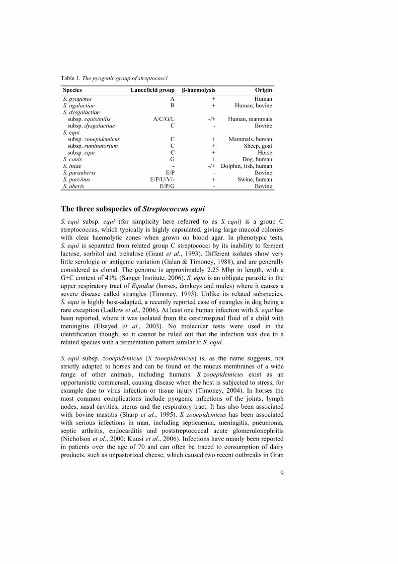
Pathogenesis The infection is initiated via the mouth or nose, where bacteria attach to the epithelium of the tonsils and adjacent lymphoid nodules. Within a few hours the bacteria have been translocated below the mucosa, reaching the deeper tissues of the tonsils. Bacteria can be detected in the lymph nodes that drain the pharyngeal and tonsillar region 18 hours after experimental infection (Kumar et al., 2004; Timoney et al., 2006). Once inside the lymph nodes, bacterial multiplication leads to the release of chemotactic factors, attracting large numbers of neutrophils. High fever develops 3 to 12 days after experimental infection, possibly associated with release of pyrogenic exotoxins. By that time chains of S. equi cells, most of which are dead, can be observed inside the infected lymph nodes and tonsils (Kumar et al., 2004; Timoney et al., 2006).
Fig. 1. Nasal discharge from a horse with strangles. Photo used with the kind permission of Prof. Derek C. Knottenbelt. Transmission and persistent carriage Nasal shedding of S. equi usually begins within 2 to 3 days after onset of fever and persists for 2 to 3 weeks. In many cases, however, the horse continues to harbour S. equi for several weeks after recovery from strangles. During this period, transmission of infection can continue either by direct horse-horse contact, or via indirect routes such as contaminated water buckets. Isolation of recovering animals is therefore critical to avoid spread of the disease. Furthermore, in up to 10% of affected animals a long-term subclinical infection is established, most commonly in the guttural pouches (Newton et al., 1997). This asymptomatic carrier state can persist for months or years, and is often difficult to diagnose and treat (Newton et al., 2000; Verheyen et al., 2000). Some animals in the carrier state sporadically shed S. equi through normal nasal secretions, in that way
12
providing a source of new outbreaks. Identification of asymptomatic carriers is therefore critical to prevent transmission of disease. Antibiotic treatment Streptococcus equi is sensitive to the majority of antibiotics in vitro, but the use of antibiotics to treat animals with strangles remains controversial. Immediate delivery of antibiotics during the early acute phase of strangles usually aborts the infection. However, the treated animal is unlikely to develop protective immunity and is susceptible to reinfection after the treatment is withdrawn. At later stages of infection the use of antibiotics is usually not effective. This is probably due to a lack of sufficient vascularity in the abscess to enable effective antibiotic penetration (Harrington et al., 2002).
Vaccination against S. equi The majority of animals recovering from strangles acquire a solid immunity to reinfection that persists for 5 years or longer (Sweeney et al., 2005; Waller & Jolley, 2006). This observation raised hopes for the development of an effective vaccine against strangles, but in spite of intensive research the progress in vaccine development has been slow. Early vaccines based on whole inactivated cells or extracts failed to provide adequate protection and often induced adverse reactions (Sweeney et al., 2005; Waller & Jolley, 2006). An attenuated, unencapsulated strain, (Pinnacle I.N.) has been on the market in the USA since 1998 (Timoney, 2004). The Pinnacle strain has a proven protective effect, but unwanted strangles-like symptoms have been reported following intranasal vaccination and it has not been licensed for sale in Europe for this reason. Another drawback with the Pinnacle strain is that it was developed by random chemical mutagenesis, which means it is prone to back-mutations that would revert the strain into an encapsulated phenotype. To prevent this, an improved unencapsulated mutant (Pinnacle HasNeg) has been generated by deleting a defined part of the has operon (Walker & Timoney, 2002). Another live attenuated vaccine (Equilis StrepE) has recently been introduced on the European market (Jacobs et al., 2000; Newton et al., 2005; Intervet, 2006). In order to minimise adverse reactions, the vaccine is injected submucosally under the upper lip of the horse. The vaccine appears to be safe, but the immunity is limited to a three-month period and only about 50% of vaccinates receive full protection following experimental infection. For this reason, the vaccine is only recommended for horses that are at high or moderate risk of infection. Protein subunit vaccines could offer an alternative to vaccines based on killed or attenuated bacteria. Acquired immunity appears to be primarily due to antibodies directed to the SeM protein and other unidentified antigens (Timoney, 2004). However, vaccines based on purified or recombinant SeM offer little or no protection (Sheoran et al., 2002; Timoney, 2004). A combination of different antigens is therefore likely to be necessary for a protective subunit vaccine. The protective immunity in horses recovered from strangles is believed to depend on a
13
combination of mucosal and serum immunoglobulins, and the failure of M protein-based vaccines could in part be due to the lack of an adequate mucosal response (Timoney & Eggers, 1985; Timoney & Trachman, 1985; Hamlen et al., 1994; Sheoran et al., 1997). Vaccination trials have also been performed with a combination of three cell surface proteins (CNE, EAG and SclC) which showed promising results in a mouse model (Flock et al., 2004; Flock et al., 2006). Follow-up trials in horses vaccinated with these antigens combined with different adjuvants are currently being performed and will hopefully be evaluated soon.
S. equi subsp. equi evolved through gene acquisition and gene loss Comparisons between the genomes of S. equi and S. zooepidemicus have shown about 97% sequence identity. When analysing the genomes of these closely related bacteria, two major factors seem to be associated with the evolution of S. equi; a large number of gene disruptions caused by mobile genetic elements and four prophage-like insertions, containing a number of potentially virulence-related genes. So far no genome-scale study of S. equi has been published, and for that reason the following discussion is largely based on conference abstracts from two independent groups, Artiushin et al. (2006) and Mitchell et al (2006). At present, 71 IS elements have been identified in the genome of S. equi and the number is likely to be somewhat higher in the final annotation of the genome (Matt Holden, unpublished results). These mobile genetic elements have caused numerous lesions in genes that are predicted to be important for the ability of the bacteria to adapt to changing environments, including genes involved in transcription regulation, quorum sensing and two component regulatory systems (Artiushin et al., 2006). Genetic interruptions responsible for various defects in nutrient transport and metabolism – such as the inability of the bacteria to ferment lactose, sorbitol and ribose – have also been identified (Artiushin et al., 2006; Mitchell et al., 2006). In this respect clear parallels can be seen in studies of the evolution of the genus Bordetella (Preston et al., 2004). The three bacterial species Bordetella pertussis, B. parapertussis and B. bronchiseptica are all closely related, but have different host ranges and cause different diseases. Bordetella bronchiseptica is closest to the evolutionary ancestor of these bordetellae and is asymptomatically carried by a wide range of mammals. The primarily human pathogens B. parapertussis and B. pertussis are independent derivatives of B. bronchiseptica, and genome analysis suggests that they evolved primarily due to loss of gene function caused by insertion of a large number of IS elements. Another more closely related species that evolved mainly through loss-of-function events is Streptococcus thermophilus (Bolotin et al., 2004), the only ‘Generally Recognized as Safe’ species within the genus Streptococcus. In this species the adaptation to a constant milk environment has led to the inactivation of most of the streptococcal virulence-related genes. It has long been known that phage-mediated gene transfer plays an important role in the evolution and virulence of pathogens (Brussow et al., 2004). With the
14
advent of bacterial genome sequencing, a large number of new prophages have been discovered, which has led to increased interest in this area of research. In the genome of S. equi, the majority of the genes that have no homologues in S. zooepidemicus have been located in four prophage-like insertions. These genes include previously reported virulence factors such as the pyrogenic mitogens SeeH, SeeI, SpeL, SpeM, but also a putative phospholipase with high similarity to the Sla toxin of S. pyogenes M3, a potential mitogen with similarity to the streptodornase of S. pyogenes M3 and a hyaluronidase that is required for phage penetration of the bacterial capsule (Mitchell et al., 2006; Tiwari et al., 2006). Interestingly, the Sla and streptodornase homologues are located in two separate prophages in both S. equi and S. pyogenes M3, highlighting the importance of gene transfer between these closely related species. It has been proposed that the recent acquisition of Sla by S. pyogenes M3 (Beres et al., 2002) may have occurred via phage-mediated transfer from S. equi (Mitchell et al., 2006). To summarise, the rapid IS element expansion in the genome of S. equi has led to numerous interruptions of genes with important functions for the ability of the bacteria to adapt to changing environments. Loss of gene function therefore seems largely accountable for the inability of S. equi to colonise mucosal surfaces. The acquisition of virulence-related genes through phage-mediated transfer, on the other hand, has most likely been essential for the bacteria in its adaptation to a new and unique ecological niche.
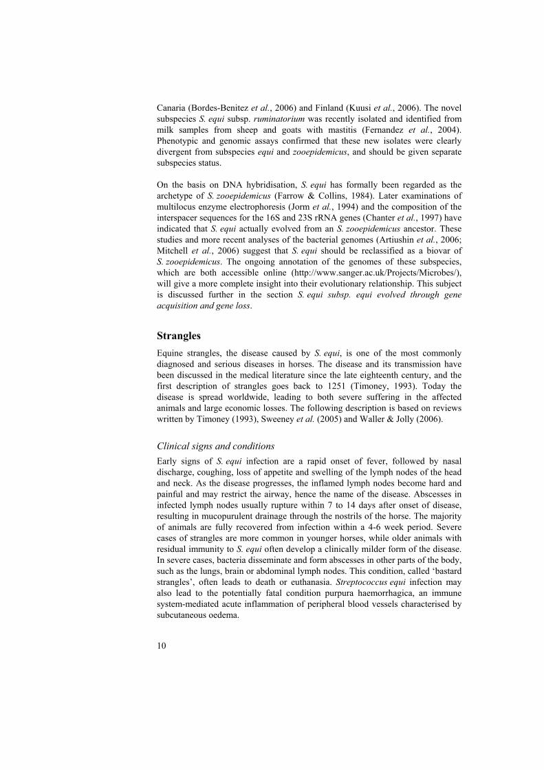
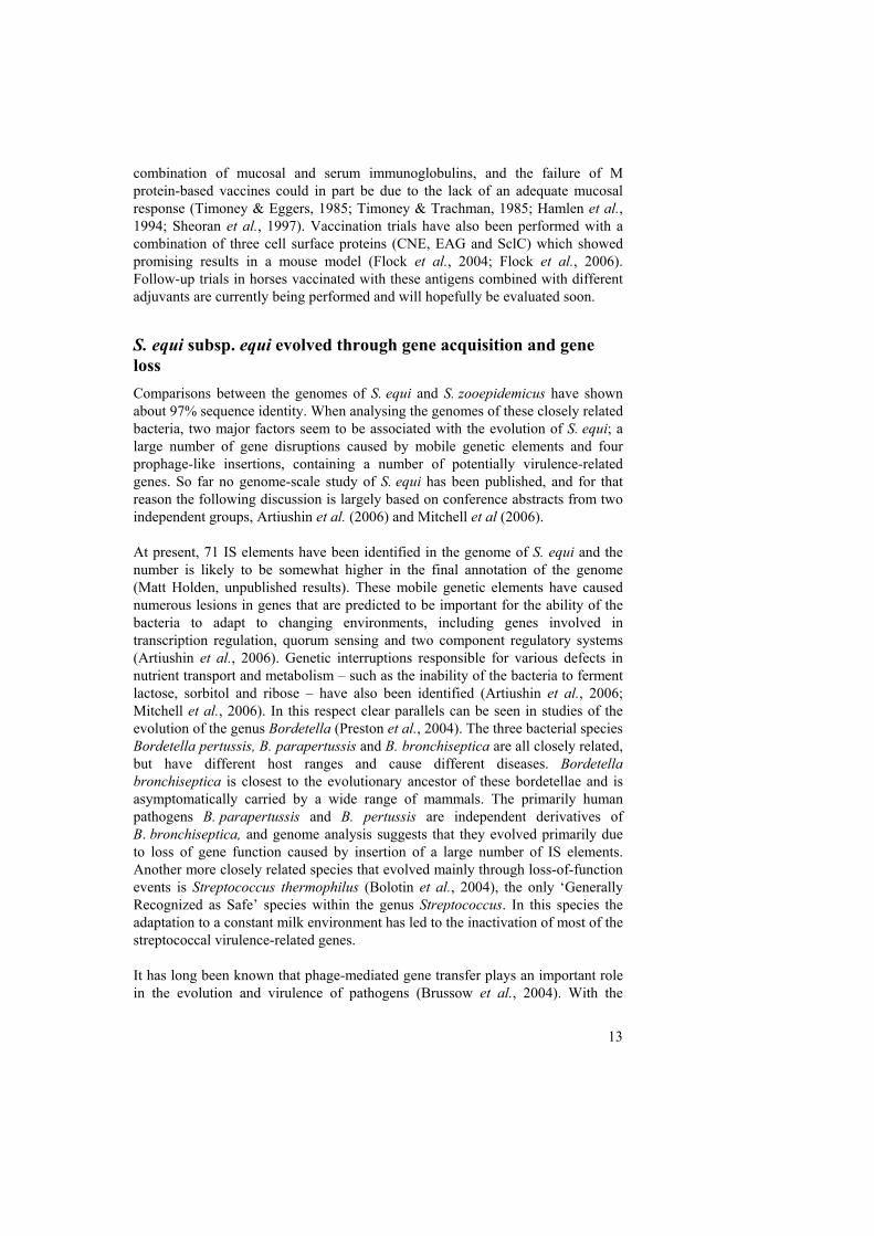
Extracellular proteins of streptococci Extracellular proteins, which here collectively refers to secreted and cell surface-located proteins, are believed to play an important role in the process of infection and in bacterial survival inside the host. This section describes the general characteristics of streptococcal cell surface-anchored proteins, while the next section discusses the importance of some of these proteins in more detail. The streptococcal cell wall The streptococci, like other Gram-positive bacteria, are surrounded by a rigid cell envelope made from cross-linked peptidoglycan (Navarre & Schneewind, 1999). This large macromolecule works as a physical barrier, protecting the bacteria against both mechanical and osmotic lysis. The cell wall also serves as a scaffold for the attachment of proteins, teichoic acids and polysaccharides that are exposed from the bacterial surface. Three different mechanisms of anchoring proteins to the cell surface are common among the streptococci (Ton-That et al., 2004) (Fig. 2): (i) Sortase-mediated attachment of proteins with LPxTG-type motifs to the cell wall; (ii) N-terminal linkage to lipids of the underlying cell membrane (lipoproteins); and (iii) binding by charge or hydrophobic interaction. In S. equi, as in the other streptococci, the majority of all cell surface proteins studied are predicted to be sortase-attached.
15
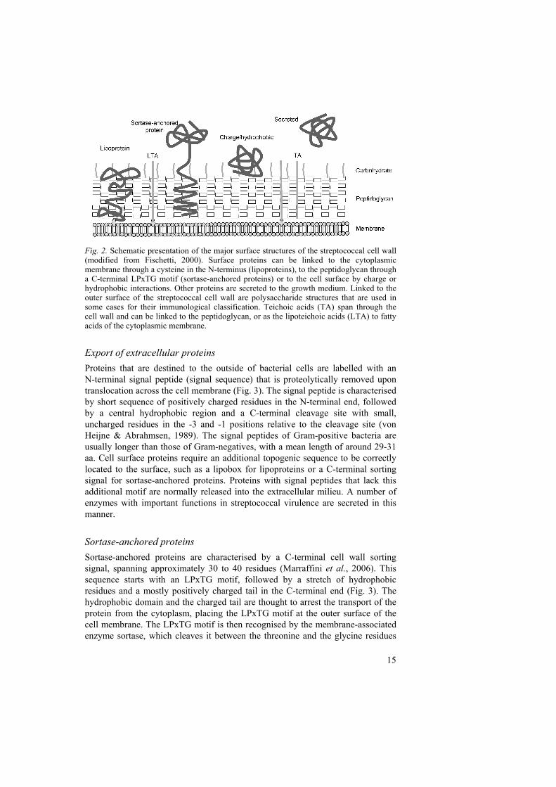
Fig. 2. Schematic presentation of the major surface structures of the streptococcal cell wall (modified from Fischetti, 2000). Surface proteins can be linked to the cytoplasmic membrane through a cysteine in the N-terminus (lipoproteins), to the peptidoglycan through a C-terminal LPxTG motif (sortase-anchored proteins) or to the cell surface by charge or hydrophobic interactions. Other proteins are secreted to the growth medium. Linked to the outer surface of the streptococcal cell wall are polysaccharide structures that are used in some cases for their immunological classification. Teichoic acids (TA) span through the cell wall and can be linked to the peptidoglycan, or as the lipoteichoic acids (LTA) to fatty acids of the cytoplasmic membrane. Export of extracellular proteins Proteins that are destined to the outside of bacterial cells are labelled with an N-terminal signal peptide (signal sequence) that is proteolytically removed upon translocation across the cell membrane (Fig. 3). The signal peptide is characterised by short sequence of positively charged residues in the N-terminal end, followed by a central hydrophobic region and a C-terminal cleavage site with small, uncharged residues in the -3 and -1 positions relative to the cleavage site (von Heijne & Abrahmsen, 1989). The signal peptides of Gram-positive bacteria are usually longer than those of Gram-negatives, with a mean length of around 29-31 aa. Cell surface proteins require an additional topogenic sequence to be correctly located to the surface, such as a lipobox for lipoproteins or a C-terminal sorting signal for sortase-anchored proteins. Proteins with signal peptides that lack this additional motif are normally released into the extracellular milieu. A number of enzymes with important functions in streptococcal virulence are secreted in this manner. Sortase-anchored proteins Sortase-anchored proteins are characterised by a C-terminal cell wall sorting signal, spanning approximately 30 to 40 residues (Marraffini et al., 2006). This sequence starts with an LPxTG motif, followed by a stretch of hydrophobic residues and a mostly positively charged tail in the C-terminal end (Fig. 3). The hydrophobic domain and the charged tail are thought to arrest the transport of the protein from the cytoplasm, placing the LPxTG motif at the outer surface of the cell membrane. The LPxTG motif is then recognised by the membrane-associated enzyme sortase, which cleaves it between the threonine and the glycine residues
16
and covalently links the carboxy-end of the threonine to the peptidoglycan (Navarre & Schneewind, 1999). The cell wall sorting signal, including the LPxTG motif, is useful in the bioinformatic identification of genes for cell wall-bound proteins. In many bacterial species, however, the sortase substrate motifs tend to deviate from the LPxTG pattern (Boekhorst et al., 2005). Among the predicted cell surface proteins of S. equi, LPxTN appears to be a common variant (IV). A recently discovered function of sortases that has received great attention is their participation in the assembly of pili in Gram-positive bacteria (Ton-That & Schneewind, 2004; Marraffini et al., 2006). In this process, the sortase is believed to catalyse the polymerisation of proteins with LPxTG motifs into an extended filament, often with an adhesive component at the tip. Pili of this type have been identified in a number of streptococcal species, including S. pyogenes and S. agalactiae (Mora et al., 2005; Dramsi et al., 2006).
Fig. 3. Domain organisation of a characteristic sortase-anchored surface protein. SS, signal sequence; W, wall spanning region; M, membrane spanning region. Common structures and functions of cell wall-anchored proteins Streptococcal cell wall-anchored proteins display a wide variety of different structures and functions. A common domain in almost all cell wall-anchored proteins is a 50-125 aa long proline/glycine or threonine/serine-rich sequence, located next to the LPxTG motif (Fischetti, 2000). This part of the protein is positioned within the cell wall. The part of the protein that is exposed outside the cell contains the functional domain(s) and is therefore typically unique. In many proteins, however, the C-terminal part contains a set of repeated sequence blocks of varying lengths (Navarre & Schneewind, 1999). In some proteins this repetitive region displays binding activity, but in others it is believed to function merely as a spacer region, exposing the functional domain from the cell. The most commonly studied functions of cell wall-anchored proteins are the binding to host tissues, plasma proteins and specific immune system components. In this respect they stand in contrast to lipoproteins, which are more commonly involved in transport systems, often involving uptake of nutrients (Sutcliffe & Russell, 1995). Bacterial proteins, cell-bound or soluble, that specifically interact with host tissue or plasma components are generally called receptins (Kronvall & Jonsson, 1999). The commonly used term adhesin is more narrow, specifically referring to proteins that the bacteria use to adhere to host tissues. Other cell wall-anchored proteins function as enzymes, often proteases, with the catalytic site exposed to the surrounding environment.
17
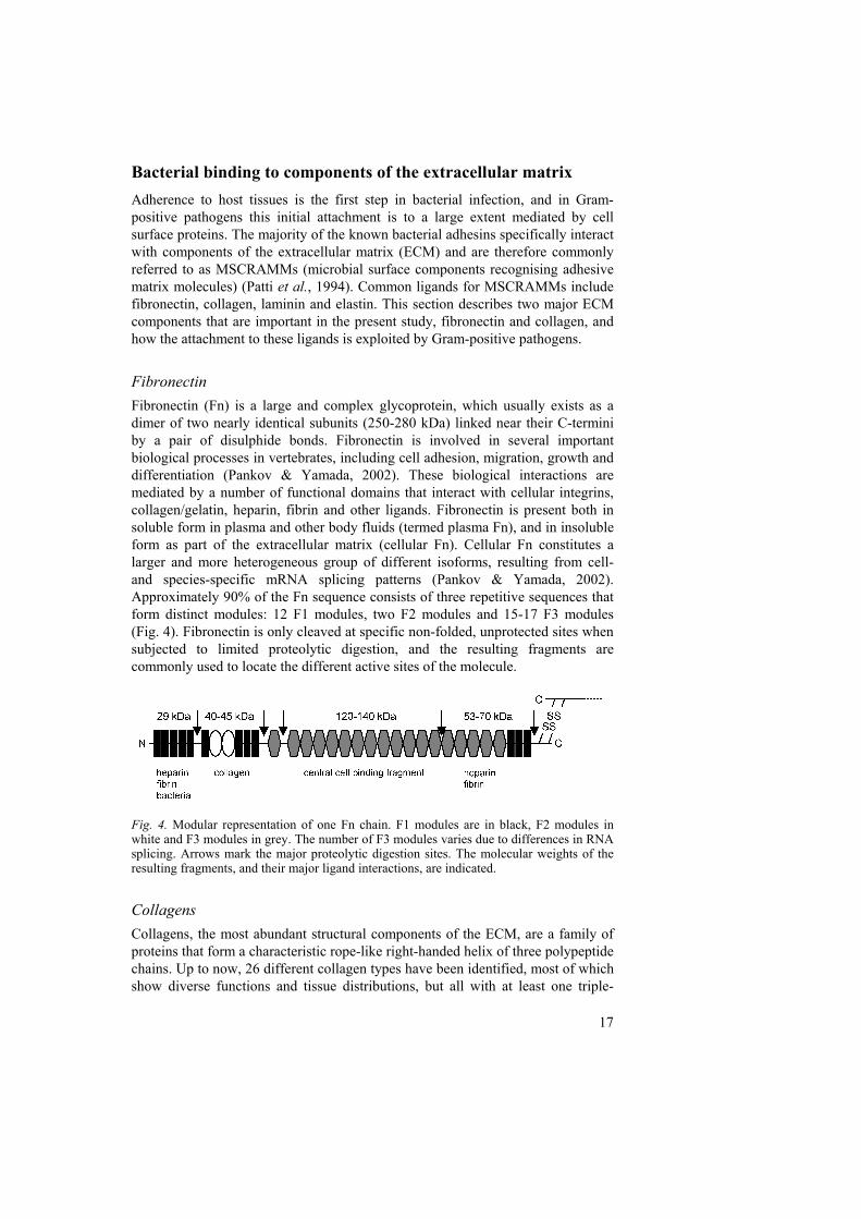
Bacterial binding to components of the extracellular matrix Adherence to host tissues is the first step in bacterial infection, and in Gram-positive pathogens this initial attachment is to a large extent mediated by cell surface proteins. The majority of the known bacterial adhesins specifically interact with components of the extracellular matrix (ECM) and are therefore commonly referred to as MSCRAMMs (microbial surface components recognising adhesive matrix molecules) (Patti et al., 1994). Common ligands for MSCRAMMs include fibronectin, collagen, laminin and elastin. This section describes two major ECM components that are important in the present study, fibronectin and collagen, and how the attachment to these ligands is exploited by Gram-positive pathogens. Fibronectin Fibronectin (Fn) is a large and complex glycoprotein, which usually exists as a dimer of two nearly identical subunits (250-280 kDa) linked near their C-termini by a pair of disulphide bonds. Fibronectin is involved in several important biological processes in vertebrates, including cell adhesion, migration, growth and differentiation (Pankov & Yamada, 2002). These biological interactions are mediated by a number of functional domains that interact with cellular integrins, collagen/gelatin, heparin, fibrin and other ligands. Fibronectin is present both in soluble form in plasma and other body fluids (termed plasma Fn), and in insoluble form as part of the extracellular matrix (cellular Fn). Cellular Fn constitutes a larger and more heterogeneous group of different isoforms, resulting from cell- and species-specific mRNA splicing patterns (Pankov & Yamada, 2002). Approximately 90% of the Fn sequence consists of three repetitive sequences that form distinct modules: 12 F1 modules, two F2 modules and 15-17 F3 modules (Fig. 4). Fibronectin is only cleaved at specific non-folded, unprotected sites when subjected to limited proteolytic digestion, and the resulting fragments are commonly used to locate the different active sites of the molecule.
Fig. 4. Modular representation of one Fn chain. F1 modules are in black, F2 modules in white and F3 modules in grey. The number of F3 modules varies due to differences in RNA splicing. Arrows mark the major proteolytic digestion sites. The molecular weights of the resulting fragments, and their major ligand interactions, are indicated. Collagens Collagens, the most abundant structural components of the ECM, are a family of proteins that form a characteristic rope-like right-handed helix of three polypeptide chains. Up to now, 26 different collagen types have been identified, most of which show diverse functions and tissue distributions, but all with at least one triple-
18
helical domain formed by a number of Gly-x-y repeats (Gelse et al., 2003; Ricard-Blum & Ruggiero, 2005). Collagens can be assembled by three identical peptide chains (homotrimers) or by a combination of different chains (heterotrimers). The collagen superfamily serves a number of different biological functions, but most noticeably they function by forming highly organised polymers that contribute to the structural integrity of the extracellular matrix. Collagens can be grouped in two main classes based on the type of polymers formed; the fibril-forming collagens (collagens I, II, III, V and XI), which represent a rather homogeneous group, and the more heterogeneous group of nonfibrillar collagens (Aumailley & Gayraud, 1998). Collagen fibrils are supramolecular aggregates that give tissues such as tendons, bone, cartilage and skin the ability to resist tear, tensile stress and pressure (Bosman & Stamenkovic, 2003). A major nonfibrillar sub-group is the network-forming collagens (IV, VIII and X), which form a large part of the basement membrane (Bosman & Stamenkovic, 2003).
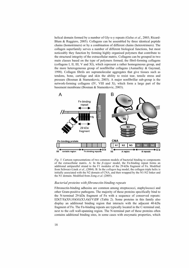
Fig. 5. Cartoon representations of two common models of bacterial binding to components of the extracellular matrix. A: In the β-zipper model, the Fn-binding repeat forms an additional antiparallel strand to the F1 modules of the 29-kDa fragment of Fn. Modified from Schwarz-Linek et al., (2004). B: In the collagen hug model, the collagen triple helix is initially associated with the N2 domain of CNA, and then wrapped by the N1-N2 linker and the N1 domain. Modified from Zong et al. (2005). Bacterial proteins with fibronectin-binding repeats Fibronectin-binding adhesins are common among streptococci, staphylococci and other Gram-positive pathogens. The majority of these proteins specifically bind to the N-terminal 29-kDa fragment of Fn with a sequence of conserved repeats: ED(T/S)(X9,10)GG(X3,4)(I/V)DF (Table 2). Some proteins in this family also display an additional binding region that interacts with the adjacent 40-kDa fragment of Fn. The Fn-binding repeats are typically located in the C-terminal end, next to the cell wall-spanning region. The N-terminal part of these proteins often contains additional binding sites, in some cases with enzymatic properties, which
19
gives them mosaic-like structures of different functional domains. The properties of this family of Fn-binding proteins have been discussed in a number of recent reviews (Schwarz-Linek et al., 2004; Hauck & Ohlsen, 2006; Schwarz-Linek et al., 2006). The structure of typical Fn-binding repeats in complex with the 29-kDa Fn domain has been determined using NMR spectroscopy (Schwarz-Linek et al., 2003; Pilka et al., 2006). An unusual feature of this model is that the Fn-binding repetitive part of the protein has a free, linear conformation in solution, which upon binding to Fn forms an additional antiparallel strand to the β-sheet structure of the N-terminal F1 modules (Fig. 5). Interestingly, Fn-binding proteins of S. pyogenes and Staphylococcus aureus have both been confirmed to mediate internalisation in several non-professional phagocytes, including epithelial and endothelial cells (Ozeri et al., 1998; Sinha et al., 1999). This process starts with the recruitment of plasma Fn to the bacterial surface. Upon binding, a conformational change is triggered in Fn, leading to the exposure of the integrin-binding RGD motif, which is cryptic in soluble Fn (Tomasini-Johansson et al., 2001). Fn-integrin interactions lead to integrin clustering on the cell surface, which triggers signalling pathways leading to cytoskeleton rearrangements and bacterial uptake through membrane invaginations (Schwarz-Linek et al., 2004; Hauck & Ohlsen, 2006). This mechanism of invasion might help the bacteria evade host immune responses and has been linked to persistent bacterial infections. It has also been suggested that intracellular survival of S. pyogenes contributes to penicillin treatment failures. Studies to evaluate the role of Fn-binding proteins in experimental infections have led to unexpected and seemingly conflicting results. In two separate experiments, S. pyogenes strains lacking Fn-binding proteins (SOF and FbaB, respectively) were used in experimental mouse infections, both resulting in decreased mouse mortality (Courtney et al., 1999; Terao et al., 2002). However, the observed effect could not be conclusively linked to a lack of cellular binding to Fn in either of these studies. In a more recent study, the gene for protein F1 was introduced into a S. pyogenes strain lacking this gene (Nyberg et al., 2004). Surprisingly, these F1-expressing bacteria were less virulent than the F1-negative strain in a mouse model. The virulence of these bacteria was partially restored when the bacteria were used to infect mice lacking plasma Fn, confirming that the decrease in virulence could be attributed to the Fn-binding activity of F1. Furthermore, infections with F1-negative bacteria caused an increased bacterial dissemination to the spleen, compared to the more localised infections caused by F1-expressing bacteria. A similar enhancement of virulence has been observed in a FnBP-negative strain of S. aureus when used in a rat model of pneumonia (McElroy et al., 2002). Together, these experiments indicate that the Fn-binding activity of S. pyogenes and S. aureus leads to less virulent infections, as the bacteria are being immobilised by host cells and tissues.
20
Table 2. Proteins with Fn-binding repeats recognising the 29-kDa fragment of Fn. Most of these proteins are multi-functional with additional binding or enzymatic properties
Species Name Additional function ReferenceS. aureus FnBPA Elastin- and Fg-
binding(Signas et al., 1989; Wann et al., 2000; Roche et al., 2004)
FnBPB Elastin-binding (Jonsson et al., 1991; Roche et al., 2004)
S. pyogenes SfbI / F1 Fibrinogen-binding (Hanski & Caparon, 1992; Talay et al., 1992; Katerov et
al., 1998) Pfbp / F2 (Jaffe et al., 1996; Rocha &
Fischetti, 1999; Ramachandran et al., 2004)
SOF/SfbII Lipoproteinase (Rakonjac et al., 1995; Kreikemeyer et al., 1999)
FbaB / F2 (Jaffe et al., 1996; Ramachandran et al., 2004)
SfbX (Jeng et al., 2003)S. dysgalactiae SOF/FnBA Lipoproteinase (Lindgren et al., 1992;
Katerov et al., 2000) FnBB (Lindgren et al., 1992)S. zooepidemicus FNZ Collagen-binding (Lindmark et al., 1996) FNZ2 Collagen-binding (Hong, 2005)S. equi FNEB (II)Borrelia burgdorferi BBK032 (Probert et al., 2001)
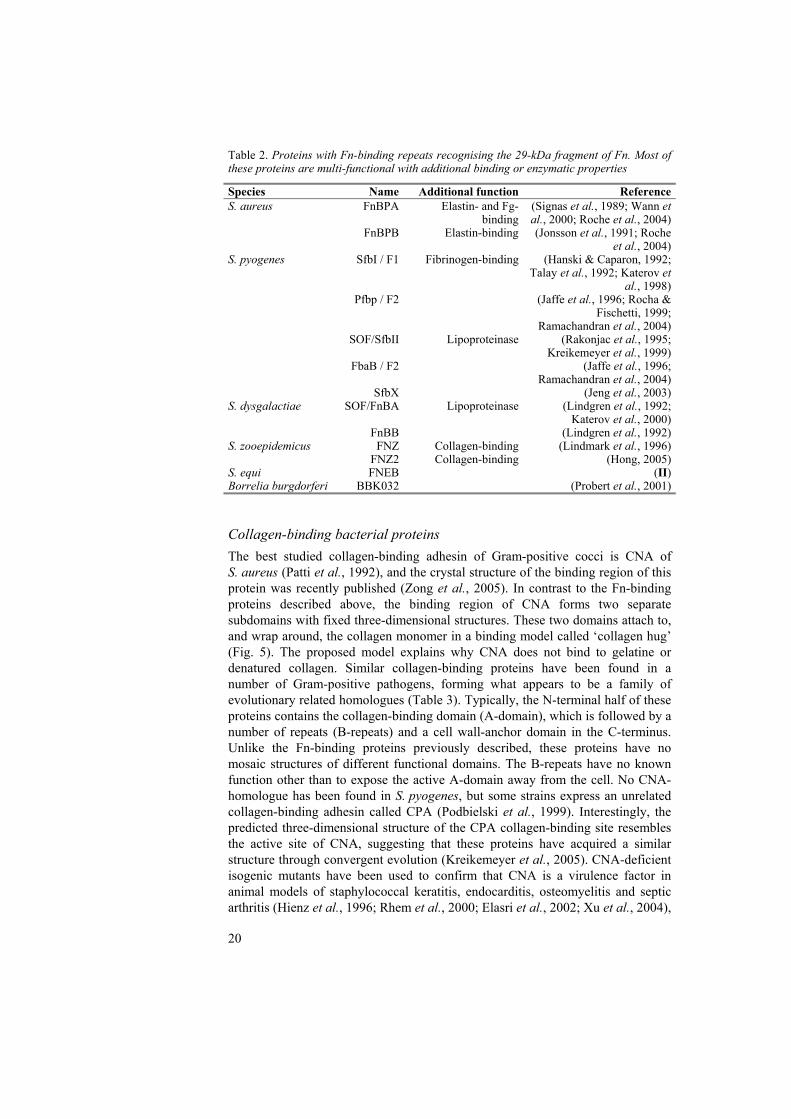
Collagen-binding bacterial proteins The best studied collagen-binding adhesin of Gram-positive cocci is CNA of S. aureus (Patti et al., 1992), and the crystal structure of the binding region of this protein was recently published (Zong et al., 2005). In contrast to the Fn-binding proteins described above, the binding region of CNA forms two separate subdomains with fixed three-dimensional structures. These two domains attach to, and wrap around, the collagen monomer in a binding model called ‘collagen hug’ (Fig. 5). The proposed model explains why CNA does not bind to gelatine or denatured collagen. Similar collagen-binding proteins have been found in a number of Gram-positive pathogens, forming what appears to be a family of evolutionary related homologues (Table 3). Typically, the N-terminal half of these proteins contains the collagen-binding domain (A-domain), which is followed by a number of repeats (B-repeats) and a cell wall-anchor domain in the C-terminus. Unlike the Fn-binding proteins previously described, these proteins have no mosaic structures of different functional domains. The B-repeats have no known function other than to expose the active A-domain away from the cell. No CNA-homologue has been found in S. pyogenes, but some strains express an unrelated collagen-binding adhesin called CPA (Podbielski et al., 1999). Interestingly, the predicted three-dimensional structure of the CPA collagen-binding site resembles the active site of CNA, suggesting that these proteins have acquired a similar structure through convergent evolution (Kreikemeyer et al., 2005). CNA-deficient isogenic mutants have been used to confirm that CNA is a virulence factor in animal models of staphylococcal keratitis, endocarditis, osteomyelitis and septic arthritis (Hienz et al., 1996; Rhem et al., 2000; Elasri et al., 2002; Xu et al., 2004),
21
which indicates that the ability to bind collagen is advantageous for the bacteria in several different types of infection. Table 3. The CNA family of collagen-binding proteins
Species Name Reference S. aureus CNA (Patti et al., 1992) S. equi CNE (I) Streptococcus mutans CNM (Sato et al., 2004) Enterococcus faecalis ACE (Rich et al., 1999) Enterococcus faecium ACM (Nallapareddy et al., 2003) Bacillus anthrasis BA0871 (Xu et al., 2004) BA5258 (Xu et al., 2004)
Fibronectin- and collagen-binding proteins as vaccine components The important functions of bacterial adhesins in the early phase of bacterial infections make them promising as components in subunit vaccines (Wizemann et al., 1999). The proposed strategy has been to block the initial bacterial attachment with adhesin-specific antibodies at the site of infection. However, the level of success for adhesin-based vaccines or for subunit vaccines in general has been very limited to date (Moxon & Rappuoli, 2002; Grandi, 2003; Scarselli et al., 2005; Serruto & Rappuoli, 2006). A common difficulty in vaccination trials is to induce a strong and lasting immune response at the mucosal level. Furthermore, most bacteria express a large number of adhesins that can compensate for each other during infection. An immune response against a specific combination of surface antigens might therefore be necessary to successfully block bacterial binding. To complicate matters further, several bacterial pathogens display strain-specific antigenic variations in their surface proteins. A broad-range vaccine must therefore induce an immune response directed to conserved epitopes of proteins that are common among different strains of the bacteria in question. This has been a great obstacle in the development of vaccines against S. pyogenes and other pathogens with great antigenic variation, but will probably not be as much of a problem with more homologous bacterial species such as S. equi. Proteins with Fn-binding repeats have shown potential as vaccine components in several animal studies, including a rat model for S. aureus endocarditis and a mouse model for S. pyogenes infection (Rennermalm et al., 2001; Schulze et al., 2003). In contrast, no protection was observed in a mouse model for S. equi infection (Flock et al., 2006). The observed protection is however most likely due to an antibody-mediated bacterial clearance by phagocytes (opsonisation). It has been reported that these antibodies actually recognise the Fn-binding proteins in complex with Fn and therefore do not prevent, but could actually enhance, the binding (Speziale et al., 1996; Casolini et al., 1998). The reason for this failure to generate neutralising antibodies could be that the ‘free’ Fn-binding region is poorly immunogenic due to the lack a secondary structure, or simply that Fn-binding proteins quickly form complexes with plasma Fn before they are recognised by the immune system.
22
A recombinant fragment of CNA has been shown to be effective in protecting mice against S. aureus-induced septic death (Nilsson et al., 1998). Antibodies against CNA have also been shown to effectively interfere with the attachment of S. aureus to a collagen substrate (Visai et al., 2000). Likewise, the use of CNE in a strangles model in mice gave encouraging results, and the same study showed that serum antibodies against CNE were able to block the binding of S. equi to immobilised collagen (Flock et al., 2006). Potential virulence factors of Streptococcus equi subsp. equi The exact definition of the term virulence factor has been a matter of debate, but it is generally defined as a component of a pathogen that fulfils molecular Koch’s postulate, i.e. the deletion of the gene associated with the trait specifically impairs virulence (Casadevall & Pirofski, 1999; Falkow, 2004). To separate virulence factors from fitness factors, it is often implied that the deletion should not lead to a decrease in general viability. Factors that are believed to be important in the virulence of S. equi include the carbohydrate capsule, several surface proteins and a number of toxins. To date, however, only a few S. equi deletion mutants have been made and used in infection models. For that reason these factors are here collectively referred to as potential virulence factors. Capsule S. equi display an highly encapsulated phenotype when grown on blood agar, giving the colonies a ‘honeydew’ appearance. This hyaluronic acid capsule is non-antigenic and has in several studies been associated with virulence and resistance to phagocytosis. The encapsulation in liquid growth media is greatly influenced by culture age, as young cultures (4-6 h) are highly encapsulated, whereas the encapsulation of overnight cultures is barely detectable (Harrington et al., 2002). Infection experiments in mice and foals have both shown that young cultures are significantly more virulent and also more resistant to phagocytosis (Harrington et al., 2002). A study by Anzai et al. (1999) confirmed that resistance to phagocytosis is also correlated with levels of encapsulation when strains with different expression of capsule are compared. Mutants completely lacking a capsule are unable to colonise the lymph nodes of experimentally infected horses (Timoney et al., 2006). Hyaluronidase treatment of S. equi cells results in a slight increase in adherence to tongue cells of ponies, suggesting that heavy encapsulation partially covers the active sites of adhesins that are expressed on the cell surface (Srivastava & Barnum, 1983). M-like proteins The M protein family of S. pyogenes is an extensively studied class of cell surface-anchored proteins that extend from the surface as alpha-helical coiled-coil dimers (Cunningham, 2000). The N-terminal region is often hypervariable and induces opsonic antibodies that normally do not cross-react with other M types. An important function of the M proteins is to enable the bacteria to resist
23
phagocytosis, or even facilitate intracellular survival inside neutrophils (Staali et al., 2003). The antiphagocytic properties of the M proteins depend on binding to fibrinogen (Fg) or to human C4b-binding protein, which in turn prevents the deposition of complement factors on the bacterial surface (Carlsson et al., 2005). The ‘M-like proteins’ of related streptococci often refer to Fg-binding proteins with antiphagocytic activity, and these proteins may or may not be structurally related to the M proteins of S. pyogenes.
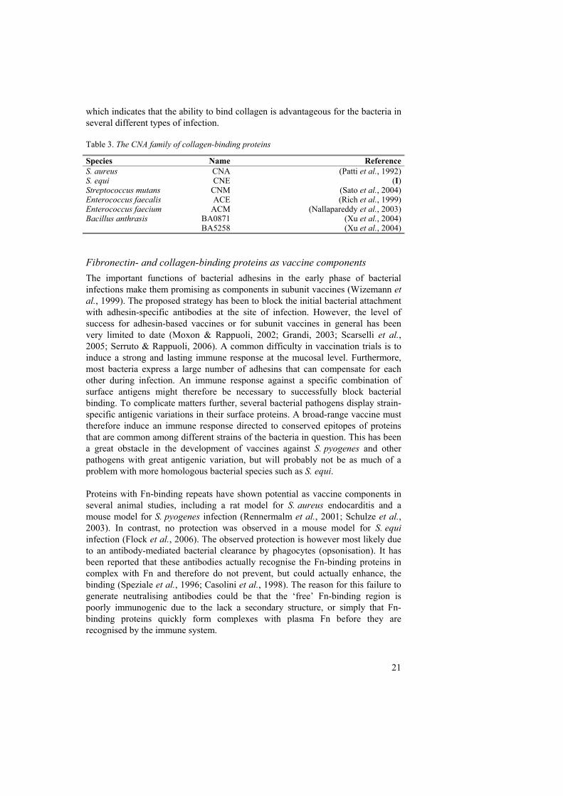
Fig. 6. Schematic representation of the SeM protein, adapted from Kelly et al. (2006). The region found deleted in 24% of S. equi strains isolated from subclinical carriers, the variable region used in subtyping of strains and the Fg- and IgG-binding regions are indicated. In S. equi two M-like proteins have been studied, SeM (also known as FgBP) (Fig. 6) and SzPSe. These proteins show no significant sequence similarity between each other, or to the M proteins of S. pyogenes, but both have extended regions of alpha-helical coiled-coils and bind strongly to equine Fg (Timoney et al., 1997; Meehan et al., 2002). In addition, SeM binds to IgG-Fc (Meehan et al., 2001). SzPSe is the S. equi homologue of the S. zooepidemicus protein SzP, but SeM has been regarded as unique to S. equi, and the acquisition of the gene encoding this protein has been proposed to be an important step in the evolution of this subspecies (Timoney et al., 1997). However, recent genome studies have shown that S. zooepidemicus indeed encodes an allelic variant of SeM, denoted SzM (Kelly et al., 2006). This protein shows no significant similarity to SeM in the N-terminal region, which is necessary for the binding of SeM to Fg and IgG-Fc, so it is possible that the replacement or modification of this region, rather than the acquisition of the whole protein, represents the important evolutionary step. SeM is the major cell wall-associated protein of S. equi cells grown in vitro (Meehan et al., 1998) and antibodies to this protein are strongly opsonogenic in whole blood or with purified neutrophils (Boschwitz & Timoney, 1994) Antibodies to SzPSe, on the contrary, have no opsonic effect and the protein is not believed to be as important in resistance to phagocytosis (Timoney et al., 1995). The importance of SeM as a virulence factor was demonstrated in a study where a knockout mutant lacking this protein was employed in a number of experiments (Meehan et al., 2001). The mutant failed to autoaggregate when it reached the stationary phase under the conditions of laboratory growth, bound no Fg or IgG, was rapidly killed in horse blood and showed decreased virulence in a mouse model. Mutants lacking SeM are also unable to colonise the lymph nodes of experimentally infected horses (Timoney et al., 2006). Studies on the cross-reactivity of convalescent horse sera and Southern blots with a sem probe have suggested that SeM is highly homogeneous across different
24
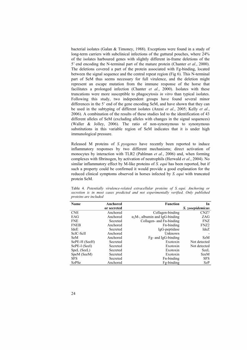
bacterial isolates (Galan & Timoney, 1988). Exceptions were found in a study of long-term carriers with subclinical infections of the guttural pouches, where 24% of the isolates harboured genes with slightly different in-frame deletions of the 5’ end encoding the N-terminal part of the mature protein (Chanter et al., 2000). The deletions covered a part of the protein associated with Fg-binding, located between the signal sequence and the central repeat region (Fig 6). This N-terminal part of SeM thus seems necessary for full virulence, and the deletion might represent an escape mutation from the immune response of the horse that facilitates a prolonged infection (Chanter et al., 2000). Isolates with these truncations were more susceptible to phagocytosis in vitro than typical isolates. Following this study, two independent groups have found several minor differences in the 5’ end of the gene encoding SeM, and have shown that they can be used in the subtyping of different isolates (Anzai et al., 2005; Kelly et al., 2006). A combination of the results of these studies led to the identification of 43 different alleles of SeM (excluding alleles with changes in the signal sequences) (Waller & Jolley, 2006). The ratio of non-synonymous to synonymous substitutions in this variable region of SeM indicates that it is under high immunological pressure. Released M proteins of S. pyogenes have recently been reported to induce inflammatory responses by two different mechanisms; direct activation of monocytes by interaction with TLR2 (Pahlman et al., 2006) and, when forming complexes with fibrinogen, by activation of neutrophils (Herwald et al., 2004). No similar inflammatory effect by M-like proteins of S. equi has been reported, but if such a property could be confirmed it would provide a good explanation for the reduced clinical symptoms observed in horses infected by S. equi with truncated protein SeM. Table 4. Potentially virulence-related extracellular proteins of S. equi. Anchoring or secretion is in most cases predicted and not experimentally verified. Only published proteins are included
Name Anchored or secreted
Function In S. zooepidemicus
CNE Anchored Collagen-binding CNZ?EAG Anchored α2M-, albumin and IgG-binding ZAGFNE Secreted Collagen- and Fn-binding FNZFNEB Anchored Fn-binding FNZ2IdeE Secreted IgG-peptidase IdeZSclC-SclI Anchored Unknown -SeM Anchored Fg- and IgG-binding SzMSePE-H (SeeH) Secreted Exotoxin Not detectedSePE-I (SeeI) Secreted Exotoxin Not detectedSpeL (SeeL) Secreted Exotoxin SzeLSpeM (SeeM) Secreted Exotoxin SzeMSFS Secreted Fn-binding SFSSzPSe Anchored Fg-binding SzP
25
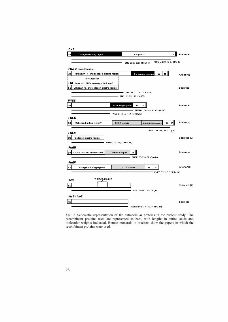
Other receptins and adhesins EAG is the S. equi equivalent of ZAG, a plasma protein receptin of S. zooepidemicus that specifically binds to albumin, IgG and the protease inhibitor α2-macroglobulin (α2M) (Jonsson et al., 1995). In Northern blots no transcripts of the gene encoding EAG could be detected from cells grown in laboratory cultures (Lindmark, 1999). Consequently, the binding of S. equi cells grown in vitro to IgG is reported to be very low (Lindmark et al., 1999) and mediated primarily by SeM (Meehan et al., 2001). Based on these data, the importance of EAG as a virulence factor could be questioned. However, measurements of antibody titres specific to EAG in convalescent horses can provide indirect evidence for the in vivo expression of EAG during S. equi infection (Flock et al., 2004). Three different Fn-binding proteins of S. equi have so far been published. FNE is a homologue of FNZ of S. zooepidemicus. FNZ contains two different Fn-binding domains: a region with typical repeats in the C-terminal end that interacts with the 29-kDa fragment of Fn, and an hitherto uncharacterised 40-kDa fragment binding domain in the N-terminal half of the protein (Lindmark et al., 1996; Lindmark et al., 2001) (II). However, the gene encoding FNE is interrupted by a frameshift mutation, leading to the transcription of a truncated protein without the 29-kDa binding repeats or the C-terminal cell wall anchor domain of FNZ (Fig. 7) (Lindmark et al., 2001). FNE has consequently been found released in the medium during S. equi cultivation. Interestingly, the N-terminal half of FNZ, which is identical to the entire protein FNE, was recently found to bind collagen in addition to Fn (Liden et al., 2006). Four new putative paralogues to FNE have been found in S. equi, all of which bind collagen to different degrees and one that binds Fn. These are discussed in the section Present investigation. Another Fn-binding protein (SFS) also binds the 40-kDa fragment (Lindmark & Guss, 1999) (II). SFS lacks any putative cell-wall anchoring domains, and has for that reason been predicted to be a secreted protein. However, the in vitro expression of SFS is low, and no protein has been detected in growth cultures. Finally, FNEB is the only published Fn-binding protein of S. equi that appears to be anchored to the cell wall (II). FNEB contains typical Fn-binding repeats and specifically interacts with the 29-kDa fragment. This protein is further described in the section Present investigation. Seven different proteins with collagen-like repeats (SclC-SclI) have been studied in S. equi (Karlstrom et al., 2004; Karlstrom et al., 2006). The C-terminal parts of these proteins contain similar collagen-like repeats, and in the N-terminal ends each have a unique A-domain with no significant similarities to any known proteins. Two proteins with similar collagen-like repeats have been found in S. pyogenes, and these have been shown to form triple helices and interact with mammalian collagen-binding integrins (Xu et al., 2002; Humtsoe et al., 2005). Wether the Scl-family of proteins in S. equi have similar structures and biological functions remains to be investigated.
26
Secreted toxins and enzymes Streptococcus equi has acquired genes for at least four mitogenic exotoxins by phage-mediated transfer. These toxins are believed to function as superantigens and misdirect the immune response by activating a large number of T cells. This activation leads to a massive release of pro-inflammatory cytokines, which is likely to contribute to the severe symptoms of strangles, such as high fever and neutrophilia (Anzai et al., 1999). Two of these toxins (SePE-H and SepE-I) are located in the same phage-associated locus and are almost identical to their counterparts in S. pyogenes. Both proteins have been shown to induce a strong mitogenic response in horse peripheral blood monocytes and pyrexia in rabbits, but only SEPE-I is pyrogenic in ponies (Artiushin et al., 2002). The two other superantigens (SPE-L and SPE-M) also share high sequence similarity to orthologues found in S. pyogenes (Proft et al., 2003). In a recent study, the genes for these two superantigens were also found in one out of 31 strains of S. zooepidemicus (Alber et al., 2005). Studies of the β-haemolysis in S. equi cultures under different growth conditions have suggested that this activity is due to a Streptolysin S-like toxin (Flanagan et al., 1998). Production of the haemolysin is induced during logarithmic growth and is dependent on a serum supplement. However, the gene(s) responsible for the observed activity have not yet been published. The precise role of this toxin is not known, but the lysis of neutrophils and macrophages is expected to be important in immune evasion (Harrington et al., 2002). This lytic activity is also likely to make essential nutrients available to the bacteria. Streptococcus equi also secretes a streptokinase that specifically activates equine plasminogen and thereby induces the hydrolysis of fibrin (McCoy et al., 1991). The function of this streptokinase in infection has not been proven, but it is possible that the hydrolysis of fibrin clots could aid bacterial spread.
27
“Sometimes you make the right decision, sometimes you make the decision right”
Dr Phil
Present investigation
The main objective of this study was to identify and characterise new potential virulence factors expressed by S. equi. The focus was mainly on cell surface proteins that specifically adhere to collagen and fibronectin (I, II, IV), two major extracellular matrix proteins of vertebrates, but a secreted immunoglobulin-specific protease was also studied (III). A useful tool in this research was the publicly available S. equi genome, which can be used to identify homologues to virulence-associated proteins of related pathogens such as S. pyogenes and S. aureus. This genome database was also useful in identifying potential paralogues to previously characterised proteins (IV). The present study was mainly of a basic science nature, but had also the applied objective of identifying components for a future vaccine against strangles.
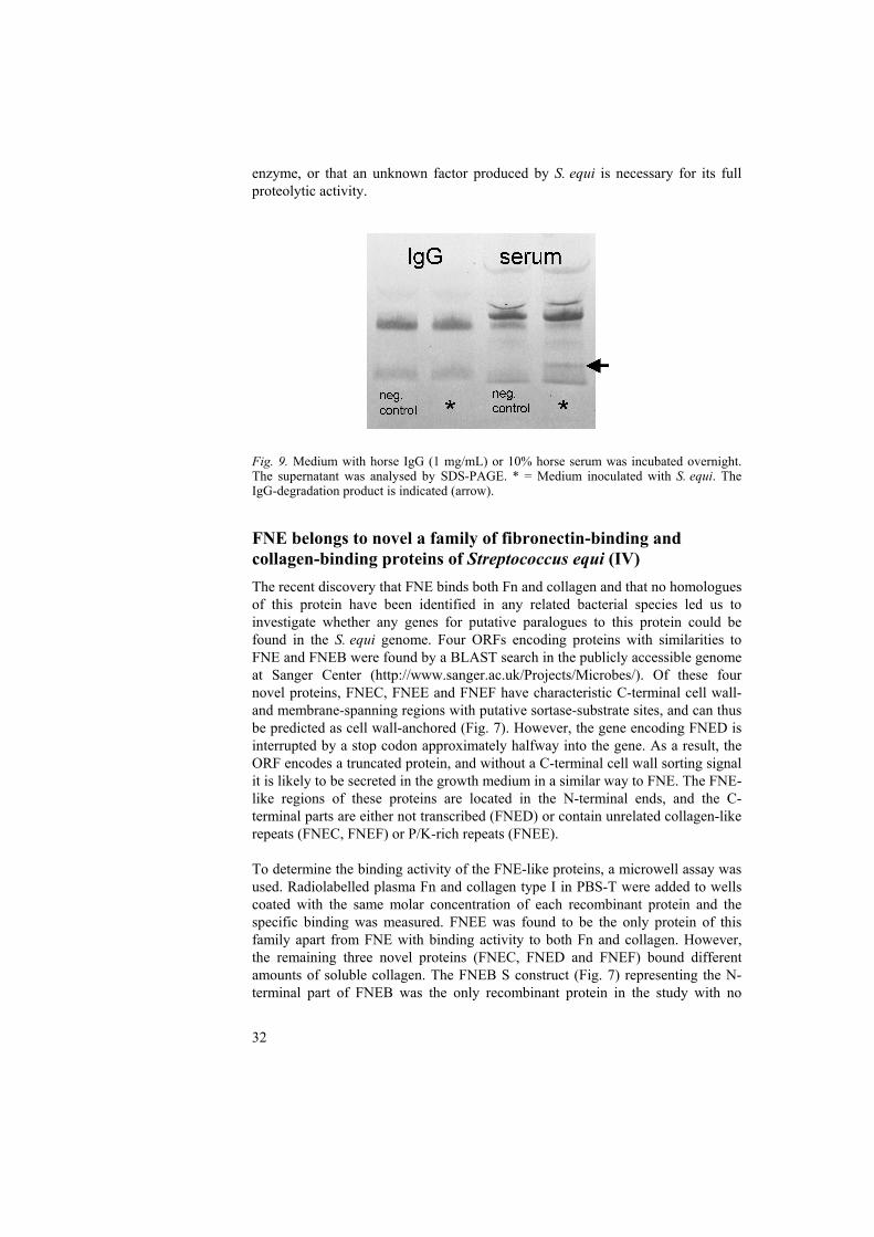
CNE, a collagen-binding protein of S. equi subsp. equi (I) The collagen-binding protein CNE belongs to a putative family of structural and functional homologues of Gram-positive cocci described in a previous section. In this study, we expressed recombinant proteins corresponding to the mature protein (CNE L) and the collagen-binding ‘A domain’ (CNE S) (Fig. 7). These recombinant proteins were used in a Western ligand blot to confirm that CNE bound to 125I-labelled bovine collagen type I, and that the binding activity was located in the A domain. It was possible to inhibit the binding of soluble radiolabelled collagen to immobilised CNE S in microwells with increasing concentrations of CNE S, indicating that the binding was specific. In a similar manner, it was possible to inhibit the binding between collagen and immobilised CNE S with recombinant CNA, confirming that the two proteins were competing for the same binding site in collagen. CNE S was used to inhibit the binding of radiolabelled collagen to S. equi cells in solution, but the binding could only be reduced to 65%. The reason for this incomplete inhibition could be that multiple binding sites on collagen in combination with a high cellular expression of CNE make complete inhibition unattainable, or that the cellular binding to collagen is due to the combined activity of CNE and an unidentified surface structure. When S. equi cells were incubated in solution with radiolabelled CNE S and increasing concentrations of collagen, the radioactivity bound to the cell pellet was proportional to the amount of added collagen, indicating that CNE binds to multiple binding sites on the collagen monomer.
28
Fig. 7. Schematic representation of the extracellular proteins in the present study. The recombinant proteins used are represented as bars, with lengths in amino acids and molecular weights indicated. Roman numerals in brackets show the papers in which the recombinant proteins were used.
29
The gene encoding CNE was detected in all isolates of S. equi and S. zooepidemicus tested. Today, the gene encoding the CNE-homologue of S. zooepidemicus can be found in the publicly available S. zooepidemicus genome database. However, the protein encoded by this gene has not been studied. Since the completion of this study, CNE has been evaluated as a vaccine component in a strangles model in the mouse (Flock et al., 2006). Vaccination with CNE prior to experimental infection resulted in a low but statistically significant decrease in clinical symptoms, measured as weight loss and nasal shedding. Interestingly, CNE had an immunostimulatory effect on the poorly immunogenic protein EAG, and a coadministration of CNE and EAG resulted in a synergistic protective effect. The same study showed that serum antibodies against CNE were able to block the binding of S. equi to immobilised collagen. Vaccination trials with a protein subunit vaccine including CNE are currently being performed in horses.
Studies of fibronectin-binding proteins of Streptococcus equi (II) In this study, a novel Fn-binding protein of S. equi (FNEB) was described (Fig. 7). FNEB displays a signal sequence and a typical cell wall-anchoring domain with an LPxTG motif. FNEB can thus be predicted as cell surface-located, contrary to the two previously characterised fibronectin-binding proteins of S. equi FNE and SFS. Western ligand blots with two recombinant proteins, one corresponding to the mature protein (FNEB L) and one corresponding to the C-terminal half (FNEB S), confirmed that the binding activity is located near the C-terminus, and that it interacts specifically with the 29-kDa fragment of Fn. This part of the protein contains a number of characteristic Fn-binding repeats described in the section Bacterial binding to components of the extracellular matrix. This study also showed that the previously characterised proteins FNE and SFS both bind to the 40-kDa collagen-binding fragment of Fn. Binding-inhibition studies with iodinated Fn added to microwells with immobilised FNEB, SFS and FNE showed a cross-inhibition between FNE and SFS, indicating that these proteins bind to overlapping, or nearby located sites on the Fn molecule. As expected, FNEB could not be used to inhibit the binding activity of FNE or SFS and vice versa. Streptococcus equi is known to bind Fn to a much lower degree than S. zooepidemicus (Lindmark et al., 1999). By measuring the binding of iodinated Fn-fragments to S. equi cells, it was shown that the observable binding is primarily directed to the 29-kDa domain. Furthermore, FNEB L could be used to inhibit the binding of both radiolabelled 29-kDa fragments and whole Fn to S. equi cells, indicating that FNEB is the primary adhesin that mediates binding of Fn to S. equi cells grown in vitro. As described previously, experiments with S. aureus and S pyogenes mutants have revealed that a decreased binding to Fn is associated with an increase in invasiveness and virulence. Considering the comparatively low ability of S. equi to bind Fn compared with the evolutionary parental S. zooepidemicus, we postulate that this decreased adhesion is a contributing factor to its more virulent phenotype.
30
Based on the low, but observable binding to Fn, it could be predicted that FNEB is expressed under laboratory conditions, albeit at a low level. To test this hypothesis, total RNA was prepared from S. equi cells at the logarithmic phase of growth, followed by cDNA synthesis using RT-PCR with short random primers. The relative amounts of transcripts of fne, sfs, and fneB were measured by gene-specific PCR, with the housekeeping gene gyrA used as a control. The highest expression was observed for gyrA (clear PCR-product after 22 cycles), followed by fne (24 cycles), fneB (27 cycles) and sfs (29 cycles). This pattern fits well with previous studies where transcripts of fne, but not sfs, were detected by Northern blots (Lindmark & Guss, 1999). As mentioned previously, native FNE has also been isolated from growth cultures (Lindmark et al., 2001), indicating a relatively high expression. Analyses of antibody titres from mice and horses with or without S. equi infections were used to assess the expression of FNEB during infection. A significant increase in antibody titres in mice with experimental infection and in convalescent horses indicates that FNEB is expressed during infection and exposed to the immune system.
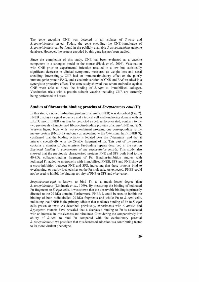
IdeE, an IgG-endopeptidase of S. equi subsp. equi (III) In all mammals, immunoglobulin G (IgG) has a key role in the defence against bacterial pathogens. By detecting antigens of the invading bacterium, the IgG molecule can mediate bacterial killing either by activating the classical complement system or by marking the bacterium for ingestion by phagocytes, an activity called opsonisation. Recognition by IgG can also inactivate bacterial toxins and adhesins. The IgG molecule is a glycoprotein made up of four subunits, two heavy chains and two light chains, linked by disulphide bridges. Its overall organisation is presented in Figure 8. Bacterial pathogens have evolved several different strategies to avoid recognition by IgG. One mechanism is to bind the Fc region (CH2-CH3) of the IgG molecule with specific surface receptors, such as the previously mentioned SeM and EAG proteins, and in that way block the interaction with Fc-receptors on phagocytes. Another strategy is to interfere with the interaction between IgG and the Fc-receptors of phagocytes by releasing enzymes that either degrade the N-linked glycans of the IgG molecule, or cleave the peptide backbone of the IgG heavy chains (Collin & Olsen, 2003). IdeS, also known as Mac, is a secreted cysteine protease of S. pyogenes that cleaves the hinge region of human IgG with high specificity (Lei et al., 2002; von Pawel-Rammingen et al., 2002; von Pawel-Rammingen & Bjorck, 2003). Genes for IdeS/Mac homologues have been found in the genomes of S. equi and S. zooepidemicus. In the present study, these genes were cloned and the proteins they encode, IdeE and IdeZ, were expressed and characterised. Purified horse IgG was incubated with recombinant IdeE and a cleavage product was visible when analysed by SDS-PAGE. The N-terminal end of the cleavage product was identified as GPSVFIFPP[K/N]PK by tandem mass spectrometry sequencing, confirming that IdeE and IdeS/Mac cleaves IgG at the same site. A
31
study of the proteolytic activity of recombinant IdeE and IdeZ on purified IgG from a selection of mammals shows that only antibodies containing the substrate site of IdeS/Mac are cleaved, indicating that the specificities of the two enzymes are very similar. Interestingly, horse IgG was cleaved less effectively than the IgG of e.g. dogs or humans. A likely explanation for this partial proteolysis would be that the hinge region of the dominating IgG isotype in horse serum (IgG4) is resistant to cleavage due to the lack of a distinct substrate site for IdeE/IdeZ. A Western blot with IgG4-specific antibodies confirmed that the heavy chain of this isotype was indeed intact after incubation with recombinant IdeE.
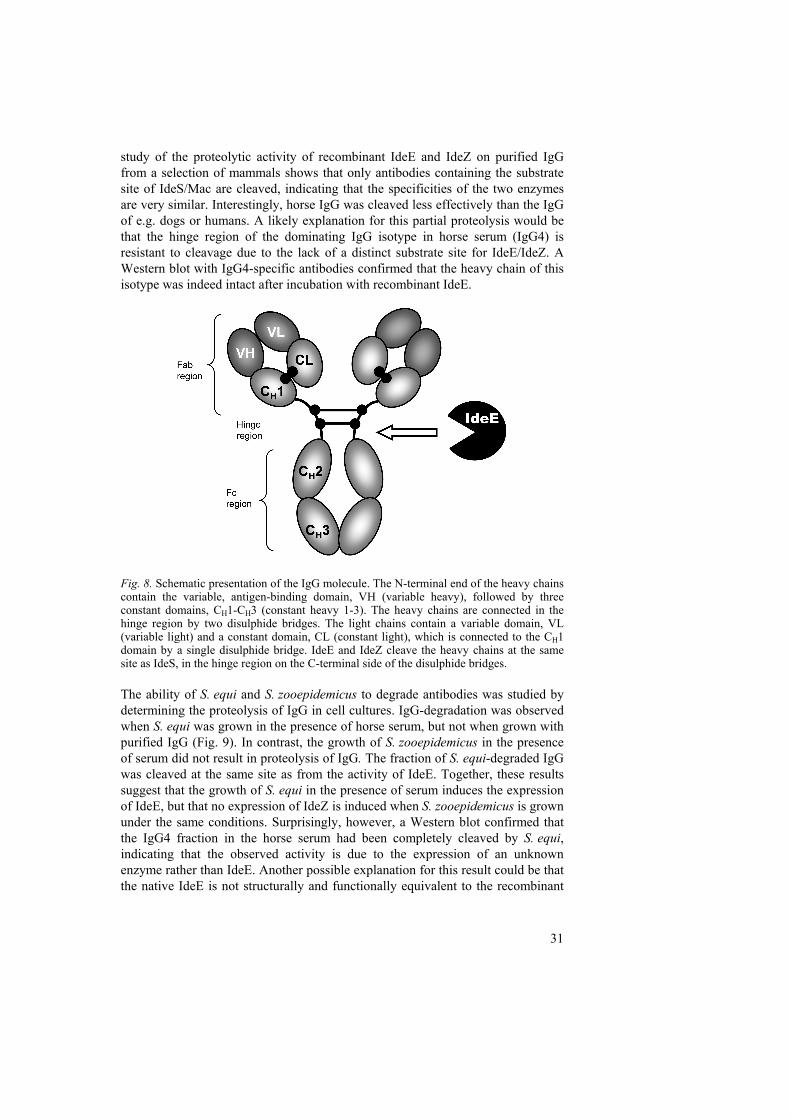
Fig. 8. Schematic presentation of the IgG molecule. The N-terminal end of the heavy chains contain the variable, antigen-binding domain, VH (variable heavy), followed by three constant domains, CH1-CH3 (constant heavy 1-3). The heavy chains are connected in the hinge region by two disulphide bridges. The light chains contain a variable domain, VL (variable light) and a constant domain, CL (constant light), which is connected to the CH1 domain by a single disulphide bridge. IdeE and IdeZ cleave the heavy chains at the same site as IdeS, in the hinge region on the C-terminal side of the disulphide bridges. The ability of S. equi and S. zooepidemicus to degrade antibodies was studied by determining the proteolysis of IgG in cell cultures. IgG-degradation was observed when S. equi was grown in the presence of horse serum, but not when grown with purified IgG (Fig. 9). In contrast, the growth of S. zooepidemicus in the presence of serum did not result in proteolysis of IgG. The fraction of S. equi-degraded IgG was cleaved at the same site as from the activity of IdeE. Together, these results suggest that the growth of S. equi in the presence of serum induces the expression of IdeE, but that no expression of IdeZ is induced when S. zooepidemicus is grown under the same conditions. Surprisingly, however, a Western blot confirmed that the IgG4 fraction in the horse serum had been completely cleaved by S. equi, indicating that the observed activity is due to the expression of an unknown enzyme rather than IdeE. Another possible explanation for this result could be that the native IdeE is not structurally and functionally equivalent to the recombinant
32
enzyme, or that an unknown factor produced by S. equi is necessary for its full proteolytic activity.
Fig. 9. Medium with horse IgG (1 mg/mL) or 10% horse serum was incubated overnight. The supernatant was analysed by SDS-PAGE. * = Medium inoculated with S. equi. The IgG-degradation product is indicated (arrow).
FNE belongs to novel a family of fibronectin-binding and collagen-binding proteins of Streptococcus equi (IV) The recent discovery that FNE binds both Fn and collagen and that no homologues of this protein have been identified in any related bacterial species led us to investigate whether any genes for putative paralogues to this protein could be found in the S. equi genome. Four ORFs encoding proteins with similarities to FNE and FNEB were found by a BLAST search in the publicly accessible genome at Sanger Center (http://www.sanger.ac.uk/Projects/Microbes/). Of these four novel proteins, FNEC, FNEE and FNEF have characteristic C-terminal cell wall- and membrane-spanning regions with putative sortase-substrate sites, and can thus be predicted as cell wall-anchored (Fig. 7). However, the gene encoding FNED is interrupted by a stop codon approximately halfway into the gene. As a result, the ORF encodes a truncated protein, and without a C-terminal cell wall sorting signal it is likely to be secreted in the growth medium in a similar way to FNE. The FNE-like regions of these proteins are located in the N-terminal ends, and the C-terminal parts are either not transcribed (FNED) or contain unrelated collagen-like repeats (FNEC, FNEF) or P/K-rich repeats (FNEE). To determine the binding activity of the FNE-like proteins, a microwell assay was used. Radiolabelled plasma Fn and collagen type I in PBS-T were added to wells coated with the same molar concentration of each recombinant protein and the specific binding was measured. FNEE was found to be the only protein of this family apart from FNE with binding activity to both Fn and collagen. However, the remaining three novel proteins (FNEC, FNED and FNEF) bound different amounts of soluble collagen. The FNEB S construct (Fig. 7) representing the N-terminal part of FNEB was the only recombinant protein in the study with no

33
binding activity to either ligand. An alignment of the FNE-like regions showed patches of conserved residues among all proteins, and did not give any obvious clues to the observed differences in binding specificities or the location of the binding sites.
34
Future perspectives
As shown in Lindmark et al (1999) and in II, a clear difference in the adhesive properties of S. equi and S. zooepidemicus is that the former binds considerably less Fn, and especially the 40-kDa fragment of Fn, when grown under standard laboratory conditions. In II, we postulated that this could be an important factor behind the invasive nature of S. equi infection. The finding that S. equi harbours at least two Fn-binding proteins that appear to be cell-wall anchored (FNEB and FNEE) merits further study of the expression of these proteins, and of the Fn-binding activity of S. equi cells, under different growth conditions and growth phases. Expression and binding studies could, for example, be performed with cells grown in the presence of blood, under anaerobic conditions, in iron-depleted medium or – even better – with cells isolated from infected horses. Studies with S. pyogenes have shown that growth in plasma or blood increases the expression of several secreted and cell surface-bound proteins (Graham et al., 2005; Johansson et al., 2005), and our own cell culture experiments in III indicate that the presence of blood components also influences the expression of virulence factors in S. equi. In a study by Meehan et al. (1998), FnBP/SeM was identified as the major cell wall-associated protein of S. equi. However, this work, like most ligand-binding studies with S. equi, was performed with cells from overnight cultures grown in standard laboratory culture medium. A large number of novel S. equi proteins with cell wall-anchoring motifs have been characterised since then, and genes for many more can be found in the genome. Given this seemingly abundant arsenal of adhesins, it is likely that different cell surface proteins are expressed during different phases of infection – such as the initial attachment to the tonsils, the proliferation inside the lymph node and during the establishment of persistent infection in the guttural pouches. The Fn- and collagen-binding proteins FNE/FNZ have several putative paralogues in S. equi and S. zooepidemicus, but no known homologues in other bacterial species. As we show in IV, the binding sites for Fn and collagen cannot easily be pinpointed by aligning the sequences of binders and non-binders within the FNE-family of proteins. With truncated recombinants of FNE, FNEC, FNED, FNEE and FNEF, this could be elucidated. In addition, it would be interesting to search for homologues to these proteins in related streptococci, especially since recent studies have suggested that FNE could alter fluid homeostasis in infected tissues (Lidén et al., 2006 and unpublished results). The binding of Fn to the proteins SfbI and FnBPA of S. pyogenes and S. aureus has been shown to trigger integrin-mediated internalisation of these bacteria by epithelial and endothelial cells (Schwarz-Linek et al., 2004; Hauck & Ohlsen, 2006). As we speculate in II, it is possible that FNEB serves a similar function in S. equi. It would therefore be interesting to evaluate wether FNEB contributes to the rapid internalisation of S. equi during the initial infection of the epithelium of the tonsil and adjacent lymphoid nodules (Timoney et al., 2006).
35
The cell culture experiments performed in III suggest that an unidentified IgG-cleaving protease, rather than IdeE, is expressed when S. equi is grown in the presence of serum. An isogenic mutant lacking the ideE gene could be used to elucidate this. An alternative strategy would be to search the S. equi genome for genes encoding other extracellular proteases that could be expressed and characterised. In recent years, pili have been found on the surface of a number of streptococci including S. pyogenes, S. agalactiae and S. pneumoniae (Ton-That & Schneewind, 2004; Mora et al., 2005; Dramsi et al., 2006; Marraffini et al., 2006). These proteinaceous filaments are assembled from proteins with LPxTG-type motifs by a specific sortase, and the genes for the building blocks and the sortase are often found within a single operon. Interestingly, the cne-gene in S. equi is located in the upstream end of a putative operon with two other genes; just downstream is a gene for a protein with (weak) similarities to several fimbrial subunit proteins in different Gram-positive bacteria, followed by a gene for a sortase-like protein. It can thus be postulated that CNE is incorporated to the end of a pilus structure with the putative fimbrial subunit proteins as the main building block (which would explain the lack of a typical cell wall-spanning region in CNE). The best way to test this hypothesis would probably be with electron microscopy, using cells coated with gold particles conjugated to antibodies recognising CNE and/or the putative fimbrial subunit protein.
36
References
Alber, J., El-Sayed, A., Estoepangestie, S., Lammler, C. & Zschock, M. 2005. Dissemination of the superantigen encoding genes seeL, seeM, szeL and szeM in Streptococcus equi subsp. equi and Streptococcus equi subsp. zooepidemicus. Vet Microbiol 109, 135-41.
Anzai, T., Kuwamoto, Y., Wada, R., Sugita, S., Kakuda, T., Takai, S., Higuchi, T. & Timoney, J.F. 2005. Variation in the N-terminal region of an M-like protein of Streptococcus equi and evaluation of its potential as a tool in epidemiologic studies. Am J Vet Res 66, 2167-71.
Anzai, T., Sheoran, A.S., Kuwamoto, Y., Kondo, T., Wada, R., Inoue, T. & Timoney, J.F. 1999. Streptococcus equi but not Streptococcus zooepidemicus produces potent mitogenic responses from equine peripheral blood mononuclear cells. Vet Immunol Immunopathol 67, 235-46.
Anzai, T., Timoney, J.F., Kuwamoto, Y., Fujita, Y., Wada, R. & Inoue, T. 1999. In vivo pathogenicity and resistance to phagocytosis of Streptococcus equi strains with different levels of capsule expression. Vet Microbiol 67, 277-86.
Artiushin, S., Walker, J. & Timoney, J. 2006. Mobile Genetic Elements in Genomes of S. equi And S. zooepidemicus. In The 7th ASM Conference on Streptococcal Genetics. Saint-Malo, France
Artiushin, S.C., Timoney, J.F., Sheoran, A.S. & Muthupalani, S.K. 2002. Characterization and immunogenicity of pyrogenic mitogens SePE-H and SePE-I of Streptococcus equi. Microb Pathog 32, 71-85.
Aumailley, M. & Gayraud, B. 1998. Structure and biological activity of the extracellular matrix. J Mol Med 76, 253-65.
Beres, S.B., Sylva, G.L., Barbian, K.D., Lei, B., Hoff, J.S., Mammarella, N.D., Liu, M.Y., Smoot, J.C., Porcella, S.F., Parkins, L.D., Campbell, D.S., Smith, T.M., McCormick, J.K., Leung, D.Y., Schlievert, P.M. & Musser, J.M. 2002. Genome sequence of a serotype M3 strain of group A Streptococcus: phage-encoded toxins, the high-virulence phenotype, and clone emergence. Proc Natl Acad Sci U S A 99, 10078-83.
Boekhorst, J., de Been, M.W., Kleerebezem, M. & Siezen, R.J. 2005. Genome-wide detection and analysis of cell wall-bound proteins with LPxTG-like sorting motifs. J Bacteriol 187, 4928-34.
Bolotin, A., Quinquis, B., Renault, P., Sorokin, A., Ehrlich, S.D., Kulakauskas, S., Lapidus, A., Goltsman, E., Mazur, M., Pusch, G.D., Fonstein, M., Overbeek, R., Kyprides, N., Purnelle, B., Prozzi, D., Ngui, K., Masuy, D., Hancy, F., Burteau, S., Boutry, M., Delcour, J., Goffeau, A. & Hols, P. 2004. Complete sequence and comparative genome analysis of the dairy bacterium Streptococcus thermophilus. Nat Biotechnol 22, 1554-8.
Bordes-Benitez, A., Sanchez-Onoro, M., Suarez-Bordon, P., Garcia-Rojas, A.J., Saez-Nieto, J.A., Gonzalez-Garcia, A., Alamo-Antunez, I., Sanchez-Maroto, A. & Bolanos-Rivero, M. 2006. Outbreak of Streptococcus equi subsp. zooepidemicus infections on the island of Gran Canaria associated with the consumption of inadequately pasteurized cheese. Eur J Clin Microbiol Infect Dis 25, 242-6.
37
Boschwitz, J.S. & Timoney, J.F. 1994. Inhibition of C3 deposition on Streptococcus equi subsp. equi by M protein: a mechanism for survival in equine blood. Infect Immun 62, 3515-20.
Bosman, F.T. & Stamenkovic, I. 2003. Functional structure and composition of the extracellular matrix. J Pathol 200, 423-8.
Brussow, H., Canchaya, C. & Hardt, W.D. 2004. Phages and the evolution of bacterial pathogens: from genomic rearrangements to lysogenic conversion. Microbiol Mol Biol Rev 68, 560-602.
Carlsson, F., Sandin, C. & Lindahl, G. 2005. Human fibrinogen bound to Streptococcus pyogenes M protein inhibits complement deposition via the classical pathway. Mol Microbiol 56, 28-39.
Casadevall, A. & Pirofski, L.A. 1999. Host-pathogen interactions: redefining the basic concepts of virulence and pathogenicity. Infect Immun 67, 3703-13.
Casolini, F., Visai, L., Joh, D., Conaldi, P.G., Toniolo, A., Hook, M. & Speziale, P. 1998. Antibody response to fibronectin-binding adhesin FnbpA in patients with Staphylococcus aureus infections. Infect Immun 66, 5433-42.
Chanter, N., Collin, N., Holmes, N., Binns, M. & Mumford, J. 1997. Characterization of the Lancefield group C streptococcus 16S-23S RNA gene intergenic spacer and its potential for identification and sub-specific typing. Epidemiol Infect 118, 125-35.
Chanter, N., Talbot, N.C., Newton, J.R., Hewson, D. & Verheyen, K. 2000. Streptococcus equi with truncated M-proteins isolated from outwardly healthy horses. Microbiology 146 (Pt 6), 1361-9.
Collin, M. & Olsen, A. 2003. Extracellular enzymes with immunomodulating activities: variations on a theme in Streptococcus pyogenes. Infect Immun 71, 2983-92.
Courtney, H.S., Hasty, D.L., Li, Y., Chiang, H.C., Thacker, J.L. & Dale, J.B. 1999. Serum opacity factor is a major fibronectin-binding protein and a virulence determinant of M type 2 Streptococcus pyogenes. Mol Microbiol 32, 89-98.
Cunningham, M.W. 2000. Pathogenesis of group A streptococcal infections. Clin Microbiol Rev 13, 470-511.
Dramsi, S., Caliot, E., Bonne, I., Guadagnini, S., Prevost, M.C., Kojadinovic, M., Lalioui, L., Poyart, C. & Trieu-Cuot, P. 2006. Assembly and role of pili in group B streptococci. Mol Microbiol 60, 1401-13.
Elasri, M.O., Thomas, J.R., Skinner, R.A., Blevins, J.S., Beenken, K.E., Nelson, C.L. & Smeltzer, M.S. 2002. Staphylococcus aureus collagen adhesin contributes to the pathogenesis of osteomyelitis. Bone 30, 275-80.
Elsayed, S., Hammerberg, O., Massey, V. & Hussain, Z. 2003. Streptococcus equi subspecies equi (Lancefield group C) meningitis in a child. Clin Microbiol Infect 9, 869-72.
Facklam, R. 2002. What happened to the streptococci: overview of taxonomic and nomenclature changes. Clin Microbiol Rev 15, 613-30.
Falkow, S. 2004. Molecular Koch's postulates applied to bacterial pathogenicity--a personal recollection 15 years later. Nat Rev Microbiol 2, 67-72.
Farrow, J.A.E. & Collins, M.D. 1984. Taxonomic Studies on Streptococci of Serological Groups C, G and L and Possibly Related Taxa. System. Appl. Microbiol. 5, 483-493.
Fernandez, E., Blume, V., Garrido, P., Collins, M.D., Mateos, A., Dominguez, L. & Fernandez-Garayzabal, J.F. 2004. Streptococcus equi subsp. ruminatorum subsp.
38
nov., isolated from mastitis in small ruminants. Int J Syst Evol Microbiol 54, 2291-6.
Fischetti, V.A. 2000. Surface Proteins on Gram-Positive Bacteria. In Gram-Positive Pathogens. Edited by V.A. Fischetti, R.P. Novick, J.J. Ferretti, D.A. Portnoy & J.I. Rood. ASM Press. 11-24. pp.
Flanagan, J., Collin, N., Timoney, J., Mitchell, T., Mumford, J.A. & Chanter, N. 1998. Characterization of the haemolytic activity of Streptococcus equi. Microb Pathog 24, 211-21.
Flock, M., Jacobsson, K., Frykberg, L., Hirst, T.R., Franklin, A., Guss, B. & Flock, J.I. 2004. Recombinant Streptococcus equi proteins protect mice in challenge experiments and induce immune response in horses. Infect Immun 72, 3228-36.
Flock, M., Karlstrom, A., Lannergard, J., Guss, B. & Flock, J.I. 2006. Protective effect of vaccination with recombinant proteins from Streptococcus equi subspecies equi in a strangles model in the mouse. Vaccine 24, 4144-51.
Galan, J.E. & Timoney, J.F. 1988. Immunologic and genetic comparison of Streptococcus equi isolates from the United States and Europe. J Clin Microbiol 26, 1142-6.
Gelse, K., Poschl, E. & Aigner, T. 2003. Collagens--structure, function, and biosynthesis. Adv Drug Deliv Rev 55, 1531-46.
Graham, M.R., Virtaneva, K., Porcella, S.F., Barry, W.T., Gowen, B.B., Johnson, C.R., Wright, F.A. & Musser, J.M. 2005. Group A Streptococcus transcriptome dynamics during growth in human blood reveals bacterial adaptive and survival strategies. Am J Pathol 166, 455-65.
Grandi, G. 2003. Rational antibacterial vaccine design through genomic technologies. Int J Parasitol 33, 615-20.
Grant, S.T., Efstratiou, A. & Chanter, N. 1993. Laboratory diagnosis of strangles and the isolation of atypical Streptococcus equi. Vet Rec 133, 215-6.
Hamlen, H.J., Timoney, J.F. & Bell, R.J. 1994. Epidemiologic and immunologic characteristics of Streptococcus equi infection in foals. J Am Vet Med Assoc 204, 768-75.
Hanski, E. & Caparon, M. 1992. Protein F, a fibronectin-binding protein, is an adhesin of the group A streptococcus Streptococcus pyogenes. Proc Natl Acad Sci U S A 89, 6172-6.
Hardie, J.M. 1986. Genus Streptococcus. In Bergey's Manual of Systematic Bacteriology. Edited by J.G. Holt. Williams & Wilkins. Baltimore, USA. 1043-1056. pp.
Hardie, J.M. & Whiley, R.A. 1995. The genus Streptococcus. In The Genera of Lactic Acid Bacteria. Edited by B.J.B. Wood & W.H. Holzapfel. Blackie A&P. Glasgow, UK. 55-124. pp.
Harrington, D.J., Sutcliffe, I.C. & Chanter, N. 2002. The molecular basis of Streptococcus equi infection and disease. Microbes Infect 4, 501-10.
Hauck, C.R. & Ohlsen, K. 2006. Sticky connections: extracellular matrix protein recognition and integrin-mediated cellular invasion by Staphylococcus aureus. Curr Opin Microbiol 9, 5-11.
Herwald, H., Cramer, H., Morgelin, M., Russell, W., Sollenberg, U., Norrby-Teglund, A., Flodgaard, H., Lindbom, L. & Bjorck, L. 2004. M protein, a classical bacterial virulence determinant, forms complexes with fibrinogen that induce vascular leakage. Cell 116, 367-79.
39
Hienz, S.A., Schennings, T., Heimdahl, A. & Flock, J.I. 1996. Collagen binding of Staphylococcus aureus is a virulence factor in experimental endocarditis. J Infect Dis 174, 83-8.
Hong, K. 2005. Identification and characterization of a novel fibronectin-binding protein gene from Streptococcus equi subspecies zooepidemicus strain VTU211. FEMS Immunol Med Microbiol 45, 231-7.
Humtsoe, J.O., Kim, J.K., Xu, Y., Keene, D.R., Hook, M., Lukomski, S. & Wary, K.K. 2005. A streptococcal collagen-like protein interacts with the alpha2beta1 integrin and induces intracellular signaling. J Biol Chem 280, 13848-57.
Intervet. 2006. Equilis StrepE http://www.intervet.co.uk/Products_Public/Equilis_StrepE/Overview.asp 2006-07-18
Jacobs, A.A., Goovaerts, D., Nuijten, P.J., Theelen, R.P., Hartford, O.M. & Foster, T.J. 2000. Investigations towards an efficacious and safe strangles vaccine: submucosal vaccination with a live attenuated Streptococcus equi. Vet Rec 147, 563-7.
Jaffe, J., Natanson-Yaron, S., Caparon, M.G. & Hanski, E. 1996. Protein F2, a novel fibronectin-binding protein from Streptococcus pyogenes, possesses two binding domains. Mol Microbiol 21, 373-84.
Jeng, A., Sakota, V., Li, Z., Datta, V., Beall, B. & Nizet, V. 2003. Molecular genetic analysis of a group A Streptococcus operon encoding serum opacity factor and a novel fibronectin-binding protein, SfbX. J Bacteriol 185, 1208-17.
Johansson, B.P., Levander, F., von Pawel-Rammingen, U., Berggard, T., Bjorck, L. & James, P. 2005. The protein expression of Streptococcus pyogenes is significantly influenced by human plasma. J Proteome Res 4, 2302-11.
Jonsson, H., Lindmark, H. & Guss, B. 1995. A protein G-related cell surface protein in Streptococcus zooepidemicus. Infect Immun 63, 2968-75.
Jonsson, K., Signas, C., Muller, H.P. & Lindberg, M. 1991. Two different genes encode fibronectin binding proteins in Staphylococcus aureus. The complete nucleotide sequence and characterization of the second gene. Eur J Biochem 202, 1041-8.
Jorm, L.R., Love, D.N., Bailey, G.D., McKay, G.M. & Briscoe, D.A. 1994. Genetic structure of populations of beta-haemolytic Lancefield group C streptococci from horses and their association with disease. Res Vet Sci 57, 292-9.
Karlstrom, A., Jacobsson, K., Flock, M., Flock, J.I. & Guss, B. 2004. Identification of a novel collagen-like protein, SclC, in Streptococcus equi using signal sequence phage display. Vet Microbiol 104, 179-88.
Karlstrom, A., Jacobsson, K. & Guss, B. 2006. SclC is a member of a novel family of collagen-like proteins in Streptococcus equi subspecies equi that are recognised by antibodies against SclC. Vet Microbiol 114, 72-81.
Katerov, V., Andreev, A., Schalen, C. & Totolian, A.A. 1998. Protein F, a fibronectin-binding protein of Streptococcus pyogenes, also binds human fibrinogen: isolation of the protein and mapping of the binding region. Microbiology 144 (Pt 1), 119-26.
Katerov, V., Lindgren, P.E., Totolian, A.A. & Schalen, C. 2000. Streptococcal opacity factor: a family of bifunctional proteins with lipoproteinase and fibronectin-binding activities. Curr Microbiol 40, 149-56.
Kelly, C., Bugg, M., Robinson, C., Mitchell, Z., Davis-Poynter, N., Newton, J.R., Jolley, K.A., Maiden, M.C. & Waller, A.S. 2006. Sequence variation of the SeM gene of
40
Streptococcus equi allows discrimination of the source of strangles outbreaks. J Clin Microbiol 44, 480-6.
Kreikemeyer, B., Martin, D.R. & Chhatwal, G.S. 1999. SfbII protein, a fibronectin binding surface protein of group A streptococci, is a serum opacity factor with high serotype-specific apolipoproteinase activity. FEMS Microbiol Lett 178, 305-11.
Kreikemeyer, B., Nakata, M., Oehmcke, S., Gschwendtner, C., Normann, J. & Podbielski, A. 2005. Streptococcus pyogenes collagen type I-binding Cpa surface protein. Expression profile, binding characteristics, biological functions, and potential clinical impact. J Biol Chem 280, 33228-39.
Kronvall, G. & Jonsson, K. 1999. Receptins: a novel term for an expanding spectrum of natural and engineered microbial proteins with binding properties for mammalian proteins. J Mol Recognit 12, 38-44.
Kumar, P., Timoney, J. & Muthupalani, S.K. 2004. Early Pathogenesis of Streptococcus equi Infection in the Horse. In ASM General Meeting. New Orleans, USA
Kuusi, M., Lahti, E., Virolainen, A., Hatakka, M., Vuento, R., Rantala, L., Vuopio-Varkila, J., Seuna, E., Karppelin, M., Hakkinen, M., Takkinen, J., Gindonis, V., Siponen, K. & Huotari, K. 2006. An outbreak of Streptococcus equi subspecies zooepidemicus associated with consumption of fresh goat cheese. BMC Infect Dis 6, 36.
Ladlow, J., Scase, T. & Waller, A. 2006. Canine Strangles Case Reveals a New Host Susceptible to Infection with Streptococcus equi. J Clin Microbiol 44, 2664-5.
Lancefield, R.C. 1933. A serological differentiation of human and other groups of hemolytic streptococci. J Exp Med 57, 571–595.
Lei, B., DeLeo, F.R., Reid, S.D., Voyich, J.M., Magoun, L., Liu, M., Braughton, K.R., Ricklefs, S., Hoe, N.P., Cole, R.L., Leong, J.M. & Musser, J.M. 2002. Opsonophagocytosis-inhibiting mac protein of group a streptococcus: identification and characteristics of two genetic complexes. Infect Immun 70, 6880-90.
Liden, A., Karlstrom, A., Lannergard, J., Kalamajski, S., Guss, B., Rubin, K. & Ryden, C. 2006. A fibronectin-binding protein from Streptococcus equi binds collagen and modulates cell-mediated collagen gel contraction. Biochem Biophys Res Commun 340, 604-10.
Lindgren, P.E., Speziale, P., McGavin, M., Monstein, H.J., Hook, M., Visai, L., Kostiainen, T., Bozzini, S. & Lindberg, M. 1992. Cloning and expression of two different genes from Streptococcus dysgalactiae encoding fibronectin receptors. J Biol Chem 267, 1924-31.
Lindmark, H. (1999). Characterization of Adhesive Extracellular Proteins from Streptococcus equi, Agraria 139. Swedish University of Agricultural Sciences.
Lindmark, H. & Guss, B. 1999. SFS, a novel fibronectin-binding protein from Streptococcus equi, inhibits the binding between fibronectin and collagen. Infect Immun 67, 2383-8.
Lindmark, H., Jacobsson, K., Frykberg, L. & Guss, B. 1996. Fibronectin-binding protein of Streptococcus equi subsp. zooepidemicus. Infect Immun 64, 3993-9.
Lindmark, H., Jonsson, P., Engvall, E. & Guss, B. 1999. Pulsed-field gel electrophoresis and distribution of the genes zag and fnz in isolates of Streptococcus equi. Res Vet Sci 66, 93-9.
Lindmark, H., Nilsson, M. & Guss, B. 2001. Comparison of the fibronectin-binding protein FNE from Streptococcus equi subspecies equi with FNZ from S. equi subspecies
41
zooepidemicus reveals a major and conserved difference. Infect Immun 69, 3159-63.
Marraffini, L.A., Dedent, A.C. & Schneewind, O. 2006. Sortases and the art of anchoring proteins to the envelopes of gram-positive bacteria. Microbiol Mol Biol Rev 70, 192-221.
McCoy, H.E., Broder, C.C. & Lottenberg, R. 1991. Streptokinases produced by pathogenic group C streptococci demonstrate species-specific plasminogen activation. J Infect Dis 164, 515-21.
McElroy, M.C., Cain, D.J., Tyrrell, C., Foster, T.J. & Haslett, C. 2002. Increased virulence of a fibronectin-binding protein mutant of Staphylococcus aureus in a rat model of pneumonia. Infect Immun 70, 3865-73.
Meehan, M., Kelly, S.M., Price, N.C. & Owen, P. 2002. The C-terminal portion of the fibrinogen-binding protein of Streptococcus equi subsp. equi contains extensive alpha-helical coiled-coil structure and contributes to thermal stability. FEMS Microbiol Lett 206, 81-6.
Meehan, M., Lynagh, Y., Woods, C. & Owen, P. 2001. The fibrinogen-binding protein (FgBP) of Streptococcus equi subsp. equi additionally binds IgG and contributes to virulence in a mouse model. Microbiology 147, 3311-22.
Meehan, M., Nowlan, P. & Owen, P. 1998. Affinity purification and characterization of a fibrinogen-binding protein complex which protects mice against lethal challenge with Streptococcus equi subsp. equi. Microbiology 144 (Pt 4), 993-1003.
Mitchell, Z., Parkhill, J., Holden, M., Robinson, C., Davis-Poynter, N. & Waller, A. 2006. The Evolution of S. equi Results From Genome Comparisons With S. zooepidemicus. In The 7th ASM Conference on Streptococcal Genetics. Saint-Malo, France
Mora, M., Bensi, G., Capo, S., Falugi, F., Zingaretti, C., Manetti, A.G., Maggi, T., Taddei, A.R., Grandi, G. & Telford, J.L. 2005. Group A Streptococcus produce pilus-like structures containing protective antigens and Lancefield T antigens. Proc Natl Acad Sci U S A 102, 15641-6.
Moxon, R. & Rappuoli, R. 2002. Bacterial pathogen genomics and vaccines. Br Med Bull 62, 45-58.
Nallapareddy, S.R., Weinstock, G.M. & Murray, B.E. 2003. Clinical isolates of Enterococcus faecium exhibit strain-specific collagen binding mediated by Acm, a new member of the MSCRAMM family. Mol Microbiol 47, 1733-47.
Navarre, W.W. & Schneewind, O. 1999. Surface proteins of gram-positive bacteria and mechanisms of their targeting to the cell wall envelope. Microbiol Mol Biol Rev 63, 174-229.
Newton, J.R., Verheyen, K., Talbot, N.C., Timoney, J.F., Wood, J.L., Lakhani, K.H. & Chanter, N. 2000. Control of strangles outbreaks by isolation of guttural pouch carriers identified using PCR and culture of Streptococcus equi. Equine Vet J 32, 515-26.
Newton, J.R., Wood, J.L., Dunn, K.A., DeBrauwere, M.N. & Chanter, N. 1997. Naturally occurring persistent and asymptomatic infection of the guttural pouches of horses with Streptococcus equi. Vet Rec 140, 84-90.
Newton, R., Waller, A. & King, A. 2005. Investigation of suspected adverse reactions following strangles vaccination in horses. Vet Rec 156, 291-2.
Nicholson, M.L., Ferdinand, L., Sampson, J.S., Benin, A., Balter, S., Pinto, S.W., Dowell, S.F., Facklam, R.R., Carlone, G.M. & Beall, B. 2000. Analysis of
42
immunoreactivity to a Streptococcus equi subsp. zooepidemicus M-like protein To confirm an outbreak of poststreptococcal glomerulonephritis, and sequences of M-like proteins from isolates obtained from different host species. J Clin Microbiol 38, 4126-30.
Nilsson, I.M., Patti, J.M., Bremell, T., Hook, M. & Tarkowski, A. 1998. Vaccination with a recombinant fragment of collagen adhesin provides protection against Staphylococcus aureus-mediated septic death. J Clin Invest 101, 2640-9.
Nyberg, P., Sakai, T., Cho, K.H., Caparon, M.G., Fassler, R. & Bjorck, L. 2004. Interactions with fibronectin attenuate the virulence of Streptococcus pyogenes. Embo J 23, 2166-74.
Ozeri, V., Rosenshine, I., Mosher, D.F., Fassler, R. & Hanski, E. 1998. Roles of integrins and fibronectin in the entry of Streptococcus pyogenes into cells via protein F1. Mol Microbiol 30, 625-37.
Pahlman, L.I., Morgelin, M., Eckert, J., Johansson, L., Russell, W., Riesbeck, K., Soehnlein, O., Lindbom, L., Norrby-Teglund, A., Schumann, R.R., Bjorck, L. & Herwald, H. 2006. Streptococcal m protein: a multipotent and powerful inducer of inflammation. J Immunol 177, 1221-8.
Pankov, R. & Yamada, K.M. 2002. Fibronectin at a glance. J Cell Sci 115, 3861-3. Patti, J.M., Allen, B.L., McGavin, M.J. & Hook, M. 1994. MSCRAMM-mediated
adherence of microorganisms to host tissues. Annu Rev Microbiol 48, 585-617. Patti, J.M., Jonsson, H., Guss, B., Switalski, L.M., Wiberg, K., Lindberg, M. & Hook, M.
1992. Molecular characterization and expression of a gene encoding a Staphylococcus aureus collagen adhesin. J Biol Chem 267, 4766-72.
Pilka, E.S., Werner, J.M., Schwarz-Linek, U., Pickford, A.R., Meenan, N.A., Campbell, I.D. & Potts, J.R. 2006. Structural insight into binding of Staphylococcus aureus to human fibronectin. FEBS Lett 580, 273-7.
Podbielski, A., Woischnik, M., Leonard, B.A. & Schmidt, K.H. 1999. Characterization of nra, a global negative regulator gene in group A streptococci. Mol Microbiol 31, 1051-64.
Preston, A., Parkhill, J. & Maskell, D.J. 2004. The bordetellae: lessons from genomics. Nat Rev Microbiol 2, 379-90.
Probert, W.S., Kim, J.H., Hook, M. & Johnson, B.J. 2001. Mapping the ligand-binding region of Borrelia burgdorferi fibronectin-binding protein BBK32. Infect Immun 69, 4129-33.
Proft, T., Webb, P.D., Handley, V. & Fraser, J.D. 2003. Two novel superantigens found in both group A and group C Streptococcus. Infect Immun 71, 1361-9.
Rakonjac, J.V., Robbins, J.C. & Fischetti, V.A. 1995. DNA sequence of the serum opacity factor of group A streptococci: identification of a fibronectin-binding repeat domain. Infect Immun 63, 622-31.
Ramachandran, V., McArthur, J.D., Behm, C.E., Gutzeit, C., Dowton, M., Fagan, P.K., Towers, R., Currie, B., Sriprakash, K.S. & Walker, M.J. 2004. Two distinct genotypes of prtF2, encoding a fibronectin binding protein, and evolution of the gene family in Streptococcus pyogenes. J Bacteriol 186, 7601-9.
Rennermalm, A., Li, Y.H., Bohaufs, L., Jarstrand, C., Brauner, A., Brennan, F.R. & Flock, J.I. 2001. Antibodies against a truncated Staphylococcus aureus fibronectin-binding protein protect against dissemination of infection in the rat. Vaccine 19, 3376-83.
43
Rhem, M.N., Lech, E.M., Patti, J.M., McDevitt, D., Hook, M., Jones, D.B. & Wilhelmus, K.R. 2000. The collagen-binding adhesin is a virulence factor in Staphylococcus aureus keratitis. Infect Immun 68, 3776-9.
Ricard-Blum, S. & Ruggiero, F. 2005. The collagen superfamily: from the extracellular matrix to the cell membrane. Pathol Biol (Paris) 53, 430-42.
Rich, R.L., Kreikemeyer, B., Owens, R.T., LaBrenz, S., Narayana, S.V., Weinstock, G.M., Murray, B.E. & Hook, M. 1999. Ace is a collagen-binding MSCRAMM from Enterococcus faecalis. J Biol Chem 274, 26939-45.
Rocha, C.L. & Fischetti, V.A. 1999. Identification and characterization of a novel fibronectin-binding protein on the surface of group A streptococci. Infect Immun 67, 2720-8.
Roche, F.M., Downer, R., Keane, F., Speziale, P., Park, P.W. & Foster, T.J. 2004. The N-terminal A domain of fibronectin-binding proteins A and B promotes adhesion of Staphylococcus aureus to elastin. J Biol Chem 279, 38433-40.
Sanger. 2006. Streptococcus equi http://www.sanger.ac.uk/Projects/S_equi/ 2006.07.13 Sato, Y., Okamoto, K., Kagami, A., Yamamoto, Y., Igarashi, T. & Kizaki, H. 2004.
Streptococcus mutans strains harboring collagen-binding adhesin. J Dent Res 83, 534-9.
Scarselli, M., Giuliani, M.M., Adu-Bobie, J., Pizza, M. & Rappuoli, R. 2005. The impact of genomics on vaccine design. Trends Biotechnol 23, 84-91.
Schulze, K., Medina, E., Chhatwal, G.S. & Guzman, C.A. 2003. Stimulation of long-lasting protection against Streptococcus pyogenes after intranasal vaccination with non adjuvanted fibronectin-binding domain of the SfbI protein. Vaccine 21, 1958-64.
Schwarz-Linek, U., Hook, M. & Potts, J.R. 2004. The molecular basis of fibronectin-mediated bacterial adherence to host cells. Mol Microbiol 52, 631-41.
Schwarz-Linek, U., Hook, M. & Potts, J.R. 2006. Fibronectin-binding proteins of Gram-positive cocci. Microbes Infect 8, 2291-8.
Schwarz-Linek, U., Werner, J.M., Pickford, A.R., Gurusiddappa, S., Kim, J.H., Pilka, E.S., Briggs, J.A., Gough, T.S., Hook, M., Campbell, I.D. & Potts, J.R. 2003. Pathogenic bacteria attach to human fibronectin through a tandem beta-zipper. Nature 423, 177-81.
Serruto, D. & Rappuoli, R. 2006. Post-genomic vaccine development. FEBS Lett 580, 2985-92.
Sharp, M.W., Prince, M.J. & Gibbens, J. 1995. S zooepidemicus infection and bovine mastitis. Vet Rec 137, 128.
Sheoran, A.S., Artiushin, S. & Timoney, J.F. 2002. Nasal mucosal immunogenicity for the horse of a SeM peptide of Streptococcus equi genetically coupled to cholera toxin. Vaccine 20, 1653-9.
Sheoran, A.S., Sponseller, B.T., Holmes, M.A. & Timoney, J.F. 1997. Serum and mucosal antibody isotype responses to M-like protein (SeM) of Streptococcus equi in convalescent and vaccinated horses. Vet Immunol Immunopathol 59, 239-51.
Sherman, J.M. 1937. The Streptococci. Bacteriol Rev 1, 3-97. Signas, C., Raucci, G., Jonsson, K., Lindgren, P.E., Anantharamaiah, G.M., Hook, M. &
Lindberg, M. 1989. Nucleotide sequence of the gene for a fibronectin-binding protein from Staphylococcus aureus: use of this peptide sequence in the synthesis of biologically active peptides. Proc Natl Acad Sci U S A 86, 699-703.
Sinha, B., Francois, P.P., Nusse, O., Foti, M., Hartford, O.M., Vaudaux, P., Foster, T.J., Lew, D.P., Herrmann, M. & Krause, K.H. 1999. Fibronectin-binding protein acts
44
as Staphylococcus aureus invasin via fibronectin bridging to integrin alpha5beta1. Cell Microbiol 1, 101-17.
Speziale, P., Joh, D., Visai, L., Bozzini, S., House-Pompeo, K., Lindberg, M. & Hook, M. 1996. A monoclonal antibody enhances ligand binding of fibronectin MSCRAMM (adhesin) from Streptococcus dysgalactiae. J Biol Chem 271, 1371-8.
Srivastava, S.K. & Barnum, D.A. 1983. Adherence of Streptococcus equi on tongue, cheek and nasal epithelial cells of ponies. Vet Microbiol 8, 493-504.
Staali, L., Morgelin, M., Bjorck, L. & Tapper, H. 2003. Streptococcus pyogenes expressing M and M-like surface proteins are phagocytosed but survive inside human neutrophils. Cell Microbiol 5, 253-65.
Sutcliffe, I.C. & Russell, R.R. 1995. Lipoproteins of gram-positive bacteria. J Bacteriol 177, 1123-8.
Sweeney, C.R., Timoney, J.F., Newton, J.R. & Hines, M.T. 2005. Streptococcus equi infections in horses: guidelines for treatment, control, and prevention of strangles. J Vet Intern Med 19, 123-34.
Talay, S.R., Valentin-Weigand, P., Jerlstrom, P.G., Timmis, K.N. & Chhatwal, G.S. 1992. Fibronectin-binding protein of Streptococcus pyogenes: sequence of the binding domain involved in adherence of streptococci to epithelial cells. Infect Immun 60, 3837-44.
Terao, Y., Kawabata, S., Nakata, M., Nakagawa, I. & Hamada, S. 2002. Molecular characterization of a novel fibronectin-binding protein of Streptococcus pyogenes strains isolated from toxic shock-like syndrome patients. J Biol Chem 277, 47428-35.
Timoney, J., Kumar, P. & Muthupalani, S.K. 2006. Interaction of Streptococcus equi with the equine nasopharynx. International Congress Series 1289, 267-270.
Timoney, J.F. 1993. Strangles. Vet Clin North Am Equine Pract 9, 365-74. Timoney, J.F. 2004. The pathogenic equine streptococci. Vet Res 35, 397-409. Timoney, J.F., Artiushin, S.C. & Boschwitz, J.S. 1997. Comparison of the sequences and
functions of Streptococcus equi M-like proteins SeM and SzPSe. Infect Immun 65, 3600-5.
Timoney, J.F. & Eggers, D. 1985. Serum bactericidal responses to Streptococcus equi of horses following infection or vaccination. Equine Vet J 17, 306-10.
Timoney, J.F. & Trachman, J. 1985. Immunologically reactive proteins of Streptococcus equi. Infect Immun 48, 29-34.
Timoney, J.F., Walker, J., Zhou, M. & Ding, J. 1995. Cloning and sequence analysis of a protective M-like protein gene from Streptococcus equi subsp. zooepidemicus. Infect Immun 63, 1440-5.
Tiwari, R., Artiushin, S. & Timoney, J. 2006. P9, a temperate bacteriophage of Streptococcus equi. International Congress Series 1289, 165-168.
Tomasini-Johansson, B.R., Kaufman, N.R., Ensenberger, M.G., Ozeri, V., Hanski, E. & Mosher, D.F. 2001. A 49-residue peptide from adhesin F1 of Streptococcus pyogenes inhibits fibronectin matrix assembly. J Biol Chem 276, 23430-9.
Ton-That, H., Marraffini, L.A. & Schneewind, O. 2004. Protein sorting to the cell wall envelope of Gram-positive bacteria. Biochim Biophys Acta 1694, 269-78.
Ton-That, H. & Schneewind, O. 2004. Assembly of pili in Gram-positive bacteria. Trends Microbiol 12, 228-34.
45
Walker, J.A. & Timoney, J.F. 2002. Construction of a stable non-mucoid deletion mutant of the Streptococcus equi Pinnacle vaccine strain. Vet Microbiol 89, 311-21.
Waller, A.S. & Jolley, K.A. 2006. Getting a grip on strangles: Recent progress towards improved diagnostics and vaccines. Vet J
Wann, E.R., Gurusiddappa, S. & Hook, M. 2000. The fibronectin-binding MSCRAMM FnbpA of Staphylococcus aureus is a bifunctional protein that also binds to fibrinogen. J Biol Chem 275, 13863-71.
Verheyen, K., Newton, J.R., Talbot, N.C., de Brauwere, M.N. & Chanter, N. 2000. Elimination of guttural pouch infection and inflammation in asymptomatic carriers of Streptococcus equi. Equine Vet J 32, 527-32.
Visai, L., Xu, Y., Casolini, F., Rindi, S., Hook, M. & Speziale, P. 2000. Monoclonal antibodies to CNA, a collagen-binding microbial surface component recognizing adhesive matrix molecules, detach Staphylococcus aureus from a collagen substrate. J Biol Chem 275, 39837-45.
Wizemann, T.M., Adamou, J.E. & Langermann, S. 1999. Adhesins as targets for vaccine development. Emerg Infect Dis 5, 395-403.
von Heijne, G. & Abrahmsen, L. 1989. Species-specific variation in signal peptide design. Implications for protein secretion in foreign hosts. FEBS Lett 244, 439-46.
von Pawel-Rammingen, U. & Bjorck, L. 2003. IdeS and SpeB: immunoglobulin-degrading cysteine proteinases of Streptococcus pyogenes. Curr Opin Microbiol 6, 50-5.
von Pawel-Rammingen, U., Johansson, B.P. & Bjorck, L. 2002. IdeS, a novel streptococcal cysteine proteinase with unique specificity for immunoglobulin G. Embo J 21, 1607-15.
Xu, Y., Keene, D.R., Bujnicki, J.M., Hook, M. & Lukomski, S. 2002. Streptococcal Scl1 and Scl2 proteins form collagen-like triple helices. J Biol Chem 277, 27312-8.
Xu, Y., Liang, X., Chen, Y., Koehler, T.M. & Hook, M. 2004. Identification and biochemical characterization of two novel collagen binding MSCRAMMs of Bacillus anthracis. J Biol Chem 279, 51760-8.
Xu, Y., Rivas, J.M., Brown, E.L., Liang, X. & Hook, M. 2004. Virulence potential of the staphylococcal adhesin CNA in experimental arthritis is determined by its affinity for collagen. J Infect Dis 189, 2323-33.
Zong, Y., Xu, Y., Liang, X., Keene, D.R., Hook, A., Gurusiddappa, S., Hook, M. & Narayana, S.V. 2005. A 'Collagen Hug' model for Staphylococcus aureus CNA binding to collagen. Embo J 24, 4224-36.
46
Acknowledgements
This work was supported by the AgriFunGen programme at the Swedish University of Agricultural Sciences, Stiftelsen Svensk Hästforskning and NordVac Läkemedel AB. Over the last few years, many people have contributed to this work in different ways. I would in particular like to thank the following: My supervisor Bengt Guss for giving me the opportunity to work within the exciting area of bacterial pathogenesis, for always taking the time to discuss things whenever I came knocking at your door and for your undying optimism during the years. Your intuitive feeling for kvalitetssäkring is something I always will strive for! My co-supervisor Karin Jacobsson and former co-supervisor Lars Frykberg for honest and constructive criticism of my projects and manuscripts, as well as technical help in the lab. And, of course, for the spring barbecues and boule games. My former room-mate and co-author Åsa Karlström, good luck in Memphis! All the other past members of the Adhesion group, from back in the days when it actually was a group: Jocke, Anna, Martin, Azar, Signäs, Nora and the late Prof. Martin Lindberg for your help and guidance during my early days as a PhD student. All fellow PhD students, senior scientists and administrative personnel at the Department of Microbiology – thank you all for the help, encouragement, and inspiration. Thomas Eberhard for the last minute proofreading. My colleagues and co-authors at Karolinska Institutet, Prof. Jan-Ingemar Flock and Margareta Flock for fruitful collaborations. My co-authors at BMC, Prof. Kristofer Rubin, Åsa and Tijs for giving our studies of bacterial proteins a whole new dimension. Researchers and fellow PhD students at the AgriFunGen programme for inspiring meetings and courses. Johan Meijer for dedication in the research school. Agr. Dr Anders Johansson, “The Angriest Scientist in the World” for lots of fun and laughs during these pre- and postgraduate years (at Edinburgh Airport, for example). Magnus “Lerprinsen” and Karl-Johan for staying in touch, and for the summer trips abroad. Erland Ljunggren for being a great host in Warsaw, helping out with RT-PCR and just being a guy out of the ordinary. Nazdrowie! Monsterklubben at SVA and Svensk Skog for surreal lunches at Kronkrogen: Anders and Erland (again), Karin, David and Skogsdirren. Thanks also to Anna and Susanne at ‘Kronken’ for the hospitality and prime quality keso. All the good people at Uppsala Shotokan Karateklubb for stress relief, friendship, the after training falafels and interdisciplinary scientific discussions. Björn, the only non-biologist I believe will read further than this in the thesis, and Bibi for always being supportive and interested in my work. And last but not least, my family, Gun-Britt, Kalle, Magnus, Karin and Lisa for all their patience and support.