practical manual of tricuspid valve diseases
TRANSCRIPT
Practical Manualof Tricuspid ValveDiseases
Osama I. SolimanFolkert J. ten CateEditors
123
Practical Manual of Tricuspid Valve Diseases
Osama I. Soliman • Folkert J. ten CateEditors
Practical Manual of Tricuspid Valve Diseases
ISBN 978-3-319-58228-3 ISBN 978-3-319-58229-0 (eBook)DOI 10.1007/978-3-319-58229-0
Library of Congress Control Number: 2017955549
© Springer International Publishing AG 2018This work is subject to copyright. All rights are reserved by the Publisher, whether the whole or part of the material is concerned, specifically the rights of translation, reprinting, reuse of illustrations, recitation, broadcasting, reproduction on microfilms or in any other physical way, and transmission or information storage and retrieval, electronic adaptation, computer software, or by similar or dissimilar methodology now known or hereafter developed.The use of general descriptive names, registered names, trademarks, service marks, etc. in this publication does not imply, even in the absence of a specific statement, that such names are exempt from the relevant protective laws and regulations and therefore free for general use.The publisher, the authors and the editors are safe to assume that the advice and information in this book are believed to be true and accurate at the date of publication. Neither the publisher nor the authors or the editors give a warranty, express or implied, with respect to the material contained herein or for any errors or omissions that may have been made. The publisher remains neutral with regard to jurisdictional claims in published maps and institutional affiliations.
Printed on acid-free paper
This Springer imprint is published by Springer NatureThe registered company is Springer International Publishing AGThe registered company address is: Gewerbestrasse 11, 6330 Cham, Switzerland
EditorsOsama I. SolimanThe Thoraxcenter, Erasmus MC:
University Medical Center RotterdamRotterdam The Netherlands
Folkert J. ten CateThe Thoraxcenter, Erasmus MC:
University Medical Center RotterdamRotterdam The Netherlands
To our families for their loving support and inspiration.
vii
Preface
Why Do We Need This Book?
Since the conception of the first aortic valvular intervention in 1912, it took the medical and surgical cardiology community over four decades to perform the first tricuspid valvular intervention in 1954. Technological advancement now is rela-tively faster; while the first transcatheter aortic valve replacement took place in 2002, transcatheter tricuspid valve repair/replacement was attempted in men in 2010. The tricuspid valve has become subject of interest in order to replicate the success in the aortic and pulmonary interventions. It is for sure no more the “forgot-ten valve.”
We have learned and greatly benefited from the 15 years of experience in aortic valve interventions. There is a need for understanding anatomical and imaging requirements as well as anticipating and understanding complications. Recently, first-in-man experience of completely percutaneous tricuspid valve repair to treat severe tricuspid regurgitation has been performed in a few structural heart centers of excellence. There is a need as well as an opportunity to begin with tricuspid valve interventions from where aortic valve interventions reached so far.
This book is written by world-renowned experts in cardiology and cardiothoracic surgery. It provides a comprehensive but concise overview of tricuspid valve disease in adults, which is relevant in daily practice.
This book includes 19 chapters divided into 5 parts or perspectives. In Part I, the authors provide a perspective on the anatomy and pathology of the tricuspid valve. In Part II, the authors discuss clinical perspective of tricuspid valve disease with attention to special populations of patients with TVD due to pacemakers and post-heart transplant. Part III provides detailed imaging perspective that covers noninva-sive imaging. Readers will learn about measurements of tricuspid annulus and quantification of right heart function. Part IV highlights hemodynamic approach to tricuspid valve disease as well as right heart catheterization. Therapeutic options of tricuspid valve disease are thoroughly discussed in Part V. Readers can learn about current technique, risk stratification, and outcome of current therapy for tricuspid
viii
valve disease. Furthermore, heart team discussion, tips, and tricks for tricuspid valve interventions are discussed.
The book has an educational goal. Key messages are highlighted at the end of each chapter. Review Questions at the end of each chapter is dedicated to test knowledge and give feedback on each individual chapter.We sincerely thank all contributors, who are world-renowned experts in their respective area of interest, for their hard work. This book will serve as the standard reference regarding all aspects of tricuspid valve disease in adults. We expect that this work will be helpful to the readers and ultimately have a positive impact on the care of our patients.
Rotterdam, The Netherlands Osama I. Soliman, M.D., Ph.D. Rotterdam, The Netherlands Folkert J. ten Cate, M.D., Ph.D.
Preface
ix
Contents
Part I Anatomical and Pathological Perspective
1 Anatomy and Pathology of the Tricuspid Valve . . . . . . . . . . . . . . . . . . 3Karen P. McCarthy, Jan Lukas Robertus, and S. Yen Ho
Part II Clinical Perspective
2 Clinical Recognition of Tricuspid Valve Disease . . . . . . . . . . . . . . . . . 25Ashraf M. Anwar, Folkert J. ten Cate, and Osama I. Soliman
3 Tricuspid Regurgitation in Patients with Heart Transplant . . . . . . . . 49Kadir Caliskan, Mihai Strachinaru, and Osama I. Soliman
4 Tricuspid Regurgitation in Patients with Pacemakers and Implantable Cardiac Defibrillators . . . . . . . . . . . . . . . . . . . . . . . . 59Yash Jobanputra, Jasneet Devgun, Mandeep Bhargava, and Samir Kapadia
Part III Non-invasive Imaging Perspective
5 Tricuspid Valve Disease: Imaging Using Transthoracic Echocardiography . . . . . . . . . . . . . . . . . . . . . . . . . . . . . . . . . . . . . . . . . . 79Osama I. Soliman, Jackie McGhie, Ashraf M. Anwar, Mihai Strachinaru, Marcel L. Geleijnse, and Folkert J. ten Cate
6 Imaging of the Tricuspid Valve: Transoesophageal Echocardiography . . . . . . . . . . . . . . . . . . . . . . . . . . . . . . . . . . . . . . . . . . 117Rebecca T. Hahn
7 A Surgeon’s View on Echocardiographic Imaging of the Tricuspid Valve . . . . . . . . . . . . . . . . . . . . . . . . . . . . . . . . . . . . . . . 127Kevin M. Veen, Folkert J. ten Cate, and Frans B. Oei
x
8 Imaging of the Tricuspid Valve: Magnetic Resonance Imaging . . . . . 143Soha Romeih and Sara El Fawal
9 Tricuspid Valve Disease: A Computed Tomographic Assessment . . . 179Rahatullah Muslem, Mohammed Ouhlous, Sakir Akin, Abd Alla Fares, and Osama I. Soliman
10 Tricuspid Annulus Measurements: Dynamic Changes in Health and Disease . . . . . . . . . . . . . . . . . . . . . . . . . . . . . . . . . . . . . . . . . . . . . . . 205Denisa Muraru and Luigi P. Badano
11 Imaging of the Right Ventricle: Overview of Imaging Modalities for Assessing RV Volume and Function . . . . . . . . . . . . . . . . . . . . . . . . 221Annemien van den Bosch and Roderick van Grootel
Part IV Hemodynamic Perspective
12 Echocardiographic Assessment of Pulmonary Artery Pressure, Tips and Tricks . . . . . . . . . . . . . . . . . . . . . . . . . . . . . . 235Anthonie L. Duijnhouwer and Arie P.J. van Dijk
13 Right Heart Catheterization for Hemodynamic Evaluation of Right Sided Heart Disease . . . . . . . . . . . . . . . . . . . . . . . 253Tim ten Cate, Tamara Aipassa, and Roland van Kimmenade
Part V Therapeutic Perspective
14 Pharmacotherapy of Tricuspid Valve Disease . . . . . . . . . . . . . . . . . . . 273Kadir Caliskan
15 Tricuspid Valve Interventions: Heart Team Discussion, When, Who and What? . . . . . . . . . . . . . . . . . . . . . . . . . . . . . . . . . . . . . 279Joachim Schofer
16 Tricuspid Valve Disease: Surgical Outcome . . . . . . . . . . . . . . . . . . . . . 305Kevin M. Veen, Jonathan R.G. Etnel, and Johanna J.M. Takkenberg
17 Tricuspid Valve Disease: Surgical Techniques . . . . . . . . . . . . . . . . . . . 329Michele De Bonis, Benedetto Del Forno, Teodora Nisi, Elisabetta Lapenna, and Ottavio Alfieri
18 Transcatheter Interventions for Tricuspid Regurgitation: Rationale, Overview of Current Technologies, and Future Perspectives . . . . . . . 353Mohammad Abdelghani, Joachim Schofer, and Osama I. Soliman
19 Catheter-Based Therapy for Tricuspid Valve Disease: Practical Considerations for Interventionalists . . . . . . . . . . . . . . . . . . . . . . . . . . 379Shingo Kuwata, Alberto Pozzoli, Francesco Maisano, and Maurizio Taramasso
Index . . . . . . . . . . . . . . . . . . . . . . . . . . . . . . . . . . . . . . . . . . . . . . . . . . . . . . . . . 393
Contents
Part IAnatomical and Pathological Perspective
3© Springer International Publishing AG 2018 O.I. Soliman, F.J. ten Cate (eds.), Practical Manual of Tricuspid Valve Diseases, DOI 10.1007/978-3-319-58229-0_1
Chapter 1Anatomy and Pathology of the Tricuspid Valve
Karen P. McCarthy, Jan Lukas Robertus, and S. Yen Ho
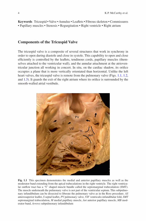
Abstract The tricuspid valve (TV), the morphologically right atrioventricular valve, guards the inflow junction between the right atrium and right ventricle. In functional anatomy, the valve does not consist only of leaflets. Instead, the valve complex is comprised of the annulus, leaflets, tendinous cords and papillary muscles occupying the inlet part of the right ventricle. Right sided structures have not had the extensive analysis when viewed in comparison to the systemic mitral valve. This is possibly due to the complexity of measuring the geometrically unusual shape of the right ventricular cavity, tricuspid valve, the curvature of the muscular ventricular septum and the right ventricular cavity wrapping around the systemic left ventricle. The route of inflow to outflow in the low pressure right ventricle (RV) is elongated compared to the left side of the heart. The right ventricle itself is an anterior struc-ture forming the sterno-costal border of the heart beneath the sternum. The tricuspid valve is always associated with a morphological right ventricle. The chamber can be arbitrarily demarcated into three regions: inlet, apical and outlet, hence the concept of a tripartite ventricle. But, there are no anatomic borders for these regions within the right ventricle. The inflow region of the tricuspid valve is separated from the outflow pulmonary valve by several muscular structures; the ventricular infundibu-lar fold (VIF), septomarginal trabeculation (SMT), septoparietal trabeculations (SPT) and the free standing subpulmonary infundibulum musculature (Fig. 1.1). Adjacent along the atrial side tricuspid valve complex are important structures like the triangle of Koch, tendon of Todaro, atrioventricular node continuing into the bundle of His and coronary sinus orifice (Fig. 1.2). Continuing improvements of imaging methods such as echocardiography, cardiac magnetic resonance imaging and computed tomography to examine in detail and analyse these structures and to measure the flow of deoxygenated blood to the lungs from the right heart allows critical analysis and on-going follow up of patients in normal and disease states.
K.P. McCarthy (*) • S. Yen Ho Brompton Cardiac Morphology Unit, Royal Brompton & Harefield NHS Trust, Sydney Street, London SW3 6NP, UKe-mail: [email protected]
J.L. Robertus Histopathology Department, Royal Brompton & Harefield NHS Trust, Sydney Street, London SW3 6NP, UK
4
Keywords Tricuspid • Valve • Annulus • Leaflets • Fibrous skeleton • Commissures • Papillary muscles • Stenosis • Regurgitation • Right ventricle • Right atrium
Components of the Tricuspid Valve
The tricuspid valve is a composite of several structures that work in synchrony in order to open during diastole and close in systole. This capability to open and close efficiently is controlled by the leaflets, tendinous cords, papillary muscles (them-selves attached to the ventricular wall), and the annular attachment at the atrioven-tricular junction all working in concert. In situ, on the cardiac shadow, its orifice occupies a plane that is more vertically orientated than horizontal. Unlike the left heart valves, the tricuspid valve is remote from the pulmonary valve (Figs. 1.1, 1.2, and 1.3). It guards the exit of the right atrium where its orifice is surrounded by the smooth-walled atrial vestibule.
Fig. 1.1 This specimen demonstrates the medial and anterior papillary muscles as well as the moderator band extending from the apical trabeculations in the right ventricle. The right ventricu-lar outflow tract has a ‘Y’ shaped muscle bundle called the septomarginal trabeculation (SMT). The muscle underneath the pulmonary valve is not part of the ventricular septum. This subpulmo-nary infundibulum can be dissected to liberate the pulmonary valve as in the Ross procedure. AS anterosuperior leaflet, S septal leaflet, PV pulmonary valve, VIF ventriculo-infundibular fold, SMT septomarginal trabeculation, M medial papillary muscle, Ant anterior papillary muscle, MB mod-erator band, Arrows subpulmonary infundibulum
K.P. McCarthy et al.
5
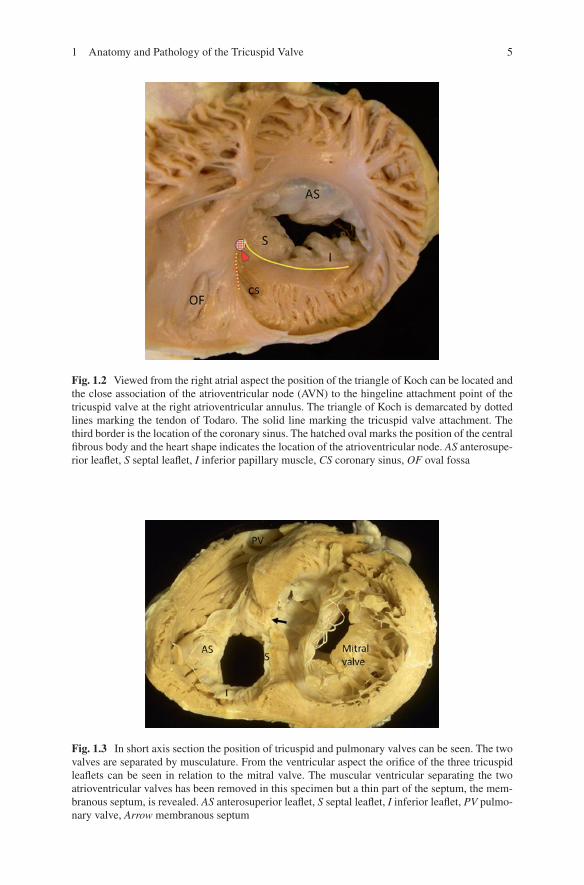
Fig. 1.2 Viewed from the right atrial aspect the position of the triangle of Koch can be located and the close association of the atrioventricular node (AVN) to the hingeline attachment point of the tricuspid valve at the right atrioventricular annulus. The triangle of Koch is demarcated by dotted lines marking the tendon of Todaro. The solid line marking the tricuspid valve attachment. The third border is the location of the coronary sinus. The hatched oval marks the position of the central fibrous body and the heart shape indicates the location of the atrioventricular node. AS anterosupe-rior leaflet, S septal leaflet, I inferior papillary muscle, CS coronary sinus, OF oval fossa
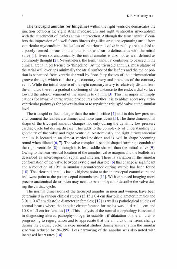
Fig. 1.3 In short axis section the position of tricuspid and pulmonary valves can be seen. The two valves are separated by musculature. From the ventricular aspect the orifice of the three tricuspid leaflets can be seen in relation to the mitral valve. The muscular ventricular separating the two atrioventricular valves has been removed in this specimen but a thin part of the septum, the mem-branous septum, is revealed. AS anterosuperior leaflet, S septal leaflet, I inferior leaflet, PV pulmo-nary valve, Arrow membranous septum
1 Anatomy and Pathology of the Tricuspid Valve
6
The tricuspid annulus (or hingeline) within the right ventricle demarcates the junction between the right atrial myocardium and right ventricular myocardium with the attachment of leaflets at this intersection. Although the term ‘annulus’ con-fers the impression of a well forms fibrous ring-like structure separating atrial from ventricular myocardium, the leaflets of the tricuspid valve in reality are attached to a poorly formed fibrous annulus that is not as clear to delineate as with the mitral valve [1]. Even so, anatomically, the mitral annulus is also not as well defined as commonly thought [2]. Nevertheless, the term, ‘annulus’ continues to be used in the clinical arena in preference to ‘hingeline’. At the tricuspid annulus, musculature of the atrial wall overlaps minimally the atrial surface of the leaflets and the major por-tion is separated from ventricular wall by fibro-fatty tissues of the atrioventricular groove through which run the right coronary artery and branches of the coronary veins. While the initial course of the right coronary artery is relatively distant from the annulus, there is a gradual shortening of the distance to the endocardial surface toward the inferior segment of the annulus to <3 mm [3]. This has important impli-cations for invasive intracardiac procedures whether it is to ablate accessory atrio-ventricular pathways for pre-excitation or to repair the tricuspid valve at the annular level.
The tricuspid orifice is larger than the mitral orifice [4] and in this low pressure environment the leaflets are thinner and more translucent [5]. The three dimensional shape of the tricuspid annulus changes not only during the dynamic low pressure cardiac cycle but during disease. This adds to the complexity of understanding the geometry of the valve and right ventricle. Anatomically, the right atrioventricular annulus is located in an almost vertical position and is oval in shape becoming round when dilated [6, 7]. The valve complex is saddle shaped forming a conduit to the right ventricle [8] although it is less saddle shaped than the mitral valve [9]. Owing to the near vertical location of the annulus, valve margins and the leaflets are described as anterosuperior, septal and inferior. There is variation in the annular conformation of the valve between systole and diastole [6] this change is significant and a reduction of 19% in annular circumference during systole has been found [10]. The tricuspid annulus has its highest point at the anteroseptal commissure and its lowest point at the posteroseptal commissure [11]. With enhanced imaging more precise anatomical description may need to be employed to describe the valve dur-ing the cardiac cycle.
The normal dimensions of the tricuspid annulus in men and women, have been determined in various clinical studies (3.15 ± 0.4 cm diastolic diameter in males and 3.01 ± 0.47 cm diastolic diameter in females) [12] as well as pathological studies of normal hearts where the annular circumference for males was 11.4 ± 1.1 cm and 10.8 ± 1.3 cm for females [13]. This analysis of the normal morphology is essential in diagnosing altered pathophysiology, to establish if dilatation of the annulus is progressing to regurgitation and to appreciate that the annulus dimensions change during the cardiac cycle. In experimental studies during sinus rhythm the annular size was reduced by 20–39%. Less narrowing of the annulus was also noted with increased heart rates [14].
K.P. McCarthy et al.
7
The Tricuspid Leaflets and Commissures
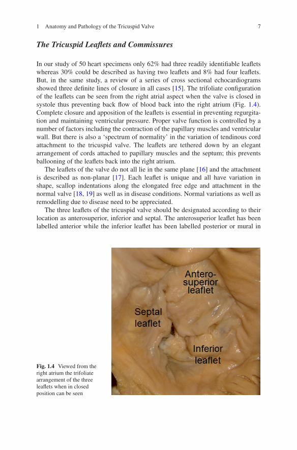
In our study of 50 heart specimens only 62% had three readily identifiable leaflets whereas 30% could be described as having two leaflets and 8% had four leaflets. But, in the same study, a review of a series of cross sectional echocardiograms showed three definite lines of closure in all cases [15]. The trifoliate configuration of the leaflets can be seen from the right atrial aspect when the valve is closed in systole thus preventing back flow of blood back into the right atrium (Fig. 1.4). Complete closure and apposition of the leaflets is essential in preventing regurgita-tion and maintaining ventricular pressure. Proper valve function is controlled by a number of factors including the contraction of the papillary muscles and ventricular wall. But there is also a ‘spectrum of normality’ in the variation of tendinous cord attachment to the tricuspid valve. The leaflets are tethered down by an elegant arrangement of cords attached to papillary muscles and the septum; this prevents ballooning of the leaflets back into the right atrium.
The leaflets of the valve do not all lie in the same plane [16] and the attachment is described as non-planar [17]. Each leaflet is unique and all have variation in shape, scallop indentations along the elongated free edge and attachment in the normal valve [18, 19] as well as in disease conditions. Normal variations as well as remodelling due to disease need to be appreciated.
The three leaflets of the tricuspid valve should be designated according to their location as anterosuperior, inferior and septal. The anterosuperior leaflet has been labelled anterior while the inferior leaflet has been labelled posterior or mural in
Fig. 1.4 Viewed from the right atrium the trifoliate arrangement of the three leaflets when in closed position can be seen
1 Anatomy and Pathology of the Tricuspid Valve
8
other reports. All leaflets are attached like a hinge at the annulus of the right atrio-ventricular junction, leading to the use of the term hinge-line. All three are notice-ably dissimilar in shape and size to each other. The posteroseptal portion of the leaflets is near to the coronary sinus. Taking the plane of the annulus as a whole, the anterolateral portions of the leaflets are nearest to the apex and posterolateral nearer the right atrium. Each leaflet has variable number of scallops along the free edge and is attached within the ventricle via the cords and papillary muscles. Some papers have previously defined the tricuspid valve as having more than three leaflets or cusps [20, 21] and subdivision by scallops of the leaflets has also been described [13]. The general agreement is that there are three tricuspid leaflets which may or may not have scallops.
Viewed from the right atrial aspect the tricuspid valve orifice is roughly triangu-lar in appearance with the three leaflets closing together resulting in a large area of coaptation. The anterosuperior leaflet is the largest and most extensive leaflet that extends curtain-like around approximately half the free wall of the right ventricle from the infundibular outflow region to the inferior part of the right ventricle. Anatomically, the inferior/mural leaflet occupies an inferior location and originates from the diaphragmatic parietal wall of the right ventricle and is also described as the inferior or in older literature as the posterior leaflet.
The septal leaflet is more complex than the anteroseptal and inferior leaflets. This leaflet has been described as occupying the smallest portion of the annulus [8, 22] while other studies describe the mural leaflet as the smallest [23]. From the septal leaflet tendinous cords attach directly to the muscular ventricular septum. This is one of the characteristic features of the tricuspid valve seen with diagnostic imaging that distinguishes it from the mitral valve which does not normally have cordal attachments to the septum. The restricted size of the septal leaflet and the multiple tendinous cord attachments along the septal leaflet directly to the septum results in a different range of movement compared to the other two tricuspid leaflets whose cord attachments have a different morphology [8, 17].
The septal leaflet attachment across the membranous septum divides it into two; a right atrial to right ventricular component which is superior (atrial) to the tricuspid valve and an interventricular component between both ventricles and inferior to the tricuspid valve leaflet. The septal leaflet has one other distinct feature seen with cross sectional imaging and that is its more apically located annular attachment in contrast to the mitral valve. This offset attachment is a key feature seen in diagnostic cross-sectional imaging in the four-chamber plane that aids identification of ven-tricular morphology in congenital heart disease.
Corresponding to the three leaflets configuration, there are three commissures or peaks of apposition between adjacent leaflets. It is important, however, to appreciate that although ‘tricuspid’ denotes three leaflets, there is no separation of one leaflet from another in that the three leaflets are arranged like a skirt and commissures between the leaflets do not extend all the way to the level of the annulus. Instead, continuity between adjacent leaflets is preserved by at least 5 mm of leaflet tissue along the annulus. This continuity is crucial for maintaining integrity of the valve when the leaflets come together in apposition. As mentioned above, the septal leaflet
K.P. McCarthy et al.
9
crosses the membranous septum. It swings ‘around the corner’, away from the sep-tum into the antero-superior leaflet. Thus, the antero-septal commissure attached to the medial papillary muscle is away from the septum. Interestingly, at the membra-nous septum, it is not uncommon to find a gap in the septal leaflet. Leaflet tissue around this area is usually adequate to provide apposition to close the orifice.
From the attachment of the leaflet at the annulus to the free edge, the tricuspid leaflet has specific zones [13]. The clear zone comprises the majority of the leaflet and is the thin translucent central portion and generally comprises two-thirds of the leaflet length from the annulus toward the free margin.
The basal zone is the portion of the leaflet attached to the annulus at the atrioven-tricular junction at this connection point there is vascularisation and innervation to the leaflet. This is the junction where atrial myocardium inserts at the annulus. The rough zone forms the free edge and the region of apposition, forming approximately a third of the leaflet length. This thicker portion of the leaflet contains more glycos-aminglycans allowing cushioning of the leaflet edges as they oppose during closure. These zones emphasise that the morphology is not uniform macroscopically or microscopically throughout the leaflet.
In cross section, each leaflet extends from the annulus hinge line proximally to the free edge distally. The normal tricuspid valve has a layered leaflet arrangement of the atrialis, spongiosa, fibrosa and ventricularis layers surrounded by a layer of endothelial cells which is continuous with the luminal surface of the atrium and the ventricle. In the tricuspid valve the leaflet comprises interstitial fibroblasts and con-nective tissue fibres within an extracellular matrix.
The atrialis is the uppermost layer. When the valve is in closed position, the atria-lis faces directly the right atrium. This layer is composed of mainly aligned elastic and collagen fibres covered with overlying endothelium. Of the layers, the atrialis has the most elastic fibres [24]. Beneath the atrialis is the spongiosa layer. This layer is composed largely of an extracellular matrix of proteoglycans and glycosamingly-cans, along with elastic fibres. The glycosaminglycans and proteoglycans are hydro-philic and attract water molecules. This causes the extracellular matrix to expand and swell at the free edge, providing a natural physical protective buffer to the leaflet. The spongiosa functions as a physiological ‘shock absorber’. It provides a structural cushion along the point of apposition to offset the effect of leaflet closure at the free edge.
Beneath the spongiosa is the fibrosa layer which is the major load-bearing layer, comprising the central structural collagenous core [5] of compact and aligned col-lagen fibres to the leaflet. The fibrosa layer extends from the annulus into two thirds of the leaflet and is absent at the free edge. The final layer of the tricuspid leaflet is the ventricularis, this layer is covered by a continuous sheet of endothelial cells that overlie elastic fibres and collagen fibres. The thickness of each layer varies from the attachment site at the annulus to the free edge. At the proximal region of the leaflet, near the annulus, the fibrosa is the thickest layer providing the structural core of the leaflet. The spongiosa and atrialis in contrast are relatively thin at the annulus attach-ment point of the leaflet, but increase in thickness distally, becoming the main com-ponent of the leaflet at the free edge.
1 Anatomy and Pathology of the Tricuspid Valve
10
Histologically, the extracellular matrix surrounds the connective tissue fibres in the leaflet and is a dynamic substrate of neutral pH [25]. The leaflet is composed of the glycosaminoglycan hyaluronic acid and proteoglycans such as aggrecan, deco-rin, veriscan [26–28] as well as fibroblast cells, some with variable phenotypes exhibiting actin filaments [29].
The maintenance of connective tissue integrity is fundamental in determining the overall strength and competence of the leaflet. This depends upon a contin-ued balance between synthesis of the matrix components, their deposition and repair within the leaflet and the degradation of the tissue elements. Metalloproteinase enzymes, synthesised from fibroblasts, regulate the renewal and turnover of matrix components in connective tissues [30] within the human cardiac valves [31].
Under normal physiological conditions, the extracellular matrix comprising fibrillar proteins of the normal leaflet undergoes constant turnover of constituents to maintain both structure and function with the expression various proteins, including the contractile proteins troponin [32] and myosin [33]. These factors contribute in determining the overall structural integrity of the leaflets. External factors, such as mechanical stresses exerted on the valve leaflet, may also trigger secondary physi-ological responses affecting the structure and function of the leaflet.
Collagen fibres are the major component of atrioventricular leaflets and have an important role in defining shape, integrity and mechanical strength. Collagen fibres are produced from interstitial fibroblasts and endothelial cells [34, 35]. The align-ment of collagen provides mechanical strength required for the repeated opening and closing of the valve. Elastic fibres interconnect with the collagen fibrils and bundles and promote recoil ensuring the leaflet and tendinous cord can return to the resting state.
The pH of the extracellular matrix environment affects the morphology of the fibres. Normal collagen fibres in the tissues are tightly packed and interspersed with smaller homogenous fibrils [36]. These are stable within a near neutral pH [37] but if the pH is altered, as seen in studies on collagen fibre development in chicks where the pH was more acidic, the collagen fibres synthesised can have variable diameters [38].
Matrix enzymes such as matrix metalloproteinases (MMP) and tissue inhibitor of matrix metalloproteinases (TIMP) can also act on collagen fibres [30] to remodel the leaflet tissue.
Tendinous Cords and Papillary Muscles
The slender and fibrous tendinous cords are the key interconnecting structure tether-ing the leaflets to the papillary muscles ensuring a functional and efficient valve. In the normal valve, leaflet cords and interleaflet cords have been identified. Fan- shaped tendinous cords insert at the junction between each leaflet, facilitating the bringing together and separation of adjacent leaflets as the valve closes and opens. These are the interleaflet cords and they have multiple connections between
K.P. McCarthy et al.
11
adjacent leaflet connecting to the papillary muscles. Leaflets cords include rough zone tendinous cords which insert directly to the ventricular surface of the leaflet. The thicker ones are sometimes referred to as strut cords as they bear the mechani-cal load during valve opening and closing. On the ventricular aspect of the leaflet there are also attachments of tendinous cords to the rough and clear zones of the leaflet and these are termed deep cords. Basal cords can be found attaching the underside of the leaflets close to the annulus directly to the ventricular wall. Further leaflet cords also attach to the free edge of the leaflet and these are simply described as free-edge cords. The multiple attachments of the tendinous cords along the leaf-lets result in greater support and control of the valve.
Although the papillary muscles are not as uniformly distributed as in the mitral valve, the anterior papillary muscle sited in the right ventricle is usually well- defined, supporting the antero-superior leaflet in its midportion. Usually, there is a cluster of smaller papillary muscles are located laterally or inferiorly in the right ventricle and these support the inferior as well as the antero-superior leaflets. Supporting the commissure between the septal and antero-superior leaflets is a small papillary muscle called the medial (or conal) papillary muscle, also known as the muscle of Lancisi. Variability in number of papillary muscles has been noted as well as the papillary muscle group having multiple heads [39, 40].
The papillary muscles are extensions of trabecular myocardium extending from the apical portion of the right ventricle and are highly innervated, highly vascular-ised with a central artery, and carry the distal ramifications of the purkinje fibre network. The most common pattern of coronary supply to the right ventricle and papillary muscles is via the dominant right coronary artery running within the right atrioventricular groove. Histologically, the cords are formed of a collagen core with elastic fibres surrounded by endothelial cells. The mechanical load is supported by the collagen fibres during systole.
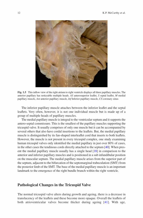
The anterior papillary muscle is the largest of the three papillary muscles and usually is composed of a distinct conical muscle with multiple heads with cords attached, hence it is described as bifid or trifid. Sometimes, there are several papillary muscles fused together or there is a dominant papillary muscle with adjoining smaller papillary muscles. They are located between the anteroseptal leaf-let and the inferior leaflet. Additionally, further support from smaller anterior mus-cles is provided to the anterior segment of the leaflet (Fig. 1.5). The anterior papillary muscle extends from the apical portion of the right ventricle. It usually arises or continues from the moderator band as the latter muscle band crosses the right ven-tricular cavity. The origin of the anterior papillary muscle from the moderator band can be midway along the band or close to where the band itself fuses with the right ventricular free wall, on cross sectional imaging, recognising the moderator band is one way of distinguishing the morphologically right ventricle from a left ventricle. Apart from the distinctive moderator band, the right ventricular chamber is criss- crossed by further muscle bundles termed trabeculations and these mainly occupy the apical portion. They interconnect extending from the septal to the apical and parietal walls and these trabeculations are course in comparison to those in the left ventricle.
1 Anatomy and Pathology of the Tricuspid Valve
12
The inferior papillary muscle attaches between the inferior leaflet and the septal leaflets. Very often, however, it is not one individual muscle but is made up of a group of multiple heads of papillary muscles.
The medial papillary muscle is integral to the ventricular septum and it supports the antero-septal commissure. This is the smallest of the papillary muscles supporting the tricuspid valve. It usually comprises of only one muscle but it can be accompanied by several others that also have cordal insertions to the leaflets. But, the medial papillary muscle is distinguished by its fan-shaped interleaflet cord that inserts to both leaflets. However, the muscle is not present in every tricuspid complex, one study examining human tricuspid valves only identified the medial papillary in just over 80% of cases, in the other cases the tendinous cords directly attached to the septum [40]. When pres-ent the medial papillary muscle usually has a single head [18] in comparison to the anterior and inferior papillary muscles and is positioned in a sub infundibular position on the muscular septum. The medial papillary muscle arises from the superior part of the septum, adjacent to the bifurcation of the septomarginal trabeculation (SMT) from the posterior limb of the SMT. The base of the medial papillary muscle is an important landmark to the emergence of the right bundle branch within the right ventricle.
Pathological Changes in the Tricuspid Valve
The normal tricuspid valve alters during growth and ageing, there is a decrease in translucency of the leaflets and these become more opaque. Overall the leaflets of both atrioventricular valves become thicker during ageing [41]. With age,
Fig. 1.5 This inflow view of the right atrium to right ventricle displays all three papillary muscles. The anterior papillary has noticeable multiple heads. AS anterosuperior leaflet, S septal leaflet, M medial papillary muscle, Ant anterior papillary muscle, Inf Inferior papillary muscle, CS coronary sinus
K.P. McCarthy et al.
13
elongation and doming of the tricuspid valve apparatus can occur along with floppy changes and this can lead to valve regurgitation [19]. Heart valve disease is an increasing clinical challenge encompassing valve replacement and reconstruc-tion of the valve to maintain appropriate physiological parameters. Newer tech-nologies, including tissue engineering, are being employed to repair valvar disease.
The Tricuspid Valve in Disease
Tricuspid Regurgitation
The purpose of an efficient tricuspid valve to the pumping of blood has been noted since ancient Greece [42]. When the valve fails tricuspid regurgitation occurs and this is a common condition in asymptomatic patients which can be associated with an adverse diagnosis. However the study of regurgitation has not been as extensive as systemic heart disease [43]. Functional regurgitation is a lesion produced by a disturbance of the efficient coordination of all the elements of the valve complex such as caused by annular dilatation, elongation of the valve cords, perforation of the leaflet, and so on. Dysfunction of the valve pri-marily leads to incompetence and regurgitation of blood back into the right atrium cardiac output decreases and this can potentially progress to atrial dilata-tion/fibrillation. There are several etiologies to cause alteration to one or more valve components resulting in regurgitation. Dilatation of the tricuspid annulus and the attachment of the leaflets are recognised as the primary mechanisms of functional TV regurgitation.
Structural anomalies of the valve apparatus include unguarded TV orifice, cord rupture, flail cords [44], parachute TV [45], double orifice TV, cleft TV, and Ebstein malformation. Acquired lesions affecting the valve such as prolapse, dysplasia, floppy TV, papillary fibroelastoma, rheumatic changes, endocarditis, carcinoid dis-ease, endomyocardial fibrosis, RV dilatation following pulmonary hypertension or cardiomyopathy and RV dysfunction as a result of myocardial disease, RV ischemia or infarction can cause tricuspid regurgitation. There are also iatrogenic lesions fol-lowing interventions in patients with permanent pacemakers or implantable defibril-lators which may damage the tricuspid valve and cause tricuspid regurgitation. Over time when the leads and guide wires of such devices become adherent to the valvar elements that may disturb the cords, papillary muscles or leaflets and be the cause of valvar insufficiency. Adhesions also put the valve at risk of damage during lead extraction. Or, the tricuspid valve can be damaged during biopsy collection follow-ing heart transplantation and this leads to regurgitation of the valve [46]. Use of circular mapping catheters of the tricuspid valve in adult congenital heart disease may also increase risk of damage. Overall, regurgitation from the tricuspid valve can be divided into functional and degenerative in origin. Functional regurgitation can be caused by an enlarged/dilated ventricle. Degenerative incompetence affects the valve and the apparatus itself such as; rupture of the cords, or papillary muscles or damage to the leaflets.
1 Anatomy and Pathology of the Tricuspid Valve
14
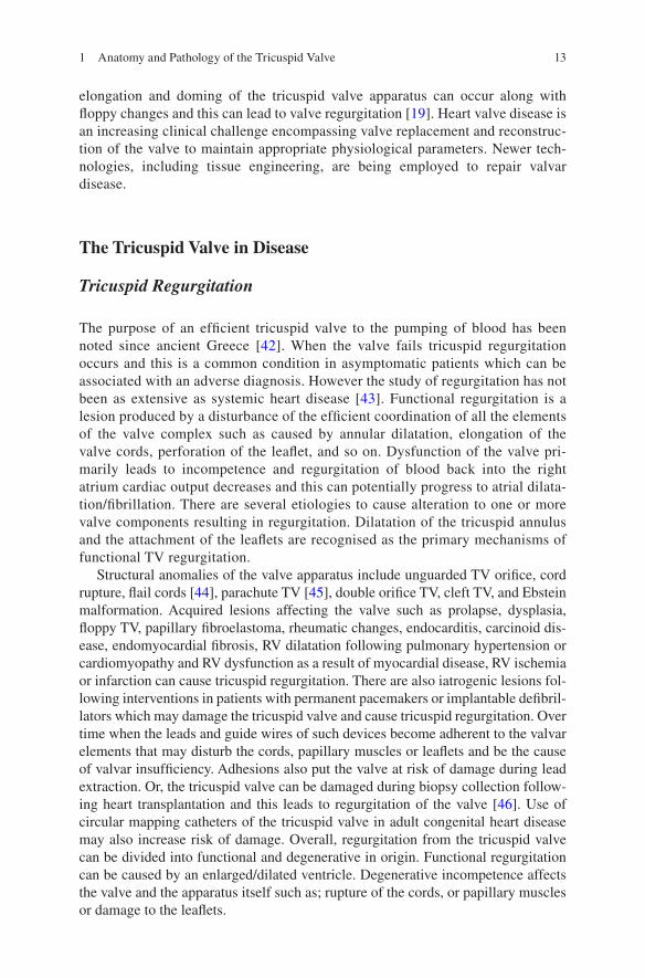
Among the congenital abnormalities of the TV causing regurgitation, Ebstein anomaly is probably the most distinctive although its milder forms may be missed. This malformation can be associated with other congenital heart defects but more commonly it co-exists with an atrial septal defect. Developmentally, there is incom-plete delamination of the septal and inferior leaflets from the ventricular wall/septum causing the hingelines of these two leaflets to be located well within the ventricular chamber, away from the level of the atrioventricular junction (Fig. 1.6). The most affected site is usually the region between the inferior and septal leaflets [47]. In cross-sectional imaging, the four chamber plane shows increase in length of offset at the septum between the mitral and tricuspid insertions, with the tricuspid more api-cally displaced than normal. In mildly affected cases, the increased offset can be difficult to recognise. In severe more forms, the septal leaflet may be appear like an bubble of tissue on the septum, or be missing altogether. The lack of delamination of the affected leaflets can result in the corresponding ventricular inferior wall becom-ing thinner, resembling the thickness of the atrial wall, and described as atrialisation of the ventricle. Indeed, in such cases the atrial chamber appears larger than it is, even taking into account the effect of tricuspid regurgitation. The antero- superior leaflet is well delaminated and hinged appropriately to the atrioventricular junction. However, it is never entirely normal. Often it is large, deeper than normal, and has abnormal attachments and abnormal papillary muscles supporting it. Sometimes, its leaflet may be muscularised, leading to suggestions that the muscle may be sub-strates for accessory atrioventricular pathways. In some severe forms, instead of attaching to papillary muscles, its free edge may be attached to a large muscle band that subdivides the right ventricle. This arrangement can result in the effective orifice of the valve that is the gap between the septum and the antero- superior leaflet being displaced toward the right ventricular outlet and becoming much reduced in size, leading to valvar incompetence and stenosis.
Fig. 1.6 This case of Ebstein malformation of the tricuspid valve particularly shows the failure of delamination of the septal leaflet and this is now a bubble-like formation on the ventricular septal surface. The attachment is not at the atrioventricular junction, indicated by the dotted line. This case of Ebstein also shows the atrialisation of a portion of the right ventricular wall. The anterosu-perior leaflet is forming the curtain of valve tissue to separate inflow from outflow in this ventricle. OF oval fossa, AS anterosuperior leaflet, S septal leaflet, Dotted line atrioventricular junction
K.P. McCarthy et al.
15
Other examples of congenital tricuspid malformations causing regurgitation include leaflet dysplasia, cordal elongations with hooding of leaflets, accessory ori-fices, breach or cleft in a leaflet, and agenesis of one or more leaflets.
Valvar Stenosis
Stenosis of the tricuspid valve involves one or more elements of the valvar appara-tus that is abnormally formed or become changed resulting in reduction of flow from the atrium to the ventricle. For instance, reduction in annular size results in narrowing of the effective atrioventricular orifice as the tricuspid valve fails to open fully in diastole thus hindering filling of the ventricle Tricuspid stenosis can take various pathologies from acquired to congenital origins.
Acquired tricuspid stenosis includes forms of degenerative disease, endocarditis, stiffening of the leaflets and calcification. Some cases of endocarditis develop perfora-tions in the leaflets leading to regurgitation. The tricuspid valve is the most frequently affected valve in carcinoid heart disease resulting in tricuspid stenosis. Furthermore, the leaflets become thickened with increased fibrosis resulting in stiffening and reduced overall motility. Vegetations on the tricuspid leaflets can also affect leaflet motility and hazard of embolic event. Rheumatic disease is also a cause of stenosis where thick leaflets have restricted motion, there is fusion at the commissures, short-ening of cords as well as reduced separation of the leaflets leading to a restricted ori-fice. Ultimately, the valve becomes stenotic as well as regurgitant. Globally the most common cause of tricuspid stenosis is rheumatic fever most often found in children.
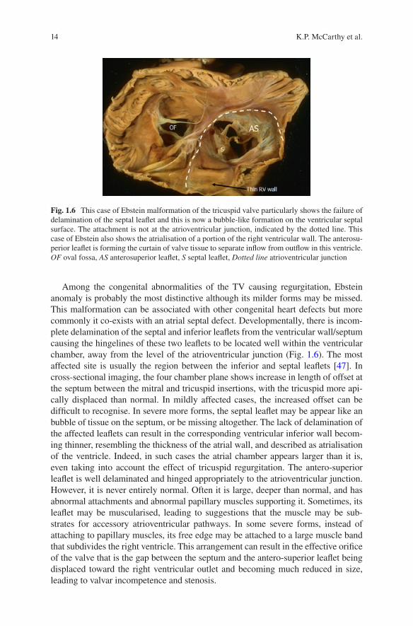
Congenitally malformed tricuspid valves obstructing inflow into the right ven-tricle can occur in isolation or with other congenital heart lesions. The underlying malformation affect one or more elements of the valve e.g. underdeveloped leaflets, short and thickened or fused tendinous cords, a small annulus or abnormal papillary muscles. The extreme example of the malformation is in the rare setting of tricuspid atresia in infants where there is complete occlusion of the tricuspid valve orifice owing to fusion of the valvar leaflets forming an imperforate membrane across the atrioventricular junction. It is pertinent to highlight that the more common forms of ‘tricuspid atresia’ are hearts without any evidence of a tricuspid valve (Fig. 1.7). These are hearts with univentricular atrioventricular connection in which the right sided atrium has no egress to the ventricular mass other than through an interatrial communication to the left side of the heart, in other words these are hearts with absence of the right atrioventricular connection.
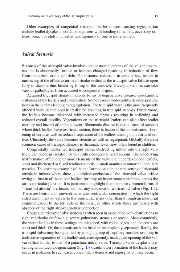
Congenital tricuspid valve stenosis is often seen in association with obstructions to right ventricular outflow e.g. severe pulmonary stenosis or atresia. Most commonly, the valvar leaflets in these settings are thickened, with rolled edges, and the cords are short and thick. Or, the commissures are fused or incompletely separated. Rarely, the tricuspid valve may be supported by a single group of papillary muscles resulting in ineffective separation of the leaflets and, consequently, inadequate opening of the val-var orifice similar to that of a parachute mitral valve. Tricuspid valve dysplasia pre-senting with mucoid degeneration (Fig. 1.8), cauliflower formation of the leaflets may occur in isolation. In such cases concomitant stenosis and regurgitation may occur.
1 Anatomy and Pathology of the Tricuspid Valve
16
Straddling Tricuspid Valve
In the presence of a ventricular septal defect the tricuspid valve can straddle the defect [48] with cords attaching within the left ventricle or the cords can be anchored to a papillary muscle within the left ventricle. If the ventricular septal defect is asso-ciated with malignment of the septums then not only is the valve leaflet straddling across the defect but the valve orifice may also override the septum resulting in
Fig. 1.7 In this case the is no evidence of the formation of a tricuspid valve. This is termed absent right atrioventricular connection (sometimes referred to as tricuspid atresia). OF oval fossa, CS coronary sinus, App. Right atrial appendage, RA right atrium
Fig. 1.8 In this example of tricuspid dysplasia the leaflets and highly thickened and not as mobile as the cords area also shortened leading to valve regurgitation. AS anterosuperior leaflet, S septal leaflet
K.P. McCarthy et al.
17
tricuspid inflow going to both ventricles. The malignment of the atrial and ventricu-lar septums at the atrioventricular junction in this setting is also associated with deviation of the atrioventricular node rightward and inferiorly.
Conclusion
The tricuspid valve, like the mitral valve, requires coordination and integrity of its various components for normal function. It is a common misconception to consider a valve solely in terms of its leaflets. For proper function, the tricuspid valve requires all its component parts i.e. the annulus with extension of atrial wall, the leaflets, tendinous cords, papillary muscles and the associated ventricular/septal walls to work in harmony. Not only that, the surrounding cardiac structures should be exam-ined prior to considering any interventional procedures on the valve. Of particular note are the locations of the atrioventricular node and His bundle of the conduction system. The course of the right coronary artery should also be taken into account.
There is still much to learn about the complex tricuspid valve. Understanding the valve features in the normal valve, the normal spectrum of variation, the natural changes that occur during ageing and those changes during disease will help in understanding the changes that occur to this dynamic and highly mobile structure.
Take Home Points
• The tricuspid valve is not simply the leaflets but a valve complex involving the cords, papillary muscles, annulus and the surrounding musculature.
• The valve remodels during aging becoming thicker and prone to regurgitation.• With numerous scallops, cord attachment and papillary muscle morphology, the
tricuspid valve has greater variation than the anatomy of the mitral valve.
Review Questions
1. Which of the following sentences is correct?
(a) The tricuspid valve has greater variation than the anatomy of the mitral valve (b) The tricuspid valve has always three clearly identifiable three leaflets as
proven on post-mortem studies (c) the tricuspid valve leaflets position is less apical than those of the mitral
valve (d) The tricuspid valve is not always associated with a morphological right
ventricle.
1 Anatomy and Pathology of the Tricuspid Valve
18
2. Which of the following sentences is correct?
(a) The tricuspid valve leaflets are uniform throughout in structure? (b) The tricuspid valve septal and anterior leaflets are uniform throughout in
structure? (c) The tricuspid valve septal and posterior leaflets are uniform throughout in
structure? (d) The tricuspid valve leaflets are not uniform throughout in structure?
3. Which of the following sentences is correct?
(a) Two of the tricuspid valve papillary muscles are attached to septum (b) Unlike the left heart valves, the tricuspid valve is remote from the pulmonary
valve (c) The tricuspid orifice is smaller than the mitral orifice (d) The tricuspid annulus has a near horizontal location
4. Which of the following sentences is correct?
(a) The attachment of the tricuspid valve leaflets crosses the atrioventricular node?
(b) The attachment of the inferior (posterior) tricuspid valve leaflet is the closest to the atrioventricular node?
(c) The attachment of the anterior (anterosuperior) tricuspid valve leaflet is the closest to the atrioventricular node?
(d) The attachment of the septal tricuspid valve leaflet is separated by the central fibrous body from the atrioventricular node?
5. Which of the following sentences is correct?
(a) The right coronary artery runs in closest proximity to the tricuspid annulus at its initial course.
(b) The right coronary artery runs always above to the tricuspid annulus. (c) The right coronary artery runs always below to the tricuspid annulus. (d) There is a gradual shortening of the distance between the right coronary
artery and the endocardial surface toward the inferior segment of the annulus to <3 mm.
References
1. Taramasso M, Vanermen H, Maisano F, Guidotti A, La Canna G, Alfieri O. The grow-ing clinical importance of secondary tricuspid regurgitation. J Am Coll Cardiol. 2012;59:703–10.
2. Angelini A, Ho SY, Anderson RH, Davies MJ, Becker AE. A histological study of the atrio-ventricular junction in hearts with normal and prolapsed leaflets of the mitral valve. Br Heart J. 1988;59(6):712–6.
K.P. McCarthy et al.
19
3. Ueda A, McCarthy KP, Sánchez-Quintana D, Ho SY. Right atrial appendage and vestibule: further anatomical insights with implications for invasive electrophysiology. Europace. 2013;15(5):728–34.
4. Yacoub MH, Cohn LH. Novel approaches to cardiac valve repair from structure to function: part I. Circulation. 2004;109:942–50.
5. Misfeld M, Sievers HH. Heart valve macro- and microstructure. Philos Trans R Soc Lond B Biol Sci. 2007;362:1421–36.
6. Kwan J, Kim GC, Jeon MJ, Kim DH, Shiota T, Thomas JD, Park KS, Lee WH. 3D geometry of a normal tricuspid annulus during systole: a comparison study with the mitral annulus using real-time 3D echocardiography. Eur J Echocardiogr. 2007;8:375–83.
7. Shah PM, Raney AA. Tricuspid valve disease. Curr Probl Cardiol. 2008;33:47–84. 8. Rogers JH, Bolling SF. The tricuspid valve. current perspective and evolving management of
tricuspid regurgitation. Circulation. 2009;119:2718–25. 9. Bateman MG, Quill JL, Hill AJ, Iaizzo PA. The clinical anatomy and pathology of the human
atrioventricular valves: implications for repair or replacement. J Cardiovasc Trans Res. 2013;6:155–65.
10. Fukuda S, Saracino G, Matsumara Y, et al. Three-dimensional geometry of the tricuspid annu-lus in healthy subjects and in patients with functional tricuspid regurgitation: a real-time, 3-dimensional echocardiographic study. Circulation. 2006;114:I492–8.
11. Fawzy H, Fukamachi K, Mazer CD, Harrington A, Latter D, Bonneau D, Errett L. Complete mapping of the tricuspid valve apparatus using three-dimensional sonomicrometry. J Thorac Cardiovasc Surg. 2011;141:1037–43.
12. Dwivedi G, Mahadevan G, Jimenez D, Frenneaux M, Steeds RP. Reference values for mitral and tricuspid annular dimensions using two-dimensional echocardiography. Echo Res Pract. 2014;1(2):43–50.
13. Silver MD, Lam JHC, Ranganathan N, Wigle ED. Morphology of the human tricuspid valve. Circulation. 1971;43:333–48.
14. Tsakiris AG, Mai DD, Seki S, et al. Motion of the tricuspid valve annulus in anesthetized intact dogs. Circ Res. 1975;36(1):43–8.
15. Sutton JP, Ho SY, Vogel M, Anderson RH. Is the morphologically right atrioventricular valve tricuspid? J Heart Valve Dis. 1995;4(6):571–5.
16. Dreyfus GD, Martin RP, KMJ C, Dulguerov F, Alexandrescu C. Functional tricuspid regurgita-tion. a need to revise our understanding. J Am Coll Cardiol. 2015;65(21):2331–6.
17. Huttin O, Voilliot D, Mandry D, Vennera C, Juillière Y, Selton-Sutya C. All you need to know about the tricuspid valve: tricuspid valve imaging and tricuspid regurgitation analysis. Arch Cardiovasc Dis. 2016;109:67–80.
18. Tretter JT, Sarwark AE, Anderson RH, Spicer DE. Assessment of the anatomical variation to be found in the normal tricuspid valve. Clin Anat. 2016;29:399–407.
19. Davies MJ. The mitral valve. In: Pathology of cardiac valves. London: Butterworths; 1980. 20. Ikegaya T, Kurata C, Hayashi H, Muro H, Yamazaki N. A case of congenital tricuspid valve
abnormality showing six leaflets. Eur Heart J. 1991;12:94–5. 21. Wafae N, Hayashi H, Gerola LR, Vieira MC. Anatomical study of the human tricuspid valve.
Surg Radiol Anat. 1990;12:37–41. 22. Antoniali F, Braile DM, Poterio GMB, da Costa CE, Lopes MM, Ribeiro GCA, Tarelho
LDS. Proportion among the segments of the normal tricuspid valve annulus: parameter for valve annuloplasty. Braz J Cardiovasc Surg. 2006;21(3):262–71.
23. Skwarek M, Hreczecha J, Dudziak M, Jerzemowski J, Szpinda M, Grzybiak M. Morphometric features of the right atrioventricular orifice in adult human hearts. Folia Morphol (Warsz). 2008;67(1):53–7.
24. Otto S, Baum T, Keller F. Sex-dependence of the relative number of elastic fibres in human heart valves. Ann Anat. 2006;188:153–8.
25. Stetler-Stevenson WG. Dynamics of matrix turnover during pathologic remodeling of the extracellular matrix. Am J Pathol. 1996;148:1345–50.
1 Anatomy and Pathology of the Tricuspid Valve
20
26. Grande-Allen KJ, Calabro A, Gupta V, Wight TN, Hascall VC, Vesely I. Glycosaminoglycans and proteoglycans in normal mitral valve leaflets and chordae: association with regions of tensile and compressive loading. Glycobiology. 2004;14:621–33.
27. Iozzo RV. The biology of the small leucine-rich proteoglycans. J Biol Chem. 1999;274(27):18843–6.
28. McDonald PC, Wilson JE, McNeill S, Gao M, Spinelli JJ, Rosenberg F, Wiebe H, McManus BM. The challenge of defining normality for human mitral and aortic valves. Geometrical and compositional analysis. Cardiovasc Pathol. 2002;11:193–209.
29. Filip DA, Radu A, Simionescu M. Interstitial cells of the heart valves possess characteristics similar to smooth muscle cells. Circ Res. 1986;59:310–20.
30. Woessner FJ, Nagase H. Introduction, overview of the individual TIMPS; Protein substrates of the MMPs. In: Matrix metalloproteinases and TIMPs. 2nd ed. New York: Oxford University Press; 2002.
31. Dreger SA, Taylor PM, Allen SP, Yacoub MH. Profile and localization of matrix metallopro-teinases (MMPs) and their tissue inhibitors (TIMPs) in human heart valves. J Heart Valve Dis. 2002;11:875–80.
32. Roy A, Brand NJ, Yacoub MH. Molecular characterization of interstitial cells isolated from human heart valves. J Heart Valve Disease. 2000;9:459–64.
33. Taylor PM, Batten P, Brand NJ, Thomas PS, Yacoub MH. The cardiac valve interstitial cell. Int J Biochem Cell Biol. 2003;35:113–8.
34. Hafizi S, Taylor PM, Chester AH, Allen SP, Yacoub MH. Mitogenic and secretory responses of human valve interstitial cells to vasoactive agents. J Heart Valve Disease. 2000;9:454–8.
35. Mulholland DL, Gotlieb AI. Cardiac valve interstitial cells: regulator of valve structure and function. Cardiovasc Pathol. 1997;6:167–74.
36. Ottani V, Raspanti M, Ruggeri A. Collagen structure and functional applications. Micron. 2001;32:251–60.
37. Zanaboni G, Rossi A, Tina Onana AM, Tenni R. Stability and networks of hydrogen bonds of the collagen triple helical structure: influence of pH and chaotrophic nature of three anions. Matrix Biol. 2000;19:511–20.
38. Bard JBL, Hulmes DJS, Purdom IF, Ross ASA. Chick corneal development in vitro: diverse effects of pH on collagen assembly. J Cell Sci. 1993;105:1045–55.
39. Aktas EO, Govsa F, Kocak A, Boydak B, Yavuz IC. Variations in the papillary muscles of normal tricuspid valve and their clinical relevance in medicolegal autopsies. Saudi Med J. 2004;25(9):1176–85.
40. Loukas M, Tubbs RS, Louis RG Jr, Apaydin N, Bartczak A, Huseng V, Alsaiegh N, Fudalej M. An endoscopic and anatomical approach to the septal papillary muscle of the conus. Surg Radiol Anat. 2009;31:701–6.
41. Gross L, Kugel MA. Topographic anatomy and histology of the valves in the human heart. Am J Pathol. 1931;7:445–73.
42. Paraskevas G, Koutsouflianiotis K, Iliou K. The first descriptions of various anatomical structures and embryological remnants of the heart: a systematic overview. Int J Cardiol. 2017;227:674–90.
43. Mascherbauer J, Maurer G. The forgotten valve: lessons to be learned in tricuspid regurgita-tion. Eur Heart J. 2010;31:2841–3.
44. D’Aloia A, Bonadei I, Vizzardi E, Sciatti E, Bugatti S, Curnis A, Metra M. Different types of tricuspid flail: case reports and review of the literature. Hell J Cardiol. 2016;57:134–7.
45. Godart F, Piot D, Rey C. Parachute tricuspid valve, supravalvar tricuspid ring, and coarctation of aorta in congenitally corrected transposition. Cardiol Young. 1997;7:337–9.
46. Fiorelli AI, Coelho GHB, Aiello VD, Benvenuti LA, Palazzo JF, Santos Júnior VP, Canizares B, Dias RR, Stolf NAG. Tricuspid valve injury after heart transplantation due to endomyocar-dial biopsy: an analysis of 3550 biopsies. Transplant Proc. 2012;44:2479–82.
K.P. McCarthy et al.

21
47. Schreiber C, Cook A, Ho SY, Augustin N, Anderson RH. Morphologic spectrum of Ebstein's malformation: revisitation relative to surgical repair. J Thorac Cardiovasc Surg. 1999;117(1):148–55.
48. Milo S, Ho SY, Macartney FJ, Wilkinson JL, Becker AE, Wenink AC, Gittenberger de Groot AC, Anderson RH. Straddling and overriding atrioventricular valves: morphology and clas-sification. Am J Cardiol. 1979;44(6):1122–34.
1 Anatomy and Pathology of the Tricuspid Valve
Part IIClinical Perspective