practical of clinical hematology collected and prepared by mr. mohammed o. jaber mr. mohammed o....
TRANSCRIPT
Practical of Clinical Practical of Clinical HematologyHematology
Collected and prepared by
Mr. Mohammed O. JaberMr. Mohammed O. Jaber
Medical Technology DepartmentMedical Technology DepartmentIslamic University-GazaIslamic University-Gaza
RBCs Abnormal morphology
Peripheral Blood MorphologyPeripheral Blood Morphology
Abnormal erythrocyte morphology Is found in pathological states that may be
abnormalities in I. Red cell distribution.II.Size (anisocytosis).III.Hemoglobin content – Color Variation .IV.Shape (poikilocytosis).V.The presence of inclusion bodies in
erythrocyte.
I.I. Erythrocyte Distribution Erythrocyte Distribution AbnormalitiesAbnormalities
Rouleaux formation Stacking of RBCs due to increased plasma proteins coating RBCs
Agglutination Antibody-mediated clumping; temperature
dependent
I.I. Erythrocyte Distribution Erythrocyte Distribution AbnormalitiesAbnormalities
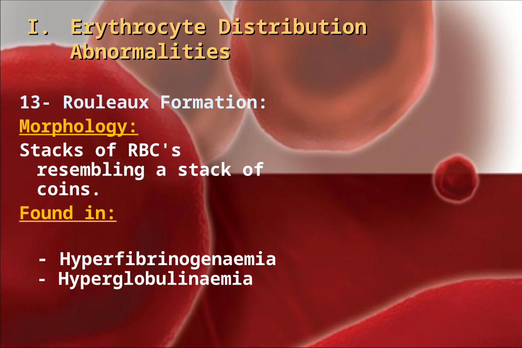
13- Rouleaux Formation:Morphology:Stacks of RBC's resembling a
stack of coins.Found in:
- Hyperfibrinogenaemia- Hyperglobulinaemia
I.I. Erythrocyte Distribution Erythrocyte Distribution AbnormalitiesAbnormalities
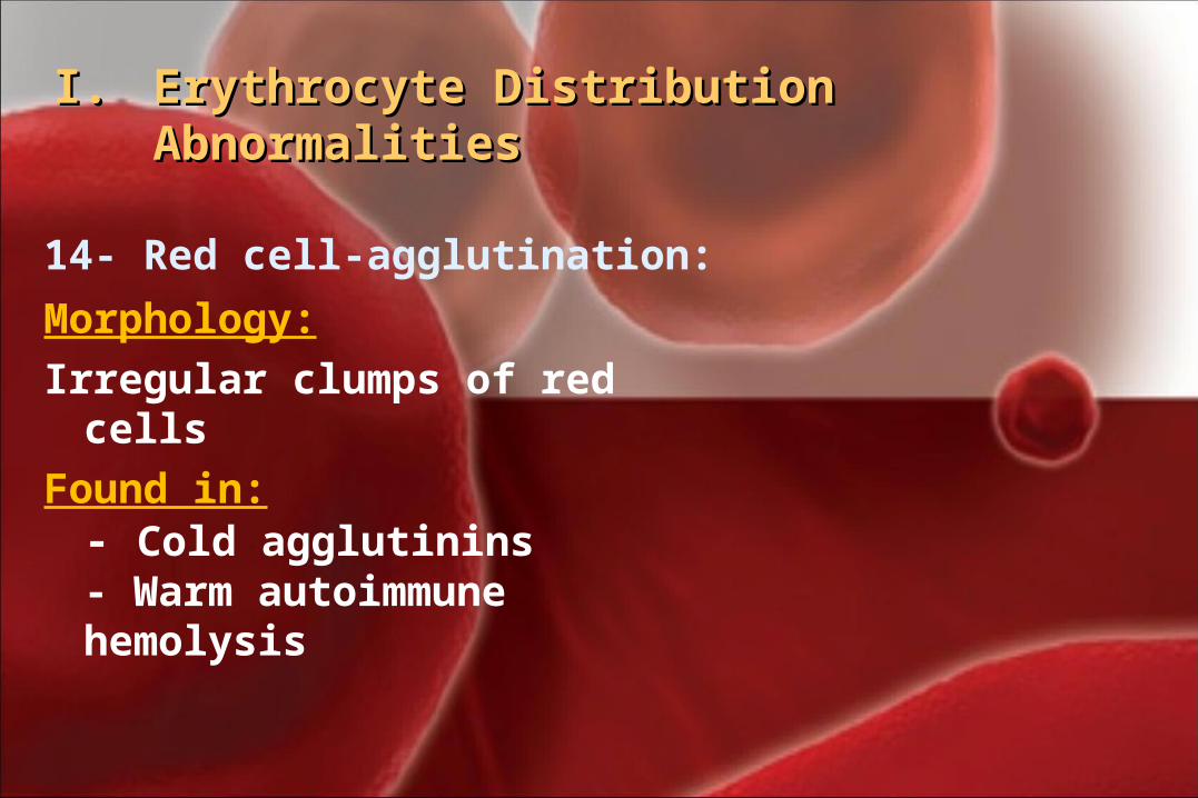
14- Red cell-agglutination:
Morphology: Irregular clumps of red cells
Found in:- Cold agglutinins- Warm autoimmune hemolysis
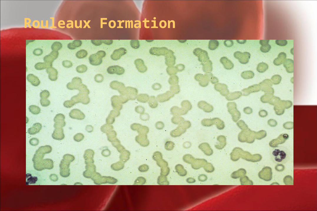
Rouleaux Formation
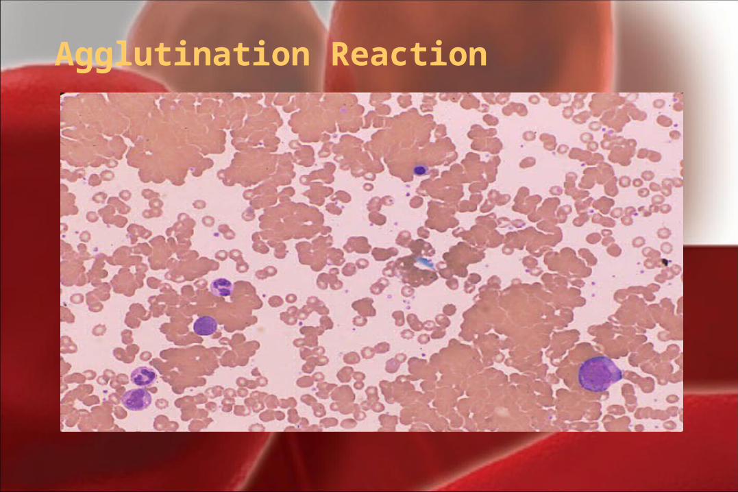
Agglutination Reaction

Schrier, S. ASH Image Bank 2002;2002:100344
Figure 2. The patient has autoimmune cold agglutinin disease and the Figure 2. The patient has autoimmune cold agglutinin disease and the red blood cells (RBC) have clumped when placed on the cold slidered blood cells (RBC) have clumped when placed on the cold slide
II. Variation in erythrocyte size (anisocytosis)
Normocyte:Normocyte: normal size of RBC, The average size of an RBC is 7.2 μm with a range of 6.8 to 7.5 μm. The nucleus of a small lymphocyte (± 8,µm) is a useful guide to the size of a red blood cell.
AnisocytosisAnisocytosis: Variations in size e.g.
1. Microcyte2. Macrocyte
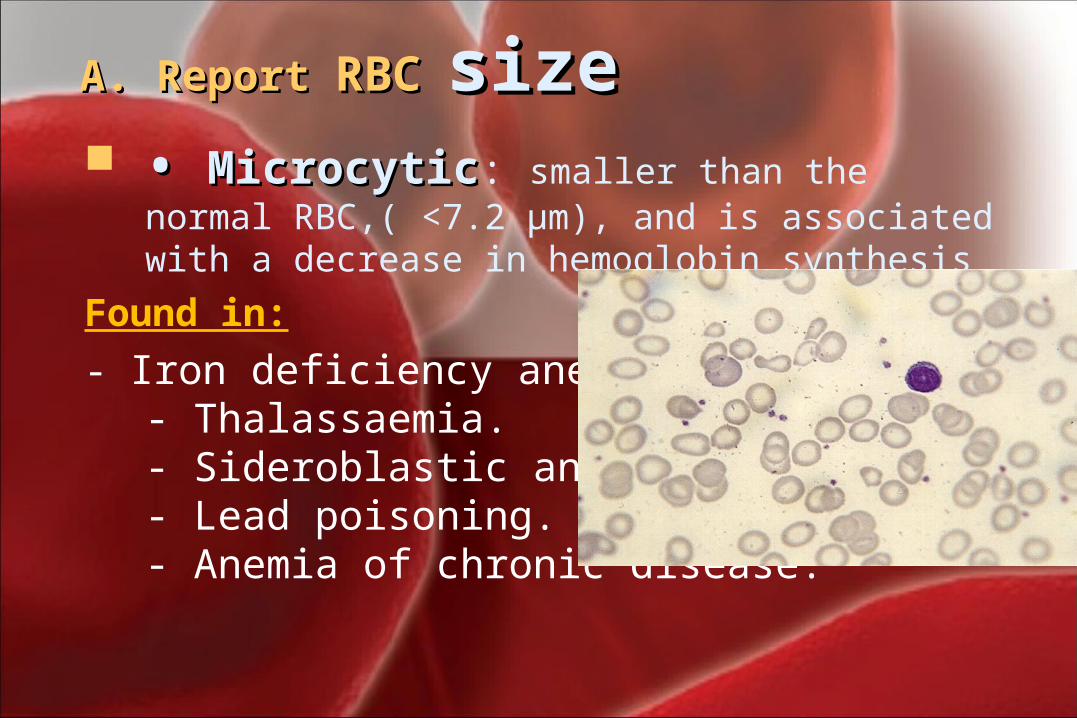
A. Report A. Report RBCRBC sizesize • • MicrocyticMicrocytic: smaller than the normal
RBC,( <7.2 μm), and is associated with a decrease in hemoglobin synthesis
Found in:
- Iron deficiency anemia.- Thalassaemia.- Sideroblastic anemia.- Lead poisoning.- Anemia of chronic disease.
II.II. Variation in erythrocyte size Variation in erythrocyte size (anisocytosis)(anisocytosis)
• • Macrocyte: Macrocyte: larger than the normal RBC (<8.2 μm) and is the result of a defect in nuclear maturation or stimulated erythropoiesis. May be round or oval in shape, the diagnostic significance being different.
Found in:- Folate and B12 deficiencies (oval)- Ethanol (round)- Liver disease (round)- Reticulocytosis (round)
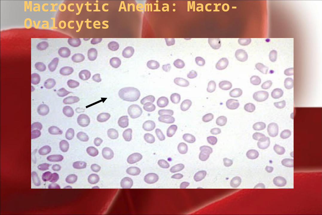
Macrocytic Anemia: Macro-Ovalocytes
II.II. Variation in erythrocyte size Variation in erythrocyte size (anisocytosis)(anisocytosis)
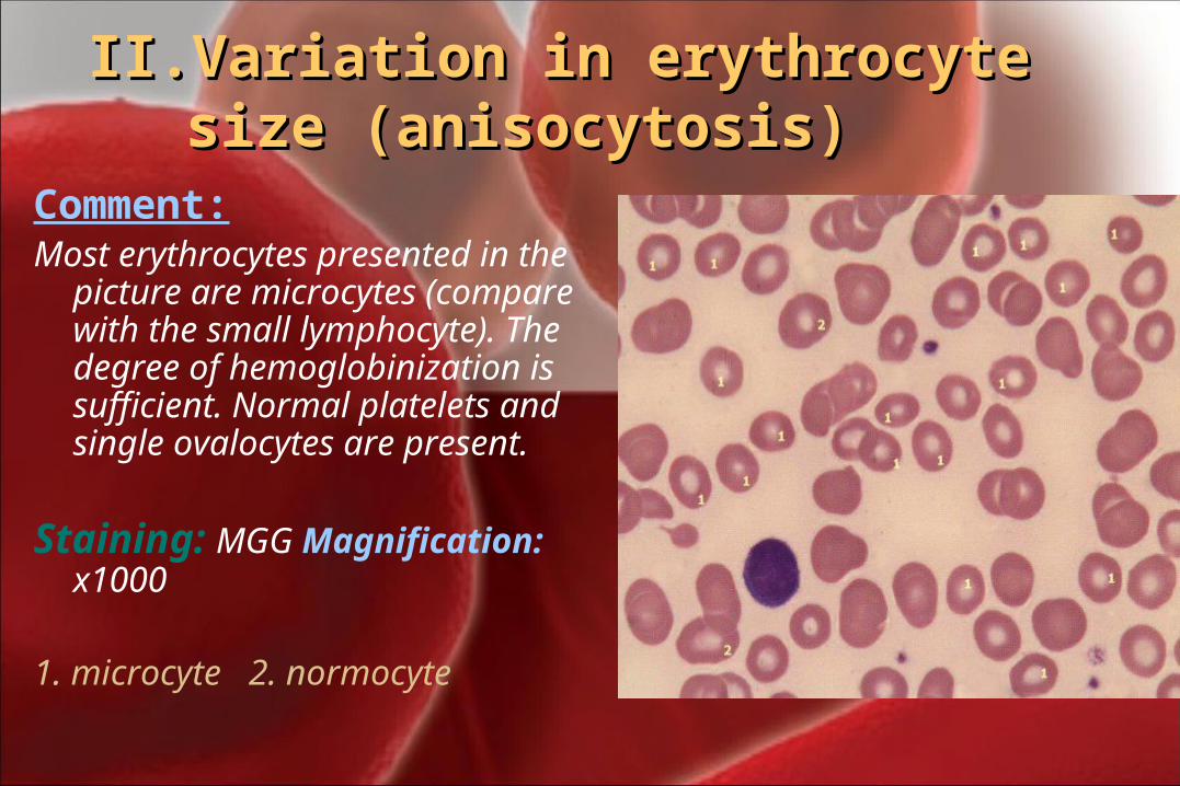
Comment: Most erythrocytes presented in
the picture are microcytes (compare with the small lymphocyte). The degree of hemoglobinization is sufficient. Normal platelets and single ovalocytes are present.
Staining: MGG Magnification: x1000
1. microcyte 2. normocyte
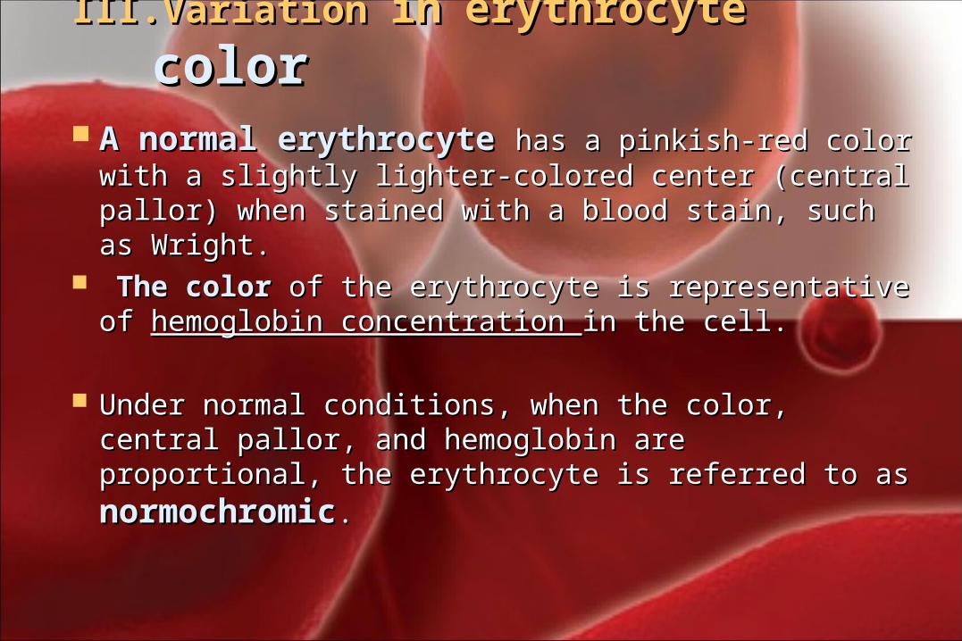
III.III. Variation Variation inin erythrocyte erythrocyte colorcolor A normal erythrocyte A normal erythrocyte has a pinkish-red has a pinkish-red
color with a slightly lighter-colored center color with a slightly lighter-colored center (central pallor) when stained with a blood stain, (central pallor) when stained with a blood stain, such as Wright.such as Wright.
The color The color of the erythrocyte is representative of of the erythrocyte is representative of hemoglobin concentration hemoglobin concentration in the cell. in the cell.
Under normal conditions, when the color, central Under normal conditions, when the color, central pallor, and hemoglobin are proportional, the pallor, and hemoglobin are proportional, the erythrocyte is referred to as erythrocyte is referred to as normochromicnormochromic..
III.III. Variation in hemoglobin Variation in hemoglobin contentcontent
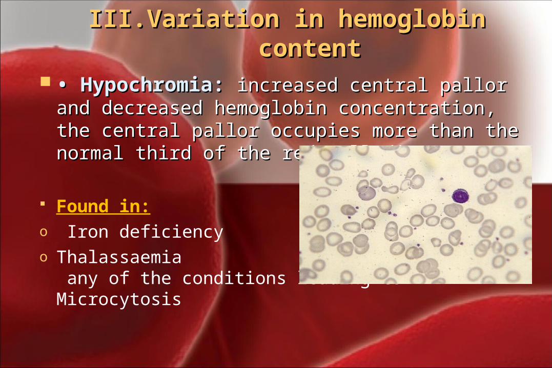
• • Hypochromia: Hypochromia: increased central pallor increased central pallor and decreased hemoglobin concentration, and decreased hemoglobin concentration, the central pallor occupies more than the the central pallor occupies more than the normal third of the red cell diameter.normal third of the red cell diameter.
Found in: o Iron deficiencyo Thalassaemia
any of the conditions leading to Microcytosis
III.III. Variation in hemoglobin Variation in hemoglobin contentcontent
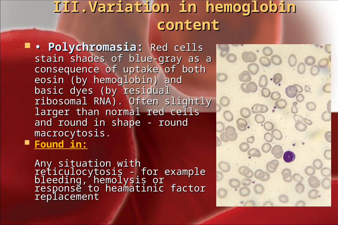
• • Polychromasia: Polychromasia: Red cells Red cells stain shades of blue-gray as a stain shades of blue-gray as a consequence of uptake of both consequence of uptake of both eosin (by hemoglobin) and basic eosin (by hemoglobin) and basic dyes (by residual ribosomal dyes (by residual ribosomal RNA). Often slightly larger than RNA). Often slightly larger than normal red cells and round in normal red cells and round in shape - round macrocytosis.shape - round macrocytosis.
Found in:
Any situation with Any situation with reticulocytosis - for example reticulocytosis - for example bleeding, hemolysis or response bleeding, hemolysis or response to heamatinic factor to heamatinic factor replacementreplacement
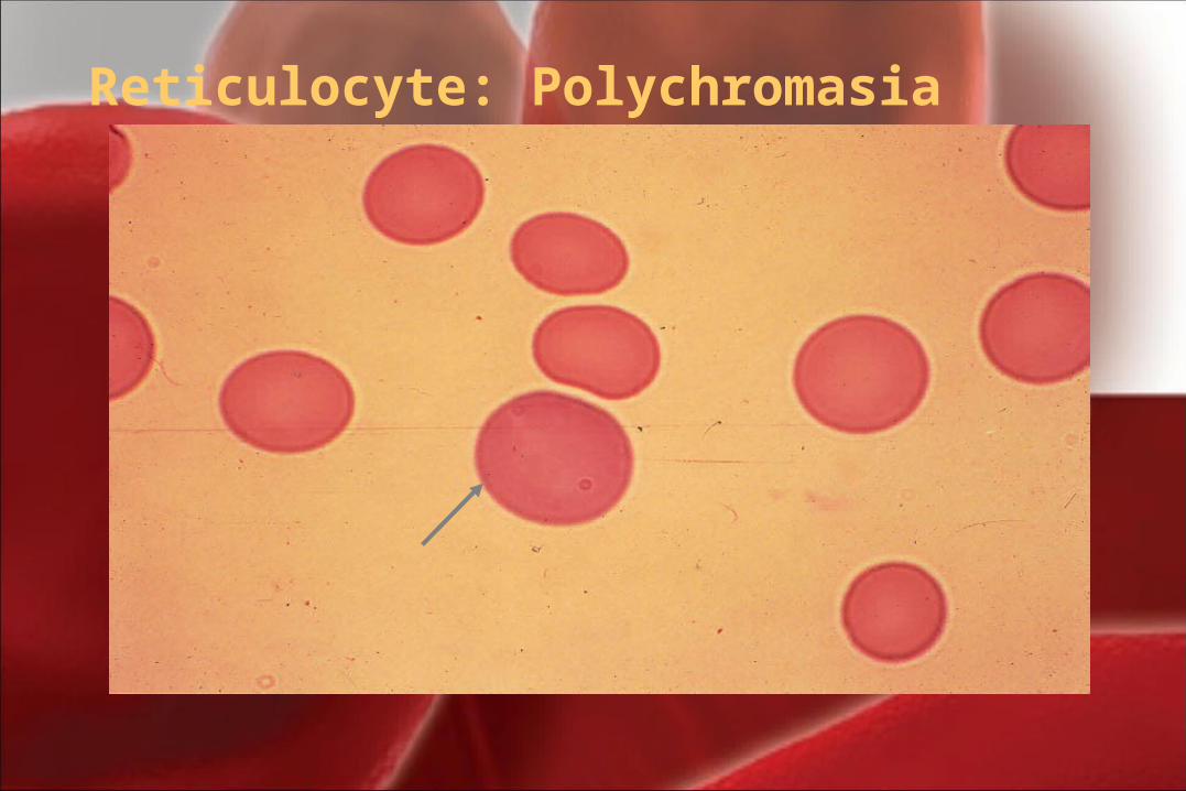
Reticulocyte: Polychromasia
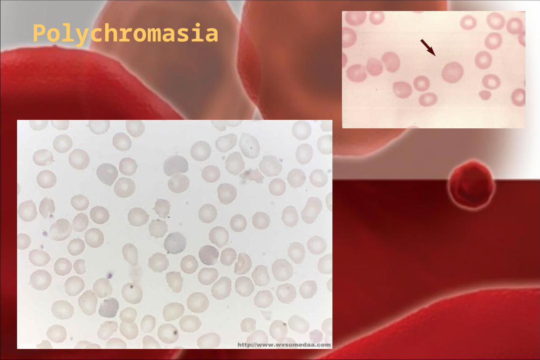
Polychromasia
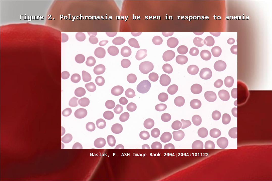
Maslak, P. ASH Image Bank 2004;2004:101122
Figure 2. Polychromasia may be seen in response to anemiaFigure 2. Polychromasia may be seen in response to anemia
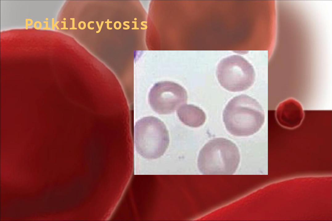
IV.IV. ShapeShape Abnormalities of ErythrocytesAbnormalities of Erythrocytes
PoikilocytosisPoikilocytosis is the general term for is the general term for mature erythrocytes that have a shape other mature erythrocytes that have a shape other than the round, biconcave disk.than the round, biconcave disk.
Poikilocytes can be seen in many shapes.(e.g. Poikilocytes can be seen in many shapes.(e.g.
Acanthocyte, Spherocytosis,……)Acanthocyte, Spherocytosis,……)
Poikilocytosis
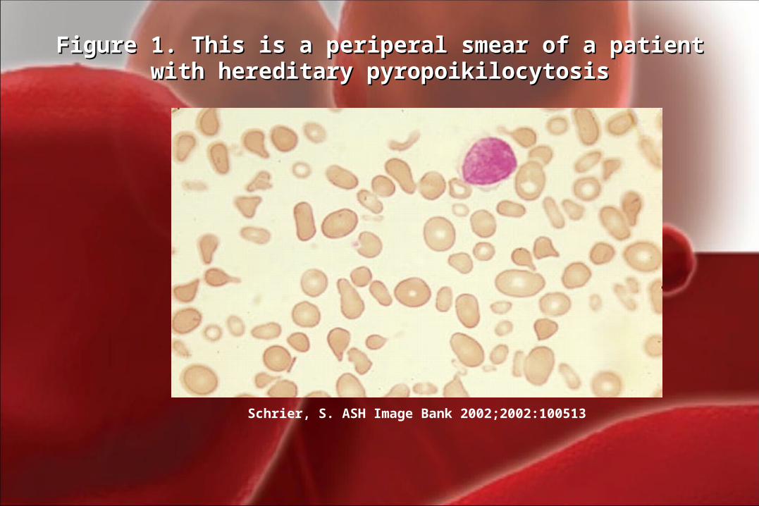
Schrier, S. ASH Image Bank 2002;2002:100513
Figure 1. This is a periperal smear of a patient with Figure 1. This is a periperal smear of a patient with hereditary pyropoikilocytosishereditary pyropoikilocytosis
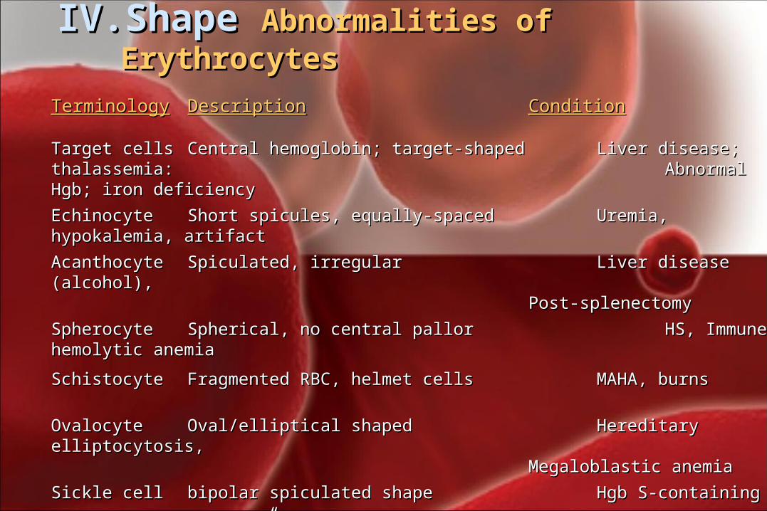
IV.IV. ShapeShape Abnormalities of ErythrocytesAbnormalities of Erythrocytes
TerminologyTerminology DescriptionDescription ConditionCondition
Target cellsTarget cells Central hemoglobin; target-shapedCentral hemoglobin; target-shaped Liver disease; thalassemia: Liver disease; thalassemia: Abnormal Hgb; iron deficiencyAbnormal Hgb; iron deficiency
EchinocyteEchinocyte Short spicules, equally-spacedShort spicules, equally-spaced Uremia, hypokalemia, artifactUremia, hypokalemia, artifact
AcanthocyteAcanthocyte Spiculated, irregularSpiculated, irregular Liver disease (alcohol),Liver disease (alcohol),Post-splenectomyPost-splenectomy
SpherocyteSpherocyte Spherical, no central pallorSpherical, no central pallor HS, Immune hemolytic anemiaHS, Immune hemolytic anemia
SchistocyteSchistocyte Fragmented RBC, helmet cells Fragmented RBC, helmet cells MAHA, burnsMAHA, burns
OvalocyteOvalocyte Oval/elliptical shapedOval/elliptical shaped Hereditary elliptocytosis,Hereditary elliptocytosis,Megaloblastic anemiaMegaloblastic anemia
Sickle cellSickle cell bipolar spiculated shapebipolar spiculated shape Hgb S-containing Hgb S-containing ““banana” shapedbanana” shaped hemoglobinopathyhemoglobinopathy
Teardrop cellTeardrop cell single elongated extremitysingle elongated extremity Myelophthistic changesMyelophthistic changes
Bite cellsBite cells Irregular gap in membrane Irregular gap in membrane G6PD deficiencyG6PD deficiency
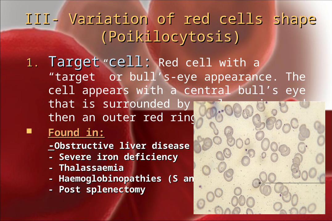
III- Variation of red cells shape III- Variation of red cells shape (Poikilocytosis)(Poikilocytosis)
1.1. Target cell:Target cell: Red cell with a “target” or bull’s-eye appearance. The cell appears with a central bull’s eye that is surrounded by a clear ring and then an outer red ring.
Found in:Found in:--Obstructive liver diseaseObstructive liver disease- Severe iron deficiency- Severe iron deficiency- Thalassaemia- Thalassaemia- Haemoglobinopathies (S and C)- Haemoglobinopathies (S and C)- Post splenectomy- Post splenectomy
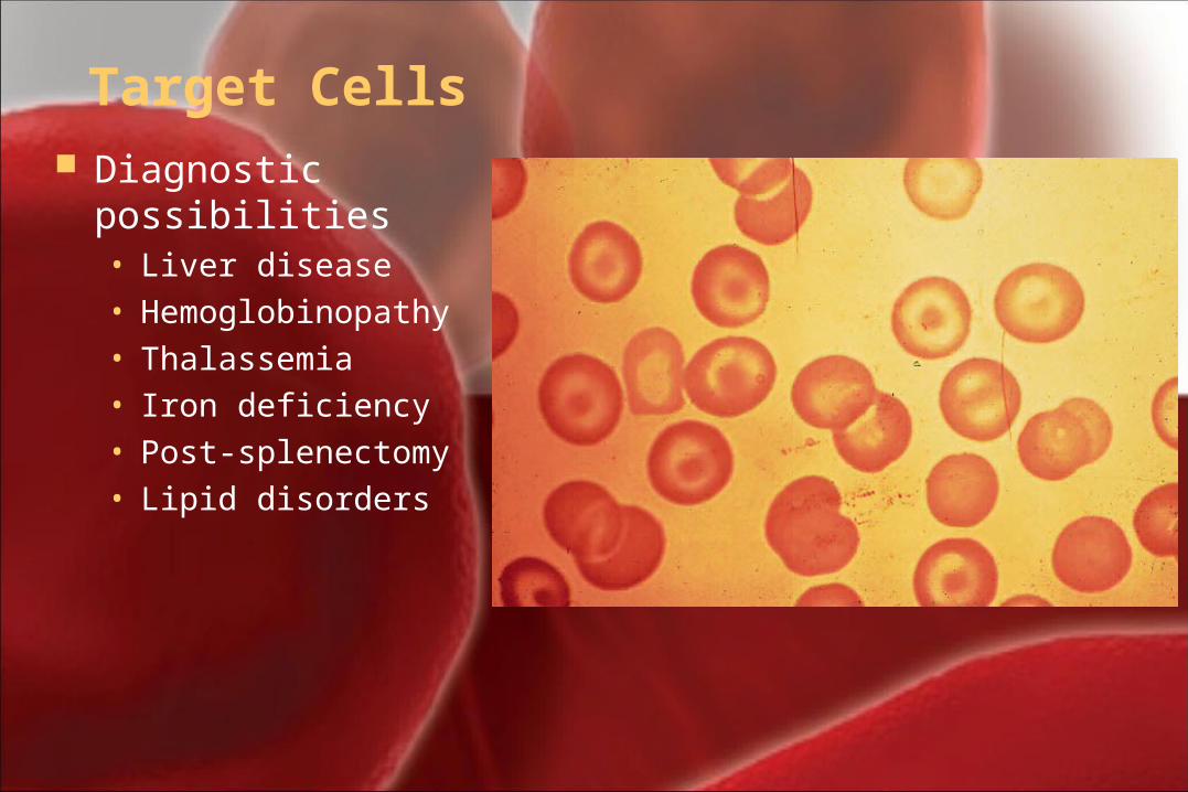
Target Cells Diagnostic possibilities
• Liver disease• Hemoglobinopathy• Thalassemia• Iron deficiency• Post-splenectomy• Lipid disorders
IV.IV. Abnormal Abnormal ShapeShape
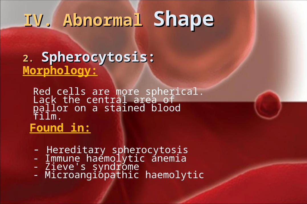
2.2. Spherocytosis:Spherocytosis: Morphology:
Red cells are more spherical. Lack the central area of pallor on a stained blood film.
Found in:
- Hereditary spherocytosis- Immune haemolytic anemia- Zieve's syndrome- Microangiopathic haemolytic
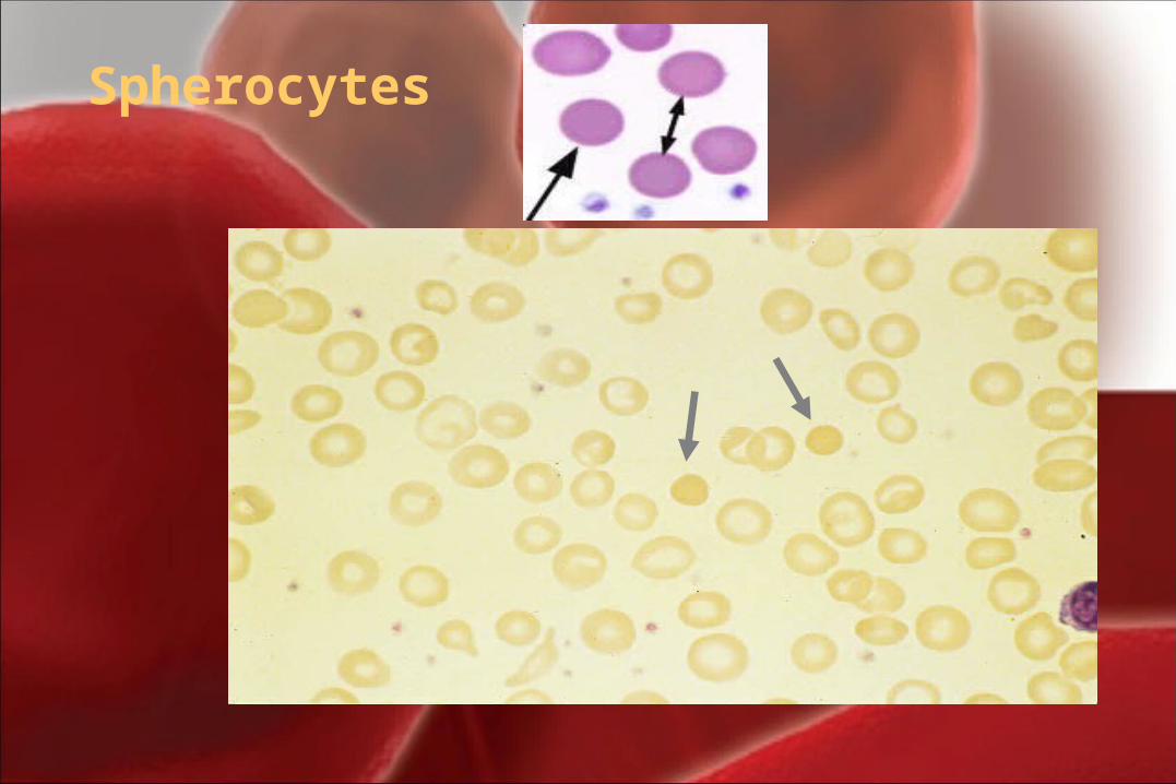
Spherocytes
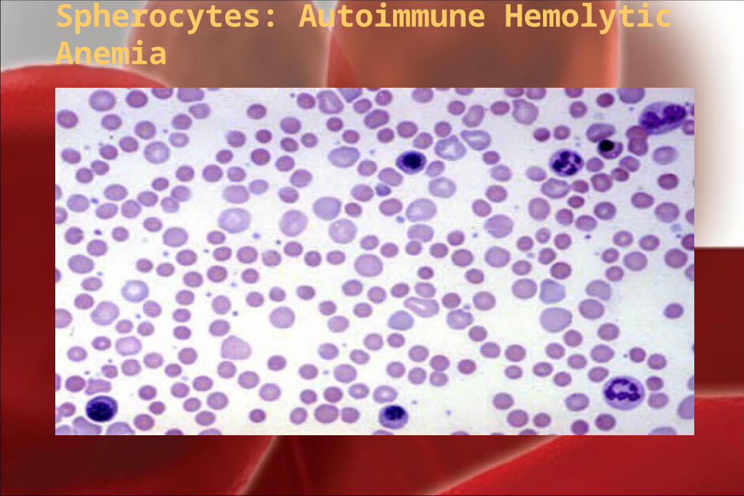
Spherocytes: Autoimmune Hemolytic Anemia
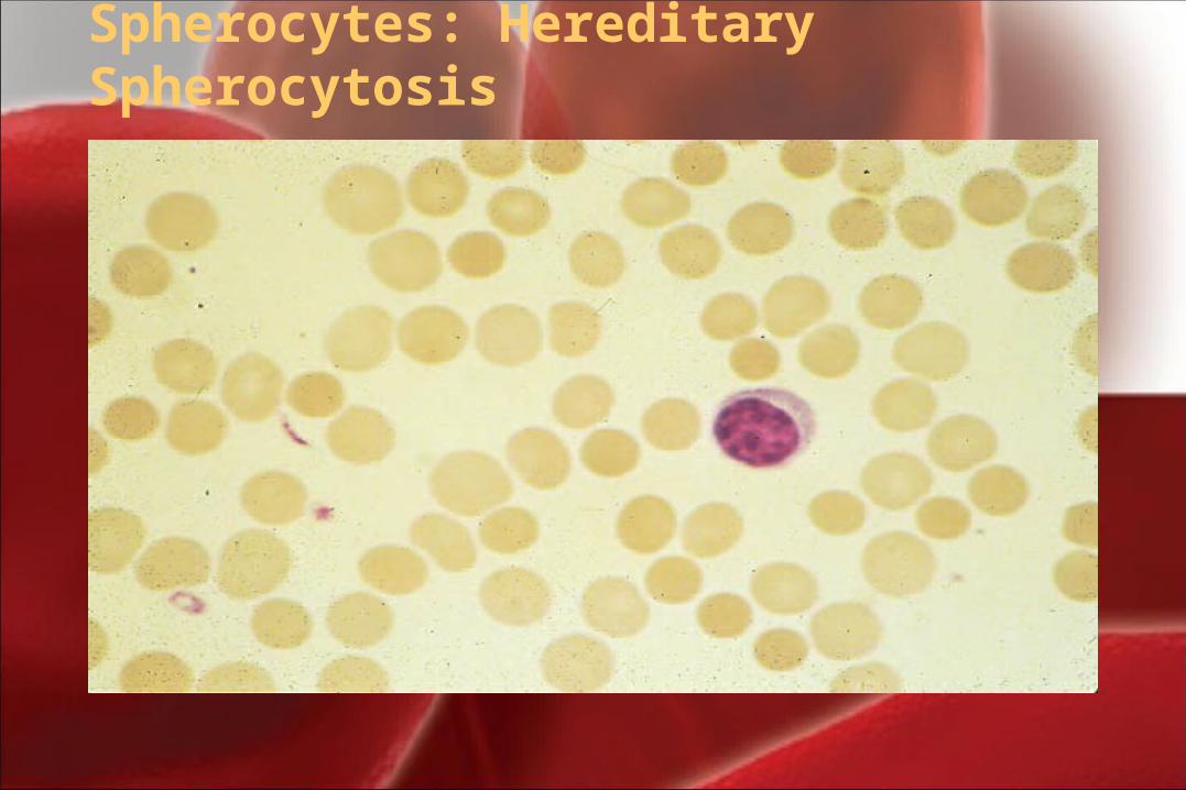
Spherocytes: Hereditary Spherocytosis
III- Variation of red cells shape III- Variation of red cells shape (Poikilocytosis)(Poikilocytosis)
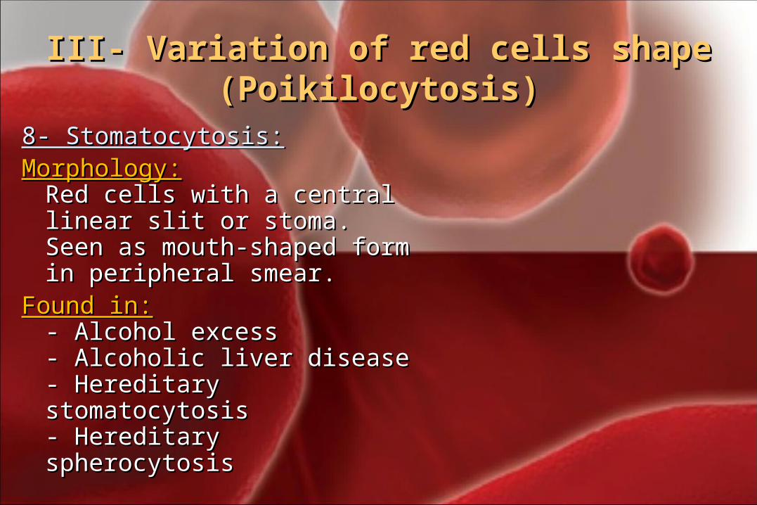
8- Stomatocytosis:8- Stomatocytosis:Morphology:Morphology:
Red cells with a central linear Red cells with a central linear slit or stoma. Seen as mouth-slit or stoma. Seen as mouth-shaped form in peripheral shaped form in peripheral smear.smear.
Found in:Found in:- - Alcohol excessAlcohol excess- Alcoholic liver disease- Alcoholic liver disease- Hereditary stomatocytosis- Hereditary stomatocytosis- Hereditary spherocytosis - Hereditary spherocytosis
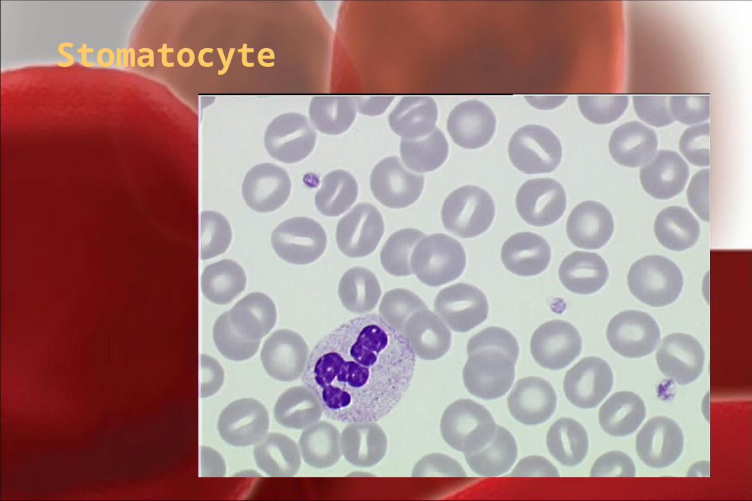
Stomatocyte
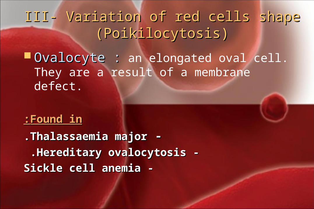
III- Variation of red cells shape III- Variation of red cells shape (Poikilocytosis)(Poikilocytosis)
Ovalocyte :Ovalocyte : an elongated oval cell. They are a result of a membrane defect.
Found inFound in::
- -Thalassaemia majorThalassaemia major..
- -Hereditary ovalocytosisHereditary ovalocytosis . .
- -Sickle cell anemiaSickle cell anemia
III- Variation of red cells III- Variation of red cells shape (Poikilocytosis)shape (Poikilocytosis)
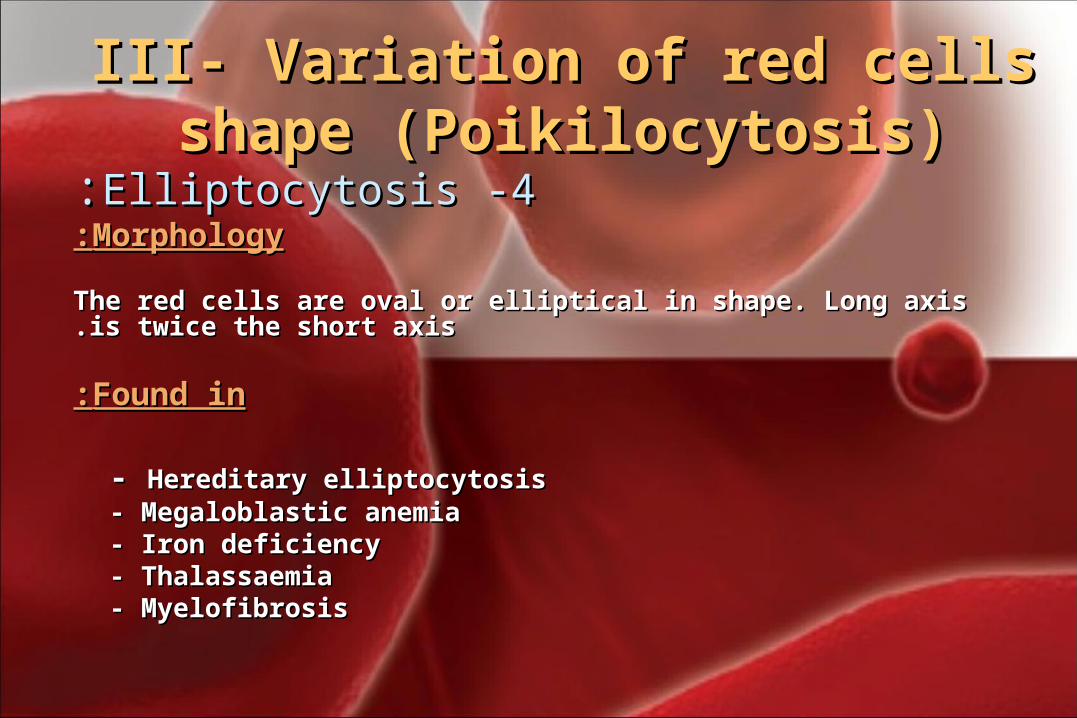
44 - -ElliptocytosisElliptocytosis::MorphologyMorphology::
The red cells are oval or elliptical in shape. Long axis is twice the short The red cells are oval or elliptical in shape. Long axis is twice the short axisaxis..
Found inFound in::
- - Hereditary elliptocytosisHereditary elliptocytosis- Megaloblastic anemia- Megaloblastic anemia- Iron deficiency - Iron deficiency - Thalassaemia- Thalassaemia- Myelofibrosis- Myelofibrosis
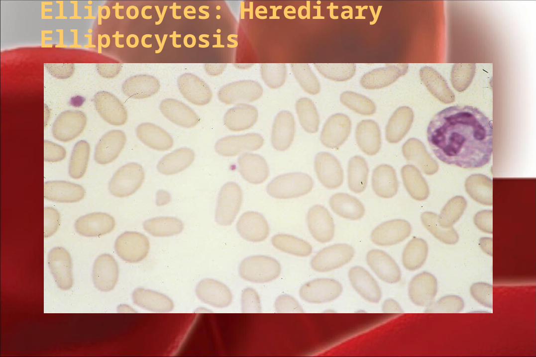
Elliptocytes: Hereditary Elliptocytosis
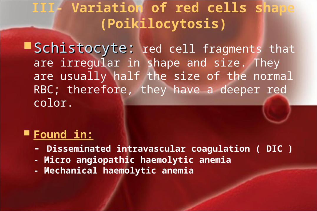
III- Variation of red cells shape (Poikilocytosis)
Schistocyte:Schistocyte: red cell fragments that are irregular in shape and size. They are usually half the size of the normal RBC; therefore, they have a deeper red color.
Found in: - Disseminated intravascular coagulation ( DIC ) - Micro angiopathic haemolytic anemia- Mechanical haemolytic anemia
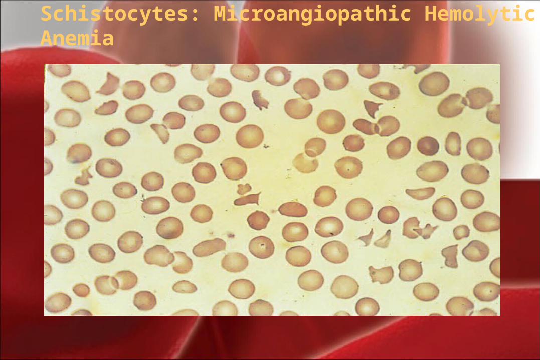
Schistocytes: Microangiopathic Hemolytic Anemia
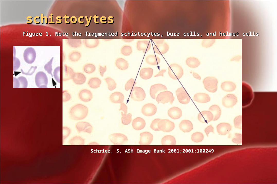
Schrier, S. ASH Image Bank 2001;2001:100249
Figure 1. Note the fragmented schistocytes, burr cells, and helmet cellsFigure 1. Note the fragmented schistocytes, burr cells, and helmet cells
schistocytesschistocytes
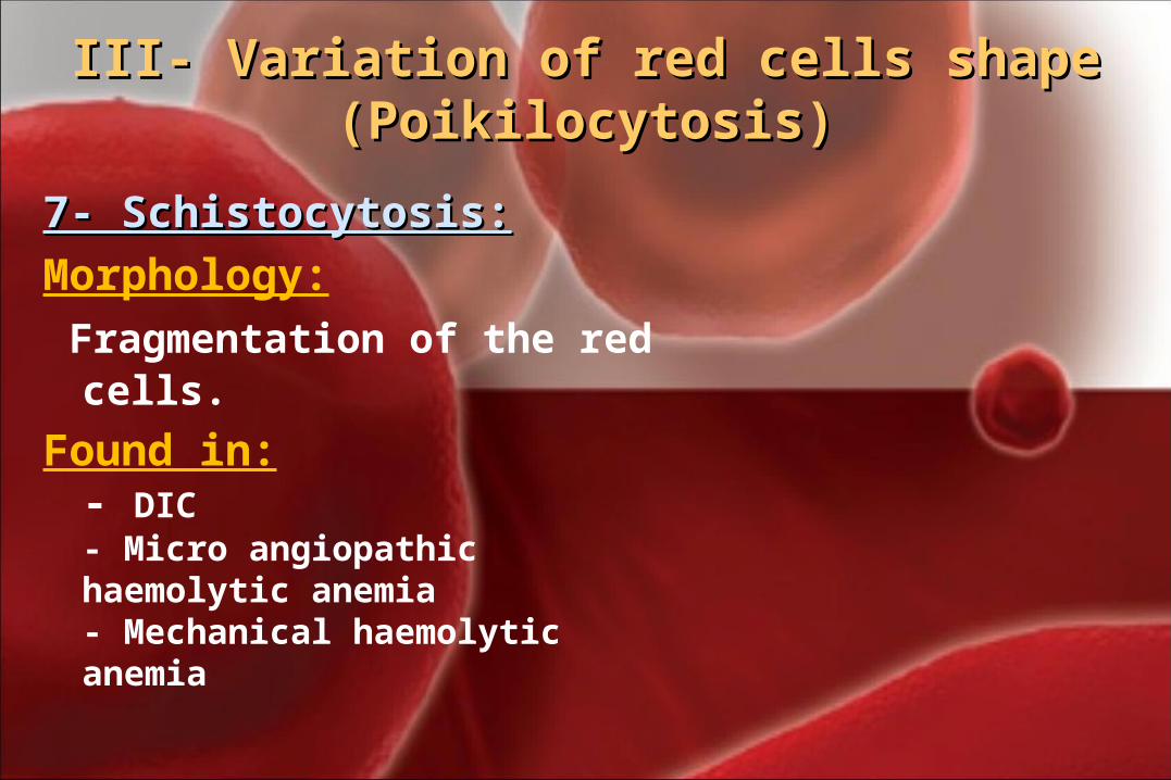
III- Variation of red cells shape III- Variation of red cells shape (Poikilocytosis)(Poikilocytosis)
7- Schistocytosis:7- Schistocytosis:Morphology: Fragmentation of the red cells. Found in:
- DIC - Micro angiopathic haemolytic anemia- Mechanical haemolytic anemia
III- Variation of red cells shape III- Variation of red cells shape (Poikilocytosis)(Poikilocytosis)
6- Blister 6- Blister cell:prekeratocytecell:prekeratocyte
MorphologyMorphology:: Have accentric hallow areaHave accentric hallow area..
Resemble a women's handbag and may be called pocket-book cell.
Found inFound in:: Microangiopathic hemolytic anemiaMicroangiopathic hemolytic anemia
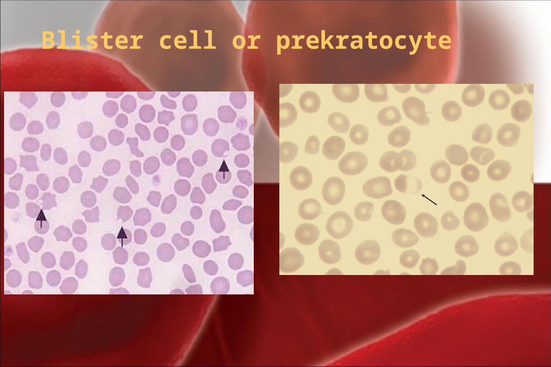
Blister cell or prekratocyte
III- Variation of red cells shape III- Variation of red cells shape (Poikilocytosis)(Poikilocytosis)
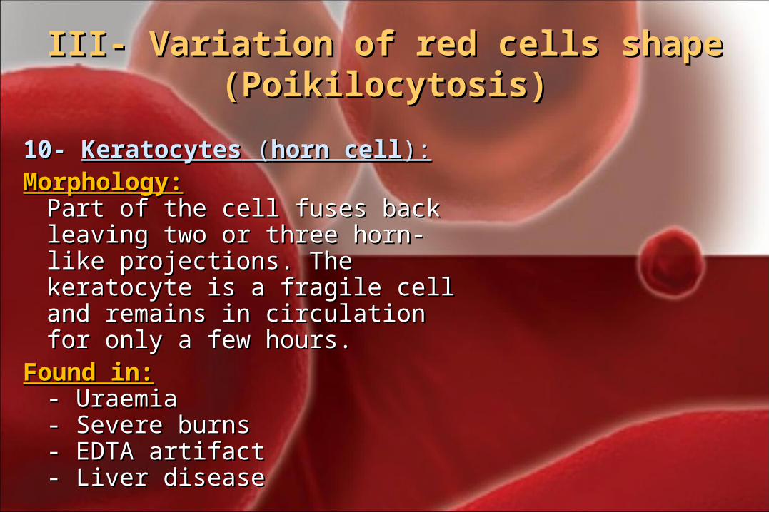
10- 10- KeratocytesKeratocytes ( (horn cellhorn cell):):Morphology:Morphology:
Part of the cell fuses back leaving Part of the cell fuses back leaving two or three horn-like projections. two or three horn-like projections. The keratocyte is a fragile cell and The keratocyte is a fragile cell and remains in circulation for only a remains in circulation for only a few hours.few hours.
Found in:Found in:- - UraemiaUraemia- Severe burns- Severe burns- EDTA artifact- EDTA artifact- Liver disease- Liver disease
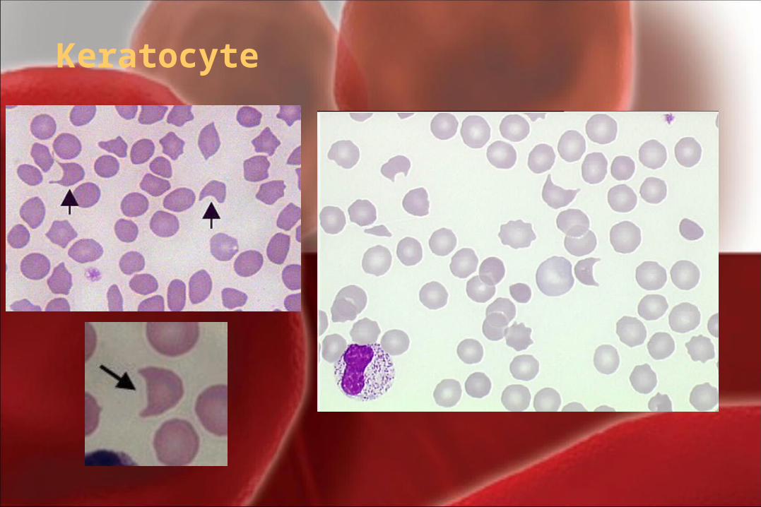
Keratocyte
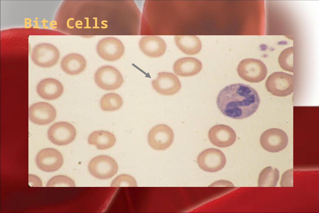
Bite Cells
III- Variation of red cells shape III- Variation of red cells shape (Poikilocytosis)(Poikilocytosis)
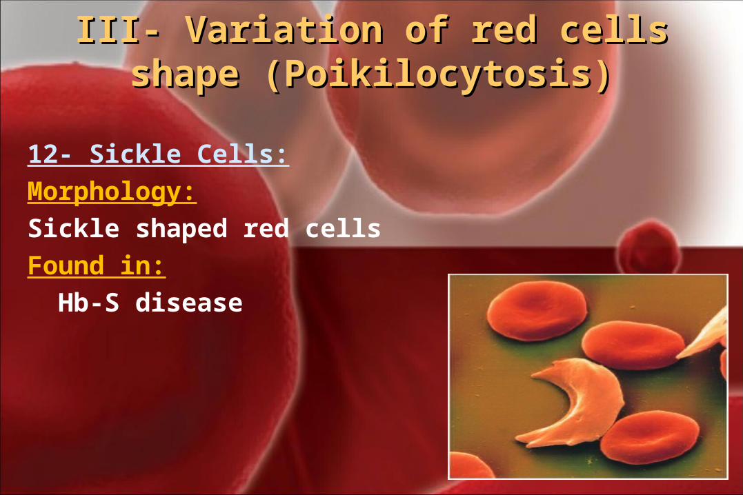
12- Sickle Cells:Morphology: Sickle shaped red cells Found in: Hb-S disease
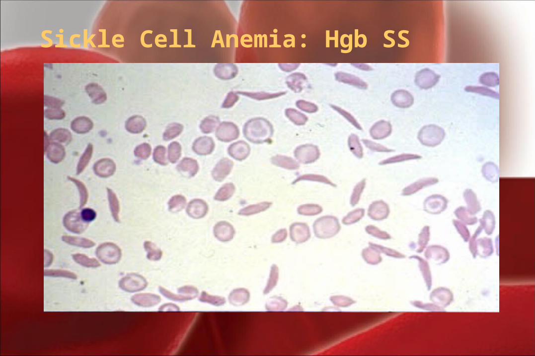
Sickle Cell Anemia: Hgb SS
III- Variation of red cells shape III- Variation of red cells shape (Poikilocytosis)(Poikilocytosis)
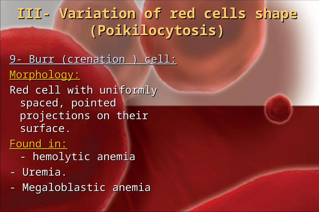
9- Burr (crenation ) cell:9- Burr (crenation ) cell:
Morphology:Morphology:
Red cell with uniformly spaced, Red cell with uniformly spaced, pointed projections on their pointed projections on their surface.surface.
Found in:Found in:- - hemolytic anemiahemolytic anemia
- Uremia.- Uremia.
- Megaloblastic anemia- Megaloblastic anemia
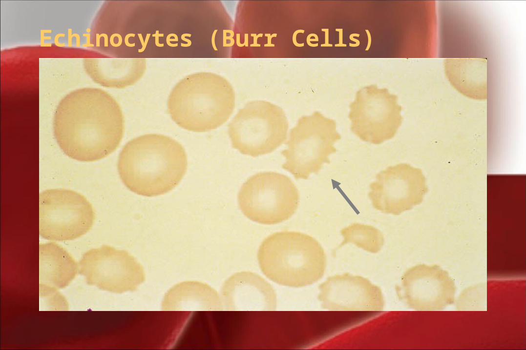
Echinocytes (Burr Cells)
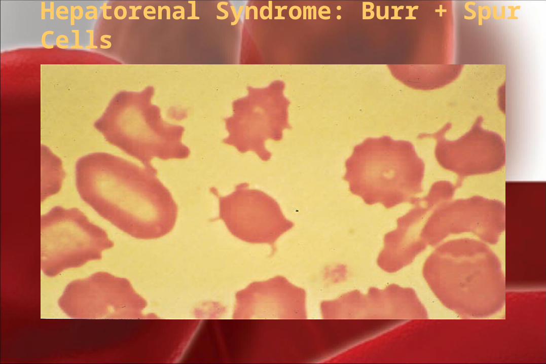
Hepatorenal Syndrome: Burr + Spur Cells
III- Variation of red cells shape III- Variation of red cells shape (Poikilocytosis)(Poikilocytosis)
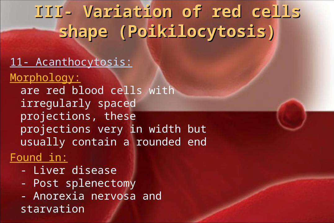
11- Acanthocytosis:Morphology:
are red blood cells with irregularly spaced projections, these projections very in width but usually contain a rounded end
Found in:- Liver disease - Post splenectomy- Anorexia nervosa and starvation
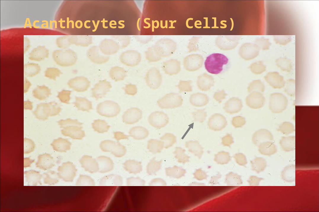
Acanthocytes (Spur Cells)
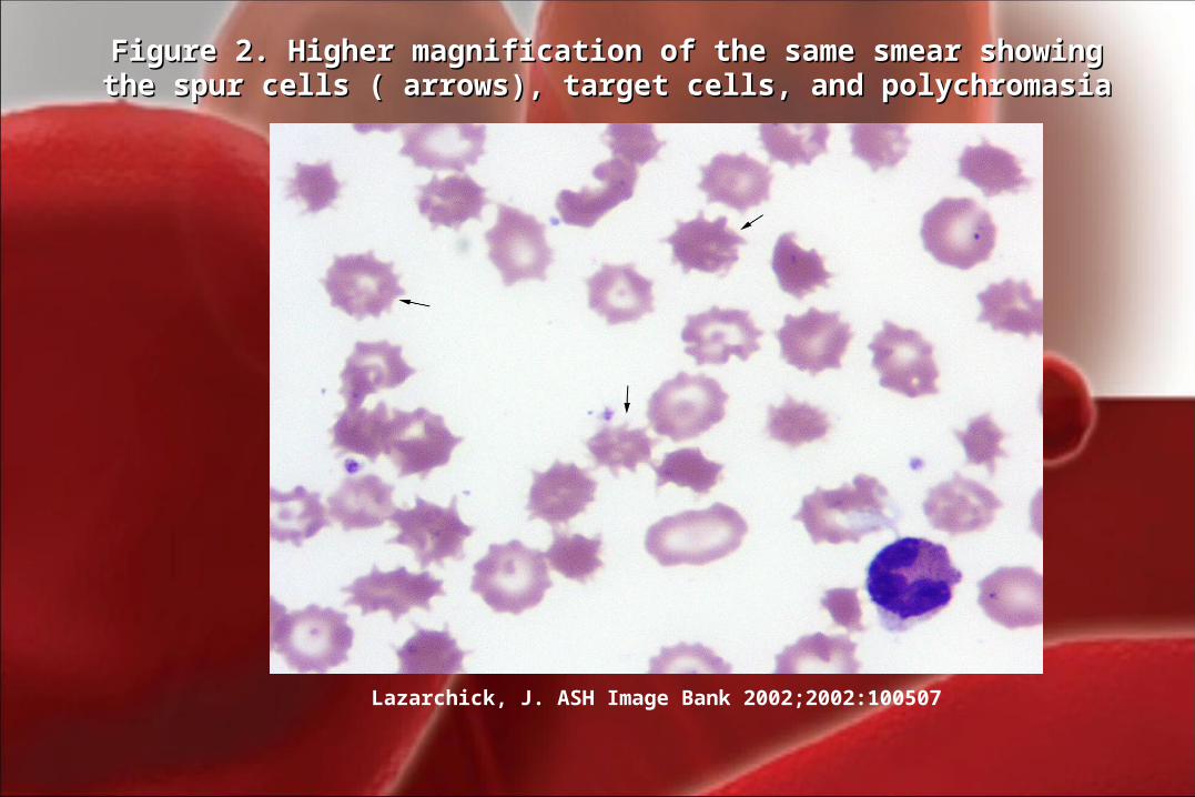
Lazarchick, J. ASH Image Bank 2002;2002:100507
Figure 2. Higher magnification of the same smear showing the spur cells Figure 2. Higher magnification of the same smear showing the spur cells ( arrows), target cells, and polychromasia( arrows), target cells, and polychromasia
III- Variation of red cells shape III- Variation of red cells shape (Poikilocytosis)(Poikilocytosis)
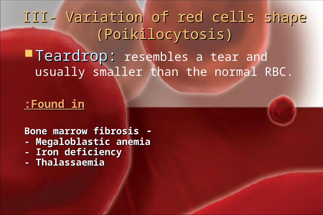
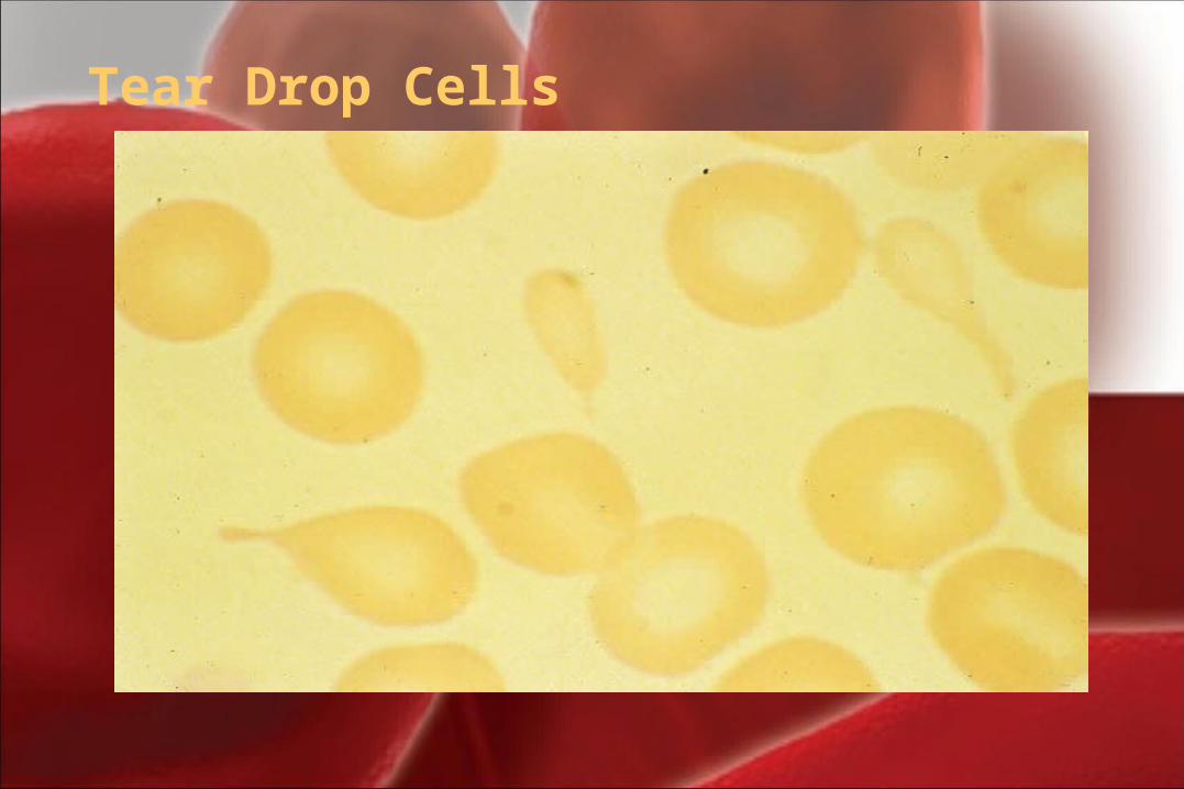
Teardrop:Teardrop: resembles a tear and usually smaller than the normal RBC.
Found inFound in::
- -Bone marrow fibrosisBone marrow fibrosis- Megaloblastic anemia- Megaloblastic anemia- Iron deficiency- Iron deficiency- Thalassaemia- Thalassaemia
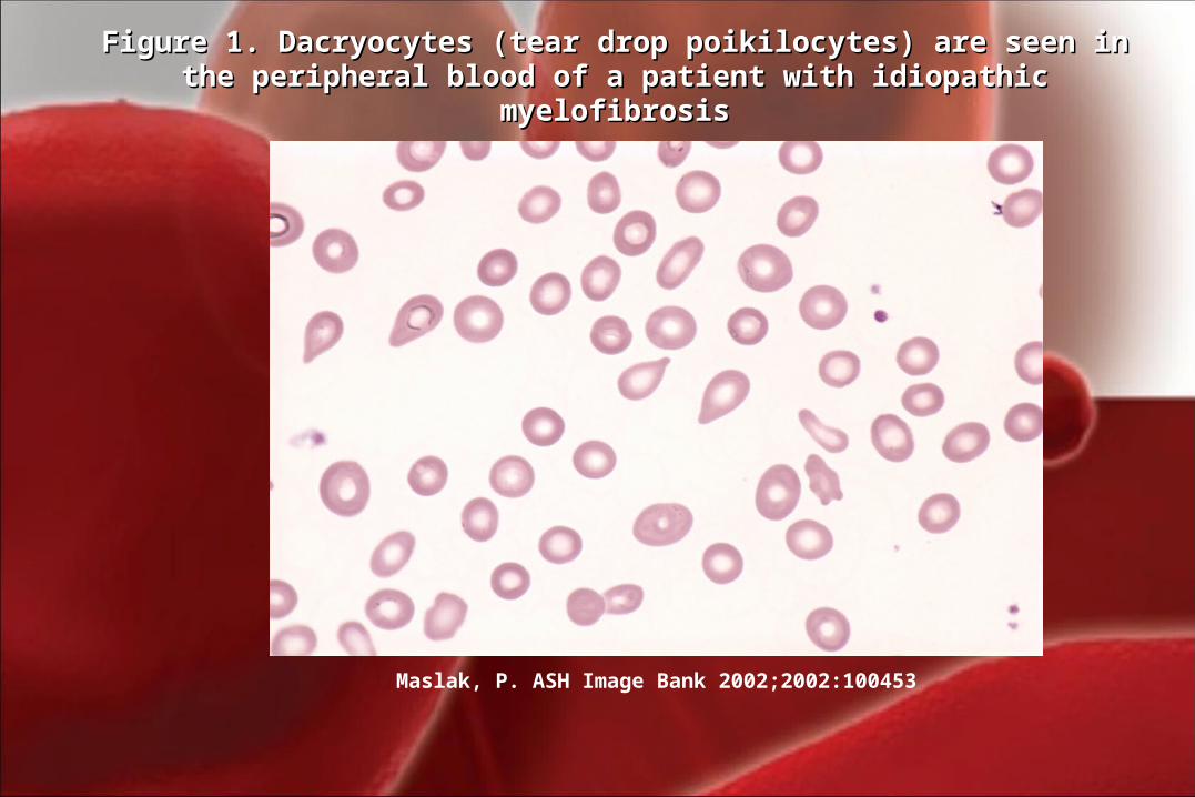
Maslak, P. ASH Image Bank 2002;2002:100453
Figure 1. Dacryocytes (tear drop poikilocytes) are seen in the peripheral Figure 1. Dacryocytes (tear drop poikilocytes) are seen in the peripheral blood of a patient with idiopathic myelofibrosisblood of a patient with idiopathic myelofibrosis
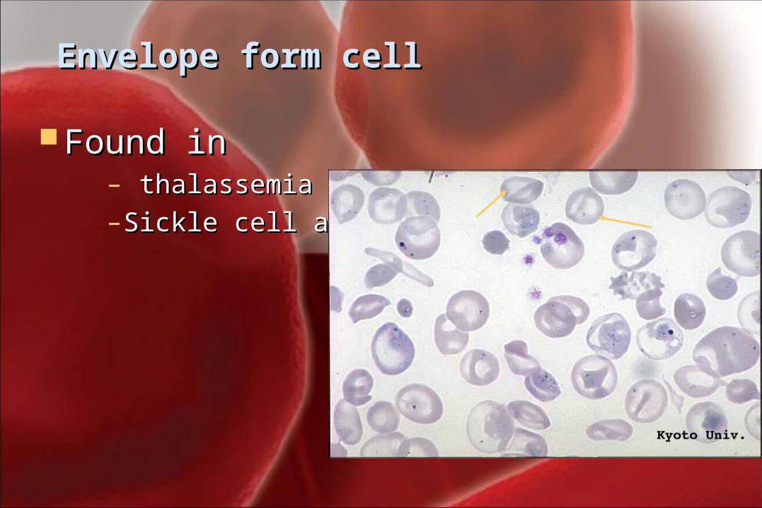
Envelope form cellEnvelope form cell
Found inFound in– thalassemia thalassemia – Sickle cell anemiaSickle cell anemia
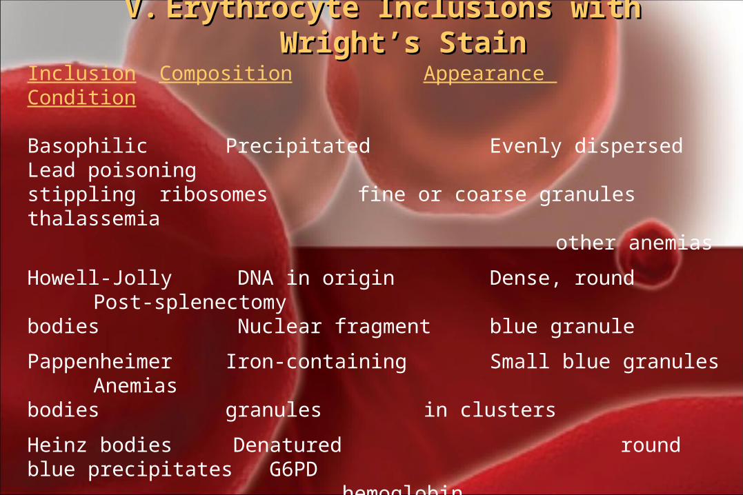
V.V. Erythrocyte Inclusions with Wright’s StainErythrocyte Inclusions with Wright’s StainInclusion Composition Appearance Condition
Basophilic Precipitated Evenly dispersed Lead poisoningstippling ribosomes fine or coarse granules thalassemia
other anemias
Howell-Jolly DNA in origin Dense, round Post-splenectomybodies Nuclear fragment blue granule
Pappenheimer Iron-containing Small blue granules Anemiasbodies granules in clusters
Heinz bodies Denatured round blue precipitates G6PD hemoglobinCabot Rings remnants of Reddish-blue threadlike Severe anemia, nuclear membrane rings Lead poisoning
Organism Small blue inclusion MalariaBabesiosis
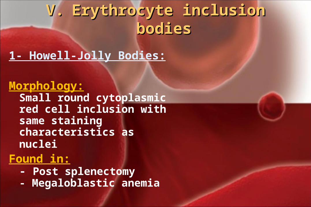
V.V. Erythrocyte inclusion bodies Erythrocyte inclusion bodies
1- Howell-Jolly Bodies:
Morphology:Small round cytoplasmic red cell inclusion with same staining characteristics as nuclei
Found in:- Post splenectomy- Megaloblastic anemia
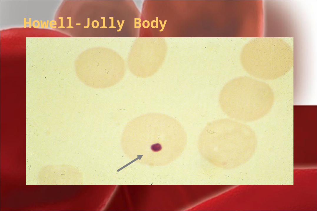
Howell-Jolly Body
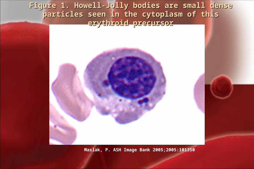
Maslak, P. ASH Image Bank 2005;2005:101350
Figure 1. Howell-Jolly bodies are small dense particles seen Figure 1. Howell-Jolly bodies are small dense particles seen in the cytoplasm of this erythroid precursorin the cytoplasm of this erythroid precursor
V.V. Erythrocyte inclusion bodiesErythrocyte inclusion bodies
2- Siderotic Granules 2- Siderotic Granules (Pappenheimer Bodies)(Pappenheimer Bodies)
RBCs which contain no hemoglobin iron granules. They appear as dense blue, irregular granules which are unevenly distributed in Wright stained RBCs. Pappenheimer bodies can be increased in hemolytic anemia, infections and post-splenectomy.
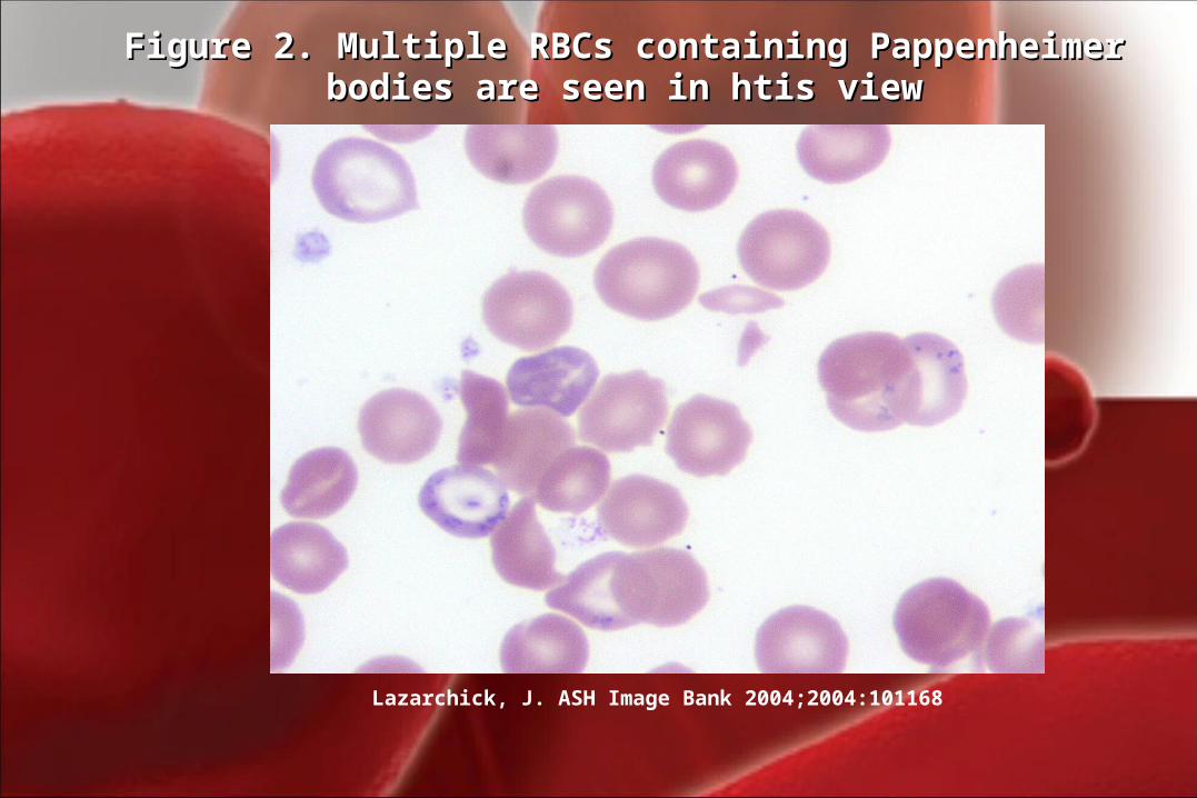
Lazarchick, J. ASH Image Bank 2004;2004:101168
Figure 2. Multiple RBCs containing Pappenheimer bodies are Figure 2. Multiple RBCs containing Pappenheimer bodies are seen in htis viewseen in htis view
V.V. Erythrocyte inclusion bodiesErythrocyte inclusion bodies
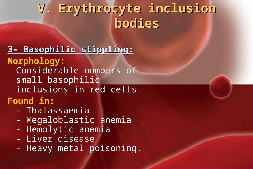
3- Basophilic stippling:3- Basophilic stippling: Morphology:
Considerable numbers of small basophilic inclusions in red cells.
Found in:- Thalassaemia- Megaloblastic anemia- Hemolytic anemia - Liver disease- Heavy metal poisoning.
Tear Drop Cells
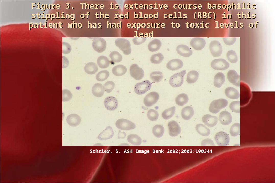
Schrier, S. ASH Image Bank 2002;2002:100344
Figure 3. There is extensive course basophilic stippling of Figure 3. There is extensive course basophilic stippling of the red blood cells (RBC) in this patient who has had the red blood cells (RBC) in this patient who has had
exposure to toxic levels of leadexposure to toxic levels of lead
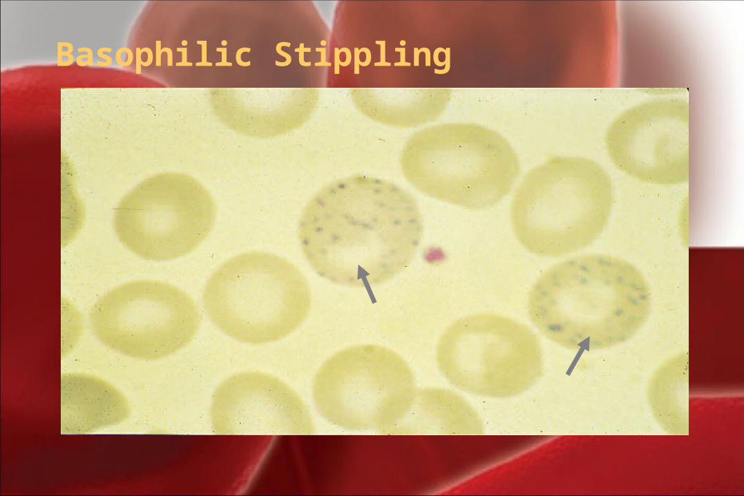
Basophilic Stippling
V.V. Erythrocyte inclusion bodiesErythrocyte inclusion bodies
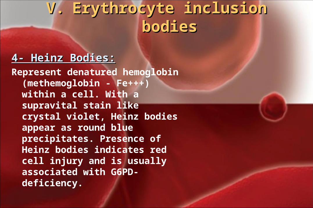
4- Heinz Bodies:4- Heinz Bodies:Represent denatured hemoglobin
(methemoglobin - Fe+++) within a cell. With a supravital stain like crystal violet, Heinz bodies appear as round blue precipitates. Presence of Heinz bodies indicates red cell injury and is usually associated with G6PD-deficiency.
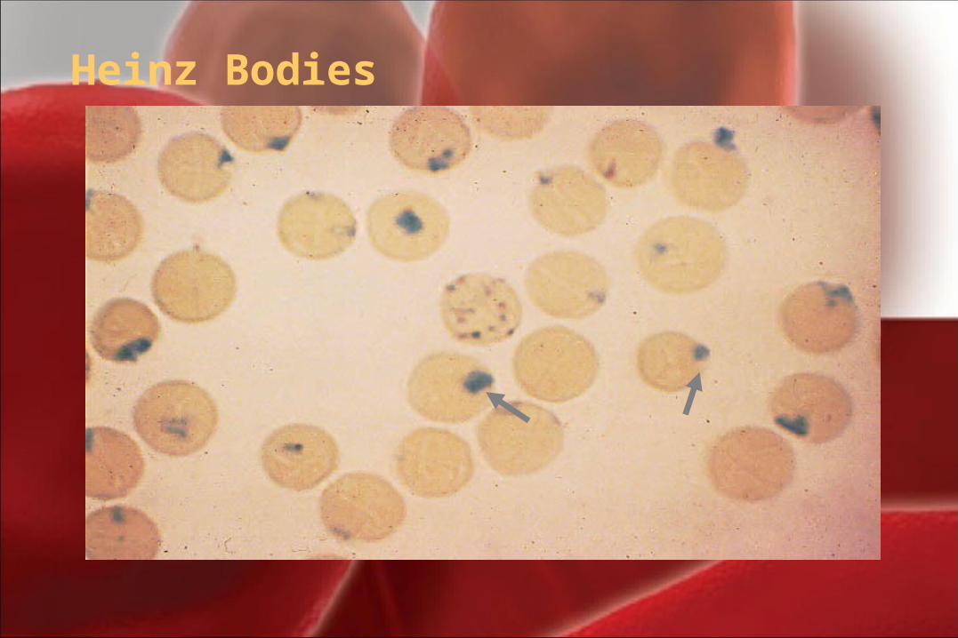
Heinz Bodies
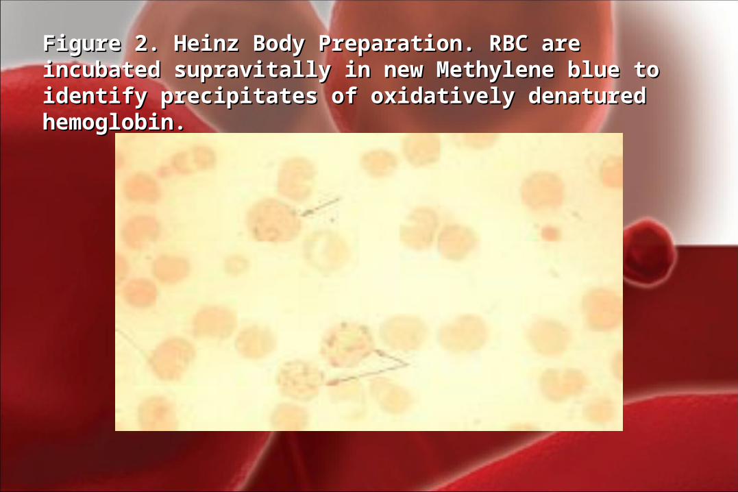
Figure 2. Heinz Body Preparation. RBC are incubated Figure 2. Heinz Body Preparation. RBC are incubated supravitally in new Methylene blue to identify precipitates of supravitally in new Methylene blue to identify precipitates of oxidatively denatured hemoglobin.oxidatively denatured hemoglobin.
IV -Erythrocyte inclusion IV -Erythrocyte inclusion bodiesbodies
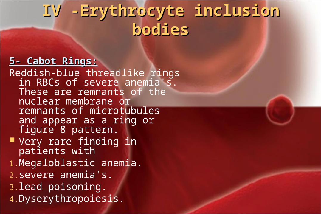
5- Cabot Rings:5- Cabot Rings: Reddish-blue threadlike rings in
RBCs of severe anemia's. These are remnants of the nuclear membrane or remnants of microtubules and appear as a ring or figure 8 pattern.
Very rare finding in patients with 1. Megaloblastic anemia. 2. severe anemia's.3. lead poisoning.4. Dyserythropoiesis.
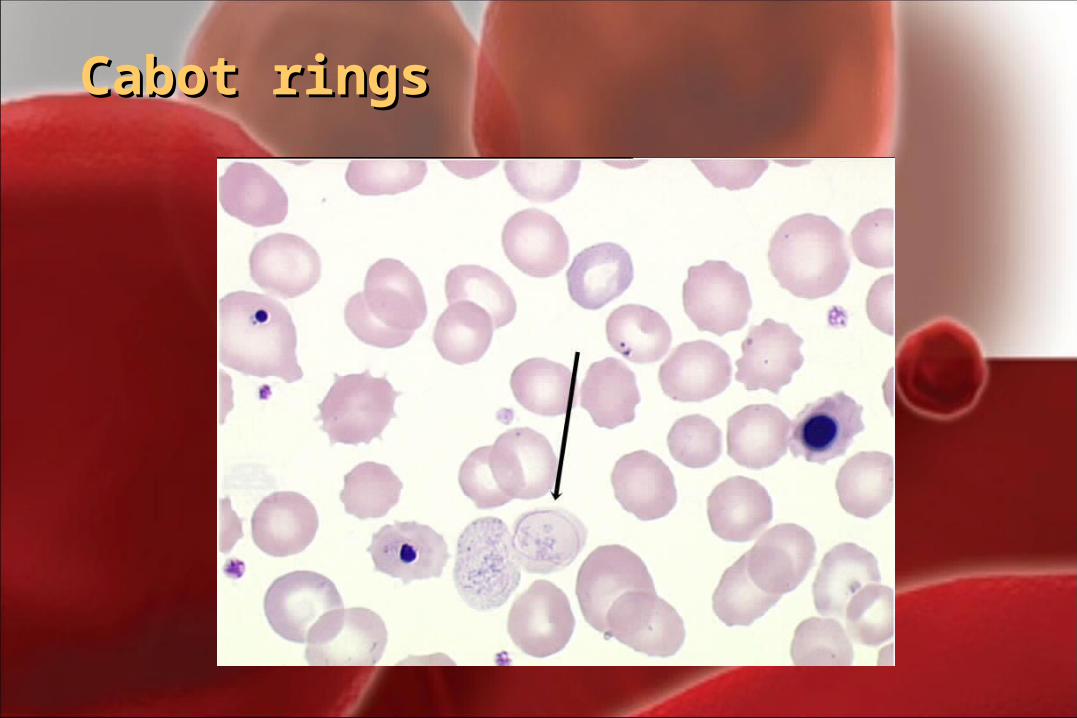
Cabot ringsCabot rings
V.V. Erythrocyte inclusion bodiesErythrocyte inclusion bodies
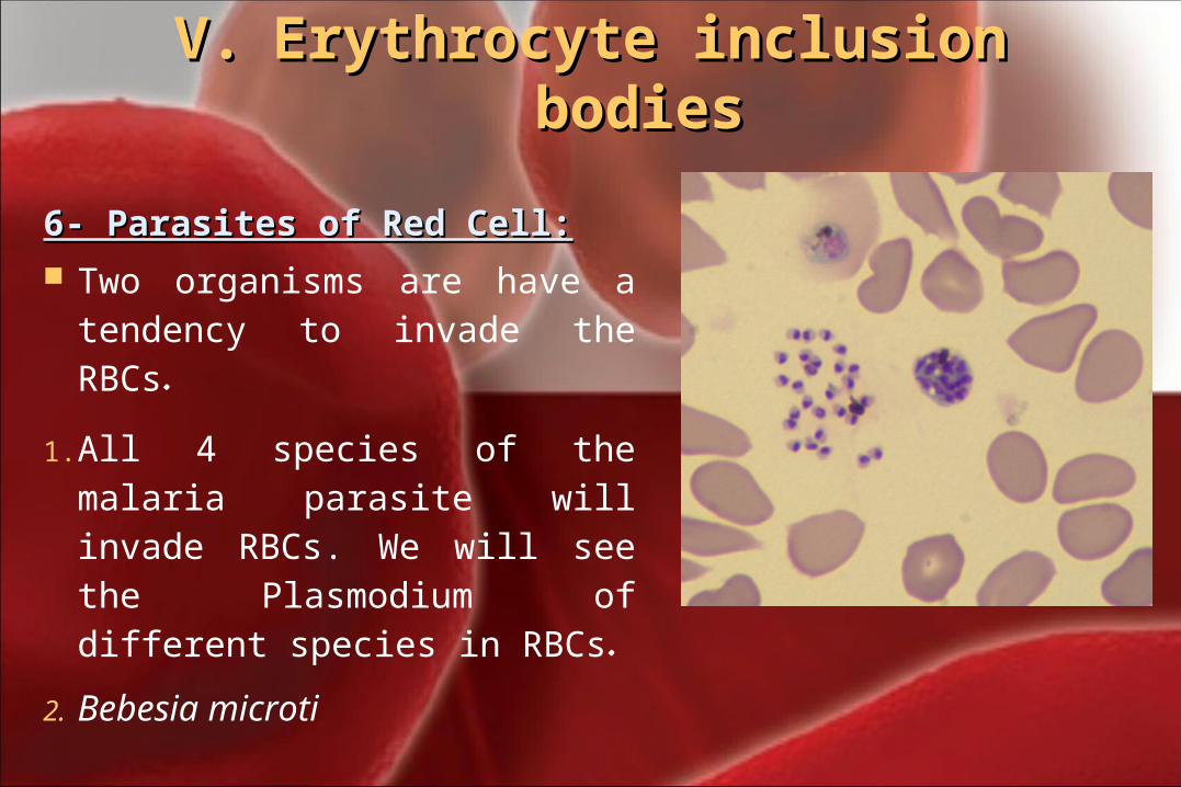
6- Parasites of Red Cell:6- Parasites of Red Cell: Two organisms are have a
tendency to invade the RBCs.
1. All 4 species of the malaria parasite will invade RBCs. We will see the Plasmodium of different species in RBCs.
2. Bebesia microti
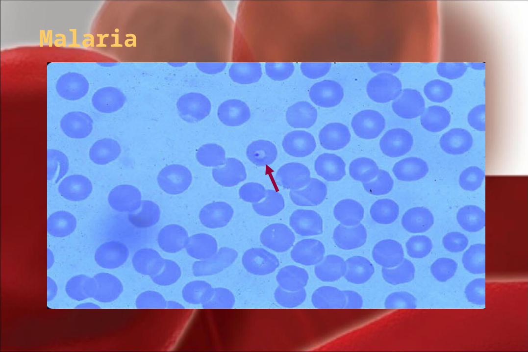
Malaria
RBCs Abnormal morphologyRBCs Abnormal morphology
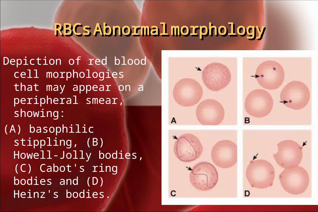
Depiction of red blood cell morphologies that may appear on a peripheral smear, showing:
(A) basophilic stippling, (B) Howell-Jolly bodies, (C) Cabot's ring bodies and (D) Heinz's bodies.
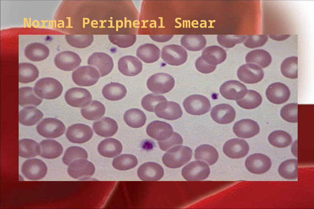
Normal Peripheral Smear
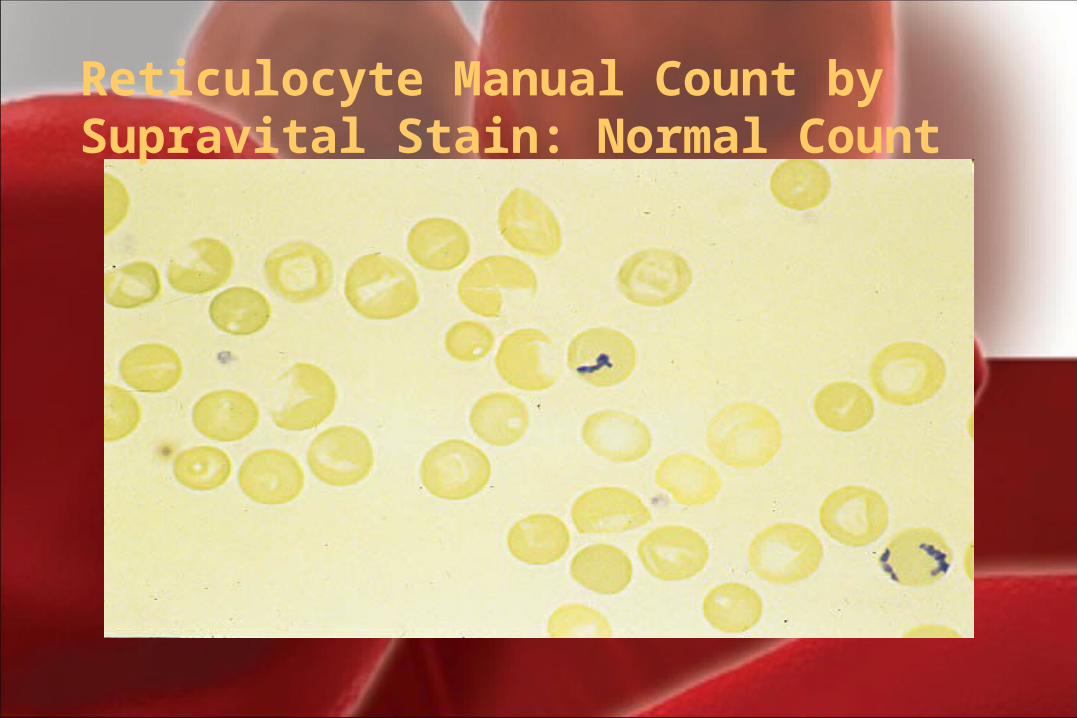
Reticulocyte Manual Count by Supravital Stain: Normal Count
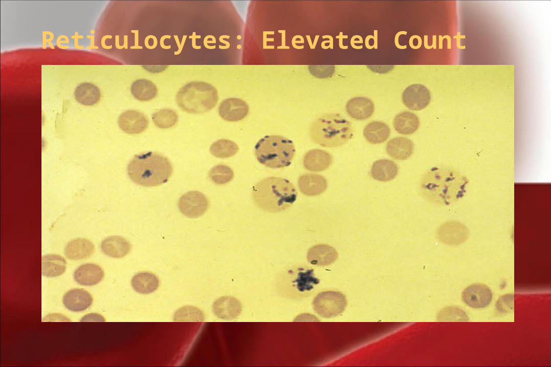
Reticulocytes: Elevated Count
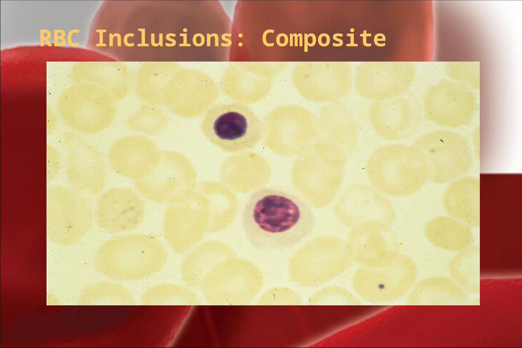
RBC Inclusions: Composite
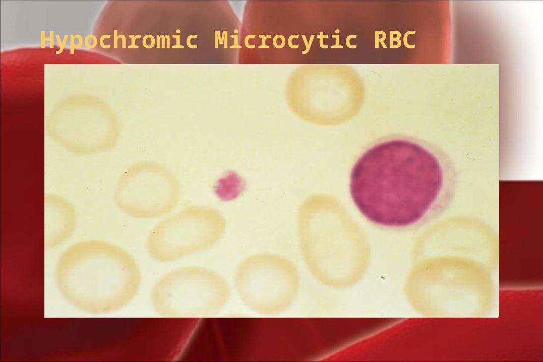
Hypochromic Microcytic RBC
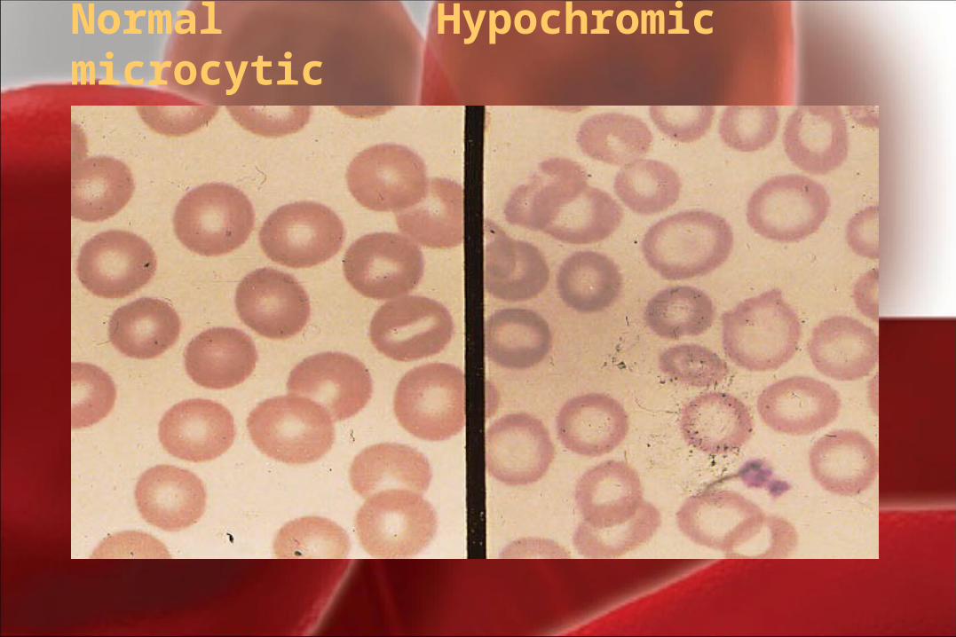
Normal Hypochromic microcytic
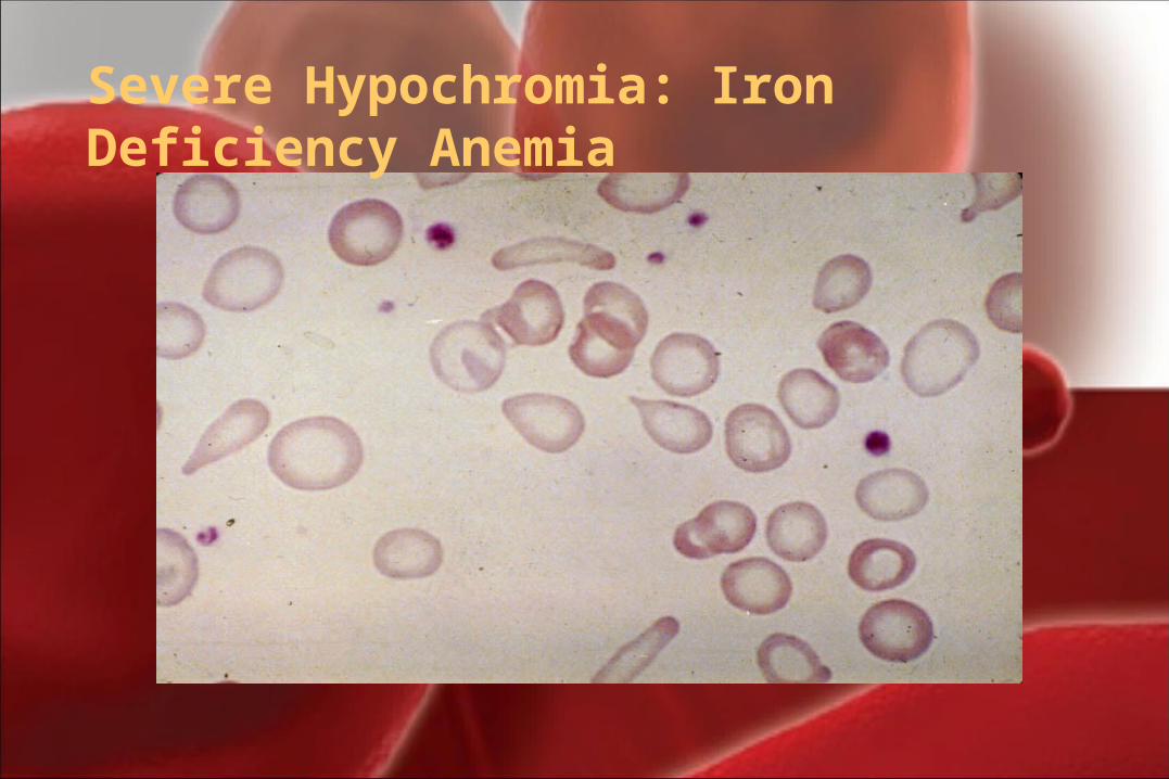
Severe Hypochromia: Iron Deficiency Anemia
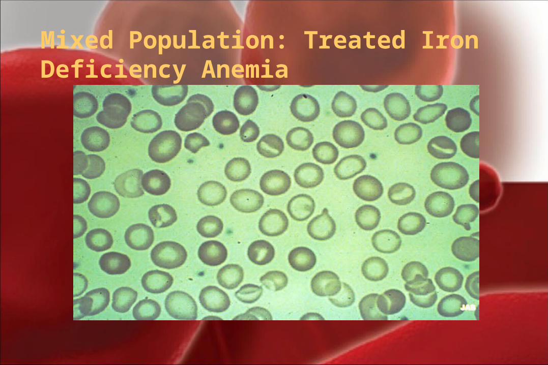
Mixed Population: Treated Iron Deficiency Anemia
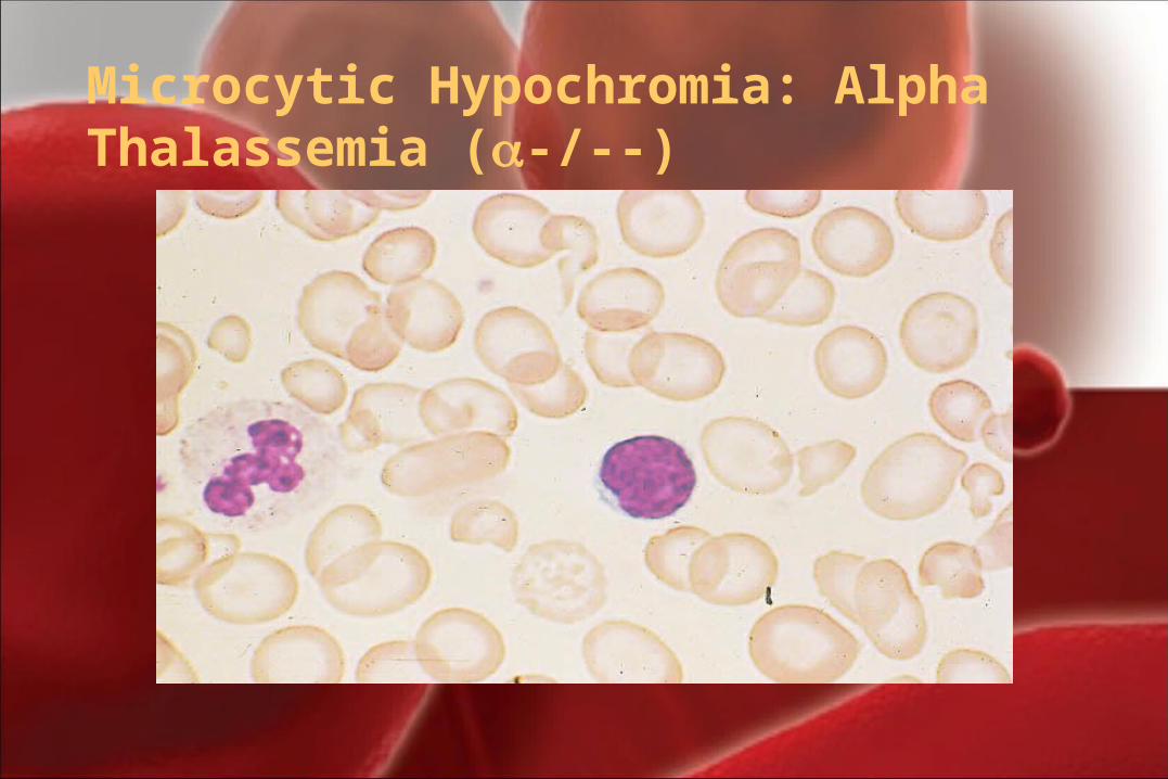
Microcytic Hypochromia: Alpha Thalassemia (-/--)
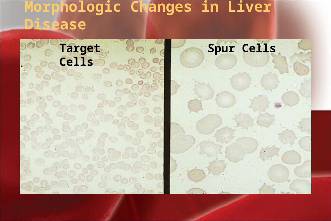
Target Cells Spur Cells
Morphologic Changes in Liver Disease