practical points in ep: when to use wearable icd, loop
TRANSCRIPT

9/18/2016
1
Practical Points in EP: When to Use Wearable ICD, Loop Recorder
John D. Hummel M.D. Director of Clinical Electrophysiology Research
Ross Heart Hospital
The Problem: Sudden Cardiac Arrest
• In people diagnosed with HF, sudden cardiac arrest occurs at 6-9 times the rate of the general population and can be mitigated by ICD implantation or recovery of the EF. Which one is needed takes time to discern.
• Genetic arrhythmia syndromes often present with syncope and can be difficult to diagnose, which takes time
• In patients suffering aborted sudden death ICD’s can be transiently contraindicated and time is needed to allow safe implantation‒ Active infection‒ Neurologic insult with unclear outcome‒ Underlying malignancy with unclear outcome

9/18/2016
2
Understanding the RiskLV Systolic Dysfunction and SCD Risk
• SCD accounted for ~50% (35-64%) of total mortality‒ EF was the single most important risk factor for SCD
0
2
4
6
8
10
12
14
16
18
MERIT-HF12 month
CIBIS-II16 month
CARVEDILOL-US6 month
Total mortality
Sudden Cardiac Death
% m
orta
lity
Post-MIThe risk of SCD post-MI is the highest in the first 30 days
‒ Post-MI patients with heart failure are at 4-6 times greater risk of SCD in the first 30 days after MI
‒ 83% of SCA occurred after hospital discharge.‒ 74% of those resuscitated in the first 30 days were alive at 1 year
1 Solomon SD, et al. Sudden Death in Patients with Myocardial Infarction and Left Ventricular Dysfunction, Heart Failure, or Both. NEJM 2005; 352: 2581-2588.

9/18/2016
3
The Wearable Cardiac Defibrillator
FDA Labeling:
The LifeVest System is indicated for adult patients who are at risk for sudden cardiac arrest and are not candidates for or who refuse an implantable defibrillator.
How It Works:
• Default 150 bpm for VT and 200 bpm for VF
• Discrimination for Afib and SVT up to 200 bpm
• If criteria met it will alarm and give opportunity to suspend treatment or emit gel and deliver therapy.
• Can deliver up to five 150J biphasic shocks if necessary.
• Can deliver this repetitively for up to 20 treatments
• Comes with two batteries and the fully charged battery changed every day.
• Lifevestpatient.com
6

9/18/2016
4
Diagnostics Downloaded FeaturesEfficiently Triage LifeVest Patients
• Tailor alerts & notifications to be notified for events, such as:
• Treatments• Patient Recorded
ECG• Detected arrhythmia
where no treatment was given
• ECG recordings automatically recorded by the LifeVest or manually captured by the patient provide additional information that can be used to decide future care paths.
WEARIT-II Registry: Prospective Registry Of Patients Using WCD
• N= 2,000 patients enrolled in the US
• Data collection: August 2011 – February 2014
ICMICM
805 pts(40.3%)
NICMNICM
927 pts(46.4%)
Cong/InheritedCong/Inherited
268 pts (13.4%)
Kutyifa, et al. Use of the Wearable Cardioverter Defibrillator in High-Risk Cardiac Patients: Data from the Prospective Registry of Patients Using the Wearable Cardioverter Defibrillator (WEARIT-II Registry). Circulation 2015; DOI: 10.1161/CIRCULATIONAHA.115.015677 [epubahead of print].

9/18/2016
5
WEARIT-IIArrhythmic Events
* Treated VT/VF and sustained VT's that spontaneously terminated during the use of the response button or during the extended detection time
1 in 14 patients diagnosed with an arrhythmia requiring intervention while wearing the LifeVest
Patients (%)Events
(events/pt)Event Rate
Per 100 Pt-Year
Any Sustained VT/VF * 41 (2.1%) 120 (2.9) 22
WCD Therapy for VT/VF 22 (1.1%) 30 (1.36) 5
Non-sustained VT 28 (1.4%) 164 (5.9) 30
Atrial arrhythmias/SVT 72 (3.6%) 561 (7.8) 121
Asystole 6 (0.3%) 9 (1.5) 2
7.4%or
1/14
Outcomes Following LifeVest UseWEARIT-II Registry
• Results in the HF population‒ 41%: LVEF improved and patients did not need an
ICD‒ 42%: LVEF did not improve >35% and patients
received an ICD
Kutyifa, V et al., Results From The Prospective Registry Of Patients Using The Wearable Defibrillator (WEARIT-II Registry), presented as Late Breaking
Clinical Trial at European Society of Cardiology, August 30, 2014. Available at http://congress365.escardio.org/Presentation/106358#.VES87vmsUmk
9/18/2016
6
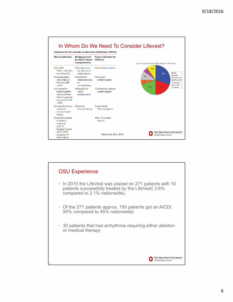
In Whom Do We Need To Consider Lifevest?
Klein et al, EHJ, 2013
OSU Experience
• In 2015 the LifeVest was placed on 271 patients with 10 patients successfully treated by the LifeVest( 3.6% compared to 2.1% nationwide).
• Of the 271 patients approx. 150 patients got an AICD( 56% compared to 45% nationwide)
• 30 patients that had arrhythmia requiring either ablation or medical therapy
12

9/18/2016
7
When To Use Implantable Loop Recorders
Implantable Loop Recorders
Indications for use
• Patients with clinical syndromes or situations at increased risk of cardiac arrhythmias
• Patients who experience transient symptoms such as dizziness, palpitation, syncope and chest pain, that may suggest a cardiac arrhythmia

9/18/2016
8
Syncope
• 7814 participants followed for an average of 17 years, 822 reported syncope
• In one-third of participants, a cause for syncope could not be assigned
• Risk of death was increased by 31% among all participants with syncope
• Risk of death was doubledamong participants with cardiac syncope
• Neurologic syncope (CVA, TIA, seizure) also associated with three-fold risk of stroke Soteriades et al. NEJM 2002; 347: 878
Syncope: Diagnostic Methods & Yield
Test/Procedure Yieldbased on mean time to diagnosis of 5.1
months7
History and Physical (including carotid sinus massage)
49-85% 1, 2
ECG 2-11% 2
Electrophysiology Study without SHD* 11% 3
Electrophysiology Study with SHD 49% 3
Tilt Table Test (without SHD) 11-87% 4, 5
Ambulatory ECG Monitors:
- Holter 2% 7
- External Loop Recorder (2-3 weeks duration) 20% 7
-Insertable Loop Recorder (up to 18 months duration)
65-88% 6, 7
Neurological † (Head CT Scan, EEG, Carotid Doppler) 0-4% 4,5,8,9,10
* Structural Heart Disease† MRI not studied
1 Kapoor, et al N Eng J Med, 1983.2 Kapoor, Am J Med, 1991.3 Linzer, et al. Ann Int. Med, 1997.4 Kapoor, Medicine, 1990.5 Kapoor, JAMA, 1992
6 Krahn, Circulation, 19957 Krahn, Cardiology Clinics, 1997.8 Eagle K,, et al. The Yale J Biol and Medicine. 1983; 56: 1-8.9 Day S, et al. Am J Med. 1982; 73: 15-23.10 Stetson P, et al. PACE. 1999; 22 (part II): 782.

9/18/2016
9
Symptom-Rhythm Correlation
Auto Activation Point
Patient Activation Point
Diagnosis of AF in stroke patientchanges treatment protocol (currently aspirin)
61%of patients who had both AF and a stroke did not know they had AF prior to their stroke
1/3of Ischemic Strokes are Cryptogenic
20%Cardioembolic
30%Cryptogenic Stroke
30%Large Vessel
15%Small Vessel
5%Other
CRYPTOGENIC STROKE

9/18/2016
10
CRYPTOGENIC STROKE
Detection of AF allows for treatment with OAC therapy
More patients with AF detected at 12 months with ILR
Median number of days to AF detection over 12 months
7×
84Percent of patients prescribed OAC once AF was detected
97%
7X
Crystal-AF Study, NEJM
ILR
ILR AF monitoring
Duration-based performance metrics
98.4%SENSITIVITY
99.5%SPECIFICITY
99.4%ACCURACY
97.2%PPV
99.7%NPV
Comparison of AF burden detected by ICM and Holter

9/18/2016
11
Detailed AF Data
21
All patient and clinical data are fictitious and for demonstration purposes only.
Daily AF Burden
Day/Night HR
Heart Rate Variability
Ventricular Rate During AF
Patient Activity
22
AF MANAGEMENTOptimize decisions pre- and post-ablation
PRE
Document Baseline AF burden.
Provide objective data for follow-up comparisons (burden, symptoms).
Case Planning
PVI when Paroxysmal AF is documented?
PVI + additional techniques if substrate ablation is needed (atrial flutter, rotor)
Work Flow Planning .
Triage patients on your waiting list (poor rate control, frequent Sx)
POST
Monitor patients for up to 3 years.
Optimize medical therapy.
Discontinue OAC or AARx?
Re-ablate to disrupt progression over time.

9/18/2016
12
Summary: Current Trends in Arrhythmia Monitoring
• Greater Use of Implantable Loop Recorders:‒ Diagnosis of Syncope‒ Management of Atrial Fibrillation‒ Evaluation of Cyptogenic Stroke
Insurance Coverage
• Primary prevention (EF≤35% and MI, NICM, or other DCM) including:‒ After recent MI (Coverage during the 40 day ICD waiting period).‒ Before and after CABG or PTCA (Coverage during the 90 day ICD waiting
period).‒ Listed for cardiac transplant.‒ Recently diagnosed nonischemic cardiomyopathy (Coverage during the 3 to
9 month ICD waiting period).‒ NYHA class IV heart failure.‒ Terminal disease with life expectancy of less than 1 year.
• ICD indications when patient condition delays or prohibits ICD implantation
• ICD explantation

9/18/2016
13
Syncope Diagnosis: Role of an ILR
A. Strickberger et al. Circulation 2006; 113: 316-327
AHA/ACC Scientific Statement on the Evaluation of Syncope:
“This approach (ILRs) is
more likely to identify
the mechanism of
syncope than is a
conventional approach
that uses Holter or
event monitors and EP
testing and is cost-
effective.”
ISSUE Study Implications
• HUT outcome was not predictive of vasodepressor vs. cardioinhibitory response‒ Bradycardia is common in spontaneous VVS -
independent of HUT outcome
• Bradycardia is more prevalent in spontaneous events vs. HUT induced VVS
• Clinical Implication: Consider a strategy of ILR guided evaluation in positive TTT patients unresponsive to medication
Moya A. Circulation. 2001; 104:1261-1267
9/18/2016
14
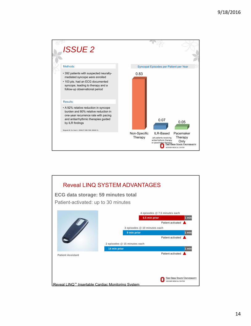
ISSUE 2
Syncopal Episodes per Patient per Year
0.83
0.07 0.05
Non-Specific Therapy
ILR-Based (all patients receivingantiarrhythmic therapyor pacemaker therapy)
Pacemaker Therapy
Only
Brignole M. Eur Heart J. 2006;27:1085-1092 (ISSUE 2).
Methods:
• A 92% relative reduction in syncope burden and 80% relative reduction in one-year recurrence rate with pacing and antiarrhythmic therapies guided by ILR findings
• 392 patients with suspected neurally-mediated syncope were enrolled
• 103 pts. had an ECG documented syncope, leading to therapy and a follow-up observational period
Results:
Patient Assistant
4 episodes @ 7.5 minutes each
3 episodes @ 10 minutes each
2 episodes @ 15 minutes each
Patient-activated
Patient-activated
Patient-activated
9 min prior
14 min prior 1 min
1 min
1 min6.5 min prior
Reveal LINQ SYSTEM ADVANTAGES
28
ECG data storage: 59 minutes total
Patient-activated: up to 30 minutes
Reveal LINQ™ Insertable Cardiac Monitoring System

9/18/2016
15
Reveal LINQ SYSTEM ADVANTAGES
29
ECG data storage: 59 minutes total
Automatically detected: 27 minutes
Reveal LINQ™ Insertable Cardiac Monitoring System
Pause, Brady, Tachy
End of episode
Start of episode
30 sec 27 sec
Automatic detection
AT/AF
2 minutes of longest AF episode stored since last interrogation in addition to the 27 minutes of automatically detected episodes.
Reveal LINQ SYSTEM ADVANTAGES• PROVEN ARRYTHMIA DETECTION.
• INFORMED CLINICAL DECISIONS.
30
Highest published AF detection accuracy on the market, at 99.4%, streamlines data review8
63% fewer false positives than shown in other ICM published data14
As the most clinically-validated ICM, with 50+ detection performance papers, Reveal LINQ is the reliable choice for arrhythmia management15
Reveal LINQ™ Insertable Cardiac Monitoring System
99.4%
63%
50+
Reveal LINQ is proven to find AF

9/18/2016
16
PROVEN ACCURACY
• AF FALSE POSITIVE COMPARISON
31Reveal LINQ™ Insertable Cardiac Monitoring System
63%fewer false positives with Reveal LINQ than shown in other ICM published data14
50%
Confirm-AF16
BioMonitor 2-AF17
Reveal LINQ8Fal
se P
ositi
ve R
ate*
0%25
%
9.6%
26.3%
40.9%
*% of False Positives = (1 –Episode PPV). Episode PPV may vary (gross, patient average).
CLINICAL RIGOR
• EVIDENCE SUPERIORITY.
• REAL-WORLD IMPACT.
32
• With an evidence portfolio of 500+ published clinical articles and abstracts1
• Across Cryptogenic Stroke, Syncope, and Atrial Fibrillation patient populations8-10
• Published in multiple premier journals, including Heart Rhythm, The New England Journal of Medicine and JACC 8,9,11
Reveal LINQ™ Insertable Cardiac Monitoring System
Most Studied ICM
Most Clinically Validated ICM
Only ICM with Premier Clinical Evidence

9/18/2016
17
Clinical rigor
• CRYPTOGENIC STROKE
33
Real-world practice with Reveal LINQ ICMSuperiority of Reveal LINQ system to detect AF in Cryptogenic Stroke patients
Real-world practice validates superiority of the Reveal LINQ System to detect AF in cryptogenic stroke (CS) patients12,13
Everyday use of Reveal LINQ confirms findings in landmark CRYSTAL AF study (NEJM,Reveal LINQ™ Insertable Cardiac Monitoring System
72%of AF patients would be missed if monitoring ends at 30 days12
AF
12
CLINICAL RIGOR
• SYNCOPE
34
40% of the population will experience syncope21
Cardiac syncope doubles the risk of death
Reveal LINQ™ Insertable Cardiac Monitoring System
MORTALITY
Cardiac condition doubles the risk of death and increasesthe 6-month mortality rate by 10%22
MAGNITUDE
3-5% of all ER visits23
1-3% of all hospitalizations23
UNDIAGNOSED
50% of all patients leave the hospital without a diagnosis24
FRUSTRATION
13 different tests during diagnosis period10
Syncope patient visits 3 specialists on average10

9/18/2016
18
CLINICAL RIGOR• SYNCOPE
35
Limited yield of many diagnostic testsDiagnostic yield of non implantable diagnostic tests is less than 50%
Reveal LINQ™ Insertable Cardiac Monitoring System
11%2%
20%11%
49%
4%
78%
0%
20%
40%
60%
80%
100%
ECG Holter Monitor External LoopRecorder
EP Study w/oStructural
Heart Disease
EP Study WithStructural
Heart Disease
Neurological(CT, Carotid
Doppler)
Reveal ICM
Dia
gn
ost
ic Y
ield
*
25 26
11
27 28
29
10
*Based on mean diagnosis time of 5.1 mos.
CLINICAL RIGOR
SYNCOPE GUIDELINES, European Heart Journal 20092
Recommendations for the use of ICM monitoringGuidelines for the diagnosis and management of syncope (version 2009)
The Task Force for the Diagnosis and Management of Syncope of the European Society of Cardiology (ESC)
Developed in collaboration with European Heart Rhythm Association (EHRA),
Heart Failure Association (HFA), and Heart Rhythm Society (HRS)
Monitoring Choice by Frequency of Symptoms3Frequency of symptoms Suggested ECG monitoring
technique
Daily
Every 2-3 days
Every week
Every month
Less than once per month
24 h Holter, in-hospitaltelemetric monitoring
48-72 h Holter, in-hospitaltelemetric monitoring
7 days Holter or external loop recorder
14-30 days external loop recorder
Implantable loop recorder
Monitoring Choice by Patient Risk2

9/18/2016
19
SUSPECTED AF MONITORING
Continuous monitoring can reveal AF
• Symptoms are not a good indicator for presence of AF30-34
• Continuous monitoring with ICMs can identify AF in patients at high risk35, 36
Study Method of Monitoring% Asymptomatic AF
Page et al. 199430 External monitors:1 day/week (5×)
92.3% of episodes
Strickberger et al. 200531
Implantable Pacemakers 94% of episodes
Quirino et al. 200932 Implantable Pacemakers 81% of episodes
Orlov et al. 200733 Implantable Pacemakers 94.7% of episodes
Verma et al. 201334 Implantable Loop Recorders 79% of episodes
22% of a patient population at high risk was found to have > 6min of AF within 12 months36
Advanced monitoring
• Innovative solutions.
• Simplified experience.
38Reveal LINQ™ Insertable Cardiac Monitoring System
STREAMLINED INSERTION WORK FLOW
ACTIONABLE REPORTS SIMPLIFIED PATIENT MANAGEMENT Supported by an
enhanced Medtronic CareLink Network
Industry’s highest diagnostic yields, with actionable reports10,39-41
Simple, minimally invasive outpatient insertion procedure
New app-based device management with the Reveal LINQ Mobile Manager
Resources to support clinic efficiency and data review
New Medtronic Academy Learning Plan
New Patient Education Resources
New Reveal LINQSM
Monitoring Service*
*Available in select U.S. markets.
9/18/2016
20
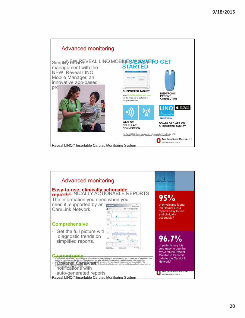
Advanced monitoring
• NEW REVEAL LINQ MOBILE MANAGER
39
Simplify device management with the NEW Reveal LINQ Mobile Manager, an innovative app-based programming system
Reveal LINQ™ Insertable Cardiac Monitoring System
IT’S EASY TO GET STARTED
The Reveal LINQ Mobile Manager can only be used with the Reveal LINQ ICM and the Medtronic patient connector, available from Medtronic.
SUPPORTED TABLET
Visit LINQMobileManager.comfor the most up-to-date list of supported tablets
WI-FI OR CELLULAR CONNECTION
MEDTRONIC PATIENT CONNECTOR
DOWNLOAD APP ON SUPPORTED TABLET
Advanced monitoring
• CLINICALLY ACTIONABLE REPORTS
40
Easy-to-use, clinically actionable reports4
The information you need when you need it, supported by an enhanced CareLink Network
Comprehensive
• Get the full picture withdiagnostic trends on simplified reports.
Customizable
• Optional CareAlert™notifications with auto-generated reports
Reveal LINQ™ Insertable Cardiac Monitoring System
95%of physicians found the Reveal LINQ reports easy to use and clinically actionable 6
96.7%of patients say it is very easy to use the MyCareLink Patient Monitor to transmit data to the CareLink Network6
The Medtronic MyCareLink Patient Monitor and the Medtronic CareLink Network are indicated for use in the transfer of patient data from Medtronic implantable cardiac devices. These products are not a substitute for appropriate medical attention in the event of an emergency. Data availability and alert notifications are subject to Internet connectivity and access, and service availability. The MyCareLink Patient Monitor must be on and in range of the device. Alert notifications are not intended to be used as the sole basis for making decisions about patient medical care.

9/18/2016
21
Medical Therapy Optimization RequiredPrior To Managing Long-Term Arrhythmic Risk
• Medical optimization and stabilization can take 3 months or more.‒ Beta blocker doses effective in HF are generally
achieved in 8 to 12 weeks and do not impart any mortality benefit until at least 3 months
CIBIS-II3
Car
diov
ascu
lar
Mor
talit
y (%
)
5
10
15
3 6 9 12 15 18
Placebo
Metoprolol CR/XL
Months
Merit-HF1
00
% S
urvi
val
3 6 9 12 15 18 21
Months
100
90
80
60
70
Carvedilol
Placebo
COPERNICUS2
0 200 400 600 800
100
80
60
Bisoprolol
Placebo
Days
% S
urvi
val
1 Merit-HF Study Group. Effect of metoprolol CR/XL in chronic heart failure: Metoprolol CR/XL Randomized Intervention Trial in Congestive Heart Failure (MERIT-HF) Lancet 1999;353:2001-7.
2 Packer M, et al. Effect of Carvedilol on survival in severe chronic heart failure. NEJM 2001;344:1651-8.3 CIBIS-II Investigators. The Cardiac Insufficiency Bisoprolol Study II (CIBIS-II). Lancet 1999;353:9-13.
LifeVest by the Numbers
• 98% first shock success rate
• 92% shocked event survival (conscious ER arrival or stayed at home)
• Most (73%) treated within 60 seconds (remaining delayed from response button use or VT programming)

9/18/2016
22
LifeVest Offers Protection From SCDTime To Recovery and Assess Long-Term Risk
‒ Allows physician to assess long-term arrhythmic risk at the end of the Medicare ICD waiting period (40 days post-MI and 90 days post-PTCA/post-CABG).
‒ Patients return to the prescribing or referring physician at the term of their LifeVest prescription for follow-up to determine if:
1) LifeVest usage should be extended,
2) Long-term arrhythmic risk requires ICD implantation, or
3) Cardiac recovery has mitigated arrhythmic risk, and ongoing follow-up with optimized medical therapy is appropriate.
WCD allows time to develop long-term risk management strategies
Figure 1. Risk stratification for ICD therapy: The role of the WCD (Klein et al. Eur Heart J 2013;34:2230-2242)

9/18/2016
23
Heart Rhythm SocietySudden Cardiac Death Primary Prevention Protocols
Conclusions
• Post-MI patients with heart failure are at 4-6 times greater risk of SCD in the first 30 days after MI
• OMT optimization takes time and is different for every patient
• The AHA (endorsed by the HRS) recommends the WCD as a Class IIb indication for a broad range of patients
• SCD screening tools can help to identify those patients at the highest risk
• The LifeVest is an effective tool to protect HF patients with low EF during medication titration while long-term risk is being determined

9/18/2016
24
2016 AHA Science AdvisoryWearable Cardioverter Defibrillator Therapy for the Prevention of Sudden Cardiac Death
• WCDs can serve as temporary means of preventing arrhythmic death without the need for bystander response to cardiac arrest.
• WCD use may be appropriate in clinical circumstances associated with transient increased arrhythmic risk
Piccini JP Sr, Allen LA, Kudenchuk PJ, Page RL, Patel MR, Turakhia MP; on behalf of the American Heart Association Electrocardiography and Arrhythmias Committee of the Council on Clinical Cardiology and Council on Cardiovascular and Stroke Nursing. Wearable cardioverter-defibrillator therapy for the prevention of sudden cardiac death: a science advisory from the
American Heart Association. Circulation. 2016;133:XXX–XXX. DOI 10.1161/CIR.0000000000000394.
Indication ClassLevel of Evidence
Use of WCDs is reasonable when there is a clear indication for an implanted/ permanent device accompanied by a transient contraindication or interruption in ICD care such as infection.
IIa C
Use of WCDs is reasonable as a bridge to more definitive therapy such as cardiac transplantation. IIa C
Use of WCDs may be reasonable when there is concern about a heightened riskof SCD that may resolve over time or with treatment of left ventricular dysfunction; for example, in ischemic heart disease with recent revascularization, newly diagnosed non-ischemic dilated cardiomyopathy in patients starting guideline-directed medical therapy, or secondary cardiomyopathy (tachycardia mediated, thyroid mediated, etc.) in which the underlying cause is potentially treatable
IIb C
WCDs may be appropriate as bridging therapy in situations associated with increased risk of death in which ICDs have been shown to reduce SCD but not overall survival such as within 40 days of MI.
IIb C
WCDs should not be used when non-arrhythmic risk is expected to significantly exceed arrhythmic risk, particularly in patients who are not expected to survive >6 mo.
III: No benefit
C

9/18/2016
25
LifeVest Patient Example
50

9/18/2016
26
Remote Patient Data Management with the Wearable Cardioverter Defibrillator
Agenda
• WEARIT II Registry
• Overview of the LifeVest Network and LifeVest Trends
• Clinical Case Studies – Remote Patient Management in Action

9/18/2016
27
Remote Patient Data Management with the LifeVest Network
• Information about Clinical Events in the earliest period after a cardiac event
• Automatic Event Recordings‒ Shock Treatment
• VT/VF detection‒ Arrhythmia Detected but Not Treated
• Sustained VT/VF that terminated prior to treatment• Non-sustained VT/VF
‒ Asystole• Asystole• Bradycardia that meets asystole criteria
LifeVest Network:Customized Reports

9/18/2016
28
TrendsFeatures Overview
84 bpm
1 2 3
90
45
30
0
• Avg daily heart rate
• Avg heart rate in 5 min increments for each day
• Total steps per day
• Steps in 5 min increments for each day
• Overall body position (movement, upright, reclined, lying)
• Body angle while reclined or lying
• Body position while reclined or lying (prone, supine, left, right)
• Clinicians can select up to 12 questions for patients to answer on a daily or weekly basis
TrendsFeatures – Health Survey
Clinicians can select up to 12 preset Health Survey questions for patients to answer on a daily or weekly basis

9/18/2016
29
TrendsFeatures – Health Survey
Users can review patient survey responses by day
Survey Answers by Individual Day
Or view trending data of patient survey responses for each question over time
Trend of Survey Answers by Question
TrendsFeatures – Health Survey
Patients can respond to clinician selected Health Survey questions directly on the LifeVest monitor. Patient survey responses are transmitted with patient downloads.

9/18/2016
30
TrendsFeatures – Trends Reports
Users can create custom reports to include any of the Trends features
Conclusions
• In the WEARIT II Registry, 1 in 14 patients were diagnosed with an arrhythmia requiring intervention while wearing the LifeVest.
• In addition to protection from SCD, the LifeVest captures valuable data about the patient’s broader cardiac function and health status that offers meaningful clinical value during the early period following a cardiac event when a patient’s condition is changing.

9/18/2016
31
Case Studies Remote Patient Data Management
Case 1: Syncope

9/18/2016
32
Patient Experiences Unexplained Syncope
Patient History and Plan
• A conscious 64-year-old female presented to the ER after suffering a syncope event that resulted in minor injuries. Lab work and CT scan of head were normal.
• History: ‒ 2.5 months post large anterior STEMI.
‒ LAD had 100% de novo stenosis requiring DES placement. Following intervention, there was 0% stenosis with TIMI grade 3 flow.
‒ Discharge LVEF = 30%.
• Pharmacy:‒ Patient is euvolemic; continue carvedilol, sprironolactone, lisinopril, add
sublingual nitroglycerin prn
• Recent hospitalization noted continued low LVEF = 30% with dilated cardiomyopathy. ‒ Suspected Ventricular Tachycardia (VT) cause for syncope
• Electrophysiology (EP) consult resulted in patient discharge with the LifeVest wearable defibrillator for protection from SCD

9/18/2016
33
LifeVest Network Configuration
• LifeVest Network Configuration‒ Red (high-level) alert for treatments‒ Orange (mid-level) alert for asystole event‒ Orange (mid-level) alert for patient initiated ECG recording.
• The LifeVest detection algorithm is programmed to declare asystole when the heart rate falls below 10 beats per minute (bpm) for 16 seconds and automatically records the event, with 120 seconds of onset.
• Patients can perform manual recordings by pressing the response buttons for three seconds, which records the previous 30 seconds plus the next 15 seconds.
LifeVest Network Configuration

9/18/2016
34
Results
• ECG review by the EP revealed bradycardia at rate below 20 bpmwith increasing R-R intervals. In-office follow-up by the EP resulted in permanent pacemaker implantation.
• One (1) day post-discharge, the LifeVest captured 2 patient-initiated recordings and 5 asystole events with no tachyarrhythmia detections or shocks.
• The asystole events occurred while the patient was sleeping.
Identification of Bradycardia through Remote Patient Monitoring
• The clinic regularly reviews notifications and patient recordings on the LifeVest Network.
• Upon review of the ECG recordings in the LifeVestNetwork, it was determined that the patient experienced several bradycardia events, identified by the LifeVest as asystole events, that required additional in-office work up.
• In this case, information captured by the LifeVest directly impacted the patient’s care path.
9/18/2016
35
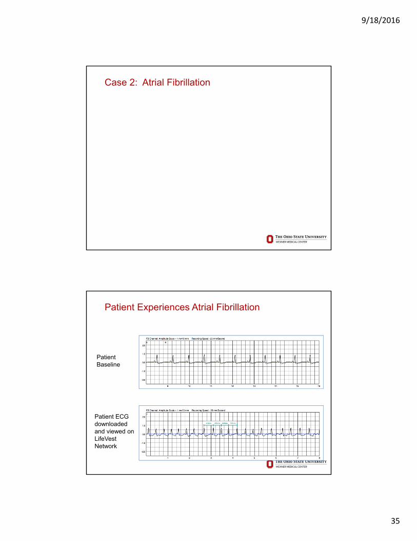
Case 2: Atrial Fibrillation
Patient Experiences Atrial Fibrillation
Patient Baseline
Patient ECG downloaded and viewed on LifeVest Network

9/18/2016
36
History and Plan
• 68 year-old man with no known past medical history was admitted to the ER due to a one week history of shortness of breath and chest discomfort
• Chest x-ray showed cardiomegaly; labs indicated mildly elevated BNP
• Enzymes were negative for acute MI
• 2D echogram:‒ LVEF = 20%
‒ Global hypokinesis of left ventricle
‒ Mild mitral valve regurgitation
• Pharmacy:‒ Patient was Dx with Lasix 20 mg QD, carvedilol 1.5625 mg BID, low salt diet, and
a plan to follow up with a cardiologist in 3-5 days.
• Upon follow-up, patient complained of continued non-exertional chest “tightening” lasting 30-45 minutes.
• Cardiologist prescribed the LifeVest wearable defibrillator for protection from SCD.
BK7
LifeVest Network Configuration
• LifeVest Network Configuration‒ Orange (mid-level) alert for asystole event‒ Orange (mid-level) alert for at least 2 patient-initiated recordings
per day, detected but not treated events
• The LifeVest allows patients to manually record ECG strips by pressing the response buttons for three seconds.
BK8

Slide 71
BK7 correct carvedilol spellingBernard Komoroski, 11/5/2015
Slide 72
BK8 font colorBernard Komoroski, 11/5/2015
9/18/2016
37
Results
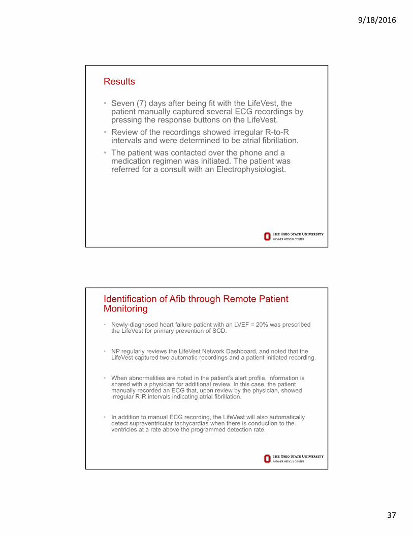
• Seven (7) days after being fit with the LifeVest, the patient manually captured several ECG recordings by pressing the response buttons on the LifeVest.
• Review of the recordings showed irregular R-to-R intervals and were determined to be atrial fibrillation.
• The patient was contacted over the phone and a medication regimen was initiated. The patient was referred for a consult with an Electrophysiologist.
Identification of Afib through Remote Patient Monitoring
• Newly-diagnosed heart failure patient with an LVEF = 20% was prescribed the LifeVest for primary prevention of SCD.
• NP regularly reviews the LifeVest Network Dashboard, and noted that the LifeVest captured two automatic recordings and a patient-initiated recording.
• When abnormalities are noted in the patient’s alert profile, information is shared with a physician for additional review. In this case, the patient manually recorded an ECG that, upon review by the physician, showed irregular R-R intervals indicating atrial fibrillation.
• In addition to manual ECG recording, the LifeVest will also automatically detect supraventricular tachycardias when there is conduction to the ventricles at a rate above the programmed detection rate.

9/18/2016
38
Current Trends: Development of Subcutaneous Sensors for Disease Management
Signal Company
ECG •Medtronic•St. Jude•Transoma Medical•AJ Medical
Arterial Pressure •Transoma Medical
Gross Motor Activity •Medtronic•St. Jude•Transoma Medical
Heart Sounds •Boston Scientific
Respiration •Apnex•Transoma Medical
Case 3: Bradycardia

9/18/2016
39
Patient Experiences Bradycardia
History and Plan
• A 61-year-old woman presented to the ER with complaints of acute chest pain x2 hours radiating to the left side of chest that improved with nitroglycerin.
• Significant history of rheumatoid arthritis, hypertension, hyperlipidemia, and osteoporosis.
• No known surgical history.
• Cardiac enzymes and ECG were both negative for STEMI.
• ECG indicated RBBB.
• Cardiac echo: ‒ Mild left ventricular hypertrophy
‒ Mild mitral annular calcification
‒ LVEF = 30%
• SPECT imaging showed lack of activity throughout the inferior wall extending into the inferiolateral region. Cardiomegaly with large areas of infarction also noted.
• The patient was discharged with the LifeVest wearable defibrillator for protection from SCD.

9/18/2016
40
LifeVest Network Configuration
• LifeVest Network Configuration‒ Orange (mid-level) alert for asystole events, at least 2 patient-
initiated recordings per day, detected but not treated events
• The LifeVest detection algorithm is programmed to declare asystole when the heart rate falls below 10 beats per minute (bpm) for 16 seconds and automatically records the event, including 120 seconds of onset.
BK9
Results
• On day 29 post discharge, the LifeVest detected an asystole event and captured an ECG recording. Upon ECG review on the LifeVest Network, the event was determined to be bradycardia.
• The patient was contacted at home and instructed to seek immediate medical care in the ER.
• Cardiac catheterization and angiography showed apical and inferior hypokinesia of left ventricle with triple vessel disease involving left anterior descending (LAD) with its first diagonal branch (D1), left circumflex (LCx) with its first diagonal branch, and right coronary artery (RCA).
• Post-CABG, the patient’s LVEF remained ≤ 35% and the patient was discharged home with instructions to continue LifeVest use.

Slide 79
BK9 font colorBernard Komoroski, 11/5/2015

9/18/2016
41
Sinus Tachycardia
Identification of Bradycardia through Remote Patient Monitoring
• The patient was diagnosed with ischemic cardiomyopathy following a NSTEMI with a LVEF = 30% and prescribed the LifeVest for primary prevention of SCD.
• The nurse in this practice reviews the LifeVest Network dashboard weekly, monitoring the practice’s active LifeVest patients.
• The discovery of several bradycardia events in this case revealed a significant underlying ischemic disease requiring CABG, potentially avoiding significant future myocardial injury.
9/18/2016
42
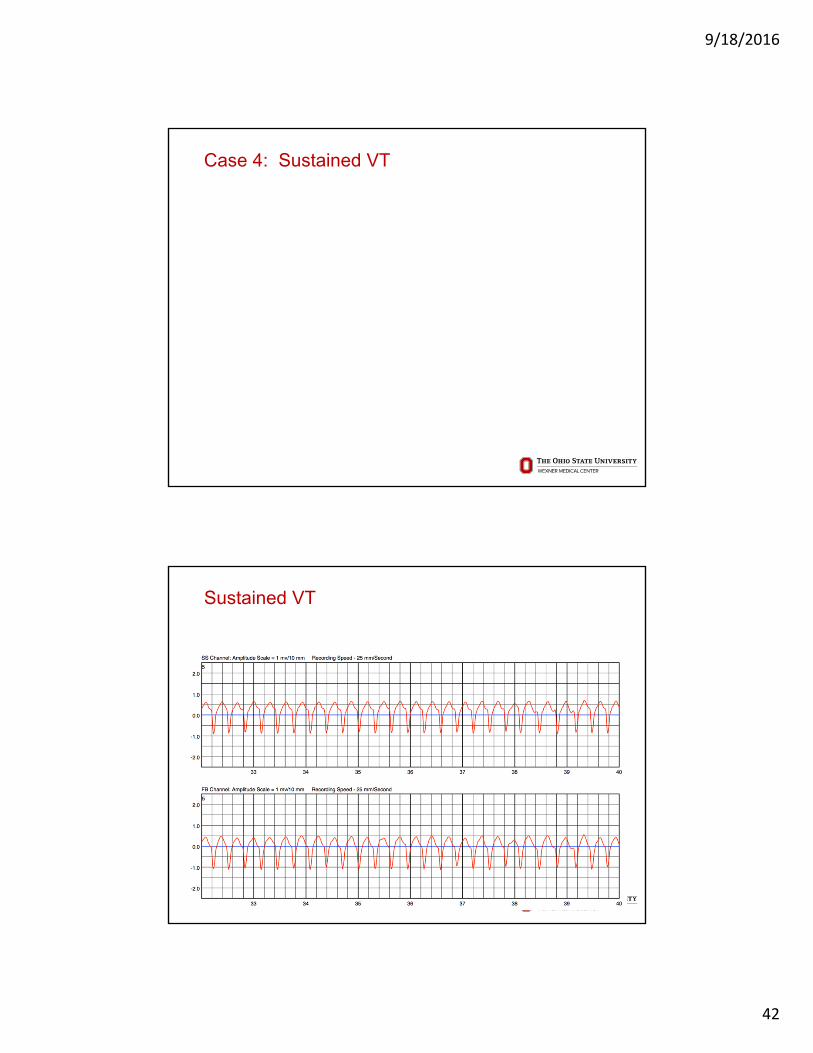
Case 4: Sustained VT
Sustained VT
9/18/2016
43
History and Plan
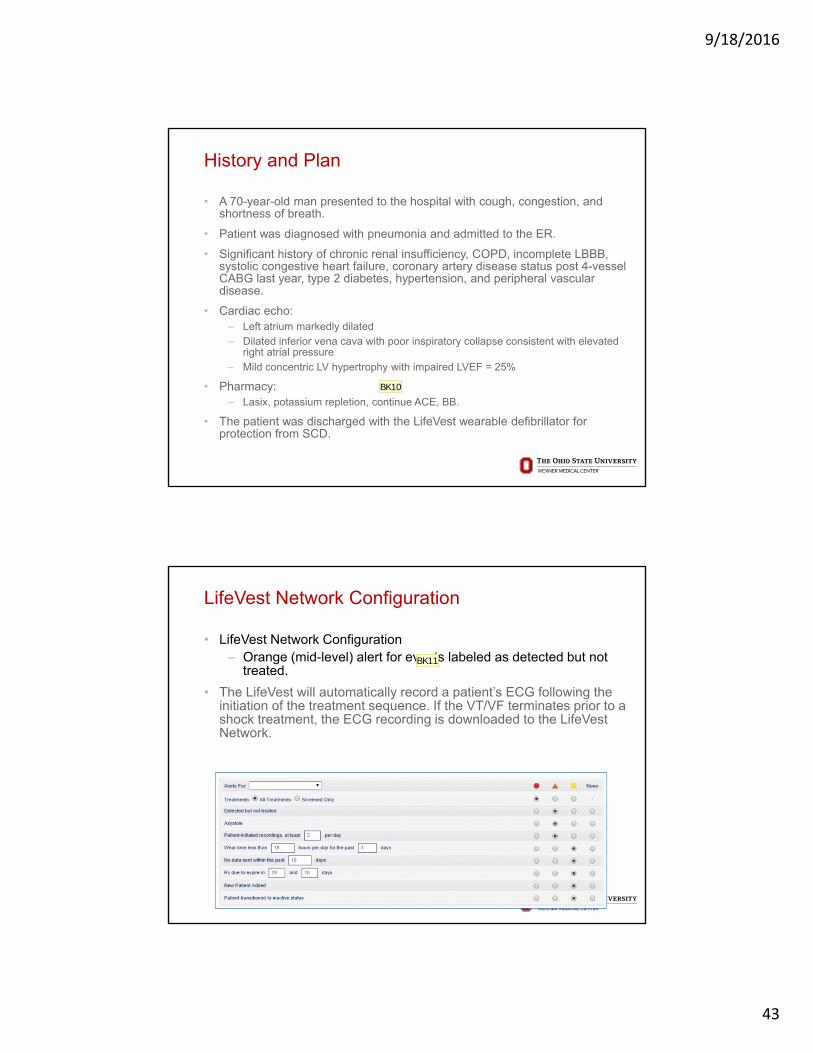
• A 70-year-old man presented to the hospital with cough, congestion, and shortness of breath.
• Patient was diagnosed with pneumonia and admitted to the ER.
• Significant history of chronic renal insufficiency, COPD, incomplete LBBB, systolic congestive heart failure, coronary artery disease status post 4-vessel CABG last year, type 2 diabetes, hypertension, and peripheral vascular disease.
• Cardiac echo: ‒ Left atrium markedly dilated
‒ Dilated inferior vena cava with poor inspiratory collapse consistent with elevated right atrial pressure
‒ Mild concentric LV hypertrophy with impaired LVEF = 25%
• Pharmacy: ‒ Lasix, potassium repletion, continue ACE, BB.
• The patient was discharged with the LifeVest wearable defibrillator for protection from SCD.
BK10
LifeVest Network Configuration
• LifeVest Network Configuration‒ Orange (mid-level) alert for events labeled as detected but not
treated.
• The LifeVest will automatically record a patient’s ECG following the initiation of the treatment sequence. If the VT/VF terminates prior to a shock treatment, the ECG recording is downloaded to the LifeVest Network.
BK11

Slide 85
BK10 fixed misspelled hypertrophyBernard Komoroski, 11/5/2015
Slide 86
BK11 font colorBernard Komoroski, 11/5/2015

9/18/2016
44
Results
• Twelve (12) days after discharge, the patient experienced an episode of ventricular tachycardia (VT) at a rate of 240 bpm.
• The LifeVest appropriately detected the VT, and the treatment sequence was initiated. Treatment was delayed through proper patient use of the response buttons.
• The VT spontaneously terminated after 35 seconds.
• The patient was referred to an Electrophysiologist (EP) and a permanent implantable cardioverter defibrillator (ICD) was later implanted.
Identification of Sustained VT through Remote Monitoring
• The patient was diagnosed with a DCM and a LVEF = 25% and prescribed the LifeVest for primary prevention of SCD.
• The NP regularly reviews the LifeVest Network dashboard to monitor for new alerts for the practice’s current active patients.
• Upon dashboard review, a “detected but not treated” event was noted for the patient. Review of the ECG recordings revealed that the LifeVest had recorded a sustained VT event.
• Subsequent consult with an EP resulted in the implantation of an ICD.

9/18/2016
45
Case 5: Sinus Tachycardia
History and Plan
• A 63-year-old male presented to the emergency department with severe chest pain and pressure.
• Significant history of hypertension, hyperlipidemia, chronic kidney disease and tobacco use.
• Coronary Angiography: ‒ 80% ostial stenosis of the large first diagonal of the left anterior
descending (LAD) artery. ‒ 80% bifurcation lesion affecting both the LAD and the ostium of the
diagonal branch. ‒ Two (2) drug-eluting stents were placed.
• Pharmacy: ‒ Patient was prescribed aspirin 81 mg QD, ticagrelor 90 mg BID, lisinopril 10 mg
QD and metoprolol succinate 25 mg QD.
• The patient was discharged with the LifeVest wearable defibrillator for protection from SCD.

9/18/2016
46
LifeVest Network Configuration
• LifeVest Network Configuration‒ Red (high-level) alert for notifications when the patient manually
captured ECG recordings‒ Orange (mid-level) alert for events labeled as ‘detected but not
treated’
• The LifeVest detection algorithm is programmed to initiate a treatment sequence and automatically record an ECG if it meets the programmed rate threshold (default setting of ≥150 beats per minute (bpm)) and morphology criteria. Once a treatment sequence is initiated, treatments can be prevented by appropriate patient use of the response buttons. Symptomatic events can also be captured by the patient via a manual recording. Manual recordings are initiated by pressing and holding the response buttons for three seconds.
BK12
LifeVest Network Configuration

Slide 91
BK12 font colorBernard Komoroski, 11/5/2015

9/18/2016
47
Results
• Two (2) weeks post-discharge, the LifeVest captured several ‘detected but not treated’ events. The ECG recordings of these events were transmitted to the LifeVest Network.
• Physician review of the ECG recordings revealed sinus tachycardia at rates between 150 and 170 bpm. The patient was then contacted to assess any symptoms, and reported that he “felt his heart racing.” To control the patient’s heart rate, the dose of metoprolol succinate was increased to 50 mg QD with plans for further up-titration as tolerated.
• In order to assess the intended effect, the physician instructed the patient to complete two manual ECG recordings on the LifeVest device by pressing the response buttons.
• Review of the manual ECG recordings revealed the patient’s heart rate returned to a normal sinus rhythm with a rate of 83 bpm.
Identification of Sinus Tachycardia through Remote Monitoring
• The clinic regularly reviews notifications and patient recordings on the LifeVest Network. Upon review of the ECG recordings, it was determined that the patient experienced several sinus tachycardia events that were detected by the LifeVest but did not result in a treatment shock.
• The cardiologist was able to remotely review the ECG recordings, make the necessary medication adjustments and confirm the intended results.
9/18/2016
48
Conclusions
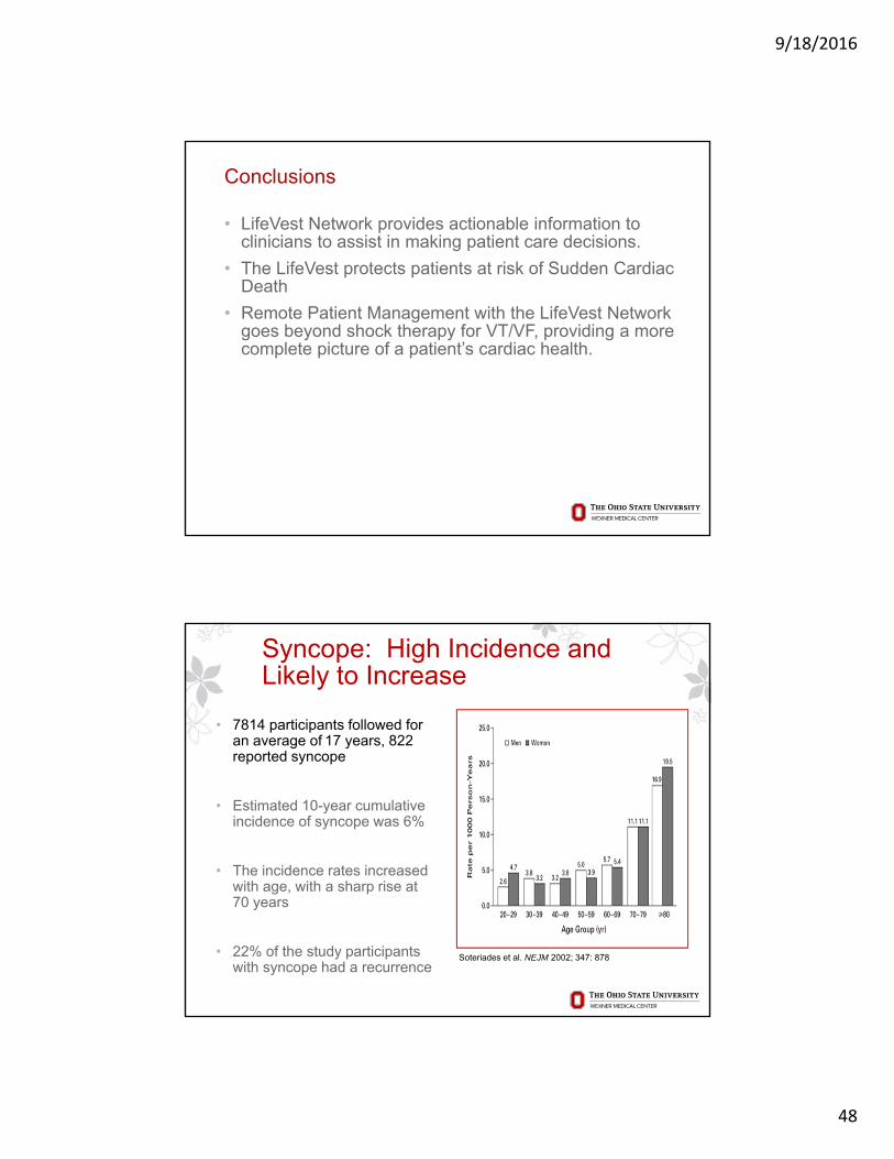
• LifeVest Network provides actionable information to clinicians to assist in making patient care decisions.
• The LifeVest protects patients at risk of Sudden Cardiac Death
• Remote Patient Management with the LifeVest Network goes beyond shock therapy for VT/VF, providing a more complete picture of a patient’s cardiac health.
Syncope: High Incidence and Likely to Increase
• 7814 participants followed for an average of 17 years, 822 reported syncope
• Estimated 10-year cumulative incidence of syncope was 6%
• The incidence rates increased with age, with a sharp rise at 70 years
• 22% of the study participants with syncope had a recurrence
Soteriades et al. NEJM 2002; 347: 878
9/18/2016
49
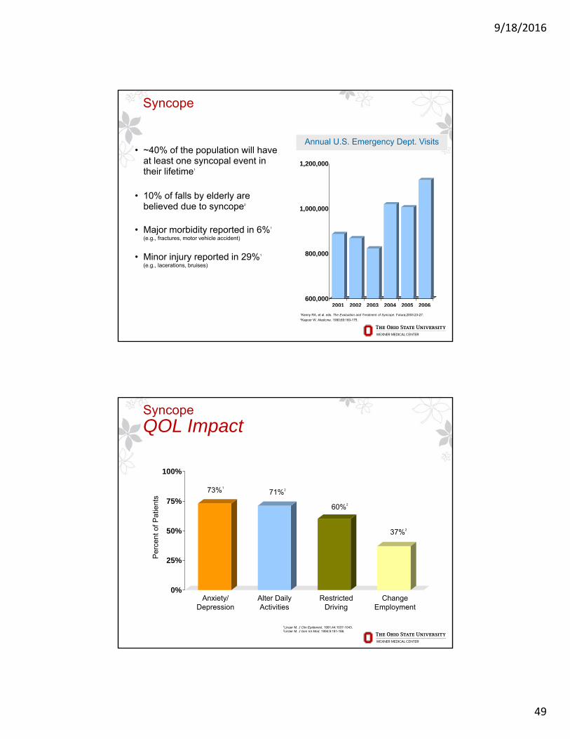
Syncope
Annual U.S. Emergency Dept. Visits
600,000
800,000
1,000,000
1,200,000
2001 2002 2003 2004 2005 2006
• ~40% of the population will have at least one syncopal event in their lifetime1
• 10% of falls by elderly are believed due to syncope2
• Major morbidity reported in 6%1
(e.g., fractures, motor vehicle accident)
• Minor injury reported in 29%1
(e.g., lacerations, bruises)
1Kenny RA, et al. eds. The Evaluation and Treatment of Syncope. Futura;2003:23-27.2Kapoor W. Medicine. 1990;69:160-175.
SyncopeQOL Impact
0%
25%
50%
75%
100%
73%1
71%2
60%2
37%2
Anxiety/Depression
Alter DailyActivities
RestrictedDriving
ChangeEmployment
1Linzer M. J Clin Epidemiol, 1991;44:1037-1043.2Linzer M. J Gen Int Med, 1994;9:181-186.
Per
cent
of
Pat
ient
s

9/18/2016
50
Types of External Arrhythmia Monitors
• Electrocardiogram: snapshot in time
• Holter monitor : 24 to 48 hours of continuous outpatient electrocardiographic (ECG) recording ‒ Shortcoming: repeated monitoring if an arrhythmia not occur 24-48 hours‒ Processing can delay action on malignant arrhythmias
• Event recorder: stores 1 to 2 minutes of ECG as soon as the patient activates‒ Enables much longer period of monitoring‒ Misses asymptomatic arrhythmias and some symptomatic arrhythmias when pt fails
to activate
• Automatic-trigger loop monitors: Records in continuous loop and automatically captures certain arrhythmias or can be manually activated during symptoms‒ Devices can capture detect several types of arrhythmias. ‒ Typically worn for up to 30 days. ‒ Download data to base station or phone
• Real-time cardiac surveillance: continuous outpatient ECG monitoring for periods ranging up to several weeks, if necessary. ‒ Cardiac activity detected by 3 electrodes attached to a ~2 ounce pager-sized
sensor/telephone transmitter ‒ Continuously analyzes the heart rhythm data. If an arrhythmia detected, the monitor
automatically transmits data to a central monitoring station for analysis/action‒ Any symptoms recorded by the patient are also transmitted.
Kinlay, S. et. al. Ann Intern Med 1996;124:16-20
Cumulative number of patients who sent an electrocardiogram from an event recorder by the number of days needed to record an electrocardiogram during palpitations
Prospective, randomized crossover comparison 48 hour holter to 30 day EVM
Twice as many symptomatic recordings from EVM as holter
19% of events recorded on EVM required intervention, none from the holter
9/18/2016
51
MCOT Study
• Multicenter randomized controlled trial
• 266 pts with palpitations, presyncope, syncope and nondiagnostic Holter
• Randomized to 30 days of MCOT (Cardionet) or external loop (Loop Group).
• Results‒ Clinically significant arrhythmias
• 55 (41%) pts in the MCOT Group • 19 (14%) patients in the Loop Group (p< 0.001).
Rothman SA, Laughlin JC, Seltzer J, et al. J Cardiovasc Electrophysiol. 2007;18(3):241-247
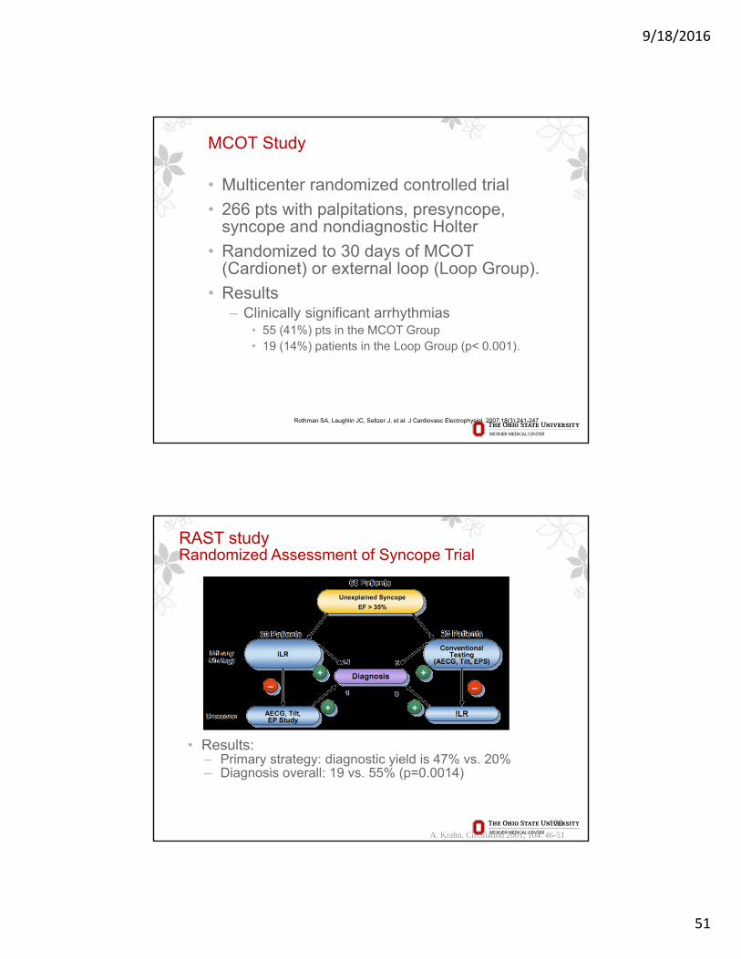
RAST studyRandomized Assessment of Syncope Trial
• Results:‒ Primary strategy: diagnostic yield is 47% vs. 20%‒ Diagnosis overall: 19 vs. 55% (p=0.0014)
102A. Krahn. Circulation 2001; 104: 46-51

9/18/2016
52
Ideal System for Long Term Cardiac Monitoring
• Subcutaneous placement, simple and fast to implant, excellent safety profile.
• Reliably provides information that can aid selection and titration of therapies‒ High sensitivity detector in ILR‒ Signal processing software to remove false positives and extract
information at monitoring center‒ Human over-read at service center to assure information delivered
to physician is clinically relevant
• Simple for the patient – requires little or no compliance‒ Long-range telemetry for automated data transfer
• Simple for the physician – maximizes practice efficiency, follow up requires minimal work load‒ Data download tailored to institution/practice
Sub-Q ILR’s: Evolution
Reveal DX2007
Reveal XT, Sleuth ATConfirm
2009
Reveal1998
Reveal Plus2000
Automatic detection added
Longevity and ECG memory increased (to 3 yrs., 49.5 minutes), with episode logs, ICD sensing technology, MRI labeling, and
remote monitoring added
AF detection and long-term trended diagnostics (the Cardiac Compass and AF Summary Reports) added
Reveal is developed to help diagnose unexplained syncope
Transoma: the first wireless and automated monitoring system
TransomaSleuth

9/18/2016
53
Transoma‘Sleuth AT”
Medtronic‘Reveal XT’
St. Jude‘Confirm’
Asystole Any single pause >3.0 sec. >1.5, >3.0, >4.5 sec >1.5, >3.0, >4.5 sec
Bradycardia Detection Settings
30, 40, 50 bpm 30, 40, 60 bpm 30, 40, 50 bpm
Tachycardia Detection Settings
120 to 220 bpm, Off 4/4, 6/8, 8/8, 16/16, 32/32 beats
VT = 250 to 520 ms
(115 to 240 bpm)
FVT = 240 to 400 ms
(150 to 250 bpm)
VT = 250 to 520 ms
(115 to 240 bpm)
FVT = 240 to 400 ms
(150 to 250 bpm
AF Detection 20 sec. ECG strips sent to Monitoring Center every 7.5 min.Analysis of ECG data and arrhythmia classification, by Certified Cardiac Techs at Monitoring Center
On board detection algorithm based on R to R variability
On board detection algorithm based on R to R variability but can’t turn on until clinical study is completed and approved by FDA for AF detection
Memory for ECG Data
673 min. between automatic, wireless transmission to the Monitoring Center
ILR: 43 min.
PDM: 630 min.
49.5 minutes total between scheduled office visits
Memory available on ILR only
Download to Carelink
48 minutes total between scheduled office visits
Memory available on ILR only
Download to TTM
ILR Battery Life 18 to 30 months 36 months 36months
Competing ILR’s
RUP Study: Importance of Wireless Download
1.Franco Giada et al.. J Am Coll Cardiol 2007;49:1951–6
Automatic detection mode in the REVEAL was activated, but no significant arrhythmias were recorded:because ILR memory “was always saturated by inappropriate activations.”

9/18/2016
54
Krahn et al, PACE 2004; 27: 657
Loss of tissue contact from shape/form factor
Importance of an Antenna
Medicomp Arrhythmia Access
Patient Activated Report Page

9/18/2016
55
Arrhythmia Access-Sleuth Patient Summary Report
The Value of Advanced Diagnostics
• Daily AF burden
• V-rate during AF
• Avg. day/night HR
• Patient activity
• Heart rate variability (HRV)
Note: All clinic, physician, and patient names and data in this document are fictitious

9/18/2016
56
Value of Advanced Diagnostics
How long do the episodes last?
When did the episodes start?
What is the AF burden?
Note: All clinic, physician, and patient names and data in this document are fictitious