practical training in histology and embryology - is.muni.cz · from the lesson or course ... short...
TRANSCRIPT

Practical training in Histology and Embryology

Organization issues • Beginning - strictly on time
• Change your shoes - you will not be allowed to enter the hall w/o indoor shoes
• Lockers – Jackets, coats, bags etc.
• Cell phone – switched off or in silent mode
• Microscopic hall = laboratory – eating, drinking, smoking not allowed – smoking strictly forbidden anywhere in LF – students have to follow the instructions – academic misconducts or inappropriate behavior result in excluding from the lesson or course
• Follow safety rules
• You have dedicated working place
• You are responsible for microscope, slide set, EM atlas

• Practical lesson
Introduction; the images aree available through MedAtlas
your individual work = study of the slides, schematic but precise drawing of tissue architecture, careful description. You make your own „study atlas“
students come prepared for practices - schedules and syllables – pin-boards or dpt. webpage
your knowledge is verified during semester
break – 10 minutes
• Attendance
100% attendance
substitution only in exceptional cases, after permissions from both the teacher of your group and the lesson where you plan to substitute
sign in to the list
make a protocol, let it check and signed by the lecturer

Registration of substitution: Datum Jméno Ročník Skupina Č. praktika Č. místa Vyučující - podpis Date Name Year Group Nr. of practice Nr. of place Teacher- signature

• Protocols
you have to make paper protocols (no tablets, laptops)
A4 size, blank, without lines, according to the template (can be downloaded from www.med.muni.cz/histology - Education)
(color)pencil handdrawings (no pen)
complete set of signed protocols is required for getting the credits
the quality of the protocol is approved by your teacher’s signature at the end of practical lesson
incomplete or low-quality protocols cannot be approved and you have to substitute the respective practical lesson


• Testing your knowledge
every student is examined 4 per semester
testing the knowledge of structures of the previous practical lesson, including the theory (their English and Latin names, functions, development and biological context) AND the theory for the curent practical lesson
short written test with images or schemes, results: „Passed“ or „Failed“
all images and schemes are made public (MedAtlas, IS)
you have to successfully pass all 4 tests
if you fail in partial test, you can repeat it once per semester
failing in the partial tests result in the overal Credit test at the end of semester

• Credits
100% attendance
complete set of signed protocols from all lessons
passed four tests
• End of practical lesson:
the practice is closed by the lecturer
you are allowed to leave your working place only after checking the microscope and slides
if you leave before the check you may be responsible for any damages/losses recognized later

http://www.med.muni.cz/histology
Department of Histology and Embryology
Faculty of Medicine MU

RECOMMENDED LITERATURE Čech, S. a D. Horký. Přehled obecné histologie. 1. vyd. Brno: Masarykova univerzita, 2005. 140 s. ISBN 8021038543. Horký, D. a S. Čech. Mikroskopická anatomie. 2. nezm. vyd. Brno: Masarykova univerzita, 2005. 353 s. ISBN 802103775X. Čech, S., D. Horký a M. Sedláčková. Přehled embryologie člověka. 1. vyd. Brno: Masarykova univerzita, 2011. 187 s. ISBN 978-80-210-5414-1. Mescher, A.L. Junqueira's basic histology :text and atlas. 13th ed. New York: McGraw-Hill Medical, 2013. xi, 544. ISBN 9781259072321. Moore, K.L., T.V.N. Persaud a M.G. Torchia. The developing human :clinically oriented embryology. 9th ed. Philadelphia, PA: Saunders/Elsevier, 2013. xix, 540. ISBN 9781437720020. Vacek, Z. Embryologie :učebnice pro studenty lékařství a oborů všeobecná sestra a porodní aistentka. 1. vyd. Praha: Grada, 2006. 255 s. ISBN 9788024712673. Sadler, T.W. Langmanova lékařská embryologie. 1. české vyd. Praha: Grada, 2011. xviii, 414. ISBN 9788024726403. Kapeller, K. a V. Pospíšilová. Embryológia človeka: učebnica pre lekárske fakulty. Martin: Osveta, 2001. 370 s. ISBN 80-8063-072-0. Lüllmann-Rauch, R. Histologie. Translated by Radomír Čihák. 1. české vyd. Praha: Grada, 2012. xx, 556. ISBN 9788024737294. Ovalle, W.K., P.C. Nahirney a F.H. Netter. Netter's essential histology. 2nd ed. Philadelphia, PA: Elsevier/Saunders, 2013. xv, 517. ISBN 9781455706310. Young, B. Wheater's functional histology :a text and colour atlas. 5th ed. [Oxford]: Churchill Livingstone, 2006. x, 437. ISBN 044306850X. Sadler, T.W. a J. Langman. Langman's medical embryology. Illustrated by Jill Leland. 11th ed. Baltimore, Md.: Lippincott William & Wilkins, 2010. ix, 385. ISBN 9781605476568. Lowe, J.S. a P.G. Anderson. Stevens and Lowe
s Human Histology. 4th. : Elsevier, 2015. ISBN 978-0-7234-3502-0.
Lectures Protocols

http://www.med.muni.cz/histology
HISTOLOGY
• structure and ultrastructure of normal cells and tissues,
• cytology and general histology
• special histology = microscopic anatomy of individual organs
• relevance: oncology, surgery, hematology, pathology, forensic,…

EMBRYOLOGY
– prenatal (intra uterine) development
• General embryology (until 2nd month – EMBRYO )
gametogenesis and early embryonic development
• Special embryology (since 3rd month to birth – FETUS )
organogenesis
• Teratology – defects in organ development, malformations, anomalies; prenatal screening – ultrasonography, amniocentesis, genetic and karyotype screening
• Relevance: gynecology and obstetrics, pediatrics, assisted reproduction

Histology and ulstrastructural
architecture
Cell biology Molecular
biology
Physiology
Biochemistry
Gene, cell and tissue
engineering
Quantitative analyses of cells and tissues - Genomics - Transcriptomics - Proteomics - Metabolomics - …
Understaning of the whole
system
- Personalized medicine
- Regenerative medicne
- Assisted reproduction
- Gene therapy
- Cancer therapy
- Biomedical research
Histology cannot be put out of the biological and functional context

Histology
• Resolution of naked eye – 0,1 mm
• Resolution of light microscopy – 10 nm • Resolution of electron microscopy – 0,1 nm

Tissue processing for the light microscopy (LM)
(making of permanent preparations – slides)
• SAMPLING (obtaining of material – cells, tissue pieces)
• FIXATION of samples (tissue blocks)
• RINSING (washing) of samples
• EMBEDDING of samples - embedded blocks
• CUTTING of blocks - sections
• AFFIXING of sections
• STAINING of sections
• MOUNTING of sections
SAMPLING
• A small piece of organ (tissue) is sampled and quickly put into the fixative medium.
• Biopsy during surgical dissection of organs in living organism
= excision
= puncture (liver or kidney parenchyma, bone marrow)
= curettage (uterine endometrium, adenoid vegetation)
• Necropsy from dead individual (sections); in experiments laboratory animals are used and tissue have to be sampled as soon as possible after the break of blood circulation
• The specimens shouldn't be more than 5 – 10 mm3 thick and fixation should follow immediately.

FIXATION • Definition: denaturation and stabilization of cell proteins with minimum artifacts
• The reason of fixation: freshly removed tissues are chemically unstable – dry, shrink, undergo hypoxia, autolysis and bacteriological changes
• To stop or prevent these changes and preserve the structure tissue samples have to be fixed. During the fixation, all tissue proteins are converted into inactive denaturized (stable) form.
• 3 main requirements on fixatives:
- good preservation of structure
- quick penetration into tissue block
- no negative effects on tissue staining

• Fixatives: solutions of different chemicals
- organic fixatives – ALDEHYDES – formaldehyde (most frequently used for LM)
– glutaraldehyde (used for EM)
– ALCOHOLS – 96 – 100 % (absolute) ethylalcohol
– ORGANIC ACIDS – glacial acetic acid, picric acid,
trichloracetic acid
- inorganic fixatives – INORGANIC ACIDS – chromic acid, osmium tetraoxide (OsO4)
– SALTS OF HEAVY METALS – mercuric chloride HgCl2
- compound fixatives – mixtures (two or more chemical components to offset
undesirable effects fo indiviual (simple) fixatives.
FLEMMING´s fluid – with OsO4
ZENKER´s and HELLY´s fluid, SUSA fluid – with HgCl2
BOUIN´s fluid – with picric acid
CARNOY´s fluid – with alcohol
Fixation is carried out at the room temperature, the time varies between 12 – 24 hours, specimen must be overlayed by 20 – 50 times fixative volume:
Ratio of tissue block volume to fixative volume 1 cm3 : 20 – 50 cm3

RINSING and EMBEDDING
• All samples should be washed to remove the excess of fixative; the choice of rinsing medium is determined by type of fixative: running tap-water or 70-80% ethanol
• Relevance of embedding: tissues and organs are brittle and unequal in density, they must be hardened before cutting

Embedding media
water soluble – gelatine, celodal, water soluble waxes
anhydrous – paraffin, celoidin
EMBEDDING into PARAFFIN
• dehydration – to remove water from fixed samples by
ascending series of ethanol is used (50%, 70%, 90%, 96%. each step - 2 – 6 hours
• clearing – the ethanol must be replaced with organic solvatant that dissolves paraffin – benzene or xylene
• infiltration – melted paraffin wax (56°C) is used; 3 x 6 hours.
• casting (blocking out) – moulds (plastic, paper or metal chambers) are used for embedding.
- The moulds are filled with melted paraffin, tissue samples are then placed inside and immediately immersed in cold water to cool paraffin quickly down.
- These paraffin blocks are ready for trimming

Automated device for tissue dehydration
Leica TP 1020
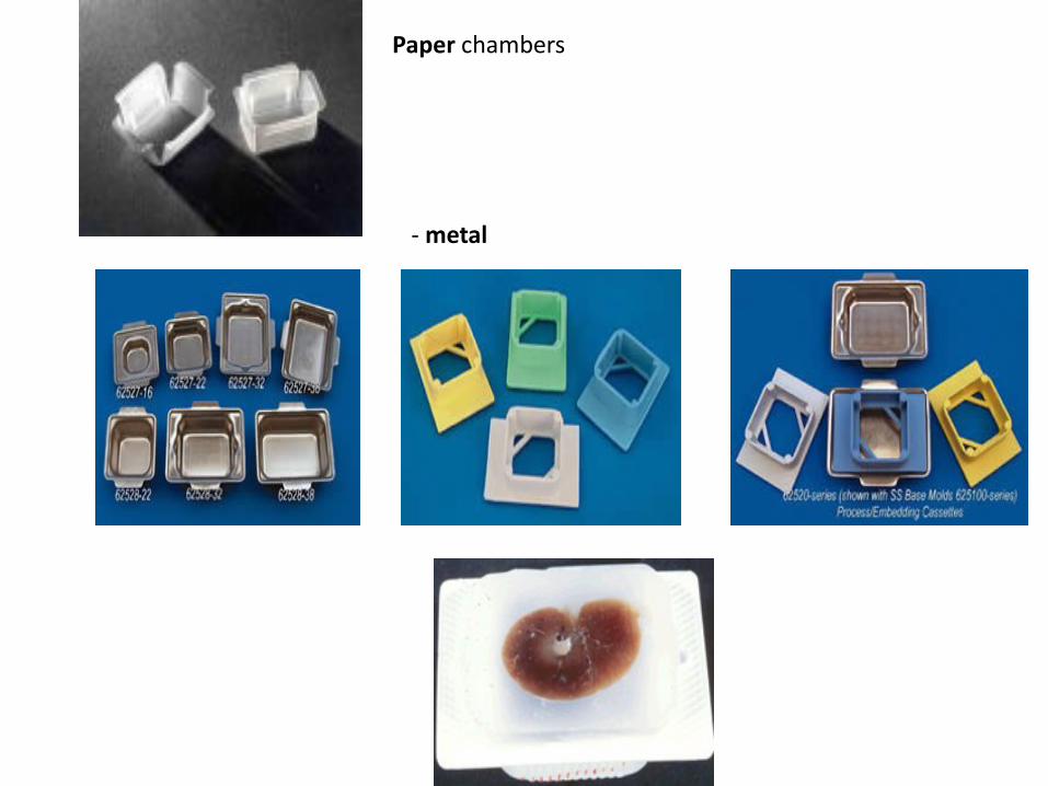
Paper chambers
- metal

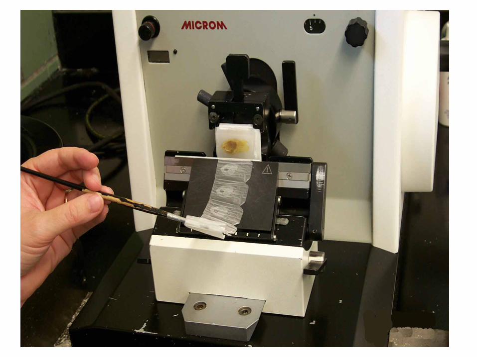
CUTTING
• Microtome – a machine with automatic regulation of section thickness: 5 – 10 μm is optimum.
sliding microtome – block is fixed in holder, knife or razor moves horizontally
rotary microtome – knife is fixed, block holder moves vertically
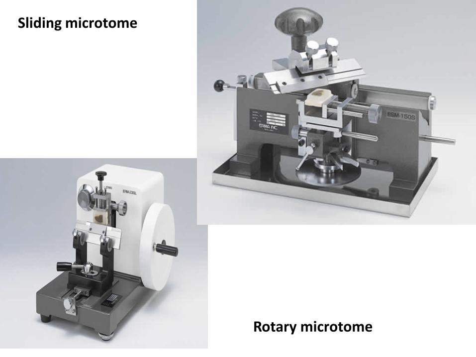
Rotary microtome
Sliding microtome
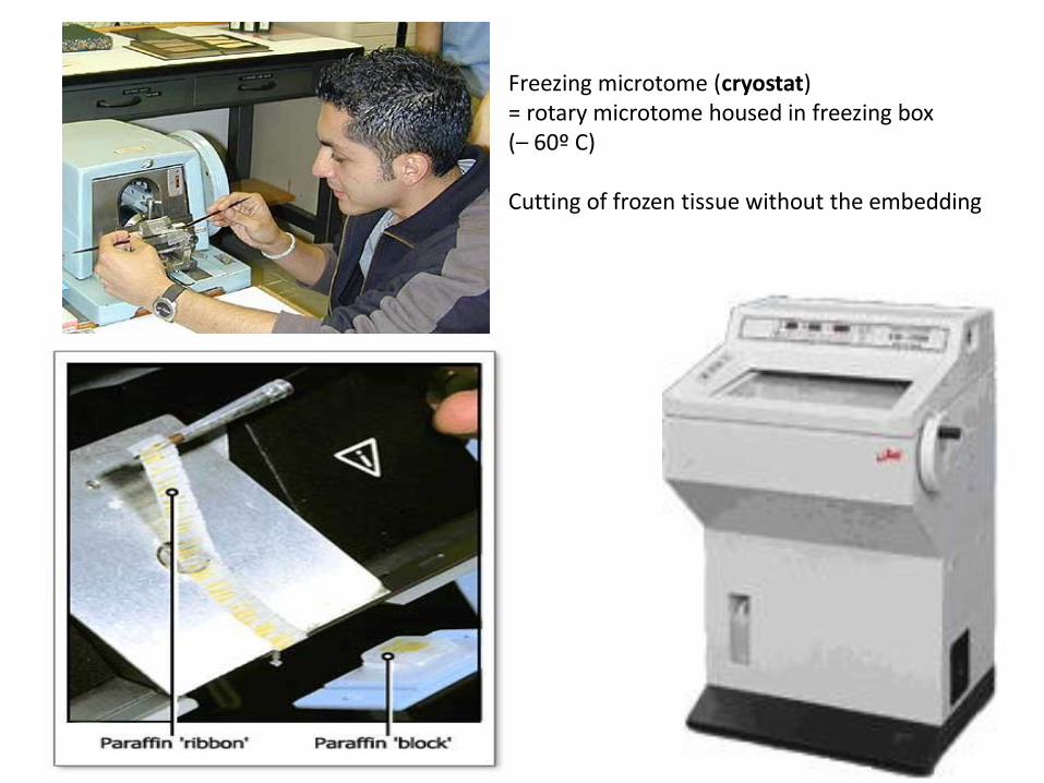
Freezing microtome (cryostat) = rotary microtome housed in freezing box (– 60º C) Cutting of frozen tissue without the embedding

1 2 3
4 5
6 7
sampling fixation fixation
cutting cutting
section affixing section affixing


AFFIXING
• Mixture of glycerin and egg albumin or gelatin
• Section are transferred from microtome razor or knife on the level of warm water (45º C), where they are stretched; then they are put on slides coated with adhesive mixture; excess of water is drained and slides are put in incubator (thermostat, 37º C) over night to affixing of sections.

Stretching of sections on warm water
Stretching on a warm plate

STAINING
• Different cell or tissue structures are not apparent without staining.
• Cellular structures exhibit different affinity to staining dyes
alkaline dyes (basic or nuclear) – react with anionic groups of cell and tissue components
basophilia – basophilic structures in the cell
acid dyes (cytoplasmic) – react with cationic groups
acidophilia – acidophilic structures in the cell
neutrophilia – no reaction

Staining methods: routine – HE, AZAN (demonstrate all components of tissue) special visualizes only special structures impregnation by silver salt for detection of nerve or reticular fibers
HE – the most frequent used method
Lipid droplets detected by oil red

ROUTINE STAINING with
HEMATOXYLINE – EOSIN (HE) Hematoxyline – basic (nuclear) dye
Eosin – acid (cytoplasmic dye
• Staining procedure:
• paraffin must be removed (dissolved) by xylene
• sections are rehydrated in descending series of ethanol (100% 96% 80%)
• staining with hematoxyline
• differentiation in acid ethanol and water (excess of dye is removed)
• staining with eosin
• rinsing in water (excess of dye is removed)
• dehydration in graded ethanol series (80% 96% 100%)
• clearing in xylene

Xylen I XylenII 100% 96% H2O hematoxyline acid ethanol ethanol ethanol
Xylen IV xylen III 100% 96% H2O eosin H2O ethanol ethanol
HEMATOXYLINE – EOSIN (HE)
Deparaffination Rehydration Washing Staining Differentiation
Clearing Dehydration Washing Staining Washing
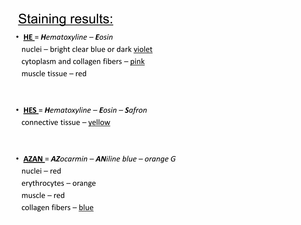
Staining results:
• HE = Hematoxyline – Eosin
nuclei – bright clear blue or dark violet
cytoplasm and collagen fibers – pink
muscle tissue – red
• HES = Hematoxyline – Eosin – Safron
connective tissue – yellow
• AZAN = AZocarmin – ANiline blue – orange G
nuclei – red
erythrocytes – orange
muscle – red
collagen fibers – blue

cuvette
flask
slides holder (basket)
Staining tools:

Automatic slide stainer
staining set of boxes with media

MOUNTING • Finally, preparates are closed with coverslip (coverglass) to form a permanent
preparate. Small amount of mounting medium must be placed between stained section and the coverslip.
• Mounting media: soluble in xylene – canada balsam soluble in water – glycerin-gelatine, arabic gum
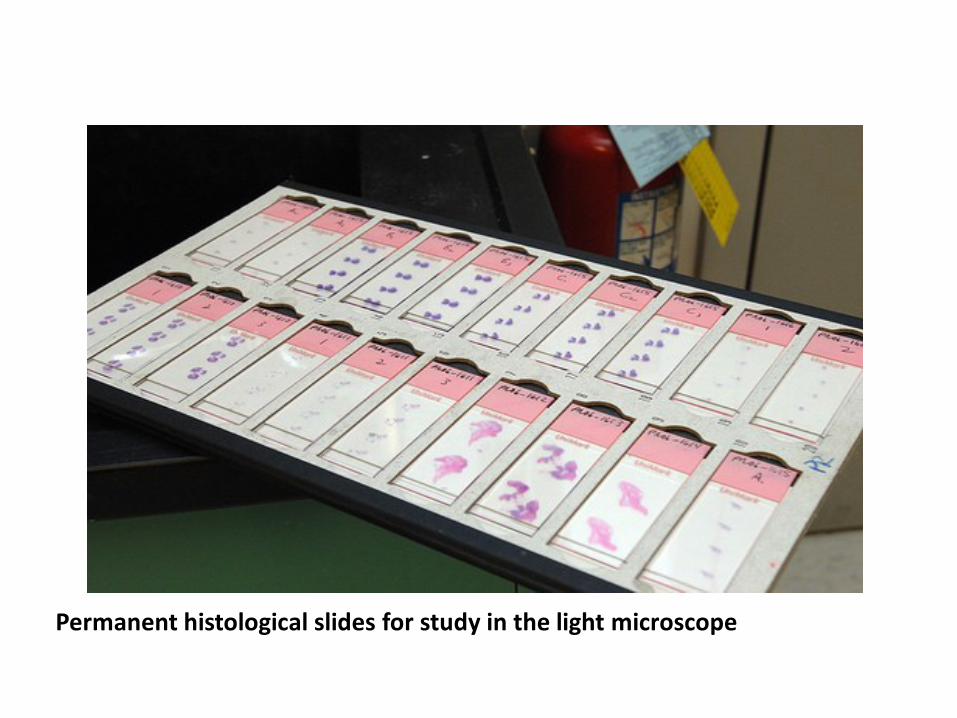
Permanent histological slides for study in the light microscope

Hematoxyline and eosin (HE)

Hematoxyline and eosin (HE)
cell cytoplasm
cell nuclei
collagenous connective tissue

Hematoxyline and eosin (HE)
basophilic cytoplasm of glandular cells (contains ribosomes with RNA)
acidophilic cytoplasm of epithelial cells

Hematoxyline, eosin and saffron
(HES)
cartillage
Collagenous fibers of connective tissue are yellow after staining with saffron
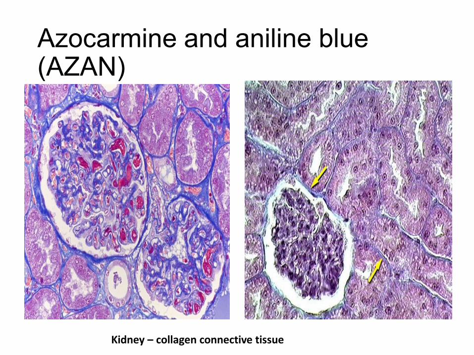
Azocarmine and aniline blue (AZAN)
Kidney – collagen connective tissue

Impregnation of tissue with silver
Lien - reticular fibers Cerebellum – nerve fibers

Iron hematoxyline
Skeletal muscle cells (fibers)

Iron hematoxyline
Mitochondria in hepatocytes


Histochemistry and Immunohistochemistry
• Relevance:
various chemical compounds detected „in situ“ (proteins, AA, NA, saccharides, lipids, enzymes, pigments, inorganic substances – Fe, Ca, Zn)
Various epitopes detected by immunotechniques

Antigen
Primary Ab specific against epitope of the particular antigen
Secondary Ab specific against primary Ab
Enzyme conjugated with secondary Ab - visualization

Actin (cytoskeleton)
DAPI (nucleus)
Microtubules (cytoskeleton)
KI-67

Tissue processing for the EM
• pH of all solutions (media) must be buffered on 7.2 – 7.4
Cacodylate or phosphate buffer is frequently used.
• Absolutely dustfree environment
• Solutions (media) have to be precise (artifacts)

Tissue processing for the EM
• SAMPLING – immediatelly after arresting of blood circulation, tissue block sized no more than 1mm3
• FIXATION – glutaraldehyde (binds amine groups) + OsO4 (binds lipids) are used as double fixation
• RINSING – distilled water
• DEHYDRATION - ethanol
• EMBEDDING – gelatin capsule or plastic forms are filled with some medium (which can be polymerized from liquid to solid form) and pieces of fixed tissue are placed into this medium. Epoxyd resins (Epon, Durcupan, Araldite) are usually used as in water insoluble media.
• CUTTING – ultrathin sections (in ultramictomes)
• CONTRASTING ≈ staining

Embedding tools: gelatin (1) or plastic (2) capsules capsule holder (3) embedding plates (4, 5)
Embedded blocks prepared for cutting
1 2
3
4,5

By trimming, using ultramicrotome, an excess of hard medium is removed and pyramide with minimal cut surface (0.1 mm2) is prepared. Minimum of tissue (black) is in the top of pyramid
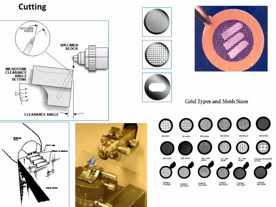
Cutting

Cutting
Ultrathin sections (70 – 100 nm) - ultramicrotomes.
Glass or diamond (b) knives with water
reservoir are used Sections slide flow on water in small
container attached to the knive
Supporting grids

Ultramicrotom knives:
glass
diamond

CONTRASTING (=STAINING)
• principle of differentiation of structures – different dispersion of beam of electrons depending on atomic weight of elements. „electron dyes“ are thus mixtures of heavy metals: uranylacetate or lead citrate
stain droplet with floating grid placed section-side down on the droplet


Differences between LM and EM
LM EM
Sampling 1 cm3
minutes
1 mm3
seconds
Fixation formaldehyde
12 – 24 hours
glutaraldehyde
1 – 3 hours
Embedding paraffin epoxid resins (Durcupan)
Cutting
Thickness of sections
microtome
5 – 10 m
Ultramicrotomes
50 – 100 nm
Staining (LM)
contrasting (EM)
dyes
(hematoxyline – eosin)
heavy metals
(uranylacetate,lead citrate)
Mounting (only LM) ---
Result histological slide (preparate) photograph of ultrathin section

Visit us at:
http://www.med.muni.cz/histology
Thank you for attention