pre-existing brain function predicts subsequent practice of mindfulness and compassion meditation
TRANSCRIPT
NeuroImage 69 (2013) 35–42
Contents lists available at SciVerse ScienceDirect
NeuroImage
j ourna l homepage: www.e lsev ie r .com/ locate /yn img
Pre-existing brain function predicts subsequent practice of mindfulness andcompassion meditation
Jennifer S. Mascaro a,b,c, James K. Rilling a,b,c,d, Lobsang Tenzin Negi e, Charles L. Raison f,g,⁎a Department of Anthropology, Emory University, 207 Anthropology Building, 1557 Dickey Drive, Atlanta, GA 30322, USAb Department of Psychiatry and Behavioral Sciences, Emory University School of Medicine, 1639 Pierce Drive, Suite 4000, Atlanta, GA 30322, USAc Center for Behavioral Neuroscience, Emory University, P.O. Box 3966, Atlanta, GA 30302, USAd Center for Translational Social Neuroscience, Emory University, GA 30322, USAe Emory–Tibet Partnership, Department of Religion, Emory College, Callaway Center S306A, Atlanta, GA 30322, USAf Department of Psychiatry, University of Arizona College of Medicine, 1501 Campbell Ave, P.O. Box 245017 Tucson, AZ 85724, USAg The John and Doris Norton School of Family and Consumer Sciences, College of Agriculture and Life Sciences, 650 North Park Avenue, Tucson, AZ 85721650 N. Park Ave Tucson,Arizona 85721650 N. Park Ave Tucson, AZ 85721, USA
⁎ Corresponding author at: Department of Psychiatryof Medicine, 1501 Campbell Ave, P.O. Box 245017 Tucso
E-mail address: [email protected] (C.L
1053-8119/$ – see front matter © 2012 Elsevier Inc. Allhttp://dx.doi.org/10.1016/j.neuroimage.2012.12.021
a b s t r a c t
a r t i c l e i n f oArticle history:Accepted 14 December 2012Available online 22 December 2012
Keywords:MeditationCompassionMindfulnessAnterior insulaAmygdalaEmpathy
While a variety of meditation techniques are increasingly employed as health interventions, the fact thatmeditation requires a significant commitment of time and effort may limit its potential widespread utility.In the current study, we ask whether baseline subjective reports or brain activity in response to a “Pain forSelf and Others” paradigm predicts subsequent engagement in mindfulness and compassion meditation.The study also investigated whether compassion training would impact neural responses when comparedto an active health education control group. Prior to training, activation of the left and right anterior insula,an area thought to be important for empathy, in response to the Other pain task was positively related to en-gagement with compassion meditation as measured by practice time (n=13). On the other hand, activity inthe left amygdala during the Self pain task was negatively correlated with mindfulness practice time. Follow-ing the study intervention, there was no difference between the compassion group (n=13), and the controlgroup (n=8), in brain responses to either the Self or Other task. These results are the first to indicate thatbaseline neural responses may predict engagement with meditation training and suggest that pre-existingneurobiological profiles differentially predispose individuals to engage with disparate meditation techniques.
© 2012 Elsevier Inc. All rights reserved.
Introduction
Meditation is increasingly incorporated into clinical treatmentsfor a variety of mental and physical ailments (Hofmann et al., 2011;Marchand, 2012) and is widely believed to enhance well-being evenin individuals not suffering from any specific mental or physical disor-der (Sedlmeier et al., 2012). However, despite its apparent promise,the commitment of time and effort required to learn meditative tech-niques may limit their potential widespread utility. This may be partic-ularly true of meditative practices designed to enhance compassionatefeelings and behaviors toward others since these practices pose the ad-ditional challenge of requiring trainees to contemplate deeply and forextended periods about the suffering of other people, including thosethey love. Because health-relevant emotional and physiological effectsof compassion meditation appear to be positively associated with prac-tice time (Fredrickson et al., 2008; Pace et al., 2009, 2010, 2012), identi-fying pre-existing variables that predict people's differential ability or
, University of Arizona Collegen, AZ 85724, USA.. Raison).
rights reserved.
willingness to engage with the practice has clear therapeutic relevance.The current study sought to identify whether self-report or neurobio-logical responses to suffering in oneself or others prior to learning com-passion meditation would predict subsequent engagement with thepractice over an8-week training period. An additional aimwas to inves-tigate whether the training program impacted neural responses uponrepeated exposure to “Pain for Self and Others” when compared to anactive health education control group.
Cognitively-Based Compassion Training (CBCT), the protocol used inthe current investigation, reduces stress responsivity (Pace et al., 2009),depression (Desbordes et al., 2012), and enhances empathic accuracyand related neural activity (Mascaro et al., 2012; Pace et al., 2009). In-creased amount of practice time has also been shown to reduce levels ofc-reactive protein—an important biomarker for disease development—in traumatized/neglected youth (Pace et al., 2012). CBCT is a secularizedtraining derived from the 11th century Tibetan Buddhist lojong tradi-tion. As such, it commences with two-weeks of training in focused at-tention (shamatha) and non-judgmental awareness (vipassana) priorto one week of self compassion training and five weeks of specific com-passion training designed to enhance interpersonal equanimity, in-crease feelings of gratitude toward others, and finally to induce strong
36 J.S. Mascaro et al. / NeuroImage 69 (2013) 35–42
feelings of empathy toward all people. Importantly, these two aspects ofthe training (i.e. shamatha/vipassana during the first two weeks andcompassion during the last five weeks), are widely divergent in termsof themeditative techniques they employ. Because of this, CBCT provid-ed a novel opportunity to examine the specificitywith which subjectiveand neurobiological responses to the suffering of self and others predictthe ability to practice mindfulness based techniques (i.e. shamatha/vipassana) vs. compassion techniques.
Themindfulness-based technique employed in CBCT (i.e. shamatha/vipassana) has been a target of extensive scientific investigation in thelast decade, in part because of its apparent promise as a relativelybrief and cost-effective practice for alleviating anxiety as well as for en-hancing well-being (Baer, 2003; Grossman et al., 2004; Marchand,2012; Sedlmeier et al., 2012). In particular, it is thought that by promot-ing a type of awareness in which phenomena are experienced in anon-analytical and non-evaluative manner, mindfulness techniquesallow individuals to experience aversive events with less emotional re-activity (Creswell et al., 2007). The mindfulness component of the pro-tocol used in this study was designed to be explicitly devoid of anycompassion related content and required that practitioners attend totheir sensations, thoughts, and emotions in a non-evaluative manner.We therefore hypothesized that baseline aversiveness ratings and neu-ral activity in regions important for the affective and evaluative re-sponse to pain, such as the mid cingulate cortex (MCC), anteriorinsula, and amygdala (Peyron et al., 1999; Price, 2000), would be in-versely associated with the ability or willingness to subsequently en-gage with the mindfulness component of the CBCT protocol.
Given that the compassion-specific elements within CBCT require ex-tensive contemplation of the suffering of others,wehypothesized that in-dividuals with high levels of baseline empathy would be more likely toengage in this portion of the CBCT protocol. The neural systems relatedto empathy, defined as an affective reaction similar to, and evoked by,another's affective state (de Vignemont and Singer, 2006) have been in-vestigated using an empathy for pain (EFP) paradigm. This paradigm, inwhich participants imagine or observe other people receiving a painfulstimulus, commonly elicits neural activation in the affective componentof the pain matrix, including the anterior mid-cingulate cortex (aMCC),as well as the bilateral anterior insula (AI) and the ventral frontal opercu-lum (Botvinick et al., 2005; Jackson et al., 2005; Lamm et al., 2010; Simonet al., 2006; Singer et al., 2004). Activity in the AI may represent a simu-lated mapping of the observed individual's body state onto one's own(Singer et al., 2009),which is important for an empathic response. Impor-tantly, activity in the AI predicts later helping behavior, suggesting that itsactivity is related to prosocial emotions andmotivation rather than to thetype of distress that has been shown to precedemore self-serving behav-ior (Batson, 1998; Hein et al., 2010). We therefore hypothesized that theactivity in the AI in response to an empathy inducing task as well asself-reported empathy levels would predict amount of subsequent en-gagement with the compassion-specific elements of the CBCT protocol.
Materials and methods
This studywas approved by the Institutional Review Board of EmoryUniversity and all participants gave written informed consent prior toinclusion. To test the study hypotheses participants underwent func-tional magnetic resonance imaging (fMRI) while both receiving (Self)and watching videos of others receiving (Other) painful stimulationsboth prior to and upon completion of the study interventions (for de-sign, see Supplementary figure S1).
Study participants
Twenty-nine (16 males) participants from the Atlanta area wererecruited using a combination of fliers and electronic notificationsposted at several local universities, as well as electronic advertise-ments on Craigslist as part of a larger study that assessed the effects
of meditation on stress physiology and social cognition. Participantswere aged 25–55 (M=31.0; SD=6.02) and were screened and ex-cluded for (self-reported) use of any psychotropic medication within1 year of screening, for regular use of any medications that mightinfluence activity of the autonomic nervous system, HPA axis, orinflammatory pathways, and for any ongoing medical or psychiatriccondition.
Compassion meditation
The compassion meditation training protocol used here (Cognitively-Based Compassion Training [CBCT]) was designed by one of us (LTN).Although secular in presentation, CBCT derives from the 11th centuryTibetan Buddhist lojong tradition. In its operationalization, CBCT madetwo important modifications to traditional lojong teachings. First, alldiscussions of soteriological or existential themes (e.g. the attainmentof Buddhahood, Karma)were omitted. Second, participantswere taughtoneweek each of concentrative (i.e. shamatha) andmindful-awareness(i.e. vipassana) practices at the beginning of the course. While not spe-cifically included in traditional lojong curricula, these basic meditationpractices were an assumed prerequisite for commencing lojong trainingin a traditional Buddhist context (HHDL, 2001). For simplicity, we havesubsumed these attention practices under the term ‘mindfulness prac-tice’, in accordance with the general Western and clinical understand-ing of mindfulness, although it is important to note that the attentionalpractices that are trained in the first two weeks of CBCT are differentthan those employed when MBSR training is taken as a whole, becausethey are without any compassion related content. A complete descrip-tion of theweekly schedule can be found in the Supporting information.
The compassion meditation courses were taught by two graduatestudents from the Emory Religion department who are experiencedmeditators and who had undergone extensive training with LobsangTenzin Negi. Study participants were asked to attend 2 h of class timeper week for eight weeks. Class sessions combined a didactic teachingand discussion section with approximately 20 min of meditation perhour class time. Participants were provided with a meditation compactdisk to guide “at-home” practice sessions that reflected in-class materi-al, andwere asked to keep track of practice time each day. In calculatingpractice time for the current study, only “at-home” practice was includ-ed, as it was determined a priori that in-class practice did not reflect en-gagement with the material because participants did not know thespecific techniques they would practice each week prior to attendingclass.
Health discussion control group
Participants randomized to the control condition attended2 hof a dis-cussion group per week. Classes were designed and taught by graduatestudents from the Emory Rollins School of Public Health. Topics includedhistory of medicine, nutrition, sleep, mental health, exercise, stress, infec-tious disease, and complementary and alternative medicine. The healthdiscussion group was designed to control for the non-specific effects ofthe meditation class, including education and social engagement with acollective group. Subjects were not asked to do any “at home”work.
Protocol for preparing the Other pain stimuli video
The empathy for pain video stimuli set was created using the fol-lowing protocol. Twenty participants (10 males) were recruitedfrom the Emory campus and we explicitly solicited a diverse popula-tion in terms of age and ethnicity (11 Caucasian, 9 non-Caucasian).Volunteers were seated such that they could both view a laptop com-puter and face directly toward the video camera. Participants weretold that the video clips would be used as stimuli in an fMRI studyof empathy and were asked to make facial expressions that camenaturally.
37J.S. Mascaro et al. / NeuroImage 69 (2013) 35–42
Volunteers were outfitted with 2 electrode pads on the insideof their right wrist and connected to the GRASS SD-9 stimulator.First, we indexed participants' pain tolerance by asking them to ratestimulations on a 10-point intensity rating scale (0= ‘don't feel any-thing’, 1= ‘can feel something but not painful’, 8= ‘maximum tolerablepain’, and 10= ‘worst imaginable pain’). The 1 setting was used for the‘no pain’ stimuli and the 8 setting was used for the ‘pain’ stimuli. Uponfinding the settings, 3 of each were administered in pseudorandomorder. Prior to each stimulation, the laptop screen next to the subjectsshowed a colored screen for 6 s indicating which level they wereabout to receive (a red screen indicated that they would receive a pain-ful stimulation, a blue screen indicated that they would receive anon-painful stimulation). Each stimulation lasted approximately 3 s.
Data collection
After providing informed consent, participants completed theInterpersonal Reactivity Index (IRI) (Davis, 1983a, 1983b), a 28 itemLikert-scale measure (0=Does not describe me very well, 4=Describesme verywell). The primary IRI subscale of interestwas the empathic con-cern subscale (example item, ‘I often have tender, concerned feelings forpeople less fortunate than me’), which assesses other-oriented affectiveresponses to those who are suffering.
Experimental paradigm
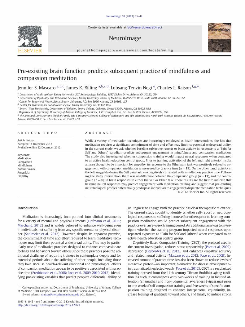
The EFP task used here followed other fMRI paradigms that havesuccessfully identified an empathy-for-pain response (Botvinick et al.,2005; Singer et al., 2004) (Fig. 1). First, subjects completed the Selfpain task, in which they received moderately painful and nonpainfulstimulations to the inside of their wrist. Prior to entering the scanner,subjects' individual pain levels were found using the same method aswas used with the video subjects. During the Self pain task, the subjectsaw either a green or a yellow colored screen (the anticipation cue) for6 s, which indicated whether they were about to receive a painful or anon-painful stimulus. The stimulus was then delivered for 3 s, andwas followed by a fixation period of 12–16 s (14+/−2 jittered). Painand no-pain stimuli were each presented 10 times. Null trials werealso included (3painful, 3 non-painful), inwhich subjects saw the antic-ipation cue but did not receive a stimulation.
Fig. 1. Schematic of the empathy for pain (EFP) (Self pain and Other pain) paradigm. (a.) Trial
Following the Self pain task, subjects completed the Other pain task,in which they saw the previously described video clips of other peopleanticipating and receiving the same stimuli that they received. Boththe subject and the person in the video saw the anticipation cue(red=painful, blue=nonpainful), signifying whether the pendingstimulus would be painful or not. The video clips then showed the per-son receiving the stimulus for 3 s, followed by a 12–16 s (14+/−2 sjittered) fixation period. The Other pain task consisted of 2 blocks ofstimuli, each comprised 10 pain and 10 no-pain events presentedpseudo-randomly. Thus, each subject viewed 20 other pain and 20other no-pain events in total. Subjects saw 1 pain event and 1 no-painevent for each person. There was a one minute break between blocks,during which the subject saw a fixation cross. Again, 12 null trials (6pain, 6 non-pain) were included in which the subject saw a video clipof a person viewing the anticipation cue, but they did not see the painepoch of the trial.
Upon completion of the Self and Other tasks, subjects were asked torate on a scale from 1 to 5 how aversive they found it to: 1. Receive thenonpainful stimulations, 2. Receive the painful stimulations, 3. Watchothers receive the nonpainful stimulations, and 4.Watch others receivethe painful stimulations.
During a second visit approximately 10 weeks after the first, subjectswere scanned a second time using the same EFP paradigm. In the interim,participants were randomized to 8 weeks of either compassion medita-tion or to a health control course (described in more detail below).Sixteen participants were randomized to the meditation group and13 to the control group. Due to subject attrition, 21 (12 males)subjects were scanned the second time (M age=31.9; SD=6.70). Ofthese, 13 (M age=29.4; SD=4.43) were in the meditation group and8 (M age=35.9; SD=8.06) were in the control group. Study drop-outsare described in detail in the Supplemental information.
Image acquisition
AllMR imageswere acquired on a Siemens 3 T Trio scanner. Function-al imageswere acquiredusing anEPI sequencewith the followingparam-eters: TR=2000 ms, TE=28 ms, matrix=64×64, FOV=192 mm, slicethickness=3 mm, gap=0.45 mm, and 34 axial slices. A 4.5 minuteT1-weighted MPRAGE scan (TR=2600 ms, TE=3.02 ms, matrix=256×256, FOV=256 mm, slice thickness=1.00 mm, gap=0 mm)was acquired for anatomical localization of fMRI activations.
structure for Self pain and Other pain trials; (b.) Design of Self pain and Other pain tasks.
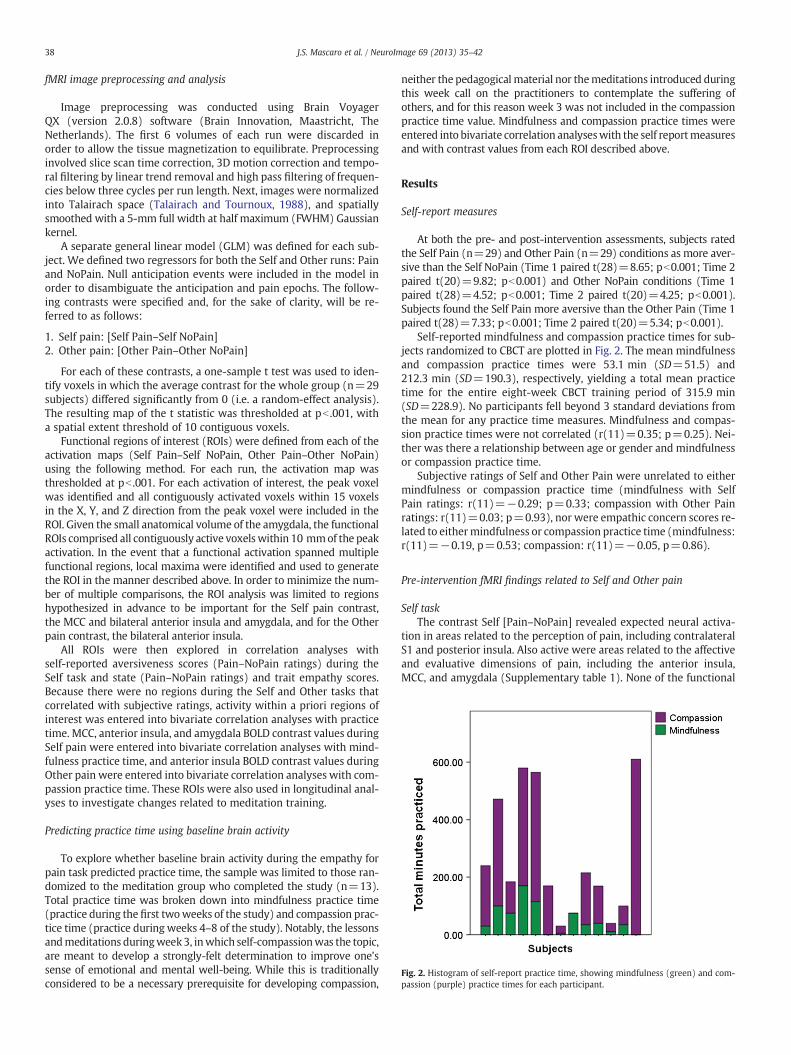
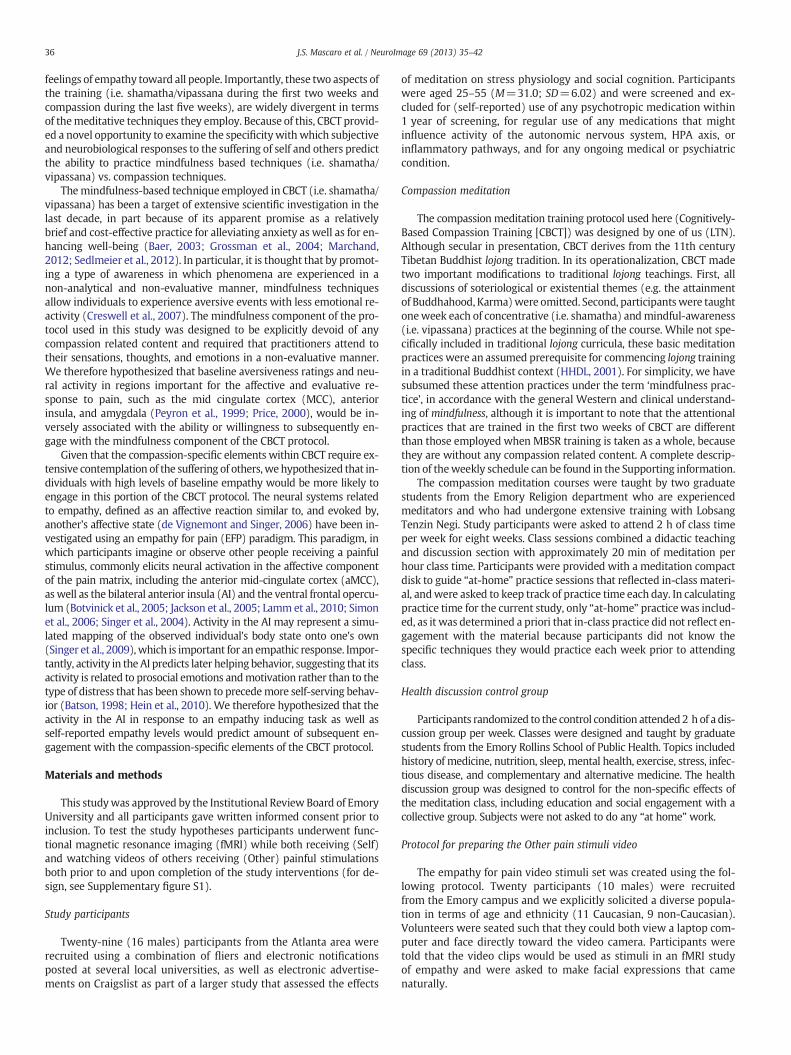
Fig. 2. Histogram of self-report practice time, showing mindfulness (green) and com-passion (purple) practice times for each participant.
38 J.S. Mascaro et al. / NeuroImage 69 (2013) 35–42
fMRI image preprocessing and analysis
Image preprocessing was conducted using Brain VoyagerQX (version 2.0.8) software (Brain Innovation, Maastricht, TheNetherlands). The first 6 volumes of each run were discarded inorder to allow the tissue magnetization to equilibrate. Preprocessinginvolved slice scan time correction, 3D motion correction and tempo-ral filtering by linear trend removal and high pass filtering of frequen-cies below three cycles per run length. Next, images were normalizedinto Talairach space (Talairach and Tournoux, 1988), and spatiallysmoothed with a 5-mm full width at half maximum (FWHM) Gaussiankernel.
A separate general linear model (GLM) was defined for each sub-ject. We defined two regressors for both the Self and Other runs: Painand NoPain. Null anticipation events were included in the model inorder to disambiguate the anticipation and pain epochs. The follow-ing contrasts were specified and, for the sake of clarity, will be re-ferred to as follows:
1. Self pain: [Self Pain–Self NoPain]2. Other pain: [Other Pain–Other NoPain]
For each of these contrasts, a one-sample t test was used to iden-tify voxels in which the average contrast for the whole group (n=29subjects) differed significantly from 0 (i.e. a random-effect analysis).The resulting map of the t statistic was thresholded at pb .001, witha spatial extent threshold of 10 contiguous voxels.
Functional regions of interest (ROIs) were defined from each of theactivation maps (Self Pain–Self NoPain, Other Pain–Other NoPain)using the following method. For each run, the activation map wasthresholded at pb .001. For each activation of interest, the peak voxelwas identified and all contiguously activated voxels within 15 voxelsin the X, Y, and Z direction from the peak voxel were included in theROI. Given the small anatomical volume of the amygdala, the functionalROIs comprised all contiguously active voxelswithin 10 mmof the peakactivation. In the event that a functional activation spanned multiplefunctional regions, local maxima were identified and used to generatethe ROI in the manner described above. In order to minimize the num-ber of multiple comparisons, the ROI analysis was limited to regionshypothesized in advance to be important for the Self pain contrast,the MCC and bilateral anterior insula and amygdala, and for the Otherpain contrast, the bilateral anterior insula.
All ROIs were then explored in correlation analyses withself-reported aversiveness scores (Pain–NoPain ratings) during theSelf task and state (Pain–NoPain ratings) and trait empathy scores.Because there were no regions during the Self and Other tasks thatcorrelated with subjective ratings, activity within a priori regions ofinterest was entered into bivariate correlation analyses with practicetime. MCC, anterior insula, and amygdala BOLD contrast values duringSelf pain were entered into bivariate correlation analyses with mind-fulness practice time, and anterior insula BOLD contrast values duringOther pain were entered into bivariate correlation analyses with com-passion practice time. These ROIs were also used in longitudinal anal-yses to investigate changes related to meditation training.
Predicting practice time using baseline brain activity
To explore whether baseline brain activity during the empathy forpain task predicted practice time, the sample was limited to those ran-domized to the meditation group who completed the study (n=13).Total practice time was broken down into mindfulness practice time(practice during the first twoweeks of the study) and compassion prac-tice time (practice during weeks 4–8 of the study). Notably, the lessonsandmeditations duringweek3, inwhich self-compassionwas the topic,are meant to develop a strongly-felt determination to improve one'ssense of emotional and mental well-being. While this is traditionallyconsidered to be a necessary prerequisite for developing compassion,
neither the pedagogical material nor themeditations introduced duringthis week call on the practitioners to contemplate the suffering ofothers, and for this reason week 3 was not included in the compassionpractice time value. Mindfulness and compassion practice times wereentered into bivariate correlation analyseswith the self reportmeasuresand with contrast values from each ROI described above.
Results
Self-report measures
At both the pre- and post-intervention assessments, subjects ratedthe Self Pain (n=29) and Other Pain (n=29) conditions as more aver-sive than the Self NoPain (Time 1 paired t(28)=8.65; pb0.001; Time 2paired t(20)=9.82; pb0.001) and Other NoPain conditions (Time 1paired t(28)=4.52; pb0.001; Time 2 paired t(20)=4.25; pb0.001).Subjects found the Self Pain more aversive than the Other Pain (Time 1paired t(28)=7.33; pb0.001; Time 2 paired t(20)=5.34; pb0.001).
Self-reported mindfulness and compassion practice times for sub-jects randomized to CBCT are plotted in Fig. 2. The mean mindfulnessand compassion practice times were 53.1 min (SD=51.5) and212.3 min (SD=190.3), respectively, yielding a total mean practicetime for the entire eight-week CBCT training period of 315.9 min(SD=228.9). No participants fell beyond 3 standard deviations fromthe mean for any practice time measures. Mindfulness and compas-sion practice times were not correlated (r(11)=0.35; p=0.25). Nei-ther was there a relationship between age or gender and mindfulnessor compassion practice time.
Subjective ratings of Self and Other Pain were unrelated to eithermindfulness or compassion practice time (mindfulness with SelfPain ratings: r(11)=−0.29; p=0.33; compassion with Other Painratings: r(11)=0.03; p=0.93), nor were empathic concern scores re-lated to eithermindfulness or compassion practice time (mindfulness:r(11)=−0.19, p=0.53; compassion: r(11)=−0.05, p=0.86).
Pre-intervention fMRI findings related to Self and Other pain
Self taskThe contrast Self [Pain–NoPain] revealed expected neural activa-
tion in areas related to the perception of pain, including contralateralS1 and posterior insula. Also active were areas related to the affectiveand evaluative dimensions of pain, including the anterior insula,MCC, and amygdala (Supplementary table 1). None of the functional
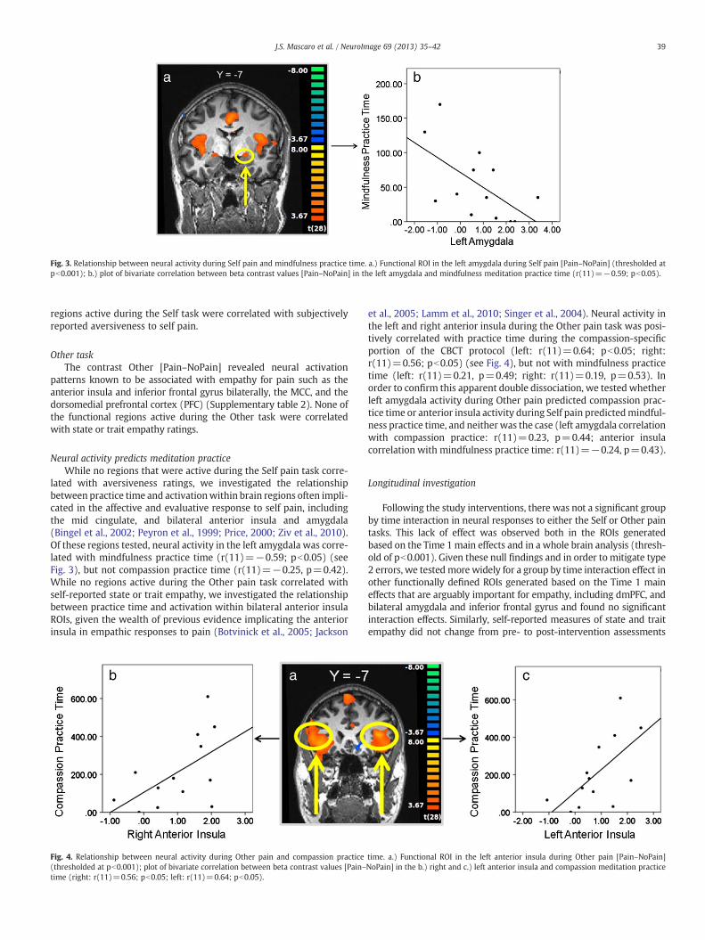
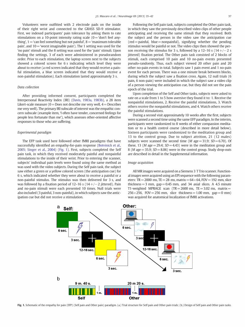
Fig. 3. Relationship between neural activity during Self pain and mindfulness practice time. a.) Functional ROI in the left amygdala during Self pain [Pain–NoPain] (thresholded atpb0.001); b.) plot of bivariate correlation between beta contrast values [Pain–NoPain] in the left amygdala and mindfulness meditation practice time (r(11)=−0.59; pb0.05).
39J.S. Mascaro et al. / NeuroImage 69 (2013) 35–42
regions active during the Self task were correlated with subjectivelyreported aversiveness to self pain.
Other taskThe contrast Other [Pain–NoPain] revealed neural activation
patterns known to be associated with empathy for pain such as theanterior insula and inferior frontal gyrus bilaterally, the MCC, and thedorsomedial prefrontal cortex (PFC) (Supplementary table 2). None ofthe functional regions active during the Other task were correlatedwith state or trait empathy ratings.
Neural activity predicts meditation practiceWhile no regions that were active during the Self pain task corre-
lated with aversiveness ratings, we investigated the relationshipbetween practice time and activationwithin brain regions often impli-cated in the affective and evaluative response to self pain, includingthe mid cingulate, and bilateral anterior insula and amygdala(Bingel et al., 2002; Peyron et al., 1999; Price, 2000; Ziv et al., 2010).Of these regions tested, neural activity in the left amygdala was corre-lated with mindfulness practice time (r(11)=−0.59; pb0.05) (seeFig. 3), but not compassion practice time (r(11)=−0.25, p=0.42).While no regions active during the Other pain task correlated withself-reported state or trait empathy, we investigated the relationshipbetween practice time and activation within bilateral anterior insulaROIs, given the wealth of previous evidence implicating the anteriorinsula in empathic responses to pain (Botvinick et al., 2005; Jackson
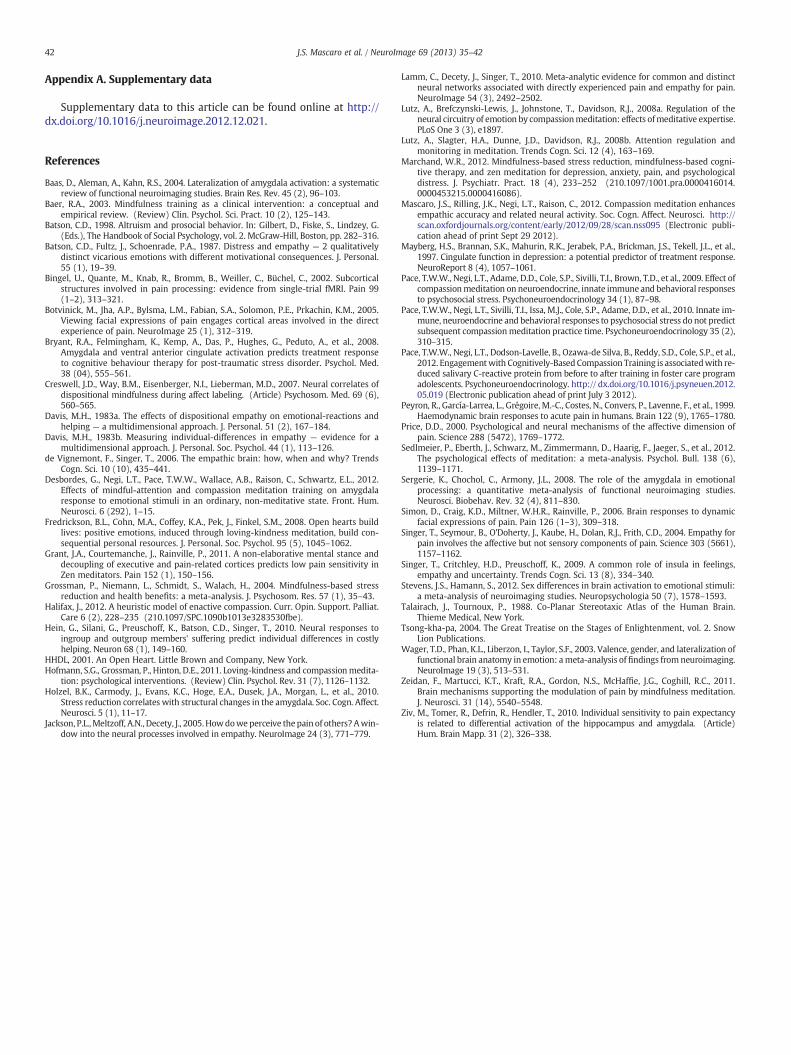
Fig. 4. Relationship between neural activity during Other pain and compassion practice(thresholded at pb0.001); plot of bivariate correlation between beta contrast values [Pain–time (right: r(11)=0.56; pb0.05; left: r(11)=0.64; pb0.05).
et al., 2005; Lamm et al., 2010; Singer et al., 2004). Neural activity inthe left and right anterior insula during the Other pain task was posi-tively correlated with practice time during the compassion-specificportion of the CBCT protocol (left: r(11)=0.64; pb0.05; right:r(11)=0.56; pb0.05) (see Fig. 4), but not with mindfulness practicetime (left: r(11)=0.21, p=0.49; right: r(11)=0.19, p=0.53). Inorder to confirm this apparent double dissociation, we tested whetherleft amygdala activity during Other pain predicted compassion prac-tice time or anterior insula activity during Self pain predictedmindful-ness practice time, and neither was the case (left amygdala correlationwith compassion practice: r(11)=0.23, p=0.44; anterior insulacorrelation with mindfulness practice time: r(11)=−0.24, p=0.43).
Longitudinal investigation
Following the study interventions, there was not a significant groupby time interaction in neural responses to either the Self or Other paintasks. This lack of effect was observed both in the ROIs generatedbased on the Time 1main effects and in a whole brain analysis (thresh-old of pb0.001). Given these null findings and in order to mitigate type2 errors, we tested more widely for a group by time interaction effect inother functionally defined ROIs generated based on the Time 1 maineffects that are arguably important for empathy, including dmPFC, andbilateral amygdala and inferior frontal gyrus and found no significantinteraction effects. Similarly, self-reported measures of state and traitempathy did not change from pre- to post-intervention assessments
time. a.) Functional ROI in the left anterior insula during Other pain [Pain–NoPain]NoPain] in the b.) right and c.) left anterior insula and compassion meditation practice
40 J.S. Mascaro et al. / NeuroImage 69 (2013) 35–42
in the study population as a whole, and no differences were observedbetween those randomized to CBCT vs. the control condition. Linear re-gression analyses indicated that neither mindfulness nor compassionpractice time accounted for a significant amount of the variance inpost-intervention amygdala activation during the Self task (b=0.00,R2 change=0.01, F (1, 10)=0.12, p=0.73) or anterior insula activationduring the Other task (left: b=0.00, R2 change=0.02, F (1, 10)=0.17,p=0.69; right: b=0.00, R2 change=0.06, F (1, 10)=0.63, p=0.45),when controlling for Time 1 brain activity.
Discussion
In this study, we tested the hypothesis that different patterns ofneural activity in response to receiving painful stimulations orwatching others receive a similar painful stimulus would uniquelypredict subsequent engagement with different types of meditationtraining (mindfulness vs. compassion). Activation of the left amygda-la during Self pain was inversely associated with subsequent mindful-ness practice time. During the Other pain task, activity in the anteriorinsula was positively correlated with subsequent compassion practicetime. Interestingly, however, training in CBCT had no effect on brainactivity in any of these areas and did not impact self-reported mea-sures of either pain aversiveness or empathy. Moreover, within thegroup randomized to CBCT, practice time had no effect on patternsof brain activity or self-reportedmeasures of pain aversiveness or em-pathy as assessed following training. Despite these negative findings,other evidence speaks to the effectiveness of CBCT (Desbordes et al.,2012; Mascaro et al., 2012; Pace et al., 2009, 2010, 2012) and the im-portance of practice time in attaining these effects. These findings high-light the potential importance of identifying predictors of practice time,because these predictors might help identify individuals most likely tobenefit from learning CBCT and/or other mediation-based interventions.
Self-report
The hypothesis that baseline levels of self-reported aversivenessto pain would predict subsequent mindfulness practice time is notsupported by the data presented here, nor was there a relationshipbetween state or trait empathy and subsequent compassion practicetime. These data are consistent with a larger trend found here,which was a general lack of correlation between self-report dataand objective measures, since brain activity was also unrelated toany of the self report measures. While the finding that brain activitypredicted subsequent behavior is striking (discussed in more detailbelow), the fact that subjective reports were not related to brain ac-tivity or to practice time means that we cannot definitively determinehow these neural states were relevant to the subjective states of theparticipants. However a large body of previous studies that usedvery similar tasks found relationships between amygdala and anteriorinsula activation and subjective states. The discrepancy between pre-vious studies and the data presented here may be due to limitationsin our self-report data. For example, it may be that because subjectivereports were probed upon completion of both tasks, participants' re-ports did not actually reflectwhat they felt during the task.With respectto the empathy for pain task, the state empathymeasure, in which par-ticipants were asked how “aversive” they found it to watch others re-ceive the painful stimulations, may have probed something more akinto personal distress than to empathy (Batson et al., 1987). In addition,while state empathy measures are often found to correlate with anteri-or insula activity, trait empathy, as measured by the empathic concernsubscale of the IRI (usedhere) is far less reliably correlatedwith anteriorinsula activity (Lamm et al., 2010). Future studies should include morecomprehensive assessments of state and trait empathy, mindfulness,and pain-induced reactivity to more systematically investigate the rela-tionship between subjective states and meditation engagement as wellas the eventual effects of meditation training.
Amygdala activity predicts mindfulness practice
The finding that left amygdala activation during the Self taskpredicted subsequent mindfulness meditation practice time is consis-tent with previous mindfulness meditation research. Individuals whoscored higher on a dispositional mindfulness scale had less bilateralamygdala activation during an affect labeling task (Creswell et al.,2007), and a recent longitudinal study found that participants in amind-fulness based stress reduction (MBSR) intervention reported decreasesin perceived life stress that correlated with decreases in gray matter inthe right amygdala (Holzel et al., 2010). Moreover, studies of Zen, a sim-ilar style of non-evaluative meditation, show that long-term practi-tioners had less activity in the bilateral amygdala while receivingpainful stimuli (Grant et al., 2011). These previous studies suggest thatthe amygdala is likely an important region for many of the evaluativejudgments that mindfulness training is intended to mitigate, and maybe one of the key neural regions mediating the stress ameliorating ef-fects of mindfulness practices. The findings presented here elaborateon the role of the amygdala in mindfulness practices by indicating thatleft amygdala hyperreactivity may impede an individual's ability topractice mindfulness meditation. While these results do not indicatethat mindfulness training is harmful to those who are particularlyreactive to their own painful sensations, the data point to a potential“Catch-22”; specifically that mindfulness training may be most difficultprecisely for those people most in need of its positive effects.
While the correlation betweenmindfulness practice time and activ-ity in the right amygdala was in the same direction, it did not reach thelevel of significance. This is not surprising given the extensive evidencefor lateralized amygdala function in human subjects (Baas et al., 2004;Sergerie et al., 2008; Stevens and Hamann, 2012; Wager et al., 2003).Consistent with our findings, a meta-analysis concluded that the leftamygdala is more strongly implicated in negative emotion, particularlythose negative emotions that tend to elicit a withdrawal response(Wager et al., 2003). The specific subregion identified in that meta-analyses closely matches the dorsal amygdala activation we reporthere. The idea that individuals with a more robust withdrawal-relatedresponse to their own pain are not as likely to engage in mindfulnesshas face validity and is a hypothesis that warrants more careful testingin the future.
Anterior insula activity predicts compassion practice
Despite the fact that there were no observed correlations in the cur-rent study between self-reported state or trait empathy and neural ac-tivity when observing another person receive a painful stimulus, theempathy for pain task elicited robust activity in regions previously iden-tified as important for empathy. Given the consistency with whichneuroimaging investigations have implicated the anterior insula in em-pathic responses (Lammet al., 2010), thefinding that anterior insula ac-tivity was positively correlated with subsequent compassion-specificmeditation practice time may suggest that more empathic individualsare able to engagemore fully with compassionmeditation. This correla-tion between anterior insula responses to observing others in pain andsubsequent practice time is particularly interesting in light of the tradi-tional Tibetanworks uponwhich CBCT is based. In the late 14th CenturyTsong-Kha-Pa (Tsong-kha-pa, 2004) emphasized the importance ofhaving empathy and compassion at the beginning of a practice,since one will not be moved to commit to being compassionate towardothers if his or her empathy and compassion are weak to begin with.Our findings support and sharpen Tsong-Kha-Pa's observation bysuggesting that a certain amount of activity in a brain region involvedin empathic responses may be important to successfully embark oncompassion training.
Several broad points should be made about these practice-timefindings. First, the fact that neural activity differentially predictsan individual's propensity to practice two different meditative

41J.S. Mascaro et al. / NeuroImage 69 (2013) 35–42
techniques supports the assertion that the term ‘meditation’ subsumesheterogeneous techniques with a broad variety of methods andgoals (Lutz et al., 2008b). In fact, these data point to a double dissociationof predictive effects since anterior insula activity during the Self paintask does not predict subsequent mindfulness practice time nor doesamygdala activity during the Other pain task predict subsequent com-passion practice time. Because of this, future meditation studies willbenefit from the direct comparison of multiple, well-characterized andoperationalized styles of meditation training. Moreover, while this isthe first study to show that pre-existing neural activity predicts medita-tion practice time, the findings presented here are analogous to thosecoming out of psychiatric research, in which neuroimaging has beenused to predict responses to both pharmacological and behavioral treat-ments (Bryant et al., 2008; Mayberg et al., 1997). Given the increasinguse of meditation for clinical indications, we believe the study designutilized here has similar utility for determining the efficacy of medita-tion for the treatment of psychiatric pathology.
Longitudinal investigation
Longitudinal findings from the current study suggest that theeight-week CBCT protocol used here does not attenuate neural re-sponses associated with pain aversiveness. With respect to the Selfpain findings, these results are consistent with a recent study byZeidan et al. (2011). While this group found that a 4-day mindfulnesstraining program altered brain activation during a meditation pluspain condition, the amygdala was not one of the regions modifiedby the brief training. While we are duly cautious to lump studies ofZen and mindfulness, the fact that long-term Zen practitioners hadless amygdala activity during pain (Grant et al., 2011), while the cur-rent study and that by Zeidan et al. (2011) did not find amygdalachanges after shorter training periods, may suggest that amygdalachanges require a relatively longer period of training.
Additionally, longitudinal analysis of the empathy task suggests thatCBCT does not amplify the neural correlates of empathy for pain. Withrespect to our ability to detect meditation-induced changes in the neu-ral systems important for empathy, we believe that the fMRI paradigmused here was sufficient for investigating the potential of CBCT tochange the neurobiology of empathy for pain given the robust patternof activations elicited by the EFP task at Time 1. In fact, it appears thatindividuals habituated to the video clips of others in pain, given that ac-tivation in the anterior insula, independent of group, showed significantattenuation from the pre- to the post-intervention assessments(see Supplementary figure S5). It remains possible that participantswould not have habituated had they been presented at both assess-ments with people experiencing pain in real time rather than a video.At the least, however, the data presented here suggest that compassiontraining did not diminish habituation to seeing video clips of others inpain, which in itself suggests the practice had no effect on either theself-reported or neural correlates of empathy. Nonetheless, it is impor-tant to note that empathy is a broad construct subserved by complexneural systems, and the empathy measures used here do not tap intothe related but distinct prosocial emotion of compassion. One recenttheoretical discussion of compassion training suggests that compassionis an emergent process that may be enhanced by training one or moreof the multiple “noncompassion” processes from which it arises(Halifax, 2012). If so, then CBCT may differentially affect these underly-ing, noncompassion processes such that the ability to identify relevantchange will rest squarely on the use of appropriate assessments in fu-ture studies. Importantly, CBCT does appear to affect empathic accuracy(Mascaro et al., 2012), depressive symptoms (Desbordes et al., 2012),and stress physiology (Pace et al., 2009, 2010, 2012).
Results from the current studymay also provide a novel perspectiveon previous cross-sectional studies reporting an association betweenamount of engagement with compassion meditation and enhancedactivation in neural circuits important for empathy, including in the
anterior insula (Lutz et al., 2008a). Our results suggest that ratherthan being a result of compassion meditation training, high levels ofanterior insula activation in response to the suffering of others may ac-tually contribute to the foundation for engaging in compassion medita-tion in the first place. Indeed, while the population studied in thecurrent project is different in numerous ways from the meditation ad-epts investigated in other studies, our findings are consistent with thenotion that adepts may be a self-selected population who likely beginwith extraordinary personality and biological profiles. Studies exploringthe underlying features that render such individuals able to attain highlevels of expertise are an unexplored but highly promising avenue formeditation research.
Limitations
Some limitations of the present study are worth noting. First, prac-tice time was self-reported and it remains possible that some partici-pants were biased or inaccurate in their reporting. In addition, thecontrol groupwas relatively small and the studymay have been under-powered to find a group by time interaction effect. However, given thatboth groups showed significantly attenuated brain activity to the EFPtask at Time 2, we do not believe that the small sample size contributedto the null results of the longitudinal analyses. Moreover, it is importantto note that the same study participants showed a group by time inter-action effect for neural activity during an empathic accuracy task andparticipants randomized to CBCT were significantly more likely tohave enhanced empathic accuracy scores (Mascaro et al., 2012), reduc-ing the likelihood that the studywas underpowered. Additionally,whilethis investigation was an initial attempt at investigating individual dif-ferences in the propensity to adoptmeditation techniques, there are im-portant aspects related to this theme that were glossed for the currentstudy. For example, it remains possible that the pedagogical materialor training was differently understood or received across individuals,or that practice time differed in importance at different points of CBCTtraining. While we use amount of practice time here as a measure ofthe level of engagement with CBCT due to the fact that other studieshave shown the importance of practice for outcomes of interest(Fredrickson et al., 2008; Pace et al., 2009, 2012), future studies shouldanalyze other metrics of engagement such as self-reported connectionto the practice and physiological changes that occur during the practice.A fascinating prospective study could interrogate the relationship be-tween practice time and individual differences in the subjective mean-ing of the training and investigate whether baseline variables aredifferentially related to these various measures of meditation engage-ment. Current findings highlight the need to conduct such a study.Moreover, while the control group for the current study was designedto help account for expectancy bias and non-specific effects of group in-teraction and health-relevant education, future studies should include arandomized comparison of CBCT training with another, similar mentaltraining intervention. These important inquiries will be critical for fu-ture investigations of CBCT and for meditation research more broadly.
Acknowledgments
We thank Carol Worthman and Nathan Mascaro for the designand statistical advice. We also thank Brooke Dodson-Lavelle andBrendan Ozawa-de Silva for training participants using CBCT protocol,Teri Sivilli for the help in recruiting and screening study participants,and Todd Preuss for the helpful comments on this paper. This workwas supported by an Emory University Neuroscience Initiative SeedGrant and by NIH NCCAM (AT004698) (C.R.). J. S. M. was supportedby the Center for Behavioral Neuroscience and by a Ruth L. KirschsteinNational Research Service Award (NRSA) for Individual PredoctoralFellows through the National Center for Complementary and Alterna-tive Medicine (NCCAM) (1F31AT004878-01).
42 J.S. Mascaro et al. / NeuroImage 69 (2013) 35–42
Appendix A. Supplementary data
Supplementary data to this article can be found online at http://dx.doi.org/10.1016/j.neuroimage.2012.12.021.
References
Baas, D., Aleman, A., Kahn, R.S., 2004. Lateralization of amygdala activation: a systematicreview of functional neuroimaging studies. Brain Res. Rev. 45 (2), 96–103.
Baer, R.A., 2003. Mindfulness training as a clinical intervention: a conceptual andempirical review. (Review) Clin. Psychol. Sci. Pract. 10 (2), 125–143.
Batson, C.D., 1998. Altruism and prosocial behavior. In: Gilbert, D., Fiske, S., Lindzey, G.(Eds.), The Handbook of Social Psychology, vol. 2. McGraw-Hill, Boston, pp. 282–316.
Batson, C.D., Fultz, J., Schoenrade, P.A., 1987. Distress and empathy — 2 qualitativelydistinct vicarious emotions with different motivational consequences. J. Personal.55 (1), 19–39.
Bingel, U., Quante, M., Knab, R., Bromm, B., Weiller, C., Büchel, C., 2002. Subcorticalstructures involved in pain processing: evidence from single-trial fMRI. Pain 99(1–2), 313–321.
Botvinick, M., Jha, A.P., Bylsma, L.M., Fabian, S.A., Solomon, P.E., Prkachin, K.M., 2005.Viewing facial expressions of pain engages cortical areas involved in the directexperience of pain. NeuroImage 25 (1), 312–319.
Bryant, R.A., Felmingham, K., Kemp, A., Das, P., Hughes, G., Peduto, A., et al., 2008.Amygdala and ventral anterior cingulate activation predicts treatment responseto cognitive behaviour therapy for post-traumatic stress disorder. Psychol. Med.38 (04), 555–561.
Creswell, J.D., Way, B.M., Eisenberger, N.I., Lieberman, M.D., 2007. Neural correlates ofdispositional mindfulness during affect labeling. (Article) Psychosom. Med. 69 (6),560–565.
Davis, M.H., 1983a. The effects of dispositional empathy on emotional-reactions andhelping — a multidimensional approach. J. Personal. 51 (2), 167–184.
Davis, M.H., 1983b. Measuring individual-differences in empathy — evidence for amultidimensional approach. J. Personal. Soc. Psychol. 44 (1), 113–126.
de Vignemont, F., Singer, T., 2006. The empathic brain: how, when and why? TrendsCogn. Sci. 10 (10), 435–441.
Desbordes, G., Negi, L.T., Pace, T.W.W., Wallace, A.B., Raison, C., Schwartz, E.L., 2012.Effects of mindful-attention and compassion meditation training on amygdalaresponse to emotional stimuli in an ordinary, non-meditative state. Front. Hum.Neurosci. 6 (292), 1–15.
Fredrickson, B.L., Cohn, M.A., Coffey, K.A., Pek, J., Finkel, S.M., 2008. Open hearts buildlives: positive emotions, induced through loving-kindness meditation, build con-sequential personal resources. J. Personal. Soc. Psychol. 95 (5), 1045–1062.
Grant, J.A., Courtemanche, J., Rainville, P., 2011. A non-elaborative mental stance anddecoupling of executive and pain-related cortices predicts low pain sensitivity inZen meditators. Pain 152 (1), 150–156.
Grossman, P., Niemann, L., Schmidt, S., Walach, H., 2004. Mindfulness-based stressreduction and health benefits: a meta-analysis. J. Psychosom. Res. 57 (1), 35–43.
Halifax, J., 2012. A heuristic model of enactive compassion. Curr. Opin. Support. Palliat.Care 6 (2), 228–235 (210.1097/SPC.1090b1013e3283530fbe).
Hein, G., Silani, G., Preuschoff, K., Batson, C.D., Singer, T., 2010. Neural responses toingroup and outgroup members' suffering predict individual differences in costlyhelping. Neuron 68 (1), 149–160.
HHDL, 2001. An Open Heart. Little Brown and Company, New York.Hofmann, S.G., Grossman, P., Hinton, D.E., 2011. Loving-kindness and compassionmedita-
tion: psychological interventions. (Review) Clin. Psychol. Rev. 31 (7), 1126–1132.Holzel, B.K., Carmody, J., Evans, K.C., Hoge, E.A., Dusek, J.A., Morgan, L., et al., 2010.
Stress reduction correlates with structural changes in the amygdala. Soc. Cogn. Affect.Neurosci. 5 (1), 11–17.
Jackson, P.L.,Meltzoff, A.N., Decety, J., 2005.Howdoweperceive thepain of others?Awin-dow into the neural processes involved in empathy. NeuroImage 24 (3), 771–779.
Lamm, C., Decety, J., Singer, T., 2010. Meta-analytic evidence for common and distinctneural networks associated with directly experienced pain and empathy for pain.NeuroImage 54 (3), 2492–2502.
Lutz, A., Brefczynski-Lewis, J., Johnstone, T., Davidson, R.J., 2008a. Regulation of theneural circuitry of emotion by compassionmeditation: effects ofmeditative expertise.PLoS One 3 (3), e1897.
Lutz, A., Slagter, H.A., Dunne, J.D., Davidson, R.J., 2008b. Attention regulation andmonitoring in meditation. Trends Cogn. Sci. 12 (4), 163–169.
Marchand, W.R., 2012. Mindfulness-based stress reduction, mindfulness-based cogni-tive therapy, and zen meditation for depression, anxiety, pain, and psychologicaldistress. J. Psychiatr. Pract. 18 (4), 233–252 (210.1097/1001.pra.0000416014.0000453215.0000416086).
Mascaro, J.S., Rilling, J.K., Negi, L.T., Raison, C., 2012. Compassion meditation enhancesempathic accuracy and related neural activity. Soc. Cogn. Affect. Neurosci. http://scan.oxfordjournals.org/content/early/2012/09/28/scan.nss095 (Electronic publi-cation ahead of print Sept 29 2012).
Mayberg, H.S., Brannan, S.K., Mahurin, R.K., Jerabek, P.A., Brickman, J.S., Tekell, J.L., et al.,1997. Cingulate function in depression: a potential predictor of treatment response.NeuroReport 8 (4), 1057–1061.
Pace, T.W.W., Negi, L.T., Adame, D.D., Cole, S.P., Sivilli, T.I., Brown, T.D., et al., 2009. Effect ofcompassionmeditation on neuroendocrine, innate immune and behavioral responsesto psychosocial stress. Psychoneuroendocrinology 34 (1), 87–98.
Pace, T.W.W., Negi, L.T., Sivilli, T.I., Issa, M.J., Cole, S.P., Adame, D.D., et al., 2010. Innate im-mune, neuroendocrine and behavioral responses to psychosocial stress do not predictsubsequent compassionmeditation practice time. Psychoneuroendocrinology 35 (2),310–315.
Pace, T.W.W., Negi, L.T., Dodson-Lavelle, B., Ozawa-de Silva, B., Reddy, S.D., Cole, S.P., et al.,2012. Engagementwith Cognitively-Based Compassion Training is associatedwith re-duced salivary C-reactive protein from before to after training in foster care programadolescents. Psychoneuroendocrinology. http:// dx.doi.org/10.1016/j.psyneuen.2012.05.019 (Electronic publication ahead of print July 3 2012).
Peyron, R., García-Larrea, L., Grégoire, M.-C., Costes, N., Convers, P., Lavenne, F., et al., 1999.Haemodynamic brain responses to acute pain in humans. Brain 122 (9), 1765–1780.
Price, D.D., 2000. Psychological and neural mechanisms of the affective dimension ofpain. Science 288 (5472), 1769–1772.
Sedlmeier, P., Eberth, J., Schwarz, M., Zimmermann, D., Haarig, F., Jaeger, S., et al., 2012.The psychological effects of meditation: a meta-analysis. Psychol. Bull. 138 (6),1139–1171.
Sergerie, K., Chochol, C., Armony, J.L., 2008. The role of the amygdala in emotionalprocessing: a quantitative meta-analysis of functional neuroimaging studies.Neurosci. Biobehav. Rev. 32 (4), 811–830.
Simon, D., Craig, K.D., Miltner, W.H.R., Rainville, P., 2006. Brain responses to dynamicfacial expressions of pain. Pain 126 (1–3), 309–318.
Singer, T., Seymour, B., O'Doherty, J., Kaube, H., Dolan, R.J., Frith, C.D., 2004. Empathy forpain involves the affective but not sensory components of pain. Science 303 (5661),1157–1162.
Singer, T., Critchley, H.D., Preuschoff, K., 2009. A common role of insula in feelings,empathy and uncertainty. Trends Cogn. Sci. 13 (8), 334–340.
Stevens, J.S., Hamann, S., 2012. Sex differences in brain activation to emotional stimuli:a meta-analysis of neuroimaging studies. Neuropsychologia 50 (7), 1578–1593.
Talairach, J., Tournoux, P., 1988. Co-Planar Stereotaxic Atlas of the Human Brain.Thieme Medical, New York.
Tsong-kha-pa, 2004. The Great Treatise on the Stages of Enlightenment, vol. 2. SnowLion Publications.
Wager, T.D., Phan, K.L., Liberzon, I., Taylor, S.F., 2003. Valence, gender, and lateralization offunctional brain anatomy in emotion: ameta-analysis of findings fromneuroimaging.NeuroImage 19 (3), 513–531.
Zeidan, F., Martucci, K.T., Kraft, R.A., Gordon, N.S., McHaffie, J.G., Coghill, R.C., 2011.Brain mechanisms supporting the modulation of pain by mindfulness meditation.J. Neurosci. 31 (14), 5540–5548.
Ziv, M., Tomer, R., Defrin, R., Hendler, T., 2010. Individual sensitivity to pain expectancyis related to differential activation of the hippocampus and amygdala. (Article)Hum. Brain Mapp. 31 (2), 326–338.