precipio, inc. instructions for useprecipiodx.com/enrichment_only/1011-e.pdfgreen 4 360 -20...
TRANSCRIPT
Precipio, Inc.
Instructions for Use NRAS Exon 3 Mutation Enrichment using ICE COLD-PCR Research Use Only
1 IFU1011E - Revision 28Jun2019
Table of Contents Manufacturer ............................................................................................................................................................ 2
Intended Use ............................................................................................................................................................. 2
Reagent Preparation .................................................................................................................................................. 2
Components and Storage Conditions ........................................................................................................................ 2
Reagents Required but not Supplied ......................................................................................................................... 3
ICE COLD-PCR Coupled to Downstream Analysis ................................................................................................... 3
Primary Sample Collection, Handling and Storage .................................................................................................... 4
Notes for this Assay ................................................................................................................................................... 5
ICE COLD-PCR Protocol .............................................................................................................................................. 5
Thermal Cycler Program for ICE COLD-PCR ............................................................................................................... 8
Technical Support/Questions .................................................................................................................................... 8
Appendix .................................................................................................................................................................... 9
Copyright © 2019 by Precipio, Inc. All rights reserved. This Instruction manual or any portion thereof may not be reproduced in any form, or transmitted outside of the recipient’s organization in any form by any means — electronic, mechanical, photocopy, recording, or otherwise — or used in any manner whatsoever without the express written permission of the Company.
2 IFU1011E - Revision 28Jun2019
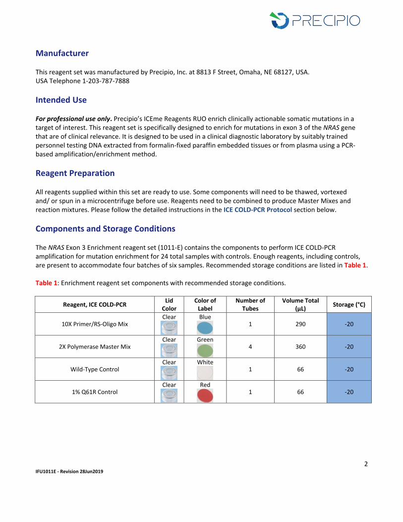
Manufacturer This reagent set was manufactured by Precipio, Inc. at 8813 F Street, Omaha, NE 68127, USA. USA Telephone 1-203-787-7888
Intended Use For professional use only. Precipio’s ICEme Reagents RUO enrich clinically actionable somatic mutations in a target of interest. This reagent set is specifically designed to enrich for mutations in exon 3 of the NRAS gene that are of clinical relevance. It is designed to be used in a clinical diagnostic laboratory by suitably trained personnel testing DNA extracted from formalin-fixed paraffin embedded tissues or from plasma using a PCR-based amplification/enrichment method.
Reagent Preparation All reagents supplied within this set are ready to use. Some components will need to be thawed, vortexed and/ or spun in a microcentrifuge before use. Reagents need to be combined to produce Master Mixes and reaction mixtures. Please follow the detailed instructions in the ICE COLD-PCR Protocol section below.
Components and Storage Conditions The NRAS Exon 3 Enrichment reagent set (1011-E) contains the components to perform ICE COLD-PCR amplification for mutation enrichment for 24 total samples with controls. Enough reagents, including controls, are present to accommodate four batches of six samples. Recommended storage conditions are listed in Table 1. Table 1: Enrichment reagent set components with recommended storage conditions.
Reagent, ICE COLD-PCR Lid
Color Color of
Label Number of
Tubes Volume Total
(μL) Storage (°C)
10X Primer/RS-Oligo Mix Clear
Blue
1 290 -20
2X Polymerase Master Mix Clear
Green
4 360 -20
Wild-Type Control Clear
White
1 66 -20
1% Q61R Control Clear
Red
1 66 -20
3 IFU1011E - Revision 28Jun2019
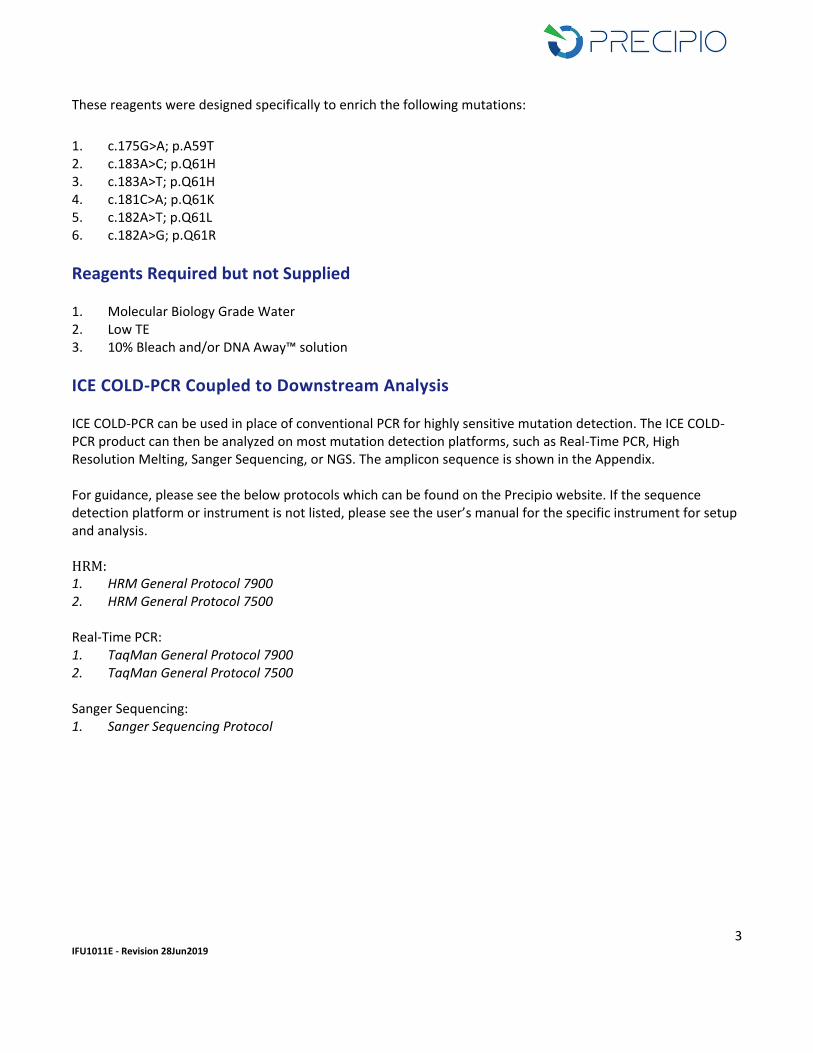
These reagents were designed specifically to enrich the following mutations:
1. c.175G>A; p.A59T 2. c.183A>C; p.Q61H 3. c.183A>T; p.Q61H 4. c.181C>A; p.Q61K 5. c.182A>T; p.Q61L 6. c.182A>G; p.Q61R
Reagents Required but not Supplied 1. Molecular Biology Grade Water 2. Low TE 3. 10% Bleach and/or DNA Away™ solution
ICE COLD-PCR Coupled to Downstream Analysis ICE COLD-PCR can be used in place of conventional PCR for highly sensitive mutation detection. The ICE COLD-PCR product can then be analyzed on most mutation detection platforms, such as Real-Time PCR, High Resolution Melting, Sanger Sequencing, or NGS. The amplicon sequence is shown in the Appendix. For guidance, please see the below protocols which can be found on the Precipio website. If the sequence detection platform or instrument is not listed, please see the user’s manual for the specific instrument for setup and analysis. HRM: 1. HRM General Protocol 7900 2. HRM General Protocol 7500 Real-Time PCR: 1. TaqMan General Protocol 7900 2. TaqMan General Protocol 7500 Sanger Sequencing: 1. Sanger Sequencing Protocol
4 IFU1011E - Revision 28Jun2019
Primary Sample Collection, Handling and Storage This reagent set can be used with the following: - DNA extracted from formalin-fixed paraffin-embedded tumor samples (FFPE slides & blocks) or fine
needle aspirations (FNAs) o For optimal DNA extraction from FFPE, the tissue should be fixed in neutral buffered formalin for
14 – 24 hours, placed in ethanol and then embedded in paraffin following standard histological practices.
o Tumor biopsies are a heterogeneous mixture of tumor cells and non-tumor cells. In addition, the tumor itself is a heterogeneous mixture of tumor cells with mutations and tumor cells without mutations. Because these somatic mutations may not be evenly distributed throughout the tumor, the resultant mutational analysis of different sections from the same tumor may be different.
o To increase the probability of detecting a mutation, DNA from the tumor region of the tissue should be isolated by scraping only the tumor area from the glass slide using a fresh, sterile scalpel for each new slide. It is recommended that at least two independent analyses are performed for each sample.
o The formalin-fixation process used in preparing FFPE tumor biopsy samples may result in deamination of cytosines. Deamination converts cytosine to uracil. The polymerase will recognize this uracil as a thymine and incorporate an adenine in the copied strands. The correct allele G is then replaced with an A causing an artifact mutation due to the fixation process and not a true somatic mutation. It is highly suggested that an extraction protocol which incorporates Uracil-N-Glycosylase be used for FFPE Extractions.
- Circulating free DNA (cfDNA) from plasma or serum o The following collection tubes can be used to collect whole blood for subsequent extraction of
cfDNA from plasma: Lavender top EDTA tube Streck Cell Free DNA BCT® Tube NOTE: Extraction of cfDNA should be performed within 24 hrs of collection with the lavender top EDTA tube. See manufacturer’s guidance for each tube type. NOTE: Upon separation via centrifugation, if the sample is red or orange in color, this denotes the sample is hemolyzed and therefore not suitable for downstream plasma analysis. A redraw is required. Plasma should have a yellow or light yellow color and show distinct separation on top of the blood. NOTE: Storage and shipping conditions of the whole blood may affect quantity and quality of cfDNA in relation to full length DNA.
o The preferred extraction kit for ICE COLD-PCR applications is the Bioo Scientific NextPrep-Mag™ cfDNA Isolation Kit (Refer to general protocol MagBind Plasma Extraction Protocol on the Precipio website). The Qiagen QIAamp® Circulating Nucleic Acid Kit (QIAGEN Plasma Extraction Protocol) was also tested.
- DNA isolated from other body fluids. Other considerations:
5 IFU1011E - Revision 28Jun2019
1. It is recommended that all samples and controls are extracted with the same protocol to avoid any variations due to reagents and/or buffers.
2. All DNA samples should be diluted to the same starting concentration with UV-treated low TE. 3. Ensure sample to sample uniformity. 4. Any additional control DNA to be analyzed should be of the same quality and quantity as the sample DNA.
Notes for this Assay
- The ICE COLD-PCR assay has been optimized using the Bio-Rad C1000 thermal cycler; however, the ICE COLD-PCR assay can be optimized/run on most other thermal cyclers.
- Due to heterogeneity associated with tumors, biopsy samples may contain normal cells as well as Wild-Type and mutant tumor cells.
- The Limit of Detection (LOD) of any mutations present in the sample DNA following ICE COLD-PCR is dependent on the sensitivity of the downstream sequence detection platform used.
- Only the DNA polymerase(s) supplied within this reagent set should be used as indicated for the specific assay or downstream application.
- This is a mutation enrichment assay. Any mutation or mismatch covered by the RS-Oligo will be enriched during the ICE COLD-PCR process. There are two possible sources of false positive results related to this assay: o Polymerase errors. While the polymerase included in this set is a high-fidelity enzyme, there is the
possibility of a polymerase-induced error. o The formalin-fixation process used in preparing FFPE tumor biopsy samples
NOTE: Variants caused by polymerase errors or deamination of cytosine do not repeat upon re-analysis. Therefore, it is recommended that any such mutation be confirmed by duplicate analysis using the same extracted genomic DNA.
ICE COLD-PCR Protocol IMPORTANT! Use dedicated hood/room for ICE COLD-PCR reaction setup to avoid contamination from post-PCR products. IMPORTANT! The following procedures are optional but highly recommended prior to PCR setup: - Turn on UV light inside hood or a UV crosslinker. - Prior to preparing Master Mixes, UV crosslink all empty Master Mix tubes. Also UV crosslink 1.7 mL tubes
containing appropriate volume of Molecular Biology Grade Water needed for Master Mix preparation. These tubes should be UV irradiated for 10 min.
- Make sure all work areas are prepared for analysis of low level mutations. This includes correct use of the PCR Workstation, dedicated pipettes, tips, 10% bleach solution and/or DNA Away solutions.
1. Remove controls, 10X Primer/RS-Oligo Mix and 2X Polymerase Master Mix from freezer and thaw on ice.
2. Once thawed, vortex all components ~3 - 5 sec to mix thoroughly. Briefly centrifuge 5 sec to ensure no liquid remains on tube lids and place on ice.
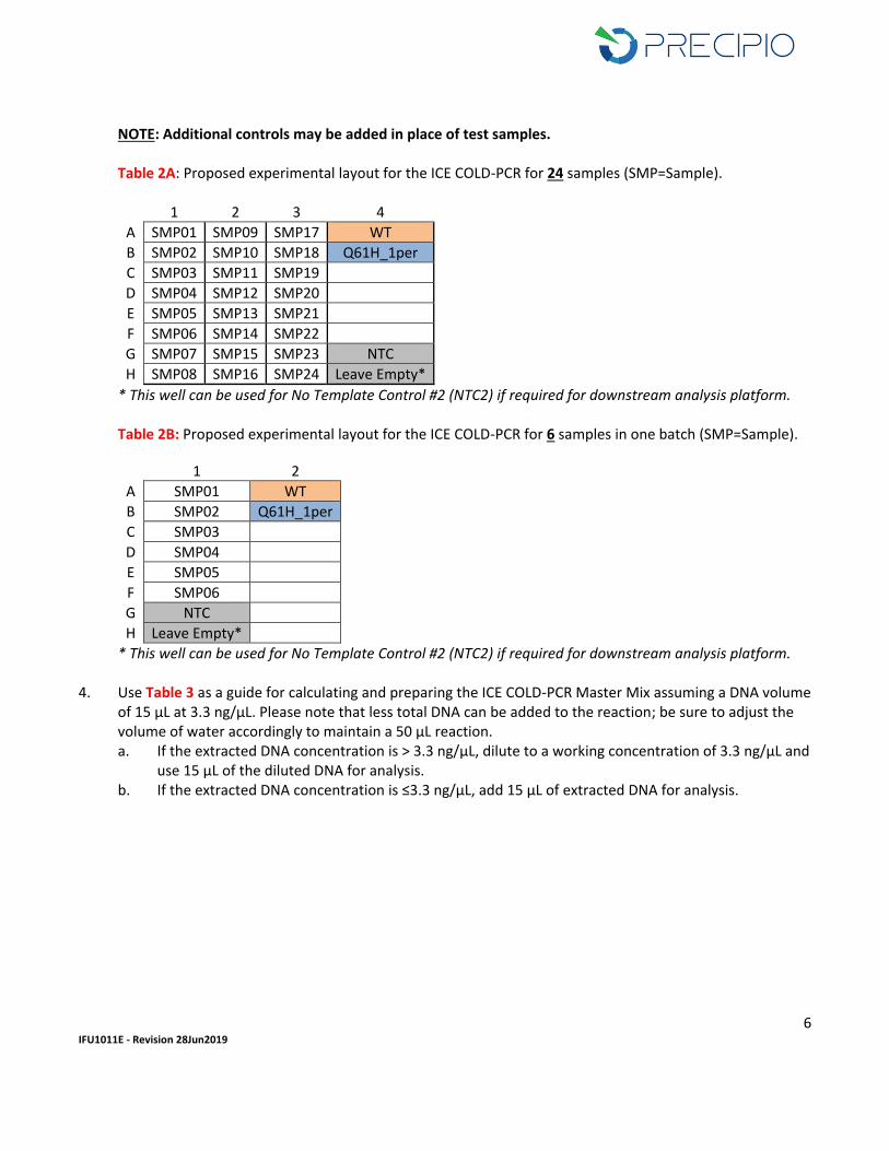
3. Example experiment layout: Follow Table 2 for layout to simplify later steps of the procedure. The following three controls are required for each setup: NTC (No Template Control) and the control DNAs provided in this reagent set, including WT (NRAS Wild-Type) and Q61H A>C_1per (1% NRAS- Q61H).
6 IFU1011E - Revision 28Jun2019
NOTE: Additional controls may be added in place of test samples. Table 2A: Proposed experimental layout for the ICE COLD-PCR for 24 samples (SMP=Sample).
1 2 3 4
A SMP01 SMP09 SMP17 WT
B SMP02 SMP10 SMP18 Q61H_1per
C SMP03 SMP11 SMP19
D SMP04 SMP12 SMP20
E SMP05 SMP13 SMP21
F SMP06 SMP14 SMP22
G SMP07 SMP15 SMP23 NTC
H SMP08 SMP16 SMP24 Leave Empty*
* This well can be used for No Template Control #2 (NTC2) if required for downstream analysis platform. Table 2B: Proposed experimental layout for the ICE COLD-PCR for 6 samples in one batch (SMP=Sample).
1 2
A SMP01 WT
B SMP02 Q61H_1per
C SMP03
D SMP04
E SMP05
F SMP06
G NTC
H Leave Empty*
* This well can be used for No Template Control #2 (NTC2) if required for downstream analysis platform.
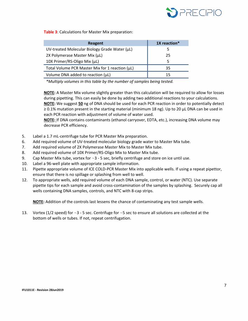
4. Use Table 3 as a guide for calculating and preparing the ICE COLD-PCR Master Mix assuming a DNA volume of 15 µL at 3.3 ng/µL. Please note that less total DNA can be added to the reaction; be sure to adjust the volume of water accordingly to maintain a 50 µL reaction. a. If the extracted DNA concentration is > 3.3 ng/µL, dilute to a working concentration of 3.3 ng/µL and
use 15 µL of the diluted DNA for analysis. b. If the extracted DNA concentration is ≤3.3 ng/µL, add 15 µL of extracted DNA for analysis.
7 IFU1011E - Revision 28Jun2019
Table 3: Calculations for Master Mix preparation:
Reagent 1X reaction*
UV-treated Molecular Biology Grade Water (µL) 5
2X Polymerase Master Mix (µL) 25
10X Primer/RS-Oligo Mix (µL) 5
Total Volume PCR Master Mix for 1 reaction (µL) 35
Volume DNA added to reaction (µL) 15
*Multiply volumes in this table by the number of samples being tested.
NOTE: A Master Mix volume slightly greater than this calculation will be required to allow for losses during pipetting. This can easily be done by adding two additional reactions to your calculations. NOTE: We suggest 50 ng of DNA should be used for each PCR reaction in order to potentially detect ≥ 0.1% mutation present in the starting material (minimum 18 ng). Up to 20 µL DNA can be used in each PCR reaction with adjustment of volume of water used. NOTE: If DNA contains contaminants (ethanol carryover, EDTA, etc.), increasing DNA volume may decrease PCR efficiency.
5. Label a 1.7 mL-centrifuge tube for PCR Master Mix preparation. 6. Add required volume of UV-treated molecular biology grade water to Master Mix tube. 7. Add required volume of 2X Polymerase Master Mix to Master Mix tube. 8. Add required volume of 10X Primer/RS-Oligo Mix to Master Mix tube.
9. Cap Master Mix tube, vortex for ~3 - 5 sec, briefly centrifuge and store on ice until use. 10. Label a 96-well plate with appropriate sample information. 11. Pipette appropriate volume of ICE COLD-PCR Master Mix into applicable wells. If using a repeat pipettor,
ensure that there is no spillage or splashing from well to well. 12. To appropriate wells, add required volume of each DNA sample, control, or water (NTC). Use separate
pipette tips for each sample and avoid cross-contamination of the samples by splashing. Securely cap all wells containing DNA samples, controls, and NTC with 8-cap strips.
NOTE: Addition of the controls last lessens the chance of contaminating any test sample wells.
13. Vortex (1/2 speed) for ~3 - 5 sec. Centrifuge for ~5 sec to ensure all solutions are collected at the bottom of wells or tubes. If not, repeat centrifugation.
8 IFU1011E - Revision 28Jun2019
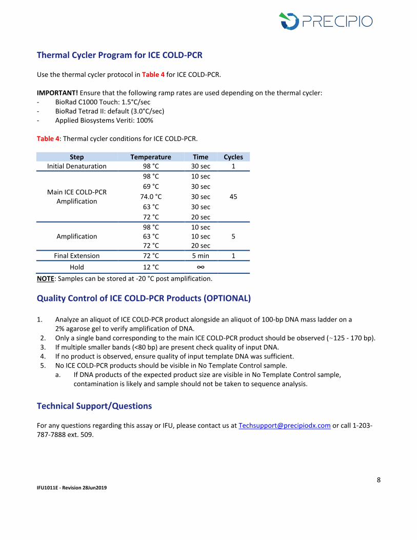
Thermal Cycler Program for ICE COLD-PCR
Use the thermal cycler protocol in Table 4 for ICE COLD-PCR. IMPORTANT! Ensure that the following ramp rates are used depending on the thermal cycler: - BioRad C1000 Touch: 1.5°C/sec - BioRad Tetrad II: default (3.0°C/sec) - Applied Biosystems Veriti: 100% Table 4: Thermal cycler conditions for ICE COLD-PCR.
Step Temperature Time Cycles Initial Denaturation 98 °C 30 sec 1
Main ICE COLD-PCR Amplification
98 °C 10 sec
45
69 °C 30 sec
74.0 °C 30 sec
63 °C 30 sec
72 °C 20 sec
Amplification 98 °C 10 sec
5 63 °C 10 sec 72 °C 20 sec
Final Extension 72 °C 5 min 1
Hold 12 °C ∞
NOTE: Samples can be stored at -20 °C post amplification.
Quality Control of ICE COLD-PCR Products (OPTIONAL)
1. Analyze an aliquot of ICE COLD-PCR product alongside an aliquot of 100-bp DNA mass ladder on a 2% agarose gel to verify amplification of DNA.
2. Only a single band corresponding to the main ICE COLD-PCR product should be observed (~125 - 170 bp). 3. If multiple smaller bands (<80 bp) are present check quality of input DNA. 4. If no product is observed, ensure quality of input template DNA was sufficient. 5. No ICE COLD-PCR products should be visible in No Template Control sample.
a. If DNA products of the expected product size are visible in No Template Control sample, contamination is likely and sample should not be taken to sequence analysis.
Technical Support/Questions For any questions regarding this assay or IFU, please contact us at [email protected] or call 1-203-787-7888 ext. 509.
9 IFU1011E - Revision 28Jun2019
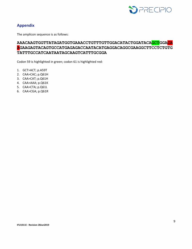
Appendix The amplicon sequence is as follows:
AAACAAGTGGTTATAGATGGTGAAACCTGTTTGTTGGACATACTGGATACAGCTGGACA
AGAAGAGTACAGTGCCATGAGAGACCAATACATGAGGACAGGCGAAGGCTTCCTCTGTG
TATTTGCCATCAATAATAGCAAGTCATTTGCGGA Codon 59 is highlighted in green; codon 61 is highlighted red: 1. GCT>ACT; p.A59T 2. CAA>CAC; p.Q61H 3. CAA>CAT; p.Q61H 4. CAA>AAA; p.Q61K 5. CAA>CTA; p.Q61L 6. CAA>CGA; p.Q61R