predators and parasites of the - oregon state university
TRANSCRIPT

OCTOBER 1964
DEC 1964
LIBRARYION STATE
NNiVOSTV .
TECHNICAL BULLETIN 79
Predators and Parasites of the
Douglas-fir Beetle:
Description and Identification
of the Immature Stages
Agricultural Experiment StationOregon State UniversityCorvallis

CONTENTS
Page
3
4
5
5
7
9
9
19
25
32
37
44
46
49
50
50
AUTHORS : L. N. Kline is a former research assistant and J. A.Rudinsky is a professor of forest entomology, Department of Ento-mology, Oregon State University. This bulletin is based on the seniorauthor's Master's thesis submitted to OSU.
INTRODUCTION
LITERATURE REVIEW . .
IDENTIFICATION OF THE INSECT ENEMIES OF.DENDROCTONUS PSEUDOTSUGAE
Methods and Procedures of StudyKnown and Possible Predators and Parasites .
Field Key to the Insect Larvae of the Known Predatorsand Parasites
Description and Illustration of the Egg, Larva, and Pupaof the Known Predators and Parasites .
Cleridae : Enoclcrus sphegeus Fabricus .
Cleridae: Enoclcrus lecontei Wolcott .
Cleridae: Thanasimus undatulus Say
Ostomatidae : Temnochila virescens chlorodia Mannerheim
Dolichopodidae: NVcdetcra spp
Lonchaeidae : Lonchaca spp
Braconidae: Coeloides brunneri Viereck .
Pteromalidae : Roptrocerus eccoptogasteri Ratz, Cecidostibaburkci Craw., and C dendroctoni Ash
ACKNOWLEDGMENTS .
REFERENCES CITED. .
Predators and Parasites
of the Douglas-fir Beetle:
Description and Identification
of the Immature StagesL. N. KLINE AND J. A. RUDINSKY
INTRODUCTION
The Douglas-fir beetle, Dendroctonus pseudotsugae Hopkins(Coleoptera: Scolytidae), is the most destructive insect enemy ofDouglas-fir, Pseudotsuga menziesii (Mirb.) Franco, throughout therange of this important tree species. During the epidemic of 1951-1954, for instance, it killed over three billion board feet of greentimber in western Oregon and Washington alone.
The beetle is native to North America, and is preyed upon bya number of important predators and parasites. Some of theseundoubtedly play a significant role in reducing the epidemic popula-tion of the beetle or in preventing its increase to destructive numbers.
Making a prognosis of the trend of the Douglas-fir beetle popu-lation in the field or forecasting the economic damage requiressampling not only Douglas-fir beetles, but also numbers and speciesof the immature stages of predators and parasites present under thebark. Past studies have been concerned more with the biology ofthe Douglas-fir beetle than with its natural enemies. The biology andidentification, especially of the immature stages of the predators andparasites, are incomplete or in some instances virtually unknown.
This bulletin presents identifications, descriptions, and drawingsof the immature stages of the main predators and parasites of theDouglas-fir beetle in the Intermountain and Pacific Northwest regionsof the United States. Brief notes on the life histories of the preda-tors and parasites, and a key separating the larvae are also included.
Emphasis is placed on the following species : Enoclerus sphegeus,E. lecontei, Thanasimus undatulus, and Temnochila virescens. The re-maining species, Medetera spp., Lonchaea spp., Coeloides brunneri,and the pteromalids, are treated in a more general manner.
Three instars exist for the clerids, with five to six for theostomatid.

LITERATURE REVIEW
A review of the literature shows that only limited studies havebeen made of the biology, habits, and identification of a few of theinsect enemies of the Douglas-fir beetle.
In northern Idaho and Montana, Bedard (1, pp. 27-61) workedbriefly on the biology, habits, and identification of Enoclerus sphegeusFab. (Coleoptera : Cleridae) ; E. lecontei Wolc. (Coleoptera : Cleri-dae) ; T hanasivnus dubius (Fab.) (Coleoptera: Cleridae), a mistakenidentity for T undatulus Say; Teznnochila virescens chlorodia Mann.(Coleoptera : Ostomatidae) ; Medetera aldrichii Wh. (Diptera :Doli-chopodidae); Lonchaea corticis Tay. (Diptera: Lonchaeidae); Coe-
loides brunneri Vier. ('Hymenoptera; Braconidae); Roptrocerus ec-co ptogasteri Ratz. (Hymenoptera : Pteromalidae) ; and Cecidostibadendroctoni Ashm. (Hymenoptera: Pteromalidae). Bedard's de-scriptions and drawings of the larvae were only of the mature instars.
Roving, Craighead, and Champlain (4, 5, 6) published on theidentification of the mature larval instars of E. sphegeus, E. lecontei,and T. virescens. Only a few drawings were made for T. virescens.These workers also included brief notes on the biology of someof the clerids preying on bark beetles in general. Habits of the adultsand larvae and identification of four instars of E. sphegeus in rela-tion to Ips pini (Say) and Ips perrotti Sw. on lodgepole pine slashin Alberta, Canada, have been reported by Reid (20). Person (17)worked on the life history, habits, importance as a natural controlfactor, and possibilities of increasing the effectiveness of E. leconteiin association with western pine beetle, Dendroctonus brevicosnisLec., in California. Some work has been done by Struble and Car-pelan on the identification, biology, habits, and laboratory propaga-tion of E. sphegeus and T. virescens in an attempt to rear thesepredators for liberation in the field as control agents of bark beetles(24, 25, 26).
A fairly complete study was made by DeLeon (8) on thebiology, habits, and internal and external morphology of the larvaof Medetera aldrichii Wh. in relation to mountain pine beetle, D.nionticolae Hopk., in Montana and northeastern Washington. Also,several notes were made by Hopping (9) on the seasonal develop-ment of M. aldrichii as a predator of D pseudotsugae in BritishColumbia.
Recently, Johnsey (10) studied the biology of M. aldrichii andLonchaea sp. associated with the Douglas-fir beetle in western Oregonand Washington.

Ryan and Rudinsky (22) published on the biology, habits, anddescriptions of the five larval instars, development of the immaturestadia at various temperatures, and factors contributing to the effec-tiveness of Coeloides brunneri Vier., a parasite of the Douglas-firbeetle in western Oregon.
IDENTIFICATION OF THE INSECT ENEMIES OFDENDROCTONUS PSEUDOTSUGAE
As just noted, previous studies, with the exception of those byDeLeon (8, pp. 62-67), Reid (20), and Ryan (21), were concernedonly with the identification of the mature larval stage. Past andpresent biological appraisals or population sampling of bark beetleshave been based on immature stages found under the bark. There-fore, in this type of sampling, since all stages of the insect concernedmay be encountered, all should be easily identified.
Discussed here are the methods and procedures used in rearingcertain species in order to obtain specimens for study, and also themethods of measurement and drawing. To facilitate future identifica-tions, a field key, descriptions, and illustrations of the immaturestages of the most important species are included. The generalbiology, geographical distribution, and relative importance of eachspecies are given, based on general observation.
Methods and Procedures of StudyBecause of the scant taxonomical knowledge of the adults of
the hymenopterous parasites and dipterous predators, it was possiblein this study to identify the immature stages only to the family andgeneric levels. Available literature was used for the descriptionswhenever possible, and other characters and material were includedfrom personal observation.
The taxonomy of the adult coleopterous predators has beenworked on more intensively but still presents a problem in associatingadults with their immature forms. Therefore, each species was rearedin order to obtain known specimens of each stage for study. Theserearings were conducted in a manner similar to those reared byStruble (25).
Briefly, the method of rearing was as follows : Known identifiedadults of Enoclerus sphegeus, E lecontei, Thanasirnus undatulus, andTemnochila virescens were collected in the field on the bark andaround Douglas-fir trees infested by Dendroctonus pseudotsugae. The

sex of the adults was determined whenever possible, using the sexcharacters described by Struble and Carpelan (26). The submentalpit present only on the males of T. virescens was easy to observe witha 10x hand lens and was very dependable. However, the sex charactersuggested by the same workers for E sphegeus was not easy to ob-serve even under a microscope. It was found that the large size ofthe abdomen of certain clerids usually indicated the condition of agravid female This character, although not too dependable at times,was used for E. sphegeus, E. lecontei, and T. undatulus. No othersecondary sex characteristic could be found. The adults of eachspecies were originally placed in pint jars, and later in clear, plasticboxes, measuring 1 by 2.5 by 3.5 inches. A coarse piece of paperwas glued to the bottom of the box to provide traction for the adults.Usually there was only one pair of each species per container, butoccasionally more. This increased the probability of obtaining eggsin instances where sex determinations were not definite; however,there was the disadvantage of increasing mortality since the adults,as well as the larvae, are cannibalistic, especially under crowdedconditions.
A spiral of blotting paper was made by rolling a triangularpiece one to two inches wide at the base. One spiral was placed ineach container. It served fairly well as an oviposition site whenrolled to about one-quarter inch diameter, and most of the eggs weredeposited between the overlapping layers. At times, especially withT virescens, the eggs were laid around the edges of the paper bot-tom of the box, but these eggs were usually damaged by the activityof the adults. The spiral pieces of paper were examined at variousintervals by unrolling the spiral. Portions of the spiral containingeggs were cut to minimize the amount of damage to the eggs. Theywere then placed in another small plastic box and left there duringthe incubation period.
Living adults of the Douglas-fir beetle were supplied in variousnumbers and at different intervals of time as food for the ovipositingpredators.
As soon as eclosion took place, the larvae were removed bymeans of a camel's-hair brush and placed individually in a 3- by - byft-inch clear plastic box. Only one larva could be placed in a containerbecause all species are cannibalistic. A piece of blotting paper wasplaced over the larva to simulate bark, as it was found that the smalllarvae had difficulty in obtaining a purchase on their prey when thispaper was absent. Each larva was fed enough Douglas-fir beetlelarvae to insure survival. As in the case of the adults, no attemptwas made to record the number of hosts consumed or to maintain

a constant feeding schedule. All rearings were made in the laboratorywithout attempting to control or measure temperature and humidity.
Measurements were taken at 15x, 30x, and 90x magnifications,depending on the size of the subject, with a dissecting microscopehaving a calibrated-micrometer eyepiece. Each micrometer unit atthe three magnifications was equivalent to 0.096 mm, 0.034 mm, and0.017 mm, respectively. All measurements were taken while thesubject was immersed in alcohol.
Microscope slides were prepared for certain structures in orderto make more detailed observations and drawings.
Drawings were made by using the same microscope, but with aneyepiece having a 10 by 10 grid. Tracing paper was placed overdifferent scales of graph paper, and the image of the subject trans-ferred from the microscope to the tracing paper by eye with the aidof the grid lines. All drawings were made from preserved specimensby L. N. Kline with the exception of the two of Coeloides brunneri,which were redrawn from Ryan (21).
Terminology in the descriptions follows Knull (11), Peterson(18, 19), and Torre-Bueno (27).
Known and Possible Predators and ParasitesPast studies concerning the Douglas-fir beetle have reported
many species of insects that were supposedly predators or parasites.However, more recent studies have reduced the list of insects believedto cause appreciable mortality. The species which are known preda-tors and parasites of the Douglas-fir beetle and those which are com-mensals or possibly predaceous or parasitic are listed in this section.The associated arthropods are believed to prey upon the secondaryinsects associated with the Douglas-fir beetle (Cerambycidae, Bupres-tidae, and Scolytidae), or they may be scavengers. Even some of thespecies which are listed as being known predators and parasites maycause only a very small percentage of mortality.
Known predators and parasitesCLASS INSECTA:
Coleoptera : Cleridae-Enoclerus sphegeus Fab.Enoclerus lecontei WolcThanasimus undatulus Say
Ostomatidae-Temnochila virescens chlorodia Mann.Diptera : Dolichopodidae-Medetera aldrichii Wh.
Medetera sp (near nigripes Lev )Medetera sp. (near oregonensis Van Duzee)
Lonchaeidae-Lonchaea sp. (near corticis Taylor)Lonchaea sn. (near watsoni Curran)

Hymenoptera: Braconidae-Coeloides brunneri Vi&.Pteromalidae-Roptrocerus eccoptogasteri Ratz
Cecidostiba burkei CrawfordCecidostiba dendroctoni Ashm
Commensals or possible predators and parasitesCLASS TNSECTA:
Coleoptera : Ostomatidae-Tenebroides spHisteridae-Undetermined speciesStaphylinidae-Undetermined speciesOthniidae-Undetermined speciesTenebrionidae-Corticeus sp.Melandryidae-Rushia sp.Colydiidae-Lasconotus sp
Hemiptera: Anthocoridae-Lyctocoris spDiptera: Scenopinidae-Undetermined species
Stratiomylidae-Undetermined speciesItonididae-Undetermined speciesEmpididae-Undetermined species
CLASS ARACHNIDA: ORDER PSEUDOSCORPIONIDA
Field Key to the Insect Larvae of the Known Predatorsand Parasites
The following key is based on mature larvae and is designedfor use with a 10x hand lens. No attempt has been made to separatethe different instars, eggs, or pupae. Reference can be made to thedescriptions of the species if-separation of these stages is needed.Although color is generally undesirable as a primary separation char-acter, it is often the most conspicuous feature of a larva and is there-fore useful in field determinations. For this reason, color is used inseparating the ostomatids from the clerids.
FIELD KEY1 Larvae with legs ------------------------------------------------------- ----------------------------------------------- 2- Larvae without legs ---------------------------------------- --------------------------------------------- ----------- 52 Ventral mouthparts retracted; mesothoracic plates, except for first instar,
superficially resembling one plate; all thoracic plates very visible; ab-dominal segments one to six with dorsal ampullae; apex of mandiblesdentate; head, thoracic plates, basal plate and urogomphi very dark brownto black; abdomen white to blue gray at maturity. (Plate XII, Figure A;Plate XIII, Figures B, C, G, and H; Plate XIV, Figure A) ....................
-------------------------------------------------------------------------------------- Temnochila virescens- Ventral mouthparts protracted; mesothoracic plates distinctly separated;
all thoracic plates not very visible; abdominal segments without dorsalampullae ; apex of mandibles entire ; head, thoracic plates, basal plate andurogomphi yellow ocher to dark brown ; abdomen pink to lilac at maturity(Plate III, Figures B, C, and E; Plate VI, Figures J and K; Plate VII,Figure B; Plate IX, Figures D, E, and F)---------------------------------------------------- 3

Epicranium with a dorsal tubercle on each side. (Plate I, Figure ti; PlateII, Figures C and F) ----------------------------------------------------------------Enoclerus sphegeusEpicranium without dorsal tubercle. (Plate VI, Figures B and L; PlateVIII, Figure F; Plate IX, Figure C) ---------------------------------------------------------------- 4
Urogomphi somewhat swollen near apex. (Plate VI, Figure H) ----------------Enoclerus lecontei
Urogomphi not swollen near apex. (Plate IX, Figure A) -------------------------_-
........................... Thanasimus undatulus
Body slender and cylindrical; with ventral pseudopodia. (Plate XV, Fig-ures B and C)Body crescent-shaped, fusiform, or cyphosomatic; without ventral pseudo-podia (Plate XVI, Figures A and D) ------------------------------------------------------------ 7
6 Body cylindrical, tapering slightly at each end ; internal cylindrical, meta-cephalic and tentorial rods black; head subdivided; a small sclerotizedplate on posterior region of head and anterior margin of prothorax;prothoracic spiracles small, circular ; caudal pair larger, oval, with twolarge oval openings (Plate XV, Figure B) ------- _---------------------------- Medetera spBody wedge-shaped or muscidiform, tapering gradually to a sharplypointed cephalic end; internal rods dark brown; fused at two locations,branching caudally; head not subdivided; no sclerotized plates on head orprothorax; prothoracic spiracles nine-lobed; caudal pair containing threeslits situated at right angles to each other. (Plate XV, Figure C) ---------------------------------------------------- ----------------------------------------------------------------------- Lonchaea sp.
7 Body fusiform to cyphosomatic; spiracles located on prothorax and ab-dominal segments one to eight ; lines between mouthparts and parts ofhead capsule heavily sclerotized, stripital sclerome between cardo andstripes; seven dorsal protrusile areas in the successive intersegmentalareas, the most anterior between the metathoracic and first abdominalsegment. (Plate XVI, Figures C and D)....(Braconidae) Coeloides brunneriBody wedge-shaped or muscidiform, tapering gradually to a sharplyend; spiracles located on mesothorax, metathorax and abdominal segmentsone to seven; lines between mouthparts and parts of head capsule not orfeebly sclerotized, cardo and stripes fused or nearly so, without a con-spicuous stipital sclerome between them ; without dorsal protrusile areas inthe successive intersegmented areas. (Plate XVI, Figures A and B)--------
---------------------- ---------------- Pteromalidae
Description and Illustration of the Egg, Larva, andPupa of the Known Predators and Parasites
Cleridae : Enoclerus sphegeus FabrieiusThe adult was originally described by Fabricius in 1787 (12, p.
385: 16, p. 84), with a more detailed description by Brown in 1957(20). The mature larva was briefly described and illustrated byRoving (5, pp. 632-633) in 1920. Bedard (1, pp. 33-34) made verysketchy descriptions and illustrations of the egg and mature larvain 1933. In 1957, Reid (20) described the egg and four instars withsome drawings.

This species is one of the more abundant and widely distributedclerids in the coniferous forests of western North America. It is animportant enemy of many species of Scolytidae, principally Dendroc-tonus and Ips. Records indicate that the species is found as far southas Mexico and north into British Columbia, with its eastern boundaryin Colorado and New Mexico (12, p. 150; 16, p. 84). Because of itsabundance and apparent aggressiveness, this insect can be rated themost important coleopterous predator of D. pseudotsugae.
Briefly, the life cycle with special reference to the Douglas-firbeetle may be summarized as follows : There is only one generationper year, and the emergence of adults is closely synchronized withthe emergence of the Douglas-fir beetle in April or May.
Mating takes place on and around material being invaded oralready infested by the Douglas-fir beetle, and eggs are laid in clustersunder scales of the outer bark. The first larval instars apparentlyenter the cambial region of the tree through the entrance and "ven-tilation" holes constructed by the bark beetles. The larvae developunder the bark, feeding on the eggs, larvae, pupae, and callow adultsof its host. All three instars are present throughout the summer. Alarge percentage of the mature larvae (third instar) emerge in mid-summer and migrate on the outer bark to the base of the tree. Pupalcells are formed in the outer bark or in the duff around the rootcollar. The clerid overwinters as a prepupal larva. When this specieswas reared in the laboratory, it appeared not to require diapause.
Two generations of this species were reared for identificationin the laboratory from adults collected in Idaho and Oregon.
Descriptions of immature stages. The egg, three larval instars,and pupa are described.
Egg: (Plate I, Figure A). Average length, 3.1 mm; range inlength, 2.5 mm to 3.5 mm. Average width, 0.7 mm; range in width,0.6 mm to 0.8 mm. Form subcylindrical, with a slightly curved longi-tudinal axis, tapering slightly at each end, broadest at the middle, alittle wider at one end than at the other; margins slightly unequal;chorion smooth, shining, transparent, without ornamentation, be-coming slightly rugose and dull as the embryo approaches maturity;color at first pale orange pink, with development gradually becomingbright salmon pink with translucent or opaque white portions ateach end and lateral margins.
First instar: (Plate I, Figures B to E). Maximum length oflarva, 7.30 mm; minimum length, 3.25 mm. Maximum width of body,1.11 mm; maximum thickness of body, 1.11 mm. Average width ofhead capsule, 0.69 mm; range of width of head capsule, 0.64 mm
,in
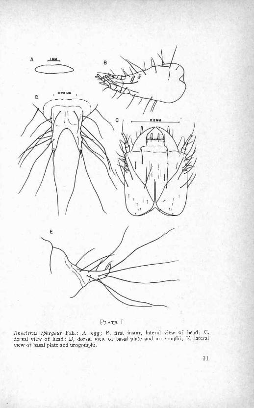
E
PLATE I
Enoclerus sphegeus Fab.: A, egg; B, first instar, lateral view of head; C,dorsal view of head; D, dorsal view of basal plate and urogomphi; E, lateralview of basal plate and urogomphi.
to 0.72 mm. Anterior width of prothoracic shield, about 0.72 mm.Width of basal plate of urogomphi, about 0.35 mm. Form orthoso-matic; abdomen membranous, pale pink, with a few long, strong,scattered setae; ten abdominal segments with segments four to sevenslightly wider; ninth abdominal segment crescentiform, dorsally witha basal plate and paired urogomphi; tenth segment located below theninth, developed as a locomotive organ, with an ambulatory wart andanal opening; ambulatory wart surrounded anteriorly by four smallpapillae and posteriorly by one large, liplike lobe; papillae indistinct.
Head prognathous, exserted or slightly inserted; dorsal sur-face somewhat flattened, ventral surface somewhat convex, lateralmargins parallel; as wide as long; with scattered setae, majority samelength as mandible; heavily sclerotized, dark brown. Frons triangular,delimited by slightly curved frontal sutures which posteriorly forman acute angle; medially and internally with a well sclerotized, darkbrown longitudinal endocarnia, one-half the length of head, branch-ing posteriorly to form a "Y"; two parallel, slightly elevated ridges,one on each side of anterior portion of endocarina. Epicraniumdorsally separated by frons into two epicranial halves, ventrallyseparated by an elongate, rectangular gula; slightly rugose; a verysmall tubercle located dorsally on each epicronial half near the middle-posterior portion of the frontal sutures. Ocelli located on epicraniumbehind ventrolateral part of antennal ring; arranged in an ante-rior row of three and a posterior row of two; rows parallel. Clypeusand labrum lightly sclerotized, yellow ocher, not distinct; anteriormargin of labrum with a row of very small setae and posteriorlywith a row of four to six longer setae. Antenna lightly sclerotized,yellow ocher; projecting from an antennal ring; extending beyondanterior margin of labrum; basal membrane large, whitish, trans-parent, and enclosing about one-half of basal segment; basal segmenttwice as long as second segment; second segment with a small ap-pendix, three setae around distal margin; apical segment cylindrical,about three-fourths the length of basal segment, apex with one longseta surrounded by three very short setae. Mandible subtriangular,apex pointed, about three-fourths the length of frons, width at baseslightly more than one-half length of mandible; retinaculum slightlycloser to apex than to base of mandible; two short setae on the lateralmandibular face; with a longitudinal groove on the ventral surface.Ventral mouthparts protracted with distal half directed obliquely up-wards; lightly sclerotized, yellow ocher.
Prothorax dorsally with a tergal shield or plate; heavily sclero-tized, dark brown; anterior margin straight, curving ventrocephalad;rounded, posteriorly oblique side margins which end at the dorsal

notch; a longitudinal endocarnia starting from dorsal notch and con-tinuing medially and anteriorly for three-fourths of the length ofprothoracic plate, dark brown; long setae around outer margin withshorter setae internally. Ventrally, with a pair of subtriangular,presternal plates, one on each side of a narrow, lanceolate, sternalplate; all plates lightly sclerotized and very light yellow ocher orlight brown.
Mesothorax dorsally with two, subtriangular plates; one smallseta on each; ventrally with a very small, obtuse plate posterior toprothoracic lanceolate plate; a small, oblong plate posterior to obtuseplate.
Metathorax dorsally with two pentagonal plates ; plates smallerand farther apart than mesothoracic plates; one small seta on each;ventrally with a small, oblong plate. Mesothoracic and metathoracicdorsal plates lightly sclerotized and light brown, less distinct on theliving specimen than the prothoracic dorsal plate.
Thoracic legs five-segmented, no free claws.Spiracles annular-biforous, located laterally on the mesothorax
and abdominal segments one to eight; mesothoracic spiracle slightlylarger than abdominal spiracles; metathoracic spiracle, rudimentary.
Basal plate of urogomphi lying at an angle on ninth abdominalsegment; not well defined, fading into and surrounded by a lightlysclerotized, yellow ocher, crescent-shaped sclerite; length about one-half as long as frons, slightly wider than long; lightly sclerotized,dark brown ocher. Urogomphi subconical; directed slightly upwards,markedly divergent at apex, apex not recurved; outer marginsunequal, inner margins equal; about as long as length of basal plate;heavily sclerotized, dark brown. A few scattered setae on basal plate,about as long as width of basal plate; setae on urogomphi morenumerous and up to three times as long as urogomphi.
Second instar: (Plate II, Figures A to C). Maximum length oflarva, about 17 mm; minimum length, about 14 mm. Maximum widthof body, about 1.5 mm; maximum thickness of body, about 1.3 mm.Average width of head capsule, 0.99 mm; range of width of headcapsule, 0.80 mm to 1.02 mm. Anterior width of prothoracic shield,about 1.04 mm. Width of basal plate of urogomphi, about 0.69 mm.Abdomen bright pink to light lilac color at maturity; dorsally thelilac color appearing as a mottled pattern with intervenient areas andventral side being whitish lilac; setae more abundant. Head darkbrown to black; dorsal epicranial tubercles becoming more prominent;apical segment of antenna about same length as second segment;anterior row of ocelli subparallel to slightly curved in relation toposterior row. Prothoracic shield darker brown. Basal plate of uro-
13

A B0.0 MM
0.5 MM
PLATE 11
Enoclerus sphegeus Fab.: A, second instar, dorsal view of basal plate andurogomphi ; B, second instar, lateral view of basal plate and urogomphi ; C,second instar, lateral view of head; D, third instar, dorsal view of basal plateand urogomphi ; E, third instar, lateral view of basal plate and urogomphi ;F, third instar, lateral view of head.
gomphi well defined; anterior margin with a thin, subparallel, lightlysclerotized sclerite; length slightly less than one-half as long as frons,wider than long; heavily sclerotized, dark brown ocher. Urogomphisubconical; directed distinctly upwards, slightly divergent at apex,apex sharply recurved and turned slightly inwards; outer marginsunequal, inner margins slightly unequal; about two-thirds to samelength as basal plate; heavily sclerotized, dark brown. Setae morenumerous on basal plate and urogomphi, scattered, about as longas width of basal plate; few small.
Third instar: (Plate IT, Figures D to F; Plate III, Figures Ato G; Plate IV, Figures A and B; Plate V, Figure D). Maximumlength of larva, about 22 mm; minimum length, about 15 mm. Maxi-mum width of body, about 3.0 mm; maximum thickness of body,about 2.7 mm. Average width of head capsule, 1.53 mm; range ofwidth of head capsule, 1.40 mm to 1.72 mm. Anterior width of pro-thoracic shield, about 1.77 mm. Width of basal plate of urogomphi,about 1.19 mm. Abdomen dorsally light lilac to deep lilac color atpupation; ventrally more bluish gray; lilac color appearing as amottled pattern with intervening areas very light blue; thorax morepinkish; setae very numerous and of varying lengths. Head darkbrown to black. Frons with two circular, cushionlike elevations, oneat base of each mandible; also with four parallel, slightly elevatedridges, two on each side of endocarina; posterior portion of fronsrugose. Dorsal epicranial tubercles very large and prominent; epi-cranial halves very rugose; anterior and posterior rows of ocelli sub-parallel. Labrum and clypeus distinct. Antenna brown; basal segmentabout two to three times as long as second segment; apical segmentabout two-thirds the length of second segment. Metathoracic plateseach bearing one long centrally located seta surrounded by foursmaller setae. Basal plate of urogomphi about one-half the length offrons, twice as wide as long. Urogomphi not divergent at apex, apexturned more inwards; outer and inner margins more nearly equal;about the length of basal plate; heavily sclerotized, dark brown withapex and margins black; setae numerous, a few small, with remainderas long as urogomphi. The four small, anterior papillae of ambula-tory wart very distinct.
Pupa: (Plate V, Figure B). Total length 8 mm to 10 mm; typi-cally exarate; abdomen with nine visible segments dorsally and sevenventrally; head, thorax, and abdomen pink with white appendages,abdomen becoming lilac at maturity; a few, small, strong, scatteredsetae on head, legs, and abdomen; antennae bent downward alongpleura; lateral spiracles on abdominal segments one to seven; apicesof wings subequal, extending to middle of fourth abdominal segment;
15

q
1
li
lp,
05 MM
C
PLATE III
Enoclerus sphegeus Fab., third instar: A, dorsal view of head; B, dorsal viewof mandible ; C, ventral view of mandible ; D, mesothoracic leg ; E, ventral viewof head; F, dorsal view of antenna; G, abdominal spiracle

T r - },x.\'A
PLATE IV
Enoclerus sphegeus Fab., third instar: A, dorsal view of complete larva;ventral view of complete larva

PLATE V
A, ventral view of pupa of Enoclerus lecontei Wolc.; B, ventral view of pupaof Enoclerus sphegeus Fab. ; C, third instar, lateral view of complete larva ofEnoclerus sphegeus Fab. ; D, ventral view of pupa of Thanasimus undatulusSay.
18

apices of prothoracic tarsi extending to apex of thorax, those ofmesothoracic tarsi to apex of second abdominal segment, those ofmetathoracic tarsi to apex of fifth abdominal segment; anal cercipresent.
Cleridae: Enoclertiis lecontei WolcottThe adult was originally described by LeConte in 1861 as Clerus
nigriventris from specimens collected near Fort Colville, Washington,and from the Bitterroot Valley, Montana (2, p. 33; 17). Schenklingin 1910 placed LeConte's nigriventris in the genus Thanasimus andcalled it T. nigriventris The species was redescribed by Wolcott asClerus lecontei in 1910.1 Wolcott (4) stated, "The name nigriventrisis preoccupied in Clerus by C. nigriventris Blanchard (an Argentinaspecies described in 1842). Should the species be reassigned to thegenus Clerus, the specific name of lecontei is suggested for it." Inthe same year, the genus Clerus was changed to Enoclerus (12).Boving (4) in 1928 called the clerid Enoclerus lecontei Wolc. Theclerid was referred to as Thanasimus lecontei (Wolc.) by Person in1940 (17), but he recently concluded that this was a mistaken identi-fication.2 Blackwelder in 1945 cited the species as T. nigriventris(Lee.) and gave lecontei Wolc. as a synonym.' Apparently, Black-welder missed Blanchard's species nigriventris because there is nomention of it in his checklist. The species was called Enocleruslecontei Wolc. by Wolcott in his catalog of 1947 (28, p. 79). Black-welder (3, p. 14) in 1948 followed Wolcott. Apparently the correctname at present is Enoclerus lecontei Wolcott.
The mature larva was described and illustrated by Boving (4)in 1928 from material collected from Ips galleries near Coeur d'Alene,Idaho. The following is the first known description and illustrationof all immature stages of this species. One generation was rearedin the laboratory from adults collected in Oregon.
The known distribution of this species extends from BritishColumbia to Michigan and south to Guatemala (16, p. 84). It hasbeen reported mainly as an enemy of bark beetles infesting Pinus.Bedard (1, p. 36) reports that it was associated with the Douglas-firbeetle, Dendroctonus pseudotsugae, in Montana. He states, "Thescarcity of this insect in Douglas-fir prohibits it from being of anygreat value in the control of the Douglas-fir beetle." The clerid wasnot observed to be associated with the Douglas-fir beetle in southern
1 Personal communication, P. 1. Spangler, United States National Museum,Washington, D C
2 Personal communication, H. L Person, Foreign Forestry Services, Wash-ington, D. C.

Idaho. As far as is known, there has been no earlier report of thepresence of this species in association with D. pseudotsugae inwestern Oregon. Again, it is believed that E. lecontei is of no greatimportance in controlling the Douglas-fir beetle in this region. Theclerid seems to be more abundant in association with the smaller barkbeetles, such as Scolytus unispinosus Lec. and various species of thegenus Pseudohylesinus Its life cycle appears to be similar to E.sphegeus in Oregon.
Descriptions of immature stages. The egg, three larval in-stars, and pupa are described.
Egg: (Plate VI, Figure A). Average length, 1.3 mm; range inlength, 1.2 mm to 1.6 mm. Average width, 0.4 mm; range in width0.3 mm to 0.4 mm. Form subcylindrical, with a slightly curved longi-tudinal axis, tapering slightly at each end, broadest at the middle,a little wider at one end than at the other; margins equal; chorionsmooth, shining, transparent, without ornamentation, becomingslightly rugose and dull as the embryo approaches maturity; color atfirst pale pink, with development gradually becoming brighter pinkwith translucent or opaque white portions at each end and lateralmargins.
First instar: (Plate VI, Figures B to E). Maximum length oflarva, 2.18 mm; minimum length, 1.34 mm. Maximum width of body,0.37 mm; maximum thickness of body, 0.34 mm. Average width ofhead capsule, 0.27 mm; range of width of head capsule, 0.23 mm to0.30 mm. Anterior width of prothoracic shield, about 0.29 mm. Widthof basal plate of urogomphi, about 0.18 mm. Form orthosomatic;abdomen membranous, very pale pink, with long, strong, scatteredsetae; ten abdominal segments with segments four to six slightlywider; ninth abdominal segment somewhat crescentiform, dorsallywith a basal plate and paired urogomphi; tenth segment located belowthe ninth, developed as a locomotive organ, with an ambulatory wartand anal opening; papillae of ambulatory wart indistinct.
Head prognathous, exserted or slightly inserted; dorsal surfaceflattened to slightly concave, ventral surface and lateral marginsslightly convex; as wide as long; with scattered setae, majority samelength as mandible; heavily sclerotized, light brown. Frons triangular,delimited by slightly curved frontal sutures which posteriorly forman acute angle; medially and internally with a well sclerotized, darkbrown longitudinal endocarnia, two-thirds length of head, branchingposteriorly to form a "Y." Epicranium dorsally separated by fronsinto two epicranial halves, ventrally separated by an elongate, rec-tangular gula; epicranial halves without tubercles. Ocelli located on

A 1MM
coC
0.25 MM
0.25 MM
PLATE VI
Enocicrus lecontci Wole.: A, egg; B, first instar, lateral view of head; C, firstinstar, dorsal view of head; D, first instar, lateral view of basal plate andurogomphi ; E, first instar, dorsal view of basal plate and urogomphi ; F, secondinstar, lateral view of basal plate and urogomphi; G, second instar, dorsal viewof basal plate and urogomphi; H, third instar, dorsal view of basal plate andurogomphi; I, third instar, lateral view of basal plate and urogomphi; J, thirdinstar, ventral view of mandible; K, third instar, dorsal view of mandible; L,third instar, lateral view of head.
21

epicranium behind ventrolateral part of antennal ring; arranged inan anterior row of three and a posterior row of two; rows parallel.Clypeus and labrum not distinct; anterior margin of labrum with arow of small setae. Antenna slightly sclerotized, light yellow ocher ;projecting from an antenna] ring; extending beyond anterior marginof labrum; basal membrane large, whitish, transparent, and en-closing about one-half of basal segment; basal segment about threeto four times as long as second segment; second segment with asmall appendix, three setae around distal margin; apical segmentcylindrical, same length as second segment, apex with one long seta.Mandible subtriangular, apex pointed, about three-fourths the lengthof frons, as wide at base as long; retinaculum near middle ofmandible; two short setae on the lateral mandibular face; with alongitudinal groove on the ventral surface. Ventral mouthparts pro-tracted with distal half directed somewhat obliquely upwards; lightlysclerotized, yellow ocher.
Prothorax dorsally with a tergal shield or plate; heavily sclero-tized, light brown; anterior margin straight, curving ventrocephalad;rounded, posteriorly oblique side margins which end at the dorsalnotch; a longitudinal endocarnia starting from dorsal notch andcontinuing medially and anteriorly three-fourths length of prothor-acic plate, (lark brown; with scattered setae. Ventrally, with a pair ofsubtriangular, presternal plates, one on each side of a narrow, lan-ceolate, sternal plate; all plates lightly sclerotized and very lightyellow ocher; not too distinct on living specimen.
Mesothorax dorsally with two subtriangular plates; one smallseta on each; ventrally with a very small, obtuse plate posterior toprothoracic lanceolate plate; a small, oblong plate posterior to theobtuse plate; ventral plates not distinct on living specimen.
Metathorax dorsally with two subquadrangular plates; platessmaller and farther apart than mesothoracic plates; one small setaon each; ventrally with a small, nondistinct, oblong plate. Meso-thoracic and metathoracic dorsal plates lightly sclerotized and lightbrown, less distinct on the living specimen than the prothoracic dorsalplate.
Thoracic legs five-segmented, no free claws.Spiracles annular-biforous, located laterally on the mesothorax
and abdominal segments one to eight; mesothoracic spiracle slightlylarger than abdominal spiracles; metathoracic spiracle, rudimentary.
Basal plate of urogomphi lying at an angle on ninth abdominalsegment; surrounded by a lightly sclerotized, yellow ocher, crescent-shaped sclerite; length slightly shorter than frons, wider than long;lightly sclerotized, light brown ocher. Urogomphi subconical; strongly

divergent at apex, apex not recurved; outer and inner marginsunequal; slightly longer than length of basal plate; heavily sclero-tized, light brown. A few scattered setae on basal plate, about aslong as basal plate; setae on urogomphi more numerous and up tofour times as long as urogomphi.
Second instar: (Plate VI, Figure F and G). Maximum lengthof larva, about 5 mm; minimum length, about 4 mm. Maximumwidth of body, about 0.80 mm; maximum thickness of body, about0.77 mm. Average width of head capsule, 0.47 mm; range of widthof head capsule, 0.37 mm to 0.52 mm. Anterior width of prothoracicshield, about 0.52 mm. Width of basal plate of urogomphi, about0.40 mm. Abdomen bright pink; setae more abundant. Head and pro-thoracic shield dark brown. Basal plate of urogomphi well defined;surrounded by a narrow, lightly sclerotized sclerite; length about two-thirds as long as frons, slightly wider than long; somewhat sculpturedwith fine subparallel depressions running length of plate; heavilysclerotized, light brown ocher. Urogomphi subconical; swollen atbase, not divergent at apex; apex not recurved but turned slightlyinwards; outer margins unequal, inner margins equal; length aboutone-half as long as basal plate; heavily sclerotized, dark brown. Setaescattered, slightly longer than length of basal plate.
Third instar: (Plate VI, Figures H to L; Plate VII, FiguresA to E). Maximum length of larva, about 11 mm; minimum length,about 8 mm. Maximum width of body, about 1.6 mm; maximumthickness of body, about 1.3 mm. Average width of head capsule,0.80 mm; range of width of head capsule, 0.77 mm to 0.82 mm.Anterior width of prothoracic shield, about 0.91 mm. Width of basalplate of urogomphi, about 0.75 mm. Abdomen bright pink, becominglilac color starting from the posterior end, very dark lilac at pupation;dorsally very dark mottled lilac pattern, ventrally more gray lilac;thorax more reddish brown; setae very numerous and about the samelength. Head very dark brown to black. Frons with two circular,cushion-like elevations, one at base of each mandible; also with twoparallel, slightly elevated ridges, one on each side of endocarnia.Anterior and posterior rows of ocelli subparallel. Labrum and clypeusdistinct. Basal plate of urogomphi surrounded by a narrow, lightlysclerotized sclerite; slightly less than one-half the length of frons,twice as wide as long; somewhat sculptured with irregular shapeddepressions scattered over plate; dark brown. Urogomphi subconicalto subtriangular; swollen from base to near apex; apex turnedsharply inwards; outer and inner margins unequal; length abouttwo-thirds as long as basal plate; very dark brown to black. Setaescattered, a few very small with remainder about one-half to three-
23

PLATE VII
Enoclerus lecontei Wolc, third instar : A, dorsal view of head ; B, ventral viewof head ; C, dorsal view of complete larva ; D, ventral view of complete larva
lateral view of complete larva

fourths width of basal plate. Ambulatory wart surrounded anteriorlyby four small papillae and posteriorly by one large lobe and analopening; papillae and lobe very distinct.
Pupa: (Plate V, Figure A). Total length, about 6 mm; typicallyexarate; abdomen with nine visible segments dorsally and sevenventrally; head and thorax pink, abdomen dark pink with mottledlilac areas; appendages white to gray; a few small, strong, scatteredsetae on head, legs, and abdomen; antennae bent downward alongpleura; apices of wings equal, extending to apex of fourth abdominalsegment; apices of prothoracic tarsi extending to apex of thorax,those of mesothoracic tarsi to middle of third abdominal segment,those of metathoracic tarsi to apex of fifth abdominal segment; analcerci present.
Cleridae : Thanasimus undatulus SayThe adult of this species was described by Say in 1835 (12, p.
149) as Thanasimus undatulus. Leng in 1920 (12, p. 149) listed thespecies as T. undulatus Say. In 1927 (13, p. 28) it was reported thatundulatus was a typographical error for undatulus. This insect wasmistakenly identified as T. dubius (Fab.) by Bedard (1, p. 28). Theforms of the species appear different throughout its geographicaldistribution. Barr states, "I prefer for the present to call everythingT. undatulus."3
It has been reported that the insect ranges from Alaska to NewMexico and eastward to New Hampshire (12, p. 149; 16, p. 82). Theclerid occurs occasionally in association with the Douglas-fir beetlein Montana, Idaho, and Oregon; its preferred prey are smaller barkbeetles such as various species of Pseudohylesinus, Scolytus, etc. Thelife cycle of this species appears to be similar to that of E. sphegeus,with the exception that the larvae do not migrate to the base of thetree. Two generations were reared in the laboratory from adultscollected in Idaho.
The following is the first known description and illustration ofall of the immature stages.
Descriptions of immature stages. The egg, three larval instars,and pupa are described.
Eggs: (Plate VIII, Figure E). Average length, 2.3 mm; range inlength, 2.0 mm to 2.2 mm. Average width, 0.5 mm; range in width,0.5 mm to 0.6 mm. Form subcylindrical, with a slightly curved longi-tudinal axis, tapering slightly at each end, broadest at the middle, oneend slightly wider than the other, margins equal; chorion smooth,
'Personal communication, W. F. Barr, University of Idaho, Moscow.

E
IYY
A 0.25 mm C , 0.5 YY
0.26 mm
PLATE VIII
Thanasimus undatulus Say : A, first instar, dorsal view of basal plate andurogomphi ; B, first instar, lateral view of basal plate and urogomphi ; C, secondinstar, dorsal view of basal plate and urogomphi ; D, second instar, lateral viewof basal plate and urogomphi; E, egg; F, first instar, lateral view of head; G,first instar, dorsal view of head; H, third instar, dorsal view of head.

shining, transparent, without ornamentation; color opaque whitethroughout development.
First instar: (Plate VIII, Figure A, B, F, and G). Maximumlength of larva, 3.77 mm; minimum length, 2.63 mm. Maximumwidth of body, 0.53 mm; maximum thickness of body, 0.50 mm.Average width of head capsule, 0.36 mm; range of width of headcapsule, 0.35 mm to 0.38 mm. Anterior width of prothoracic shield,about 0.40 mm. Width of basal plate of urogomphi, about 0.27 mm.Form orthosomatic; abdomen membranous, very pale pink, with afew, long, strong, scattered setae; ten abdominal segments with seg-ments four to seven slightly wider; ninth abdominal segment some-what crescentiform, dorsally with a basal plate and paired urogomphi;tenth segment located below the ninth, developed as a locomotiveorgan, with an ambulatory wart and anal opening; ambulatory wartsurrounded anteriorly by four small papillae and posteriorly by onelarge, liplike lobe; papillae indistinct.
Head prognathous, exserted or slightly inserted; dorsal surfaceflattened, ventral surface and lateral margins slightly convex; as wideas long; with scattered setae, majority same length as mandible;heavily sclerotized, dark brown. Frons triangular, delimited byslightly curved frontal sutures which posteriorly form an acute angle;medially and internally with a well sclerotized, dark brown, longi-tudinal endocarnia, one-third length of head, branching posteriorly toform a "Y." Epicranium dorsally separated by frons into two epi-cranial halves, ventrally separated by an elongate, rectangular gula;epicranial halves without tubercles. Ocelli located on epicranium be-hind ventrolateral part of antennal ring; arranged in an anterior rowof three and a posterior row of two; rows subparallel. Clypeus andlabrum lightly sclerotized, yellow ocher, not distinct; anterior marginof labrum with a row of very small setae. Antenna lightly sclerotized,yellow ocher; projecting from an antennal ring; extending beyondanterior margin of labrum; basal membrane large, whitish, trans-parent, and enclosing about one-fourth of basal segment; basal seg-ment about three times as long as second segment; second segmentwith a small appendix, three setae around distal margin; apical seg-ment cylindrical, about twice the length of second segment, apex withone long seta. Mandible subtriangular, apex pointed, about three-fourths the length of frons, width at base about two-thirds lengthof mandible; retinaculum slightly closer to apex than to base ofmandible; two short setae on the lateral mandibular face; with alongitudinal groove on the ventral surface. Ventral mouthparts pro-tracted with distal half directed obliquely upwards; lightly sclerotized,yellow ocher.
27
Prothorax dorsally with a tergal shield or plate; heavily sclero-tized, light brown, anterior margin straight, curving slightly ventro-caudad; rounded, posteriorly oblique side margins which end at thedorsal notch; a longitudinal endocarnia starting from dorsal notchand continuing medially and anteriorly three-fourths length of pro-thoracic plate, dark brown; with scattered setae. Ventrally, with apair of triangular, presternal plates, one on each side of a narrow,lanceolate, sternal plate; all plates lightly sclerotized and very lightyellow ocher.
Mesothorax dorsally with two subtriangular plates; three smallseta on each; ventrally with a very small, obtuse plate posterior toprothoracic lanceolate plate; a small, oblong plate posterior to obtuseplate.
Metathorax dorsally with two subquadrangular plates; platessmaller and farther apart than mesothoracic plates; two small setae oneach; ventrally with a small, nondistinct, oblong plate. Mesothoracicand metathoracic dorsal plates lightly sclerotized and light brown,less distinct on the living specimen than the prothoracic dorsal plate.
Thoracic legs five-segmented, no free claws.Spiracles annular-biforous, located laterally on the mesothorax
and abdominal segments one to eight; mesothoracic spiracle slightlylarger than abdominal spiracles; metathoracic spiracle rudimentary.
Basal plate of urogomphi lying horizontally on ninth abdominalsegment; anterior margin at times not well defined; two longitudinalgrooves extending length of plate; length about two-thirds as longas frons, wider than long; lightly sclerotized, light brown ocher.L rogomphi subcylindrical; markedly divergent at apex, apex notrecurved; outer margins slightly unequal, inner margins equal; abouttwo-thirds length of basal plate; heavily sclerotized, dark yellowbrown. A few scattered setae on basal plate, about as long as widthof basal plate; setae on urogomphi more numerous and up to threetimes as long as urogomphi.
Second instar: (Plate VIII, Figures C and D). Maximum lengthof larva, about 13 mm; minimum length, about 8 mm. Maximumwidth of body, about 2.0 mm; maximum thickness of body, about1.8 mm. Average width of head capsule, 0.63 mm; range of width ofhead capsule, 0.60 mm to 0.68 mm. Anterior width of prothoracicshield, about 0.69 mm. Width of basal plate of urogomphi, about0.56 mm. Abdomen bright pink to light lilac at maturity; lilac colorstarting to appear at posterior end and progressing anteriorly; dor-sally the lilac color appearing as a mottled pattern with intervenientareas and ventral side light blue; setae more abundant. Head andprothoracic shield dark brown. Basal plate of urogomphi lying at
28

an angle on ninth abdominal segment; well defined; surrounded by alightly sclerotized, light brown sclerite; a large depression, centrallylocated and extending length'of plate; length about as long as frons,wider than long; heavily sclerotized, dark brown ocher. Urogomphisubcylindrical; directed distinctly upwards, not divergent at apex,apex slightly recurved; outer margins unequal, inner margins equal;slightly more than one-half the length of basal plate; heavily sclero-tized, dark brown. Setae more numerous on basal plate and uro-gomphi, scattered, about as long as width of basal plate; few small.
Third instar: (Plate VIII, Figure H; Plate IX, Figures A toF; Plate X, Figures A to Q. Maximum length of larva, about19 mm; minimum length, about 11 mm. Maximum width of body,about 2.8 mm; maximum thickness of body, about 2.7 mm. Averagewidth of head capsule, 1.12 mm; range of width of head capsule,1.09 mm to 1.19 mm. Anterior width of prothoracic shield, about1.24 mm. Width of basal plate of urogomphi, about 0.99 mm.Abdomen dorsally lilac to deep lilac color at pupation; ventrally morebluish; lilac color appearing as a mottled pattern with intervenientareas light blue; thorax more whitish; setae very numerous and ofvarying lengths. Head dark reddish brown to black. Frons with twocircular, cushionlike elevations, one at base of each mandible ; alsowith four parallel, slightly elevated ridges, two on each side ofendocarnia. Posterior portion of head slightly rugose. Labrum andclypeus distinct. Antenna brown; basal membrane enclosing aboutone-half to two-thirds of basal segment; apical segment about aslong as second segment. Basal plate of urogomphi with several de-pressions extending length of plate; length three-fourths the widthof plate, and slightly shorter than frons; dark reddish brown. Uro-gomphi subcylindrical; apex sharply recurved and turned inwards;outer and inner margins unequal; about one-half the length of basalplate; very dark brown to black; setae numerous, few small, withremainder slightly less than length of urogomphi. Four small anteriorpapillae of ambulatory wart very distinct.
Pupa: (Plate V, Figure D). Total length, about 10 mm; typicallyexarate; abdomen with nine visible segments dorsally and sevenventrally; head and thorax pinkish, appendages white, abdomen darklilac; a few small, strong, scattered setae on head, legs, and abdomen;antennae bent downward along pleura; lateral spiracles on abdominalsegments one to seven; apices of wings subequal, extending betweenthe fourth and fifth abdominal segments; apices of prothoracic tarsiextending to apex of thorax, those of mesothoracic tarsi to apex ofthird abdominal segment, those of metathoracic tarsi to apex of fifthabdominal segment; anal cerci present.

A
io
0.25 MM
PLATE IX
Thanasimus undatulus Say, third instar : A, dorsal view of basal plate andurogomphi; B, lateral view of basal plate and urogomphi; C, lateral view ofhead; D, ventral view of head; E, dorsal view of mandible; F, ventral viewof mandible.

IYM
I
PLATE X
Thanasimus undatulus Say, third instar: A, dorsal view of complete larva; B,ventral view of complete larva ; C, lateral view of complete larva

a
17
Temrwochila zirdsce#&s was' described by Fabricius in 1775 (12,,p. 193). A subspecies, T. virescens chlorodia, was described in 1$431
by Mannerheim (12, p. 193). The mature larva of T. vi.rescens Fab.Was illustrated by Roving and Craighead in 1930 (6, pp. 272-274).Bedard (1, pp. 38-39) and Struble (23, pp. 100-101) made some very.general descriptions of the mature larva. Records indicate that T.virescens is. found in the eastern states, while T. viresceis ch rod'i
I
s
I t
uh
Ostomatidae : Temnochila virescens chlorodiaMannerheim
is found in the western states (12, p. 193).This species is very abundant and widely distributed in conif-
erous forests of North America. It is an important enemy of manyspecies of Scolytidae, principally Dendroctonus and Ips. Because ofits abundance and apparent aggressiveness, this insect can be ratedvery close to Enoclerus sphegus in importance as a predator ofD. pseudotsugae in the Intermountain Region. Its importance as apredator of D. pseudotsugae in the Pacific Northwest is less. Herethe species is less abundant and seems to be somewhat aggregated inits distribution through the forest. It has been observed that theinsect prefers more exposed, warmer sites of windthrown Douglas-fir in western Oregon. The life cycle of T. virescens follows closelythat of the clerids with the exception that the larvae do not migrate.
The following is the first known description and illustration ofthe immature stages of this species. The insect was found to be moredifficult to rear in the laboratory than the clerids. Apparently, thequantity of food, temperature, and humidity are very important inthe development of the larvae. The number of times the larvae moltedvaried considerably, but the maximum number of instars observedwas seven. There was a gradual increase in size of most structures,especially the head capsule, until the seventh molt. At that time thelarva were about the size of the fifth instars, and this fact may indi-cate that the usual number of instars under field conditions is aroundfive or six. There were no major morphological changes after thethird instar. Therefore, the drawings of the sixth instar representthe third to sixth instars with the exception of change in size. Nopupae were obtained. Two generations were reared in the laboratoryfrom adults collected in Idaho and Oregon.
Descriptions of immature stages. The egg and six larvalinstars are described.
Egg: (Plate XI, Figure A). Average length, 2.3 mm; range inlength, 2.2 mm to 2.5 mm. Average width, 0.4 mm; range in width,0.4 mm to 0.5 mm. Form cylindrical, with a slightly curved longi-
32

I
01;
li
/// \ 111 nI-,
F
33
0.25 MM
PLATE XI
Temnochila virescens chlorodia Mann : A, egg ; B, first instar, lateral view ofhead; C, first instar, dorsal view of head and thorax; D, first instar, dorsalview of basal plate and urogomphi ; E, first instar, lateral view of basal plateand urogomphi ; F, second instar, lateral view of basal plate and urogomphi

First instar: (Plate XI, Figures B to E). Maximum length oflarva, 3.42 nun; minimum length, 2.45 mm. Maximum width ofbody, 0.50 mm; maximum thickness of body, 0.44 mm. Aver-age width of head capsule. 0.42 mm; range of width of headcapsule. 0.40 nun to 0.43 mm. Anterior width of prothoracicplates, about 0.46 nom. Width of basal plate of urogomphi,about 0.28 mm. Form orthosomatic; abdomen membranous, gray
teriorly by one large, liplike lobe; papillae indistinct.Head prognathous, exserted or slightly inserted: dorsal surface
with small, scattered grooves, ventral surface somewhat flattened.lateral margins parallel; about as wide as long: scattered setae smallexcept for a few along the lateral margins of the epicranium whichare about as long as the mandible; heavily sclerotized, (lark reddishbrown. Frons triangular, delimited by slightly curved frontal sutureswhich posteriorly form an acute angle: medially and internally witha well sclerotized, (lark brown, longitudinal endocarnia, about one-third length of head, branching posteriorly to form a "Y." Epi-cranium dorsally separated by frons into two epicranial halves,ventrally separated by the retracted mouthparts and it short, broad},rata surrounded by a pair of paragula: rugose; epicranial halveswithout tubercles. Ocelli located on epicranium behind ventrolateral
sclerotized, yellow ocher, not distinct; anterior margin of labrumwith a row of very small setae. Antenna lightly sclerotized, yellowOcher; projecting from an antennal ring; extending beyond anteriormargin of labrum: basal membrane large, whitish, transparent, andenclosing about one-fourth of basal segment; basal segment one-halfthe length of second segment: second segment with a small appendix.three setae around distal margin; apical segment cyclindrical, about
tudinal axis, ends rounded; chorion smooth, dull, transparent, with-out ornamentation; color very pale, orange pink throughout develop-ment.
pink; setae strong and scattered, more abundant on ventral sidethan dorsal side, consisting of three lengths; all setae surrounded bya very small, black papilla, the longer setae raised on a small chalaza;ten abdominal segments with segments two to seven slightly wider;segments without dorsal ambulatory warts; ninth abdominal segmentsomewhat crescentiform, dorsally with a basal plate and pairedurogomphi; tenth segment located below the ninth, developed as alocomotive organ, with an ambulatory wart and anal opening; ambu-latory wart surrounded anteriorly by six small papillae and pos-
part of antennal ring; arranged in an anterior row of three and aposterior row of two, rows subparallel; a single ocellus locatedventrad of the two rows of ocelli. Clypeus and labrum lightly
34

three-fourths the length of second segment, apex with one long setasurrounded by two very short setae. Mandible subtriangular, apexdentate with distal ends pointed, about two-thirds the length of frons,width at base about two-thirds the length of mandible; retinaculumsituated near the middle of mesal edge of mandible; a penicillus onthe edge of a pseudomolar; mesal edge of mandible coarsely serrate;ventral surface somewhat grooved along the distal-mesal edge; twoshort setae on the lateral mandibular face. Ventral mouthparts re-tracted with distal half either horizontal or directed slightly down-wards; lightly sclerotized, yellow ocher.
Prothorax dorsally with two tergal shields or plates, superficiallyresembling one plate; heavily sclerotized, anterior-mesal portion ofplates light brown with the lateral margins and posterior portionsdark brown to black; anterior margins straight, curving ventro-cephalad; rounded, posteriorly oblique side margins which end atthe dorsal notch; a shallow depression running the length of eachplate; long setae around outer margins, with shorter setae internally;a pair of very small, black, subtriangular plates posterior to the largeprothoracic plates. Ventrally, three pairs of subtriangular, presternalplates, one pair on each side of a narrow, lancelolate, sternal plate;all plates lightly sclerotized, light brown.
Mesothorax dorsally with two, distinctly separated subquad-rangular plates; heavily sclerotized, dark brown to black with anteriormargin light brown; seven small setae on each plate; a pair of verysmall, black, subtriangular plates posterior to large, mesothoracicplates. Ventrally, spatulate plate posterior to the prothoracic lanceolateplate, lightly sclerotized, light brown; an oblong plate posterior tothe spatulate plate, lightly sclerotized, yellow ocher, not too distincton living specimen.
Metathorax dorsally with two subtriangular plates; platessmaller and farther apart than mesothoracic plates; four small setaeon each; heavily sclerotized, dark brown to black. Ventrally with asmall, obtuse plate; an oblong plate posterior to obtuse plate; lightlysclerotized, yellow ocher, not too distinct on living specimen.
Thoracic legs five-segmented, no free claws.Spiracles annular-biforous, located laterally on the mesothorax
and abdominal segments one to eight; mesothoracic spiracle slightlylarger than abdominal spiracles; metathoracic spiracle, rudimentary.
Basal plate of urogomphi lying at an angle on ninth abdominalsegment; well-defined; anterior margin sinuous; plate with two lightbrown tubercles, each with one seta; length about one-half as longas frons, twice as wide as long; heavily sclerotized, dark brown toblack. Urogomphi subcylindrical; directed slightly upwards, slightly
35

divergent at apex; apex not recurved, circular in cross-sectional view;outer and inner margins equal; about one-half the length of basalplate; heavily sclerotized, brown. Scattered setae on basal plate, afew small with remainder slightly longer than abdominal setae; setaeon urogomphi scattered, few small, with remainder up to eight timesas long as urogomphi.
Second instar: (Plate XI, Figure F; Plate XII, Figures A andB). Maximum length of larva, about 5.40 mm; minimum length,about 3.69 mm. Maximum width of body, about 0.77 mm; maximumthickness of body, about 0.64 mm. Average width of head capsule,0.72 mm; range of width of head capsule, 0.60 mm to 0.89 mm.Anterior width of prothoracic shields, about 0.80 mm. Width of basalplate of urogomphi, about 0.57 mm. Anterior margin of prothoracicplates straight, curving ventrocaudad. Mesothorax dorsally with twosubquadrangular plates; plates not very close together, superficiallyresembling one plate; anterior margin of plates with a lightly sclero-tized, yellow ocher sclerite; each plate with five small setae. Basalplate of urogomphi with the anterior-mesal portion of marginsinuous; lateral margins rounded, curving ventrocaudad; length aboutthree-fourths as long as frons, width nearly twice the length; setaemore numerous than on urogomphi; dark brown to black. Urogomphisubconical; not divergent at apex; apex turned sharply upwards andslightly inwards; outer margin unequal, inner margin equal; one-half the length of basal plate; with small setae, a few up to seventimes as long as urogomphi; brown with apex black.
Third instar: Average length of larva, about 10 mm. Maximumwidth of body, about 2.01 mm; maximum thickness of body, about1.68 mm. Average width of head capsule, 1.30 mm; range of width ofhead capsule, 1.22 mm to 1.37 mm. Anterior width of prothoracicshields, about 1.53 mm. Width of basal plate of urogomphi, about1.10 mm. Head capsule ovate; posterior portion of endocarnia ofhead branching to form a small rectangle; posterior portion ofepicranial halves nearly meeting each other. Basal segment of antennaabout three-fourths the length of second segment; apical segmentvery short. Clypeus and labrum light brown, distinct. Abdomen be-coming blue white. Dorsal ambulatory warts present on abdominalsegments two to seven; papillae on second abdominal, ambulatorywart not too distinct or numerous; papillae on remaining ambulatorywarts more distinct and numerous, arranged in two rows curvingventrocephalad; few papillae scattered on lateral margins. Ventralprothoracic plates light brown, distinct; ventral mesothoracic andmetathoracic plates yellow ocher, more distinct. Papillae of ventral,tenth abdominal, ambulatory wart distinct. Basal plate of urogomphi
36

crescentiform, anterior and lateral margins rounded, curving ventro-caudal; mesal portion slightly concave, somewhat sculptured with afew, irregularly shaped depressions; length about two-thirds thewidth of plate and slightly shorter than frons. Urogomphi subconical;directed distinctly upwards and turned slightly inwards; apex sharplyrecurved and pointed; outer margins unequal, inner margins slightlyequal; about one-third as long as basal plate; dark brown to black.
Fourth instar: Average length of larva, about 18 mm. Maximumwidth of body, about 2.35 mm; maximum thickness of body, about2.35 mm. Average width of head capsule, 1.46 mm; range of widthof head capsule, 1.24 mm to 1.64 mm. Anterior width of prothoracicshields, about 1.76 mm. Width of basal plate of urogomphi, about1.29 mm. Setae on mesothoracic and metathoracic plates nearly lack-ing. Two small papillae appearing, one on each side of dorsal mesalline of first abdominal segment. Papillae on second abdominal, ambu-latory wart more numerous and distinct.
Fifth instar: Average length of larva, about 17 mm. Maximumwidth of body, about 2.68 mm; maximum thickness of body, about2.52 mm. Average width of head capsule, 1.63 mm; range of widthof head capsule, 1.41 mm to 1.81 mm. Anterior width of prothoracicshields, about 1.95 mm. Width of basal plate of urogomphi, about1.43 mm. Abdomen becoming light blue gray.
Sixth instar: (Plate XII, Figures C to E; Plate XIII, FiguresA to H; Plate XIV, Figures A and B; Plate XV, Figure A). Aver-age length of larva, about 16 mm. Maximum width of body, about2.85 mm; maximum thickness of body, about 2.35 mm. Averagewidth of head capsule, 1.87 mm; range of width of head capsule,1.68 mm to 2.05 mm. Anterior width of prothoracic shields, about2.27 mm. Width of basal plate of urogomphi, about 1.02 mm. Epi-cranial halves of head capsule slightly rugose. First abdominalspiracle slightly larger than remaining abdominal spiracles. Abdomen,blue gray.
Dolichopodidae: Medetera spp.Specimens of adults from the Intermountain Region were identi-
fied by Dr. R. H. Foote, United States National Museum, as mostlyM. aldrichii Wh., but some were identified as M. sp. (near nigripesLev.) and M. sp. (near oregonensis Van Duzee).4 Specimens fromthe Northwest were identified by G. Steyskal, United States Depart-ment of Agriculture, as M. aldrichii Since the classification of this
'Personal communication, M. M Furniss, United States Forest Service,Ogden, Utah
37

0.25 mm
PLATE XII
Temnochila virescens chlorodia Mann : A, second instar, dorsal view of thorax;B, second instar, dorsal view of basal plate and urogomphi; C, sixth instar,dorsal view of basal plate and urogomphi ; D, sixth instar, lateral view of basalplate and urogomphi ; E, sixth instar, mesothoracic leg.

e
`02 um
C)
'A..
Vu
C.
1
t
I
7 Tom
'I
`
1
M,
OImm
IMM
0.5 MM
PLATE X I I I
Temnochila virescens chlorodia Mann., sixth instar: A, dorsal view of head;B, lateral view of head; C, ventral view of head; D, abdominal spiracle; E,dorsal view of antenna; F, chalaza and base of abdominal seta; G, ventralview of mandible: H, dorsal view of mandible.

PLATE XIV
Temnochila virescens chlorodia Mann., sixth instar: A, dorsal view of com-plete larva; B, lateral view of complete larva.

group of flies is in a state of revision, no effort was made to identifythe larvae down to the species level (10).
Species of this genus have been reported as being predaceouson many species of Scolytidae. mainly Ilcndroctonus, Scoh'tus, andI ps: and on Ceranlbvcidae, Ruprestidae, and Curculionidae. Recordsindicate that Al. aldrichii is widely distributed throughout the conif-erous forests of western United States and British Columbia (1,i, 9).
their prey fortuitously. Having no legs, the larvae are not very mobileand hence not very efficient in finding their hosts.
The observed life cycle of this insect is briefly as follows: Adultshave been observed moving very rapidly over the hark of trees whichthe Douglas-fir beetle in the Intermountain Region is initially invad-ing. The emergence of the fly in the Pacific Northwest appears to beconsiderably later than the initial attack by the bark beetle. Matingapparently takes place on the bark of infested material and the eggsare laid in small clusters under scales of the bark. Adults are presentthroughout the summer. There is only one generation per year withall three instars present throughout the summer (10). Pupation usu-ally occurs under the bark in spring.
Egg: Average length, 0.86 nun: average width, 0.18 mm, Formsuhcylindrical, with a slightly curved longitudinal axis, convex dor-sally, concave ventrally, pointed at one end: margins equal: chorionsmooth and shining: color at first pearly white, becoming brownishorange with development.
Maggots of this insect are very abundant under the bark of treesinfested by D. pseudotsugae. It is the most numerous of all of thepredators, and ranks second in number only to the parasite C. brun-neri. Despite its large numbers, this species is probably not so ef-fective as E. sphegeus in the control of the Douglas-fir beetle popula-tion. DeLeon (8, p. 73) reported that the larvae of Al. aldrichii find
The following descriptions were taken from the literature onM. aldrichii Wh. by Bedard (1, pp. 40-42), DeLeon (8, pp. 62-67),and Hopping (9) with some additional observations. A drawing(Plate XV, Figure B) was made illustrating a complete, mature larvaof Medetera sp.
Descriptions of immature stages. The egg, three larval instars,and pupa are described.
First instar: Average length of larva, about 0.7 mm. Formslender, cylindrical; creamy white in color; metapneustic; number ofbody segments not determined; no sclerotized areas on the head orfirst thoracic segment.
41

B C
PLATE XV
A, sixth instar, ventral view of complete larva of Temnochila virescens chlor-odia Mann. ; B, mature larva, lateral view of complete larva of Medetera sp.;C, mature larva, lateral view of complete larva of Lonchaea sp

Second instar: DeLeon (8, p. 62) states, "The second instar wasnot observed for this species. The larvae of Al. signaticornis Loew,which were studied by the writer with greater care, went throughthree instars, and it is probable that there is the same number forM. aldrichii. The second instar of the former species resembles thefirst instar in lacking the sclerotized areas of the head and firstthoracic segment." Johnsey observed a second instar for Medeterasp. associated with Dendroctonus pseudotsugae in Washington andOregon. Again, the sclerotized areas of the head and first thoracicsegment were not present (10).
Third instar: (Plate XV, Figure B). Average length, 8.5 mm.Form cylindrical with a slightly curved longitudinal axis, taperingslightly at each end with the tapering more pronounced cephalad;abdomen with a rather rigid cuticle, glabrous (although DeLeonmentions very minute sensillae on the body segments), grayish white;three thoracic and eight abdominal segments; abdominal segmentsfive to ten are subdivided at the anterior margin, forming a narrowring on all sides except the ventral; tenth segment is about 1.4 timesas long as segment nine, and about 2.5 times as long as segment11; each of the abdominal segments, except the eighth, with a ventralpseudopod; eighth abdominal segment with the anal opening andabdominal spiracle.
Head subdivided, exserted; caudal region of the dorsum of theposterior region of the head with a brownish, sclerotized, oval plate.Anterior region of head protruding forward in the form of two laterallobes; lobes with the mandibles, mandibular plates, palpi, and othersensillae; a median spine extending anteriorly between the two lobes;internally and extending back into the mesothorax are two meta-cephalic and two tentorial black rods. Antennae apparently one-segmented, located on a pair of lateral lobes of the anterior headregion.
Anterior margin of prothorax dorsally with a small crescent-shaped, sclerotized plate.
Amphipneustic type of respiratory system; two pair of spiracles;cephalic pair small, located below the median lateral, longitudinalline and behind the median transverse line of the prothorax; caudalpair located in the center of the four posterior protuberances of theeighth abdominal segment, larger than the cephalic pair, morestrongly pigmented with brown, roughly oval in shape with two largeoval openings into the stigmatic chamber; dorsal and lateral areasaround the caudal pair of spiracles are slightly sclerotized with fourbranched spines located in this region close to the margin of thespiracles.
4

Eighth abdominal segment divided dorso-ventrally by a mediangroove and laterally by a transverse groove, thus forming four pro-tuberances; two ventral protuberances project beyond the dorsal onesso that the former can be seen when viewed from above; alsocloser together and more sharply tapered than the dorsal pair ofprotuberances.
Anus opens as a longitudinal slit on the ventral surface of, andslightly anterior to, the eighth abdominal segment, in a more or lesscircular padlike swelling of the cuticula.
Pupa: Average length, about 4.5 mm; average width, about2 mm; typically exarate; creamy white except for brownish sclero-tized areas. Pair of sclerotized respiratory spines or horns directlybehind the eyes with their bases extending parallel to the posteriormargin of the eyes beneath the cuticula; pair of spiracles or vestigialspiracles on all but seventh and eighth abdominal segments. Singletransverse row of spines located dorsally on all but eighth abdominalsegment. Anterior to each row of dorsal abdominal spines an areaof innumerable, minute, sharply pointed spines that diminish innumber towards anterior margin of each segment; laterad, spinesbecome smaller but are larger on the lateral swellings, forming dis-tinct scabrous areas. Eighth abdominal segment with a series of fiveto seven pair of elongated spines; an inner, much shorter pair; allbut inner and shortest pair hooked at end. Thorax with three pairof small spines; anterior spine in some instances doubled, so that onone side of pupa two spines may be present where there is normallyone. Wings extend to apex of second abdominal segment, apices ofprothoracic tarsi to apex of third abdominal segment, those of meso-thoracic tarsi to apex of fourth abdominal segment, those of meta-thoracic tarsi to apex of fifth abdominal segment. Usually pupates ina white, silky-lined pupal cell.
Lonchaeidae : Lonchaea spp.Adult specimens from the Intermountain Region were deter-
mined by Dr. C. W. Sabrosky of the United States National Museumand Dr. J. F. McAlpine of Ottawa, Canada. As in the case ofMedetera, there was more than one species involved. The Lonchaeahave been identified as L. sp. (near corticis Taylor) and L. sp. (nearwatsoni'Curran),' and those from the Northwest as belonging to theL. watsoni group (10).
Very little has been reported concerning this group of flies.Bedard (1, p. 49-49) included very brief descriptions and illustra-
s Personal communication, M. M Furniss.

tions of the egg, mature larva, and pupa of L. corticis. Species of thisgenus have been observed in association with D. pseudotsugae inDouglas-fir and D. engelmanni in Engelmann spruce in the Inter-mountain Region. It has been found under Douglas-fir bark in thePacific Northwest. Bedard (1, p. 47) in 1933 stated, "The larvae ofL. corticis unquestionably destroy much of the D pseudotsugaebroods, possibly even more than Medetera, as the larvae are moreabundant in the infested trees than those of the latter species." Fromobservations made in western Oregon, however, it appears that thelarvae of this fly are more scavengers than predators. There are twogenerations per year and the adults of the first generation begin toemerge during the last week of June.
The following descriptions were taken from the literature byBedard (1. p. 48-49) on L. corticis, with additional information fromobservation. A drawing (Plate XV, Figure C) of a complete larva isincluded.
Descriptions of immature stages. The egg, mature larva,and pupa are described.
Egg: Average length, 0.86 mm; range in length, 0.80 mm to0.88 mm. Average width, 0.17 mm; range in width, 0.15 mm to0.20 mm. Form subcylindrical, with a slightly curved longitudinalaxis, tapering at one end, tapered end truncated; margins unequal;chorion smooth and shining; color pearly white.
Mature larva: (Plate XV, Figure C). Average length, aboutmm. Form wedge-shaped or muscidiform, with a slightly curved
longitudinal axis, tapering gradually to a sharply pointed cephalicend; creamy white and glabrous; three thoracic and eight abdominalsegments; pseudopodia situated ventrally on second to eighth abdomi-nal segments; setulae present about margins of anal area on eighthabdominal segment.
Head may be retractile; with small antennae and buccal ap-pendages; mouthparts mainly internal, visible; anterior hooks black;posterior rods extending into the prothorax and mesothorax, darkbrown, fused at two locations, branching caudally.
Head and prothorax without sclerotized plates.Amphipneustic type of respiratory system; two pair of spiracles;
cephalic pair small, nine-lobed, located laterad and caudal on theprothorax; caudal pair located on dorso-caudal end of bluntly pointedeighth abdominal segment, containing three slits situated at rightangles to each other, heavily sclerotized on dorsal portion extendingoutward and upward to form a heavy, blunt, spine curving above eachspiracle.
45
Pupa: Average length, about 5.0 mm; typically coarctate;puparium reddish brown, average length, about 5.2 mm; pupa creamywhite, slightly shining; apices of wings extend to apex of third andlast abdominal segment; apices of prothoracic tarsi to middle of samesegment; apices of mesothoracic tarsi extend to apex of penultimateabdominal segment; those of metathoracic tarsi to middle of apicalabdominal segment.
Braconidae: Coeloides brunneri ViereckIdentification of the immature stages of Coeloides brunneri Vier.
was very briefly worked on by Bedard (1, p. 52). Recently, Ryan(21) published descriptions of the five larval instars.
This insect has been reported as being parasitic mainly onDendroctonus pseudotsugae (1, p. 51; 22). Ryan and Rudinsky (22,p. 755) reported that a small percentage of parasitism occurs onMelanophila drummondi. It is the most abundant of all of the preda-tors and parasites of D. pseudotsugae. In small trees and towards thetop of larger trees, C. brunneri can be very effective in parasitizinga large percentage of the Douglas-fir brood.
Virtually nothing is known about the biology of this species inrelation to the Douglas-fir beetle in southern Idaho, although adultshave been observed on the bark of infested trees in early July. Ryanand Rudinsky (22, p. 755) reported that C. brunneri in Oregon "suc-cessfully parasitizes the Douglas-fir beetle only when the host is inthe second, third, and fourth instars." There are three generationsper year in western Oregon with varied incidence of diapause. Theincidence of diapause increases as the season progresses, being only5% in the first generation, 45%o in the second one, and 95% in thethird.
Records indicate that the parasite is widely distributed fromBritish Columbia to California and eastward to Montana andColorado (15, p. 160).
The following descriptions were taken from the literature onC. brunneri by Bedard (1, p. 52) and Ryan (21). Illustrations (PlateXVI, Figures C and D) were redrawn from Ryan.
Descriptions of immature stages. The egg, five larval instars,and pupa are described.
Egg: Average length, 1.3 mm; average width at widest point,0.15 mm. Form cylindrical with a long tapering tail; widest portionapproximately one-fifth of distance from broad, rounded anteriorend; tail usually slightly curved and tapering to a small acute point;
46

A
0N
PLATE XVI
A, mature larva, lateral view of complete larva of Pteromalidae; B, maturelarva, frontal view of head of Pteromalidae ; C, fifth instar, frontal view ofhead of Coeloides brunneri Vier. ; D, fifth instar, lateral view of complete larvaof C. brunneri Vier.

chorion thin, elastic, translucent, without ornamentation; color white,ends becoming clear and transparent.
First instar: Average length, about 1.1 mm. Form cylindrical,tapering gradually towards caudal end; distinct head and 13 bodysegments; first 11 body segments with a prominent band of sensorysetae around the middle in addition to several smaller scattered setae;twelfth and thirteenth body segments have only a few scattered setae;body membranous and white.
Head exserted; lateral margins i ndorsal view nearly straight forapproximately two-thirds the length of head, converging slightlyanteriorly and merging with the broadly rounded cephalic margin;antennae conical in a cephaloteral position; labrum, maxillae, andlabium grouped about mouth opening as a cup-shaped, prominent,cephaloventral projection from the head capsule proper.
Spiracles circular; one pair on prothorax and abdominal seg-ments one to eight; all located slightly dorsad of midlateral line, thaton thoracic segment just cephalad of caudal margin of that segment,while those on the abdominal segments lie just caudad of the cephalicmargins of their respective segment.
Second, third, and fourth instars: Average length of second,third, and fourth instars, 1.4, 2.3, and 3.3 mm, respectively. Bodfusiform; no prominent bands of setae present, although from fourto eight individual setae aligned around center of each thoracic seg-ment; abdominal segments with single setae present laterad of mid-dorsal and midventral lines and at the midlateral position. Head cap-sule spherical; outline of head in dorsal view curved with widest partin the center; mouth parts do not project.
Fifth instar: (Plate XVI, Figures C and D). Average length,about 6 mm. Body fusiform to cyphosomatic; seven dorsal pro-trusile areas in the successive intersegmental areas, the most anteriorbetween the metathoracic and first abdominal segment; in livinglarvae the first eight abdominal segments have swellings in the mid-lateral areas; sensory setae slightly more numerous than precedinginstars; numerous cuticular spines covering the first 12 body seg-ments except along the intersegmental lines and on the dorsal pro-trusile areas, also not present on terminal body segment or on thehead except ventral surface. Head in frontal view obovate, con-stricted slightly below the midline by the hypostoma, a slight depres-sion on either side of the dorsal midline of the epicranium; bases ofantennae surrounded by a broadly elliptic antennal foramina; six pairof setae dorsad of mouth armature; labiobase with three pair ofsetae dorsally and many cuticular spines ventrally; lines between
48
Personal communication from M. M. Furniss' Personal communication, D. E Parker, United States Forest Service,
Ogden, Utah
mouthparts and parts of head capsule heavily sclerotized, stipitalsclerome between cardo and stipes.
Pupa: Average length, about 5.3 mm; typically exarate pupa;white in color, becoming darker with maturity; usually pupates ina white silken cocoon.
Pteromalidae: Roptrocerus eccoptogasteri Ratz., Ceci-dostiba burkei Craw., and C. dendroctoni Ash.
Adult specimens of this family which emerged from Douglas-fir slabs were determined by B. D. Burks, United States NationalMuseum.' The parasites were identified as Roptrocerus eccoptogasteriRatzeburg, Cecidostiba burkei Crawford, and C. dendroctoni Ash-mead.
R. eccoptogasteri has been reported as occurring in some ofthe Atlantic States and the Pacific Northwest. Its hosts are Dendroc-tonus and Ips (15, p. 549). C. burkei appears to parasitize species ofDendroctonus and occurs in the Pacific Northwest (15, p. 557).Records on C. dendroctonus state that this species is parasitic onDendroctonus, Ips, and Cylindrocopturus and occurs in West Vir-ginia and the Pacific Northwest (15, p. 557). These species are notvery abundant in association with D. pseudotsugae. The biology andhabits of this group are presumably similar to those of C. brunneri.
The following description is from characters by Michener (14,pp. 993-995), and Parker,' and from observation. Drawings (PlateXVI, Figures A and B) were made from preserved specimens.
Mature larva : (Plate XVI, Figures A and B). Body formsomewhat crescent-shaped to cyphosomatic, tapering sharply towardscaudal end; distinct head and 13 body segments; abdominal segmentsone to four wider than remaining abdominal and thoracic segments;no dorsal protrusile areas in the successive intersegmental areas; afew sensory setae located dorsally on thoracic and abdominal seg-ments; numerous cuticular spines scattered over the first 12 body seg-ments; six setae around anus; body membranous and white. Headexserted, in frontal view broadly obovate, constricted slightly belowthe midline by the hypostoma; short, conical antennae with basessurrounded by a broadly elliptic antennal foramina; lines betweenmouthparts and parts of head capsule not (or feebly) sclerotized,cardo and stipes fused or nearly so, without a conspicuous stipital
under the direction of J. A. Rudinskv. Specimens from the Inter-mountain Region were collected from the Boise National Forest andlayette National Forest in Idaho, while L. N. ]\-line was employedby the United States Forest Service, and Pacific Northwest specimenswere taken from the Marys Peak watershed near Corvallis, Oregon.
The authors acknowledge the assistance of D. H. Parker, UnitedStates Forest Service, Ogden, Utah, who originated the distribution
r L.I"
.7,
sclerome between them. Spiracles circular; one pair on mesothorax,metathorax and abdominal segments one to seven; all except firstabdominal spiracle located on midlateral line, that of first abdominalsegment slightly dorsad of midlateral line; all spiracles near cephalicmargin of their respective segment.
ACKNOWLEDGMENTSThis study is an outgrowth of a 1960 cooperative undertaking
between Oregon State University and the Intermountain Forest andRange Experiment Station, United States Forest Service, Ogden,Utah, on the distribution of the parasites and predators of theDouglas-fir beetle infesting standing trees. Financial support foridentification and description was provided by the Oregon Agricul-tural Experiment Station and National Science Foundation, grantNo. G-14296. The work was clone by research assistant L. N. Kline,
project; M. M. Furniss, of the same agency, who helped in planningand in the field; and P. O. Ritcher, head of the Department of Ento-mology, Oregon State University, who checked many of the de-scriptions.
REFERENCES CITED
1. Bedard, W. D., The Douglas-fir beetle, its seasonal history, biology, habits,and control. USDA, Forest Service, Forest Insect Field Station, Coeur d'Alene, Idaho, 1933 (Processed.)
2. Blackwelder, Richard E, Fourth supplement to the Leng Catalogue ofColeoptera of America, North of Mexico Mount Vernon, N. Y.: Frey-bourg, 1939.
3. Blackwelder, Richard E., and Ruth M. Blackwelder, Fifth supplement tothe Leng Catalogue of Coleoptera of America, North of Mexico. Burling-ton, Vt.: Lane Press, 1948
4. Roving, A G., The larvae of Enoclerus lecontei Wolcott and Callimerusarcufer Chapin, of the beetle family Cleridae. Proc. of the Ent. Soc. ofWash., 30:94-100, 1928.
5. Roving, A. G., and A. B. Champlain, Larvae of North American beetles ofthe family Cleridae. Proc U. S. Natl Museum, 57:575-649, 1920.
50

6 Roving, A. G., and F. C. Craighead, An illustrated synopsis of the principallarval forms of the order Coleoptera Entomologica Americana, 11:1-348,1930
7. DeLeon, Donald, The morphology of Coeloides dendroctoni Cushman(Hymenoptera: Braconidae), Jour. of the N Y. Ent. Soc., 42:297-317, 1934.
8. DeLeon, Donald, A study of Medetera aldrichii Wh (Diptera: Dolicho-podidae), a predator of the mountain pine beetle (Dendroctonus monticolaeHopk., Coleoptera : Scolytidae) Entomologica Americana, 15:59-91, 1935.
9 Hopping, George R, Notes on the seasonal development of Medetera al-drichii Wheeler (Diptera: Dolichopodidae) as a predator of the Douglas-firbark beetle, Dendroctonus pseudotsugae Hopkins The Canadian Ento-mologist, 79:150-153, 1947
10. Johnsey, Richard L., The biologies of two dipterous predators of Dendroc-tonus pseudotsugae Hopkins (Coleoptera: Scolytidae) in western Oregonand Washington Masters thesis, Oregon State University, Corvallis, 1964
It. Knull, Josef N., The checkered beetles of Ohio (Coleoptera : Cleridea)Ohio State Univ Studies, Ohio Biological Surv. Bull. 42, 1951.
12 Leng, Charles W., Catalogue of the Coleoptera of America, North ofMexico. Cambridge, Mass.: Cosmos Press, 1920.
13. Leng, Charles W., and Andrew T. Mutchler, supplement to Catalogue of theColeoptera of America, North of Mexico. Cambridge, Mass.: CosmosPress. 1927.
14 Michener, C D, Comparative morphological and systematic studies of beelarvae with a key to the families of Hymenopterous larvae. Univ. of KansasSci Bull, 35:987-1102, 1953.
15. Muesebeck, C. F. W., Karl V. Krombein, and Henry K. Townes, Hymenop-tera of America North of Mexico, USDA, Agriculture Monograph No. 2,1951.
16. Papp, Charles S., The Cleridae of North America, Part I, The geographicaldistribution of Cleridae of North America, north of the Panama Canal.Bull. Southern Calif. Acad. Sciences, 59:76-88, 1960
17. Person, Hubert L., The clerid Thanasimus lecontei (Wole.) as a factorin the control of the western pine beetle. Jour. of For., 38:390-396, 1940.
18. Peterson, Alvah, Larvae of Insects: An Introduction to Nearctic Species.Part I, Lepidoptera and Plant Infesting Hymenoptera Ann Arbor, Mich.:Edwards Brothers, 1959.
19. Peterson, Alvah, Larvae of Insects: An Introduction to Nearctic Species,Part II, Coleoptera, Diptera, Neuroptera, Siphonapera, Mecoptera, Tri-choptera. Ann Arbor, Mich.: Edwards Brothers, 1960.
20. Reid, R. W., The bark beetle complex associated with lodgepole pine slashin Alberta, Part III-Notes on the biologies of several predators with spe-cial reference to Enoclerus sphegeus Fab. (Coleoptera: Cleridae) and twospecies of mites. The Canadian Entomologist, 89:111-120, 1957.
21. Ryan, Roger B , Durations of the immature stadia of Coeloides brunneri(Hymenoptera: Braconidae) at various constant temperatures, with descrip-tions of the five larval instars. Annals of the Ent. Soc. of Amer., 55:403-409, 1962.
51

2
—
t I
:
22. Ryan, Roger B., and Julius A. Rudinsky, Biology and habits of the Douglas-fir beetle parasite, Coeloides brunneri Viereck (Hymenoptera: Braconidae),in western Oregon, The Canadian Entomologist, 94:748-763, 1962.
23 Struble, George R., The biology of certain Coleoptera associated with barkbeetles in western yellow pine, Univ. of Calif. Publications in Ent. 5(6)105-134, 1930.
24. Struble, George R, Biology of two native coleopterous predators of themountain pine beetle in sugar pine, The Pan-Pacific Entomologist, 18:97-107, 1942.
25. Struble, George R, Laboratory propagation of two predators of the moun-tain pine beetle. Jour. of Econ. Ent, 35:841-844, 1942.
26. Struble, George R., and L. H. Carpelan, External sex characters of twoimportant native predators of the mountain pine beetle in sugar pine. ThePan-Pacific Entomologist, 17:153-156, 1941.
27. Torre-Bueno, J. R., De La, ed., A Glossary of Entomology. Brooklyn,N. Y.: Brooklyn Entomological Society, 1950.
28. Wolcott, Albert B., Catalogue of North American beetles of the familyCleridae. Fieldiana : Zoology, 32:61-105. 1947.
52