prediction of alzheimer’s disease using multi-variants
TRANSCRIPT

Prediction of Alzheimer’s disease usingmulti-variants from a Chinese genome-wideassociation study
Longfei Jia,1 Fangyu Li,1 Cuibai Wei,1 Min Zhu,1 Qiumin Qu,2 Wei Qin,1 Yi Tang,1
Luxi Shen,1 Yanjiang Wang,3 Lu Shen,4 Honglei Li,5 Dantao Peng,6 Lan Tan,7 Benyan Luo,8
Qihao Guo,9 Muni Tang,10 Yifeng Du,11 Jiewen Zhang,12 Junjian Zhang,13 Jihui Lyu,14
Ying Li,1 Aihong Zhou,1 Fen Wang,1 Changbiao Chu,1 Haiqing Song,1 Liyong Wu,1
Xiumei Zuo,1 Yue Han,1 Junhua Liang,1 Qi Wang,1 Hongmei Jin,1 Wei Wang,1 Yang Lu,15
Fang Li,16 Yuying Zhou,17 Wei Zhang,18,19 Zhengluan Liao,20 Qiongqiong Qiu,1 Yan Li,1
Chaojun Kong,1 Yan Li,1 Haishan Jiao,1 Jie Lu21,22 and Jianping Jia1,23,24,25
Previous genome-wide association studies have identified dozens of susceptibility loci for sporadic Alzheimer’s disease, but few of
these loci have been validated in longitudinal cohorts. Establishing predictive models of Alzheimer’s disease based on these novel
variants is clinically important for verifying whether they have pathological functions and provide a useful tool for screening of dis-
ease risk. In the current study, we performed a two-stage genome-wide association study of 3913 patients with Alzheimer’s disease
and 7593 controls and identified four novel variants (rs3777215, rs6859823, rs234434, and rs2255835; Pcombined = 3.07 � 10–19,
2.49 � 10–23, 1.35 � 10–67, and 4.81 � 10–9, respectively) as well as nine variants in the apolipoprotein E region with genome-
wide significance (P55.0 � 10–8). Literature mining suggested that these novel single nucleotide polymorphisms are related to
amyloid precursor protein transport and metabolism, antioxidation, and neurogenesis. Based on their possible roles in the develop-
ment of Alzheimer’s disease, we used different combinations of these variants and the apolipoprotein E status and successively built
11 predictive models. The predictive models include relatively few single nucleotide polymorphisms useful for clinical practice, in
which the maximum number was 13 and the minimum was only four. These predictive models were all significant and their peak
of area under the curve reached 0.73 both in the first and second stages. Finally, these models were validated using a separate lon-
gitudinal cohort of 5474 individuals. The results showed that individuals carrying risk variants included in the models had a
shorter latency and higher incidence of Alzheimer’s disease, suggesting that our models can predict Alzheimer’s disease onset in a
population with genetic susceptibility. The effectiveness of the models for predicting Alzheimer’s disease onset confirmed the contri-
butions of these identified variants to disease pathogenesis. In conclusion, this is the first study to validate genome-wide association
study-based predictive models for evaluating the risk of Alzheimer’s disease onset in a large Chinese population. The clinical appli-
cation of these models will be beneficial for individuals harbouring these risk variants, and particularly for young individuals seek-
ing genetic consultation.
1 Innovation Center for Neurological Disorders and Department of Neurology, Xuanwu Hospital, Capital Medical University,National Clinical Research Center for Geriatric Diseases, Beijing, China
2 Department of Neurology, The First Affiliated Hospital of Xi’an Jiaotong University, Shaanxi, China3 Department of Neurology and Center for Clinical Neuroscience, Daping Hospital, Third Military Medical University, Chongqing,
China4 Department of Neurology, Xiangya Hospital, Central South University, Changsha, China5 Laboratory of Medical Neurobiology of Zhejiang Province, Zhejiang University School of Medicine, Zhejiang, China6 Department of Neurology, China-Japan Friendship Hospital, Beijing, China
Received February 14, 2020. Revised July 30, 2020. Accepted August 14, 2020.VC The Author(s) (2020). Published by Oxford University Press on behalf of the Guarantors of Brain.
This is an Open Access article distributed under the terms of the Creative Commons Attribution Non-Commercial License (http://creativecommons.org/licenses/by-nc/4.0/), which
permits non-commercial re-use, distribution, and reproduction in any medium, provided the original work is properly cited. For commercial re-use, please contact
doi:10.1093/brain/awaa364 BRAIN 2020: Page 1 of 14 | 1
Dow
nloaded from https://academ
ic.oup.com/brain/advance-article/doi/10.1093/brain/aw
aa364/5981992 by guest on 15 Novem
ber 2020
7 Department of Neurology, Qingdao Municipal Hospital, School of Medicine, Qingdao University, Shandong, China8 Department of Neurology, The First Affiliated Hospital, Zhejiang University, Zhejiang, China9 Department of Gerontology, Shanghai Jiaotong University Affiliated Sixth People’s Hospital, Shanghai, China
10 Department of Geriatrics, Guangzhou Huiai Hospital, Affiliated Hospital of Guangzhou Medical College, Guangzhou, China11 Department of Neurology, Shandong Provincial Hospital Affiliated to Shandong University, Shandong, China12 Department of Neurology, Henan Provincial People’s Hospital, Zhengzhou University People’s Hospital, Henan, China13 Department of Neurology, Zhongnan Hospital, Wuhan University, Hubei, China14 Center for Cognitive Disorders, Beijing Geriatric Hospital, Beijing, China15 Department of Geriatrics, The First Affiliated Hospital of Chongqing Medical University, Chongqing, China16 Department of Geriatric, Fuxing Hospital, Capital Medical University, Beijing, China17 Department of Neurology, Tianjin Huanhu Hospital, Tianjin, China18 Department of Neurology, Beijing Tiantan Hospital, Capital Medical University, Beijing, China19 Center for Cognitive Neurology, Beijing Tiantan Hospital, Capital Medical University, Beijing, China20 Department of Psychiatry, Zhejiang Provincial People’s Hospital, People’s Hospital of Hangzhou Medical College, Hangzhou,
Zhejiang, China21 Department of Radiology, Xuanwu Hospital, Capital Medical University, Beijing, China22 Department of Nuclear Medicine, Xuanwu Hospital, Capital Medical University, Beijing, China23 Beijing Key Laboratory of Geriatric Cognitive Disorders, Beijing, China24 Clinical Center for Neurodegenerative Disease and Memory Impairment, Capital Medical University, Beijing, China25 Center of Alzheimer’s Disease, Beijing Institute for Brain Disorders, Beijing, China
Correspondence to: Jianping Jia, MD, PhD Innovation Center for Neurological Disorders and Department of
Neurology, Xuanwu Hospital, Capital Medical University, National Clinical Research Center for Geriatric
Diseases, Changchun Street 45, Xicheng District, Beijing, China, 100053
E-mail: [email protected]
Correspondence may also be addressed to: Jie Lu, MD, PhD
Department of Radiology, Department of Nuclear Medicine, Xuanwu Hospital, Capital
Medical University, Changchun Street 45, Xicheng District, Beijing, China, 100053
E-mail: [email protected]
Keywords: Alzheimer’s disease; genome-wide association study; Chinese; predictive model; longitudinal cohort
Abbreviations: AUC = area under the curve; eQTL = expression quantitative trait loci; GWAS = genome-wide association study;SNP = single nucleotide polymorphism
IntroductionAlzheimer’s disease is the most common type of dementia
and is genetically complex with an estimated heritability of
60–80% (Gatz et al., 1997). Previous genome-wide associ-
ation studies (GWASs) of Alzheimer’s disease in Caucasian,
African-American, and Asian populations have identified
genetic risk variants in ABCA7, BIN1, CASS4, CD2AP,
CD33, CDK5RAP2, CELF1, CLU, COBL, CR1,
ECHDC3, EPHA1, EXOC3L2, FERMT2, HLA-DRB5,
HLA-DRB1, HS3ST1, INPP5D, KANSL1, MEF2C, MS4A,
NME8, PICALM, PM20D1, PTK2B, SLC10A2, SLC24A4,
SORL1, TREM2, and ZCWPW1 (Harold et al., 2009;
Lambert et al., 2009, 2013a; Seshadri et al., 2010;
Hollingworth et al., 2011; Naj et al., 2011; Guerreiro et al.,
2013; Miyashita et al., 2013; Reitz et al., 2013; Desikan
et al., 2015; Jun et al., 2016; Lacour et al., 2017; Miron
et al., 2018; Sanchez-Mut et al., 2018; Kunkle et al., 2019).
These variants affect several Alzheimer’s disease-related
processes, such as lipid metabolism, inflammation, innate
immunity, production and clearance of amyloid-b, and
endosomal vesicle recycling (Selkoe and Hardy, 2016).
However, few of the variants reported in Caucasians have
been identified in the Chinese population (Wang et al.,
2016). A recent whole genome sequencing study in a
Chinese population identified variants in GCH1 and
KCNJ15, in addition to the well-known apolipoprotein E
(APOE) locus; however, the sample size of this study was
relatively small (Zhou et al., 2018).
Recently, genetic predictive models have been established
for predicting the onset of Alzheimer’s disease using a poly-
genic risk score approach, which was used to reveal polygen-
etic contributions to Alzheimer’s disease risk of common
single nucleotide polymorphisms (SNPs) that show a disease
association but fail to meet the accepted P-value threshold
for genome-wide significance (Escott-Price et al., 2015,
2017a, b, 2019; Chouraki et al., 2016; Stocker et al., 2018;
Leonenko et al., 2019). These studies showed variable
results. Specifically, Escott-Price et al. reported that the area
under the curve (AUC) of their predictive models, which
included APOE, 480 000 SNPs, age, and sex as predictors,
was 0.78, whereas in their other study, the AUC of their
models including 420 000 SNPs and APOE as predictors
increased to 0.84 as the included individuals were patho-
logically but not clinically confirmed (Escott-Price et al.,
2015, 2017a). However, despite the high predictive accuracy
2 | BRAIN 2020: Page 2 of 14 L. Jia et al.
Dow
nloaded from https://academ
ic.oup.com/brain/advance-article/doi/10.1093/brain/aw
aa364/5981992 by guest on 15 Novem
ber 2020

of these polygenic risk score-based models, it may not be
easy to use these models in a clinical setting because an indi-
vidual may not carry so many risk variants. Thus, simple
and effective Alzheimer’s disease predictive models are
needed for use as tools to screen for the genetic risk of
Alzheimer’s disease, particularly in young individuals who
carry the risk variants.
The current study aimed to investigate novel Alzheimer’s
disease-related genetic variants in a GWAS, to establish pre-
dictive models based on these variants, and to validate the
models in a longitudinal cohort. This approach can be
applied for early intervention in individuals who are at a
risk of developing Alzheimer’s disease.
Materials and methods
Subjects
The two-stage GWAS study involved 3913 patients withAlzheimer’s disease and 7593 controls from a Chinese popula-tion. The cohorts used in the two stages were independent ofeach other. Patients with Alzheimer’s disease were recruitedfrom the outpatient memory clinics at the Department ofNeurology, Xuanwu Hospital, Capital Medical University,Beijing, China and 46 other participating hospitals across Chinafrom 2013 to 2018. All diagnoses of Alzheimer’s disease in thisstudy were based on the recommendations of the NationalInstitute on Aging and the Alzheimer’s Association workgroup(McKhann et al., 2011) or National Institute of Neurologicaland Communicative Disorders and Stroke and the Alzheimer’sDisease and Related Disorders Association criteria (McKhannet al., 1984), with an age-at-onset 560 years and no family his-tory of dementia. Controls were recruited from the aforemen-tioned medical centre hospitals. All controls were 560 years ofage, cognitively normal (without subjective memory complaints,a Mini-Mental State Examination score of 26–30, and ClinicalDementia Rating Scale score of 0), and free of any general or la-boratory evidence of diseases that could impact cognition.Demographic information was collected from each subject usinga structured questionnaire.
Furthermore, using associated SNPs from the GWAS data,predictive models of Alzheimer’s disease were generated by com-bining risk variants. To estimate the effectiveness of the predict-ive models, participants from a longitudinal cohort of the ChinaCognition and Aging Study (China COAST) (Jia et al., 2014)were selected. China COAST was a longitudinal study estab-lished in 2008 as a multicentre cohort study comprising normal,mild cognitive impairment-, and Alzheimer’s disease-affectedindividuals across 30 of 34 provinces in China with yearly fol-low-up. The inclusion criteria were as follows: (i) the individualwas cognitively normal 10 years ago at baseline with indicativeblood samples; (ii) the individual developed Alzheimer’s diseaseat the time of sample collection for the present study 10 yearslater; and (iii) the individual had a detailed clinical data profileincluding psychometric evaluation every year during follow-up.Finally, 5474 participants were recruited, from among which2358 developed Alzheimer’s disease and 3116 were cognitivelynormal in 2019 (Supplementary Table 1). The study wasapproved by the Ethical Committees of Xuanwu Hospital,
Capital Medical University. Written informed consent wasobtained from either the subjects or their legal guardiansaccording to the Declaration of Helsinki.
GWAS study
First stage
Genomic DNA was extracted from peripheral blood samplesusing a modified salting-out procedure (Nasiri et al., 2005). Inthe first stage, we performed genome-wide genotyping of 1679patients with Alzheimer’s disease and 2508 controls usingIllumina HumanOmniZhongHua-8 Bead Chips (Illumina). Aftergenotyping, systematic quality control analyses were conductedusing PLINK 1.90 software (http://www.cog-genomics.org/plink2) (Purcell et al., 2007; Chang et al., 2015). First, 118 sam-ples (84 patients with Alzheimer’s disease and 34 controls) wereomitted because of sample duplicates or cryptic relatedness(PI_HAT 4 0.1875, which is the identity-by-descent expectedbetween third- and second-degree relatives) (Ellingson andFardo, 2016), or low individual call rate (50.95). The remain-ing samples were assessed for population outliers and stratifica-tion in principal component analysis using EIGENSTRAT(Patterson et al., 2006). All non-autosomal variants wereexcluded from statistical analyses, as well as SNPs with a callrate 598%, minor allele frequency 50.01, and/or significantdeviation from Hardy-Weinberg equilibrium in controls(P51.0 � 10–4) (Supplementary Table 2). Following qualitycontrol processing, the genotypes of 765 144 SNPs in 4069Chinese individuals (1595 patients with Alzheimer’s disease and2474 controls) were further analysed.
Phasing and imputation were performed by SHAPEIT(Delaneau et al., 2011) and IMPUTE2 (Howie et al., 2009), re-spectively, and version 3 of the 1000 Genomes Project data wasused as the reference set (Genomes Project et al., 2012).Variants with r2 values 5 0.80 or impute information measures5 0.50 from IMPUTE2, missing frequency 4 0.02, deviationfrom Hardy-Weinberg equilibrium (P51.0 � 10–4), and minorallele frequency 5 0.01 were excluded from post-imputationquality control analysis. Logistic regression analysis of GWASdata was conducted before and after imputation to test the dif-ferences in allele dosage between cases with Alzheimer’s diseaseand controls under an additive genetic model, adjusted for sex,APOE status, age (defined as age-at-onset for cases and age-at-last exam for controls), and population substructure using thefirst two principal components with PLINK 1.90 software.Manhattan and quantile-quantile plots of the first stage beforeand after imputation and adjustments for sex and APOE statuswere generated using the R qqman package (Version 3.4.2,https://www.r-project.org/). Regional association plots were gen-erated via LocusZoom (http://locuszoom.sph.umich.edu/locuszoom/) (Pruim et al., 2010). Linkage disequilibrium plots of var-iants in chromosome 19 were generated using Haploview soft-ware (https://www.broadinstitute.org/haploview/haploview).Conditional analysis was performed to assess the independenceof the novel associations of the genotyped SNPs. In addition,stratified analysis was performed by gender and disease status.
Power calculations with Quanto software were applied to cal-culate the power of the results from the discovery stage(Gauderman et al., 2006). Alzheimer’s disease prevalence wasset to 3.21% in accordance with epidemiological studies ofAlzheimer’s disease in Chinese subjects aged 565 years (Jia
Predictive models for Alzheimer’s disease BRAIN 2020: Page 3 of 14 | 3
Dow
nloaded from https://academ
ic.oup.com/brain/advance-article/doi/10.1093/brain/aw
aa364/5981992 by guest on 15 Novem
ber 2020

et al., 2014). Parameters included outcome (disease), design (un-matched case-control ratio of 1:1.5), hypothesis (gene only),sample size (n = 1679 cases), significance (1.0 � 10–5, two-sided), mode of inheritance (log-additive), and population risk(0.0321).
Second stage
To replicate the first stage association results, the top 34 var-iants showing an association with a P51.0 � 10–5 after adjust-ing for age, sex, APOE status and the first two principalcomponents were selected and analysed as part of an independ-ent cohort of 7319 Chinese individuals consisting of 2234 caseswith Alzheimer’s disease and 5085 controls (Table 1 andSupplementary Table 1). These 34 SNPs were genotyped atBioMiao Biological Technology Beijing Co. using theMassArray System (Agena iPLEXassay).
Combined analysis of the first and second stages
To improve statistical power, a meta-analysis was applied tocombine the associated results from the first two stages usingMETAL (Willer et al., 2010) with an inverse variance-basedmodel. Heterogeneity tests between the two groups were per-formed using the Breslow-Day test (Higgins and Thompson,2002), and the extent of heterogeneity was assessed using the I2
and P-values of the Q statistics calculated by METAL (Higginset al., 2003).
Single nucleotide polymorphismannotation
SNPnexus was used for SNP annotation (https://www.snp-nexus.org/v4/) (Chelala et al., 2009; Dayem Ullah et al., 2012,2013, 2018). For co-localization of SNPs with significant associ-ations in both stages of the study, we conducted expressionquantitative trait loci (eQTL) analysis using the dataset pre-sented by Ramasamy et al. (2014), COLOC analysis (http://coloc.cs.ucl.ac.uk) (Giambartolomei et al., 2014) using thebrain-eQTL datasets (Trabzuni et al., 2011; Ramasamy et al.,2013), and summary Mendelian randomization-Heidi analysis(https://cnsgenomics.com/software/smr/) (Zhu et al., 2016) usingsummary eQTL data from the brain and blood (Westra et al.,2013; Lloyd-Jones et al., 2017; Qi et al., 2018). The expressionof novel Alzheimer’s disease-associated genes was analysedusing data from the National Center for BiotechnologyInformation Gene Expression Omnibus dataset (http://www.ncbi.nlm.nih.gov/geo). These included the expression of genes inthe frontal cortex, hippocampus, and temporal cortex of con-trols and patients with Alzheimer’s disease (SupplementaryTable 3). Prism software (version 8.0.0, GraphPad Software,Inc., CA, USA) was used to compare gene expression betweencognitively normal and Alzheimer’s disease groups (unpaired t-test and Welch’s t-test) and to generate figures. For theAlzheimer’s disease-associated genes in this study, STRING ana-lysis was performed to evaluate protein-protein interactions(Szklarczyk et al., 2015). Medium confidence (0.400) was usedas the minimum required interaction score and no more than 50interactors were shown in the first shell. The exported networkwas analysed using the bioinformatics software platformCytoscape (Version: 3.7.1, https://cytoscape.org/). In addition,we exported the Gene Ontology information, including molecu-lar function, biological process, and cellular component, as well
as the Kyoto Encyclopedia of Genes and Genomes (KEGG)pathways for the network, from STRING analysis.
APOE genotyping
The APOE genotypes for haplotypes derived from rs7412 andrs429358 in samples from both stages of the study were deter-mined using the Sanger sequencing method (Sanger et al.,1977).
Validation of gene associations inCaucasian populations
To determine whether there is underlying heterogeneity in thecontributors of genetic risk between Chinese and Caucasianpopulations, the eligible novel SNPs were examined in the datafrom ‘The International Genomics of Alzheimer’s Project sum-mary statistics from stage 1 data’ (Lambert et al., 2013b). Thegenetic correlation between the Chinese GWAS and publiclyavailable International Genomics of Alzheimer’s Project (IGAP)summary statistics was estimated using linkage disequilibriumscore regression implemented in the online software LD Hub(http://ldsc.broadinstitute.org/) (Zheng et al., 2017).
Predictive model study
We performed predictive modelling using the polygenic riskscore based on SNP significance in combined analysis andAPOE status as predictor variables, based on the data of thefirst stage. The individual polygenic risk scores were generatedas sums of the risk variants weighted by effect sizes derivedfrom logistic regression. We also ran the predictive analyses onsecond-stage data using the same factors. Furthermore, wetested different predictive models with different combinations ofSNPs in a population negative for APOE e4. Areas under thereceiver operating characteristic curve were calculated by com-paring the observed case/control status and polygenic risk scorecalculated using PRSice2 (Choi and O’Reilly, 2019) profiling ina standard weighted allele-dose manner.
To confirm the capacity of the models to predict Alzheimer’sdisease, we applied the models to individuals who wererecruited in a longitudinal study from 2009 to 2019. To esti-mate the effectiveness of the GWAS-based predictive models, bymeasuring the fraction of individuals living without Alzheimer’sdisease for a certain amount of time from baseline, survivalcurve analyses were performed using the follow-up data in thislongitudinal cohort.
Data availability
The data that support the findings of this study are available onrequest from the corresponding author.
Results
Demographics of three cohorts
In the GWAS, a total of 11 506 individuals participated
in this two-stage study, including 3913 patients with
Alzheimer’s disease and 7593 controls (Supplementary
4 | BRAIN 2020: Page 4 of 14 L. Jia et al.
Dow
nloaded from https://academ
ic.oup.com/brain/advance-article/doi/10.1093/brain/aw
aa364/5981992 by guest on 15 Novem
ber 2020

Tab
le1
Ass
ocia
tio
no
f13
SN
Ps
wit
hA
lzh
eim
er’
sd
isease
inth
efi
rst
an
dse
co
nd
stages
Ch
r.S
NP
Neare
st
gen
e
Po
siti
on
Min
or
allele
Fir
stst
age
Seco
nd
stage
Pco
mO
Rco
m
(95%
CI)
Ph
et
(1679
case
s,2508
co
ntr
ols
)(2
234
case
s,5085
co
ntr
ols
)
MA
F_C
A,
%
MA
F_C
L,
%
OR
Pgw
as
MA
F_C
A,
%
MA
F_C
L,
%
OR
Pre
5rs
3777215
RH
OBTB3,G
LRX
95122000
A12.9
17.1
0.6
99.3
4�
10
–8
15.5
20.9
0.6
91.3
1�
10
–13
3.0
7�
10
–19
0.6
9(0
.64–0.7
4)
0.7
2
5rs
6859823
CTC-2
78L1
.1105554384
T29.2
35.8
0.7
43.2
8�
10
–9
27.1
33.9
0.7
25.3
2�
10
–15
2.4
9�
10
–23
0.7
4(0
.69–0.7
8)
0.7
9
14
rs234434
CTD
-2506J1
4.1
97821020
G36.6
25.8
1.4
23.7
0�
10
–11
37.2
25.4
1.7
51.0
7�
10
–44
1.3
5�
10
–67
1.7
1(1
.61–1.8
2)
0.8
7
19
rs11668861
NECTIN
245380970
G33.1
22.7
1.7
61.7
5�
10
–10
34.9
22.4
1.8
62.9
9�
10
–52
4.1
3�
10
–63
1.8
1(1
.69–1.9
4)
0.3
2
19
rs6859
NECTIN
245382034
A40.9
31.6
1.5
52.9
2�
10
–8
43.7
32.6
1.6
19.6
7�
10
–35
3.7
0�
10
–42
1.5
8(1
.48–1.6
9)
0.4
8
19
rs3852860
NECTIN
245382966
C33.5
23.6
1.7
21.8
7�
10
–9
35.6
24.1
1.7
53.0
7�
10
–44
4.2
0�
10
–54
1.7
2(1
.60–1.8
4)
0.4
8
19
rs71352238
TOM
M40
45394336
C23.3
9.6
3.1
35.6
4�
10
–15
21.8
8.7
2.9
12.4
0�
10
–100
1.1
6�
10
–131
2.9
0(2
.66–3.1
7)
0.6
9
19
rs157580
TOM
M40
45395266
A53.9
45.4
1.4
81.1
0�
10
–6
54.9
46.6
1.4
01.6
0�
10
–19
2.9
9�
10
–25
1.4
0(1
.31–1.4
9)
0.8
6
19
rs2075650
TOM
M40
45395619
G23.3
9.4
3.2
73.0
5�
10
–18
25.2
9.4
3.2
55.3
0�
10
–132
2.5
7�
10
–164
3.1
7(2
.91–3.4
5)
0.3
6
19
rs157582
TOM
M40
45396219
T32.0
18.9
2.3
32.5
3�
10
–10
33.7
18.9
2.1
96.5
7�
10
–76
2.9
7�
10
–95
2.1
5(2
.00–2.3
1)
0.6
2
19
rs439401
APO
E45414451
C50.9
43.3
1.5
02.0
6�
10
–6
52.8
42.1
1.5
42.0
8�
10
–31
1.1
7�
10
–35
1.5
0(1
.40–1.5
9)
0.1
4
19
rs4420638
APO
C1
45422946
G27.2
12.2
2.9
82.6
2�
10
–17
28.5
11.0
3.2
03.4
0�
10
–140
8.3
2�
10
–171
3.0
7(2
.83–3.3
2)
0.1
3
21
rs2255835
CH
OD
L19491664
C33.6
27.7
1.3
38.6
1�
10
–6
26.2
23.0
1.1
96.7
8�
10
–05
4.8
1�
10
–9
1.2
3(1
.16–1.3
1)
0.5
1
Chr.
=ch
rom
oso
me;M
AF_
CA
=m
inor
alle
lefr
equency
ofca
ses;
MA
F_C
L=
min
or
alle
lefr
equency
ofco
ntr
ols
;Min
or
alle
le=
min
or
alle
leofth
eco
ntr
ols
;OR
=odds
ratios
calc
ula
ted
acco
rdin
gto
the
min
or
alle
le;O
Rcom
=th
eodds
ratio
of
the
com
bin
ed
anal
ysis
;Pcom
=th
eP-
valu
eofth
eco
mbin
ed
stag
eusi
ng
meta
-anal
ysis
;Pgw
as,
the
P-va
lue
ofth
efirs
tst
age
adju
sted
for
sex,A
POE
stat
us,
age
(defined
asag
eat
onse
tfo
rca
ses
and
age
atla
stexam
for
contr
ols
)an
dtw
oan
cest
rypri
n-
cipal
com
ponents
;P h
et
=th
eP-
valu
eofhete
roge
neity;
Posi
tion
=bas
epai
rposi
tion
acco
rdin
gto
hg
19;P r
e=
the
P-va
lue
ofth
ese
cond
stag
e.
Predictive models for Alzheimer’s disease BRAIN 2020: Page 5 of 14 | 5
Dow
nloaded from https://academ
ic.oup.com/brain/advance-article/doi/10.1093/brain/aw
aa364/5981992 by guest on 15 Novem
ber 2020

Table 1). In the first stage, the mean age at onset of 1679
patients with Alzheimer’s disease was 72.07 ±6.4 years,
of which 949 (56.5%) were female, whereas the mean
age at examination of 2508 controls was 72.95 ± 19.5
years, of which 1398 (55.7%) were female. In the second
stage, the mean age at onset of 2234 patients with
Alzheimer’s disease was 72.35 ± 7.2 years of which 1,253
(56.1%) were female, whereas the mean age at examin-
ation of 5085 controls was 69.22 ± 7.0 years, of which
2931 (57.6%) were female. In the longitudinal cohort,
5474 individuals were recruited, comprising 2358
patients with Alzheimer’s disease and 3116 controls
(Supplementary Table 1). The mean age at onset of 2358
patients with Alzheimer’s disease was 71.70 ±6.8 years,
of which 1351 (57.3%) were female, whereas the mean
age at examination of 3116 controls was 74.70 ± 7.5
years, of which 1676 (53.8%) were female. The flow
chart of the current study is shown in Fig. 1.
Results from genome-wide
association studies
In the first stage, 1679 patients with Alzheimer’s disease and
2508 controls were genotyped (Supplementary Table 1).
Overall, 765 144 autosomal SNPs passed the quality control
standards and were included for further analysis. Principal
component analysis confirmed that the patients with
Alzheimer’s disease and controls were well-matched
(Supplementary Fig. 1). A quantile-quantile plot indicated
that population stratification had negligible effects on the
statistical results (kGC = 1.08; Supplementary Fig. 2). After
adjusting for age, sex, and APOE status along with the first
two principal components, several markers on various chro-
mosomes exhibited genome-wide significance, with the senti-
nel markers occurring on chromosome 19 (Supplementary
Fig. 3). Power calculations indicated that the sample size
used in this GWAS provided sufficient statistical power to
detect Alzheimer’s disease-associated variants. In the second
stage, the top 34 SNPs with evidence of associations with
Alzheimer’s disease (Supplementary Table 4) were selected
for genotyping as part of an independent Chinese cohort of
2234 patients with Alzheimer’s disease and 5085 controls
(Supplementary Table 1). Of these 34 Alzheimer’s disease-
associated SNPs, 13 surpassed the Bonferroni correction
threshold (P5 1.47 � 10–3) with no detectable heterogen-
eity between stages. This included nine SNPs on chromo-
some 19 in the APOE region (Jun et al., 2012) (APOE
rs439401, APOC1 rs4420638, TOMM40 rs2075650,
Figure 1 Study flow chart. AD = Alzheimer’s disease.
6 | BRAIN 2020: Page 6 of 14 L. Jia et al.
Dow
nloaded from https://academ
ic.oup.com/brain/advance-article/doi/10.1093/brain/aw
aa364/5981992 by guest on 15 Novem
ber 2020

TOMM40 rs71352238, TOMM40 rs157582, TOMM40
rs157580, NECTIN2 rs11668861, NECTIN2 rs3852860,
and NECTIN2 rs6859; Table 1). The other four SNPs were
not located on chromosome 19 (rs3777215, rs6859823,
rs234434, and rs2255835; P = 1.31 � 10–13, 5.32 � 10–15,
1.07 � 10––44, and 6.78 � 10–05, respectively). In combined
analysis, all 13 of these SNPs showed associations exceeding
the genome-wide significance threshold (P5 5.0 � 10–8;
Table 1). Furthermore, all four novel variants remained sig-
nificant after adjusting for the effect of APOE e4. Stratified
analyses indicated that the 34 top SNPs in the first stage
remained significant after stratification by gender
(Supplementary Table 5). Conditional analyses indicated
that the four novel SNPs we identified were independent sig-
nals (Supplementary Table 6).
Functional annotation of the fournovel single nucleotidepolymorphisms
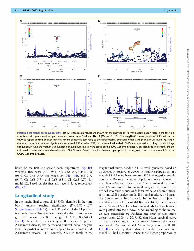
Variant rs3777215 was significantly associated with
Alzheimer’s disease [Pcombined = 3.07 � 10–19, odds ratio
(OR) = 0.69] and was located on chromosome 5 in the in-
tron regions of RHOBTB3 and GLRX (Fig. 2A, Table 1
and Supplementary Table 7). As shown in Fig. 3,
RHOBTB3 was significantly upregulated in the frontal cor-
tex, hippocampus, and temporal cortex of patients with
Alzheimer’s disease compared to cognitively normal individ-
uals (P50.05, 0.01 or 0.0001; Fig. 3A). GLRX showed
lower expression levels in the brain tissues of patients with
Alzheimer’s disease than in that of cognitively normal indi-
viduals (P5 0.05, 0.01 or 0.0001; Fig. 3B). Another SNP
detected on chromosome 5 was rs6859823 (Pcombined =
2.49 � 10–23, OR = 0.74; Fig. 2B and Table 1). Variant
rs6859823 was intergenic and located between RNA5SP189
and CTC-278L1.1 (Supplementary Table 7). RNA5SP189
and CTC-278L1.1 were both identified as pseudogenes
according to GeneCards and SNPnexus, and neither have
been previously reported to be associated with Alzheimer’s
disease or any other disease. SNP rs234434 (Pcombined =
1.35 � 10–67, OR = 1.71; Fig. 2C and Table 1) was inter-
genic and was between two long intergenic non-coding
RNAs known as RP11-359N5.1 and CTD-2506J14.1. SNP
rs2255835 was located on chromosome 21 in the intron re-
gion of CHODL and showed genome-wide significance
(Pcombined = 4.81 � 10–9, OR = 1.23; Fig. 2D and Table 1).
CHODL expression was higher in the hippocampus
(P5 0.05; Fig. 3C) and lower in the temporal cortex of
patients with Alzheimer’s disease compared to that in nor-
mal individuals (P5 0.05; Fig. 3C). Co-localization analyses
indicated that the four SNPs were related to various genes
being expressed in the blood and different brain regions
(Supplementary Tables 8–11). Roadmap epigenomics
showed that rs3777215 and rs2255835 were related to tran-
scriptional activation (H3K36me3, H3K4me1, and
H3K14ac) in neurons or neuronal progenitor cells, whereas
rs234434 was associated with transcriptional repression
(H3K27me3) in neurons (Supplementary Table 7). STRING
analysis demonstrated that the proteins encoded by the vari-
ant genes, except for CHODL, interact with APOE
(Supplementary Fig. 4). Gene Ontology enrichments of the
genes in the STRING network suggested that these genes are
involved in several biological processes. For example,
RHOBTB3 was suggested to be involved in the ‘establish-
ment of localization’ and ‘transport’ (Supplementary Table
12). Genes enriched in Gene Ontology cellular components
and molecular functions, as well as KEGG pathways, are
listed in Supplementary Tables 13–15.
Validation in Caucasian genome-wide association studies datasets
Overall, the APOE e4 allele frequency in Chinese subjects in
the present study was lower than that in Caucasians for
both, patients with Alzheimer’s disease and controls
(Supplementary Table 1). Despite these differences, nine
SNPs with significant associations in the APOE region in
the present study were either reported previously or found in
strong linkage disequilibrium with nearby SNPs in
Caucasians (Supplementary Fig. 5). One of the four novel
SNPs outside chromosome 19 reached genome-wide signifi-
cance in the IGAP stage 1 data for non-Asian populations
(rs6859823; Supplementary Table 16). Moreover, linkage
disequilibrium score regression analysis of Chinese GWAS
and publicly available IGAP summary statistics revealed a
genetic correlation of –0.14 (P = 0.73).
Predictive models
We tested 11 predictive models; four models were used to
analyse all populations (Fig. 4A–D) and seven models were
used to analyse subjects who were negative for APOE e4
(Fig. 4E–K) in this study. The number of SNPs in our mod-
els was relatively low, and even for the maximum, the num-
ber of SNPs for model A3 was only 13 with an AUC of
0.73 [95% confidence interval (CI): 0.70–0.75] in the first
stage. The AUCs of all 11 models were significant
(P5 0.05; range of AUC: 0.63–0.73), and the specific AUC
values for the 11 models are presented in Fig. 4. For model
A1, with the four novel SNPs found in this study and
APOE e4 status as predictors, training on the first-stage
data, prediction accuracy AUC = 0.69 (95% CI: 0.67–0.71)
was achieved based on a logistic regression model.
However, the prediction accuracy AUC reached 0.73 (95%
CI: 0.71–0.74) when using second-stage data (Fig. 4A).
Model B1 (with our four novel SNPs found in this study as
predictors) and model B4 (with two novel SNPs and two
SNPs in the APOE region in this study as predictors) cov-
ered more individuals who were negative for APOE e4 in
our study, and the number of model B2 was maximum in B
models with four novel SNPs and three APOE-region SNPs.
For model B1, the prediction accuracy AUC values were
0.63 (95% CI: 0.61–0.66) and 0.66 (95% CI: 0.63–0.69)
Predictive models for Alzheimer’s disease BRAIN 2020: Page 7 of 14 | 7
Dow
nloaded from https://academ
ic.oup.com/brain/advance-article/doi/10.1093/brain/aw
aa364/5981992 by guest on 15 Novem
ber 2020
based on the first and second data, respectively (Fig. 4E);
whereas, they were 0.71 (95% CI: 0.68–0.73) and 0.68
(95% CI: 0.65–0.70) for model B4 (Fig. 4H), and 0.72
(95% CI: 0.69–0.74) and 0.68 (95% CI: 0.65–0.70) for
model B2, based on the first and second data, respectively
(Fig. 4E).
Longitudinal study
In the longitudinal cohort, all 13 SNPs identified in the com-
bined analysis reached significance (P5 3.85 � 10–3;
Supplementary Table 17). The AUC values of the 11 predict-
ive models were also significant using the data from the lon-
gitudinal cohort (P5 0.05; range of AUC: 0.67–0.73;
Fig. 4). To confirm the capacity of the models to predict
Alzheimer’s disease, we performed survival curve analysis.
First, the predictive models were applied to individuals (2358
Alzheimer’s disease, 3116 controls, 5474 in total) in the
longitudinal study. Models A1–A4 were generated based on
an APOE e4-positive or APOE e4-negative population, and
models B1–B7 were based on an APOE e4-negative popula-
tion only. Because the same populations were included in
models A1–A4, and models B1–B7, we combined them into
model A and model B for survival analysis. Individuals were
divided into three groups as follows: model A positive (model
A + ), model B positive (model B + ), and model A or B nega-
tive (model A– or B–). In total, the number of subjects in
model A + was 213, in model B + was 1035, and in model
A– or B– was 4226. Data from individuals from each group
were plotted into the survival curve according to the follow-
up data comprising the incidence and onset of Alzheimer’s
disease from 2009 to 2019. Kaplan-Meier survival curve
analysis revealed significant differences between the model
A + , model B + , and model A– or B– groups (P5 0.001;
Fig. 4L), indicating that individuals with model A + and
model B + had a shorter latency and a higher proportion of
Figure 2 Regional association plots. (A–D) Association results are shown for the analysed SNPs with recombination rates in the four loci
associated with genome-wide significance at chromosome 5 (A and B), 14 (C), and 21 (D). The –log10 (P-values) (y-axis) of SNPs within the
±500 kb region centred on each marker SNP are presented according to the chromosomal positions of the SNPs (x-axis; NCBI Build 37). Purple
diamonds represent the most significantly associated SNP (marker SNP) in the combined analysis. SNPs are coloured according to their linkage
disequilibrium with the marker SNP. Linkage disequilibrium values were based on the 1000 Genome Project Asian data. Blue lines represent the
estimated recombination rates based on the 1000 Genome Project samples. Arrows depict genes in the regions of interest annotated from the
UCSC Genome Browser.
8 | BRAIN 2020: Page 8 of 14 L. Jia et al.
Dow
nloaded from https://academ
ic.oup.com/brain/advance-article/doi/10.1093/brain/aw
aa364/5981992 by guest on 15 Novem
ber 2020

Figure 3 Differential expression of the annotated genes in Gene Expression Omnibus datasets. (A–C) shows the differential ex-
pression of RHOBTB3 (A), GLRX (B), CHODL (C) in frontal cortex, hippocampus, and temporal cortex. The bold red line indicates the median
of each group, and the black dotted lines show the quartiles. AD = Alzheimer’s disease; CN = cognitively normal; FC = fold change; GSE =
Gene Expression Omnibus Series; ns = no significance; *P5 0.05; **P5 0.01; ***P5 0.001; ****P5 0.0001.
Predictive models for Alzheimer’s disease BRAIN 2020: Page 9 of 14 | 9
Dow
nloaded from https://academ
ic.oup.com/brain/advance-article/doi/10.1093/brain/aw
aa364/5981992 by guest on 15 Novem
ber 2020

Alzheimer’s disease during the follow-up period. Our data
suggest that the 11 predictive models have sufficient capacity
for predicting Alzheimer’s disease risk.
DiscussionWe found four novel variants in addition to nine APOE-
region variants that were correlated with Alzheimer’s dis-
ease risk in a Chinese population. Using different
combinations of these variants and the APOE status, we
established 11 predictive models with significant AUC
values. Validation of these models in a longitudinal co-
hort indicated the genetic power of SNPs for Alzheimer’s
disease prediction. Overall, these findings may improve
the understanding of how genetic variants impact the ini-
tiation of Alzheimer’s disease.
The novel variants identified are functionally involved in
the pathogenesis of Alzheimer’s disease. One of the anno-
tated genes for the novel SNP rs3777215 was RHOBTB3.
Figure 4 ROC curves for 11 predictive models with different predictors in the three cohorts and survival curves in a longitu-
dinal cohort. The factors included in the 11 models are as follows. (A) A1: APOE e4 status, rs3777215, rs6859823, rs234434 and rs2255835;
(B) A2: APOE e4 status, rs3777215, rs6859823, rs234434, rs2255835, rs11668861, rs71352238 and rs4420638; (C) A3: APOE e4 status,
rs3777215, rs6859823, rs234434, rs2255835, rs11668861, rs6859, rs3852860, rs71352238, rs157580, rs2075650, rs157582, rs439401 and
rs4420638; (D) A4: rs3777215, rs6859823, rs234434, rs2255835, rs11668861, rs6859, rs3852860, rs71352238, rs157580, rs2075650, rs157582,
rs439401 and rs4420638; (E) B1: rs3777215, rs6859823, rs234434 and rs2255835; (F) B2: rs3777215, rs6859823, rs234434, rs2255835,
rs11668861, rs71352238 and rs4420638; (G) B3: rs3777215, rs6859823, rs234434, rs71352238 and rs4420638; (H) B4: rs3777215, rs234434,
rs71352238 and rs4420638; (I) B5: rs6859823, rs234434, rs71352238 and rs4420638; (J) B6: rs3777215, rs6859823, rs234434 and rs71352238;
(K) B7: rs3777215, rs6859823, rs234434 and rs4420638. (L) Survival curves of the longitudinal cohort, *P5 0.001. AUC1 indicates AUC of the
first stage; AUC2 indicates AUC of the second stage; AUC3 indicates AUC of the longitudinal cohort; ROC = receiver operating characteristic
curve.
10 | BRAIN 2020: Page 10 of 14 L. Jia et al.
Dow
nloaded from https://academ
ic.oup.com/brain/advance-article/doi/10.1093/brain/aw
aa364/5981992 by guest on 15 Novem
ber 2020

Reported as a candidate Alzheimer’s disease vulnerability
gene through transcription analysis, this gene was found to
be overexpressed in the CA1 region following Alzheimer’s
disease progression (Miller et al., 2013). RHOBTB3 encodes
Rho-related BTB domain-containing protein 3 (RHOBTB3),
which is involved in endosome-to-Golgi transport and retro-
grade transport (Espinosa et al., 2009). Thus, RHOBTB3
may affect APP processing and provide a pathological basis
for the development of Alzheimer’s disease. The other anno-
tated gene for the novel SNP rs3777215 was GLRX, which
encodes glutaredoxin-1 (GRX1), an oxidoreductase that
contributes greatly to the antioxidant defence system and in-
ternal environment homeostasis. Functionally, GRX1 and
thioredoxin-1 (TRX1) are antioxidants and their reduced
forms can inhibit apoptosis signal regulating kinase (ASK1).
It has been reported that amyloid-b can oxidize GRX1 and
TRX1, resulting in apoptosis induction via ASK1 (Akterin
et al., 2006). Another study found that increasing GRX1
levels in the brain of an Alzheimer’s disease mouse model
can reverse synaptic dysfunction and cognitive deficits, sug-
gesting GRX1 as a target for Alzheimer’s disease interven-
tion (Kommaddi et al., 2019). For the novel SNP
rs2255835, the annotated gene was CHODL. This gene
encodes chondrolectin (CHODL), which is involved in the
endocytosis of glycoproteins and exogenous sugar-bearing
pathogens (Zelensky and Gready, 2005). CHODL affects
cell survival and neuronal outgrowth in animal models
(Sleigh et al., 2014). Interestingly, in some early-onset
patients with Alzheimer’s disease induced by APP duplica-
tion, the duplicated region also contains CHODL, as well as
eQTL-associated genes of rs2255835, such as BTG3,
C21orf91, and TMPRSS15, which may participate in neuro-
genesis and/or APP metabolism (McNaughton et al., 2012;
Wiseman et al., 2015).
Using different combinations of variants identified in the
current study, we established 11 predictive models. Building
predictive models based on GWAS data to distinguish
asymptomatic population at a high-risk of developing
Alzheimer’s disease from ‘normal’ individuals has gained at-
tention (Escott-Price et al., 2015, 2017a, b, 2019; Chouraki
et al., 2016; Stocker et al., 2018), and some recent studies
have focused on predicting the conversion from mild cogni-
tive impairment to Alzheimer’s disease via models based on
genetic factors (Lacour et al., 2017; Chaudhury et al.,
2019). Escott-Price and colleagues demonstrated a prediction
accuracy of 75–84% for Alzheimer’s disease risk with cer-
tain predictors (APOE, polygenic risk score calculated from
more than 20 000 SNPs, sex, and age) (Escott-Price et al.,
2015). In addition, Sultan and colleagues reported that the
prediction accuracy of their models, with APOE SNPs
(rs7412 and rs429358), 165 non-APOE SNPs, sex, and age
as predictors was 82.5% for predicting the conversion from
mild cognitive impairment to Alzheimer’s disease
(Chaudhury et al., 2019). However, most of these models
included too many SNPs, preventing their use in clinics.
Therefore, we established predictive models to determine the
risks of the possibility of developing Alzheimer’s disease. In
our A models, models A1 and A2 may be easier to use in
clinics, as the number of SNPs in model A1 or model A2
was smaller than that in model A3, and the AUC of model
A1 or model A2 was similar to that of model A3. However,
these models are no longer suitable for populations negative
for APOE e4 comprising a more important group. Thus, we
constructed models B1–B7. These models incorporate fewer
SNPs and show significant AUC values. In the B models, the
AUC of model B4 was similar to that of model B2, but the
number of SNPs in model B4 was approximately half of
that of model B2. Therefore, models B4–B7 are recom-
mended as more of the population can be covered by these
models in clinical practice. Overall, the 11 predictive models
appear to be useful for identifying the indications of
Alzheimer’s disease risk in the sectional datasets. To confirm
the capacity of the models to predict Alzheimer’s disease, we
performed survival curve analysis on a longitudinal cohort.
The results showed that individuals carrying risk variants
included in either model A or model B had a shorter latency
and higher incidence of Alzheimer’s disease, suggesting that
our models can predict Alzheimer’s disease onset in a popu-
lation with genetic susceptibility. The mechanism of this gen-
etic susceptibility requires further analysis.
The current study had some limitations. First, the novel
variants we identified are involved in the pathogenesis of
Alzheimer’s disease based on bioinformatic analysis and lit-
erature mining, but we did not conduct functional research
on these novel variants. However, validation of the predict-
ive models indicated the contributions of these variants to
sporadic Alzheimer’s disease development. Second, the pre-
diction accuracies of our models were relatively low com-
pared to those of other predictive models but the models
were verified to be effective and accessible for predicting
Alzheimer’s disease onset based on a 10-year longitudinal
cohort and sectional datasets. Third, although the longitu-
dinal study (COAST) we used was prospective, the use of
current diagnoses of individuals was compared with the ini-
tial condition at baseline and was used to validate our pre-
dictive model and to confirm the accuracy and effectiveness
of our models to predict Alzheimer’s disease. This is rational
because the DNA of individuals at baseline may reflect the
true genetic conditions. Fourth, the significant SNPs in the
discovery stage, which were not replicated in the second
stage, did not pass the heterogeneity test. This may be be-
cause of differences in the genetic background of the
Chinese population (Supplementary Table 18), which is sup-
ported by other studies (Chen et al., 2009; Tan et al., 2013;
Tao et al., 2017; Wang et al., 2018). Finally, despite the
power calculation indicating that our sample size was suffi-
cient to detect associations, larger sample sizes based on
Chinese populations are required in future studies, as well as
analyses of populations of other ethnicities, to generate more
reliable results.
In conclusion, we identified four novel susceptibility var-
iants for Alzheimer’s disease, improving the understanding
of the genetic predisposition to Alzheimer’s disease.
Annotated genes of these variants were related to APP
Predictive models for Alzheimer’s disease BRAIN 2020: Page 11 of 14 | 11
Dow
nloaded from https://academ
ic.oup.com/brain/advance-article/doi/10.1093/brain/aw
aa364/5981992 by guest on 15 Novem
ber 2020

metabolism, antioxidation, and neurogenesis. The contribu-
tions of these variants to sporadic Alzheimer’s disease devel-
opment were confirmed to be efficient based on validation
of the predictive models in a longitudinal study. This is the
first study to validate GWAS-based predictive models for
evaluating the risk of Alzheimer’s disease onset in a Chinese
population. The clinical application of these models is of po-
tential use for individuals harbouring these risk variants but
must be validated in a larger population.
AcknowledgementsWe thank all participants in this study and all neurologists at
relevant academic centres for their help in the recruitment of
subjects. In particular, we thank the participating hospitals
which were not listed in author list including Tianjin
Medical University General Hospital; No. 88 Hospital of
PLA; The First Affiliated Hospital of Wenzhou Medical
University; The First Bethune Hospital of Jilin University;
University Hospital of Hubei University for Nationalities;
The Second Hospital of Shandong University; The First
Affiliated Hospital of Anhui Medical University; Sir Run
Run Shaw Hospital of Zhejiang University; Beijing
Chaoyang Hospital, Capital Medical University.
FundingThis study was supported by the Key Project of the National
Natural Science Foundation of China (81530036); the
National Key Scientific Instrument and Equipment
Development Project (31627803); Mission Program of
Beijing Municipal Administration of Hospitals
(SML20150801); Beijing Scholars Program; Beijing Brain
Initiative from Beijing Municipal Science & Technology
Commission (Z201100005520016, Z201100005520017,
Z161100000216137); Project for Outstanding Doctor with
Combined Ability of Western and Chinese Medicine; and
Beijing Municipal Commission of Health and Family
Planning (PXM2019_026283_000003).
Competing interestsThe authors report no competing interests.
Supplementary materialSupplementary material is available at Brain online.
ReferencesAkterin S, Cowburn RF, Miranda-Vizuete A, Jimenez A, Bogdanovic
N, Winblad B, et al. Involvement of glutaredoxin-1 and thioredoxin-
1 in beta-amyloid toxicity and Alzheimer’s disease. Cell Death Differ
2006; 13: 1454–65.
Chang CC, Chow CC, Tellier LC, Vattikuti S, Purcell SM, Lee JJ.
Second-generation PLINK: rising to the challenge of larger and
richer datasets. GigaSci 2015; 4: 7.Chaudhury S, Brookes KJ, Patel T, Fallows A, Guetta-Baranes T,
Turton JC, et al. Alzheimer’s disease polygenic risk score as a pre-
dictor of conversion from mild-cognitive impairment. Transl
Psychiatry 2019; 9: 154.
Chelala C, Khan A, Lemoine NR. SNPnexus: a web database for func-
tional annotation of newly discovered and public domain single nu-
cleotide polymorphisms. Bioinformatics 2009; 25: 655–61.Chen J, Zheng H, Bei JX, Sun L, Jia WH, Li T, et al. Genetic structure
of the Han Chinese population revealed by genome-wide SNP vari-
ation. Am J Hum Genet 2009; 85: 775–85.Choi SW, O’Reilly PF. PRSice-2: polygenic Risk Score software for
biobank-scale data. Gigascience 2019; 8:1–6. doi:
10.1093/gigascience/giz082.Chouraki V, Reitz C, Maury F, Bis JC, Bellenguez C, Yu L, et al.
Evaluation of a genetic risk score to improve risk prediction for
Alzheimer’s disease. J Alzheimers Dis 2016; 53: 921–32.
Dayem Ullah AZ, Lemoine NR, Chelala C. SNPnexus: a web server
for functional annotation of novel and publicly known genetic var-
iants (2012 update). Nucleic Acids Res 2012; 40(Web Server issue):
W65–70.Dayem Ullah AZ, Lemoine NR, Chelala C. A practical guide for the
functional annotation of genetic variations using SNPnexus. Brief
Bioinform 2013; 14: 437–47.
Dayem Ullah AZ, Oscanoa J, Wang J, Nagano A, Lemoine NR,
Chelala C. SNPnexus: assessing the functional relevance of genetic
variation to facilitate the promise of precision medicine. Nucleic
Acids Res 2018; 46: W109–W13.Delaneau O, Marchini J, Zagury JF. A linear complexity phasing
method for thousands of genomes. Nat Methods 2011; 9: 179–81.Desikan RS, Schork AJ, Wang Y, Thompson WK, Dehghan A, Ridker
PM, et al. Polygenic overlap between C-reactive protein, plasma lip-
ids, and Alzheimer disease. Circulation 2015; 131: 2061–9.Ellingson SR, Fardo DW. Automated quality control for genome wide
association studies. F1000Res 2016; 5: 1889.Escott-Price V, Myers A, Huentelman M, Shoai M, Hardy J. Polygenic
risk score analysis of Alzheimer’s disease in cases without APOE4 or
APOE2 alleles. J Prev Alzheimers Dis 2019; 6: 16–9.Escott-Price V, Myers AJ, Huentelman M, Hardy J. Polygenic risk
score analysis of pathologically confirmed Alzheimer disease. Ann
Neurol 2017a; 82: 311–4.Escott-Price V, Shoai M, Pither R, Williams J, Hardy J. Polygenic score
prediction captures nearly all common genetic risk for Alzheimer’s
disease. Neurobiol Aging 2017b; 49: 214 e7–e11.
Escott-Price V, Sims R, Bannister C, Harold D, Vronskaya M,
Majounie E, et al. Common polygenic variation enhances risk pre-
diction for Alzheimer’s disease. Brain 2015; 138: 3673–84.Espinosa EJ, Calero M, Sridevi K, Pfeffer SR. RhoBTB3: a Rho
GTPase-family ATPase required for endosome to Golgi transport.
Cell 2009; 137: 938–48.Gatz M, Pedersen NL, Berg S, Johansson B, Johansson K, Mortimer
JA, et al. Heritability for Alzheimer’s disease: the study of dementia
in Swedish twins. J Gerontol A Biol Sci Med Sci 1997; 52:
M117–M25.
Gauderman WJ, Morrison JM, Morrison WGJ. QUANTO 1.1: A
computer program for power and sample size calculations for genet-
ic-epidemiology studies [Internet]. 2006. Available from: https://pre
ventivemedicine.usc.edu/download-quanto/Genomes Project C, Abecasis GR, Auton A, Brooks LD, DePristo MA,
Durbin RM, et al. An integrated map of genetic variation from
1,092 human genomes. Nature 2012; 491: 56–65.
Giambartolomei C, Vukcevic D, Schadt EE, Franke L, Hingorani AD,
Wallace C, et al. Bayesian test for colocalisation between pairs of
genetic association studies using summary statistics. PLoS Genet
2014; 10: e1004383.
12 | BRAIN 2020: Page 12 of 14 L. Jia et al.
Dow
nloaded from https://academ
ic.oup.com/brain/advance-article/doi/10.1093/brain/aw
aa364/5981992 by guest on 15 Novem
ber 2020

Guerreiro R, Wojtas A, Bras J, Carrasquillo M, Rogaeva E, Majounie
E, et al. TREM2 variants in Alzheimer’s disease. N Engl J Med
2013; 368: 117–27.Harold D, Abraham R, Hollingworth P, Sims R, Gerrish A, Hamshere
ML, et al. Genome-wide association study identifies variants at CLU
and PICALM associated with Alzheimer’s disease. Nat Genet 2009;
41: 1088–93.Higgins JP, Thompson SG. Quantifying heterogeneity in a meta-ana-
lysis. Stat Med 2002; 21: 1539–58.
Higgins JPT, Thompson SG, Deeks JJ, Altman DG. Measuring incon-
sistency in meta-analyses. Bmj 2003; 327: 557–60.
Hollingworth P, Harold D, Sims R, Gerrish A, Lambert J-C,
Carrasquillo MM, et al. Common variants at ABCA7, MS4A6A/
MS4A4E, EPHA1, CD33 and CD2AP are associated with
Alzheimer’s disease. Nat Genet 2011; 43: 429–35.Howie BN, Donnelly P, Marchini J. A flexible and accurate genotype
imputation method for the next generation of genome-wide associ-
ation studies. PLoS Genet 2009; 5: e1000529.
Jia J, Wang F, Wei C, Zhou A, Jia X, Li F, et al. The prevalence of de-
mentia in urban and rural areas of China. Alzheimers Dement 2014;
10: 1–9.Jun G, Ibrahim-Verbaas CA, Vronskaya M, Lambert JC, Chung J, Naj
AC, et al. A novel Alzheimer disease locus located near the gene
encoding tau protein. Mol Psychiatry 2016; 21: 108–17.Jun G, Vardarajan BN, Buros J, Yu CE, Hawk MV, Dombroski BA, et
al. Comprehensive search for Alzheimer disease susceptibility loci in
the APOE region. Arch Neurol 2012; 69: 1270–9.Kommaddi RP, Tomar DS, Karunakaran S, Bapat D, Nanguneri S,
Ray A, et al. Glutaredoxin1 diminishes amyloid beta-mediated oxi-
dation of F-actin and reverses cognitive deficits in an Alzheimer’s
disease mouse model. Antioxid Redox Signal 2019; 31: 1321–38.Kunkle BW, Grenier-Boley B, Sims R, Bis JC, Damotte V, Naj AC, et
al. Genetic meta-analysis of diagnosed Alzheimer’s disease identifies
new risk loci and implicates Abeta, tau, immunity and lipid process-
ing. Nat Genet 2019; 51: 414–30.Lacour A, Espinosa A, Louwersheimer E, Heilmann S, Hernandez I,
Wolfsgruber S, et al. Genome-wide significant risk factors for
Alzheimer’s disease: role in progression to dementia due to
Alzheimer’s disease among subjects with mild cognitive impairment.
Mol Psychiatry 2017; 22: 153–60.Lambert J-C, Heath S, Even G, Campion D, Sleegers K, Hiltunen M,
et al. Genome-wide association study identifies variants at CLU and
CR1 associated with Alzheimer’s disease. Nat Genet 2009; 41:
1094–9.
Lambert JC, Ibrahim-Verbaas CA, Harold D, Naj AC, Sims R,
Bellenguez C, et al. Meta-analysis of 74,046 individuals identifies 11
new susceptibility loci for Alzheimer’s disease. Nat Genet 2013a; 45:
1452–8.
Lambert JC, Ibrahim-Verbaas CA, Harold D, Naj AC, Sims R,
Bellenguez C, et al. Meta-analysis of 74,046 individuals identifies 11
new susceptibility loci for Alzheimer’s disease. Nat Genet 2013b;
45: 1452–8.Leonenko G, Sims R, Shoai M, Frizzati A, Bossu P, Spalletta G, et al.
Polygenic risk and hazard scores for Alzheimer’s disease prediction.
Ann Clin Transl Neurol 2019; 6: 456–65.
Lloyd-Jones LR, Holloway A, McRae A, Yang J, Small K, Zhao J, et
al. The genetic architecture of gene expression in peripheral blood.
Am J Hum Genet 2017; 100: 228–37.
McKhann G, Drachman D, Folstein M, Katzman R, Price D, Stadlan
EM. Clinical diagnosis of Alzheimer’s disease: report of the
NINCDS-ADRDA Work Group under the auspices of Department
of Health and Human Services Task Force on Alzheimer’s Disease.
Neurology 1984; 34: 939–44.McKhann GM, Knopman DS, Chertkow H, Hyman BT, Jack CR, Jr.,
Kawas CH, et al. The diagnosis of dementia due to Alzheimer’s dis-
ease: recommendations from the National Institute on Aging-
Alzheimer’s Association workgroups on diagnostic guidelines for
Alzheimer’s disease. Alzheimers Dement 2011; 7: 263–9.
McNaughton D, Knight W, Guerreiro R, Ryan N, Lowe J, Poulter M,
et al. Duplication of amyloid precursor protein (APP), but not prion
protein (PRNP) gene is a significant cause of early onset dementia in
a large UK series. Neurobiol Aging 2012; 33: 426 e13.
Miller JA, Woltjer RL, Goodenbour JM, Horvath S, Geschwind DH.
Genes and pathways underlying regional and cell type changes in
Alzheimer’s disease. Genome Med 2013; 5: 48.
Miron J, Picard C, Nilsson N, Frappier J, Dea D, Theroux L, et al.
CDK5RAP2 gene and tau pathophysiology in late-onset sporadic
Alzheimer’s disease. Alzheimers Dement 2018; 14: 787–96.Miyashita A, Koike A, Jun G, Wang L-S, Takahashi S, Matsubara E,
et al. SORL1 is genetically associated with late-onset Alzheimer’s
disease in Japanese, Koreans and Caucasians. PLoS One 2013; 8:
e58618.
Naj AC, Jun G, Beecham GW, Wang LS, Vardarajan BN, Buros J, et
al. Common variants at MS4A4/MS4A6E, CD2AP, CD33 and
EPHA1 are associated with late-onset Alzheimer’s disease. Nat
Genet 2011; 43: 436–41.Nasiri H, Forouzandeh M, Rasaee MJ, Rahbarizadeh F. Modified salt-
ing-out method: high-yield, high-quality genomic DNA extraction
from whole blood using laundry detergent. J Clin Lab Anal 2005;
19: 229–32.Patterson N, Price AL, Reich D. Population structure and eigenanaly-
sis. PLoS Genet 2006; 2: e190.
Pruim RJ, Welch RP, Sanna S, Teslovich TM, Chines PS, Gliedt TP, et
al. LocusZoom: regional visualization of genome-wide association
scan results. Bioinformatics 2010; 26: 2336–7.Purcell S, Neale B, Todd-Brown K, Thomas L, Ferreira MA, Bender D,
et al. PLINK: a tool set for whole-genome association and popula-
tion-based linkage analyses. Am J Hum Genet 2007; 81: 559–75.Qi T, Wu Y, Zeng J, Zhang F, Xue A, Jiang L, et al. Identifying gene
targets for brain-related traits using transcriptomic and methylomic
data from blood. Nat Commun 2018; 9: 2282.Ramasamy A, Trabzuni D, Gibbs JR, Dillman A, Hernandez DG,
Arepalli S, et al. Resolving the polymorphism-in-probe problem is
critical for correct interpretation of expression QTL studies. Nucleic
Acids Res 2013; 41: e88.Ramasamy A, Trabzuni D, Guelfi S, Varghese V, Smith C, Walker R,
et al. Genetic variability in the regulation of gene expression in ten
regions of the human brain. Nat Neurosci 2014; 17: 1418–28.Reitz C, Jun G, Naj A, Rajbhandary R, Vardarajan BN, Wang L-S, et
al. Variants in the ATP-binding cassette transporter (ABCA7), apoli-
poprotein E e4, and the risk of late-onset Alzheimer disease in
African Americans. Jama 2013; 309: 1483–92.Sanchez-Mut JV, Heyn H, Silva BA, Dixsaut L, Garcia-Esparcia P,
Vidal E, et al. PM20D1 is a quantitative trait locus associated with
Alzheimer’s disease. Nat Med 2018; 24: 598–603.Sanger F, Nicklen S, Coulson AR. DNA sequencing with chain-termi-
nating inhibitors. Proc Natl Acad Sci USA 1977; 74: 5463–7.Selkoe DJ, Hardy J. The amyloid hypothesis of Alzheimer’s disease at
25 years. EMBO Mol Med 2016; 8: 595–608.
Seshadri S, Fitzpatrick AL, Ikram MA, DeStefano AL, Gudnason V,
Boada M, et al. Genome-wide analysis of genetic loci associated
with Alzheimer disease. Jama 2010; 303: 1832–40.Sleigh JN, Barreiro-Iglesias A, Oliver PL, Biba A, Becker T, Davies KE,
et al. Chondrolectin affects cell survival and neuronal outgrowth in
in vitro and in vivo models of spinal muscular atrophy. Hum Mol
Genet 2014; 23: 855–69.
Stocker H, Mollers T, Perna L, Brenner H. The genetic risk of
Alzheimer’s disease beyond APOE epsilon4: systematic review of
Alzheimer’s genetic risk scores. Transl Psychiatry 2018; 8: 166.
Szklarczyk D, Franceschini A, Wyder S, Forslund K, Heller D, Huerta-
Cepas J, et al. STRING v10: protein-protein interaction networks,
integrated over the tree of life. Nucleic Acids Res 2015; 43(Database
issue): D447–52.Tan L, Yu JT, Zhang W, Wu ZC, Zhang Q, Liu QY, et al.
Association of GWAS-linked loci with late-onset Alzheimer’s disease
Predictive models for Alzheimer’s disease BRAIN 2020: Page 13 of 14 | 13
Dow
nloaded from https://academ
ic.oup.com/brain/advance-article/doi/10.1093/brain/aw
aa364/5981992 by guest on 15 Novem
ber 2020
in a northern Han Chinese population. Alzheimers Dement 2013; 9:546–53.
Tao QQ, Liu ZJ, Sun YM, Li HL, Yang P, Liu DS, et al.Decreased gene expression of CD2AP in Chinese patients with
sporadic Alzheimer’s disease. Neurobiol Aging 2017; 56: 212e5–e10.
Trabzuni D, Ryten M, Walker R, Smith C, Imran S, Ramasamy A,
et al. Quality control parameters on a large dataset of regionally dis-sected human control brains for whole genome expression studies.J Neurochem 2011; 119: 275–82.
Wang H-Z, Bi R, Hu Q-X, Xiang Q, Zhang C, Zhang D-F, et al.Validating GWAS-identified risk loci for Alzheimer’s disease in Han
Chinese populations. Mol Neurobiol 2016; 53: 379–90.Wang Y, Lu D, Chung YJ, Xu S. Genetic structure, divergence and ad-
mixture of Han Chinese, Japanese and Korean populations.
Hereditas 2018; 155: 19.Westra HJ, Peters MJ, Esko T, Yaghootkar H, Schurmann C,
Kettunen J, et al. Systematic identification of trans eQTLs as puta-tive drivers of known disease associations. Nat Genet 2013; 45:1238–43.
Willer CJ, Li Y, Abecasis GR. METAL: fast and efficient meta-analysisof genomewide association scans. Bioinformatics 2010; 26: 2190–1.
Wiseman FK, Al-Janabi T, Hardy J, Karmiloff-Smith A, Nizetic D,Tybulewicz VL, et al. A genetic cause of Alzheimer disease: mechan-
istic insights from Down syndrome. Nat Rev Neurosci 2015; 16:564–74.
Zelensky AN, Gready JE. The C-type lectin-like domain superfamily.
FEBS J 2005; 272: 6179–217.Zheng J, Erzurumluoglu AM, Elsworth BL, Kemp JP, Howe L,
Haycock PC, et al. LD Hub: a centralized database and web inter-
face to perform LD score regression that maximizes the potential ofsummary level GWAS data for SNP heritability and genetic correl-
ation analysis. Bioinformatics 2017; 33: 272–9.Zhou X, Chen Y, Mok KY, Zhao Q, Chen K, Chen Y, et al.
Identification of genetic risk factors in the Chinese population impli-
cates a role of immune system in Alzheimer’s disease pathogenesis.Proc Natl Acad Sci USA 2018; 115: 1697–706.
Zhu Z, Zhang F, Hu H, Bakshi A, Robinson MR, Powell JE, et al.Integration of summary data from GWAS and eQTL studies predictscomplex trait gene targets. Nat Genet 2016; 48: 481–7.
14 | BRAIN 2020: Page 14 of 14 L. Jia et al.
Dow
nloaded from https://academ
ic.oup.com/brain/advance-article/doi/10.1093/brain/aw
aa364/5981992 by guest on 15 Novem
ber 2020