prediction of the post-translational modification sites on ... · prediction of the...
TRANSCRIPT
508 Int. J. Bioinformatics Research and Applications, Vol. 6, No. 5, 2010
Copyright © 2010 Inderscience Enterprises Ltd.
Prediction of the post-translational modification sites on dengue virus E protein and deciphering their role in pathogenesis
M. Ruhul Amin and A.H.M. Mahbub Department of Microbiology, University of Dhaka, Dhaka 1000, Bangladesh E-mail: [email protected] E-mail: [email protected]
Abdur R. Sikder School of Information Technologies, University of Sydney, NSW 2006, Australia E-mail: [email protected]
M. Manjurul Karim* Department of Microbiology, University of Dhaka, Dhaka 1000, Bangladesh Fax: 880 2 861 5583 E-mail: [email protected] *Corresponding author
Abstract: Dengue virus, a member of the flavivirus family, is a mosquito-borne viral pathogen for which any specific treatment or control of infection by vaccination is yet to be conclusive. The envelope glycoprotein, E, mediates viral entry by membrane fusion. Elucidation of post-translational modification sites in E protein followed by sequence alignment produced stretches of residues which are conserved in most of the members of flaviviruses. Presence of protein kinase A (PKA) and protein kinase G (PKG) phosphorylation sites predicts that E protein may activate PKA and PKG through phosphorylation which is responsible for inhibition of platelet activation, and thereby causing thrombocytopenia. Here, we attempt to decipher the novel role of Dengue virus E protein in pathogenesis.
Keywords: dengue, E protein, post-translational modification.
Reference to this paper should be made as follows: Ruhul Amin, M., Mahbub, A.H.M., Sikder, A.R. and Karim, M.M. (2010) ‘Prediction of the post-translational modification sites on dengue virus E protein and deciphering their role in pathogenesis’, Int. J. Bioinformatics Research and Applications, Vol. 6, No. 5, pp.508–521.
Biographical notes: M. Ruhul Amin has obtained his Bachelor and Masters Degrees in Microbiology from the Department of Microbiology, University of

Prediction of the post-translational modification sites 509
Dhaka, Bangladesh. Currently, he is working as a Research Officer in ICDDR,B. He is a Researcher at Global Enteric Multi-center Study (GEMS) in collaboration with Center for Vaccine Development, University of Maryland. His research interest includes computational genomics, gene prediction and analysis, RNA secondary structure prediction, protein structure–function prediction and analysis, post-translational modification, protein–protein, protein–ligand docking, Epitope mapping etc. He has produced articles working on identification of viral pathogens from fecal samples, their phylogenetic analysis and model building for vaccine development.
Abu H.M. Mahbub graduated with Bachelor and Masters Degrees from the Department of Microbiology, University of Dhaka. He has a great passion for research works which correspond to the current research trends of the world and have impressionable outcomes as well. Additionally, he wants to make some contributions in areas of research which will bring benefit to his country. In pursuit of his thirst for knowledge, he has kept pace with the current scientific environment and kept up to date with rapid scientific advances. Currently, he is working on metagenomics.
Abdur R. Sikder is currently working as a Visiting Scientist, University of Dhaka; Honorary Associate, University of Sydney, Australia. He has received his PhD in Bioinformatics from the School of Information Technologies at The University of Sydney, Australia, in 2007. He has also a Master of Information Technology Degree and a Graduate Certificate in Educational Studies (Higher Education) from The University of Sydney. He also worked for a few years in the industry as an analyst programmer in Australia and New Zealand. He is a Member of IEEE, ISCB, APBioNET, BSB (Bangladesh Society for Bioinformatics) and Life Science Society.
Muhammad M. Karim received his PhD from UMIST, Manchester, UK in Molecular Biology in 2001, and then did a post-doc in McGill University, Canada; thanks to his NIH fellowship. He studied various mechanisms of human gene expression and its control strategies, and published six original articles in peer-reviewed journals. He is a recipient of Third World Academy of Sciences (TWAS) Young Scientist Award, Gold Medal Prize by the Bangladesh Academy of Sciences (BAS), and University Grant Commission award for his contribution in molecular biology and genomics. He has co-edited a book titled Food Safety & Hygiene, published by BAS.
1 Introduction
Dengue is one of the most common mosquito-borne viral diseases where one-third of the world population remains at risk of infection (Rigau-Perez et al., 1998). Infection may be asymptomatic or severe causing Dengue Haemorrhagic Fever (DHF), Dengue Shock Syndrome (DSS) and life-threatening complications typified by vascular leakage. Dengue epidemic activity is spread throughout the tropical and sub-tropical regions of the world with most cases occurring in South-East Asia and Central America (Gubler, 1997).
Dengue is a small single-stranded RNA virus of the family Flaviviridae composed of four distinct serotypes (DEN1–4) within the flavivirus genus. Its genome consists of a single open reading frame directing the synthesis of a polyprotein, which is cleaved by viral and host proteases into 10 viral proteins. These include three structural

510 M.R. Amin et al.
proteins, namely core (C), envelope (E) and membrane (M), synthesised in precursor form (prM), and seven non-structural (NS) proteins (Rigau-Perez et al., 1998; Mukhopadhyay et al., 2005).
Dengue infection is initiated by the bite of a mosquito in the skin, where the virus is thought to interact with resident dendritic cells, known as Langerhans cells, shown to be up to ten times more permissive to dengue infection than monocytes or macrophages (Wu et al., 2000). These cells express DC-SIGN (dendritic-cell-specific ICAM3-grabbing non-integrin receptor), a mannose-specific, C-type lectin that binds to all four serotypes of dengue envelope glycoprotein (Navarro-Sanchez et al., 2003; Tassaneetrithep et al., 2003). DC-SIGN mediates dengue virus entry, allowing for subsequent viral infection and leading to the release of infectious virions from the cell (Tassaneetrithep et al., 2003).
Post-Translational Modification (PTM) of protein can regulate the protein function by causing changes in protein activity, their cellular locations and dynamic interactions with other proteins. A number of PTMs are involved in the signalling pathway from membrane to nucleus in response to external stimuli or other proteins (Seo and Lee, 2004). PTMs of a particular protein activate or deactivate other effector proteins and play roles in modulating host cell signalling. Therefore, prediction of PTMs is an important research tool for the study of system biology. Among different PTMs, N-glycosylation, myristoylation and phosphorylation are important for viral pathogenesis. Besides, protein phosphorylation plays crucial regulatory roles in a variety of biological cellular processes, including transcription, translation, mitosis/cell cycle (Xue et al., 2006a).
The main purpose of this study is to predict the different PTMs on dengue virus envelope glycoprotein E, and observation of the conservation of PTM sites in all serotypes of dengue and other flaviviruses. We have found several conserved PTM sites on E protein that may attribute a new dimension of dengue virus pathogenesis.
2 Materials and methods
2.1 Multiple sequence alignment
Multiple sequence alignment was carried out to find the conserved regions in most serotypes of dengue and other flavivirus E protein. The sequences are taken for alignment under the following accession numbers AAZ43214.1, AAM75198.1, AAV70545.1 and ABO27189.1 for Dengue virus types 1, 2, 3 and 4, respectively. These sequences were compared with that of other members of the flavivirus family such as Japanese encephalitis virus, Tick-borne encephalitis virus, West Niles virus and yellow fever virus under the accession numbers NP059434, NP043135, YP001527877 and NP041726, respectively. Multiple sequence alignment was performed using ClustalW (2) in EBI server (http://www.ebi.ac.uk/ClustalW) (Larkin et al., 2007).
2.2 Prediction of post-translational modification
Post-translational modification sites/motifs found in PROSITE (www.expasy.ch/prosite) database are searched using the PredictProtein Server (Rost et al., 2004). Dengue type 3

Prediction of the post-translational modification sites 511
E protein amino acid sequence was used for this prediction. PredictProtein (http://cubic.bioc.columbia.edu/pp) is an internet service for sequence analysis and the prediction of aspects of protein structure and function.
2.3 Prediction of PK-specific phosphorylation sites
Several other PK-specific phosphorylation sites on dengue virus envelope glycoprotein were predicted using PPSP server (Xue et al., 2006a). PPSP is a novel, versatile and comprehensive program, deployed with approach of Bayesian Decision Theory (BDT). PPSP could predict the potential phosphorylation sites accurately for ~70 PK (Protein kinase) groups. The accuracy of the prediction of these PK phosphorylation sites is also satisfying (Sikder and Zomaya, 2009).
2.4 Prediction of SUMO (small ubiquitin-like modifier) protein attachment site
Small ubiquitin-like modifier (SUMO) protein attachment site in the dengue virus envelope glycoprotein is determined by using the SUMOPlotTM server (Xue et al., 2006b). SUMOplot™ is a program that predicts the probability for the SUMO consensus sequence (SUMO-CS) to be engaged in SUMO attachment. SUMOplot™ server can help explain why some proteins produce larger MWs than expected while migrating on SDS polyacrylamide gels, what could be due to attachment of SUMO protein (11 kDa) at multiple positions of protein sequences. The SUMOplot™ score system is based on two criteria:
• direct amino acid match to the SUMO-CS observed and shown to bind Ubiquitin conjugating enzyme (Ubc9)
• substitution of the consensus amino acid residues with amino acid residues exhibiting similar hydrophobicity.
2.5 Analysis of transmembrane topology of E protein
Transmembrane (TM) topology of the E protein is predicted using ConPred II server (Arai et al., 2004). ConPred II (http://bioinfo.si.hirosaki-u.ac.jp/~ConPred2) is a server for the prediction of TM topology based on a consensus approach by combining the results of several proposed methods. The prediction methods used in ConPred II are KKD, TMpred, TopPredII, DAS,TMAP, MEMSAT 1.8, SOSUI, TMHMM 2.0 and HMMTOP 2.0.
3 Result
3.1 Multiple Sequence Alignment of 4 Serotypes
Multiple sequence alignment of four serotypes of dengue virus and some important flavivirus such as Japanese encephalitis virus, Tick-borne encephalitis virus, West Niles virus and Yellow fever virus showed lot of conserved residues throughout the sequences. There are some stretches of conserved residues in both N-terminal and C-terminal portions that may be motifs. The most conserved stretch of residues, which is present in

512 M.R. Amin et al.
all serotypes, lies between amino acid positions 97 and 111 in the N-terminal region (Figure 1). This conserved region (shown in green in Figure 2) particularly acts as a fusion loop during membrane fusion of dengue virus (Modis et al., 2004). Conservation of this loop in all flaviviruses indicates that this particular region is essential in all flaviviruses for their infectivity, and could be used as a target for antiviral drugs.
Figure 1 Multiple sequence alignment of E protein of all serotypes of dengue virus and some important flaviviruses, Japanese Encephalitis Virus (JEV), Tick-borne Encephalitis virus (TBE), West Niles Virus (WNV), Yellow Fever Virus (YFV). Yellow shaded regions are myristoylation site, while cyan-shaded region denotes Protein Kinase C phosphorylation site. Box-shaded region is conserved in all important Flavivirus genera (see online version for colours)

Prediction of the post-translational modification sites 513
Figure 2 Location of the most conserved region, the fusion loop (Green colour) of E protein dimer (PDB code-1UZG) (Modis et al., 2005). Orange colour denotes E protein chain A and blue colour denotes chain B. Illustrations were generated using PyMol (see online version for colours)
3.2 Prediction of post-translational modification
It has been known for a long time that potential N-glycosylation sites are specific to the consensus sequence Asn-Xaa-Ser/Thr. It must be noted that the presence of the consensus tripeptide is not sufficient to conclude that an asparagine residue is glycosylated because folding of the protein plays an important role in the regulation of N-glycosylation. The consensus pattern of N-glycosylation site in prosite database is N-{P}-[S/T]-{P} where N is the glycosylation site.
cAMP- and cGMP-dependent PKs appear to share a preference for the phosphorylation of serine or threonine residues found close to at least two consecutive N-terminal basic residues. The consensus pattern of this site in prosite database is: [R/K](2)-x-[S/T] where S or T is the phosphorylation site. In vivo, protein kinase C exhibits a preference for the phosphorylation of serine or threonine residues found close to a C-terminal basic residue (Glass and Smith, 1983; Kishimoto et al., 1985). The presence of additional basic residues at the N- or C-terminal of the target amino acid enhances the Vmax and Km of the phosphorylation reaction. The consensus pattern in prosite database is: [S/T]-x-[R/K] where S or T is the phosphorylation site. Casein kinase II (CK-2) is a protein serine/threonine kinase whose activity is independent of cyclic nucleotides and calcium. CK-2 phosphorylates many different proteins. The consensus pattern in prosite database is: [S/T]-x(2)-[D/E] where S or T is the phosphorylation site (Pinna, 1990).
An appreciable number of eukaryotic proteins are acylated by the covalent addition of myristate (a C14-saturated fatty acid) to their N-terminal residue via an amide linkage. The sequence specificity of the enzyme responsible for this modification, myristoyl CoA: N-Myristoyl Transferase (NMT), has been derived from the sequence of known N-myristoylated proteins and from studies using synthetic peptides (Towler et al., 1988). The sequence of E protein was analysed with PredictProtein server. Results obtained from the server are described in Table 1.

514 M.R. Amin et al.
Table 1 Post-translational modification sites predicted by PredictProtein Server
Name of the site Pattern Position in sequence Sequence N-glycosylation site N[^P][ST][^P] 67 NITT 153 NDTQ 470 NTSM cAMP- and cGMP-dependent protein kinase phosphorylation site
[RK]{2}.[ST] 391 KKGS
Protein kinase C phosphorylation [ST].[RK] 55 TLR 184 SPR 198 TMK 237 TFK 357 TKK 403 TAR CK-II phosphorylation site [ST].{2}[DE] 19 TWVD 76 TQGE 95 TYVD 167 STVE 187 TGLD 336 STED 357 TKKE 416 TAWD N-myristoylation site G[^EDRKHPFYW]
.{2}[STAGCN][^P]14 GLSGAT
28 GGCVTT 102 GNGCGL 111 GSLVTC 188 GLDFNE 252 GSQEGA 294 GMSYAM 340 GQGKAH 393 GSSIGK 421 GSVGGV 439 GSAYTA 458 GVLLTW
3.3 Other protein-kinase-specific phosphorylation sites
Several other PK-specific phosphorylation sites that are not present in PredictProtein server are identified using the PPSP server. PKG, PKA, Platelet-Derived Growth Factor Receptor (PDGFR) kinase, Janus Kinase (JAK), Mitogen-Activated Protein (MAP) kinase and Inhibitor Kappa Kinase (IKK) phosphorylation sites are identified on dengue

Prediction of the post-translational modification sites 515
virus envelope glycoprotein E. Results obtained from the PPSP server are depicted in Table 2.
Table 2 PK-specific phosphorylation sites predicted by PPSP server
Name Position Sequence Protein Kinase A 311 KKEVSETQH 394 YKKGSSIGK 395 KKGSSIGKM 472 SKNTSMSFS Protein Kinase G 40 KNKPTLDIE 66 EGKITNITT 112 FGKGSLVTC 167 TPQASTVEA 311 KKEVSETQH 351 GRLITANPV 394 YKKGSSIGK PDGFR Kinase 90 QDQNYVCKH 96 CKHTYVDRG 442 FGSAYTALF JAK Kinase 90 QDQNYVCKH 96 CKHTYVDRG 442 FGSAYTALF MAP Kinase 163 TVEITPQAS 184 GLECSPRTG 226 ATTETPTWN IK Kinase 72 ITTDSRCPT 112 FGKGSLVTC 472 SKNTSMSFS
3.4 Prediction of SUMO (small ubiquitin-like modifier) protein attachment site
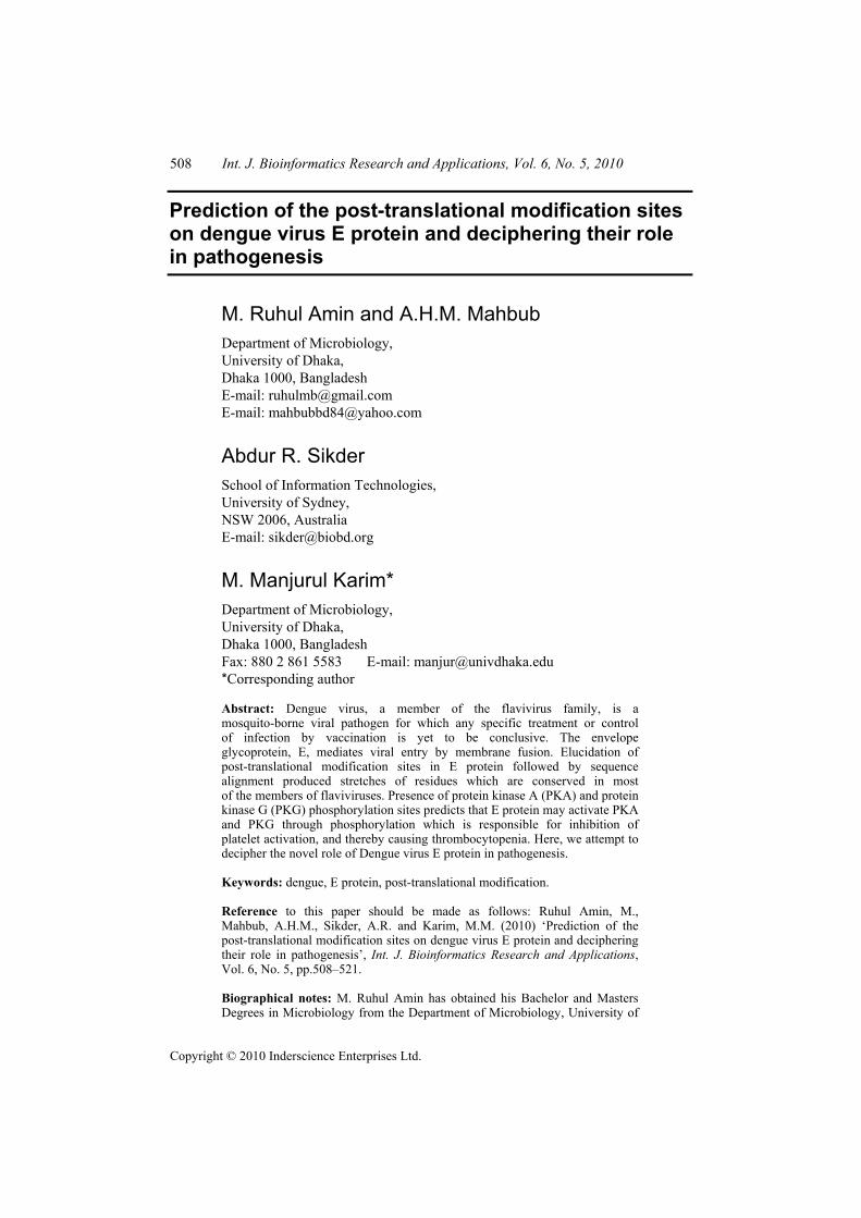
SUMO-1 (small ubiquitin-related modifier; also known as PIC1, UBL1, Sentrin, GMP1 and Smt3) is a member of the ubiquitin and ubiquitin-like superfamily. Most SUMO-modified proteins contain the tetrapeptide motif B-K-x-D/E where B is a hydrophobic residue, K is the lysine conjugated to SUMO, x is any amino acid, D or E is an acidic residue. Substrate specificity appears to be derived directly from Ubc9 and the respective substrate motif. Results obtained from the SUMO plot server are described in Table 3. The program identified nine potential SUMO binding motifs within the dengue E protein. Among them, 3 motifs are found to be highly probable (score above 0.90).
516 M.R. Amin et al.
Table 3 SUMO binding motif predicted by SUMO plot server
Motif Position Motif Sequence Score 1 K321 HGTIL IKVE YKGED 0.94 2 K307 LNTFV LKKE VSETQ 0.91 3 K286 HLKCR LKMD KLELK 0.91 4 K454 GVSWI MKIG IGVLL 0.63 5 K289 CRLKM DKLE LKGMS 0.50 6 K47 LDIEL QKTE ATQLA 0.50 7 K245 KNAHA KKQE VVVLG 0.48 8 K359 NPVVT KKEE PVNIE 0.48 9 K398 KGSSI GKMF EATAR 0.32
3.5 Analysis of transmembrane topology
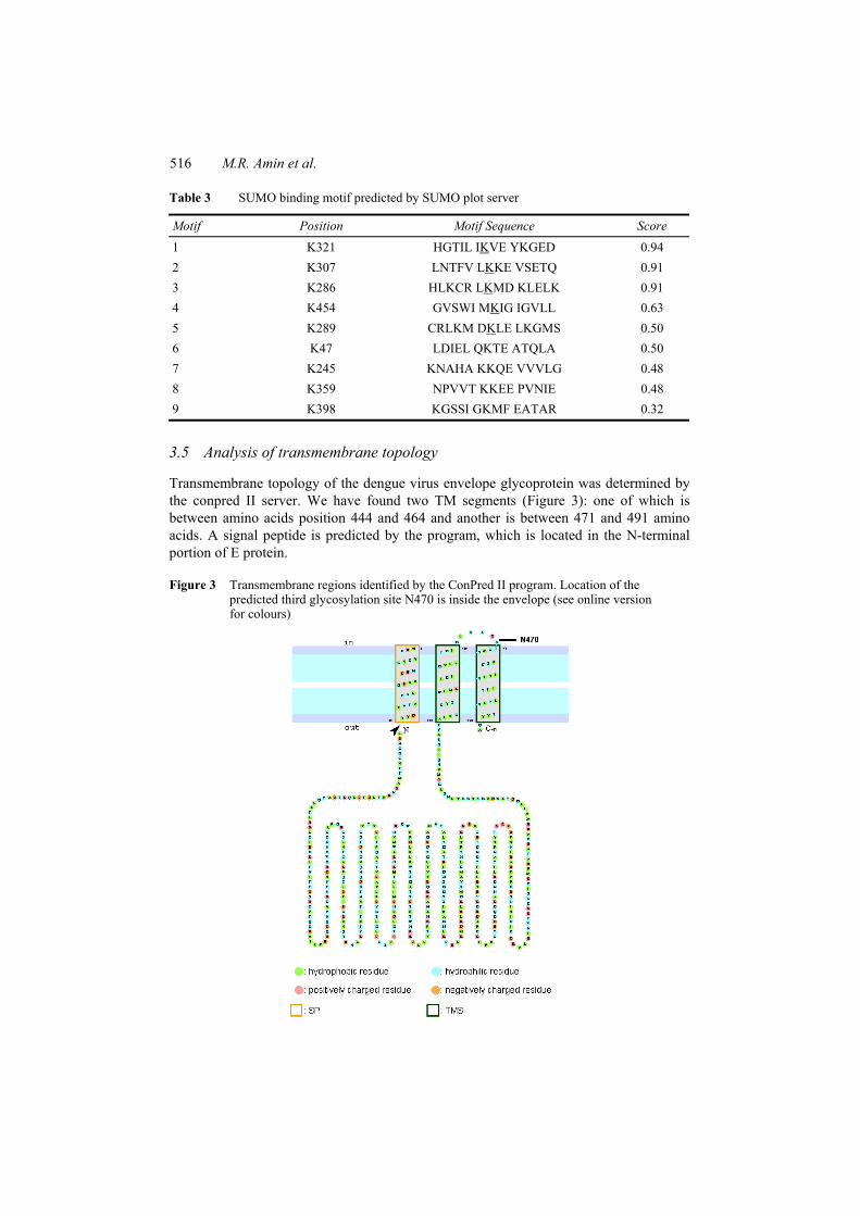
Transmembrane topology of the dengue virus envelope glycoprotein was determined by the conpred II server. We have found two TM segments (Figure 3): one of which is between amino acids position 444 and 464 and another is between 471 and 491 amino acids. A signal peptide is predicted by the program, which is located in the N-terminal portion of E protein.
Figure 3 Transmembrane regions identified by the ConPred II program. Location of the predicted third glycosylation site N470 is inside the envelope (see online version for colours)

Prediction of the post-translational modification sites 517
4 Discussion
This study is based on absolute computational analysis of the proteins such as multiple sequence alignment with other serotypes including some important flavivirus such as Japanese encephalitis virus, Tick-borne encephalitis virus, West Niles virus and Yellow Fever virus. We have further identified several PTM sites on E protein and mapped their conservation among flaviviruses. The TM topology and hydrophobicity of the E protein was also analysed. From this study, some interesting findings are surfaced.
4.1 Prediction of post-translational modification sites
The E protein has two potential N-linked glycosylation sites at Asn-67 and Asn-153. The glycosylation site at Asn-153 is conserved in most flaviviruses, while glycosylation at Asn-67 is unique for dengue virus (Heinz and Allison, 2003). Recently, a cryoelectron microscopy reconstruction of dengue virus complexed with the carbohydrate-binding domain of DC-SIGN has shown interaction of the lectin with the N-glycan at Asn-67 (Pokidysheva et al., 2006). Previous analysis of the crystal structure of E from dengue virus and other flaviviruses indicated that the carbohydrate moiety at Asn-153 extends across the dimer interface covering the fusion peptide. Stabilisation of the E dimer by this oligosaccharide is consistent with the properties of dengue virus mutants at position 153, which fuse with target membranes at a higher pH (Modis et al., 2003; Rey et al., 1995). From our computational analysis, we found three N-glycosylation sites in the sequence. Two of them, which are in Asn-67 and Asn-153 position, agree with previous analysis. Program has found an extra N-glycosylation site at Asn-470, which is not previously discussed. Through TM topology analysis, we found that this region is located adjacent to the TM segment, just inside the envelope. That is why although there is sequence similarity with the pattern of N-glycosylation site, this may not be a potential glycosylation site. Further analysis is required to know the real scenario. Multiple sequence alignment shows that glycosylation site at Asn 67 is not conserved in JEV, WNV, TBE and YFV.
Program also predicts a cAMP- and cGMP-dependent PK phosphorylation site at position 391. This may occur just by chance or it may have some functions that we do not know yet. Protein kinase C phosphorylation is important in many cellular processes such as signal transduction or other processes. Through prosite pattern, we have found some protein kinase C phosphorylation site at positions 55, 184, 198, 237, 357 and 403. Most of these sites are well conserved in dengue virus and some of them are conserved in other flaviviruses as well. The exact role of this phosphorylation is yet to be reported.
The participation of casein kinases II in regulation of different cellular processes is shown in carbohydrate and lipid metabolism, in transcription, translation and enzyme synthesis. Several casein-kinase II phosphorylation sites have been predicted by the program at different positions of the protein sequence. Among them, pattern TWVD at position 19, TQGE at position 76, TGLD at 187, TAWD at position 416 are highly conserved in most of the serotypes of dengue and some flaviviruses.

518 M.R. Amin et al.
4.2 N-myristoylation of E protein may be required for viral viability and host cell attachment
Post-translational addition of lipids to polypeptide chains has been thought to be a process akin to glycosylation and one whose sole role is the permanent localisation of the protein in a lipid bilayer in the cell. Acylation of viral protein is an important event. Lipid moiety is essential for the transport and localisation of protein in the membrane and thus for viral viability. Myristoylation is a process whereby essentially cytoplasmic proteins/enzymes can become membrane-bound, thus locating them at their site of action.
Although the exact function of myristoylation is not clear yet, the addition of a relatively large hydrophobic moiety to a protein will increase its overall hydrophobicity somewhat, and the hydrophobicity of the particular area to which it is attached appreciably. Myristate can play a role in mediating protein–protein interactions within the virus and in organising, both temporally and spatially, some of the polypeptide chains during the later stages of infection (Grand, 1989). Result obtained from the programs predicts abundance (10 sites) of myristoylation site in the dengue virus E protein. It is very interesting to note that pattern GNGCGL at position 102 is highly conserved (Figure 1) and located on the region of fusion loop. Thus, myristoylation may play an important role in viral membrane localisation. By using the myristate from host cell, it may interact with other proteins in host cells.
4.3 Dengue virus envelope glycoprotein may have impact on signalling pathways
We are predicting that the dengue virus envelope glycoprotein may be a substrate of phosphorylation with several proteins in host cell signalling pathways. This study, therefore, attempts to find out the phosphorylation sites on dengue envelope protein by using the PPSP program. We have found a substantial number of different phosphorylation sites such as PKG, PKA, PDGFR kinase, Janus Kinase (JAK), MAP kinase, Inhibitor of Kappa Beta (IKK) kinase phosphorylation sites. It is noted that all of these proteins are part of the signalling pathways in host cells particularly human. Among them, most of these PKs are involved in regulating platelet activation and inactivation directly or indirectly.
It was found that PKA and PKG inhibit platelet activation through a number of shared pathways, including inhibition of phosphoinositide metabolism and through Ca2+ extrusion (Hoffbrand et al., 2005). Our computational analysis revealed several PKG and PKA phosphorylation sites on the dengue virus E protein (Table 2). It indicates that dengue virus E protein may be responsible for activation of PKG and PKA through phosphorylation, and thereby prevents platelet activation. This may be a mechanism why patients of dengue infection suffer from thrombocytopenia.
4.4 Small Ubiquitin-like modifier attachment site of E protein is not conserved in all flavivirus
Small Ubiquitin-like Modifier is a ubiquitin-related protein that covalently binds to other proteins. Conjugation of ubiquitin and ubiquitin-related proteins (Ublps) to cellular target proteins is involved in many aspects of eukaryotic gene expression by regulating the signalling for degradation or modifying the functions of target proteins. Some evidence

Prediction of the post-translational modification sites 519
has suggested that ubiquitin conjugating enzyme (Ubc9) can itself bind specifically to substrates presenting a consensus SUMO modification motif, wKxE (w represents the hydrophobic resides, K is lysine, x is any residue, and E is glutamic acid). Recently, structural analysis has revealed that Ubc9 can recognise this sequence directly (Xue et al., 2006b). Ubc9 binding may play an important role in substrate recognition as well as in substrate modification. In recent years, growing numbers of viral proteins have been found to conjugate with SUMO-1.
To assess the direct interaction between Dengue virus E and Ubc9, we analysed the sumoylation consensus motif of E protein by SUMOplotTM (ABGENE); this system is based on two criteria: the first is the direct amino acid match to the observed SUMO consensus sequence that binds Ubc9; the second is the substitution of the consensus amino acid residues with other residues exhibiting similar hydrophobicity. The system recommended three high probability binding sites, residues 286, 307 and 321 (Table 3) among nine potential candidates. None of these binding sites are conserved in all serotypes of dengue virus and other flavivirus. These binding sites are totally different than the dengue virus type 2 E protein (Chiu et al., 2007). E protein consists of three separate domains, the central domain I, the dimerisation domain II and the domain III responsible for the receptor binding and endosomal uptake during viral infection (Modis et al., 2005). In Dengue type 2 E protein, residue 393 located too close to the TM domain and therefore, due to stereo hindrance, it should not have the opportunity to interact with Ubc9 during viral infection. Hence, it was ranked to be a less likely candidate. But in case of type 3 E protein, all three binding residues are located in domain III and are apart from the TM region. That is why three of them have chance of being the attachment site. By site-directed mutagenesis, the amino acids at these positions can be changed from K to R either individually or in pair for confirmation of correct interaction site.
In conclusion, the study identifies several PTM sites on dengue virus envelope glycoprotein E and their conservation in all serotypes of dengue and other important flaviviruses. This PTM of envelope protein may be another mechanism of dengue virus pathogenesis, as this modification is very important in regulating host cell signalling pathways. However, experimental verification of this modification site is required for deciphering the actual mechanism.
References Arai, M., Mitsuke, H., Ikeda, M., Xia, J.X., Kikuchi, T., Satake, M. and Shimizu, T. (2004)
‘ConPred II: a consensus prediction method for obtaining transmembrane topology models with high reliability’, Nucleic Acids Res., Vol. 32, pp.W390–W393.
Chiu, M.W, Shih, H.M, Yang, T.H. and Yang, Y.L. (2007) ‘The type 2 dengue virus envelope protein interacts with small ubiquitin-like modifier-1 (SUMO-1) conjugating enzyme 9 (Ubc9)’, J. Biomed. Sci., Vol. 14, No. 3, pp.429–444.
Glass, D.B. and Smith, S.B. (1983) ‘Phosphorylation by cyclic GMP-dependent protein kinase of a synthetic peptide corresponding to the autophosphorylation site in the enzyme’, J. Biol. Chem., Vol. 258, pp.14797–14803.
Grand, R.J.A. (1989) ‘Acylation of viral and eukaryotic proteins’, Biochem. J., Vol. 258, pp.625–638.
Gubler, D.J. (1997) ‘Dengue and dengue hemorrhagic fever: Its history and resurgence as a global public health problem’, in Gubler, D.J. and Kuno, G. (Eds.): Dengue and Dengue Hemorrhagic Fever, CAB International, Wallingford, UK, pp.1–22.

520 M.R. Amin et al.
Heinz, F.X. and Allison, S.L. (2003) ‘Flavivirus structure and membrane fusion’, Adv. Virus Res., Vol. 59, pp.63–97.
Hoffbrand, A.V., Catovsky, D. and Tuddenham, E.G.D. (2005) Postgraduate Haematology, 5th ed., ISBN: 978-1-4051-0821-8, pp.820–821.
Kishimoto, A., Nishiyama, K., Nakanishi, H., Uratsuji, Y., Nomura, H., Takeyama, Y. and Nishizuka, Y. (1985) ‘Studies on the phosphorylation of myelin basic protein by protein kinase C and adenosine 3′:5′-monophosphate-dependent protein kinase’, J. Biol. Chem., Vol. 260, pp.12492–12499.
Larkin, M.A., Blackshields, G., Brown, N.P., Chenna, R., McGettigan, P.A., McWilliam, H., Valentin, F, Wallace, I.M., Wilm, A., Lopez, R., Thompson, J.D., Gibson, T.J. and Higgins, D.G. (2007) ‘ClustalW and Clustalx version 2’, Bioinformatics, Vol. 23, No. 21, pp.2947–2948.
Modis, Y., Ogata, S., Clements, D. and Harrison, S.C. (2003) ‘A ligand-binding pocket in the dengue virus envelope glycoprotein’, Proc. Natl. Acad. Sci. USA, Vol. 100, pp.6986–6991.
Modis, Y., Ogata, S., Clements, D. and Harrison, S.C. (2004) ‘Structure of dengue virus envelope protein after membrane fusion’, Nature, Vol. 427, pp.313–319.
Modis, Y., Ogata, S., Clements, D. and Harrison, S.C. (2005) ‘Variable surface epitopes in the crystal structure of Dengue virus type 3 envelope glycoprotein’, J. Virol., Vol. 79, No. 2, pp.1223–1231.
Mukhopadhyay, S., Kuhn, R.J. and Rossmann, M.G. (2005) ‘A structural perspective of the flavivirus life cycle’, Nat. Rev. Microbiol., Vol. 3, pp.13–22.
Navarro-Sanchez, E., Altmeyer, R., Amara, A., Schwartz, O., Fieschi, F., Virelizier, J.L., Arenzana-Seisdedos, F. and Despres, P. (2003) ‘Dendritic-cell-specific ICAM3-grabbing non-integrin is essential for the productive infection of human dendritic cells by mosquito-cell-derived dengue viruses’, EMBO Rep., Vol. 4, pp.723–728.
Pinna, L.A. (1990) ‘Casein kinase 2: an eminence rise in cellular regulation?’, Biochim. Biophys. Acta, Vol. 1054, pp.267–284.
Pokidysheva, E., Zhang, Y., Battisti, A.J., Bator-Kelly, C.M., Chipman, P.R., Xiao, C., Gregorio, G.G., Hendrickson, W.A., Kuhn, R.J. and Rossmann, M.G. (2006) ‘Cryo-EM reconstruction of dengue virus in complex with the carbohydrate recognition domain of DC-SIGN’, Cell, Vol. 124, pp.485–493.
Rey, F.A., Heinz, F.X., Mandl, C., Kunz, C. and Harrison, S.C. (1995) ‘The envelope glycoprotein from tick-borne encephalitis virus at 2 A resolution’, Nature, Vol. 375, pp.291–298.
Rigau-Perez, J.G., Clark, G.G., Gubler, D.J., Reiter, P., Sanders, E.J. and Vorndam, A.V. (1998) ‘Dengue and dengue haemorrhagic fever’, Lancet, Vol. 352, pp.971–977.
Rost, B., Yachdav, G. and Liu, J. (2004) ‘The predictprotein server’, Nucleic Acids Res., Vol. 32, pp.W321–W326.
Seo, J. and Lee, K.J. (2004) ‘Post-translational modifications and their biological functions: proteomic analysis and systematic approaches’, J. Biochem. Mol. Biol., Vol. 37, No. 1, pp.35–44.
Sikder, A.R. and Zomaya, A.Y. (2009) ‘Analysis of protein phosphorylation site predictors with an independent dataset’, Int. J. Bioinf. Res. Appl., Vol. 5, pp.20–37.
Tassaneetrithep, B., Burgess, T.H., Granelli-Piperno, A., Trumpfheller, C., Finke, J., Sun, W., Eller, M.A., Pattanapanyasat, K., Sarasombath, S., Birx, D.L., Steinman, R.M., Schlesinger, S. and Marovich, M.A. (2003) ‘DC-SIGN (CD209) mediates dengue virus infection of human dendritic cells’, J. Exp. Med., Vol. 197, pp.823–829.
Towler, D.A., Gordon, J.I., Adams, S.P. and Glaser, L. (1988) ‘The biology and enzymology of eukaryotic protein acylation’, Annu. Rev. Biochem., Vol. 57, pp.69–99.

Prediction of the post-translational modification sites 521
Wu, S.J., Grouard-Vogel, G., Sun, W., Mascola, J.R., Brachtel, E., Putvatana, R., Louder, M.K., Filgueira, L., Marovich, M.A., Wong, H.K., Blauvelt, A., Murphy, G.S., Robb, M.L., Innes, B.L., Birx, D.L., Hayes, C.G. and Frankel, S.S. (2000) ‘Human skin Langerhans cells are targets of dengue virus infection’, Nat. Med., Vol. 6, pp.816–820.
Xue, Y, Li, A., Wang, L., Feng, H. and Yao, X. (2006a) ‘PPSP: prediction of PK-specific phosphorylation site with Bayesian decision theory’, BMC Bioinformatics, Vol. 7, p.163.
Xue, Y., Zhou, F., Fu, C., Xu, Y. and Yao, X. (2006b) ‘SUMOsp: a web server for sumoylation site prediction’, Nucleic Acids Res., Vol. 34, pp.W254–W257.