preface - inb.unam.mx · neuroglia represents the major nerve cell group in the brain....
TRANSCRIPT
2
PREFACE Neuroglia represents the major nerve cell group in the brain. Nevertheless, for a long time
it was only considered a support element for neural function. At present, neuroglia research
is fundamental to learn about the physiology of the central nervous system. Consequently,
the organization of the 2nd Symposium on Neuroglia Physiology and Pathology represents
an opportunity for our University to promote and disseminate the most recent findings
made in this field by national and international leading researchers. In this occasion, our
Institute has the honor of hosting this event. In this edition the students will have a chance
to present and discuss their results directly with the speakers. Finally, we are grateful to our
University, CONACYT, SFN, and IBRO for their valuable support which made possible the
organization of this Symposium. Our best wishes for the organizers and participants; we are
sure that this event will be successful and will become a reference in the field.
DR. ALFREDO VARELA ECHAVARRIA
DIRECTOR – INB-UNAM

3
ORGANIZING COMMITTEE
Dr. Pavel Montes de Oca Balderas
Instituto Nacional de
Neurología y Neurocirugía
Dr. Octavio C. García González
Facultad de Psicología
UNAM
Dr. Daniel Reyes Haro
Instituto de
Neurobiología UNAM
Technical Support & Administrative Staff
M. en C. Leonor Casanova Rico
Lic. Nora Ivette Ramírez Reséndiz Ing. Ramón Martínez Olvera
Act. Guadalupe Calderón Alzati Lic. Rogelio Rocha García
C.P. Marco Alberto Olguín Araujo C.P. Antonio González Cruz
Lic. Felipe P. Pedroza Montes de Oca
4
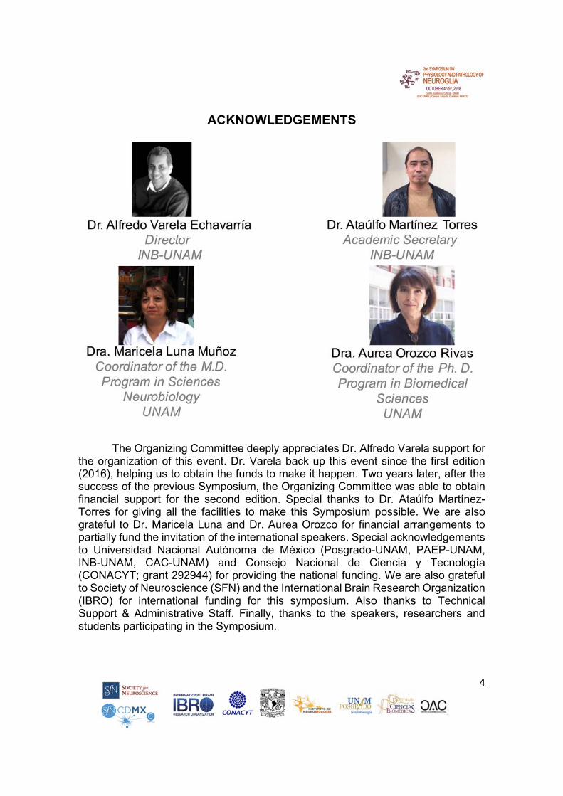
ACKNOWLEDGEMENTS
The Organizing Committee deeply appreciates Dr. Alfredo Varela support for the organization of this event. Dr. Varela back up this event since the first edition (2016), helping us to obtain the funds to make it happen. Two years later, after the success of the previous Symposium, the Organizing Committee was able to obtain financial support for the second edition. Special thanks to Dr. Ataúlfo Martínez-Torres for giving all the facilities to make this Symposium possible. We are also grateful to Dr. Maricela Luna and Dr. Aurea Orozco for financial arrangements to partially fund the invitation of the international speakers. Special acknowledgements to Universidad Nacional Autónoma de México (Posgrado-UNAM, PAEP-UNAM, INB-UNAM, CAC-UNAM) and Consejo Nacional de Ciencia y Tecnología (CONACYT; grant 292944) for providing the national funding. We are also grateful to Society of Neuroscience (SFN) and the International Brain Research Organization (IBRO) for international funding for this symposium. Also thanks to Technical Support & Administrative Staff. Finally, thanks to the speakers, researchers and students participating in the Symposium.

5
I N D E X
pages
I. PROGRAM 6-15
II. SPEAKERS, ABSTRACTS (October, 4th)
16-23
III. POSTERS, ABSTRACTS
(October 4th) 24-35
IV. SPEAKERS ABSTRACTS
(October, 5th) 36-43
V. POSTERS ABSTRACTS
(October 5th)
44-56

6
SYMPOSIUM-TALKS 08:30 am – 13:30 pm
October 4th, 2018
Hour
Speaker
Title
Moderador: Dra. Mónica López Hidalgo
8:30 – 9:00 am Dr. Rogelio Arellano
Ostoa INB-UNAM
A novel GABAA receptor expressed in
oligodendrocytes
9:00 – 9:30 am
Dr. Pavel Montes de Oca
Balderas INNN
NMDA receptor in astrocytes: functions,
controversies and contradictions
Coffee break (9:30 – 10:00 am)Moderador: Dr. Pavel Montes de Oca
10:00 – 10:30 am
Dr. Gerardo B. Ramírez
Rodríguez INP
Modulation of neuroglial cells in the dentate gyrus
during depression like behavior.
10:30 – 11:00 am
Dra. Mónica López Hidalgo FM-UAQ
Astrocytes subdomains respond independently in
vivo
11:00 – 11:30 am
Dr. Adrián Rodríguez Contreras
CUNY
A postnatal period of vascular and glia cell
expansion in the auditory brainstem of rats and mice: Implications for neuronal development
and synaptic refinement before the onset of
hearing.
Coffee break (11:30 am – 12:00 pm)

7
LECTURE 1 12:00 – 13:30 pm October 4th, 2018
Centro Académico Cultural (CAC) Universidad Nacional Autónoma de México
Campus Juriquilla, Querétaro, QRO
CIRCUIT-SPECIFIC SYNAPTIC REGULATION BY ASTROCYTES
DR. ALFONSO ARAQUE UNIVERSITY OF MINNESOTA
(Lunch: 13:30 – 15:30 pm)
8
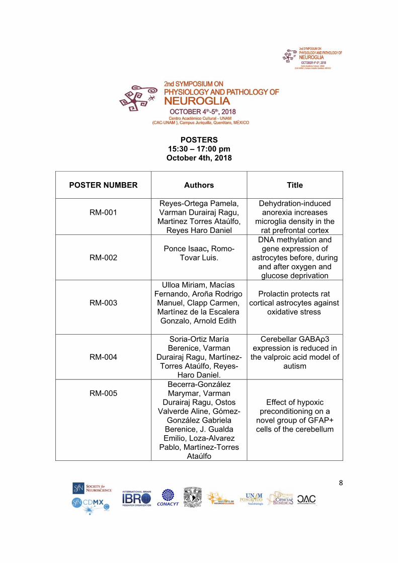
POSTERS 15:30 – 17:00 pm October 4th, 2018
POSTER NUMBER
Authors
Title
RM-001
Reyes-Ortega Pamela, Varman Durairaj Ragu, Martinez Torres Ataúlfo,
Reyes Haro Daniel
Dehydration-induced anorexia increases
microglia density in the rat prefrontal cortex
RM-002
Ponce Isaac, Romo-
Tovar Luis.
DNA methylation and gene expression of
astrocytes before, during and after oxygen and glucose deprivation
RM-003
Ulloa Miriam, Macías Fernando, Aroña Rodrigo Manuel, Clapp Carmen, Martínez de la Escalera Gonzalo, Arnold Edith
Prolactin protects rat
cortical astrocytes against oxidative stress
RM-004
Soria-Ortiz María Berenice, Varman
Durairaj Ragu, Martínez-Torres Ataúlfo, Reyes-
Haro Daniel.
Cerebellar GABAρ3 expression is reduced in
the valproic acid model of autism
RM-005 Becerra-González Marymar, Varman
Durairaj Ragu, Ostos Valverde Aline, Gómez-
González Gabriela Berenice, J. Gualda Emilio, Loza-Alvarez
Pablo, Martínez-Torres Ataúlfo
Effect of hypoxic preconditioning on a
novel group of GFAP+ cells of the cerebellum

9
RM-006
Arzate Dulce María, Guerra Crespo
Magdalena, Covarrubias Luis
Glial Cells are a Source of New Neurons in the Adult Substantia NigrA
RM-007
Labrada-Moncada Francisco Emmanuel,
Martínez-Torres Ataulfo, Reyes-Haro Daniel.
Functional expression of GABAA receptors in glial cells of cerebellar white
matter
RM-008
Zinzun Ixta Guadalupe, Saavedra Pimentel Luis Miguel, Ochoa Zarzosa Alejandra, Torner Luz
Neonatal prolactin decreases glial
population and affects cytokine expression of
the hippocampus
RM-009
Segura Anaya Edith,
Martínez Gómez Alejandro, Roberta Dent
Myrna Alexandra
Aquaporin 1 is localized differentially between mice and rat, in both
normal sciatic nerve and during Wallerian
degeneration
RM-010
Tovar Cueva José
Rubén, Larriva Sahd Jorge
Visual deafferentation effects on astrocytic
processes of the lateral geniculate nucleus of the
thalamus
RM-011
Medrano-Jiménez Elisa, Pedraza-Escalona Martha, Álvarez-Arellano Lourdes, Zamilpa-Alvarez Alejandro,
Herrera-Ruíz Maribel, Jiménez-Ferrer Enrique, Tortoriello-García Jaime, Pedraza-Alva Gustavo, Pérez-Martínez Leonor
Malva parviflora extract
regulates the phagocytosis of microglia via PPAR in an Alzheimer s disease
model

10
LECTURE 2 17:00 – 18:00 pm October 4th, 2018
ASTROCYTE DYSFUNCTION AND NEURODEGENERATION IN DOWN SYNDROME
DR. JORGE BUSCIGLIO UNIVERSITY OF CALIFORNIA, IRVINE
11
SYMPOSIUM-TALKS 08:30 am – 13:30 pm
October 5th, 2018
Hour
Speaker
Title
Moderador Dr. Lenin Ochoa de la Paz
8:30 – 9:00 am Dr. Ataúlfo Martínez
Torres INB-UNAM
Cell diversity of ependymal glial layer of
the roof of the fourth ventricle
9:00 – 9:30 am
Dr. Héctor Lópes Valdés
FM-UNAM
Astrogliosis in stroke
Coffe break (9:30 – 10:00 am)Moderador Dr. Gerardo Ramírez Rodríguez
10:00 – 10:30 am
Dr. Lenin Ochoa de la Paz FM-UNAM
Taurine-GABA receptors interaction in the
proliferative processes of GFAP+ progenitor cells from the subventricular
zone of mice
10:30 – 11:00 am Dr. Daniel Reyes Haro
INB-UNAM GABA receptors in
astroglia
11:00 – 11:30 am Dr. Edith Arnold INB-UNAM
Insights into the role of Prolactin in Astrocytes
Coffee break (11:30 am – 12:00 pm)

12
LECTURE 1 12:00 – 13:30 pm October 5th, 2018
HOW DO GLIAL CELLS CONTROL CNS FUNCTION?
DR. AXEL NIMMERJAHN SALK INSTITUTE, LA JOLLA, USA
(Lunch: 13:30 – 15:30 pm)

13
POSTERS
15:30 – 17:00 pm October 5th, 2018
POSTER NUMBER
Authors
Title
RM-012
Saavedra Pimentel Luis Miguel, Fenton Navarro Bertha, Ochoa Zarzosa Alejandra, Torner Luz
Early life stress and lipopolysaccaride affect hippocampal glial cells and induce long term behavioral alterations
RM-013
Ojeda García Francisco Gerardo, Cruz Martínez
María Yolanda, Arias-Santiago Stella Viviana,
Díaz Munguía Isabel, Gálvez Susano Jessica
Vanessa, Ibarra Arias José Juan Antonio
Cop-1 effect on interleukins and
neurotrophins expression in choroid plexus in rats with cerebral ischemia
RM-014
Hernández Velázquez Martha Guadalupe, Duran
Juárez Sergio Gabriel, Saavedra Pimentel Luis
Miguel, Torner Luz
Changes in emotional and learning behavior in female rats exposed to a
double challenge neonatal immune-stress
RM-015
Castro-Ramírez Mariana, Arias-Santiago Stella, Cruz Yolanda, García
Vences Elisa, Domínguez Adriana, Saavedra-Navarrete
Melanie, Ibarra Antonio
Characterization of lymphocytes in
cerebrospinal fluid of rats with cerebral ischemia
immunized with Copolymer-1
RM-016
Nava Gómez Laura Jaqueline, Calero Vargas
Isnarhazni Alejandra, Ramírez Hernández Noemi,
Ortiz Retana Juan José, Alcauter Solórzano Sarael,
López Hidalgo Mónica
D-serine modify brain
functional connectivity in aged rats

14
RM-017
Ramírez-Hernández Noemi, Roque-Roque
Georgina, Calero-Vargas Isnarhazni Alejandra, Nava-
Gómez Laura Jaqueline, Reyes-López Julián,
Hernández-Montiel Hebert Luis, Arias-García Nallely
Amaranta, Rodríguez-Córdoba Verónica, López-
Hidalgo Mónica
Serum D-serine levels correlates with the
cognitive performance of aged humans
RM-018
Ordaz Ramos Rainald Pablo, Cisneros Mejorado Abraham, García García
Cindy Lucero, Garay Rojas Edith, Arellano Ostoa
Rogelio
A novel GABAA receptor
expressed in oligodendrocytes
RM-019
Cadena Reynoso Omar Alejandro de Jesús,
García González Octavio César
Environmental enrichment modifies the morphology of mouse cortical astrocytes
RM-020
Calero Vargas Isnarhazni Alejandra, Ramírez
Hernández Noemi, Nava Gómez Laura Jaqueline, López-Hidalgo Mónica
Oral administration of D-serine improves the
cognitive flexibility and attention in aged rats
RM-021
Meneses San Juan David, Ortiz López
Leonardo, Reyes-Haro Daniel, González Olvera
Jorge Julio, Ramírez Rodríguez Gerardo
Bernabé
Repetitive transcranial magnetic stimulation (5Hz)
modulates hippocampal neurogenesis and glial cells in chronic mild stress mice
RM-022
Rodríguez Arzate Cynthia Alejandra, Reyes-
Haro Daniel, Martínez-Torres Ataúlfo
Organization and functional characteristics of Bergmann
glial cells in an experimental model of
cortical dysplasia RM-023 Montes de Oca Balderas
Pavel, Montes de Oca Balderas Horacio
An Order of Magnitude Analysis of Inositol tris-
phosphate Diffusion at the Nanoscale in a Model of Peri-synaptic Astrocyte
Projection
15
LECTURE 2 17:00 – 18:00 pm October 5th, 2018
THE PATHOPHYSIOLOGY OF CNS WHITE MATTER INJURY DEPENDS ON THE INSULT: ANOXIA VS. AGLYCEMIA VS. ISCHEMIA
PROF. BRUCE R. RANSOM UNIVERSITY OF WASHINGTON

16
SYMPOSIUM ABSTRACTS (SPEAKERS)
OCTOBER 4TH, 2018

17
RM-S001 A novel GABAA receptor expressed in oligodendrocytes Arellano Ostoa Rogelio, Ordaz Ramos Rainald Pablo, Cisneros-Mejorado Abraham, García García Cindy Lucero, Garay Rojas Edith. Departamento de Neurobiología Celular y Molecular, Instituto de Neurobiología, UNAM Campus Juriquilla, Queretaro, Mexico.
The γ-aminobutyric acid (GABA) is a key neurotransmitter in the central nervous
system, GABA acts through activation of pentameric receptor-channels permeable
to Cl- ions, known as GABAA receptors, and through metabotropic receptors named
GABAB. In oligodendrocytes (OL), GABAA receptor expression is controlled by their
interaction with neurons, thus, it has been proposed a role of GABAergic signaling
during the myelination process. Functional characteristics of the GABA-response in
OL from the rat optic nerve, have indicated a specific subunits combination
conforming the GABAA receptor, being different to receptors expressed in other
neural cells. Determination of the subunits combination expressed in OL is essential
for its pharmacologic and genetic control. Here, GABAA subunits (α3, β2, β3, γ1-γ3)
were cloned from OL and, heterologously expressed in Xenopus laevis oocytes in
the distinct combinations, then GABA-response for each combination was studied
electrophysiologically. Results showed that α3β2γ1 co-expression, mimicked the
functional and pharmacological pattern described for the endogenous response in
OL. For example, this ensemble compared with the endogenous receptor, showed
similar sensitivity to GABA, as well as to a variety of positive and negative allosteric
modulators; including, a distinctive robust potentiation by butyl β-carboline-3-
carboxylate, a β-carboline that has only a weak effect on the GABAA receptor
expressed in cortical neurons. The putative GABAA receptor α3β2γ1 represents a
novel subunits combination expressed in OL, which displays distinctive
pharmacological characteristics, this information might be used to analyze in detail
the GABAergic signaling role in myelination, and its probable involvement in various
pathologies.
18
RM-S002 Montes de Oca Balderas Pavel Departamento de Neurociencia Cognitiva, Instituto de Fisiología Celular, UNAM y Unidad de Neurobiología Dinámica del Dpto. de Neuroquímica, INNN.
In the framework of the neurocentric theory the role of glia in information handling
within the Central Nervous System (CNS) was dismissed for almost a century. Accordingly,
the NMDA receptor (NMDAR), a critical player in synapse communication, neuronal
plasticity and CNS function, was mainly studied in the neuronal context. Nevertheless, this
receptor is widely expressed in cells within and beyond the CNS including astrocytes, whose
role, as that of other neuroglial cells, has been remodeling since almost three decades ago.
It turns out that in order to have an integrative perspective of the CNS function it is not
possible to ignore neuroglial cells. In particular astrocytes have been found to play a relevant
role in different aspects of CNS function, including information handling in the tripartite
synapse model, as they “listen” to neuronal synaptic communication, regulate it and respond
at the cellular and synctitial level.
Despite the expression and function of ionotropic glutamate NMDAR in astrocytes
was matter of different controversies and apparent contradictions for almost 30 years, today
it is known that these cells have functional receptors, although its role has not been deeply
investigated. One of the main reasons for these controversies and apparent contradictions
was the a priori expectation that this receptor should work in a similar manner to its neuronal
counterpart. However, the diversity of subunits that may be assembled into NMDAR results
in receptors with different biophysical, transport and signaling properties, as it is the case of
the NMDAR in astrocytes.
In this talk I briefly outline NMDAR general features, pinpointing those that are the
source of NMDAR diversity and complexity. Then, I will show our results that indicate that
the NMDAR in cultured astrocytes mainly acts as a metabotropic-like flux-independent
receptor, function that still has to be confirmed in tissue astrocytes, but that some reports
have demonstrated for neuronal cells. Given the complex molecular nature of NMDAR, its
critical role and the relevance of astrocytes, the study of astrocytic NMDAR promise to
provide further understanding of CNS physiology and pathology.
Acknowldgements: Proyectos CONACyT de Repatriación # MOD-ORD-1-09-PCI-010-03-
10 y de Ciencia Básica #132706.

19
RM-S003 Modulation of neuroglial cells in the dentate gyrus during depression like behavior. Ramírez-Rodríguez GB. Instituto Nacional de Psiquiatría “Ramón de la Fuente Muñiz”, Ciudad de México, México. Stress is a key factor for some neuropsychiatric disorders including depression. In
fact, several types of stressors have relevant impact on the brain plasticity with
important alterations in behavior and cognition. Among the type of stressors widely
studied exists the chronic mild stress (CMS). The CMS paradigm has proven effects
on plasticity and behavioral alterations related to depression. Moreover, CMS affects
neuroplasticity but has also shown impressive negative effects on the
microenvironment of the dentate gyrus (DG). Then, CMS also affects the generation
of new neurons in the DG of the hippocampus region of the limbic system implicated
for learning and memory but also for mood related behavior. Interestingly, some
clinical and preclinical studies have pointed to the direction of alterations in glial cells.
In this regard, the effects of key modulators of the neuroglial cells (such as
environmental enrichment, melatonin, antidepressant drugs and the repetitive
transcranial magnetic stimulation) in the context of adult hippocampal neurogenesis
in a murine model of depression will be presented.

20
RM-S004 Astrocytes subdomains respond independently in vivo López-Hidalgo Mónica, Vered Kellner, Schummers James. Facultad de Medicina,
Universidad Autónoma de Querétaro, Qro., México.
Astrocytes are a prominent non-neuronal cell type in the brain that play important
roles in modulating the activity of neural circuits. Astrocytes cover the gray matter
with bushy process where they detect and respond to neuronal activity with
intracellular calcium changes. In particular, astrocytes respond to sensory
stimulation with increases in somatic calcium concentration, although the spatial
relationship of these calcium events at the process remains unknown. Furthermore,
it remains unclear whether astrocytes behave as a single functional unit that
integrates all of the inputs, or if multiple functional subdomains reside within an
individual astrocyte. Here we utilized the columnar organization of ferret visual cortex
to analyze the spatial scale of neuron-astrocyte communication in vivo. To this end,
we monitored calcium activity throughout the bushy processes of visual cortical
astrocytes using a sensitive calcium indicator (GCaMP6s) and a two-photon
microscope during visual stimulation with parametrically varied visual stimuli. We
found that there is strong visually-driven calcium activity within the entire extent of
astrocyte processes, which is specific to the visual input. Furthermore, that astrocyte
responses to neural circuit activity are dominated by functional subdomains that
respond locally and independently to neuronal activity with high spatial precision and
are therefore suited to communicate with neuronal circuits at a fine spatial scale.

21
RM-S005 A postnatal period of vascular and glia cell expansion in the auditory brainstem of rats and mice: Implications for neuronal development and synaptic refinement before the onset of hearing. Rodríguez-Contreras Adrián1, Lingyan Shi1,2, Geng Pan1, Chaya Sussman1, Quetanya Brown1, Daphne Chang1, Grace Tsui1, Bao Vuong1. 1The City University of New York, City College, Biology Department and Center for Discovery and Innovation. 85 Saint Nicholas Terrace, New York, NY 10031. 2Present address: Chemistry Department, Columbia University, New York, NY. Although microglia have recently been implicated as key regulators of activity-dependent
synapse elimination in somatosensory and visual systems, little is known about their roles
in synaptic refinement and developmental plasticity in the auditory system. This is important
to determine if the mechanisms of microglia function are universal or dependent on brain
region and life stage. In this study we examined microglia development in the auditory
brainstem circuit formed between the medial nucleus of the trapezoid body (MNTB) and the
lateral superior olive (LSO) in rats and mice. Based on previous observations in the
somatosensory system (Arnoux et al. 2013. Glia 61, 1582-1594), we first examined the
relationship between microglia cells and vascular development using Isolectin B4 and EdU
histochemistry in rats, and with fluorescent reporters in mTmG and CX3CR1-GFP mice,
which express red and green fluorescent proteins in perivascular and micro glial cells,
respectively. We report that during the postnatal stage between birth (P0) and P10 there is
a correlated increase in vascular volume and microglia density in the auditory MNTB-LSO
circuit. We hypothesize that microglia cells use the vascular niche to undergo expansion in
preparation for the synaptic refinement of the MNTB-LSO circuit. Ongoing experiments are
testing this hypothesis using 1) transcranial two-photon microscopy in neonate mice that
express fluorescent reporters to determine the dynamic behavior of microglia cells
associated with blood vessels in vivo, and 2) by developing engulfment assays to determine
if microglia cells are involved in synaptic refinement in the MNTB-LSO connection.
Acknowledments. Supported by a Harvey L. Karp award in the Sciences.

22
RM-S006 Circuit-specific synaptic regulation by astrocytes. Araque Alfonso. Department of Neuroscience, University of Minnesota Astrocytes respond to synaptically released neurotransmitters and release
gliotransmitters that regulate synaptic transmission and plasticity. The signaling
exchange between astrocytes and neuronal synaptic elements have led to the
establishment of the functional concept of tripartite synapse. Combining calcium
imaging techniques and multiple whole-cell recordings from neurons in different
brain areas, our lab aims to determine the spatial as well as the synapse-specific
properties of the astrocyte-mediated synaptic regulation in tripartite synapses. We
have recently shown that astrocyte-neuron interaction at tripartite synapses is circuit
specific (Martin et al., Science 2015), suggesting that the bidirectional astrocyte-
neuron signaling selectively occurs between specific subpopulations of astrocytes,
neurons, and synapses. In line with these findings, I will present the most recent
evidence obtained in the lab indicating that the activity of astrocytes induces a
differential synapse-specific regulation of excitatory and inhibitory synapses in the
amygdala. Selective activation of astrocytes by DREADDs or endocannabinoids
differentially regulates neurotransmission in the CentroMedial (CeM) amygdala, the
major effector amygdala subnucleus. Astrocyte stimulation depresses excitatory
glutamatergic transmission by activating A1 adenosine receptors and enhances
inhibitory GABAergic transmission by activating A2A adenosine receptors.
Consistent with these differential synaptic effects, astrocyte stimulation inhibits in
vivo CeM neuronal firing rate and reduces the freezing responses in the auditory fear
conditioning paradigm. These results indicate that astrocyte activity influences
animal behavior through synapse-specific regulation of neuronal activity.
Acknowledgments: NIH-NINDS (R01NS097312-01) and Human Frontier Science Program
(RGP0036/2014).
23
RM-S007 Astrocyte dysfunction and neurodegeneration in Down Syndrome Busciglio Jorge, Demuro Angelo, Lioudyno María. Department of Neurobiology
and Behavior, University of California, Irvine, CA, USA.
Normal astrocytic function is essential to effectively support neuronal survival,
synaptic transmission and synaptic integrity. Several lines of evidence indicate that
primary astrocytes with trisomy 21 (Down syndrome, DS) exhibit functional
alterations that directly impact neuronal function and survival including defects in 1-
mitochondrial function; 2- protein secretion, and 3- calcium homeostasis. all of which
critically affect neuronal development and function. Disruption of calcium
homeostasis is a common feature of several neurological disorders including
Alzheimer’s disease, which has a very high prevalence in older persons with
DS. Our results showed that although trisomic and euploid astrocytes maintained
similar resting cytosolic calcium levels and store contents, trisomic astrocytes were
severely deficient in IP3R-dependent global and local calcium signaling. The number
of regions containing clusters of local calcium signals, as well as the number of
individual events per cluster were significantly smaller in trisomic astrocytes.
Analysis of local calcium signals revealed that events generated by DS astrocytes
were significantly lower in amplitude, consistent with a reduced number of activated
IP3R channels within the cluster. The results suggest that deficient IP3R mediated
calcium responses may underlie, at least partially, the reduced ability of trisomic
astrocytes to support neuronal function and survival.

24
SYMPOSIUM ABSTRACTS POSTERS
OCTOBER 4TH, 2018
25
RM-P001 Dehydration-induced anorexia increases microglia density in the rat prefrontal cortex Reyes-Ortega Pamela, Varman Durairaj Ragu, Martinez Torres Ataúlfo, Reyes
Haro Daniel Departamento de Neurobiología Celular y Molecular. Instituto de
Neurobiología - UNAM Campus Juriquilla, Querétaro, Mexico.
Anorexia involves restrictive caloric intake that induces profound weight loss.
Macroglia deficits were recently associated to prefrontal cortex. Our aim was to test
if dehydration induced anorexia (DIA) disturbs microglia density. Three independent
experimental series of seven female Wistar rats (180-200g) per group were used for
this study: a) Control: received food and water ad libitum, b) DIA: received saline
solution (2.5 % NaCl) and food ad libitum, c) Forced Food Restricted (FFR) group
received water and the same amount of food as the DIA group. Subsequently,
histological sections (30 m) were immunostained with Iba-1, a microglial marker.
Microglial density as well as TNFα and IL-6 expression were estimated for all
experimental groups. Microglia/nuclei ratio was significantly increased in medial
prefrontal cortex of DIA and FFR groups (Control 0.10±0.01, DIA 0.18±0.02, FFR
0.22±0.03; p = 0.003; n=7). Likewise, reactive/resting microglia ratio was significantly
increased for DIA and FFR (Control 0.70±0.06, DIA 2.14±0.42, 1.89±0.40; p = 0.04;
n=7). Additionally, Western blots showed that DIA and FFR increase the expression
of the TNFα (DIA 1.98±0.1, FFR 1.81±0.08; p = 0.02; n = 3) and IL-6 (DIA 1.85±0.05,
FFR 1.69±0.09; p = 0.04; n = 3). We conclude that DIA and FFR increase microglia
density and expression of TNFα and IL-6 in prefrontal cortex.
Acknowledgements: Thanks to N. Hernández-Ríos, AE Espino-Saldaña, Dr. A. Castilla, MVZ M. García-Servín and M.C. L. Casanova. PRO has a CONACYT scholarship (554231) through PDCB. This work was supported by PAPIIT-UNAM (IN200913, IN201915, IN205718) to AMT and DRH.

26
RM-P002 DNA methylation and gene expression of astrocytes before, during and after oxygen and glucose deprivation
Ponce Isaac, Romo-Tovar Luis. Departamento de Patología Molecular, Instituto
de Fisiología Celular, Universidad Nacional Autónoma de Méxco, Ciudad de
México, México
Epigenetic mechanisms such as DNA methylation are well known regulators of genetic
expression and they play key roles in the development of neurodegenerative diseases,
mainly through a substantial transcriptional regulation of active and inactive promoters and
by modifying transcription elogation and splicing in CpG islands located intra and inter-
genically. It is also widely recognized that astrocytes, that are critical regulators of neuronal
function, play a crucial role in neurovascular-related disorders like ischemic stroke.
However, few studies have addressed at the molecular resolution the overall genetic and
epigenetic changes of these complex phenomena, and in events like reperfusion damage
that occurs after ischemic stroke, these processes are practically unknown. We performed
RNA-seq and methylated DNA immunoprecipitation sequencing (MeDIP-seq) analysis of
cultured human astrocyte cells derived from grade I non-tumorigenic glioblastoma subjected
to oxygen and glucose deprivation (OGD), in order to establish a relationship between DNA
methylation and gene expression under normoxia, OGD and recovery. We identified several
genomic features including proximal and distal promoters whose methylation levels change
not only during OGD but also after 8 h of recovery that showed statistically significant
differences in both; high (house-keeping and ubiquitous genes) and low (cell lineage-specific
genes) CG promoters. Moreover, DNA methylation remodeling was correlate with gene
expression of several genes under OGD and recovery, and the organization of the
transcriptome and methylome resulted different under normoxia, OGD and recovery. These
results can help to elucidate the overall transformation of cells in terms of transcription and
DNA methylation in pathological occurrences involving ischemia and characterize the
damage that occurs during reperfusion at the genomic scale that has ben incompletely
described until now.
27
RM-P003 Prolactin protects rat cortical astrocytes against oxidative stress Ulloa Miriam1, Macías Fernando1, Aroña Rodrigo Manuel1, Clapp Carmen1, Martínez de la Escalera Gonzalo1, Arnold Edith1,2. 1Department of Cellular and Molecular Neurobiology; 2CONACYT, INB, UNAM. Astrocytes provide protection and metabolic support for neurons under oxidant
conditions. Prolactin (PRL) is a stress-related hormone that limits gliosis and
degeneration of the neural retina. In this work, we investigated whether PRL protects
cortical astrocytes against oxidative stress and cell death. Primary cultures of cortical
astrocytes were isolated from the brain of neonatal rats, and characterized by an
immunocytochemistry for GFAP. The long isoform of the PRL receptor was detected
in cortical astrocytes by qRT-PCR. The astrocytes were incubated with increasing
concentrations of PRL during 24 h, and exposed to oxidative stress induced by
hydrogen peroxide (H2O2) for 3 h. PRL inhibited the H2O2-induced cytotoxicity in
cortical astrocytes in a dose-dependent manner, as evaluated by the MTT assay. In
addition, PRL increases the expression of its receptor and GFAP under oxidative
conditions, as well as increases the expression of the Mn and Cu/Zn superoxide
dismutases, peroxirredoxins 1 and 6, glutathione peroxidase 1 and glutathione S-
transferases μ1 under basal conditions, and these changes were exacerbated after
H2O2-induced oxidative stress. Catalase, glutathione S-transferases μ3 and
hemoxygenase 1 expression increased only by effect of H2O2. These changes were
evaluated by qRT-PCR. PRL increased the total antioxidant capacity of the cultures in
basal and oxidative conditions. PRL receptor activation was determine through the
evaluation of phosphorylated forms of STAT5 and STAT3, by Western blot. These
results indicate that PRL can act through its receptor to protect astrocytes against
injuries due to oxidative stress. Acknowledgments: Gabriel Nava and Fernando López. Financial support: CONACYT CB-254728.
28
RM-P004 Cerebellar GABAρ3 expression is reduced in the valproic acid model of autism Soria-Ortiz María Berenice, Varman Durairaj Ragu, Martínez-Torres Ataúlfo,
Reyes-Haro Daniel. Departamento de Neurobiología Celular y Molecular. Instituto
de Neurobiología – UNAM, Campus Juriquilla, Querétaro, México.
Autism is a neurodevelopmental disorder that display an imbalance of
excitatory/inhibitory activity and cerebellar dysfunctions, including a reduced density
of Purkinje cells (PCs), and the abnormal expression of GABAA receptor subunits.
GABAρ3 forms homomeric receptors with high affinity for the agonist (GABA EC50
about 3 M) and little desensitization. This GABAA subunit is expressed in the early
postnatal development of the murine cerebellum and is considered potentially
relevant to modulate developmental signaling cues. Thus, we tested if the expression
of GABAρ3 was modified by prenatal exposure to valproic acid (VPA), a well-known
animal model of autism. Pregnant mice were injected intraperitoneally at embryonic
day 12.5 with either VPA (500 mg/kg) or saline solution (0.9%) (CTL). Latency to
reach the nest is increased in VPA-mice suggesting sensorimotor deficits. Western
blot from cerebella of VPA-mice revealed a reduced expression of calbindin and
GABAρ3, while the immunofluorescence studies showed loss of PCs in lobule X.
Finally, ependymal cell layer also showed a reduced expression of GABAρ3. We
conclude that the expression of GABAρ3 is reduced in lobule X by prenatal exposure
to VPA. Thus, GABAρ3 may be a relevant marker for ASD etiology.
Acknowledgements - M. B. Soria-Ortiz is a doctoral student from Programa de Doctorado en Ciencias Biomédicas, Universidad Nacional Autónoma de México (UNAM) and receive a fellowship from CONACyT (175835) through PDCB. D.R. Varman was a postdoctoral researcher supported by DGAPA-UNAM fellowship. This work was supported by PAPIIT-UNAM grants (IN201913 and IN201915) to A. Martínez-Torres and D. Reyes-Haro.

29
RM-P005 Effect of hypoxic preconditioning on a novel group of GFAP+ cells of the cerebellum Becerra-González Marymar1, Varman Durairaj Ragu1, Ostos Valverde Aline1, Gómez-González Gabriela Berenice1, J. Gualda Emilio2, Loza-Alvarez Pablo2, Martínez-Torres Ataúlfo1. 1Departamento de Neurobiología Celular y Molecular, INB, UNAM Juriquilla, Querétaro. 2ICFO - Institut de Ciencies Fotoniques, The Barcelona Institute of Science and Technology The cerebellum harbors a specialized area named the ventromedial cord (VMC)
which contacts the ventricular system at the lobule I. Cells that form the VMC express
GFAP from early development to adult stage. The area includes also neuronal and
glial cell lineages, defined by their unique electrical signature and expression of
specific identity markers. Nevertheless, the function of the VMC remains to be
determined. We show here the effects of hypoxic preconditioning (HPC), a stimulus
known to exert neuroprotection. Brain clarification of HPC-treated GFAP-eGFP
transgenic mice observed by light sheet microscopy showed a reduction of GFP
expression in the VMC. In addition, Western blot analysis showed a reduction of the
expression of GFAP and ALDH1L1 after HPC; in contrast, NeuN, nestin and Iba1
increased their expression. Immunofluorescence showed that HPC induced
changes in the morphology of microglia as assessed by immunofluorescence. On
the other hand, cell proliferation was tested by BrdU administration; however, only
little incorporation was detected in cerebellum. Golgi staining revealed that
Bergmann glial cells increased the protrusions and reduced the area of soma after
HPC. We conclude the VMC responds to low oxygen levels, which can be quickly
sensed by its anatomical location, probably allowing the triggering of mechanisms
that enhance neuroprotective mechanisms.
Acknowledgments. CONACYT and PAPIIT-DGAPA. A.E. Espino and M. Ramirez Romero for technical assistance. EJG and PLA: “Severo Ochoa” (SEV-2015-0522), Fundación Privada Cellex, Fundación Mig-Puig and the CERCA program", EJG also thanks the MINECO/FEDER founded programs (BIO2014-59614-JIN and RYC-2015-17935). EJG thanks he MINECO/FEDER founded programs (BIO2014-59614-JIN and RYC-2015-17935).
30
RM-P006
Glial Cells are a Source of New Neurons in the Adult Substantia Nigra
Arzate Dulce María1, Guerra Crespo Magdalena2, Covarrubias Luis1. 1Instituto de
Biotecnología, UNAM; 2Instituto de Fisiología Celular, UNAM
Neurogenesis in the substantia nigra (SN) has been a controversial issue. Here we
report that neurogenesis can be induced in the adult rodent SN by transplantation of
embryoid body cells (EBCs) derived from mouse embryonic stem cells. The
detection of Sox2+ dividing (BrdU+) putative host neural precursor cells (NPCs)
between 1-6 days post-transplantation (dpt) supported the neurogenic capacity of
the adult SN. In agreement with the awakening of NPCs by EBCs, only host cells
from implant-bearing SN were able to generate neurosphere-like aggregates in the
presence of Egf and Fgf2. Later, at 15 dpt, a significant number of SN Dcx+
neuroblasts were detected. However, a continuous BrdU administration after
transplantation showed that only a fraction (about 20-30%) of those host Dcx+
progeny derived from dividing cells and few BrdU+ cells, some of them NeuN+,
survived up to 30 dpt. Unexpectedly, 25-30% of Dcx+ or Psa-Ncam+ cells at 15 dpt
displayed astrocytic markers such as Gfap and S100b. Using a genetic lineage
tracing strategy, we demonstrated that a large proportion of host Dcx+ and/or Tubb3+
neuroblasts originated from astrocytes. Remarkably, new blood vessels formed in
association with the neurogenic process that, when precluded, caused a reduction
in neuroblast production. Accordingly, two proteins secreted by EBCs, Fgf2 and
Vegf, were able to promote the emergence of Dcx+, Tubb3+ and NeuN+/BrdU+ cells
in vivo in the absence of EBCs. We propose that the adult SN is a mostly silent
neurogenic niche with the ability to generate new neurons by typical and atypical
mechanisms.
31
RM-P007 Functional expression of GABAA receptors in glial cells of cerebellar white matter Labrada-Moncada Emmanuel, Martínez-Torres Ataulfo, Reyes-Haro Daniel.
Departamento Neurobiología Celular y Molecular, Instituto de Neurobiología,
Universidad Nacional Autónoma de México, Campus Juriquilla, Querétaro, México.
The cerebellum is involved in the coordination of movement; its cellular composition is
dominated by GABAergic neuronal types. The white matter (WM) includes glial cell bodies
of oligodendrocytes, microglia, NG2 glia and astrocytes. Glial cells are depolarized by GABA
through GABA-A receptors. During early development GABA signaling is known to regulate
cell proliferation, differentiation and migration. The aim of this study was to test whether glial
cells express functional GABAA receptors during postnatal development of cerebellar WM.
GFAP-EGFP transgenic mice at P7-P9 were used. The brain was isolated and 250 µm thick
cerebellar coronal slices were obtained. Glial cells responses to GABAA agonists were
recorded by whole-cell voltage-clamp. The recording micropipette was filled with 0.5%
biocytin to determine cell morphology and cell-coupling. GFAP+ cells showed dye-coupling,
a passive current-voltage relation and did not respond to the GABAA receptor agonist
muscimol (N=9). Two additional current profiles were identified in GFAP- cells (N=11). The
first population showed an outwardly rectifying current-voltage relation, responded to the
agonists GABA (100uM) and muscimol (100uM) and dye-coupling was absent (N=7); while
the remaining GFAP- population showed dye-coupling, a linear current-voltage relation and
did not respond to the GABAA agonists (N=8). We conclude that only a population of GFAP-
cells without cell coupling express functional GABAA receptors in the cerebellar WM.
. Acknowledgments: AE. Espino, A. Castilla León, N. Hernández Ríos, M García Servín, L. Casanova Rico for technical support. LME is a scholarship holder from CONACYT (640190). This work was supported by PAPIIT-UNAM (IN200913 e IN205718) to AMT and DRH.

32
RM-P008 Neonatal prolactin decreases glial population and affects cytokine expression of the hippocampus
Zinzun Ixta Guadalupe1,2, Saavedra Pimentel Luis Miguel1,2, Ochoa Zarzosa
Alejandra1, Torner Luz2. Centro Multidisciplinario de Estudios en Biotecnología,
UMSNH1. Centro de investigación Biomédica de Michoacán, IMSS2
Neonatal administration of prolactin (PRL) to rat pups was previously shown to
reduce hippocampal neurogenesis by postnatal day (PN) 15. Since PRL is a
cytokine, it probably exerts its actions affecting the neuroimmune system. Here we
analyzed the effects of neonatal PRL on the hipocampal glial cells and its cytokine
expression in the hippocampus and in the periphery at PN15. Sprague Dawley male
rat pups were used. PRL (13 mg/kg bw) or vehicle were (PN1-14) injected to pups.
Pups were sacrificed on PN15 under basal conditions or 3h after stress exposure.
Brains and hippocampi were isolated and trunk blood was collected.
Immunocytochemistry for GFAP and Iba1 was performed to analyze glial cells;
qPCR was used to assess TNF-α, IL-1ᵦ, e IL-6 expression in the hippocampus and
ELISA techniques to evaluate plasma cytokine concentrations. We show that
neonatal PRL administration had no effect on microglial cell density in the
hippocampal hilus, or the amount of the activated microglia. However, PRL induced
a significant decrease in the astrocyte population of the hilus. PRL increased the
hippocampal expression of IL-1ᵦ and IL-6, but not of TNF-α under basal conditions.
Importantly, PRL attenuated the expression of these pro inflammatory cytokines after
stress exposure. Peripheral concentrations of TNF-α increased in PRL-treated pups
under basal and stressed conditions, while IL-1ᵦ and IL-6 levels were not affected.
We conclude that a chronic neonatal administration of PRL induces a pro-
inflammatory response in the hippocampus, and alters the neuroimmune response
to stress.
33
RM-P009 Aquaporin 1 is localized differentially between mice and rat, in both normal sciatic nerve and during Wallerian degeneration Segura Anaya Edith, Martínez Gómez Alejandro, Roberta Dent Myrna Alexandra. Laboratorio de Neurociencias, UAEMéx. Toluca, Estado de México, México. Aquaporins (AQPs) are a family of small, integral membrane water-transporting
proteins, found in prokaryotes and eukaryotes implicated in mediating bidirectional
movement of water across cell membranes in response to osmotic gradients. There
are at least 13 different members of the AQP protein family described in mammals.
In the nervous system, most of the work has been focused in the central nervous
system, but very little in the peripheral nervous system (PNS). In the PNS, AQP1,
AQP2 and AQP4 have been reported in both peripheral neurons and glial cells. In
this work we studied the expression of AQP1 in rat and mouse in normal sciatic
nerve and during Wallerian degeneration by reverse transcription polymerase chain
reaction (RT-PCR), Western blot analysis and immunohistochemistry. The results
show that in the rat sciatic nerve, AQP1 is present in both myelinating and non-
myelinating Schwann cells. In myelin internodes AQP1 is enriched in the Schmidt–
Lanterman incisures and in some internodes it is also present in the abaxonal
membrane. AQP1 is also present in the paranodal regions of the nodes of Ranvier.
However in the mouse sciatic nerve, AQP1 is only present in non-myelinating
Schwann cells. The fact that AQP1 is localized differentially between two close
species of rodents, suggests that AQP1 might have a different role in nerve
homeostasis. In crushed nerves, there are no changes in the levels of mRNA at
different times of Wallerian degeneration in both mouse and rat, however at the
protein level changes are observed. Therefore, it is likely that AQP1 is regulated
similar in both rodents, not at the mRNA but at the protein level.

34
RM-P010 Visual deafferentation effects on astrocytic processes of the lateral geniculate nucleus of the thalamus Tovar Cueva José Rubén, Larriva Sahd Jorge. Instituto de Neurobiología,
Universidad Nacional Autónoma de México, Campus Juriquilla, Querétaro, Qro,
México
A set of AC processes in the normal CNS, or paraxial processes (PAPs) was
previously found in direct interaction with myelin envelope with varying degrees of
structural damage; additionally, cytoplasmic areas of the PAP-myelin interaction
displayed confluent lysosomes opening next to the altered myelin envelop. We now
test the possibility that axonal damage enhances the process of lysosome-mediated
myelin dissolution. The retina of rats was lesioned by applying positive pressure to
the eye. Following a seven days survival, animals were perfused and samples from
the dorsolateral geniculate nucleus processed for transmission electron microscopy.
Observations were focused to the PAP-myelin interphase. Three-dimensional
assemblies (n=5) were performed in series of successive sections and 3D electron
microscopic images processed with the “Reconstruct” software. From the total
volume of PAPs studied, lysosome-volume was determined and expressed as the
ratio PAP /lysosomal volume. Mann-Whitney U- test disclosed that differences are
significant (p <0.038) between the groups. These results indicate that the AC-PAP
mediates both normal and lesion-triggered myelin remodeling utilizing lysosomes as
the secretory (i.e., extracellular) route.

35
RM-P011 Malva parviflora extract regulates the phagocytosis of microglia via PPAR in an Alzheimer s disease model Medrano-Jiménez Elisa1, Pedraza-Escalona Martha1, Álvarez-Arellano Lourdes1, Zamilpa-Alvarez Alejandro2, Herrera-Ruíz Maribel2, Jiménez-Ferrer Enrique2, Tortoriello-García Jaime2, Pedraza-Alva Gustavo1, Pérez-Martínez Leonor1. 1Laboratorio de Neuroinmunobiología, Departamento de Medicina Molecular y Bioprocesos, IBt, UNAM. 2Centro de Investigación Biomédica del Sur, IMSS.
Microglia are immune cells of the CNS that participate both in normal CNS function
and in disease. Recent evidences indicate a role for activated microglia in the
Alzheimer’s disease (AD) progression since these cells release pro-inflammatory
cytokines that induce neuroinflammation which compromises microglial clearance
functions. Therefore, the microglia have been proposed as a therapeutic target.
Agonists of PPARγ exert anti-inflammatory functions and stimulate phagocytosis; via
CD36, PPARγ increases Aβ uptake by the microglia. Nevertheless, the side effects
produced by these agonists limit their use. Here we investigated the effects of a
hydroalcoholic extract (HE) of Malva parviflora (M. parviflora) in microglia. Primary
microglial cells were isolated from wild-type CD1 mice and from 5XFAD, an AD
mouse model. We demonstrated that the HE of M. parviflora possesses anti-
inflammatory properties in neonatal mice microglia as it reversed the amoeboid
phenotype (associated with activated microglia), inhibited the activation of NF-kB
resulting from LPS exposure and decreased the expression of pro-inflammatory
markers (CD86 and TNF-α) in the cortex of 5XFAD mice. Likewise, the M. parviflora
HE rescues the phagocytic capacity of microglial cells via a PPAR-γ/CD36
dependent mechanism that correlates with decreased load of β amyloid plaques in
the cortex of 5XFAD mice and improved learning and memory. These results
suggest the therapeutic use of the HE of M. parviflora to slow down the progression
of AD by restoring microglial function. This work was supported by the CONACYT grant numbers 155290, IFC 2016-2282, and 154542) and by the DGAPA-PAPIIT (IN209212 and IN213316, IN227510 and IA203416).

36
SYMPOSIUM ABSTRACTS (SPEAKERS)
OCTOBER 5TH, 2018

37
RM-S008 ORGANIZATION OF THE VENTRICULAR ZONE OF THE CEREBELLUM
Martínez-Torres Ataúlfo, Becerra-González M, Mendoza M, Rodríguez-Arzate C,
Gómez-González GB. Departamento de Neurobiología Celular y Molecular. Instituto
de Neurobiología, Universidad Nacional Autónoma de México, Campus Juriquilla,
Querétaro, México
While exploring the expression of GABA-A receptors in glial cells of the cerebellum
we found the presence of two novel clusters of cells in the periventricular zone, that
is the roof of the fourth ventricle. The first one was named the subventricular cellular
cluster (SVCC) and is composed of cells that express glial and neuronal markers.
The second was named the ventromedial cord (VMC) and appears as a streak of
biciliated cells with microvillosities facing the ventricle, that includes GFAP+ and
nestin+ cells organized along the periventricular vasculature. The dorsal limit of the
SVCC is associated with myelinated axons that originate from the fastigial nucleus.
The cell clusters can be observed from late embryonic development and expand
during early postnatal development but are restricted to the central area of the
ventricle in adulthood. We did not find evidence of cell proliferation, cell migration or
the presence of fenestrated blood vessels. Cells from the VMC respond to mild
hypoxic conditions by changing the expression of glial and neuronal markers. These
findings provide new insights into the knowledge of the cellular composition and
structural organization of the periventricular zone of cerebellum.

38
RM-S009 Astrogliosis in Stroke López-Valdés, Héctor E.1,3, Mendoza-Rojas Martha X.3 and Martínez-Coria
Hilda1,2. 1División de Investigación, Facultad de Medicine, Universidad Nacional
Autónoma de México (UNAM), Ciudad de México, México. 2Unidad periférica de
Neurociencia, Facultad de medicina, UNAM / Instituto Nacional de Neurología y
Neurocirugía. Ciudad de México, México. 3Laboratorio Experimental de
Enfermedades Neurodegenerativas, Facultad de medicina, UNAM / Instituto
Nacional de Neurología y Neurocirugía. Ciudad de México, México.
Stroke like most of the central nervous system pathologies, share a common feature
known as reactive astrogliosis. Reactive astrogliosis is the response of the
astrocytes to tissue injury and is a finely regulated process involves molecular and
cellular changes, from transient to permanent. This astrocytic response can be
modulated by different signaling molecules released by the different cell types
present in and around the lesion site. Stroke is caused by bleeding or blocked blood
vessel in the brain. The more common kind is by blocked blood vessel and is known
as ischemic stroke. In this pathology, there are two different damage areas, one
known as core, in which the cells die quickly and the other (peri-infarct), is around
the core and the cells are functionally weakened but can be recovery. Unresolved
peri-infarct reactive astrogliosis during chronic stage of stroke, contribute to establish
a non-permissive environment to functional recovery. Evidence from several studies
suggest that the decrease in chronic reactive astrogliosis without disrupting the glial
scar, is a necessary process for functional recovery. Understand the mechanism of
this modulation might serve to develop new therapeutic targets.
39
RM-S010 Taurine-GABA receptors interaction in the proliferative processes of GFAP+ progenitor cells from the subventricular zone of mice. Ochoa-de la Paz Lenin1, Torres-Rosas C1, Pasantes-Morales Herminia2. 1Facultad
de Medicina-UNAM. 2Instituto de Fisiología Celular, Universidad Nacional Autónoma
de México, Ciudad de México, México
Degenerative diseases of the nervous system have become a public health problem
worldwide so the search for molecules that can help in the treatment or even with
the prevention it is imperative. Taurine is a non-protein sulfur-containing amino acid
which has been found to promote proliferation in GFAP+ neural progenitor cells from
the subventricular zone (NPCs-SVZ). However, the mechanism of these action of
taurine are poorly understood. It is well known that taurine is a partial agonist of
GABA receptors (GABAr). GABAr are a family of specific receptors that are activated
by the main inhibitory neurotransmitter -amino-butiric acid in central nervous
system. In NPCs-SVZ, the activation of receptors leads to the activation of different
pathways that regulate proliferation process. Therefore, it is likely that the effect
caused by taurine could be mediated by the activation that GABAr expressed in
NPCs-SVZ. The aim of this study, was to determine whether taurine, through
interaction with GABAr present in NPCs-SVZ, modulates the proliferation process.
Our results showed that the application of bicuculine and picrotoxin, in the presence
of taurine, generated a reduction in the NPCs-SVZ proliferation processes. On the
other hand, the CGP55485 (inhibitor of the metabotropic receptors) did not have a
significant effect on the cellular number in the presence of taurine. These results
suggest that the effect of taurine, in the proliferation process of neural progenitor
cells from sub-ventricular zone, is partially mediated by ionotropic GABA receptors.

40
RM-S011 GABA receptors in astroglia
Reyes-Haro Daniel, Rosas-Arellano Abraham, Estrada-Mondragón Argel,
Martínez-Torres Ataúlfo. Departamento de Neurobiología Celular y Molecular.
Instituto de Neurobiología, Universidad Nacional Autónoma de México, Campus
Juriquilla, Querétaro, Qro. CP
Gamma-amino butyric acid (GABA) is the main inhibitory neurotransmitter in the
central nervous system of vertebrates. The essential function of GABA is to inhibit
the initiation of an action potential, in mature neurons. However, in astrocytes and
neural precursors GABA produce a depolarization through activation of ionotropic
receptors known as GABA-A receptors, pentameric proteins that are modulated by
clinical compounds such as barbiturates and benzodiazepines. GABAA receptors
mediating synaptic (phasic) or extra-synaptic (tonic) transmission are molecularly
and functionally distinct, for both, neurons and glial cells. These differences are
particularly evident in glial cells from the cerebellum and striatum, where specific
expression GABA subunits lead to a lack of modulation by clinical compounds. Thus,
astroglia express functional GABAA- receptors, but their role in CNS physiology is
intriguing, since glial cells do not produce action potentials. Overall, the role of
GABAA- subunit in cerebellar or neostriatal glial cells during early postnatal
development or in the control of precise movements in adults remains to be explored.
Acknowledgements: Thanks to N. Hernández-Ríos, AE Espino-Saldaña, Dr. A. Castilla, MVZ M.
García-Servín and M.C. L. Casanova. This work was supported by PAPIIT-UNAM (IN200913,
IN201915, IN205718) to AMT and DRH.
RM-S012
41
Insights into the role of Prolactin in Astrocytes
Arnold Edith 1,2. 1Department of Cellular and Molecular Neurobiology; 2CONACYT-
Instituto de Neurobiología, Universidad Nacional Autónoma de México, Campus
Juriquilla, Querétaro, México.
The brain is one of the organs with the highest energy expenditure and oxygen
consumption. The extensive oxidative metabolism is accompanied by a concomitant
generation of high amounts of reactive oxygen (ROS) and nitrogen (RNS) species,
which in combination with a high content of polyunsaturated fatty acids increase
brain susceptibility to oxidative damage. Under normal conditions, brain cells
maintain a delicate balance between generation and elimination of ROS. Cells are
normally equipped with antioxidant scavengers and enzymes that prevent high ROS-
mediated damage, but neurons express them at a very low concentration and
reduced activity, respectively. It is therefore crucial that neurons receive support from
surrounding cells, particularly astrocytes, for a complete ROS detoxification and
neuroprotection. Astrocytes contain higher levels of endogenous antioxidants and
antioxidant systems. Thus, all forms of stimuli promoting astrocytic antioxidant
functions will protect neuronal functionality and viability. Interestingly, prolactin
(PRL), a peptide hormone secreted by the anterior pituitary gland, stimulates the
proliferation and viability of astrocytes. Moreover, we recently showed that PRL is
neuroprotective in the retina exposed to oxidative conditions (constant bright light)
and raised the levels of endogenous antioxidants in retinal pigment epithelium cells
(RPE) in culture. Therefore, the question of whether PRL promotes the
neuroprotective capacity of astrocytes by enhancing their antioxidant capacity is
relevant, and could lead to neuronal therapeutic approaches. Here we present an
overview of the role of PRL as antioxidant in the brain. Acknowledgments: Financial support by CONACYT CB-254728.
RM-S013
42
How do glial cells control CNS function?
Nimmerjahn Axel. Waitt Advanced Biophotonics Center, The Salk Institute for
Biological Studies, 10010 North Torrey Pines Road, La Jolla, CA 92037, USA.
The central nervous system (CNS) controls most of our bodily functions. Damage due to
injury or disease can have devastating consequences, as evident in patients with spinal cord
injury, stroke, or HIV-associated dementia. These examples also remind us of the limited
regenerative capacity of the CNS. Glial cells are tissue-resident cells involved in protecting,
regulating, and maintaining CNS function throughout life. In the adult human CNS, they are
as numerous as neurons. My laboratory is interested in elucidating the cellular and
molecular mechanisms that allow glial cells to exert their diverse physiological functions,
and how disruption or dysregulation of their functional properties contributes to injury or
disease. In particular, our efforts have focused on the primary two immunocompetent glial
cell types within the CNS, astrocytes and microglia. Astrocytes are one of the most abundant
glial cell types. They extend highly ramified processes that interact with synapses, nodes of
Ranvier, and blood vessels. Astrocytes are key players in regulating neuronal excitability,
antioxidant production, and blood-brain barrier maintenance, and form protective scars after
injury. They exhibit structural and functional dynamics on spatial and temporal scales that
span several orders of magnitude, from micrometers to millimeters and from milliseconds to
weeks. One of the major challenges in the field is to determine how these dynamic events
relate to defined CNS functions. Microglia are the innate immune sentinels of the CNS. They
structurally and functionally interact with both neuronal and non-neuronal elements. Their
interactions, including synaptic pruning and phagocytosis of stressed or injured cells, can
influence cellular and circuit function directly or indirectly. Because microglia undergo rapid
functional changes when extracted from tissue identifying the mechanisms that control their
in vivo function has remained a significant challenge. To address these challenges we have
developed a variety of molecular genetic, transgenic, imaging, behavioral, and
computational approaches for in vivo investigation of how glial cell dynamics relates to CNS
function at molecular, cellular, and circuit levels in both physiological and pathological
contexts. In this talk, I will present some of our latest findings on this topic.
RM-S014

43
The Pathophysiology of CNS White Matter Injury Depends on the Insult: Anoxia vs. Aglycemia vs. Ischemia Ransom Bruce R. University of Washington School of Medicine, Department of Neurology, Seattle, Washington, USA We studied the pathophysiology of central nervous system (CNS) white matter (WM) injury
using acutely isolated mouse optic nerve (MON), a typical CNS WM tract. This
advantageous preparation allows experimental assessment with quantitative
electrophysiological techniques. CNS WM is clinically important and unexpectedly complex.
It’s metabolic rate is estimated to be less than that of gray matter (GM) by about 50%. WM
comprises slightly more than 50% of total forebrain volume in humans (less than 15% of
forebrain volume in rodents). It is finally becoming clear that CNS WM is very sensitive to
disruptions in energy metabolism and that it is critically involved in the clinical manifestations
of conditions such as stroke or oxygen deprivation. Moreover, the mechanisms of injury are
different in WM than in GM. Thus, unique treatment strategies must be developed to treat
metabolically challenged WM. We have systematically analyzed the cellular mechanisms
of CNS WM injury due to three forms of energy disturbance: restriction of 1) oxygen and
glucose (i.e., ischemia), 2) glucose alone (i.e., aglycemia), 3) oxygen alone (i.e., anoxia).
Ischemia acutely injures WM by causing toxic Ca2+ overload in axons due to intracellular
Na+ accumulation. It also causes a special form of ‘excitotoxicity’ due to glutamate release
(from astrocytes probably) that activates AMPA/kainate (not NMDA) receptors damaging
oligodendrocytes. Anoxia does not cause damaging glutamate release because astrocytes
can function in the absence of oxygen. Instead, anoxia is associated with severe acidosis
that activates ‘acid sensing ion channels’ (ASICs) permeable to Ca2+, leading to toxic Ca2+
overload. ASICs are mainly located on oligodendrocytes and axons. Aglycemia acutely
injures WM due to excitotoxicity involving NMDA receptors. NMDA receptors are activated
not by glutamate (which is consumed as a fuel) but by the release of aspartate. Because
aglycemia is associated with reduced lactate release, the pH rises and this releases the H+
block of the NMDA pore allowing toxic Ca2+ influx, probably mainly involving
oligodendrocytes. More than one of these unique pathophysiological pathways can operate
during an acute stroke where different areas of the lesion are subject to different metabolic
conditions.
44
SYMPOSIUM ABSTRACTS POSTERS
OCTOBER 4TH, 2018
RM-P012
45
Early life stress and lipopolysaccaride affect hippocampal glial cells and induce long term behavioral alterations Saavedra Pimentel Luis Miguel1,2,3 Fenton Navarro Bertha 1Ochoa Zarzosa2 Alejandra, Torner Luz3. Universidad Michoacana de San Nicolás de Hidalgo, Facultad de ciencias médicas y biológicas “Dr. Ignacio Chávez”1. Universidad Michoacana de San Nicolás de Hidalgo, (CMEB)2 Centro de investigación Biomédica de Michoacán (CIBIMI), Instituto Mexicano del Seguro Social3 Early life stress activates the neuroimmune hippocampal system, and associates
with a depressive – like behavior in adulthood. Early severe infections also activate
glial cells and alter cognitive processes. Here we analyzed the immediate effects of
a neonatal double stress-immune challenge on the neuroimmune system of the
hippocampus and its long term effects on behavior. Male Sprague Dawley rats were
used: 1) control + vehicle group, 2) maternal separation (MS, 3 hours/day postnatal
days (PN) 1 to 14 + vehicle, 3) control + Lipopolysaccharide (LPS, 0.5mg / kg, PN14),
4) MS + LPS group. Some groups were sacrificed on PD15 for histological and
cytokine analyses. Starting at PN120, the emotional state of the remaining groups
was analyzed with elevated plus maze, open field and forced swimming tests. To
evaluate spatial and non-spatial memory, the object recognition and object
placement tests were used. We observed a decrease in the cellular density of
astrocytes, but an increase in the activation of microglial cells in MS and LPS pups,
and was maximal in MS-LPS pups in the hippocampal CA3 and hilus regions. LPS
induced an increased secretion of plasmatic and hippocampal IL-1b. However, MS
attenuated this cytokine response. Adult MS-vehicle and MS-LPS male rats showed
depressive - like behavior. The Control-LPS and MS-LPS groups showed an anxious
like behavior in adults. SM and LPS challenges had no effects on learning. Exposure
to LPS and early MS leads to alterations in the neuroimmune system and long term
emotional state but does not affect memory.
RM-P013

46
Cop-1 effect on interleukins and neurotrophins expression in choroid plexus in rats with cerebral ischemia
Ojeda García Francisco Gerardo, Cruz Martínez María Yolanda, Arias-Santiago
Stella Viviana, Díaz Munguía Isabel, Gálvez Susano Jessica Vanessa, Ibarra Arias
José Juan Antonio. Laboratorio de Neuroinmunología. Facultad de Ciencias de la
Salud, UAMN.
Copolymer-1 (Cop-1) has shown to exert neuroprotection and induce neurogenesis
in the focal cerebral ischemia (FCI) model. Cop-1 modulates the immune response
by inducing a shift towards the Th-2 phenotype by promoting the secretion of anti-
inflammatory cytokines and trophic factors. Recent studies have shown that
physiological communication between the immune system and the central nervous
system takes place in the choroid plexus (CP) through cytokines and neurotrophic
factors, so it is important to verify if in this area of the brain cop-1 is also able to
mediate on said expression in rats with FCI. To evaluate the expression of cytokines
and neurotrophic factors in CP of rats with FCI immunized with Cop-1 at 7 days post-
ischemia. Twenty male Sprague Dawley rats with randomized FCI were used in 4
groups: 1) Control, 2) complete Freund´s adjuvant (CFA), 3) Cop-1 + saline solution,
and 4) CFA + cop-1. Seven days after ischemia the total RNA was obtained from the
CPs and the gene expression of IL-1β, IL-17, IL-4, IL-10, INF-γ, NT- 3, IGF-1 and
BDNF were analyzed by QRT-PCR. Results were analyzed by Kruskal Wallis
followed by U of Mann Whitney test. Our results show that cop-1 is able to
significantly stimulate an increase in the expression of IGF-1 and NT-3 at 7 days
post ischemia in PC; IL-10 shows a tendency to increase. Cop-1 is able to modify
the expression of growth factors and thereby potentially modify the communication
that exists between the nervous and immune system.
RM-P014
47
Changes in emotional and learning behavior in female rats exposed to a double challenge neonatal immune-stress Hernández Velázquez Martha Guadalupe, Duran Juárez Sergio Gabriel, Saavedra Pimentel Luis Miguel, Torner Luz. Biomedical research center of Michoacán, IMSS. Stress in early stages alters the emotional behavior of adult rats who, faced with aversive
stimuli, show greater negative physiological changes. Maternal separation is a method often
used in rodents to generate stress in the early stages of life, which has repercussions in
adulthood to affect the emotional behavior and learning ability of male animals. In the same
way, infections by external agents in these stages of life achieve the same effects. Therefore,
the objective of the study was to observe a double neonatal stress-immune challenge and
its effects on the behaviors of anxiety, depression and learning in female rats. The tests were
performed on four groups of female rats of the Sprague Dawley strain of 5 months of age
(150 days). The first stress-free group (CONT), one exposed to maternal separation of day
1-14 postnatal (SM-VEH), another to which lipopolysaccharide was applied to postnatal day
15 (LPS-CONT) and the last one in which they were applied the two treatments (SM-LPS).
The tests of elevated labyrinth in cross and open field were made for the observation of
anxiety behavior. The FORCED SWIM test was used to evaluate depressive behavior. While
the object recognition and placement tests were performed to analyze spatial and non-
spatial learning. The SM-VEH group presented higher rates of depression, the groups with
LPS also had significance with respect to the CONT-VEH group. None of the treatments
provoked a greater anxiety behavior, on the contrary, the SM-LPS group showed lower
anxiety indexes with respect to the other groups. In the object recognition test, only the LPS-
CONT group is affected in comparison with the CONT-VEH group since a significant
difference is observed. There were no differences between groups in the object placement
test. Maternal separation generates depressive behavior in adulthood, but attenuates
anxiety in the group subjected to the double challenge. Learning tests indicate that treatment
with LPS induces deficiency in non-spatial learning. Maternal separation seems to diminish
the effects due to LPS in females.
RM-P015
48
Characterization of lymphocytes in cerebrospinal fluid of rats with cerebral ischemia immunized with Copolymer-1
Castro-Ramírez Mariana, Arias-Santiago Stella, Cruz Yolanda, García Vences
Elisa, Domínguez Adriana, Saavedra-Navarrete Melanie, Ibarra Antonio.
Laboratorio de Neuroinmunología. Facultad de Ciencias de la Salud, Universidad
Anáhuac México
Stroke is one of the main causes of disability and mortality in adults worldwide. Within
the pathogenesis, inflammation is considered to be caused by the accumulation of
inflammatory and mediating cells in the ischemic brain. Copolymer-1, a synthetic
peptide that contains four amino acids (lysine, alanine, glutamine and tyrosine), has
the capacity to generate an immune deviation from a T Helper 1 to T Helper 2
phenotype and increase the synthesis of growth factors and anti-inflammatory
cytokines that favor neuroprotection and neurogenesis. In this study lymphocytes
present in cerebrospinal fluid were identified, using the flow cytometry technique,
from rats with cerebral ischemia immunized with copolymer-1 at 14 days, divided
into three groups: 1) ischemia without treatment (control), 2) copolymer-1 plus saline
solution, and 3) copolymer-1 plus Freund's complete adjuvant. The immunization
with copolymer-1 plus Freund’s complete adjuvant showed and increase of CD4 +
and TNK lymphocytes, while the group treated with copolymer-1 plus saline solution
showed a grater amount in CD8 + and central memory T lymphocytes. The control
group presented the least amount of lymphocytes in general. The results show that
there is an increase of lymphocytes in the groups treated with copolymer-1 and that
the presence of adjuvant does modify the infiltration of different cells, these cellular
modifications could determine the disposition to develop neurogenesis or
neuroplasticity in the peripheral areas to the Choroid plexus.
RM-P016

49
D-serine modify brain functional connectivity in aged rats Nava Gómez Laura Jaqueline1 Calero Vargas Isnarhazni Alejandra1 Ramírez
Hernández Noemi1 Ortiz Retana Juan José2 Alcauter Solórzano Sarael2 López Hidalgo
Mónica1. 1Facultad de Medicina- UAQ. 2Instituto de Neurobiología- UNAM
As age advances, there is a natural reduction in cognitive functions such as
attention, memory and cognitive flexibility. D-serine is a non-essential amino acid
which is the endogenous co-agonist of N-methyl-D-aspartate receptors (NMDAR)
and whose participation in cognitive functions has been reported. Previous results
from our laboratory have shown that chronic administration of D-serine improves the
performance of aged rats in attention and cognitive flexibility tasks as well as the
evaluation of their locomotor activity. However, the cellular mechanisms underlying
this behavioral effect are not clear. To answer this question, we analyzed the
changes in functional brain connectivity associated with chronic administration of D-
serine in 18-month-old rats. The functional connectivity between two regions was
defined as the covariance of its BOLD signal. We used a Bruker 7T magnetic
resonator and performed an independent component analysis of the Blood Oxygen
Level Dependent (BOLD) signal, (associated with neuronal activity) measured at rest
in 36 rats under low anesthesia (isoflurane & dexmedetomidine hydrochloride). We
identified 11 components distributed in cortex, thalamus and striatum. The
connectivity between the retrosplenial cortex and the identified regions was
analyzed. The rats treated with D-serine (30 mg) showed greater connectivity
between the dorsal anterior cingulate, rostral anterior cingulate, somatosensory
cortex, lateral parietal cortex and auditory cortex with the retrospenial cortex (T (22)>
2.4, p <0.023) in compare to control rats. Our results provide evidence of the
modifications in several brain circuits that underlie D-serine effect on cognitive
functions.
RM-P017
50
Serum D-serine levels correlates with the cognitive performance of aged humans
Ramírez-Hernández Noemi1, Roque-Roque Georgina1, Calero-Vargas Isnarhazni
Alejandra1, Nava-Gómez Laura Jaqueline1, Reyes-López Julián1, Hernández-
Montiel Hebert Luis1, Arias-García Nallely Amaranta2, Rodríguez-Córdoba
Verónica3, López-Hidalgo Mónica1. 1Facultad de Medicina, UAQ. 2Facultad de
psicología, UVM. 3Instituto de Neurobiología, UNAM
Demographic estimates show that in 2050 ~20% of the world population will be over
60 years. One of the characteristics that accompany aging is the decrease in
cognitive functions, a situation that has been related to a hypofunction of the NMDA
receptor (NMDAR). Previous research have shown that the function of this receptor
is essential for synaptic plasticity and cognitive functions like memory, attention
among many others. For the NMDAR to be activated, it requires the presence of a
strong depolarization, the binding of glutamate and its co-agonist, the amino acid D-
serine. It has been reported that the levels of D-serine in the hippocampus of aged
rodents is lower in compared to young ones. D-serine can cross in and out of the
brain due to transporters expressed in the brain-blood barrier, in this way, oral
administration of D-serine improve the cognitive performance of rats and humans.
Besides this evidence, it is not known if there is a relationship between the serum
levels of D-serine and cognitive performance in aged humans. Furthermore, we
wonder if this aminoacid could be used as a diagnostic tool for cognitive detriment.
In the present study, the concentration of serum D-serine was determined by HPLC
and related with cognitive performance in healthy subjects between 60 and 90 years
of age. The data show a positive correlation between D-serine and the cognitive
performance of the participants. This shows that D-serine could be used as adjuvant
tool in the diagnosis of cognitive detriment due to aging.
RM-P018
51
A novel GABAA receptor expressed in oligodendrocytes Ordaz Ramos Rainald Pablo, Cisneros Mejorado Abraham, García García Cindy
Lucero, Garay Rojas Edith, Arellano Ostoa Rogelio. Laboratorio de Neurofisiología
Celular, Departamento de Neurobiología Celular y Molecular, Instituto de
Neurobiología, UNAM, Campus Juriquilla, Querétaro, México.
Myelination is of central importance in the functioning of the nervous system; its
failure causes devastating pathologies known as demyelinating diseases. In addition
to permit the establishment of complex neuronal circuits, myelination is also an
important factor for neuronal survival. In the central nervous system (CNS) this
process requires a constant dialogue between neurons and oligodendrocytes (OL).
Knowledge about the communication mechanisms involved in myelination is
fundamental to generate tools that might permit its control. The communication
through classic neurotransmitters between neurons and OL is of importance in their
dialogue. Recent studies clearly indicate involvement of GABAergic signaling in
myelination acting through GABA receptors subtype A (GABAAR), the expression of
these receptors in the OL s membrane depends on their interaction with neurons.
The GABAA receptors expressed in the OL are distinctive; this opens an opportunity
for the discovery of specific drugs, or other molecular strategies that might potentiate
the myelination process. Our recent studies show the existence of a novel
oligodendroglial GABAA receptor subtype, with distinctive characteristics with
respect to those expressed in other neural cells, is proposed that this receptor is
composed by α3β2γ1 subunits.

52
RM-P019 Environmental enrichment modifies the morphology of mouse cortical astrocytes Cadena Reynoso Omar Alejandro de Jesús, García González Octavio César.
Laboratorio de Neurobiología del Síndrome de Down. Departamento de
Psicobiología y Neurociencias. Facultad de Psicología, Universidad Nacional
Autónoma de México
Environmental enrichment (EE) is a model used to study brain plasticity dependent
on experience. Several studies have shown that rodents exposed to EE have
changes in dendritic arborization, number of dendritic spines and synapses,
neurogenesis and neurotrophic factors. However, the effect of EE on astroglial cells
is not completely known. In this work we analyze the effect of EE on cerebral cortex
astrocytes. Briefly, C57BL/6J mice were exposed to EE for 5 weeks, after this period
the mice were sacrificed, and the brains processed for a Golgi stain and Nissl stain.
An analysis of the morphology and number of astrocytes was performed by a semi-
automated Sholl method. Our results show that EE, does not modify the number of
glial cells, but significantly increases the area of astrocytes of visual and
somatosensory cortex. The astrocytes of the motor and auditory cortex were not
affected. These results suggest that EE produces differential changes in cortical
astrocytes, studies in progress are being carried out to determine if these changes
are associated with the secretion of molecules with synaptogenic activity.
Aknowledgments: OAJCR has a CONACYT scholarship. This work was supported by PAPIIT-UNAM
(IN304817) to OG.
RM-P020

53
Oral administration of D-serine improves the cognitive flexibility and attention in aged rats
Calero Vargas Isnarhazni Alejandra, Ramírez Hernández Noemi, Nava Gómez
Laura Jaqueline, López-Hidalgo Mónica. Clínica del Sistema Nervioso. Facultad de
Medicina. Universidad Autónoma de Querétaro, Querétaro, México.
The D-serine is a D-aminoacid catalyzed by serine racemase from L-serine.
Currently, it s known as the main endogenous co-agonist of the glutamatergic NMDA
receptors. For this reason, D-serine is essential for the induction of several types of
synaptic plasticity and cognitive processes such as learning and memory. D-serine
levels are diminished in aged rats, which could account for the deterioration of
cognitive functions associated with aging. To analyze this, cognitive flexibility and
attention were analyzed in male Wistar rats of 6, 12 and 18 months old. As expected,
both functions decline with age, being the 18-month rats, the most affected.
Interestingly, 12-month old rats that were chronically administered with D-serine (2
months before evaluation of cognitive flexibility and attention) presented a higher
performance in evaluations of cognitive flexibility and attention in compare to those
that were not treated with D-serine, becoming comparable even with the
performance achieved of young rats. This result suggest that D-serine could be used
as a cognitive enhancer to restore cognitive functions that decline during healthy
aging.
RM-P021
54
Repetitive transcranial magnetic stimulation (5Hz) modulates hippocampal neurogenesis and glial cells in chronic mild stress mice
Meneses San Juan David1, Ortiz López Leonardo1, Reyes-Haro Daniel2, González Olvera
Jorge Julio1, Ramírez Rodríguez Gerardo Bernabé1. 1Department of Subclinical Research.
Neurogenesis Laboratory. Instituto Nacional de Psiquiatría “Ramón de la Fuenete Muñiz
(INPFRM). 2Instituto de Neurobiología – UNAM, Campus, Juriquilla, Querétaro, México.
Repetitive transcranial magnetic stimulation (rTMS) is a non-invasive technique that can
regulate the neuronal excitability. The properties of this technique have allowed it to be
considered as an alternative treatment to depression. However, its mechanisms of action
are still unknown. It has been proposed that the antidepressant effect of rTMS may be
associated with neuroplastic modifications including in it the changes on other cellular
components such as microglia, which is altered in depression diagnosed patients.
Therefore, the aim of this study was to evaluate whether the antidepressant like effects of
rTMS are associated to the changes in the density of microglia and on the hippocampal
neurogenic process in Balb/C mice exposed to the chronic unpredictable stress (CMS) to
generate depressive-like behaviors. After the fourth week of CMS, mice received the
treatment with rTMS for four additional weeks. Subsequently, the depresive-like behavior
was analyzed with a behavioral battery. Later, the animals were euthanized, and their brains
were dissected out to carry the labeling of some cellular populations involved in the
neurogenic process (Ki67 for cell proliferation; double-cortin for immature neurons; CD11b
for microglia) by immunohistochemistry. The results obtained indicate that rTMS reverted
depressive-like behaviors (coat state deterioration, low activity psychomotor, anhedonia and
hopelessness). In addition to increasing the number of Ki67 and DCX-labelled cells and
decrease the CD11b-labeling.These data suggest that rTMS induces an antidepressant-like
effect that is associated with the increase of adult hippocampal neurogenesis and it may be
also related to the control to the microglial cells activation.
RM-P022

55
Organization and functional characteristics of Bergmann glial cells in an experimental model of cortical dysplasia Rodríguez Arzate Cynthia Alejandra, Reyes-Haro Daniel, Martínez-Torres Ataúlfo. Departamento de Neurobiología Celular y Molecular, Instituto de Neurobiología, UNAM Campus Juriquilla, Querétaro, México. The lobule X of cerebellum is relevant for maintaining the body balance. Bergmann glia is
essential for neuronal migration during development, and in adult life the glutamatergic
transmission mediated by these cells is important for Purkinje neurons to execute the control
of fine movements. Cortical dysplasias are alterations in the organization of the layers of the
brain which rise due to problems in neuronal migration during development. Dysplasias can
generate epilepsies. Neuronal component has been widely studied in experimental models
of cortical dysplasia, in contrast very little is known about how glia is affected. The aim of
this study was to characterize the morphology and intracellular calcium dynamics of the
Bergmann glia in the lobule X of the cerebellum and its modifications in the model of cortical
dysplasia induced by carmustine (BCNU). Cerebella from P5-P7 carmustine-treated
transgenic mice GFAP-eGFP were processed by the Golgi-Cox technique to reveal the
morphology of Bergmann glia cells, Ca2+ imaging was performed in slice preparations to
determine their spontaneous activity. Golgi-Cox staining revealed no significant differences
in the area or diameter of soma, neither in the number or length of the processes but showed
significant differences in the relative length of lateral protrusions (Ctrl 3.88 ± 0.30 m, BCNU
2.05 ± 0.22 m)**. Ca2+ imaging showed that presented spontaneous intracellular Ca2+
transients (Ctrl 2±0.70, BCNU 3.75±0.85). Frequency of transients for each cell (Ctrl
0.00516± 7.31E-4 Hz, BCNU 0.00333±5.83E-4 Hz). Duration by transitory (Ctrl 13.95±1.12
s, BCNU 10.58±1.37 s). Kinetics of rise and fall of each transient (Ctrl 2.50±0.49, BCNU
1.67±0.57 s). The data is represented as the mean ± SEM. The ** indicates significant
difference (P <0.05) compared to the control group. These results show an association of
morphological changes in the protrusions of the Bergmann cells with an increase in the
number of cells that present spontaneous Ca2+ transients.
Acknowledgements. We are grateful to A.E. Espino-Saldaña, J.M. García-Servín, A. Castilla-León for
their excellent technical assistance. We thank the Programa de Doctorado en Ciencias Biomédicas
(UNAM).
56
RM-P023 An Order of Magnitude Analysis of Inositol tris-phosphate Diffusion at the Nanoscale in a Model of Peri-synaptic Astrocyte Projection Montes de Oca Balderas Pavel, Montes de Oca Balderas Horacio. Unidad de Neurobiología Dinámica, Dpto. Neuroquímica, INNN. Insurgentes Sur #3877, Col. La Fama, C.P. 14269 Ciudad de México, México.
Astrocytes were conceived for decades only as supporting cells of the brain.
However, the observation of Ca2+ waves in astrocyte synctitia, their
neurotransmitter receptor expression and gliotransmitter secretion suggested a role
in information handling, conception that has raised some controversies. Synaptic
Neuron-Astrocyte metabotropic communication mediated by Inositol tris-phosphate
(SN-AmcIP3) is supported by different reports. However, some models contradict
this idea and Ca2+ stores have been found 1000±325 nm apart from the
Postsynaptic Density in the Perisynaptic Astrocyte Projections (PAP’s), suggesting
that SN-AmcIP3 is extrasynaptic. However, this assumption does not consider IP3
Diffusion Coefficient (Dab), that activates IP3 Receptor (IP3R) releasing Ca2+ from
intracellular stores. Here we idealized a model of a PAP (PAPm) to perform an order
of magnitude analysis of IP3 diffusion using a transient mass diffusion model. This
analysis shows that IP3 forms a concentration gradient along the PAPm that reaches
the steady state in milliseconds, three orders of magnitude before IP3 degradation.
The model predicts that IP3 concentration near the Ca2+ stores may activate IP3R,
depending upon Phospholipase C (PLC) number and activity. Moreover, the PAPm
supports that IP3 and extracellular Ca2+ entry synergize to promote global Ca2+
transients in PAP’s. Thus, the model presented here indicates that SN-AmcIP3 is
not limited by Ca2+ stores position in PAP’s.