pregnane × receptor (pxr) expression in colorectal cancer - hal
TRANSCRIPT

HAL Id: inserm-00617237http://www.hal.inserm.fr/inserm-00617237
Submitted on 26 Aug 2011
HAL is a multi-disciplinary open accessarchive for the deposit and dissemination of sci-entific research documents, whether they are pub-lished or not. The documents may come fromteaching and research institutions in France orabroad, or from public or private research centers.
L’archive ouverte pluridisciplinaire HAL, estdestinée au dépôt et à la diffusion de documentsscientifiques de niveau recherche, publiés ou non,émanant des établissements d’enseignement et derecherche français ou étrangers, des laboratoirespublics ou privés.
Pregnane X Receptor (PXR) expression in colorectalcancer cells restricts irinotecan chemosensitivity through
enhanced SN-38 glucuronidation.Caroline Raynal, Jean-Marc Pascussi, Géraldine Leguelinel, Cyril Breuker,
Jovana Kantar, Benjamin Lallemant, Sylvain Poujol, Caroline Bonnans,Dominique Joubert, Frédéric Hollande, et al.
To cite this version:Caroline Raynal, Jean-Marc Pascussi, Géraldine Leguelinel, Cyril Breuker, Jovana Kantar, et al..Pregnane X Receptor (PXR) expression in colorectal cancer cells restricts irinotecan chemosensitivitythrough enhanced SN-38 glucuronidation.. Molecular Cancer, BioMed Central, 2010, 9 (1), pp.46.<10.1186/1476-4598-9-46>. <inserm-00617237>

RESEARCH Open Access
Pregnane × Receptor (PXR) expression incolorectal cancer cells restricts irinotecanchemosensitivity through enhanced SN-38glucuronidationCaroline Raynal1,2, Jean-Marc Pascussi1, Géraldine Leguelinel1, Cyril Breuker1, Jovana Kantar3, Benjamin Lallemant1,4,Sylvain Poujol5, Caroline Bonnans1, Dominique Joubert1, Frédéric Hollande1, Serge Lumbroso1,2,Jean-Paul Brouillet1,2, Alexandre Evrard1,2*
Abstract
Background: Clinical efficacy of chemotherapy in colorectal cancer is subjected to broad inter-individual variationsleading to the inability to predict outcome and toxicity. The topoisomerase I inhibitor irinotecan (CPT-11) isworldwide approved for the treatment of metastatic colorectal cancer and undergoes extensive peripheral andtumoral metabolism. PXR is a xenoreceptor activated by many drugs and environmental compounds regulatingthe expression of drug metabolism and transport genes in detoxification organs such as liver and gastrointestinaltract. Considering the metabolic pathway of irinotecan and the tissue distribution of Pregnane × Receptor (PXR),we hypothesized that PXR could play a key role in colon cancer cell response to irinotecan.
Results: PXR mRNA expression was quantified by RT-quantitative PCR in a panel of 14 colon tumor samples andtheir matched normal tissues. PXR expression was modulated in human colorectal cancer cells LS174T, SW480 andSW620 by transfection and siRNA strategies. Cellular response to irinotecan and its active metabolic SN38 wasassessed by cell viability assays, HPLC metabolic profiles and mRNA quantification of PXR target genes. We showedthat PXR was strongly expressed in colon tumor samples and displayed a great variability of expression. Expressionof hPXR in human colorectal cancer cells led to a marked chemoresistance to the active metabolite SN38correlated with PXR expression level. Metabolic profiles of SN38 showed a strong enhancement of SN38glucuronidation to the inactive SN38G metabolite in PXR-expressing cells, correlated with an increase ofUDPglucuronosyl transferases UGT1A1, UGT1A9 and UGT1A10 mRNAs. Inhibition of PXR expression by lentivirus-mediated shRNA, led to SN38 chemoresistance reversion concomitantly to a decrease of UGT1A1 expression andSN38 glucuronidation. Similarly, PXR mRNA expression levels correlated to UGT1A subfamily expression in humancolon tumor biopsies.
Conclusion: Our results demonstrate that tumoral metabolism of SN38 is affected by PXR and point to potentialtherapeutic significance of PXR quantification in the prediction of irinotecan response. Furthermore, ourobservations are pharmacologically relevant since many patients suffering from cancer diseases are often exposedto co-medications, food additives or herbal supplements able to activate PXR. A substantial part of the variabilityobserved among patients might be caused by such interactions
* Correspondence: [email protected] de Génomique Fonctionnelle, Centre National de la RechercheScientifique (CNRS) UMR5203, Institut National de la Santé et de laRecherche Médicale (INSERM) U661, Université Montpellier 1 et 2,Montpellier F-34094, France
Raynal et al. Molecular Cancer 2010, 9:46http://www.molecular-cancer.com/content/9/1/46
© 2010 Raynal et al; licensee BioMed Central Ltd. This is an Open Access article distributed under the terms of the Creative CommonsAttribution License (http://creativecommons.org/licenses/by/2.0), which permits unrestricted use, distribution, and reproduction inany medium, provided the original work is properly cited.
IntroductionColorectal cancer is the fourth most common cancer inmen and the third in women worldwide, and is cur-rently undergoing a rapid increase in incidence [1].Approximately two-thirds of patients present potentiallycurable disease but 30-40% will relapse with metastaticdisease. Despite the emergence of targeted therapies,chemotherapy based on conventional fluoropyrimidineassociated either with the platinum salt oxaliplatin orwith the topoisomerase inhibitor irinotecan remains thefirst-line treatment [2]. Yet, clinical efficacy of thesedrugs is limited by the inability to predict chemotherapyoutcome and toxicity. Notably, broad inter-individualvariations in terms of response as well as of the occur-rence of severe toxic side-effects like diarrhea and neu-tropenia are detected following treatment withcompounds such as irinotecan [3]. In this context, iden-tification of biological markers allowing the predictionof both therapeutic and toxic response is a priorityissue.Irinotecan (or CPT-11) is a water-soluble derivative of
camptothecin acting as a topoisomerase I inhibitor andcurrently registered for use in patients with metastaticcolorectal cancer. Irinotecan itself has weak, if any,pharmacological activity in vitro. It is thought to exertits antitumor activity in vivo after enzymatic cleavage bycarboxylesterases 1 and 2 (predominantly in the liverbut also partly at the tumor site) that generate the activemetabolite SN38. Irinotecan and SN38 are then sub-jected to extensive intracellular catabolism yielding inac-tive metabolites. Irinotecan undergoes phase I oxidationby cytochromes P450 3A4 and 3A5 leading to oxidizedinactive metabolites whereas SN38 is metabolised toSN38G through phase II glucuronidation by the UDP-glucuronosyl transferases 1A1, 1A6, 1A9 and 1A10 [4,5].In addition, irinotecan and its metabolites are subjectedto extracellular efflux through transporters, including P-glycoprotein (MDR1), multidrug resistance-related pro-tein-2 (MRP2) and breast cancer resistance protein(BCRP) [6,7]. Numerous studies have focused on periph-eral irinotecan metabolism, and genetic polymorphismswithin genes coding for enzyme implicated in the irino-tecan metabolic pathway have been extensivelydescribed. Notably, detection of the UGT1A1*28 geno-type, found to be predictive for SN38 peripheral glucur-onidation and irinotecan toxicity [8], is nowrecommended by the US Food and Drug Administra-tion. However, conflicting results on UGT1A1*28 andthe plethora of studies on others sequence variations inUGT1A1, but also in ABCB1, ABCC1 or HNF1A genes,suggests that reliable predictions of SN38 exposurescannot be based on the detection of a single polymorph-ism [9]. Inter-individual variation may be due to a
combination of many genetic and non-genetic factors(diet, co-medications, etc.). Indeed, irinotecan pharmaco-kinetics and disposition is affected by various com-pounds now identified as ligands of the xenosensor PXR(Pregnane × Receptor, NR1I2) such as rifampicin [10] orSt. John’s wort [11].PXR is a nuclear receptor acting as a “molecular senti-
nel” able to bind to a large variety of structurally diversecompounds included drugs, food additive or environ-mental toxics [12]. It coordinates the detoxification ofmany lipophilic xenobiotics via transcriptional regula-tion of a large number of metabolizing enzymes andtransporters [13]. Targets genes of PXR are CYP3A4[14], MDR1 [15], CYP2B6 [16], members of UGTssuperfamily [17] and transporters like the multidrugresistance-related protein-3 (MRP3) [18] or the organicanion transporting polypeptide-2 (OATP2) [19]. PXR ispredominantly expressed in liver and in intestinal tract,but little is known about its expression in tumors.Because PXR controls the expression of key genesinvolved in anticancer drugs disposition, recent workshave focused on its potential role in drug resistance[20]. For instance, PXR is suspected to play a role inboth all-trans retinoic acid [21] and etoposide [22] resis-tances through an enhancement of their CYP3A4-mediated metabolism. In addition, it has been shownthat PXR induces cell proliferation and inhibits apopto-sis in human colon cancer cells [23]. Considering themetabolic profile of irinotecan and the tissue distribu-tion of PXR, we aimed to assess to what extent PXRcould affect metabolism and colon cancer cell responseto irinotecan. We show that expression of PXR inhuman colorectal cancer cells led to irinotecan andSN38 chemoresistance through enhancement of itsglucuronidation.
Materials and methodsCell lines, plasmids and transfectionsThe human colorectal cancer cells LS174T were kindlyprovided by Dr. Pierre Martineau (IRCM, Montpellier,France). SW480, SW620, HCT116, HT29, HepG2 andHuH7 were from the cells collection of the Macromole-cular Biochemistry Research Center (Montpellier,France). All cell lines were grown in RPMI 1640 supple-mented with 10% fetal calf serum (FCS), 2 mmol/l gluta-mine, 100 units/ml penicillin and streptomycin.Selection mediums for transfected cells were supple-mented with 250 μg/ml (SW480 and SW620) or 500 μg/ml (LS174T) geneticin. Cells were maintained routinelyat 37°C in 5% CO2 humidified atmosphere.PXR expression vector was built by cloning hPXR-1
cDNA (NM_003889) [14] in a pcDNA3 vector (Invitro-gen). Stable clones overexpressing PXR were obtained
Raynal et al. Molecular Cancer 2010, 9:46http://www.molecular-cancer.com/content/9/1/46
Page 2 of 13

by transfecting cells with the pcDNA3-hPXR vectorusing lipofectamine LTX transfection reagent (Invitro-gen), according to manufacturer’s instructions. ParentLS174T cells were transfected with empty pcDNA3 vec-tor to yield control mock-transfectant. The shRNA-expressing vectors were constructed by cloning shRNAexpression cassettes into FG12 lentiviral vector [24](additional file 1). Cells were transduced with lentiviralvectors and GFP positive cells were isolated using a BDFACSAria™ cell sorter as previously reported [25].
Human specimen samplesSpecimens of liver and colon biopsies were obtainedfrom the pathologist after resection according to Frenchgovernment regulations and with approval of the ethicalcommittee (Montpellier and Nîmes Hospitals). Informedconsent was obtained from all patients. Tissue sampleswere stored in liquid nitrogen until further use.
ChemicalsIrinotecan, 5-fluorouracil (5-FU), oxaliplatin and verapa-mil chlorhydrate solutions were provided by the depart-ment of Pharmacy of the Nimes university hospital.SN38 was a kind gift from Dr E. Chatelut (ClaudiusRegaud Institute, Toulouse, France). Dimethysulfoxide(DMSO), rifampicin, ketoconazole, fumitremorgin C andL-Sulforaphane (SFN) were purchased from Sigma-Aldrich.
RNA extraction and reverse transcriptionTotal RNA were extracted using RNAeasy kit (Qiagen),according to the manufacturer’s instructions. RNAquantity and quality of samples were determined by the260:280 nm absorbance ratios using a NanoDrop spec-trophotometer (Thermo Fisher Scientific). One μg oftotal RNA from each sample was added to 8.4 μl ofreverse transcription mix containing 4 μl of first strandbuffer 5×, 0.4 μl of dNTP mix 25 mM, 2 μl of dithio-threitol 10×, 1 μl of oligodT primer solution and ofMLV-RT enzyme 200 U/μl. Solution volumes wereadjusted to 20 μl by adding RNase free water. Sampleswere placed at 37°C for 1 hour and at 65°C for 5 min-utes. cDNA solution volumes were adjusted to 100 μl byadding 80 μl of PCR grade water and stored at -20°C forfurther analysis.
Real-time quantitative PCRmRNAs expression was evaluated by RT-quantitativePCR, using a LightCycler 480 real-time PCR system andSYBRGreen PCR master mix 2× (Roche Diagnostics) in96-well plates. Quantitative PCR was done using gene-specific primers and b-actin was used as reference gene(additional file 2). Standard curves were generated forall genes by serial dilution of cDNAs. After
normalization of threshold cycle values with the amountof b-actin, gene expression levels were expressed asratios compared with that of vehicle-treated cells. Eachsample was run three times in duplicates, and data wereanalyzed using the 1.5 version of LightCycler 480 soft-ware (Roche Diagnostics). Standard curves were gener-ated for all genes by serial dilution of cDNAs fromLS174T control for relative quantification in culturedcells and from a pool of human liver biopsies for relativequantification in tumors.
Western ImmunoblottingProtein extracts were prepared from cells by using M-PER® mammalian protein extraction reagent (ThermoScientific) in presence of a protease inhibitor cocktail(Roche), according to the manufacturer’s protocol. Pro-teins (40 μg/lane) were separated by 12% SDS-polyacry-lamide gel electrophoresis and transferred to aPROTRAN® nitrocellulose membrane (Schleicher andSchuell). Membranes were sequentially incubated withanti-hPXR (G-11, Santa Cruz Biotechnology) or anti-bactin (Santa Cruz Biotechnology) primary antibody,and with peroxidase-conjugated anti-mouse IgG (SantaCruz Biotechnology). Signals were detected by chemolu-minescence using ECL Western Blotting Detectionreagents (GE Healthcare).
ImmunohistochemistryTissues were embedded in paraffin and sections (5 μm)were dewaxed in a xylene bath and rehydrated in gradedalcohols. Endogenous peroxidase activity was quenchedwith 1.5% H2O2 in methanol for 20 min. and washed inPBS. Antigen retrieval was performed by boiling slidesin 10 mM sodium citrate buffer, pH 6.0. Nonspecificbinding sites were blocked with 1% BSA, 3% normalgoat serum, and 0.2% Triton X-100 in PBS for 1 h atRT. Slides were incubated with the primary anti-humanpolyclonal PXR antibody (Lifespan Biosiences) overnightat 4°C in 50 times-diluted blocking buffer. Universalimmuno-peroxydase polymer anti-mouse Histofine®(Nichirei Biosciences, Japan) was used as a secondaryreagent, stainings were developed with DAB (brown pre-cipitate, SIGMA) and hematoxylin counterstain wasused. After dehydration, sections were mounted in Per-tex (Histolab). Then, slides were scanned with highresolution Nanozoomer (Hamamatsu).
Neutral red chemotherapeutic sensitivity assaysExperimental conditions for neutral red assays wereadapted from a previously described protocol [26].Briefly, 20.000 cells were seeded in 96-well microtiterplates. After 24 h incubation, cells were treated for 72 hwith increasing concentrations of cytotoxics. After aneutral red incubation at 37°C for four hours, cells were
Raynal et al. Molecular Cancer 2010, 9:46http://www.molecular-cancer.com/content/9/1/46
Page 3 of 13
washed with PBS and destained with 150 μl of 1% gla-cial acetic acid/50% ethanol (vol:vol). The absorbance at540 nm was measured using a microplate reader (iEMS,Labsystems). The effect of the drugs on cell survival wasexpressed as the percentage of cell viability compared tountreated cells.
Irinotecan metabolites detection assayCells were seeded in six-well plates at 106 cells/well,incubated for 24 hours, and then treated with 0.1%DMSO (solvent) or 10 μM SN38 for 24 h. Cell pelletsand supernatants were stored at -80°C for further analy-sis. Cell pellets were dissolved in 500 μl of a mixture ofmethanol-acetonitrile (50:50 vol:vol). 400 μl of culturesupernatants were added to 800 μl of the mixture ofmethanol-acetonitrile (50:50 vol:vol). After proteinsdenaturation by full-speed vortex mixing, samples werethen centrifuged at 13000 rpm for 3 minutes. 550 μl ofclear supernatants were mixed to 250 μl of 1 M HCland used for HPLC injection. Irinotecan and its metabo-lites were detected and quantified by a HPLC method aspreviously described [27].
Statistical analysisThe Mann and Whitney test was used to analyze thedifference between two groups of quantitative variables.Alpha value was set at 5%. For comparisons amongthree groups of quantitative variables, the Kruskal Wallistest was used. In cases where there was a significant dif-ference between the groups, a pairwise comparison wascarried out by adjusting the alpha risk by the method ofBonferroni. Student’s t-tests were performed when indi-cated in figures legends. All statistical analyses were car-ried out by the Department of biostatistics,epidemiology, public Health and medical information ofthe Nîmes University Hospital using the SAS software(SAS Institute Inc.).
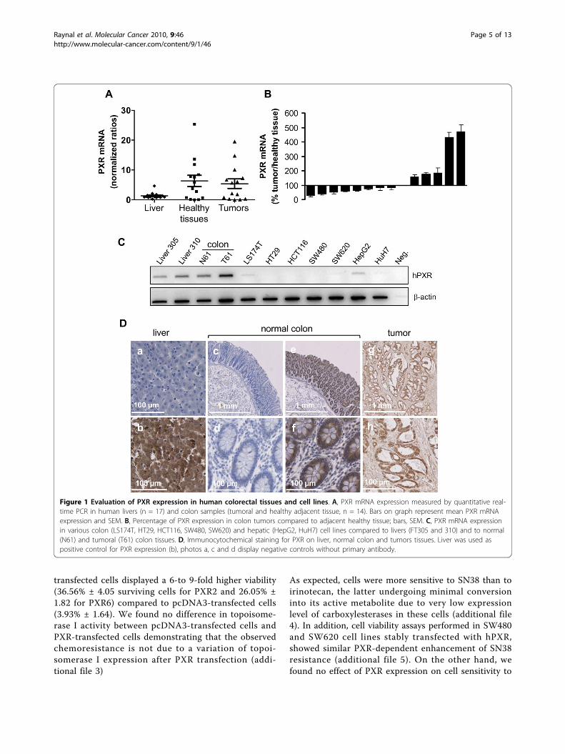
ResultsExpression of hPXR in colon tissues and colon cancercellsTo examine whether hPXR is expressed in human colonin vivo, we analyzed its mRNA expression in both nor-mal and neoplastic human colon tissues. Human livertissues, which are known to have high-PXR expression,were used as positives controls. As shown in figure 1A,PXR mRNA was detected in both normal and canceroushuman colon tissues. In our panel of 14 patients, PXRmRNA was more abundant in some colon tissues thanin liver biopsies, and displayed a greater variability incolon tissues than in liver tissues. Note that, althoughwe did observe some quantitative differences betweenthe expression of PXR in normal tissues and theirmatched colon tumour samples, we found no clear
trend of tendency (figure 1B). In addition, we observeda very low expression of PXR mRNA in both colon can-cer cells lines LS174T, SW480, SW620, or HT29 andhepatic cell lines HepG2 and HuH7 (figure 1C). Thisresult on cell lines is in accordance with the previousobservation that cultured cells often loose metabolicabilities and PXR expression [28]. Representative exam-ples of PXR immunostaining are displayed in figure 1D,liver were used as positive control for PXR expressionand negative controls without primary antibody areshown (photos a, c and d). We observed a strong immu-nostaining of PXR in both normal colon and tumorsconsistent with mRNA results.
Functional characterization of PXR transfected LS174Tcolorectal cancer cellsTo decipher the role of PXR in irinotecan-mediatedtoxicity in colorectal cancer cells, we first establishedstable clones overexpressing hPXR in LS174T. Thesecells were chosen because of their low, but detectable,PXR mRNA expression, and their well known sensitive-ness toward both irinotecan and SN38 [29]. In additionthese cells express a functional p53 [30], which is partlyinvolved in irinotecan response [31]. As shown in figure2A, we selected three clones harbouring different PXRexpression levels at both mRNA and protein levels(PXR2 ≈ PXR6 >> PXR3). Basal expression levels ofCYP3A4 mRNA were higher (up to 9.31 ± 2.40 fold forPXR6) in PXR-expressing cells compared to controls(figure 2B) and were further increased by rifampicin(48.92 ± 1.07 fold for PXR2, 19.02 ± 6.83 fold for PXR3and 92.25 ± 3.60 fold for PXR6 compared to controls).In agreement with the very low level of endogenousPXR detected in these cells, we observed a very weakincrease of CYP3A4 mRNA levels upon treatment ofparent and pcDNA3-transfected cells with rifampicin.The growth rate of PXR-transfected cells did not signifi-cantly vary from that of pcDNA3-transfected cells (fig-ure 2C)
PXR induces colorectal cancer cells resistance toirinotecan and SN38To evaluate the role of PXR onto cell sensitivity todrugs used in the treatment of advanced colorectalcancer we first carried out neutral red cell viabilityassays. For this purpose, control and PXR-transfectedLS174T clones were maintained for 72 hours in thepresence of increasing concentrations of irinotecan,SN38, 5-fluorouracil or oxaliplatin. As shown in figure3, PXR expression clearly led to an increased survivalof LS174T cells towards irinotecan and SN38, and thelevel of chemoresistance to SN38 was correlated tothe relative PXR expression level of each clone (figure3B). Upon treatment with 1 μM SN38, PXR-
Raynal et al. Molecular Cancer 2010, 9:46http://www.molecular-cancer.com/content/9/1/46
Page 4 of 13
transfected cells displayed a 6-to 9-fold higher viability(36.56% ± 4.05 surviving cells for PXR2 and 26.05% ±1.82 for PXR6) compared to pcDNA3-transfected cells(3.93% ± 1.64). We found no difference in topoisome-rase I activity between pcDNA3-transfected cells andPXR-transfected cells demonstrating that the observedchemoresistance is not due to a variation of topoi-somerase I expression after PXR transfection (addi-tional file 3)
As expected, cells were more sensitive to SN38 than toirinotecan, the latter undergoing minimal conversioninto its active metabolite due to very low expressionlevel of carboxylesterases in these cells (additional file4). In addition, cell viability assays performed in SW480and SW620 cell lines stably transfected with hPXR,showed similar PXR-dependent enhancement of SN38resistance (additional file 5). On the other hand, wefound no effect of PXR expression on cell sensitivity to
Figure 1 Evaluation of PXR expression in human colorectal tissues and cell lines. A, PXR mRNA expression measured by quantitative real-time PCR in human livers (n = 17) and colon samples (tumoral and healthy adjacent tissue, n = 14). Bars on graph represent mean PXR mRNAexpression and SEM. B, Percentage of PXR expression in colon tumors compared to adjacent healthy tissue; bars, SEM. C, PXR mRNA expressionin various colon (LS174T, HT29, HCT116, SW480, SW620) and hepatic (HepG2, HuH7) cell lines compared to livers (FT305 and 310) and to normal(N61) and tumoral (T61) colon tissues. D, Immunocytochemical staining for PXR on liver, normal colon and tumors tissues. Liver was used aspositive control for PXR expression (b), photos a, c and d display negative controls without primary antibody.
Raynal et al. Molecular Cancer 2010, 9:46http://www.molecular-cancer.com/content/9/1/46
Page 5 of 13

both 5-fluorouracil and oxaliplatin sensitivities (addi-tional file 6).Surprisingly, we observed that rifampicin did not
enhance the resistance of cells to SN38 (figure 4A).These observations suggest that activation of PXR is notrequired for these effects or that PXR is already acti-vated under these conditions. While neither SN38 noririnotecan activate PXR (additional file 7), we foundthat PXR was activated in our assays because of the pre-sence of 10% fetal calf serum, as previously observed inthe HepG2 cell line [32]. Accordingly, in presence ofserum, CYP3A4 is highly expressed in PXR2 cells, withno additional effect of rifampicin, in contrast to whatwe observed in absence of serum (figure 4B). Moreover,we found that the pharmacological PXR inhibitor L-sul-foraphane (SFN) [33] decreased the percentage of cellsurvival in PXR2 cells treated with 1 or 5 μM SN38(15.13% ± 3.99 and 1.72% ± 0.65), compared to cellstreated with SN38 alone (31.44% ± 3.31 and 9.32% ±1.14) (figure 4C). Although we cannot completelyexclude off-target effects of SFN, it is likely that inhibi-tion of PXR by SFN contributes toward decreased PXR2cell resistance.
Inhibition of PXR expression reverses chemoresistance toSN38Because SFN is known to affect several other signalingpathways, such as those involving the transcription fac-tors Nrf2 [34] and NF-kappaB [35] that are known toaffect cell sensitivity to cytotoxics, we then used a morespecific strategy to inhibit PXR expression. For this pur-pose, LS174T-CTRL or PXR2 cells were transducedwith control or shRNA-expressing lentiviral vectors asdescribed in the material and methods section. AlthoughPXR expression was partially decreased in PXR2 cellstransduced with control FG12 virus (PXR2-mock) com-pared to the untransduced PXR2 clone (figure 5A), astronger decrease was detected in PXR2 cells transducedwith shRNA constructs selectively directed against thePXR mRNA. Thus, we observed that PXR2 cells trans-duced with the sh1334 construct presented a strongdecrease of both PXR mRNA and protein levels, whilePXR2 transduced with sh2116 displayed a moderate, ifany, decrease of PXR expression. Accordingly, inductionof CYP3A4 mRNA expression after rifampicin treatmentin serum-free medium was observed in PXR2-mock and,to a lesser degree, in PXR2-sh2116 cells, but was lost inPXR2-sh1334 cells (figure 5B). In addition, we carriedout neutral red viability assays on these clones (figure5C), and detected a strong reversion of chemoresistanceto SN38 in PXR2-sh1334 cells at 0.1 μM (6.56% ± 1.75compared to 38.78 ± 5.74 in PXR2-mock), demonstrat-ing that the inhibition of PXR expression by itself is suf-ficient to enhance chemosensitivity to SN38.
Figure 2 Characterization of LS174T PXR-transfected cells. A,PXR expression level in parent LS174T, pcDNA3-transfected andstable clones PXR2, PXR3 and PXR6 (top: mRNA expression level;bottom: protein expression level). * p < 0.05, PXR expression ofstable clones compared to parent and pcDNA3-transfected cells,assayed by Mann and Whitney test. B, CYP3A4 mRNA expressionlevels of CTRL and PXR overexpressing clones treated 24 h bysolvent (0.1% DMSO) or 10 μM rifampicin in serum-free medium.Results were obtained from three separate experiments; bars, SEM.a, CYP3A4 expression of cells treated by rifampicin compared to thevehicle treated groups, b, CYP3A4 expression of vehicle treatedstable clones compared to vehicle treated; assayed by Mann andWhitney test (p < 0.05). C, proliferation rate of cell lines. Cells wereseeded in six-well plates at 5 × 10⁵cells/well and counted at theindicated times after seeding (Z1 counter, Beckman Coulter). Datafrom three separate experiments; bars, SEM.
Raynal et al. Molecular Cancer 2010, 9:46http://www.molecular-cancer.com/content/9/1/46
Page 6 of 13

Figure 3 Increased chemoresistance in PXR overexpressing cells to irinotecan and SN38. For neutral red assays, cells were treated for 72 hby increasing concentrations of irinotecan (A) or SN38 (B). Columns, mean viability as a percentage of control (i.e., cells withoutchemotherapeutics treatment, 100%) from replicates (n = 6) from six separate experiments; bars, SEM. a, viability percentages of PXR2, PXR3 andPXR6 compared to pcDNA3-transfected cells (p < 0.05) (Mann and Whitney test). b, viability percentages of PXR2, PXR3 and PXR6 compared toeach other using Kruskal Wallis test (p < 0.05).
Raynal et al. Molecular Cancer 2010, 9:46http://www.molecular-cancer.com/content/9/1/46
Page 7 of 13

Figure 4 A, cell viability assay on pcDNA3-transfected andPXR2 cells treated with rifampicin (RIF). Cells were cultured 24 hwith DMSO 0.1% (solvent) or 10 μM rifampicin in 10% serumcontaining medium and then exposed to SN38 for 72 h. Columns,mean viability as a percentage of control (i.e., cells withoutchemotherapeutics treatment, 100%) from replicates (n = 6) fromthree separate experiments; bars, SEM. B, CYP3A4 mRNA expressionlevel of pcDNA3-transfected or PXR-expressing cells cultured with(10%) or without serum and treated with solvent (0.1% DMSO) or10 μM rifampicin for 24 h. Results were obtained from threeseparate experiments; bars, SEM. * p < 0.05 (Mann and Whitneytest), CYP3A4 mRNA expression of cells cultured with serum, with orwithout rifampicin compared to vehicle treated cells. C, cell viabilityassay on pcDNA3-transfected and PXR2 cells treated with PXRantagonist L-Sulforaphane (SFN). Cells were cultured in presence ofsolvent (DMSO 0.1%) or 10 μM L-Sulforaphane in serum containingmedium for 24 h followed by the 72 h treatment of SN38 with orwithout SFN co-treatment. Columns, mean viability as a percentageof control (i.e., cells without chemotherapeutics treatment, 100%)from replicates (n = 6) from three separate experiments; bars, SEM.*** p < 0.001, * p < 0.05 (student’s t-test).
Figure 5 Reversion of chemoresistance in PXR2 by PXR shRNA.A, quantification of PXR expression by quantitative PCR (top) andwestern blot (bottom). * p < 0.05 (Mann and Whitney test), PXRexpression in PXR2-Mock, PXR2-sh1334 and PXR2-sh2116 cellscompared to pcDNA3-Mock cells. B, quantification of CYP3A4 mRNAexpression after treatment by solvent (DMSO 0.1%) or 10 μMrifampicin for 24 h in serum-free medium. * p < 0.05 (Mann andWhitney test), CYP3A4 expression of cells treated by rifampicincompared to the vehicle treated groups. C, cell viability assay onpcDNA3-Mock, PXR2-Mock, PXR2-sh1334 and PXR2-sh2116 towardSN38. Columns, mean viability as a percentage of control (i.e., cellswithout chemotherapeutics treatment, 100%) from replicates (n = 6)from six separate experiments; bars, SEM. Student’s t-test whereperformed between CTRL-mock cells to PXR2-Mock, PXR2-sh1334and PXR2-sh2116: *** p < 0.001, ** p < 0.01, * p < 0.05.
Raynal et al. Molecular Cancer 2010, 9:46http://www.molecular-cancer.com/content/9/1/46
Page 8 of 13

PXR increases SN38 glucuronidationSince PXR expression does not affect colon cancer cellsproliferation, we hypothesized that SN38 resistanceobserved in PXR-expressing cells is likely mediatedthrough transcriptional regulation of genes involved indrug metabolism. We first explored several steps of iri-notecan metabolism by using pharmacological inhibitorsof CYP3A4 (ketoconazole), MDR1 (verapamil) andBCRP (fumitremorgin C). None of these compoundswas able to reverse the PXR-dependent chemoresistance(data not shown). We next assessed the metabolic pro-file of SN38 using a previously described chromato-graphic detection [27]. Raw data of peak area forintracellular and extracellular SN38 and SN38G allowedus to calculate the metabolic ratios SN38G/SN38.Figure 6A displays relative proportions of total
amounts (intra- and extracellular) of SN38 and SN38G.We found a very significant enhancement of SN38 glu-curonidation in PXR-transfected cells. As shown in fig-ure 6B, both intracellular and extracellular SN38G/SN38ratios were significantly increased in cells overexpressingPXR concordant with PXR expression level of clones.Accordingly, PXR inhibition in PXR2-sh1334 cellsresulted in a decrease of SN38G levels compared tothose found in PXR2-mock or PXR2-sh2116 cells (figure6C). Because the SN38G/SN38 ratio is a sensitive func-tional marker of UGT1A1, 6, 9 and 10 enzymes, wemeasured their relative expression by quantitative RT-PCR. UGT1A1 mRNA expression was found to increaseproportionally to the PXR expression levels in PXR-transfected cells (figure 7A). In addition, we alsoobserved a lower, but significant overexpression of bothUGT1A9 and UGT1A10 mRNA in those cells, whileUGT1A6 mRNA was unaffected. In the same way, weobserved a decrease of UGT1A1 mRNA expression insiRNA transfected cells (figure 7B). Taken together,these data strongly suggest that PXR significantly lowersSN38 concentration by increasing SN38 metabolism toits glucuronide conjugate, mainly through induction ofUGT1A1.
DiscussionIn this work, we address whether expression of PXR inhuman colorectal cancer cells could interfere with theirsensitivity and metabolism of drugs used in treatment ofadvanced colorectal cancer. First we showed that hPXRis expressed in both normal and neoplastic humancolon tissues with a strong variability in cancer colontissues. This variability may prove clinically relevant,since a major finding of this study is that expression ofPXR in human colorectal cancer cells leads to chemore-sistance to the active metabolite of irinotecan, SN38,whereas it did not affect their sensitivity to both 5-fluor-ouracil and oxaliplatin sensitivities. The opposite effect
obtained with pharmacological inactivation of PXR orshRNA-mediated PXR down regulation confirmed thedirect involvement of PXR in SN38 chemoresistance.However, in contrast to previous studies showing thatPXR affects intrinsic cell survival through the p53 sig-naling pathway [23] and cell growth [36], we found thatPXR induced SN38 chemoresistance in LS174T (p53wt)as well as in SW480 and SW620 (p53mut) without affect-ing their intrinsic proliferation rates. Instead, weobserved that PXR expression lowered cellular SN38concentration while increasing SN38 metabolism to itsglucuronide conjugate. Accordingly, we found that sev-eral UGT1A isoenzymes were up-regulated in PXR-expressing cells, most notably UGT1A1 which is the keyenzyme responsible of the inactivation of SN38 toSN38G.Since PXR activation (or inhibition) appears to
increase (or decrease) SN38 conversion to SN38G, ourresults highlight the central role of PXR in regulatingthe cytotoxic threshold of cells to irinotecan-based che-motherapy. Several studies have demonstrated that iri-notecan pharmacokinetics and disposition are affectedby various compounds now identified as PXR ligandssuch rifampicin [10], phenobarbital [37], valproic acidand other anticonvulsivant therapies [38,39] or naturalproducts including St. John’s wort (Hypericum perfora-tum) [11]. In addition, our data are in accordance withprevious studies reporting that intratumoral expressionlevel of UGT1A isoforms may represent a mechanism ofintrinsic irinotecan resistance in colon cancer [40,41].Interestingly a significant (r2 = 0.72, p < 0.001) correla-tion between PXR and UGT1A1 mRNA levels wasfound in human colon tumors (figure 8). Takentogether, these data suggest that tumoral metabolism ispotentially affected by environmental or diet stimuli andthis should be taken into account in the prediction ofirinotecan disposition in patients. In addition, it isknown that diarrhea, a major limiting toxicity of irinote-can, is due to SN38 accumulation in enterocytes [42]and it is conceivable that in situ glucuronidation bytumors and adjacent tissues depends on PXR expressionlevels.Considering its role as master xenobiotics responsive
receptor linking DME genes expression to environmentstimuli, we think that differences in PXR expressioncontribute to the well known intra- and inter-subjectvariability in irinotecan response, and that they partici-pate in the difficulty to clearly identify factors responsi-ble for pharmacogenetics of irinotecan, the so-called“irinogenetics” [43-45]. Indeed, environmental com-pounds, nutrition and diet affecting PXR expressionand/or activation may mask or attenuate pharmacoge-netic associations. Moreover, PXR itself display stronggenetic polymorphism with more than 300 reported
Raynal et al. Molecular Cancer 2010, 9:46http://www.molecular-cancer.com/content/9/1/46
Page 9 of 13

Figure 6 HPLC quantification of SN38 and SN38G A, Relative percentage of total amounts (intra- and extracellular) of SN38 andSN38G on pcDNA3, PXR2, PXR3 and PXR6 cells after 24 h incubation with 10 μM SN38. Results were obtained from three separateexperiments; bars, SEM. B, Intracellular and extracellular SN38G/SN38 ratios on pcDNA3, PXR2, PXR3 and PXR6 cells after 24 h incubation with 10μM SN38. Results were obtained from three separate experiments; bars, SEM. *** p < 0.001, ** p < 0.01, * p < 0.05 (student’s t-test) compared toCTRL cells. C, representative chromatograms of extracellular medium of pcDNA3-Mock, PXR2-Mock, PXR2-sh1334 and PXR2-sh2116 cells exposedto SN38.
Raynal et al. Molecular Cancer 2010, 9:46http://www.molecular-cancer.com/content/9/1/46
Page 10 of 13

SNPs in the dbSNPs database, some of which well char-acterized and inducing differences in both gene expres-sion and ligand recognition [43,44]. PXR expressionlevels within tumors could be also affected by non-genetic factors such as intra-tumor inflammatory cyto-kines [46], microRNA 148a [47] and methylation statusof its exon 3 [48]. In this context, discriminating theroles of genetic influences from environmental effects indrug response, recently coined “pharmacoecology” [49],will be even harder as expected. Thus, it will be of inter-est to evaluate the relative importance of these genetic
and non-genetic factors in patient response toward iri-notecan-based chemotherapy.
ConclusionIn view of the present findings, clinical studies are nowneeded to evaluate the potential interest of PXR in per-sonalized medicine. Indeed, PXR expression and/or acti-vation level could help physicians in the choice ofappropriate chemotherapy regimen for colorectal cancerpatients, since therapeutic alternatives to irinotecanalready exist (i.e. platinum salt or targeted therapy).Finally, PXR down-regulation could be considered as anovel therapeutic approach to circumvent chemoresis-tance to chemotherapy.
Additional file 1: Sequences of shRNA cassettes used for theinhibition of PXR expression. The shRNA-expressing vectors wereconstructed by cloning shRNA expression cassettes into FG12 lentiviralvector.Click here for file[ http://www.biomedcentral.com/content/supplementary/1476-4598-9-46-S1.PDF ]
Additional file 2: Primers sequences used for the quantification ofPXR and its target genes by qPCR. mRNAs expression was evaluatedby RT-quantitative PCR using a LightCycler 480 real-time PCR system andgene-specific primers, b-actin was used as reference gene.Click here for file[ http://www.biomedcentral.com/content/supplementary/1476-4598-9-46-S2.PDF ]
Figure 7 A, UGT1As mRNA expression levels in pcDNA3, PXR2, PXR3 and PXR6 cells. Results were obtained from six separate experiments;bars, SEM. ** p < 0.01, * p < 0.05 (student’s t-test) compared to CTRL cells. B, UGT1A1 mRNA expression levels in CTRL-Mock, PXR2-Mock, PXR2-sh1334 and PXR2-sh2116 cells. Results were obtained from four separate experiments; bars, SEM. *** p < 0.001, * p < 0.05 (student’s t-test)compared to CTRL-Mock cells.
Figure 8 Correlation of PXR and total UGTs mRNA expressionin colon tumors (n = 14). Correlation coefficient R2 = 0.72, *** p <0.001. Doted lines: SEM for a 95% CI.
Raynal et al. Molecular Cancer 2010, 9:46http://www.molecular-cancer.com/content/9/1/46
Page 11 of 13

Additional file 3: Topoisomerase I activity in LS174T pcDNA3 andPXR-transfected cells. Topoisomerase I activity was assessed by using akit from TopoGen based on the ability of nuclear extracts to yield relaxedplasmid from supercoiled plasmid substrate DNA. Nuclear extracts fromLS174T pcDNA3 and PXR-transfected cells were incubated for 15, 30 and60 minutes with a supercoiled DNA marker subsequently subjected toagarose electrophoresis.Click here for file[ http://www.biomedcentral.com/content/supplementary/1476-4598-9-46-S3.PDF ]
Additional file 4: Carboxylesterases mRNA quantification.Carboxylesterases (CES1 and CES2) expression level in LS174T pcDNA3transfected-cells, stable clone PXR2 and a pool of cDNA extracted fromliver biopsies. Results were obtained from six separate experiments; bars,SEM.Click here for file[ http://www.biomedcentral.com/content/supplementary/1476-4598-9-46-S4.PDF ]
Additional file 5: Characterization of SW480 and SW620 PXR-transfected cells. A, C, PXR expression level in control and stable clonesSW480-PXR1, SW620-1L and SW620-1H (U, undetectable PXR expressionlevel). LS174T CTRL cells were taken as a calibrator for quantitative PCR.*** p < 0.001, PXR expression of stable clones compared to SW620 orSW480 control cells, assayed by Student’s t-test. B, D, Increasedchemoresistance in PXR overexpressing cells to SN38. For neutral redassays, cells were treated for 72 h by increasing concentrations of SN38.Columns, mean viability as a percentage of control (i.e., cells withoutchemotherapeutics treatment, 100%) from replicates (n = 6) from threeseparate experiments; bars, SEM. * p < 0.05, viability percentages of PXR-expressing cells compared to control cells (Student’s t-test).Click here for file[ http://www.biomedcentral.com/content/supplementary/1476-4598-9-46-S5.PDF ]
Additional file 6: Cell viability assays of LS174T control and PXRexpression cells, PXR2 and PXR6, to 5-FU and oxaliplatine. Forneutral red assays, cells were treated for 72 h by increasingconcentrations of 5-FU (A) or oxaliplatine (B). Columns, mean viability asa percentage of control (i.e., cells without chemotherapeutics treatment,100%) from replicates (n = 6) from three separate experiments; bars, SEM.Click here for file[ http://www.biomedcentral.com/content/supplementary/1476-4598-9-46-S6.PDF ]
Additional file 7: CYP3A4 mRNA quantification after drugtreatment. CYP3A4 mRNA expression levels in pcDNA3, PXR2, PXR3 andPXR6 cells after treatment with irinotecan or SN38. Results were obtainedfrom six separate experiments; bars, SEM.Click here for file[ http://www.biomedcentral.com/content/supplementary/1476-4598-9-46-S7.PDF ]
AcknowledgementsThis work was funded by La Ligue contre le Cancer, Université Montpellier Iand CHU Nîmes. We thank Jean-François Bourgaux, Christine Pignodel, JuliePannequin for providing us colon tissues, Célia Basurko for statistical analysisand Françoise Malosse for helpful technical assistance for HPLC analysis.
Author details1Institut de Génomique Fonctionnelle, Centre National de la RechercheScientifique (CNRS) UMR5203, Institut National de la Santé et de laRecherche Médicale (INSERM) U661, Université Montpellier 1 et 2,Montpellier F-34094, France. 2Laboratoire de Biochimie, Centre HospitalierUniversitaire, Nîmes, F-30029 France. 3Laboratoire de Biochimie, Faculté deMédecine, Nîmes, F-30908 France. 4Service d’Oto-rhino-laryngologie, CHUNîmes, F-30029 France. 5Service Pharmacie, Centre Régional de Lutte contrele Cancer Val d’Aurelle, Montpellier, F-34298 France.
Authors’ contributionsCR and JMP designed and carried out most of the experiments and wrotethe initial drafts of the manuscript. GL carried the cell viability assays studieson shRNA clones. CB (Breuker) carried out the western blots analysis of PXR.JK and BL provided valuable help in RT-QPCR experiments. CB (Bonnans)carried out immunohistochemistry of PXR. SP supervised HPLC experiments.DJ and FH made original observations leading to this work and contributedto the critical revision of the manuscript. JPB and SL provided valuablereagents and devices and contributed to data interpretation. AE made firstassumptions and contributed to the conception and design of the entirestudy and the final editing of the manuscript. All authors read and approvedthe final manuscript.
Competing interestsThe authors declare that they have no competing interests.
Received: 22 September 2009Accepted: 2 March 2010 Published: 2 March 2010
References1. Center MM, Jemal A, Ward E: International trends in colorectal cancer
incidence rates. Cancer Epidemiol Biomarkers Prev 2009, 18:1688-1694.2. de Gramont A, Tournigand C, Andre T, Larsen AK, Louvet C: Adjuvant
therapy for stage II and III colorectal cancer. Semin Oncol 2007, 34:S37-40.3. Anthony L: Irinotecan toxicity. Curr Opin Support Palliat Care 2007, 1:35-39.4. Ma MK, McLeod HL: Lessons learned from the irinotecan metabolic
pathway. Curr Med Chem 2003, 10:41-49.5. Mathijssen RH, van Alphen RJ, Verweij J, Loos WJ, Nooter K, Stoter G,
Sparreboom A: Clinical pharmacokinetics and metabolism of irinotecan(CPT-11). Clin Cancer Res 2001, 7:2182-2194.
6. Candeil L, Gourdier I, Peyron D, Vezzio N, Copois V, Bibeau F, Orsetti B,Scheffer GL, Ychou M, Khan QA, Pommier Y, Pau B, Martineau P, Del Rio M:ABCG2 overexpression in colon cancer cells resistant to SN38 and inirinotecan-treated metastases. Int J Cancer 2004, 109:848-854.
7. Jansen WJ, Hulscher TM, van Ark-Otte J, Giaccone G, Pinedo HM, Boven E:CPT-11 sensitivity in relation to the expression of P170-glycoprotein andmultidrug resistance-associated protein. Br J Cancer 1998, 77:359-365.
8. Innocenti F, Ratain MJ: “Irinogenetics” and UGT1A: from genotypes tohaplotypes. Clin Pharmacol Ther 2004, 75:495-500.
9. Mathijssen RH, Gurney H: Irinogenetics: how many stars are there in thesky?. J Clin Oncol 2009, 27:2578-2579.
10. Yonemori K, Takeda Y, Toyota E, Kobayashi N, Kudo K: Potentialinteractions between irinotecan and rifampin in a patient with small-celllung cancer. Int J Clin Oncol 2004, 9:206-209.
11. Mannel M: Drug interactions with St John’s wort: mechanisms andclinical implications. Drug Saf 2004, 27:773-797.
12. Bertilsson G, Heidrich J, Svensson K, Asman M, Jendeberg L, Sydow-Backman M, Ohlsson R, Postlind H, Blomquist P, Berkenstam A:Identification of a human nuclear receptor defines a new signalingpathway for CYP3A induction. Proc Natl Acad Sci USA 1998,95:12208-12213.
13. Willson TM, Kliewer SA: PXR, CAR and drug metabolism. Nat Rev DrugDiscov 2002, 1:259-266.
14. Lehmann JM, McKee DD, Watson MA, Willson TM, Moore JT, Kliewer SA:The human orphan nuclear receptor PXR is activated by compoundsthat regulate CYP3A4 gene expression and cause drug interactions. JClin Invest 1998, 102:1016-1023.
15. Geick A, Eichelbaum M, Burk O: Nuclear receptor response elementsmediate induction of intestinal MDR1 by rifampin. J Biol Chem 2001,276:14581-14587.
16. Goodwin B, Moore LB, Stoltz CM, McKee DD, Kliewer SA: Regulation of thehuman CYP2B6 gene by the nuclear pregnane × receptor. MolPharmacol 2001, 60:427-431.
17. Gardner-Stephen D, Heydel JM, Goyal A, Lu Y, Xie W, Lindblom T,Mackenzie P, Radominska-Pandya A: Human PXR variants and theirdifferential effects on the regulation of human UDP-glucuronosyltransferase gene expression. Drug Metab Dispos 2004,32:340-347.
18. Teng S, Jekerle V, Piquette-Miller M: Induction of ABCC3 (MRP3) bypregnane × receptor activators. Drug Metab Dispos 2003, 31:1296-1299.
Raynal et al. Molecular Cancer 2010, 9:46http://www.molecular-cancer.com/content/9/1/46
Page 12 of 13

19. Ma X, Idle JR, Gonzalez FJ: The pregnane × receptor: from bench tobedside. Expert Opin Drug Metab Toxicol 2008, 4:895-908.
20. Chen Y, Nie D: Pregnane × receptor and its potential role in drugresistance in cancer treatment. Recent Pat Anticancer Drug Discov 2009,4:19-27.
21. Wang T, Ma X, Krausz KW, Idle JR, Gonzalez FJ: Role of pregnane ×receptor in control of all-trans retinoic acid (ATRA) metabolism and itspotential contribution to ATRA resistance. J Pharmacol Exp Ther 2008,324:674-684.
22. Mensah-Osman EJ, Thomas DG, Tabb MM, Larios JM, Hughes DP,Giordano TJ, Lizyness ML, Rae JM, Blumberg B, Hollenberg PF, Baker LH:Expression levels and activation of a PXR variant are directly related todrug resistance in osteosarcoma cell lines. Cancer 2007, 109:957-965.
23. Zhou J, Liu M, Zhai Y, Xie W: The antiapoptotic role of pregnane ×receptor in human colon cancer cells. Mol Endocrinol 2008, 22:868-880.
24. Qin XF, An DS, Chen IS, Baltimore D: Inhibiting HIV-1 infection in human Tcells by lentiviral-mediated delivery of small interfering RNA againstCCR5. Proc Natl Acad Sci USA 2003, 100:183-188.
25. Moreau A, Teruel C, Beylot M, Albalea V, Tamasi V, Umbdenstock T,Parmentier Y, Sa-Cunha A, Suc B, Fabre JM, Navarro F, Ramos J, Meyer U,Maurel P, Vilarem MJ, Pascussi JM: A novel pregnane × receptor and S14-mediated lipogenic pathway in human hepatocyte. Hepatology 2009.
26. Evrard A, Cuq P, Ciccolini J, Vian L, Cano JP: Increased cytotoxicity andbystander effect of 5-fluorouracil and 5-deoxy-5-fluorouridine in humancolorectal cancer cells transfected with thymidine phosphorylase. Br JCancer 1999, 80:1726-1733.
27. Poujol S, Pinguet F, Malosse F, Astre C, Ychou M, Culine S, Bressolle F:Sensitive HPLC-fluorescence method for irinotecan and four majormetabolites in human plasma and saliva: application to pharmacokineticstudies. Clin Chem 2003, 49:1900-1908.
28. Pascussi JM, Jounaidi Y, Drocourt L, Domergue J, Balabaud C, Maurel P,Vilarem MJ: Evidence for the presence of a functional pregnane ×receptor response element in the CYP3A7 promoter gene. BiochemBiophys Res Commun 1999, 260:377-381.
29. Jansen WJ, Zwart B, Hulscher ST, Giaccone G, Pinedo HM, Boven E: CPT-11in human colon-cancer cell lines and xenografts: characterization ofcellular sensitivity determinants. Int J Cancer 1997, 70:335-340.
30. Arita D, Kambe M, Ishioka C, Kanamaru R: Induction ofp53-independentapoptosis associated with G2M arrest following DNA damage in humancolon cancer cell lines. Jpn J Cancer Res 1997, 88:39-43.
31. McDonald AC, Brown R: Induction of p53-dependent andp53-independent cellular responses by topoisomerase 1 inhibitors. Br JCancer 1998, 78:745-751.
32. Pascussi JM, Drocourt L, Gerbal-Chaloin S, Fabre JM, Maurel P, Vilarem MJ:Dual effect of dexamethasone on CYP3A4 gene expression in humanhepatocytes. Sequential role of glucocorticoid receptor and pregnane ×receptor. Eur J Biochem 2001, 268:6346-6358.
33. Zhou C, Poulton EJ, Grun F, Bammler TK, Blumberg B, Thummel KE,Eaton DL: The dietary isothiocyanate sulforaphane is an antagonist ofthe human steroid and xenobiotic nuclear receptor. Mol Pharmacol 2007,71:220-229.
34. Morimitsu Y, Nakagawa Y, Hayashi K, Fujii H, Kumagai T, Nakamura Y,Osawa T, Horio F, Itoh K, Iida K, Yamamoto M, Uchida K: A sulforaphaneanalogue that potently activates the Nrf2-dependent detoxificationpathway. J Biol Chem 2002, 277:3456-3463.
35. Xu C, Shen G, Chen C, Gelinas C, Kong AN: Suppression of NF-kappaB andNF-kappaB-regulated gene expression by sulforaphane and PEITCthrough IkappaBalpha, IKK pathway in human prostate cancer PC-3cells. Oncogene 2005, 24:4486-4495.
36. Masuyama H, Nakatsukasa H, Takamoto N, Hiramatsu Y: Down-regulationof pregnane × receptor contributes to cell growth inhibition andapoptosis by anticancer agents in endometrial cancer cells. MolPharmacol 2007, 72:1045-1053.
37. Gupta E, Wang X, Ramirez J, Ratain MJ: Modulation of glucuronidation ofSN-38, the active metabolite of irinotecan, by valproic acid andphenobarbital. Cancer Chemother Pharmacol 1997, 39:440-444.
38. Crews KR, Stewart CF, Jones-Wallace D, Thompson SJ, Houghton PJ,Heideman RL, Fouladi M, Bowers DC, Chintagumpala MM, Gajjar A: Alteredirinotecan pharmacokinetics in pediatric high-grade glioma patientsreceiving enzyme-inducing anticonvulsant therapy. Clin CancerRes 2002,8:2202-2209.
39. de Jong FA, Bol van der JM, Mathijssen RH, Loos WJ, Mathot RA, Kitzen JJ,Bent van den MJ, Verweij J: Irinotecan chemotherapy during valproic acidtreatment: pharmacokinetic interaction and hepatotoxicity. Cancer BiolTher 2007, 6:1368-1374.
40. Cummings J, Boyd G, Ethell BT, Macpherson JS, Burchell B, Smyth JF,Jodrell DI: Enhanced clearance of topoisomerase I inhibitors from humancolon cancer cells by glucuronidation. Biochem Pharmacol 2002,63:607-613.
41. Cummings J, Ethell BT, Jardine L, Boyd G, Macpherson JS, Burchell B,Smyth JF, Jodrell DI: Glucuronidation as a mechanism of intrinsic drugresistance in human colon cancer: reversal of resistance by foodadditives. Cancer Res 2003, 63:8443-8450.
42. Gupta E, Lestingi TM, Mick R, Ramirez J, Vokes EE, Ratain MJ: Metabolic fateof irinotecan in humans: correlation of glucuronidation with diarrhea.Cancer Res 1994, 54:3723-3725.
43. Mathijssen RH, Gurney H: Irinogenetics: How Many Stars Are There in theSky?. J Clin Oncol 2009.
44. Bosch TM, Deenen M, Pruntel R, Smits PH, Schellens JH, Beijnen JH,Meijerman I: Screening for polymorphisms in the PXR gene in a Dutchpopulation. Eur J Clin Pharmacol 2006, 62:395-399.
45. King CR, Xiao M, Yu J, Minton MR, Addleman NJ, Van Booven DJ, Kwok PY,McLeod HL, Marsh S: Identification of NR1I2 genetic variation usingresequencing. Eur J Clin Pharmacol 2007, 63:547-554.
46. Moreau A, Vilarem MJ, Maurel P, Pascussi JM: Xenoreceptors CAR and PXRactivation and consequences on lipid metabolism, glucose homeostasis,and inflammatory response. Mol Pharm 2008, 5:35-41.
47. Takagi S, Nakajima M, Mohri T, Yokoi T: Post-transcriptional regulation ofhuman pregnane × receptor by micro-RNA affects the expression ofcytochrome P450 3A4. J Biol Chem 2008, 283:9674-9680.
48. Misawa A, Inoue J, Sugino Y, Hosoi H, Sugimoto T, Hosoda F, Ohki M,Imoto I, Inazawa J: Methylation-associated silencing of the nuclearreceptor 1I2 gene in advanced-type neuroblastomas, identified bybacterial artificial chromosome array-based methylated CpG islandamplification. Cancer Res 2005, 65:10233-10242.
49. Flexner C: Pharmacoecology: a new name for an old science. ClinPharmacol Ther 2008, 83:375-379.
doi:10.1186/1476-4598-9-46Cite this article as: Raynal et al.: Pregnane × Receptor (PXR) expressionin colorectal cancer cells restricts irinotecan chemosensitivity throughenhanced SN-38 glucuronidation. Molecular Cancer 2010 9:46.
Submit your next manuscript to BioMed Centraland take full advantage of:
• Convenient online submission
• Thorough peer review
• No space constraints or color figure charges
• Immediate publication on acceptance
• Inclusion in PubMed, CAS, Scopus and Google Scholar
• Research which is freely available for redistribution
Submit your manuscript at www.biomedcentral.com/submit
Raynal et al. Molecular Cancer 2010, 9:46http://www.molecular-cancer.com/content/9/1/46
Page 13 of 13