pregnane x receptor (pxr) activation: a mechanism for ... · pregnane x receptor (pxr) activation:...
TRANSCRIPT
Pregnane X receptor (PXR) activation: A mechanismfor neuroprotection in a mouse model ofNiemann–Pick C diseaseS. Joshua Langmade*†, Sarah E. Gale*†, Andrey Frolov*, Ikuko Mohri‡, Kinuko Suzuki‡, Synthia H. Mellon§,Steven U. Walkley¶, Douglas F. Covey�, Jean E. Schaffer*, and Daniel S. Ory*,**
*Center for Cardiovascular Research, Department of Internal Medicine, and �Department of Molecular Biology and Pharmacology, Washington UniversitySchool of Medicine, St. Louis, MO 63110; ‡Department of Pathology and Laboratory Medicine, University of North Carolina, Chapel Hill, NC 27599;§Department of Obstetrics, Gynecology, and Reproductive Sciences, University of California, San Francisco, CA 94143; and ¶Department of Neuroscience,Albert Einstein College of Medicine, Bronx, NY 10461
Communicated by David M. Kipnis, Washington University School of Medicine, St. Louis, MO, July 21, 2006 (received for review June 14, 2006)
Niemann–Pick type C1 (NPC1) disease is a fatal neurodegenerativedisease characterized by neuronal lipid storage and progressivePurkinje cell loss in the cerebellum. We investigated whethertherapeutic approaches to bypass the cholesterol trafficking defectin NPC1 disease might delay disease progression in the npc1�/�
mouse model. We show that the neurosteroid allopregnanolone(ALLO) and T0901317, a synthetic oxysterol ligand, act in concert todelay onset of neurological symptoms and prolong the lifespan ofnpc1�/� mice. ALLO and T0901317 therapy preserved Purkinje cells,suppressed cerebellar expression of microglial-associated genesand inflammatory mediators, and reduced infiltration of activatedmicroglia in the cerebellar tissue. To establish whether the mech-anism of neuroprotection in npc1�/� mice involves GABAA receptoractivation, we compared treatment of natural ALLO and ent-ALLO,a stereoisomer that has identical physical properties of naturalALLO but is not a GABAA receptor agonist. ent-ALLO providedidentical functional and survival benefits as natural ALLO innpc1�/� mice, strongly supporting a GABAA receptor-independentmechanism for ALLO action. On the other hand, the efficacy ofALLO, ent-ALLO, and T0901317 therapy correlated with the abilityof these compounds to activate pregnane X receptor-dependentpathways in vivo. These findings suggest that treatment withpregnane X receptor ligands may be useful clinically in delaying theprogressive neurodegeneration in human NPC disease.
cholesterol � neurosteroid � allopregnanolone � neurodegeneration
N iemann–Pick type C (NPC) disease is an autosomal recessiveneurodegenerative disorder characterized by accumulation of
cholesterol and other lipids in the viscera and central nervoussystem and patterned Purkinje cell death in the cerebellum (1).Mutations in the NPC1 gene are responsible for �95% of humanNPC disease. NPC1 loss-of-function mutants exhibit marked im-pairment of low-density lipoprotein (LDL) cholesterol esterifica-tion and mobilization of newly hydrolyzed LDL cholesterol to theplasma membrane (2–4), resulting in lysosomal sequestration ofLDL cholesterol, delayed down-regulation of the LDL receptor andde novo cholesterol biosynthesis, and impaired ABCA1-mediatedcholesterol efflux (5–7). Despite recent progress in characterizingthe biochemical and genetic defects in NPC disease, the mecha-nisms underlying the neurodegenerative phenotype are not wellunderstood. Moreover, at present there are no effective therapiesthat delay progression of human NPC disease.
Many of the prominent neuropathological features of humanNPC disease [e.g., neuronal lipid storage and progressive loss ofPurkinje neurons (1)] are recapitulated in the BALB�c NPCnih
(npc1�/�) mouse, a naturally occurring murine model that harborsa retroposon insertion in the Npc1 gene (8, 9). In NPC1 mice,accumulation of unesterified cholesterol and gangliosides occurs inmorphologically normal neurons as early as postnatal day 9 (P9)and precedes neuronal injury and cell loss (1, 10). Concomitant with
the lipid accumulation, brains of NPC1 mice exhibit microglialactivation and infiltration and expression of proinflammatory me-diators (10–12). Recent studies in chimeric NPC mice indicate thatPurkinje cells undergo cell-autonomous neurodegeneration, sug-gesting that inflammation is not the initiating factor in Purkinje celldeath, but rather a consequence of cell degeneration (12).
The lipid trafficking defects in NPC1 disease also affect cellularutilization of lipoprotein cholesterol (13, 14). In the npc1�/� mice,disruption of the NPC1-mediated cholesterol trafficking has aprofound effect on neurosteroidogenesis, resulting in substantiallyless pregnenolone and allopregnanolone (ALLO) in the brainsof npc1�/� mice as compared with WT mice (14). Administrationof a single dose of the neurosteroid ALLO at P7 delays onset ofneurological symptoms and prolongs survival in the npc1�/� mice(14). Curiously, the efficacy of ALLO treatment is progressivelyattenuated when administered at later points in postnatal devel-opment. Because the beneficial effect of ALLO on Purkinje cellsurvival is abrogated by treatment with a specific GABAA antag-onist, it has been proposed that the survival benefit in the ALLO-treated mice may be mediated by GABAA receptor signaling.
NPC1 mutant cells also fail to appropriately use lipoproteincholesterol for synthesis of 25-hydroxycholesterol and 27-hydroxycholesterol (13). These oxysterols reduce cellular cho-lesterol levels by suppressing sterol regulatory element-bindingprotein-dependent gene expression and by transcriptional acti-vation of liver X receptor (LXR)-dependent pathways thatpromote cellular cholesterol eff lux and catabolism (15, 16).Moreover, treatment of human NPC1 fibroblasts with 25-hydroxycholesterol or 27-hydroxycholesterol corrects sterol ho-meostatic defects and mobilizes cholesterol from the aberrantlysosomal compartment (13).
The defects in sterol homeostasis and in steroid and oxysterolsynthesis led us to hypothesize that administration of a neurosteroidand a synthetic oxysterol ligand could mitigate NPC1 diseaseprogression. Here we show that ALLO and the synthetic oxysterolligand T0901317 act in concert to delay onset of neurologicalsymptoms and prolong survival in the npc1�/� mouse model. Wefound that the Purkinje cell neuroprotection afforded by ALLOand T0901317 therapies correlates with the ability of these com-pounds to activate murine pregnane X receptor (PXR) in vivo. Ourfindings suggest that PXR ligands may be useful clinically indelaying neurodegeneration in human NPC disease.
Conflict of interest statement: No conflicts declared.
Abbreviations: ALLO, allopregnanolone; LXR, liver X receptor; NPC, Niemann–Pick type C;PXR, pregnane X receptor; Pn, postnatal day n; hPXR, human PXR; mPXR, murine PXR.
†S.J.L. and S.E.G. contributed equally to this work.
**To whom correspondence should be addressed at: Center for Cardiovascular Research,Washington University School of Medicine, Box 8086, 660 South Euclid Avenue, St. Louis,MO 63110. E-mail: [email protected].
© 2006 by The National Academy of Sciences of the USA
www.pnas.org�cgi�doi�10.1073�pnas.0606218103 PNAS � September 12, 2006 � vol. 103 � no. 37 � 13807–13812
MED
ICA
LSC
IEN
CES

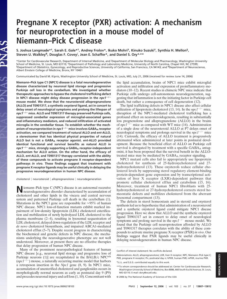
ResultsT0901317 and ALLO Therapy Improves Function and Survival innpc1�/� Mice. In cell culture studies, we found that lipoproteincholesterol-stimulated LXR gene activation is abrogated in NPC1-null cells (Fig. 7, which is published as supporting information onthe PNAS web site). However, despite deficiency of endogenousoxysterol ligands (13), NPC1 mutants respond appropriately toexogenous LXR ligand activation. To extend these findings to an invivo model of NPC1 disease, we investigated whether treatment ofnpc1�/� mice with T0901317 can promote LXR target gene ex-pression in brain tissue and thereby ameliorate disease progression.WT and npc1�/� mice were randomized at P7 to injection withALLO vs. vehicle alone and then treated at P18 with chow diet onlyor chow diet supplemented with T0901317. As shown previously byGriffin et al. (14), a single injection with ALLO at P7 improvedsurvival in npc1�/� mice by 42% (111.8 days in ALLO-treated micevs. 78.8 days in vehicle-treated mice) (Fig. 1A). Although T0901317therapy alone did not show increased survival, T0901317 treatmentprolonged survival in ALLO-treated mice by 72% (135.7 days withcombined treatment vs. 78.8 days in vehicle-treated mice). Thesurvival benefit in the treated npc1�/� mice was accompanied byimproved neurological function. Treatment with either T0901317or ALLO alone resulted in a functional improvement in the mice,delaying onset of neurological symptoms by 2 weeks and bluntingdecline in residual cerebellar function, whereas combined treat-ment with T0901317 and ALLO further slowed disease progression(Fig. 1B). Treatment with T0901317 and ALLO had no effect onsurvival or neurological function in WT mice (data not shown).Despite the significant functional and survival benefit in npc1�/�
mice, T0901317-treated mice showed evidence of toxicity, as dem-onstrated by failure to appropriately gain weight and by transienthepatic hypertriglyceridemia in P28 mice, which resolved in P49mice and was likely due to the known induction of de novolipogenesis by T0901317 (Fig. 8, which is published as supportinginformation on the PNAS web site).
T0901317 and ALLO Promote Purkinje Cell Survival. In light of theimproved neurological function and survival benefit in the treatednpc1�/� mice, we examined the effect of ALLO and T0901317therapy on preservation of cerebellar Purkinje cells. Purkinje cellnumber was reduced in vehicle-treated P63 mice by 83% ascompared with WT mice (Fig. 2), with the greatest loss of Purkinjecells occurring in cerebellar lobes I–IV. Treatment with ALLO orT0901317 improved Purkinje cell survival by 53% and 75%, re-spectively, as compared with vehicle-treated mice. The greatesteffect on Purkinje cell survival was in npc1�/� mice treated withboth ALLO and T0901317, in which the number of Purkinjeneurons was increased 118% compared with untreated mice. In WT
mice there was no difference in the number of Purkinje cell neuronsin compound-treated vs. vehicle-treated mice (data not shown).
Effect of T0901317 and ALLO on Cerebellar Gene Expression. To gaininsight into the mechanism by which T0901317 and ALLO therapyameliorate progression of NPC disease, we examined cerebellargene expression in the treated mice. Based on our cell culturestudies (Fig. 7), we anticipated that treatment with T0901317 wouldincrease expression of LXR target genes. We found that T0901317therapy, alone or in combination with ALLO, led to induction ofABCA1 (7- to 9-fold), ABCG1 (2-fold), and sterol regulatoryelement-binding protein 1c (5- to 6-fold) gene expression in P28
Fig. 1. ALLO and T0901317 improve survival and neurological function in npc1�/� mice. (A) Comparison of survival in WT and npc1�/� mice: vehicle-treated,78.8 days (n � 10); T0901317-treated, 79.9 days (n � 11, P � NS); ALLO-treated, 111.8 days (n � 10, P � 0.001); T0901317�ALLO-treated, 135.7 days (n � 10, P �0.001). (B) Cerebellar function for each of the treatment groups was evaluated by measuring retention time on a rotating drum at 15 rpm. Cumulative retentiontime on the drum was determined for three consecutive attempts (maximum 180 s per attempt). Values represent mean for each group � SEM. *, P � 0.05; **,P � 0.001 for compound-treated vs. vehicle-treated npc1�/� mice.
Fig. 2. Purkinje cell survival is increased in ALLO- and T0901317-treated npc1�/�
mice. Shown are calbindin-immunoreactive Purkinje cells in midline cerebellumsections from 63-day-old npc1�/� mice. (Scale bar: 500 �m.) Bottom Right showsquantification of calbindin-immunoreactive Purkinje cell bodies in cerebellarsections (n � 3–5 mice per treatment group). Results are means � SEM. *, P � 0.05for compound-treated vs. vehicle-treated npc1�/� mice.
13808 � www.pnas.org�cgi�doi�10.1073�pnas.0606218103 Langmade et al.

mouse cerebella (Fig. 3 and Fig. 9 A and B, which is published assupporting information on the PNAS web site). Monotherapy withALLO, which fails to activate LXR in cell-based reporter assays(data not shown), did not induce expression of the LXR targetgenes in the cerebellum. The effect of T0901317 treatment on P49mice was less pronounced, possibly because of the progressiveneuronal loss (Figs. 3 and 9B). These observations suggest thatdietary T0901317 (50 mg�kg per day) achieves sufficient drug levelsin cerebellar tissue of the npc1�/� mice to activate LXR target genesand is in agreement with earlier in vivo studies with T0901317 (17).
Because the progressive neurodegeneration in NPC disease isaccompanied by microglial infiltration in the cerebellum (11, 12),we monitored for the presence of microglia by examining expres-sion of microglial-associated genes in the npc1�/� mice. In vehicle-treated mice, there was dramatic expression of multiple microglialmarkers, which correlated with disease progression, i.e., greater inthe P49 mice than in the P28 mice (Figs. 3 and 9 C–E). Expressionof these markers was suppressed in T0901317-treated (Mac-1a) andALLO-treated (Mac-2 and Mac-1a) P49 mice, and the effects wereadditive in the combined therapy group. We also monitored forproduction of proinflammatory mediators in cerebellar tissue of thenpc1�/� mice by measuring gene expression. In vehicle-treatedmice, expression of the microglial-associated genes was accompa-nied by increased expression of proinflammatory cytokines (TNF�and IL1-�), cytokine receptors (TNFRp55), and inflammatorymediators (cyclooxygenase 2) (Figs. 3 C and D and 9 F and G).Expression of these inflammatory mediator genes was less apparentin the P28 mice than in the P49 mice, consistent with the expressionpattern for the microglial markers. Treatment with ALLO, but notT0901317, decreased expression of these genes, whereas combinedtherapy was most effective for suppression of the inflammatorymediators.
T0901317 and ALLO Attenuate Microglial Infiltration and Purkinje CellLoss. We next examined whether the attenuated expression of themicroglial-associated genes and inflammatory mediators in thetreatment groups reflected reduced infiltration of activated micro-
glia in the cerebellar tissue. Cerebellar sections from P49 npc1�/�
mice were stained for calbindin, a specific marker of Purkinje cellneurons, and CD68, a microglial marker. In vehicle-treated micethere was an �50% loss of Purkinje cell bodies in cerebellar lobesIII and IV, as compared with WT mice (Fig. 4). Strikingly, loss ofPurkinje neurons occurred precisely in regions of the Purkinje celllayer that were infiltrated with CD68-positive microglial cells. In thevehicle-treated npc1�/� mice, Purkinje cell loss and microglial cellinfiltration were least apparent in lobes IX and X, the region of thecerebellum that is generally preserved even in late-stage NPCdisease (8) (Fig. 2 and Fig. 10, which is published as supportinginformation on the PNAS web site). T0901317 and ALLO treat-ment improved Purkinje cell survival and attenuated microglial cellinfiltration in both the granule and Purkinje cell layers (Fig. 4).T0901317 or ALLO treatment alone similarly prevented Purkinjecell loss, although the effect in these groups was less pronouncedthan in the combined therapy group (data not shown).
Enantiomer of ALLO Prolongs Survival in npc1�/� Mice. ALLO hasbeen proposed to exert its anesthetic effect through binding to
Fig. 3. Cerebellar expression of LXR regulated genes and microglial andinflammatory markers in response to treatment. Expression of ABCA1, Mac-2,TNF�, and cyclooxygenase 2 in the cerebella of 28-day-old and 49-day-old micewas determined by real-time quantitative RT-PCR. Expression is shown as foldinduction relative to vehicle-treated WT mice. Values represent the mean �SEM of three mice per group (triplicate determinations for each mouse). *, P �
0.05; **, P � 0.01 for compound-treated vs. vehicle-treated npc1�/� mice.
Fig. 4. T0901317 and ALLO promote Purkinje cell survival in npc1�/� mice.Cerebellar sections from 49-day-old WT mice (Top), vehicle-treated npc1�/�
mice (Middle), and T0901317 and ALLO-treated npc1�/� mice (Bottom) werestained with antibodies to CD68 (green) and calbindin (red) and with a nuclearstain, Hoechst dye 33342 (blue in Left), and examined by immunofluorescence.The asterisk and diamond denote granule and Purkinje cell layers, respec-tively. Arrowheads indicate calbindin-stained Purkinje cell bodies. Omission ofthe nuclear stain (Right) facilitates visualization of microglia within the gran-ule cell layer. (Scale bar: 200 �m.)
Langmade et al. PNAS � September 12, 2006 � vol. 103 � no. 37 � 13809
MED
ICA
LSC
IEN
CES
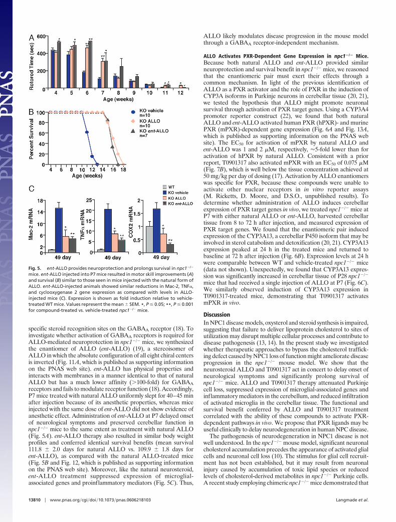
specific steroid recognition sites on the GABAA receptor (18). Toinvestigate whether activation of GABAA receptors is required forALLO-mediated neuroprotection in npc1�/� mice, we synthesizedthe enantiomer of ALLO (ent-ALLO) (19), a stereoisomer ofALLO in which the absolute configuration of all eight chiral centersis inverted (Fig. 11A, which is published as supporting informationon the PNAS web site). ent-ALLO has physical properties andinteracts with membranes in a manner identical to that of naturalALLO but has a much lower affinity (�100-fold) for GABAAreceptors and fails to modulate receptor function (18). Accordingly,P7 mice treated with natural ALLO uniformly slept for 40–45 minafter injection because of its anesthetic properties, whereas miceinjected with the same dose of ent-ALLO did not show evidence ofanesthetic effect. Administration of ent-ALLO at P7 delayed onsetof neurological symptoms and preserved cerebellar function innpc1�/� mice to the same extent as treatment with natural ALLO(Fig. 5A). ent-ALLO therapy also resulted in similar body weightprofiles and conferred identical survival benefits (mean survival111.8 � 2.0 days for natural ALLO vs. 109.9 � 1.8 days forent-ALLO), as compared with the natural ALLO-treated mice(Fig. 5B and Fig. 12, which is published as supporting informationon the PNAS web site). Moreover, like the natural neurosteroid,ent-ALLO treatment suppressed expression of microglial-associated genes and proinflammatory mediators (Fig. 5C). Thus,
ALLO likely modulates disease progression in the mouse modelthrough a GABAA receptor-independent mechanism.
ALLO Activates PXR-Dependent Gene Expression in npc1�/� Mice.Because both natural ALLO and ent-ALLO provided similarneuroprotection and survival benefit in npc1�/� mice, we reasonedthat the enantiomeric pair must exert their effects through acommon mechanism. In light of the previous identification ofALLO as a PXR activator and the role of PXR in the induction ofCYP3A isoforms in Purkinje neurons in cerebellar tissue (20, 21),we tested the hypothesis that ALLO might promote neuronalsurvival through activation of PXR target genes. Using a CYP3A4promoter reporter construct (22), we found that both naturalALLO and ent-ALLO activated human PXR (hPXR)- and murinePXR (mPXR)-dependent gene expression (Fig. 6A and Fig. 13A,which is published as supporting information on the PNAS website). The EC50 for activation of mPXR by natural ALLO andent-ALLO was 1 and 2 �M, respectively, �5-fold lower than foractivation of hPXR by natural ALLO. Consistent with a priorreport, T0901317 also activated mPXR with an EC50 of 0.075 �M(Fig. 7B), which is well below the tissue concentration achieved at50 mg�kg per day of dosing (17). Activation by ALLO enantiomerswas specific for PXR, because these compounds were unable toactivate other nuclear receptors in in vitro reporter assays(M. Ricketts, D. Moore, and D.S.O., unpublished results). Todetermine whether administration of ALLO induces cerebellarexpression of PXR target genes in vivo, we treated npc1�/� mice atP7 with either natural ALLO or ent-ALLO, harvested cerebellartissue from 8 to 72 h after injection, and measured expression ofPXR target genes. We found that the enantiomeric pair inducedexpression of the CYP3A13, a cerebellar P450 isoform that may beinvolved in sterol catabolism and detoxification (20, 21). CYP3A13expression peaked at 24 h in the treated mice and returned tobaseline at 72 h after injection (Fig. 6B). Expression levels at 24 hwere comparable between WT and vehicle-treated npc1�/� mice(data not shown). Unexpectedly, we found that CYP3A13 expres-sion was significantly increased in cerebellar tissue of P28 npc1�/�
mice that had received a single injection of ALLO at P7 (Fig. 6C).We similarly observed induction of CYP3A13 expression inT0901317-treated mice, demonstrating that T0901317 activatesmPXR in vivo.
DiscussionIn NPC1 disease models, oxysterol and steroid synthesis is impaired,suggesting that failure to deliver lipoprotein cholesterol to sites ofutilization may disrupt multiple cellular processes and contribute todisease pathogenesis (13, 14). In the present study we investigatedwhether therapeutic approaches to bypass the cholesterol traffick-ing defect caused by NPC1 loss of function might ameliorate diseaseprogression in the npc1�/� mouse model. We show that theneurosteroid ALLO and T0901317 act in concert to delay onset ofneurological symptoms and significantly prolong survival ofnpc1�/� mice. ALLO and T0901317 therapy attenuated Purkinjecell loss, suppressed expression of microglial-associated genes andinflammatory mediators in the cerebellum, and reduced infiltrationof activated microglia in the cerebellar tissue. The functional andsurvival benefit conferred by ALLO and T0901317 treatmentcorrelated with the ability of these compounds to activate PXR-dependent pathways in vivo. We propose that PXR ligands may beuseful clinically to delay neurodegeneration in human NPC disease.
The pathogenesis of neurodegeneration in NPC1 disease is notwell understood. In the npc1�/� mouse model, significant neuronalcholesterol accumulation precedes the appearance of activated glialcells and neuronal cell loss (10). The stimulus for glial cell recruit-ment has not been established, but it may result from neuronalinjury caused by accumulation of toxic lipid species or reducedlevels of cholesterol-derived metabolites in npc1�/� Purkinje cells.A recent study employing chimeric npc1�/� mice demonstrated that
Fig. 5. ent-ALLO provides neuroprotection and prolongs survival in npc1�/�
mice. ent-ALLO injected into P7 mice resulted in motor skill improvements (A)and survival (B) similar to those seen in mice injected with the natural form ofALLO. ent-ALLO-injected animals showed similar reductions in Mac-2, TNF�,and cyclooxygenase 2 gene expression as compared with levels in ALLO-injected mice (C). Expression is shown as fold induction relative to vehicle-treated WT mice. Values represent the mean � SEM. *, P � 0.05; **, P � 0.001for compound-treated vs. vehicle-treated npc1�/� mice.
13810 � www.pnas.org�cgi�doi�10.1073�pnas.0606218103 Langmade et al.

in cerebellar tissue neighboring WT Purkinje cells were unaffectedby the robust inflammatory response, suggesting that the microgli-al-mediated inflammation specifically targeted npc1�/� Purkinjecells in the cerebella of chimeric mice (12). In our study we foundextensive infiltration of activated microglia in both the Purkinje celland granule cell layers in npc1�/� mice, which is in agreement withprevious reports (10–12). We further show that the recruitment ofmicroglia and expression of proinflammatory mediators were sup-pressed by ALLO and T0901317 treatment and resulted in im-proved Purkinje cell survival. It is possible that these compounds,through modulating trafficking and utilization of lipoprotein cho-lesterol, protect npc1�/� Purkinje cells by attenuating the autono-mous signal that identifies npc1�/� neurons as ‘‘damaged’’ or bymasking recognition of npc1�/� neurons by microglia. Alterna-tively, ALLO and T0901317 may preserve npc1�/� Purkinje cells bydirectly modulating glial cell behavior with respect to activation andrecruitment to damaged neurons.
Previous studies have shown that ALLO therapy promotesPurkinje cell survival and increases lifespan in npc1�/� mice (14,23). Griffin et al. (14) proposed that ALLO may exert its effectsthrough GABAA receptor signaling because ALLO is a GABAAreceptor agonist and the beneficial effects of ALLO on survival ofcultured Purkinje cells were blocked by bicuculline, a GABAAreceptor antagonist. To establish whether the mechanism ofALLO-mediated neuroprotection involves GABAA receptor acti-vation, we compared treatment of natural ALLO and ent-ALLO innpc1�/� mice. ent-ALLO, which is not a GABAA receptor agonist(18), provided functional and survival benefits identical to those ofthe natural compound in the npc1�/� mice, strongly supporting aGABAA receptor-independent mechanism for ALLO. Given thatnatural ALLO and ent-ALLO interact with membranes in anidentical manner (18), it is possible that the neuroprotective effectsof these compounds are mediated through direct steroid–membrane interactions. On the other hand, ALLO, which has beenshown to activate PXR in vitro (21), may exert its effects in npc1�/�
mice through activation of PXR target genes. Our finding thatnatural ALLO and ent-ALLO lead to rapid induction of theCYP3A13 P450 isoform in cerebellar tissue of P7 npc1�/� micesuggests that PXR activation may be involved in Purkinje cellneuroprotection. The lack of enantioselectivity of PXR for theALLO stereoisomers is not altogether surprising. In contrast tomany other nuclear receptors, PXR exhibits broad ligand specificityfor xenobiotics and endogenous compounds and possesses a large,flexible binding cavity capable of docking single ligands in multipleorientations (24). Overlay of the three-dimensional structures of the
ALLO enantiomeric pair reveals that, when the steroid rings arekept coplanar, the polar groups at the 3 and 20 positions can besuperimposed. Thus, ent-ALLO may be able to adopt an orienta-tion similar to the natural compound that permits coordination ofthe polar residues lining the PXR binding cavity, which are criticalfor pharmacologic activation (Fig. 11B).
Activation of PXR target genes may also explain the improve-ment in functional status and preservation of Purkinje cells in thenpc1�/� mice treated with T0901317. Although the benefits of thenonselective LXR ligand may relate to induction of LXR targetgene expression involved in cholesterol efflux and catabolism or tosuppression of inflammatory mediators, T0901317 is also capable ofactivating PXR-dependent gene expression (25). In fact, we foundthat T0901317 activated mPXR in vitro at an EC50 13-fold less thanALLO and, like ALLO, induced PXR target gene expression incerebellar tissue in P28 npc1�/� mice. When T0901317 was admin-istered to ALLO-treated mice, their lifespan was further extendedby 21%. Dietary administration of T0901317, therefore, may affordneuroprotection in the npc1�/� mice through dual activation ofLXR and PXR targets or possibly by providing for sustained PXRactivation, similar to the benefits of weekly injections with ALLO(23) (S.H.M., unpublished results).
A puzzling aspect of the neuroprotective effects of ALLO is howa single injection of the neurosteroid in npc1�/� mice can prolongsurvival by �40% in P7 mice but provide no appreciable benefit inP23 mice (14). We demonstrate that PXR-regulated gene expres-sion in cerebellar tissue of ALLO-treated P7 mice is bimodal.CYP3A13 expression initially peaks at 24 h and returns to basallevels at 72 h but is reexpressed in P28 mice. A possible explanationfor this pattern of gene expression is perinatal imprinting, in whichearly exposure to a drug results in latent gene expression. Such amechanism is supported by a recent report in which neonatal ratsexposed to phenobarbital within the first 7 days of life exhibitincreased postpubertal expression of both constitutive and induc-ible P450 isoforms (26). The plasticity in neonates (e.g., P7 mice),in contrast to adults, may render them susceptible to geneticprogramming that alters levels of P450-dependent metabolizingenzymes. How would increased expression of P450 isoforms confera survival benefit in npc1�/� mice? Whereas synthesis of side-chainoxygenated cholesterol is decreased in NPC1 mutants, there is adramatic increase in npc1�/� mouse tissues in the levels of nonen-zymatic cholesterol oxidation products (27), which have beenreported to be potentially cytotoxic and proapoptotic. Thus, induc-tion of PXR-regulated P450 isoforms may serve a critical role in
Fig. 6. ALLO activates PXR target genes in vitro and in vivo. (A) Cells were transfected with XREM-CYP3A4-LUC reporter construct, pTK-Renilla, pSG5-mPXR,and hHNF4�. Vector indicates transfection with XREM-CYP3A4-LUC and pTK-Renilla alone. Cells were treated for 24 h with ALLO and ent-ALLO. Pregnenolone-16�-carbonitrile (PCN) serves as a control for activation of mPXR. (B) ALLO and ent-ALLO activate the PXR target gene CYP3A13 in cerebellar tissue of P7 npc1�/�
mice. (C) Expression of CYP3A13 is induced at 28 days in npc1�/� mice injected with ALLO at P7 and in T0901317-treated mice. Expression is shown as fold inductionrelative to vehicle-treated npc1�/� mice. Values represent the mean � SEM of four mice per group (triplicate determinations for each mouse). *, P � 0.05; **,P � 0.01 for compound-treated vs. vehicle-treated mice.
Langmade et al. PNAS � September 12, 2006 � vol. 103 � no. 37 � 13811
MED
ICA
LSC
IEN
CES

detoxification of cholesterol oxidation products, thereby mitigatingneuronal injury.
In the present study we provide insight into the mechanismthrough which ALLO delays onset of neurological symptoms andprolongs survival in npc1�/� mice. Our finding that ALLO inducesexpression of PXR targets in vivo suggests a role for PXR activationin protection of Purkinje cells. Future treatment trials will need tobe performed in npc1�/� pxr�/� double knockout mice to defini-tively establish whether PXR activation is required for ALLO-mediated neuroprotection. The possibility that PXR activation isneuroprotective in npc1�/� mice has important implications fordevelopment of therapeutic approaches to delay progression ofNPC1 disease. hPXR is activated by number of commonly useddrugs, including rifampicin, phenytoin, and hyperphorin, a constit-uent of St. John’s wort (24), and the ready availability of suchclinically approved compounds could facilitate therapeutic trials inhuman NPC subjects.
MethodsMice. BALB�c NPCnih mice were obtained from the JacksonLaboratory (Bar Harbor, ME). P7 mice received a single s.c.injection of 25 mg�kg ALLO (5�-pregnan-3�-ol-20-one; ResearchPlus, Manasquan, NJ) or 25 mg�kg ent-ALLO (19) in 20% 2-hy-droxypropyl-�-cyclodextrin as described (14). Mice were weaned atP18 and fed either a standard chow diet or a standard chow dietcontaining 50 mg�kg T0901317 compound per day (Cayman Chem-ical, Ann Arbor, MI).
Protein Preparation and Western Blot Analysis. Microsomal proteinsfrom normal (CRL-1474; American Type Culture Collection, Ma-nassas, VA) and NPC1-null fibroblasts (NPC11628delC, NIH 98.016)were prepared as previously described (3). Protein samples wereresolved by SDS�PAGE, and Western blot analysis was performedas previously described (3). For ABCA1 detection, a rabbit poly-clonal antibody to human ABCA1 (1:500; Novus Biologicals,Littleton, CO) and an anti-rabbit-HRP secondary antibody wereused.
Cholesterol Efflux Assay. Fibroblasts were labeled by 1 �Ci�ml (1Ci � 37 GBq) [1,2-3H(N)]-cholesterol (PerkinElmer, Boston, MA)incubated in the presence and absence of 10 �M T0901317 and 10�M 9-cis-retinoic acid, and efflux was performed as described (28).Cholesterol efflux was expressed as the percentage of the radio-activity released from the cells into the medium divided by totalradioactivity in cells and media.
Immunocytochemistry. Tissue sections from paraformaldehyde-fixed brains were processed and stained with anti-calbindin D-28Kas described (29). Quantification of Purkinje cells was performed bycounting the total number of calbindin-immunoreactive cell bodiesin the Purkinje cell layer.
Real-Time Quantitative RT-PCR. RNA isolation, cDNA synthesis, andreal-time quantitative RT-PCR using SYBR Green Master Mix orTaqMan Fast Universal PCR Master Mix with template-specificprimers (Table 1, which is published as supporting information onthe PNAS web site) were performed as described (30). Foldchanges in gene expression for a particular target were determinedas previously described and normalized to 36B4 expression.
Immunofluorescence Microscopy. Fixed cerebellum sections wereincubated overnight with rat polyclonal anti-mouse CD68:FITC(1:1,000; Serotec, Raleigh, NC) and rabbit polyclonal anti-calbindinD-28K (1:1,000; Chemicon International, Temecula, CA). Thecalbindin antibody was detected through incubation for 1 h withgoat anti-rabbit Alexa Fluor 647 (1:1,000; Molecular Probes, Carls-bad, CA). Sections were then incubated with Hoechst dye 33342(Molecular Probes) at a concentration of 10 �g�ml before dryingthe slides and mounting. Fluorescent images were captured by usingan Axioskop 2 microscope (Zeiss) and analyzed with Axiovision 4software.
Transient Transfection Assays. Transfection assays with Chinesehamster ovary cells were performed as described (21) with 25 ng ofXREM-CYP3A4-LUC (22), 13 ng of pTK-Renilla as a transfectioncontrol (13), 13 ng of pSG5-hPXR (31), 13 ng of pSG5-mPXR (giftof Bryan Goodwin, GlaxoSmithKline, Research Triangle Park,NC), and 13 ng of hHNF4� (21). Cells were incubated in thepresence and absence of ALLO, ent-ALLO, T0901317, or preg-nenolone-16�-carbonitrile. Cells were lysed, and luciferase activitywas determined. Values represent the average of quadruplicatedeterminations of independently transfected wells.
Statistics. All results are expressed as mean � SEM. The statisticalsignificance of differences in mean values was determined bysingle-factor ANOVA.
This work was supported by grants from the Ara Parseghian MedicalResearch Foundation (to D.S.O. and S.U.W.) and the National Institutesof Health (Grant HL04482 to D.S.O., Grant GM47969 to D.F.C., andGrant NS24453 to K.S.).
1. Walkley SU, Suzuki K (2004) Biochim Biophys Acta 1685:48–62.2. Neufeld EB, Cooney AM, Pitha J, Dawidowicz EA, Dwyer NK, Pentchev PG,
Blanchette-Mackie EJ (1996) J Biol Chem 271:21604–21613.3. Millard EE, Srivastava K, Traub L, Schaffer JE, Ory DS (2000) J Biol Chem
275:38445–38451.4. Wojtanik KM, Liscum L (2003) J Biol Chem 278:14850–14856.5. Liscum L, Faust JR (1987) J Biol Chem 262:17002–17008.6. Pentchev PG, Comly ME, Kruth HS, Vanier MT, Wenger DA, Patel S, Brady RO
(1985) Proc Natl Acad Sci USA 82:8247–8251.7. Choi HY, Karten B, Chan T, Vance JE, Greer WL, Heidenreich RA, Garver WS,
Francis GA (2003) J Biol Chem 278:32569–32577.8. Higashi Y, Murayama S, Pentchev PG, Suzuki K (1993) Acta Neuropathol
85:175–184.9. Loftus SK, Morris JA, Carstea ED, Gu JZ, Cummings C, Brown A, Ellison J, Ohno
K, Rosenfeld MA, Tagle DA, et al. (1997) Science 277:232–235.10. Reid P, Sakahita N, Sugii S, Ohno-Iwashita Y, Shimada Y, Hickey W, Chang T (2004)
J Lipid Res 45:582–591.11. Wu Y-P, Mizukami H, Matsuda J, Saito Y, Proia R, Suzuki K (2005) Mol Genet Metab
84:9–17.12. Ko D, Milenkovic L, Beier S, Manuel H, Buchanan J, Scott M (2005) PLoS Genet
1:81–95.13. Frolov A, Zielinski SE, Crowley JR, Dudley-Rucker N, Schaffer JE, Ory DS (2003)
J Biol Chem 278:25517–25525.14. Griffin L, Gong W, Verot L, Mellon S (2004) Nat Med 10:704–711.15. Janowski BA, Willy PJ, Devi TR, Falck JR, Manglesdorf DJ (1996) Nature 383:728–
731.16. Fu X, Menke JG, Chen Y, Zhou G, MacNaul KL, Wright SD, Sparrow CP, Lund EG
(2001) J Biol Chem 276:38378–38387.
17. Whitney KD, Watson MA, Collins JL, Benson WG, Stone TM, Numerick MJ, TippinTK, Wilson JG, Winegar DA, Kliewer SA (2002) Mol Endocrinol 16:1378–1385.
18. Wittmer L, Hu Y, Kalbrenner M, Evers A, Zorumski C, Covey D (1996) MolPharmacol 50:1581–1586.
19. Hu Y, Wittmer L, Kalkbrenner M, Evers A, Zorumski C, Covey D (1997) J Chem SocPerkin Trans 1 3665–3671.
20. Hagemeyer C, Rosenbrock H, Ditter M, Knoth R, Volk B (2003) Neuroscience117:521–529.
21. Lamba V, Yasuda K, Lamba J, Assem M, Davila J, Strom S, Schuetz E (2004) ToxicolAppl Pharmacol 199:251–265.
22. Goodwin B, Hodgson E, Liddle C (1999) Mol Pharmacol 56:1329–1339.23. Ahmad I, Lope-Piedrafta S, Bi X, Hicks C, Yao Y, Yu C, Chaitkin E, Howison C,
Weberg L, Trouard T, Erickson R (2005) J Neurosci Res 82:811–821.24. Watkins R, Wisely G, Moore L, Collins J, Lambert M, Williams S, Willson T, Kliewer
SA, Redinbo M (2001) Science 292:2329–2333.25. Shenoy S, Spencer T, Mercer-Haines N, Alipour M, Gargano M, Runge-Morris M,
Kocarek T (2004) Drug Metab Dispos 32:66–71.26. Agrawal A, Shapiro B (2005) FASEB J 19:470–472.27. Tint G, Pentchev P, Xu G, Batta A, Shiefer S, Salen G, Honda A (1998) J Inherited
Metab Dis 21:853–863.28. Chen W, Sun Y, Welch C, Gorelik A, Leventhal AR, Tabas I, Tall AR (2001) J Biol
Chem 276:43564–43569.29. Matsuda J, Kido M, Tadano-Aritomi K, Ishizuka I, Takeda E, Suzuki K, Kuroda Y
(2004) Hum Mol Genet 13:2709–2723.30. Millard E, Gale S, Dudley N, Zhang J, Schaffer J, Ory D (2005) J Biol Chem
280:28581–28590.31. Lehmann J, McKee D, Watson M, Willson T, Moore J, Kliewer S (1998) J Clin Invest
102:1016–1023.
13812 � www.pnas.org�cgi�doi�10.1073�pnas.0606218103 Langmade et al.