preliminary study of the ul55 gene based on infectious
TRANSCRIPT

RESEARCH Open Access
Preliminary study of the UL55 gene basedon infectious Chinese virulent duckenteritis virus bacterial artificialchromosome cloneYing Wu1,2,3, Yangguang Li1,2,3, Mingshu Wang1,2,3, Kunfeng Sun1,2,3, Renyong Jia1,2,3, Shun Chen1,2,3,Dekang Zhu2,3, Mafeng Liu1,2,3, Qiao Yang1,2,3, Xinxin Zhao1,2,3, Xiaoyue Chen2,3 and Anchun Cheng1,2,3*
Abstract
Background: Lethal Duck Enteritis Virus (DEV) infection can cause high morbidity and mortality of many species ofwaterfowl within the order Anseriformes. However, little is known about the function of viral genes including theconserved UL55 gene among alpha herpes virus due to the obstacles in maintenance and manipulation of DEVgenome in host cells.
Methods: In this paper, we constructed an infectious bacteria artificial chromosome (BAC) clone of the lethalclinical isolate duck enteritis virus Chinese virulent strain (DEV CHv) by inserting a transfer vector containing BACmini-F sequence and selection marker EGFP into UL23 gene using homologous recombination. UL55 deletion andits revertant mutant were generated by two-step RED recombination in E. coli on basis of rescued recombinantvirus. The function of UL55 gene in DEV replication and its effect on distribution of UL26.5 protein were carried outby growth characteristics and co-localization analysis.
Results: The complete genome of DEV CHv can be stably maintained in E. coli as a BAC clone and reconstitutedagain in DEF cells. The generated UL55 deletion mutant based on DEV CHv-BAC-G displayed similar growth curves,plaque morphology and virus titer of its parental virus in infected Duck Embryo Fibroblast (DEF) cells.Immunofluorescence assay indicated that the loss of UL55 gene do not affect the distribution of UL26.5 protein inintracellular. These data also suggest infectious BAC clone of DEV CHv will facilitate the gene function studies ofDEV genome.
Conclusions: We have successfully developed an infectious BAC clone of lethal clinical isolate DEV CHv for the firsttime. The generated UL55 gene mutant based on that demonstrated this platform would be a very useful tool forfunctional study of DEV genes. We found the least known DEV UL55 is dispensable for virus replication and UL26.5distribution, and it could be a very promise candidate locus for developing bivalent vaccine. Experiment are now inprogress for testifying the possibility of UL55 gene locus as an exogenous gene insertion site for developing DEVvectored vaccine.
Keywords: Duck enteritis virus, Bacteria artificial chromosome, Chinese virulent strain, UL55
* Correspondence: [email protected] of Preventive Veterinary Medicine, Sichuan Agricultural University,Chengdu, Sichuan 611130, China2Key Laboratory of Animal Diseases and Human Health of Sichuan Province,Chengdu, Sichuan 611130, ChinaFull list of author information is available at the end of the article
© The Author(s). 2017 Open Access This article is distributed under the terms of the Creative Commons Attribution 4.0International License (http://creativecommons.org/licenses/by/4.0/), which permits unrestricted use, distribution, andreproduction in any medium, provided you give appropriate credit to the original author(s) and the source, provide a link tothe Creative Commons license, and indicate if changes were made. The Creative Commons Public Domain Dedication waiver(http://creativecommons.org/publicdomain/zero/1.0/) applies to the data made available in this article, unless otherwise stated.
Wu et al. Virology Journal (2017) 14:78 DOI 10.1186/s12985-017-0748-y

BackgroundDuck Viral Enteritis (DVE), also known as Duck Plague(DP), is an acute, febrile, septic and contagious diseaseof ducks, geese, swans and many other species of birdswithin the order Anseriformes caused by Duck EnteritisVirus (DEV) [1]. The morbidity and mortality of infectedyoung ducklings or unprotected ducks reaches up to100%, resulting in huge economic losses of domestic andwild waterfowls worldwide [2–4]. In addition, afterprimary infection, the viruses persist in their host forlife. They hide from the immune system in a latent statein which their genome is almost dormant. Eventualepisodes of reactivation allow them to infect naive indi-viduals, causing a long-term of prevalence in the highdensity duck raising farms after DVE outbreak [5].Therefore, even live attenuated DEV vaccines have beenused in prevention and control of this lethal diseasesince 1960s, the DEV infection is not completely pre-vented [6–10]. Thus, investigation of DEV gene func-tions and pathogenesis will be an intelligent policy tonovel vaccine development and disease controlling.According to the reports, studying the resulting varia-
tions in phenotype of virus mutants can be very usefulfor understanding the fundamental information of virusgene function and pathogenesis, which provide theoret-ical basis for development of novel vaccines and che-motherapeutics [11]. A bacteria artificial chromosome(BAC) can take up the complete genome of a herpesvirus as an infectious clone for randomly mutagenesiswithout requirement of restriction sites or cloning stepsin E. coli have been developed for herpes virus mutagen-esis and reconstitution which extensively facilitatedfunctional studies of viral genes [11]. After the first caseof infectious herpes virus BAC clone has been suc-cessfully applied in murine cytomegalovirus (MCMV) in1997 [12], the strategy of cloning full length of herpes-virus genome as a BAC has been adopted for herpesvirus. In most recent years, infectious BAC clone hasbeen gradually applied in DEV study. Wang et al. firstlygenerated an infectious BAC clone of the European viru-lent DEV strain 2085 and expressed hemagglutinin (H5)of high pathogenicity H5N1 avian influenza virus (AIV)based on that. The insertion of BAC components intoDEV 2085 genome UL44 (gC) gene caused disruption ofgC function [13]. In China, full length of DEV vaccinestrains VAC and C-KCE were extensively cloned intoBAC to generate bivalent vaccines for protection ofducks against DEV and other poultry pathogens. DEVglycoprotein gC, junctions of UL26 and gB, regionsbetween UL15B and UL18 gene, SORF3 and US2junctions were selected for insertion of BAC Mini-Fsequence [6, 14–18]. From these literatures, we foundthat DEV infectious BAC clones were predominantlyconsidered as an ideal vector for constructing live
bivalent vaccines. However, rarely reports focused on thefunctional characterization and pathogenesis of lethalDEV that will lead to high morbidity and mortality ofinfected ducks. Although bivalent live vaccine can pro-tect ducks against DEV and other causative pathogensinfection at one time, but lacking of molecular back-ground of the DEV carrier and integration of two patho-gens may lead to potential security risks in a relativelylong period which we cannot predicted. Therefore, con-structing a platform that can be used for functionalcharacterization and pathogenesis investigation of viru-lent clinical isolate DEV will benefit the long-term devel-opment of novel vaccines and disease control.To our knowledge, previously studies about DEV
almost focused on the epidemiology and prevention ofthis disease. Limited molecular biology data are availableregarding DEV genome and its encoding proteins exceptsome significant genes related to virus infection, replica-tion and antiviral effect. As one of the least known ORFsamong total 78 genes of DEV genome, homologues ofthe DEV UL55 protein (pUL55) are encoded only amongalpha herpes viruses [19]. Reports about herpes simplexvirus 2 (HSV-2) UL55 protein revealed that the productof HSV-2 UL55 gene may play an accessory role in vir-ion assembly or maturation and has some relationshipwith UL26.5 distribution [20], but the correspondinghomologue gene of EHV-1 was supposed to mediatepersistent infection [21]. Pre-existing data suggested thatthe HSV-1 UL55 gene was not critical for intraperitonealvirulence or establishment of latent infection [22], butsubsequent documents suggested this dispensable genewas thought to be important for virus growth andspread in the natural host. However, the characteristicsof DEV UL55 gene in DEF cells remains unclear due tolimited data. Thus, carrying out some functional studiesof UL55 gene will provide some data for further studiesof DEV pathogenesis.In this study, we firstly constructed an infectious BAC
clone of the Chinese virulent DEV strain (DEV CHv) togenerate a platform that can be used for functionalcharacterization of DEV genes in DEF cells. The UL23gene of DEV, which has been demonstrated nonessentialfor DEV replication, was used for inserting BAC mini-Fsequence and screening marker EGFP [23–26]. Thefunctional studies of DEV CHv UL55 gene in DEF cellswas carried out based on this newly established platformby using two-step RED recombination for generation ofUL55 deletion and revertant mutants in E. coli. The datayielded by this study demonstrated that UL55 gene isnonessential for virus replication and has no effect ondistribution of UL26.5 protein in infected cells. As aresult, we can conclude that this DEV BAC system willfacilitate the studying of the biology and gene functionsof DEV field strain. Moreover, the nonessential UL55
Wu et al. Virology Journal (2017) 14:78 Page 2 of 14

gene could be a candidate locus for developing bivalentvaccine after attenuation.
MethodsCells, virues, plasmids and antibodiesThe DEV CHv strain (Accession NO. JQ647509) wasused for the construction of infectious BAC clone. Duckembryo fibroblast (DEF) cultures prepared from 10-day-old cherry valley duck embryos were used for the propa-gation of DEV CHv and its derived mutants, which wasmaintained in minimal essential medium (MEM, Gibico)supplemented with 100 U/mL penicillin, 100 mg/mLstreptomycin (1% P/S) and 10% new calf serum (NCS,-Gibico) at 37 °C under a 5% CO2 atmosphere. The helpplasmids pKD46, pKD4 and Pcp20 were kindly donatedby Prof. Kelly T. Hughes (University of Utah), while theplasmid pBeloBAC11 and E. coli strain DH10B wassupplied by Yunfeng Wang (Harbin veterinary researchinstitute, Chinese Academy of Agricultural Science). Ourlab prepared the antibodies against UL55 protein andUL26.5 protein by immune healthy rabbits.
Constrution of transfer vector pUC18/EGFP-TKAB-BAC11The plasmid pBeloBAC11 containing the mini-F se-quence was used for cloning of the DEV CHv completegenome. It was carried out broadly on the sameprinciple used for the construction of the BAC clones ofMDV-1, HVT, VZV, BoHV-1 and EHV-1 etc. by insert-ing the bacterial mini-F sequence and the enhancedgreen fluorescent protein (EGFP) into UL23 (TK) geneof DEV CHv through homologous recombination. Thestrategy for constructing the transfer vector containingthe essential functional components (mini-F) of BAC,EGFP and UL23 homologous sequence to facilitaterecombination was shown in Additional file 1: Figure S1.The generated transfer vector pUC18/EGFP-TKAB-BAC11harboring the homologous regions of 1357 bp on theleft and 1039 bp on the right flanking regions of TKinsertion site, mini-F sequence and a cellular screen-ing marker EGFP.
Constrution of BAC clone of DEV CHvThe cloning of DEV CHv complete genome into BACplasmid was performed by homologous recombinationin DEF cells (Additional file 2: Figure S2 a, b). To the de-tails, approximately 2.5 μg pUC18/EGFP-TKAB-BAC11was transfected into freshly seeded primary DEF cells in-fected with DEV CHv strain by using Lipofectamine3000 (Invitrogen). Meanwhile, the DEV CHv infectedcells and mock-infected cells transfected with pEGFP-ΔMCS were did in parallel as controls. When the greenfluorescence plaques appeared, several rounds of EGFPpositive plaques selection were performed for recombin-ant virus purification and enrichment. Genomic DNA of
the cultures was obtained at the next step by sodiumdodecyl sulfate (SDS)-proteinase K extraction as de-scribed earlier [27] for identification of the purifiedBAC-recombinant DEV CHv by PCR using the primerslisted in Table 1. 100 ng identified BAC-recombinantDEV CHv DNA was electroporated (1.8 KV, 200Ω,25 μF) into E. coli DH10B cells using a Bio-Rad E. coliPulser with 0.1 cm cuvettes. The colonies grown onchloramphenicol plate were detected by PCR to confirmthe presence of EGFP, essential functional componentsof BAC and some important genes related to replica-tion, virulence and structure of DEV CHv virions. Fur-thermore, plasmid extracted from the PCR identifiedchloramphenicol-resistant BAC clones was digestedwith EcoR I for RFLP analysis, the correct plasmid wasnamed pBAC-DEV and further identified by sequencing(Invitrogen).
Reconstitution of infectious virus from pBAC-DEV cloneFor the rescue of infectious recombinant virus in hostcells, 2.5 μg freshly prepared pBAC-DEV DNA by QIA-GEN Midi Kit was transfected into DEF cells usingLipofectamine 3000 (Invitrogen). Cells transfected withor without pEGFP-ΔMCS were taken as controls. Cellswith green fluorescence CPE were harvested and pas-saged for enrichment of infectious virus production. Theinfectious recombinant virus named DEV CHv-BAC-Gwill be obtained until complete cytopathic effect hadbeen developed.For determination of the reconstitute DEV CHv-BAC-
G, PCR was performed firstly for the identification ofDEV CHv gene and the functional components of BAC.A rabbit polyclonal antibody against DEV UL23 genewas used to detect the expression of TK protein in DEVCHv-BAC-G infected DEF cells by indirect immuno-fluorescence analysis (IFA). Mock and wild type DEVCHv infected DEF cells were used as controls to demon-strate the specificity of anti-TK for DEV CHv. In IFA,DEF cells were grown on glass coverslips and infectedwith either wild type DEV CHv or reconstitute DEVCHv-BAC-G at a multiplicity of infection (MOI) of0.02, 200 μl anti-TK IgG at a 1:50 dilution was usedas first antibody. The corresponding pDEV-BAC thatcan produce green fluorescence plaques was isolatedfor sequencing.
Chracterization of the rescued virus in vitroGrowth curve analyses were performed to compare thegrowth kinetics of the wild type DEV CHv with that ofthe reconstitute virus DEV CHv-BAC-G. Briefly, DEFmonolayers grown on 24-well plates were infected with0.02 MOI of wild type and reconstitute virus. Superna-tants containing virus were harvested directly at 7, 12,24, 36, 48, 72 h post infection (h.p.i), while the cells and
Wu et al. Virology Journal (2017) 14:78 Page 3 of 14
mixture of cultures were harvested at the indicated timepoints by treating with trypsin. The amount of infectiouswild type and reconstitute virus in the harvested super-natants and cells were determined by Reed-Muenchassay as previously studies described [28]. Growth curvesassay was performed in triplicate in three independentexperiments.To determine plaque areas of DEV CHv and DEV
CHv-BAC-G, DEF cells were infected at an MOI of 0.02.The inoculated viruses were discarded at 1 h postinfection before adding 500 μl MEM containing 0.5%methylcellulose and 10% NCS. After 3–5 days, the meth-ylcellulose in each well was replaced with 500 μl pre-cold 4% paraformaldehyde for cells immobilization. Cellswere then washed and stained with 500 μl crystal violetfor 5 min. Thorough washing were performed subse-quently to remove the staining solution. Plaques werephotographed, and the average plaque areas weredetermined using the Image J software (http://rsb.info.nih.gov/ij/). Values of DEV CHv-BAC-G were calculated andcompared to wild type DEV CHv plaque areas, which wereset to 100%. T-tests were used to assess the significance ofthe results (P < 0.05). Average percentages of plaque diame-ters and standard deviations were determined from at leastthree independent experiments.Transmission electron microscopy of the 0.1 MOI
DEV CHv-BAC-G infected DEF cells were performed
according to previously reports [29]. Briefly, cells werewashed with PBS at 36 h.p.i, and fixed with 2.5% glutar-aldehyde at 4 °C for 2 h. After that, the fixed adherentcells were collected by scraping from the flasks andcentrifuged at 40,000 rpm/min for 2 h. Then the pelletswere mixed with 2% low melting-temperature agarose at37 °C, and centrifuged at 6000 rpm/min for 10 min.Samples were post-fixed in 1.0% osmium tetroxide. Aftera stepwise dehydration in acetone, samples were embed-ded in epoxy resin 618 and polymerized at 80 °C for72 h. Then, 50 nm ultra-thin sections were preparedwith an LKB ultra tome (LKB Instruments, Inc.,Rockville, MD), collected on grids, and stained with ur-anyl acetate and lead citrate for subsequent examinationwith the Hitachi H-600-A2 transmission electron micro-scope at an accelerating voltage of 75 kV. Images wererecorded on Kodak electron microscope film and com-pared to that of DEV CHv virions.
Generation and characterization of UL55 deletion and itsrevertant mutant based on established recombinant DEVCHv-BAC-GCloning of wild type DEV CHv as infectious BAC clonesfacilitates easy manipulation of their genomes. In orderto test the amenability of pBAC-DEV clone to genomicmodification for DEV CHv gene study, we constructed aUL55 gene deletion and its revertant mutants by two-
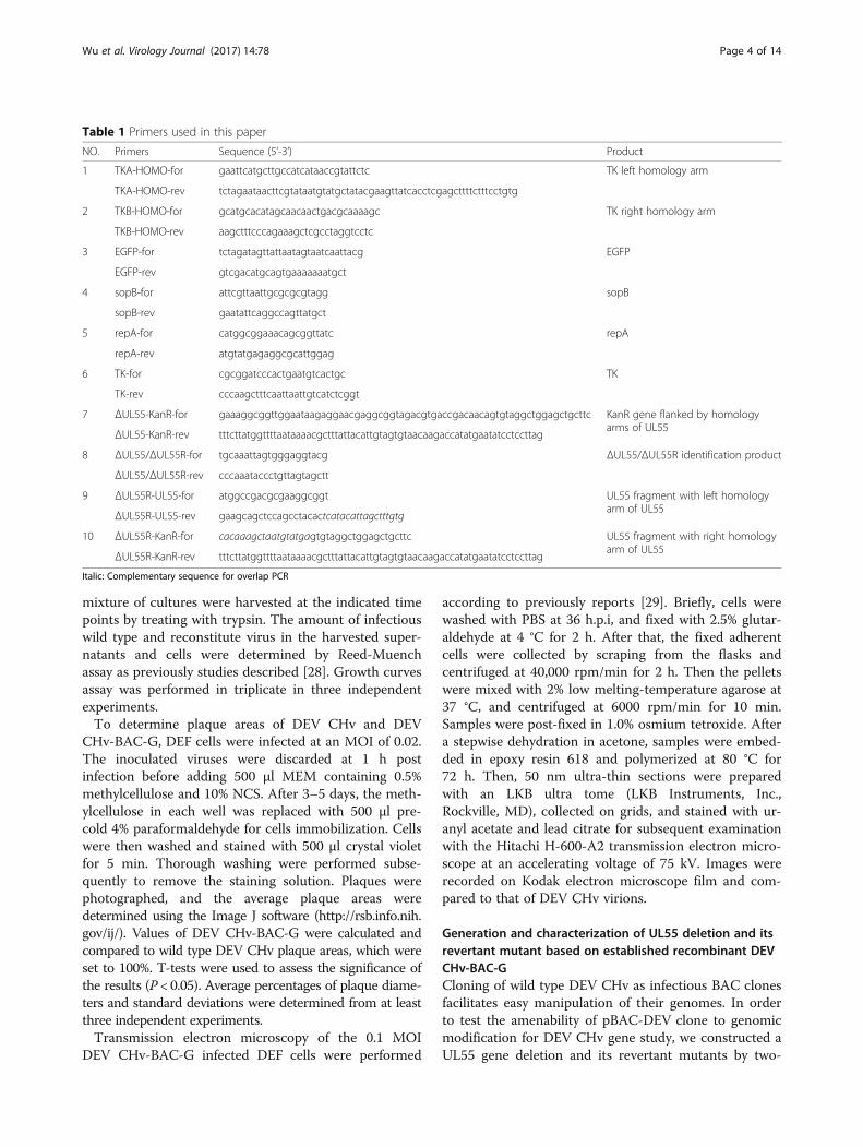
Table 1 Primers used in this paper
NO. Primers Sequence (5’-3’) Product
1 TKA-HOMO-for gaattcatgcttgccatcataaccgtattctc TK left homology arm
TKA-HOMO-rev tctagaataacttcgtataatgtatgctatacgaagttatcacctcgagcttttctttcctgtg
2 TKB-HOMO-for gcatgcacatagcaacaactgacgcaaaagc TK right homology arm
TKB-HOMO-rev aagctttcccagaaagctcgcctaggtcctc
3 EGFP-for tctagatagttattaatagtaatcaattacg EGFP
EGFP-rev gtcgacatgcagtgaaaaaaatgct
4 sopB-for attcgttaattgcgcgcgtagg sopB
sopB-rev gaatattcaggccagttatgct
5 repA-for catggcggaaacagcggttatc repA
repA-rev atgtatgagaggcgcattggag
6 TK-for cgcggatcccactgaatgtcactgc TK
TK-rev cccaagctttcaattaattgtcatctcggt
7 ΔUL55-KanR-for gaaaggcggttggaataagaggaacgaggcggtagacgtgaccgacaacagtgtaggctggagctgcttc KanR gene flanked by homologyarms of UL55
ΔUL55-KanR-rev tttcttatggttttaataaaacgctttattacattgtagtgtaacaagaccatatgaatatcctccttag
8 ΔUL55/ΔUL55R-for tgcaaattagtgggaggtacg ΔUL55/ΔUL55R identification product
ΔUL55/ΔUL55R-rev cccaaataccctgttagtagctt
9 ΔUL55R-UL55-for atggccgacgcgaaggcggt UL55 fragment with left homologyarm of UL55
ΔUL55R-UL55-rev gaagcagctccagcctacactcatacattagctttgtg
10 ΔUL55R-KanR-for cacaaagctaatgtatgagtgtaggctggagctgcttc UL55 fragment with right homologyarm of UL55
ΔUL55R-KanR-rev tttcttatggttttaataaaacgctttattacattgtagtgtaacaagaccatatgaatatcctccttag
Italic: Complementary sequence for overlap PCR
Wu et al. Virology Journal (2017) 14:78 Page 4 of 14

step RED recombination [30]. In brief, 100 ng PCR prod-uct of kanR cassette flanked by the FRT sites and hom-ology arms of UL55 gene was electroporated into DH10Bcells containing pBAC-DEV clone and pKD46 to completethe first round of recombination induced by 100 mM L-arabinose for expression of RED recombination relatedproteins exo, beta, gam in pKD46. Kanamycin resistantcolonies were isolated on LB/Cm/Kana plates. In thesecond round of RED recombination, the temperaturesensitive plasmid Pcp20 was transformed into DH10Bcells containing kanR colonies of the 1st recombination totrigger the excising of kanR FRT cassette. The inducedPcp20 can be removed at 42 °C and the positive coloniesidentified by PCR and sequencing was named as DEVCHv-BAC-GΔUL55 (Additional file 2: Figure S2 b,c).The construction of DEV CHv-BAC-GΔUL55 revert-
ant following the same mutagenesis protocol. In the 1stround of RED recombination, overlapping PCR productof complete UL55 gene and kanamycin resistance geneflanked by the homology arms of UL55 deletion regionwas inserted into the UL55 mutation locus (Additionalfile 2: Figure S2 c,d). The revertant mutant DEV CHv-BAC-GΔUL55R was obtained after excising of KanR inthe 2nd round of RED recombination (Additional file 2:Figure S2 d). Infectivity of the DEV CHv-BAC-GΔUL55and DEV CHv-BAC-GΔUL55R mutants were examinedby transfection of them into DEF cells as their parentalstrain DEV CHv-BAC-G did. PCR, IFA and RFLP ana-lysis were carried out as we described above to confirmthe presence of DEV CHv genome and BAC functionalcomponents, and the mutation of UL55 region. Growthcharacteristics of DEV CHv-BAC-GΔUL55 and DEVCHv-BAC-GΔUL55R were compared to their parentalvirus DEV CHv-BAC-G as we previously described todetermine the function of UL55 in DEV replication.
The effect of UL55 protein on the distribution of UL26.5protein in infected DEF cellsTo investigate the intracellular distribution correlationof UL55 and UL26.5 proteins, DEF cells infected with0.02 MOI wild type DEV CHv and recombinant viruseswere collected for IFA in two independent experiments.In the first experiment, DEV CHv infected DEF cellswere collected at 60 h.p.i. and fixed with 4% parafor-maldehyde. After washing step, the fixed cells wereincubated with 1:50 diluted mouse anti-UL55 or 1:100rabbit anti-UL26.5 IgG prior to addition of 1:100 dilutedTRITC-conjugated Goat anti-mouse IgG or FITC-conjugated Goat anti-rabbit IgG for the single distri-bution analysis of UL55 and UL26.5 encoded proteins inhost cells. For co-localization analysis, 1:50 diluted mouseanti-UL55 and 1:100 rabbit anti-UL26.5 IgG were addedtogether into harvested DEF cells as primary antibodywhile the mixture of 1:100 diluted TRITC-conjugated
Goat anti-mouse IgG and FITC-conjugated Goat anti-rabbit IgG were added as secondary antibody. DEF cellsinfected with 0.02 MOI parental DEV CHv-BAC-G andits derived mutants DEV CHv-BAC-GΔUL55 and DEVCHv-BAC-GΔUL55R were collected and fixed as men-tioned above to demonstrate the contribution of UL55gene to UL26.5 protein distribution. 1:100 diluted rabbitanti-UL26.5 IgG and TRITC-conjugated Goat anti-mouseIgG were used as the first and secondary antibodies forIFA Fluorescence of samples above were inspected underinvert fluorescence microscope.
ResultsCloning of the full length of DEV CHv genome into E. coliThe strategy for cloning and mutagenesis of the DEVCHv genome in E. coli was shown in Additional file 1:Figure S1 and Additional file 2: Figure S2. As a result,the sequenced transfer vector pUC18/EGFP-TKAB-BAC11 containing the mini-F sequence of BAC, EGFPand flanked two loxP sites was used for cloning of thecomplete genome of DEV CHv in pBeloBAC11 by hom-ologous recombination. As shown in Fig. 1a (A), theDEF cells containing pUC18/EGFP-TKAB-BAC11 DNAand wild type DEV CHv developed green fluorescenceafter 3 passages, indicating the successfully constructionof BAC-recombinant virus. After further purificationand enrichment of the recombinant virus, most of theBAC-recombinant virus infected cells exhibited greenCPE (Fig. 1a (B-I)). PCR was performed subsequently toconfirm the purification of BAC-recombinant virus.repA, sopB gene in BAC plasmid and selection markerEGFP gene can be amplified while TK gene is notpresented in the genome of BAC-recombinant virus asexpected (data not shown).Next, the circular DNA of BAC-recombinant virus
was then isolated from infected cells and electroporatedinto E. coli DH10B cells. Plasmid DNA extracted fromthe single chloramphenicol resistance colonies namedpBAC-DEV were subjected to RFLP analysis (Fig. 1b, c).As a result, the restriction patterns of EcoR I digestionproducts of pBAC-DEV were as same as we predicted(Fig. 1b). Further identification was carried out by PCRto determine the complete of DEV CHv genome and theexistence of inserted EGFP and mini-F sequence ofBAC. Figure 1d shows the presence of BAC functionalcomponents, EGFP gene and some important genes thatplay key roles in virus replication, structure and viru-lence in pBAC-DEV genome. Meanwhile, the absence ofthe corresponding BAC genes in DEV CHv infected cellsdemonstrated that a bacterial clone containing a BACwith a full-length DEV CHv genome was successfullyconstructed. Absence of TK gene in pBAC-DEVcould be attributed to the insertion of transfer vector inTK gene.
Wu et al. Virology Journal (2017) 14:78 Page 5 of 14

Reconstitution of infectious virus from the DEV CHv BACplasmidAn advantage of BAC cloning technology for the ma-nipulation of large DNA viruses is that infectious viruscan be reconstituted from the BAC plasmid in host cells.As shown in Fig. 2a, DNA isolated from pBAC-DEVplasmid transfected DEF cells with typical green fluores-cence cytopathic effect was used as template for PCRidentification. As a result, we found repA, sopB, EGFPand US2 can be detected in reconstitute virus, whichindicated the presence of BAC component and DEVgenome in reconstituted virus. The expression of TKprotein in wild type DEV CHv and derived recombinantDEV CHv-BAC-G infected host cells were recognized bypolyclonal rabbit anti-TK in IFA. As shown in Fig. 2b,the presence of green fluorescence in DEV CHv-BAC-Ginfected cells could attributed to the expression ofinserted EGFP gene (Fig. 2b (J)). TK protein was de-tected only in wild type DEV CHv infected cells (Fig. 2b(G)) but not in DEV CHv-BAC-G and mock infected
DEF cells (Fig. 2b (C, K)) indicated the disruption of TKregion by transfer vector insertion. Further sequencingresults also supported our assertions (data not shown).These data demonstrated that infectious DEV CHv viruscould be reconstituted efficiently from BAC plasmid inhost cells.
Characterization of the rescued recombinant viruses invitroThe recombinant viruses were characterized in vitro bydetermining replication kinetics, plaque diameters andmorphology of virions to detect the effect of BAC vectorinsertion in TK region on viral replication and assembly.Viral titers were determined by test of TCID50 ofviruses. As shown in Fig. 3a-c, the reconstitute virusDEV CHv-BAC-G displayed a slightly lower growthcurve during a 96 h period compared to that of wild typeDEV CHv due to the insertion of BAC vector into TKgene, but both growth curves exhibited the same trend.There was no increase in the yield of viruses in either
Fig. 1 Identification of recombinant DEV CHv BAC colonies. a Purification and enrichment of BAC-recombinant virus. A: The 3rd passage ofBAC-recombinant DEV after infection. B-I: Purification and enrichment of BAC-recombinant DEV by eight round of fluorescence plaque selection.b Orientation analysis of pBAC-DEV digested b EcoR I. c Real Gel analysis of pBAC-DEV digested by EcoR I. d Identification of pBAC-DEV. Lane1–14: PCR product of DEV genes in table 2; Lane 15–17: PCR product of inserting exogenous gene EGFP, repA and sopB
Wu et al. Virology Journal (2017) 14:78 Page 6 of 14

Fig. 3 Comparative characterization of DEV CHv-BAC-G and its wild type virus. a-c Growth curves of wild type DEV CHv and reconstitute virusDEV CHv-BAC-G. DEF cells were infected at an MOI of 0.02, the TCID50 titer of infected supernatant, cells and mixture of cells cultureswere titrated at the indicated time points. All titrations were carried out in three independent experiments. d Plaque area measurementof DEV CHv and DEV CHv-BAC-G. Means and standard deviations of plaques diameter of each strain were measured with ImageJ software.The Mean of plaque areas of DEV CHv was set at 100%. Standard deviations are shown with the error bar. NS: no significant difference((t-test, p > 0.05). e, f Transmission electron microscopic examination of purified wild type and reconstitute DEV virions. Red box: The virion structureof each virus was scaled up for observation
Fig. 2 Identification of the rescued DEV CHv-BAC-G in DEF cells. a Identification of rescued DEV CHv-BAC-G. repA, sopB, EGFP and US2 wereamplified by primers in table 1 and 2 using rescued DEV CHv-BAC-G as template; +:Amplicons of repA, sopB, EGFP and US2 using pBAC-DEVplasmid as template for positive control; -: Amplicons of repA, sopB, EGFP and US2 using DEF cell DNA as template for negative control. b Identificationof reconstituted DEV CHv-BAC-G by IFA. Polyclonal rabbit anti-TK IgG was taken as primary antibody for detecting of TK protein. A-D:mock infected- DEF cells; I-L: DEV CHv-BAC-G infected cells; E-H: DEV CHv infected cells
Wu et al. Virology Journal (2017) 14:78 Page 7 of 14

supernatant, cell or mixture in the first 24 h. Afterwards,a continuously increasing can be observed in each partof cell cultures. According to the obtained data and ref-erences, we speculated that the decreased activity of TKcould lead to attenuate virulence without affecting thereplication and function of virus [25, 31–33], furtherstudies will be carred out to demonstrate the hypothesis.In addition, absence of significant difference between
plaque diameters of the wild type and recombinantviruses (t-test, p > 0.05) (Fig. 3d) indicated that the dis-ruption of TK protein didn’t affected the procedures ofadsorption, replication, cell-to-cell spread or CPE forma-tion of the reconstituted virus DEV CHv-BAC-G. Trans-mission electron microscopic examination of the wildtype and reconstitute virus infected cells indicated thatthe structure of them was identical and the functionaldeficient of TK protein did not affect the structure andassembly of DEV virions (Fig. 3e, f ).
Generation of DEV CHv UL55 deletion mutant and itsrevertant by RED recombination in E. coliCloning of DEV CHv complete genome as an infectiousBAC clone allows arbitrary modification of the viralgenomes through different approaches in E. coli andreconstitution of the recombinant virus in eukaryoticcells. Based on this newly established platform, the con-struction of UL55 gene deletion and revertant mutationwere carried out by two-step RED recombination forfunctional study of DEV UL55 gene in DEF cells. Asshown in Fig. 4a, the PCR product of 510 bp and1063 bp was obtained from DEV CHv-BAC-GΔUL55and DEV CHv-BAC-GΔUL55R mutant. Further RFLPanalysis of UL55 mutants and its parental viruses dem-onstrated the mutagenesis of UL55 gene resulted in lossof a 4-kb EcoR I fragment and generation of a new 6-kbfragment (Fig. 4b,c△). Expression of UL55 protein inUL55 deletion and revertant mutants was determined byIFA. From Fig. 4d, we found the UL55 protein could bedetected in DEV CHv-BAC-G and DEV CHv-BAC-GΔUL55R infected cells but not in DEV CHv-BAC-GΔUL55 infected cells, while the green fluorescencecould be found in parental and mutants infected cells,demonstrated the successfully replication of reconstituteviruses. Thus, the mutation introduced in the DEVCHv-BAC-G plasmid was maintained after reconstitu-tion of mutant and revertant viruses. Rapid generationof infectious UL55 deletion and revertant mutants dem-onstrated the adaptability of this approach for efficientmutagenesis of DEV CHv genome in E. coli.
Growth properties of UL55 mutant and its parental virusTo determine if the deletion of the UL55 gene fromDEV CHv-BAC-G has any effect on the growth proper-ties of recombinant virus, the replication kinetics and
plaque diameters were determined and compared totheir parental virus. As a result, we found all virusesexhibited comparable growth kinetics on DEF cells insupernatants and cells during a 72 h period. The repli-cation of viruses kept quiescence at the first 24 h afterinfection, then significant increases were observed dur-ing the whole observation time in supernatants while itstopped increasing at 48 h.p.i and slightly dropped afterthat in cells (Fig. 5a, b). Meanwhile, there was no sig-nificant difference in the plaque areas of the parentaland recombinant viruses (t-test, p > 0.05) (Fig. 5c).These results suggested that the deletion of UL55 genefrom DEV genome has no effect on the growth pro-perties of DEV CHv and UL55 gene is dispensable forDEV replication.
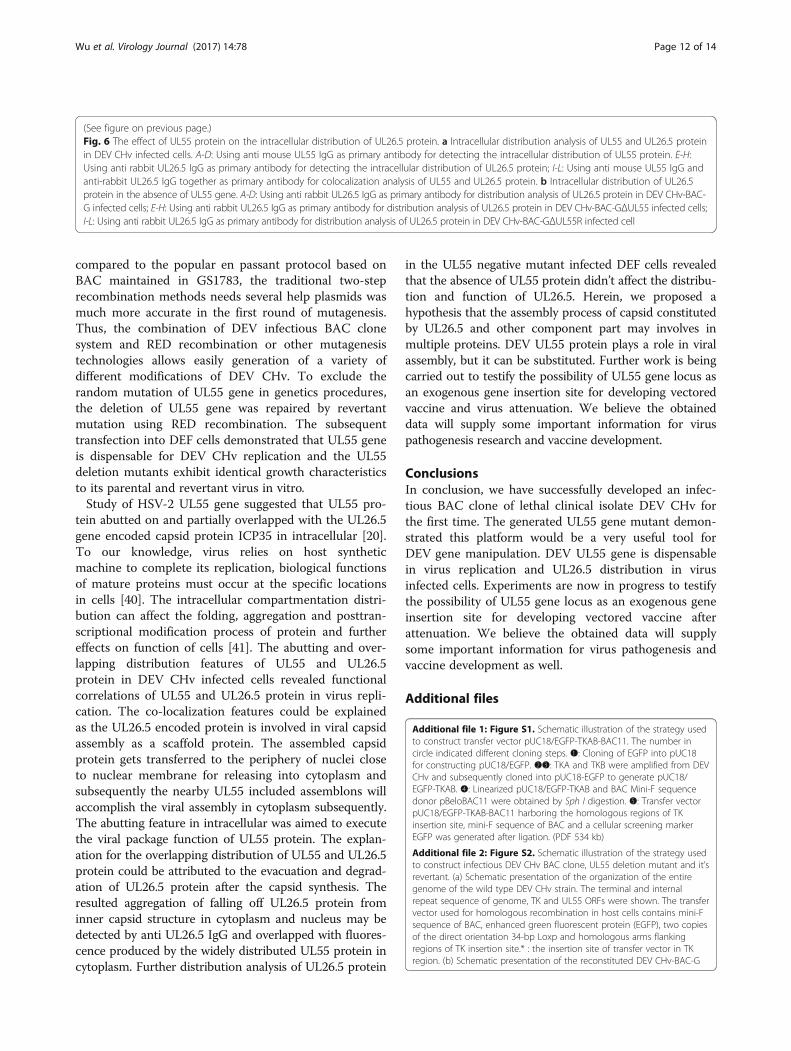
The effect of UL55 protein on the intracellulardistribution of UL26.5 proteinThe distribution of UL26.5 protein in DEF cells withpresence or absence of UL55 protein was determined byindirect immunofluorescence experiments. As shown inFig. 6a(A-D), the UL55 protein was mainly distributed inbright fluorescent granules in cytoplasm near the periph-ery of the nucleus at 60 h p.i., and a small amount of itdistributed in the nucleus, while the UL26.5 protein wasdistributed in widespread speckled structures in thenuclei of infected cells (Fig. 6a(E-H)). Co-localizationanalysis showed that the UL55 protein abutted on andpartially overlapped the UL26.5 protein, and the abuttingfeature was the predominant. The localization featuresof UL55 and UL26.5 protein were as same as that ofHSV-2 UL55 protein.UL55 deletion mutant was used to infect DEF cells to
detect the distribution of UL26.5 protein in the absenceof UL55 protein. Its parental and revertant viruses wereinfected as controls. In Fig. 6b(C, G, K), the red fluores-cence speckled structures were observed in the nucleusof DEV CHv-BAC-GΔUL55 infected cells, which werealso observed in its parental and revertant virus infectedcells. Green fluorescence was observed in all virusesinfected cells indicated the successfully replication ofviruses. These results demonstrated that the distributionof UL26.5 protein encoded by virus lacking UL55 pro-tein were identical to that of wild type virus. In spite ofthe abutting and overlapping features of UL26.5 andUL55 protein in DEV infected cells, the distribution ofUL26.5 protein is independent of UL55 protein.
DiscussionAs full genome sequence and organization of DEV havebeen published to date, specific gene functions could becharacterized. To our knowledge, however, the specificcharacteristic and function of the UL55 protein in DEVCHv are still unknown due to the limited technology of
Wu et al. Virology Journal (2017) 14:78 Page 8 of 14

Fig. 4 (See legend on next page.)
Wu et al. Virology Journal (2017) 14:78 Page 9 of 14

genome manipulation in host eukaryotic cells [15]. Re-cent advance in biologic technologies bring in manygenome editing approach including the most popularCRISPR/Cas9 system. Based on this, scientists have beensuccessfully knocking out protein-coding genes in sev-eral model organisms [34]. It is now become an import-ant approach for understanding gene functions and forengineering genetic regulatory systems. However, thesuccessful rate of gene knock out/down is not 100%because of its limitations. The non-specificity of guideRNA target sequence and the uncertainty of DNA dam-age repair both may result unknown mutations in theother locus of genome (off-target effect), especially inorganisms with large genomes [35]. Compared to this, afull length genomic cloning technology based on BACallowing the recombinants to be reconstituted as aninfectious virus in host cells after mutagenesis in E. colibecome an optimal alternative approach for herpes virusgenome manipulation. In this strategy, large herpes virusgenomes can be manipulated accurately and rapidly inE. coli without unwanted recombination events or rear-rangements due to the only homologous double-strandends will be used as a substrate [36, 37]. The stability ofthe recombinant BAC clone in E. coli can be maintainedbecause of the strict replicons in BAC mini-F sequence.On basis of that, infectious BAC clone is considered as a
preferred large-insert cloning system for genomic analysisand gene discovery in herpes virus, and undoubtedly con-tributed a lot to investigations of virus life cycles, vaccines,gene functions and pathogenesis [37–39]. Thus, we firstlyconstructed an infectious BAC clone of the Duck EnteritisVirus Chinese virulent strain (DEV CHv) for investigationof UL55 gene function in infected DEF cells in this paper.We believe outcomes will provide some clues for under-standing mechanisms of DEV life cycle and that involvedin DEV pathogenicity.Based on the established infectious DEV CHv BAC
clone, mutagenesis of UL55 gene was generated by two-step RED recombination for testing the amenability ofthis platform for DEV CHv gene study. The majoradvantage of the Red recombination system is that onlyshort homologous sequences of 30 to 50 bp are requiredfor the recombination to proceed. For the recombinationin bacteria, components of the Red or RecE/T recom-bination system can be delivered in trans by plasmidssuch as pKD46 that allow inducible expression of Alpha,Beta and Gam. Once the mutagenesis procedure iscompleted, pKD46 can be cured from bacteria by itstemperature-sensitive replication mechanism. Further-more, the unwanted selection markers introduced by 1stround RED recombination can be excised from therecognition sites by Flp recombinase [30, 37]. Besides,
(See figure on previous page.)Fig. 4 Identification of DEV CHv-BAC-GΔUL55 and its revertant DEV CHv-BAC-GΔUL55R. a Identification of DEV CHv-BAC-GΔUL55 and DEVCHv-BAC-GΔUL55R after two rounds of RED recombination. Lane1, 4: The product of UL55 region before RED recombination; Lane 2: The productof UL55 region after 1st round of recombination for constructing UL55 deletion mutant. Lane 3, 5: The product of UL55 region after 2nd round ofrecombination for constructing UL55 deletion mutant; Lane 6, 7: Identification of 1st and 2nd round of RED recombination for constructing UL55deletion revertant DEV CHv-BAC-GΔUL55R, respectively. b, c Restriction fragment length polymorphism (RFLP) analysis of rescued recombinantvirus DEV CHv-BAC-G, DEV CHv-BAC-GΔUL55, DEV CHv-BAC-GΔUL55R. (b)(c) Indicated the orientation and real Gel analysis of rescued recombinantvirus digested by EcoR I, respectively. △: The different band. d Identification of UL55 deletion mutant and revertant by IFA, DEV CHv-BAC-G infectedDEF cells were detected as parental virus. Rabbit anti-UL55 IgG were used as primary antibody. A-D: DEV CHv-BAC-G infected cells. E-H: DEV CHv-BAC-GΔUL55 infected cells. I-L: DEV CHv-BAC-GΔUL55R infected cells
Fig. 5 Growth properties of UL55 mutant and its parental virus. a, b Growth curve of the rescued recombinant DEV CHv-BAC-G, DEV CHv-BAC-GΔUL55 and DEV CHv-BAC-GΔUL55R. DEF cells were infected at an MOI of 0.02, the TCID50 titer of infected superntant and cells of cultures weretitrated at the indicated time points. All titrations were carried out in three independent experiments. c Plauqe area measurement of DEV CHv-BAC-G,DEV CHv-BAC-GΔUL55 and DEV CHv-BAC-GΔUL55R. Means and standard deviations of plaques diameter of each strain were measuredwith ImageJ software. Standard deviations are shown with the error bar. NS: no significant difference ((t-test, p > 0.05)
Wu et al. Virology Journal (2017) 14:78 Page 10 of 14

Fig. 6 (See legend on next page.)
Wu et al. Virology Journal (2017) 14:78 Page 11 of 14
compared to the popular en passant protocol based onBAC maintained in GS1783, the traditional two-steprecombination methods needs several help plasmids wasmuch more accurate in the first round of mutagenesis.Thus, the combination of DEV infectious BAC clonesystem and RED recombination or other mutagenesistechnologies allows easily generation of a variety ofdifferent modifications of DEV CHv. To exclude therandom mutation of UL55 gene in genetics procedures,the deletion of UL55 gene was repaired by revertantmutation using RED recombination. The subsequenttransfection into DEF cells demonstrated that UL55 geneis dispensable for DEV CHv replication and the UL55deletion mutants exhibit identical growth characteristicsto its parental and revertant virus in vitro.Study of HSV-2 UL55 gene suggested that UL55 pro-
tein abutted on and partially overlapped with the UL26.5gene encoded capsid protein ICP35 in intracellular [20].To our knowledge, virus relies on host syntheticmachine to complete its replication, biological functionsof mature proteins must occur at the specific locationsin cells [40]. The intracellular compartmentation distri-bution can affect the folding, aggregation and posttran-scriptional modification process of protein and furthereffects on function of cells [41]. The abutting and over-lapping distribution features of UL55 and UL26.5protein in DEV CHv infected cells revealed functionalcorrelations of UL55 and UL26.5 protein in virus repli-cation. The co-localization features could be explainedas the UL26.5 encoded protein is involved in viral capsidassembly as a scaffold protein. The assembled capsidprotein gets transferred to the periphery of nuclei closeto nuclear membrane for releasing into cytoplasm andsubsequently the nearby UL55 included assemblons willaccomplish the viral assembly in cytoplasm subsequently.The abutting feature in intracellular was aimed to executethe viral package function of UL55 protein. The explan-ation for the overlapping distribution of UL55 and UL26.5protein could be attributed to the evacuation and degrad-ation of UL26.5 protein after the capsid synthesis. Theresulted aggregation of falling off UL26.5 protein frominner capsid structure in cytoplasm and nucleus may bedetected by anti UL26.5 IgG and overlapped with fluores-cence produced by the widely distributed UL55 protein incytoplasm. Further distribution analysis of UL26.5 protein
in the UL55 negative mutant infected DEF cells revealedthat the absence of UL55 protein didn’t affect the distribu-tion and function of UL26.5. Herein, we proposed ahypothesis that the assembly process of capsid constitutedby UL26.5 and other component part may involves inmultiple proteins. DEV UL55 protein plays a role in viralassembly, but it can be substituted. Further work is beingcarried out to testify the possibility of UL55 gene locus asan exogenous gene insertion site for developing vectoredvaccine and virus attenuation. We believe the obtaineddata will supply some important information for viruspathogenesis research and vaccine development.
ConclusionsIn conclusion, we have successfully developed an infec-tious BAC clone of lethal clinical isolate DEV CHv forthe first time. The generated UL55 gene mutant demon-strated this platform would be a very useful tool forDEV gene manipulation. DEV UL55 gene is dispensablein virus replication and UL26.5 distribution in virusinfected cells. Experiments are now in progress to testifythe possibility of UL55 gene locus as an exogenous geneinsertion site for developing vectored vaccine afterattenuation. We believe the obtained data will supplysome important information for virus pathogenesis andvaccine development as well.
Additional files
Additional file 1: Figure S1. Schematic illustration of the strategy usedto construct transfer vector pUC18/EGFP-TKAB-BAC11. The number incircle indicated different cloning steps. ❶: Cloning of EGFP into pUC18for constructing pUC18/EGFP. ❷❸: TKA and TKB were amplified from DEVCHv and subsequently cloned into pUC18-EGFP to generate pUC18/EGFP-TKAB. ❹: Linearized pUC18/EGFP-TKAB and BAC Mini-F sequencedonor pBeloBAC11 were obtained by Sph I digestion. ❺: Transfer vectorpUC18/EGFP-TKAB-BAC11 harboring the homologous regions of TKinsertion site, mini-F sequence of BAC and a cellular screening markerEGFP was generated after ligation. (PDF 534 kb)
Additional file 2: Figure S2. Schematic illustration of the strategy usedto construct infectious DEV CHv BAC clone, UL55 deletion mutant and it’srevertant. (a) Schematic presentation of the organization of the entiregenome of the wild type DEV CHv strain. The terminal and internalrepeat sequence of genome, TK and UL55 ORFs were shown. The transfervector used for homologous recombination in host cells contains mini-Fsequence of BAC, enhanced green fluorescent protein (EGFP), two copiesof the direct orientation 34-bp Loxp and homologous arms flankingregions of TK insertion site.* : the insertion site of transfer vector in TKregion. (b) Schematic presentation of the reconstituted DEV CHv-BAC-G
(See figure on previous page.)Fig. 6 The effect of UL55 protein on the intracellular distribution of UL26.5 protein. a Intracellular distribution analysis of UL55 and UL26.5 proteinin DEV CHv infected cells. A-D: Using anti mouse UL55 IgG as primary antibody for detecting the intracellular distribution of UL55 protein. E-H:Using anti rabbit UL26.5 IgG as primary antibody for detecting the intracellular distribution of UL26.5 protein; I-L: Using anti mouse UL55 IgG andanti-rabbit UL26.5 IgG together as primary antibody for colocalization analysis of UL55 and UL26.5 protein. b Intracellular distribution of UL26.5protein in the absence of UL55 gene. A-D: Using anti rabbit UL26.5 IgG as primary antibody for distribution analysis of UL26.5 protein in DEV CHv-BAC-G infected cells; E-H: Using anti rabbit UL26.5 IgG as primary antibody for distribution analysis of UL26.5 protein in DEV CHv-BAC-GΔUL55 infected cells;I-L: Using anti rabbit UL26.5 IgG as primary antibody for distribution analysis of UL26.5 protein in DEV CHv-BAC-GΔUL55R infected cell
Wu et al. Virology Journal (2017) 14:78 Page 12 of 14

infectious clone after insertion of the transfer vector pUC18/EGFP-TKAB-BAC11. A kanamycin resistance cassette flanked by FRT sites and 50 bphomology arms of UL55 gene was used to replace UL55 gene in the 1stround of RED recombination induced by pKD46. Another temperaturesensitive plasmid Pcp20 was introduced into system for elimination ofKanR in the 2nd round of RED recombination by utilizing the Flplase. (c)Schematic presentation of the resulted UL55 deletion mutant DEV CHv-BAC-GΔUL55. A linear fragment contains UL55 gene and KanR cassetteflanked by homology arms of UL55 gene was used for constructing UL55deletion revertant mutant by two step RED recombination as previouslydescribed. (d) Schematic presentation of the resulted UL55 deletionrevertant mutant DEV CHv-BAC-GΔUL55R after two rounds of REDrecombination. (PDF 755 kb)
AbbreviationsBAC: Bacteria artificial chromosome; BoHv-1: Bovine herpesvirus 1;CHv: Chinese virulent strain; CPE: Cytopathic effect; DEF: Duck embryofibroblast; DEV: Duck enteritis virus; DP: Duck plague; DVE: Duck viral enteritis;EBV: Epstein–barr virus; EGFP: Enhanced green fluorescent protein; EHV-1: Equine herpesvirus 1; GPCMV: Guinea pig cytomegalovirus; h.p.i: Hourspost infection; HCMV: Human cytomegalovirus; HSV: Herpes simplex virus;HVT: Turkey herpesvirus; IFA: Indirect immunofluorescence analysis;KanR: Kanamycin-resistance; KSHV: Kaposi’s sarcoma herpesvirus;MDV: Marek’s disease virus; MEM: Minimal essential medium; MHV-68: Murinegammaherpesvirus 68; MOI: Multiplicity of infection; NCS: New calf serum; P/S: Penicillin, streptomycin; PRV: Pseudorabies virus; SDS: Sodium dodecylsulfate; VZV: Varicella zoster virus
AcknowledgementsWe would like to thank Yunfeng Wang (Harbin veterinary research institute,Chinese Academy of Agricultural Science) and Prof. Kelly T. Hughes(University of Utah) for kindly donating plasmids and E. coli strains.
FundingThis work was supported by grants from the National Natural ScienceFoundation of China (Grant No. 31272545 and No. 31602079), NationalScience and Technology Support Program (No. 2015BAD12B05), ChinaAgricultural Research System (CARS-43-8), and Special Fund for KeyLaboratory of Animal Diseases and Human Health of Sichuan Province(2016JPT0004).
Availability of data and materialsThe datasets used and/or analyzed during the current study available fromthe corresponding author on reasonable request.
Authors’ contributionsYW, MW, and AC conceived and designed the experiments; YW and YLperformed the experiments; YW wrote the paper; MW, KS, RJ, SC, QY, DZ, MLand XZ guided the experiment and helped analysis of data; XC contributedmaterials. All authors read and approved the final manuscript.
Competing interestsThe authors declare that they have no competing interests.
Consent for publicationNot applicable.
Ethics approval and consent to participateThe usage of duck embryo in this paper was approved by the Animal EthicsCommittee of Sichuan Agricultural University (approval No. 2015-016).
Publisher’s NoteSpringer Nature remains neutral with regard to jurisdictional claims inpublished maps and institutional affiliations.
Author details1Institute of Preventive Veterinary Medicine, Sichuan Agricultural University,Chengdu, Sichuan 611130, China. 2Key Laboratory of Animal Diseases andHuman Health of Sichuan Province, Chengdu, Sichuan 611130, China. 3Avian
Diseases Research Center, College of Veterinary Medicine, SichuanAgricultural University, Chengdu, Sichuan 611130, China.
Received: 1 December 2016 Accepted: 7 April 2017
References1. Swayne DE, Glisson JR, Mcdougald LR, Nolan LK, Suarez DL, Nair VL. Duck
Viral Enteritis (Duck Plague). In Diseases of Poultry, 13th Edition. Iowa: Wiley-Blackwell; 2013.
2. Kaleta E, Kuczka A, Kühnhold A, Bunzenthal C, Bönner B, Hanka K, RedmannT, Yilmaz A. Outbreak of duck plague (duck herpesvirus enteritis) innumerous species of captive ducks and geese in temporal conjunction withenforced biosecurity (in-house keeping) due to the threat of avian influenzaA virus of the subtype Asia H5N1. DTW Deutsche TierarztlicheWochenschrift. 2007;114:3–11.
3. Campagnolo ER, Banerjee M, Panigrahy B, Jones RL. An outbreak of duckviral enteritis (duck plague) in domestic Muscovy ducks (Cairina moschatadomesticus) in Illinois. Avian Dis. 2001;45(2):522–8.
4. Shen C, Cheng A, Wang M, Sun K, Jia R, Sun T, Zhang N, Zhu D, Luo Q,Zhou Y. Development and evaluation of an immunochromatographic striptest based on the recombinant UL51 protein for detecting antibody againstduck enteritis virus. Virol J. 2010;7:268.
5. Burgess E, Ossa J, Yuill T. Duck plague: a carrier state in waterfowl. Avian Dis.1979;23(4):940–9.
6. Liu J, Chen P, Jiang Y, Wu L, Zeng X, Tian G, Ge J, Kawaoka Y, Bu Z, Chen H.A duck enteritis virus-vectored bivalent live vaccine provides fast andcomplete protection against H5N1 avian influenza virus infection in ducks.J Virol. 2011;85:10989–98.
7. Huang Y, Au S, Kong F, Lin W. Investigations on duck plague virus. J SouthChina Agric College. 1980;1:21–36.
8. Jansen J, Kunst H, Wemmenhove R. The active immunization of ducksagainst duck plague. Tijdschr Diergeneesk. 1963;88:927–32.
9. Jansen J. Duck plague. J Am Vet Med Assoc. 1968;152:1009.10. Lin W, Lam K, Clark W. Isolation of an apathogenic immunogenic strain
of duck enteritis virus from waterfowl in California. Avian Dis. 1984;28(3):641–50.
11. Wagner M, Ruzsics Z, Koszinowski UH. Herpesvirus genetics has come ofage. Trends Microbiol. 2002;10:318–24.
12. Messerle M, Crnkovic I, Hammerschmidt W, Ziegler H, Koszinowski UH.Cloning and mutagenesis of a herpesvirus genome as an infectiousbacterial artificial chromosome. Proc Natl Acad Sci. 1997;94:14759–63.
13. Wang J, Osterrieder N. Generation of an infectious clone of duck enteritisvirus (DEV) and of a vectored DEV expressing hemagglutinin of H5N1 avianinfluenza virus. Virus Res. 2011;159:23–31.
14. Zou Z, Hu Y, Liu Z, Zhong W, Cao H, Chen H, Jin M. Efficient strategy forconstructing duck enteritis virus-based live attenuated vaccine againsthomologous and heterologous H5N1 avian influenza virus and duckenteritis virus infection. Vet Res. 2015;46:1.
15. Chen L, Yu B, Hua J, Ye W, Ni Z, Yun T, Deng X, Zhang C. Construction of afull-length infectious bacterial artificial chromosome clone of duck enteritisvirus vaccine strain. Virol J. 2013;10:1.
16. Liu X, Wei S, Liu Y, Fu P, Gao M, Mu X, Liu H, Xing M, Ma B, Wang J.Recombinant duck enteritis virus expressing the HA gene from goose H5subtype avian influenza virus. Vaccine. 2013;31:5953–9.
17. Wang J, Ge A, Xu M, Wang Z, Qiao Y, Gu Y, Liu C, Liu Y, Hou J. Constructionof a recombinant duck enteritis virus (DEV) expressing hemagglutinin ofH5N1 avian influenza virus based on an infectious clone of DEV vaccinestrain and evaluation of its efficacy in ducks and chickens. Virol J. 2015;12:1.
18. Zou Z, Liu Z, Jin M. Efficient strategy to generate a vectored duck enteritisvirus delivering envelope of duck tembusu virus. Viruses. 2014;6:2428–43.
19. Wu Y, Cheng A, Wang M, Zhu D, Jia R, Cui H, Luo Q, Wang Y, Xu Z, Chen Z.Molecular characterization analysis of newly identified duck enteritis virusUL55 gene. 4th International Conference on Bioinformatics and BiomedicalEngineering (iCBBE), Chengdu, China, 2010. IEEE; 2010: 1–7.
20. Yamada H, Jiang Y-M, Oshima S-i, Daikoku T, Yamashita Y, Tsurumi T,Nishiyama Y. Characterization of the UL55 gene product of herpes simplexvirus type 2. J Gen Virol. 1998;79:1989–95.
21. Harty RN, Caughman GB, Holden VR, O’Callaghan DJ. Characterization of themyristylated polypeptide encoded by the UL1 gene that is conserved in the
Wu et al. Virology Journal (2017) 14:78 Page 13 of 14
genome of defective interfering particles of equine herpesvirus 1. J Virol.1993;67:4122–32.
22. Nash TC, Spivack JG. The UL55 and UL56 genes of herpes simplex virus type1 are not required for viral replication, intraperitoneal virulence, orestablishment of latency in mice. Virology. 1994;204:794–8.
23. Azab W, Tsujimura K, Kato K, Arii J, Morimoto T, Kawaguchi Y, Tohya Y,Matsumura T, Akashi H. Characterization of a thymidine kinase-deficientmutant of equine herpesvirus 4 and in vitro susceptibility of the virus toantiviral agents. Antiviral Res. 2010;85:389–95.
24. Azab W, Kato K, Abdel-Gawad A, Tohya Y, Akashi H. Equine herpesvirus 4:Recent advances using BAC technology. Vet Microbiol. 2011;150:1–14.
25. Wu C-Y, Liao C-M, Chi J-N, Chien M-S, Huang C. Growth properties andvaccine efficacy of recombinant pseudorabies virus defective inglycoprotein E and thymidine kinase genes. J Biotechnol. 2016;229:58–64.
26. Wen Y, Cheng A, Wang M, Ge H, Shen C, Liu S, Xiang J, Jia R, Zhu D, ChenX. A Thymidine Kinase recombinant protein-based ELISA for detectingantibodies to Duck Plague Virus. 2010;7(1):77.
27. Morgan RW, Cantello JL, McDermott CH. Transfection of chicken embryofibroblasts with Marek’s disease virus DNA. Avian Dis. 1990;34(2):345–51.
28. Reed LJ, Muench H. A simple method of estimating fifty per centendpoints. Am J Epidemiol. 1938;27:493–7.
29. Guo Y, Shen C, Cheng A, Wang M, Zhang N, Chen S, Zhou Y. Anatidherpesvirus 1 CH virulent strain induces syncytium and apoptosis in duckembryo fibroblast cultures. Vet Microbiol. 2009;138:258–65.
30. Datsenko KA, Wanner BL. One-step inactivation of chromosomal genes inEscherichia coli K-12 using PCR products. Proc Natl Acad Sci. 2000;97:6640–5.
31. Zhang M, Fu S, Deng M, Xie Q, Xu H, Liu Z, Hu C, Chen H, Guo A.Attenuation of bovine herpesvirus type 1 by deletion of its glycoprotein Gand tk genes and protection against virulent viral challenge. Vaccine. 2011;29:8943–50.
32. Slater J, Gibson J, Field H. Pathogenicity of a thymidine kinase-deficientmutant of equine herpesvirus 1 in mice and specific pathogen-free foals.J Gen Virol. 1993;74:819–28.
33. Coen DM, Kosz-Vnenchak M, Jacobson JG, Leib DA, Bogard CL, Schaffer PA,Tyler KL, Knipe DM. Thymidine kinase-negative herpes simplex virusmutants establish latency in mouse trigeminal ganglia but do notreactivate. Proc Natl Acad Sci. 1989;86:4736–40.
34. Zhao Y, Dai Z, Liang Y, Yin M, Ma K, He M, Ouyang H, Teng CB.Sequence-specific inhibition of microRNA via CRISPR/CRISPRi system.Sci Rep. 2014;4:3943.
35. Larson MH, Gilbert LA, Wang X, Lim WA, Weissman JS, Qi LS. CRISPRinterference (CRISPRi) for sequence-specific control of gene expression. NatProtoc. 2013;8:2180–96.
36. Zhang Y, Buchholz F, Muyrers JP, Stewart AF. A new logic for DNA engineeringusing recombination in Escherichia coli. Nat Genet. 1998;20:123–8.
37. Tischer BK, Kaufer BB. Viral bacterial artificial chromosomes: generation,mutagenesis, and removal of mini-F sequences. Biomed Res Int. 2012;2012:472537.
38. Schumacher D, Tischer BK, Fuchs W, Osterrieder N. Reconstitution of Marek’sdisease virus serotype 1 (MDV-1) from DNA cloned as a bacterial artificialchromosome and characterization of a glycoprotein B-negative MDV-1mutant. J Virol. 2000;74:11088–98.
39. Li Y, Wang S, Zhu H, Zheng C. Cloning of the herpes simplex virus type 1genome as a novel luciferase-tagged infectious bacterial artificialchromosome. Arch Virol. 2011;156:2267–72.
40. Shen C, Guo Y, Cheng A, Wang M, Zhou Y, Lin D, Xin H, Zhang N.Characterization of subcellular localization of duck enteritis virus UL51protein. Virol J. 2009;6:92.
41. Rixon FJ, Cross AM, Addison C, Preston VG. The products of herpessimplex virus type 1 gene UL26 which are involved in DNA packagingare strongly associated with empty but not with full capsids. J GenVirol. 1988;69:2879–91.
• We accept pre-submission inquiries
• Our selector tool helps you to find the most relevant journal
• We provide round the clock customer support
• Convenient online submission
• Thorough peer review
• Inclusion in PubMed and all major indexing services
• Maximum visibility for your research
Submit your manuscript atwww.biomedcentral.com/submit
Submit your next manuscript to BioMed Central and we will help you at every step:
Wu et al. Virology Journal (2017) 14:78 Page 14 of 14